An introduction to Micro CT Scanning
An introduction to Micro CT Scanning
An introduction to Micro CT Scanning
- No tags were found...

You also want an ePaper? Increase the reach of your titles
YUMPU automatically turns print PDFs into web optimized ePapers that Google loves.
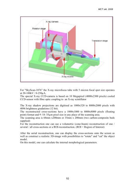
M<strong>CT</strong> okt. 2008For "SkyScan-1076” the X-ray microfocus tube with 5 micron focal spot size operatesat 20-100kV / 0-250µA.The special X-ray CCD-camera is based on 10 Megapixel (4000x2300 pixels) cooledCCD-sensor with fibre optic coupling <strong>to</strong> an X-ray scintilla<strong>to</strong>rThe X-ray shadow projections are digitised as 1000x520 <strong>to</strong> 8000x2000 pixels with4096 brightness gradations (12 bit).The reconstructed cross-sections have a 1000x1000 <strong>to</strong> 8000x8000 pixels (floatingpoint) format and 9 /18 /35µm pixel size in any place of the scanning area.The scanning area is 68mm x200mm or 35mm x 200mm (two carbon-composite bedssupplied)For the reconstruction one can use a volumetric (cone-beam) reconstruction of one /several / all cross-sections or a ROI-reconstruction. (ROI = Region of Interest)After the serial reconstruction, one can display the cross-sections on<strong>to</strong> the screen aswell as construct a realistic 3D-image with possibilities <strong>to</strong> "rotate" and "cut" the objectmodel.On this model, one can calculate the internal morphological parameters.10