

metatarsat osteotomy for the correction of metatarsus adductus
metatarsat osteotomy for the correction of metatarsus adductus
metatarsat osteotomy for the correction of metatarsus adductus
- No tags were found...

You also want an ePaper? Increase the reach of your titles
YUMPU automatically turns print PDFs into web optimized ePapers that Google loves.
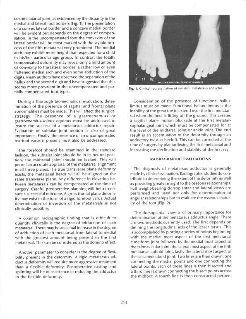
tarsometatarsal joint, as evidenced by <strong>the</strong> disparity in <strong>the</strong>medial and lateral foot borders (Fig. 1). The presentation<strong>of</strong> a convex lateral border and a concave medial borderwill be evident but depends on <strong>the</strong> degree <strong>of</strong> compensation.ln <strong>the</strong> uncompensated foot <strong>the</strong> convexity <strong>of</strong> <strong>the</strong>Iateral border will be most marked with <strong>the</strong> styloid process<strong>of</strong> <strong>the</strong> fifth metatarsal very prominent. The medialarch may exhibit more height than expected <strong>for</strong> a childin his/her particular age group. ln contrast <strong>the</strong> totallycompensated de<strong>for</strong>mity may reveal only a mild amount<strong>of</strong> convexity to <strong>the</strong> lateral border, a ra<strong>the</strong>r low or evenflattened medial arch and even some abduction <strong>of</strong> <strong>the</strong>digits. Many authors have observed <strong>the</strong> separation <strong>of</strong> <strong>the</strong>hallux and <strong>the</strong> second digit and have suggested that thisseems more prevalent in <strong>the</strong> uncompensated and partiallycompensated foot types.During a thorough biomechanical evaluation, determination<strong>of</strong> <strong>the</strong> presence <strong>of</strong> sagittal and frontal planeabnormalities must be made. This will affect <strong>the</strong> surgicalstrategy. The presence <strong>of</strong> a gastrocnemius orgastrocnemius-soleus equinus must be addressed toinsure <strong>the</strong> success <strong>of</strong> a <strong>metatarsus</strong> <strong>adductus</strong> repair.Evaluation <strong>of</strong> subtalar joint motion is also <strong>of</strong> greatimportance. Finally, <strong>the</strong> presence <strong>of</strong> an uncompensatedrearfoot varus if present must also be addressed.The <strong>for</strong>efoot should be examined in <strong>the</strong> standardfashion, <strong>the</strong> subtalar joint should be in its neutral position,<strong>the</strong> midtarsal joint should be locked. This willpermit an accurate appraisal <strong>of</strong> <strong>the</strong> metatarsal alignmentin all three planes. If a true transverse plane de<strong>for</strong>mityexists, <strong>the</strong> metatarsal heads will all be aligned on <strong>the</strong>same transverse plane. Any difference in elevation betweenmetatarsals can be compensated at <strong>the</strong> time <strong>of</strong>surgery. Careful preoperative planning will help to ensurea successful outcome. A gross frontal plane de<strong>for</strong>mitymay exist in <strong>the</strong> <strong>for</strong>m <strong>of</strong> a rigid <strong>for</strong>efoot varus. Actualdetermination <strong>of</strong> inversion <strong>of</strong> <strong>the</strong> metatarsals is notclinically possible.A common radiographic finding that is difficult toquantify clinically is <strong>the</strong> degree <strong>of</strong> adduction <strong>of</strong> eachmetatarsal. There may be an actual increase in <strong>the</strong> degree<strong>of</strong> adduction <strong>of</strong> each metatarsal from lateral to medialwith <strong>the</strong> greatest amount being present in <strong>the</strong> firstmetatarsal. This can be considered as <strong>the</strong> domino effect.Ano<strong>the</strong>r parameter to consider is <strong>the</strong> degree <strong>of</strong> flexibilitypresent in <strong>the</strong> de<strong>for</strong>mity. A rigid <strong>metatarsus</strong> <strong>adductus</strong>de<strong>for</strong>mity will require more aggressive treatmentthan a flexible de<strong>for</strong>mity. Postoperative casting andsplinting will be <strong>of</strong> assistance in reducing <strong>the</strong> <strong>adductus</strong>in <strong>the</strong> flexible de<strong>for</strong>mity.Fig. 't. Clinical representation <strong>of</strong> resistant <strong>metatarsus</strong> <strong>adductus</strong>.Consideration <strong>of</strong> <strong>the</strong> presence <strong>of</strong> functional halluxlimitus must be made. Functional hallux Iimitus is <strong>the</strong>inability <strong>of</strong> <strong>the</strong> great toe to extend over <strong>the</strong> first metatarsalwhen <strong>the</strong> heel is lifting <strong>of</strong>f <strong>the</strong> ground. This createsa sagittal plane motion blockade at <strong>the</strong> first metatarsophalangealjoint which must be compensated <strong>for</strong> at<strong>the</strong> level <strong>of</strong> <strong>the</strong> midtarsal joint or ankle joint. The endresult is an accentuation <strong>of</strong> <strong>the</strong> de<strong>for</strong>mity through anadductory twist at heel<strong>of</strong>f. This can be corrected at <strong>the</strong>time <strong>of</strong> surgery by plantarflexing <strong>the</strong> first metatarsal andincreasing <strong>the</strong> declination and stability <strong>of</strong> <strong>the</strong> first ray.RADIOGRAPHIC EVATUATIONSThe diagnosis <strong>of</strong> <strong>metatarsus</strong> <strong>adductus</strong> is generallymade by clinical evaluation. Radiographic studies do contributeto determining <strong>the</strong> extent <strong>of</strong> <strong>the</strong> de<strong>for</strong>mity as wellas providing greater insight to <strong>the</strong> osseous relationships.Full weight-bearing dorsoplantar and lateral views areper<strong>for</strong>med and used not only <strong>for</strong> determination <strong>of</strong>angular relationships but to evaluate <strong>the</strong> osseous maturity<strong>of</strong> <strong>the</strong> foot (Fig. 2).The dorsoplantar view is <strong>of</strong> primary importance <strong>for</strong>determination <strong>of</strong> <strong>the</strong> <strong>metatarsus</strong> <strong>adductus</strong> angle. Thereare two methods currently used. The first depends ondefining <strong>the</strong> longitudinal axis <strong>of</strong> <strong>the</strong> lesser tarsus. Thisis accomplished by plotting a series <strong>of</strong> points beginningwith <strong>the</strong> medial most aspect <strong>of</strong> <strong>the</strong> first metatarsalcunei<strong>for</strong>m joint followed by <strong>the</strong> medial most aspect <strong>of</strong><strong>the</strong> talonavicular joint, <strong>the</strong> lateral most aspect <strong>of</strong> <strong>the</strong> fifthmetatarsal cuboid joint, lastly <strong>the</strong> lateral most aspect <strong>of</strong><strong>the</strong> calcaneocuboid joint. Two lines are <strong>the</strong>n drawn, oneconnecting <strong>the</strong> medial points and one connecting <strong>the</strong>lateral points. Each <strong>of</strong> <strong>the</strong>se lines is <strong>the</strong>n bisected anda third line is drawn connecting <strong>the</strong> bisect points across<strong>the</strong> midfoot. A fourth line is <strong>the</strong>n constructed perpen-243


