

metatarsat osteotomy for the correction of metatarsus adductus
metatarsat osteotomy for the correction of metatarsus adductus
metatarsat osteotomy for the correction of metatarsus adductus
- No tags were found...

Create successful ePaper yourself
Turn your PDF publications into a flip-book with our unique Google optimized e-Paper software.
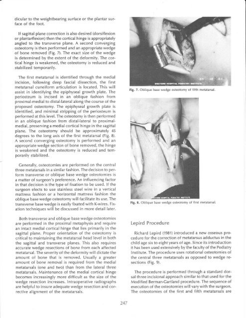
dicular to <strong>the</strong> weightbearing surface or <strong>the</strong> plantar surface<strong>of</strong> <strong>the</strong> foot.lf sagittal plane <strong>correction</strong> is also desired (dorsiflexionor plantarflexion) <strong>the</strong>n <strong>the</strong> cortical hinge is appropriatelyangled to <strong>the</strong> transverse plane. A second converging<strong>osteotomy</strong> is <strong>the</strong>n per<strong>for</strong>med and an appropriate wedge<strong>of</strong> bone removed (Fig. 7). The exact size <strong>of</strong> <strong>the</strong> wedgeis determined by <strong>the</strong> extent <strong>of</strong> <strong>the</strong> de<strong>for</strong>mity. The corticalhinge is weakened, <strong>the</strong> <strong>osteotomy</strong> is reduced andstabilized temporarily.The first metatarsal is identified through <strong>the</strong> medialincision, following deep fascial dissection, <strong>the</strong> firstmetatarsal cunei<strong>for</strong>m articulation is located. This willassist in identifying <strong>the</strong> epiphyseal growth plate. Theperiosteum is incised in an oblique fashion fromproximal-medial to distal-lateral along <strong>the</strong> course <strong>of</strong> <strong>the</strong>proposed <strong>osteotomy</strong>. The epiphyseal growth plate isidentified, and minimal stripping <strong>of</strong> <strong>the</strong> periosteum isper<strong>for</strong>med at this level. The <strong>osteotomy</strong> is <strong>the</strong>n per<strong>for</strong>medin an oblique fashion from distal-lateral to proximalmedial,preserving a medialcortical hinge in <strong>the</strong> sagittalplane. The <strong>osteotomy</strong> should be approximately 45degrees to <strong>the</strong> long axis <strong>of</strong> <strong>the</strong> first metatarsal (Fig. B).A second converging <strong>osteotomy</strong> is per<strong>for</strong>med and anappropriate wedge section <strong>of</strong> bone removed, <strong>the</strong> hingeis weakened and <strong>the</strong> <strong>osteotomy</strong> is reduced and temporarilystabilized.Cenerally, osteotomies are per<strong>for</strong>med on <strong>the</strong> centralthree metatarsals in a similar fashion. The decision to per<strong>for</strong>mtransverse or oblique base wedge osteotomies isa matter <strong>of</strong> surgeon's preference. An influencing factorin that decision is <strong>the</strong> type <strong>of</strong> fixation to be used. lf <strong>the</strong>surgeon elects to use stainless steel wire in a verticalmattress fashion or a horizontal mattress fashion <strong>the</strong>oblique base wedge <strong>osteotomy</strong> will facilitate its use. Thetransverse base wedge is easily fixated with K-wires. Fixationtechniques will be discussed in more detail later.Both transverse and oblique base wedge osteotomiesare per<strong>for</strong>med in <strong>the</strong> proximal metaphysis and requirean intact medial cortical hinge that lies primarily in <strong>the</strong>sagittal plane. Proper orientation <strong>of</strong> <strong>the</strong> <strong>osteotomy</strong> iscritical to maintaining <strong>the</strong> metatarsal head level in both<strong>the</strong> sagittal and transverse planes. This also requiresaccurate wedge resections <strong>of</strong> bone from each affectedmetatarsal. The severity <strong>of</strong> <strong>the</strong> de<strong>for</strong>mity will dictate <strong>the</strong>amount <strong>of</strong> bone that is removed. Usually a greateramount <strong>of</strong> bone removal is required from <strong>the</strong> medialmetatarsals (one and two) than from <strong>the</strong> lateral threemetatarsals. Maintenance <strong>of</strong> <strong>the</strong> medial cortical hingebecomes increasingly more difficult as <strong>the</strong> size <strong>of</strong> <strong>the</strong>wedge resection increases. lntraoperative radiographsare helpful to insure adequate wedge resection and correctivealignment <strong>of</strong> <strong>the</strong> metatarsals.Fig. 7. Oblique base wedge <strong>osteotomy</strong> <strong>of</strong> fifth metatarsal.Fig. 8. Oblique base wedge <strong>osteotomy</strong> <strong>of</strong> first metatarsal.Lepird ProcedureRichard Lepird (1981) introduced a new osseous procedure<strong>for</strong> <strong>the</strong> <strong>correction</strong> <strong>of</strong> <strong>metatarsus</strong> <strong>adductus</strong> in <strong>the</strong>child age six to eight years <strong>of</strong> age. Since its introductionit has been used extensively by <strong>the</strong> faculty <strong>of</strong> <strong>the</strong> PodiatryInstitute. The procedure uses rotational osteotomies <strong>of</strong><strong>the</strong> central three metatarsals as opposed to wedge resections(Fig. 9).The procedure is per<strong>for</strong>med through a standard dorsalthree incisional approach similar to that used <strong>for</strong> <strong>the</strong>Modified Berman-Cartland procedure. The sequence <strong>of</strong>execution <strong>of</strong> <strong>the</strong> osteotomies will vary with <strong>the</strong> surgeon.The osteotomies <strong>of</strong> <strong>the</strong> first and fifth metatarsals are247


