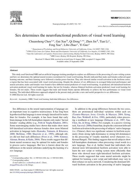
Sex determines the neurofunctional predictors of visual ... - CI Wiki
Sex determines the neurofunctional predictors of visual ... - CI Wiki
Sex determines the neurofunctional predictors of visual ... - CI Wiki
Create successful ePaper yourself
Turn your PDF publications into a flip-book with our unique Google optimized e-Paper software.
C. Chen et al. / Neuropsychologia 45 (2007) 741–747 743Fig. 1. Behavioral performance across training sessions. The upper panel (a) shows males’ and females’ reaction times on a simultaneously presented same-differentjudgment task as a function <strong>of</strong> training sessions, plotted in normal scale. The lower panels show this correlation in a log scale for males (b) and females (c),respectively; D = Day.ROI <strong>of</strong> fusiform was defined based on <strong>the</strong> group activation (including bothmales and females, p < .001, uncorrected) within <strong>the</strong> anatomical boundary<strong>of</strong> fusiform according to <strong>the</strong> automated anatomical labelling map (AAL,Tzourio-Mazoyer et al., 2002). To determine <strong>the</strong> asymmetric index (AI) in thisarea, we used <strong>the</strong> following formula: AI = (L − R)/(L + R) ×100%, where Land R represent <strong>the</strong> summed effect size 1 in <strong>the</strong> left and right ROI, respectively.A positive AI indicates left-hemispheric lateralization and a negative numberindicates right-hemispheric lateralization, and a number close to zero (i.e.,−.1 ≤ AI ≤ .1) indicates bilateral activation. The bilateral/overall fusiformactivation was calculated by combining <strong>the</strong> fusiform activation in <strong>the</strong> twohemispheres.2. Results2.1. Behavioral resultsBehavioral data indicated that our extensive training waseffective. Fig. 1a shows that <strong>the</strong> reaction time in <strong>the</strong><strong>visual</strong> judgment task decreased significantly over <strong>the</strong> trainingperiod, F(9,14) = 22.62, p < .001. The main effect for1 It should be noted that <strong>the</strong>re are o<strong>the</strong>r ways to quantify activation. In ourprevious paper (Xue et al., 2006a), we used <strong>the</strong> summed t-values as an index<strong>of</strong> activation on <strong>the</strong> basis <strong>of</strong> previous examples (e.g., Xiong et al., 1998). Bothmethods are able to characterize <strong>the</strong> activation in both intensity and volume,and result in very similar indices <strong>of</strong> brain activation. In our case, <strong>the</strong> correlationsbetween <strong>the</strong>m were .922 (<strong>the</strong> asymmetry index) and .933 (<strong>the</strong> overall activation).Fur<strong>the</strong>rmore, <strong>the</strong> pattern <strong>of</strong> sex-dependent <strong>neur<strong>of</strong>unctional</strong> <strong>predictors</strong> <strong>of</strong> <strong>visual</strong>word learning was <strong>the</strong> same no matter which method we used.sex (F(1,22) = .367, p = .551) and sex by training interaction(F(9,14) = 1.69, p = .188) were not significant.Longitudinal studies <strong>of</strong> learning have typically found thatlearning curves follow a power law (Anderson, 1983; Logan,1988), expressed in a formula form as y = x n + c, where x represents<strong>the</strong> time or <strong>the</strong> number <strong>of</strong> learning sessions, y <strong>the</strong> performance(reaction time or error rates), and c an asymptote. If<strong>the</strong> empirical data fit a power function, <strong>the</strong> correlation <strong>of</strong> log(x)and log(y − c) is close to 1. We examined <strong>the</strong> fit <strong>of</strong> male andfemale data to <strong>the</strong> power law, and <strong>the</strong> results are shown in Fig. 1band c. The correlations <strong>of</strong> log(x) (i.e., training sessions) andlog(y − c) (i.e., reaction time) were −.983 and −.986 for malesand females, respectively, suggesting a very good fit to <strong>the</strong> powerlaw.2.2. fMRI resultsFunctional MRI data indicated that, at <strong>the</strong> pre-training stage,males and females showed similar activation in <strong>the</strong> bilateraloccipital and fusiform cortices, as well as in <strong>the</strong> parietal lobule(Fig. 2a). It appeared that <strong>the</strong>re was more activation for femalesthan for males in <strong>the</strong> bilateral premotor cortex, <strong>the</strong> inferior frontalcortex, and <strong>the</strong> basal ganglia. However, masked comparisonsbetween males and females revealed no significant sex differencesat <strong>the</strong> threshold <strong>of</strong> p < .001 (uncorrected). With a lowerthreshold <strong>of</strong> p < .01 (uncorrected), more activation was foundfor females than for males in left inferior frontal cortex, right
744 C. Chen et al. / Neuropsychologia 45 (2007) 741–747Fig. 2. Brain activation during pre-training fMRI test. (a) Group-averaged brain activation for males and females when processing LAL characters relative to fixation.The threshold for this contrast is p < .001, uncorrected. (b) Direct comparison between females and males. The threshold for this comparison is p < .01, uncorrected.IFG: inferior frontal gyrus; R: right.Fig. 3. ROI results: (a) schematic representation <strong>of</strong> <strong>the</strong> bilateral fusiform ROIs defined by group-averaged activation patterns, which was overlaid onto <strong>the</strong> SPM2template. Yellow represents <strong>the</strong> left ROI and blue represents <strong>the</strong> right ROI. (b) Asymmetry index (AI) and overall activation in bilateral fusiform ROIs as a functionsex. Error bar represents standardized error <strong>of</strong> <strong>the</strong> mean. L: left; R: right.precuneus, and right lentiform (Fig. 2b). Of particular relevanceto this study, no differences were found in <strong>the</strong> fusiform cortexeven with <strong>the</strong> lowered threshold.We thus defined <strong>the</strong> functional fusiform ROIs based <strong>the</strong> groupactivation map for <strong>the</strong> whole sample (see Section 1). Consistentwith <strong>the</strong> whole-brain analysis, ROI analysis revealed nosignificant sex differences for ei<strong>the</strong>r overall fusiform activation(t(22) = .40, p = .69) or asymmetry index (t(22) = .1, p = .33)(Fig. 3).As predicted, correlational analyses showed that asymmetryin <strong>the</strong> fusiform cortex significantly predicted <strong>the</strong> post-trainingperformance for males, but not for females. Again as predicted,<strong>the</strong> overall activation in bilateral fusiform cortex predicted tosome extent <strong>the</strong> post-training performance for females, but notfor males. These correlations were relatively stable across <strong>the</strong>five post-training measurements starting with Day 6, when <strong>the</strong>rewas no obvious behavioral improvement <strong>the</strong>reafter (Fig. 4).Based on <strong>the</strong> Fisher’s r-to-z transformation test, we examinedwhe<strong>the</strong>r <strong>the</strong> correlation coefficients between asymmetryindex and reaction times for males were significantly higher(more negative) than those for females. Results showed significantsex differences at p
C. Chen et al. / Neuropsychologia 45 (2007) 741–747 745Fig. 4. Correlations and scatter plots between pre-training neural <strong>predictors</strong> (asymmetry index [a and c] and overall activation [b and d]) and post-training behavioralperformance (i.e., reaction time). D = Day; ** p < .01; * p < .05; ms: marginally significant, all two-tailed.provide compelling evidence that different neural patterns inresponse to novel stimuli are not random variations, but areactually linked to individuals’ learning ability. Fur<strong>the</strong>rmore, ourfinding that left-hemisphere dominance predicted males’ learningand bilaterality predicted females’ learning paralleled <strong>the</strong> sexdifferences in activation patterns in native language processing(i.e., left-dominance in males and bilaterality in females). Thisparallel seems to suggest that <strong>the</strong> reliance on <strong>the</strong> native languagenetwork would be optimal for <strong>the</strong> learning <strong>of</strong> new languages.As we have argued (Xue et al., 2006a), this may represent <strong>the</strong>neural tuning by native language. It appears that such tuningis beneficial to <strong>the</strong> learning <strong>of</strong> a new language, especially onethat is similar to <strong>the</strong> native language (e.g., LAL for Chinesesubjects).nificant at p < .05 even with only 6 male subjects) and −.354 for females. Thisresult seems to indicate that <strong>the</strong> sex differences we found are robust and may beindependent <strong>of</strong> training methods (<strong>visual</strong> form training in <strong>the</strong> previous study, butcomprehensive training in <strong>the</strong> present study).Results <strong>of</strong> this study also have significant methodologicalimplications for future studies <strong>of</strong> sex differences in brain functions.Using <strong>the</strong> individual-differences approach, our resultsindicated that, males and females may show similar overall neuralresponses and similar behavioral performance at <strong>the</strong> grouplevel, but <strong>the</strong>re may be different neural circuitries that lead tooptimal learning efficiency for <strong>the</strong> two sexes. Similar to previouslyreported sex differences in <strong>the</strong> associations between grayand white matters and intelligence (Haier, Jung, Yeo, Head, &Alkire, 2005), our study should help to shift <strong>the</strong> discussions onmean sex differences to individual differences within and acrosssexes and to differences in mechanisms (i.e., different mechanismsto achieve <strong>the</strong> same outcome). Such a perspective shouldhelp us to appreciate <strong>the</strong> existence <strong>of</strong> multiple “routes” (or ways<strong>of</strong> using neural resources) to <strong>the</strong> same behavioral outcome, andto avoid a simplistic examination <strong>of</strong> mean sex differences. Ofcourse, <strong>the</strong>se important implications would need much more andstronger evidence to help fur<strong>the</strong>r our understanding <strong>of</strong> sex differences,an important area <strong>of</strong> research in neuroscience (Cahill,2006).
746 C. Chen et al. / Neuropsychologia 45 (2007) 741–747Future research needs to replicate our results in o<strong>the</strong>r languagedomains (e.g., listening comprehension, production), andwith subjects speaking different languages and trying to learndifferent new languages. Future research should also address twoo<strong>the</strong>r questions. First, what is <strong>the</strong> origin <strong>of</strong> <strong>the</strong> sex differences in<strong>the</strong> differential patterns <strong>of</strong> neural resource recruitment for languagelearning and processing? One possibility is that neuraloptimization is constrained by <strong>the</strong> anatomical and physiologicalstructure <strong>of</strong> <strong>the</strong> human brain. Anatomical studies have revealedthat males have more overall volume <strong>of</strong> gray matter and whitematter than do females (Good et al., 2001), which may mean thatfemales might use more neural resources (e.g., in both hemispheres)to achieve <strong>the</strong> same cognitive performance. Consistentwith this hypo<strong>the</strong>sis, it has been revealed that females have a relativelylarger isthmus segment <strong>of</strong> <strong>the</strong> callosum, perhaps reflectinga sex-specific difference in <strong>the</strong> inter-hemispheric connectivity(Steinmetz et al., 1992; Steinmetz, Staiger, Schlaug, Huang, &Jancke, 1995). Moreover, a recent study on <strong>the</strong> relations betweenbrain structure and individual differences in general intelligencequotient (IQ) showed that, compared to men, women have morewhite matter and fewer gray matter relative to a given level <strong>of</strong>intelligence (Haier et al., 2005). In sum, it is likely <strong>the</strong> male andfemale brains are designed differently, and different operationsmight be implemented to achieve equal performance.Ano<strong>the</strong>r question is why some individuals are able to recruit<strong>the</strong> optimal neural network, whereas o<strong>the</strong>rs are not. One explanationis that neural activities may actually reflect differentcognitive strategies that correspond to different neural resourcesrecruitment. Alternatively, brain anatomy and neurophysiologymay have determined <strong>the</strong> available cognitive strategies(Breitenstein et al., 2005). Presumably, with <strong>the</strong> increase in languageexperience and fluency, <strong>the</strong> neural network will becomemore and more optimized and converge to <strong>the</strong> native languagenetwork. Long-term longitudinal studies are needed to test thishypo<strong>the</strong>sis.Taken toge<strong>the</strong>r, our study found sex-dependent neural <strong>predictors</strong><strong>of</strong> <strong>visual</strong> word learning, suggesting that sex may determine<strong>the</strong> optimal neural network for learning to read a new language.AcknowledgementsThis work was supported by <strong>the</strong> National Pandeng Program(95-special-09) to Q.D. and by School <strong>of</strong> Social Ecology, University<strong>of</strong> California, Irvine, to C.C. We would also like to thankRobert Desimone , James Fallon, and two anonymous reviewersfor <strong>the</strong>ir very insightful suggestions.ReferencesAnderson, J. R. (1983). Retrieval <strong>of</strong> information from long-term memory. Science,220(4592), 25–30.Bolger, D. J., Perfetti, C. A., & Schneider, W. (2005). Cross-cultural effect on <strong>the</strong>brain revisited: Universal structures plus writing system variation. HumanBrain Mapping, 25(1), 92–104.Breitenstein, C., Jansen, A., Deppe, M., Foerster, A. F., Sommer, J., Wolbers, T.,et al. (2005). Hippocampus activity differentiates good from poor learners<strong>of</strong> a novel lexicon. Neuroimage, 25(3), 958–968.Cahill, L. (2006). Why sex matters for neuroscience. Nature Reviews: Neuroscience,7, 477–484.Chen, Y.-P., Allport, D. A., & Marshall, J. C. (1996). What are <strong>the</strong> functionalorthographic units in Chinese word recognition: The stroke or <strong>the</strong> stroke pattern?Quarterly Journal <strong>of</strong> Experimental Psychology: Human ExperimentalPsychology, 49A(4), 1024–1043.Cohen, L., Dehaene, S., Naccache, L., Lehericy, S., Dehaene-Lambertz, G.,Henaff, M. A., et al. (2000). The <strong>visual</strong> word form area: Spatial and temporalcharacterization <strong>of</strong> an initial stage <strong>of</strong> reading in normal subjects and posteriorsplit-brain patients. Brain, 123(Pt 2), 291–307.Cohen, L., Lehericy, S., Chochon, F., Lemer, C., Rivaud, S., & Dehaene, S.(2002). Language-specific tuning <strong>of</strong> <strong>visual</strong> cortex? Functional properties <strong>of</strong><strong>the</strong> <strong>visual</strong> word form area. Brain, 125(Pt 5), 1054–1069.Dehaene, S., Dupoux, E., Mehler, J., Cohen, L., Paulesu, E., Perani, D., et al.(1997). Anatomical variability in <strong>the</strong> cortical representation <strong>of</strong> first and secondlanguage. Neuroreport, 8(17), 3809–3815.Eichelman, W. H. (1970). Familiarity effects in simultaneous matching task.Journal <strong>of</strong> Experimental Psychology, 86(2), 275–328.Friston, K. J., Ashburner, J., Frith, C. D., Poline, J. B., Hea<strong>the</strong>r, J. D., & Frackowiak,R. S. J. (1995). Spatial registration and normalization <strong>of</strong> images.Human Brain Mapping, 3(3), 165–189.Friston, K. J., Frith, C. D., Frackowiak, R. S., & Turner, R. (1995). Characterizingdynamic brain responses with fMRI: A multivariate approach. Neuroimage,2(2), 166–172.Frith, U., & Vargha-Khadem, F. (2001). Are <strong>the</strong>re sex differences in <strong>the</strong>brain basis <strong>of</strong> literacy-related skills? Evidence from reading and spellingimpairments after early unilateral brain damage. Neuropsychologia, 39(13),1485–1488.Frost, J. A., Binder, J. R., Springer, J. A., Hammeke, T. A., Bellgowan, P.S., Rao, S. M., et al. (1999). Language processing is strongly left lateralizedin both sexes: Evidence from functional MRI. Brain, 122(Pt 2), 199–208.Good, C. D., Johnsrude, I., Ashburner, J., Henson, R. N., Friston, K. J., &Frackowiak, R. S. (2001). Cerebral asymmetry and <strong>the</strong> effects <strong>of</strong> sex andhandedness on brain structure: A voxel-based morphometric analysis <strong>of</strong> 465normal adult human brains. Neuroimage, 14(3), 685–700.Haier, R. J., Jung, R. E., Yeo, R. A., Head, K., & Alkire, M. T. (2005). Theneuroanatomy <strong>of</strong> general intelligence: <strong>Sex</strong> matters. Neuroimage, 25(1),320–327.Kansaku, K., Yamaura, A., & Kitazawa, S. (2000). <strong>Sex</strong> differences in lateralizationrevealed in <strong>the</strong> posterior language areas. Cerebral Cortex, 10(9),866–872.Logan, G. D. (1988). Toward an instance <strong>the</strong>ory <strong>of</strong> automatization. PsychologicalReview, 95(4), 492–527.McGlone, J. (1980). <strong>Sex</strong> differences in human-brain asymmetry—A criticalsurvey. Behavioral and Brain Sciences, 3(2), 215–227.Shaywitz, B. A., Shaywitz, S. E., Pugh, K. R., Constable, R. T., Skudlarski, P.,Fulbright, R. K., et al. (1995). <strong>Sex</strong> differences in <strong>the</strong> functional organization<strong>of</strong> <strong>the</strong> brain for language. Nature, 373(6515), 607–609.Snyder, P. J., & Harris, L. J. (1993). Handedness, sex, and familial sinistralityeffects on spatial tasks. Cortex, 29(1), 115–134.Sommer, I. E., Aleman, A., Bouma, A., & Kahn, R. S. (2004). Do women reallyhave more bilateral language representation than men? A meta-analysis <strong>of</strong>functional imaging studies. Brain, 127(Pt 8), 1845–1852.Steinmetz, H., Jancke, L., Kleinschmidt, A., Schlaug, G., Volkmann, J., &Huang, Y. (1992). <strong>Sex</strong> but no hand difference in <strong>the</strong> isthmus <strong>of</strong> <strong>the</strong> corpuscallosum. Neurology, 42(4), 749–752.Steinmetz, H., Staiger, J. F., Schlaug, G., Huang, Y., & Jancke, L. (1995).Corpus callosum and brain volume in women and men. Neuroreport, 6(7),1002–1004.Taylor, I., & Olson, D. R. (Eds.). (1995). Scripts and literacy: Reading and learningto read alphabets, syllabaries and characters. New York, NY: KluwerAcademic/Plenum Publishers.Tzourio-Mazoyer, N., Josse, G., Crivello, F., & Mazoyer, B. (2004). Interindividualvariability in <strong>the</strong> hemispheric organization for speech. Neuroimage,21(1), 422–435.Tzourio-Mazoyer, N., Landeau, B., Papathanassiou, D., Crivello, F., Etard, O.,Delcroix, N., et al. (2002). Automated anatomical labeling <strong>of</strong> activations in
C. Chen et al. / Neuropsychologia 45 (2007) 741–747 747SPM using a macroscopic anatomical parcellation <strong>of</strong> <strong>the</strong> MNI MRI singlesubjectbrain. Neuroimage, 15(1), 273–289.Xiong, J., Rao, S., Gao, J. H., Woldorff, M., & Fox, P. T. (1998). Evaluation <strong>of</strong>hemispheric dominance for language using functional MRI: A comparisonwith positron emission tomography. Human Brain Mapping, 6(1), 42–58.Xue, G., Chen, C., Jin, Z., & Dong, Q. (2006a). Cerebral asymmetry in <strong>the</strong>fusiform areas predicted <strong>the</strong> efficiency <strong>of</strong> learning a new writing system.Journal <strong>of</strong> Cognitive Neuroscience, 18(6), 923–931.Xue, G., Chen, C., Jin, Z., & Dong, Q. (2006b). Language experience shapesfusiform activation when processing a logographic artificial language: AnfMRI training study. Neuroimage, 31(3), 1315–1326.Xue, G., Dong, Q., Chen, K., Jin, Z., Chen, C., Zeng, Y., et al. (2005). Cerebralasymmetry in children when reading Chinese characters. Brain Research.Cognitive Brain Research, 24(2), 206–214.


