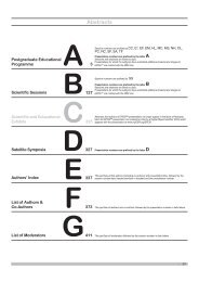
Postgraduate Educational Programme - myESR.org
Postgraduate Educational Programme - myESR.org
Postgraduate Educational Programme - myESR.org
- No tags were found...

Create successful ePaper yourself
Turn your PDF publications into a flip-book with our unique Google optimized e-Paper software.
<strong>Postgraduate</strong> <strong>Educational</strong> <strong>Programme</strong>A-180 16:30B. Musculoskeletal neoplasmsM.F. Reiser; Munich/DEDiagnosis, staging and follow‐up of musculoskeletal tumours are most importantfor selection of adequate therapeutic measures and prognosis of patients. Modernimaging technologies have greatly contributed to improvement. With modern MRIsystems high contrast of neoplastic tissue versus normal uninvolved structures isachieved. This allows for adequate delineation of intraosseous and soft tissue extensionand thereby facilitating high precision in staging. Compartmental infiltrationis readily detected. Dynamic, contrast-enhanced studies allow for assessment ofvascularity and perfusion, which provides valuable information concerning malignancyand benign character of a particular tumour. With diffusion-weighted imagingand diffusion-tensor imaging, microstructural information can be obtained, whichcorrelates with response to chemotherapy and viability of the tissue. Malignantbone tumours may spread within the bone or to distant <strong>org</strong>ans. Therefore, wholebody imaging modalities hold great potential for comprehensive assessment. Incomputer tomography, major advances have been achieved recently. The combinationof CT and PET in hybrid systems allows to assess function and morphologywithin one session. With FDG-PET, the metabolic activity of a particular lesion canbe analyzed. The standard uptake value (SUV) can be utilised for differentiationof benign malignant lesions. However, there are various benign lesions, such asNOF, fibrous dysplasia, eosinophilic granuloma and aneuysmal bone cysts as wellas inflammation and infection. False-negative results in FDG-PET may be found inlow-grade chondrosarcoma, multiple myeloma, low-grade osteosarcoma, Ewing’ssarcoma and low-grade soft tissue sarcomas.Learning Objectives:1. To become familiar with the imaging modalities which enable to detect anddifferentiate benign and malignant bone neoplasms.2. To consolidate knowledge of radiographic, CT and MRI findings which enableto classify and stage bone tumours.3. To understand the potential role of PET-CT and whole body MRI.4. To learn the signs indicative of favourable and poor response to preoperativechemotherapy and for recurrence of malignant bone tumours.A-181 16:55C. Chemo- and radiation therapy-induced toxicityH.‐U. Kauczor; Heidelberg/DE (Hans‐Ulrich.Kauczor@med.uni‐heidelberg.de)Multimodal cancer therapy, including chemotherapy, biologicals and radiotherapy, isfrequently associated with adverse toxic effects. Depending on the specific agent,different <strong>org</strong>ans, such as brain, lung, liver, bowel, bone, heart might be affected.„Inflammatory“ reactions will occur. These will be detected on imaging as whitematter lesions in the brain, reticular changes and consolidations in the lung (fibrosis,cryptogenic <strong>org</strong>anizing pneumonia), diffuse liver disease, colitis, bone necrosis, orcardiomyopathy. These direct toxic effects have to be differentiated from infectiouscomplications due to chemotherapy-induced neutropenia. These infections mainlyaffect the lung and are caused by fungi or viruses. Angioinvasive aspergillosis ofthe lung is the most frequent, but sinusitis, abscesses in brain and liver as well assepsis with haematogeneous foci are also encountered. Toxic effects of radiotherapywill mainly occur within the planned target volume and result from application ofhigh doses to radiosensitive normal tissue leading to inflammation, fibrosis andnecrosis. As imaging is routinely performed for therapy response monitoring orsurveillance in patients suffering from cancer, toxic effects have to be differentiatedfrom infectious complications, postsurgical or postradiation scar tissue or tumourrecurrence. Specific patterns of toxic effects of cancer therapy in brain, lung, liver,pelvis and their differential diagnoses will be reviewed.Learning Objectives:1. To get an overview of <strong>org</strong>an-specific toxicity and other adverse effects ofchemo- and radiotherapy.2. To review the key imaging findings of therapy-induced <strong>org</strong>an toxicity andadverse effects.3. To understand how to differentiate inflammatory, infectious, fibrotic, and necroticchanges from tumour recurrence.Author Disclosure:H. -U. Kauczor: Advisory Board; Siemens. Investigator; Boehringer Ingelheim.Speaker; Siemens, Boehringer, Bayer, Bracco.Questions 17:2016:00 - 17:30 Room L/MOrgans from A to Z: HeartMC 722Ischaemic heart diseaseModerator:C. Catalano; Rome/ITA-182 16:00A. Imaging of the coronary arteries: the Holy GrailG. Roditi; Glasgow/UK (Giles.Roditi@ggc.scot.nhs.uk)Small critical structures in the human body require high spatial resolution imaging.Coronary artery disease carries a high disease burden regarding morbidityand mortality. The coronary arteries are not only small but also move rapidly andin a complex manner predicated on movements of the beating heart with superimposedasynchronous respiratory motion. Conventional angiography has hightemporal fidelity and spatial resolution, however, this lumenogram does not fullyevaluate coronary disease, a process affecting the vessel wall. The non-invasiveacquisition of three-dimensional data combining high spatial and temporal resolutionfor coronary visualisation has proven challenging, hence the description asthe ‘Holy Grail’. The Holy Grail was sought not as an object but for what it coulddo. In similar fashion not only are the heart and related great vessels now reliablyimaged in unprecedented detail but the heart can be reincorporated into a holisticevaluation of the whole patient and the advances in technology allowing coronaryCT to enter clinical routine have benefited many other CT techniques. Advancesin temporal resolution, broad detector arrays and dual energy imaging spurred bycardiac CT have expanded the opportunities in other areas such as whole <strong>org</strong>anCT perfusion. Perhaps more importantly the high radiation doses delivered by theinitial schema for cardiac CT have led to development of dose reduction techniqueswith profound benefits in all CT applications as the risk:benefit changes comparedto plain film radiography become compelling to allow CT as a first line modalityin many situations.Learning Objectives:1. To learn about the meaning of CT coronary calcium screening for risk assessment.2. To identify suitable modalities and challenges for non-invasive coronary angiography.3. To understand the potential of coronary plaque imaging beyond calcium.Author Disclosure:G. Roditi: Advisory Board; Bayer Advisory Board member 2012. Consultant;past consulting for Toshiba Medical Visualisation Systems Europe. Speaker;In the past 5 years I have undertaken speaking engagements for the following:Bayer, Bracco, Guerbet, GE Healthcare & Toshiba Medical Systems.A-183 16:20B. The ischaemic myocardium: what to do?C. Loewe; Vienna/AT (christian.loewe@meduniwien.ac.at)In case of ischaemic heart disease, the assessment of myocardial viability is crucialfor treatment decision making. Simply, patients with viable tissue left will improveby revascularization therapy, whereas treatment of non-viable tissue by revascularizationcould be fatal. Assessment of myocardial viability by imaging is dedicatedto and follows different distinct pathophysiological mechanisms. It includes theassessment of regional wall motion abnormalities, regional perfusion deficits,abnormal metabolism or the direct demonstration of myocardial scars. Differentmethods have been established so far for demonstration of the different abnormalitiesincluding SPECT (perfusion), PET (metabolism), PET/CT (metabolism) andcardiac MR (perfusion, function, scar). Recently, even cardiac CT was introducedas comparative method for the assessment of myocardial viability and function ofischaemic myocardium combined with the possibility of direct demonstration of thecoronary pathologies. Given the number of different modalities and their differentapproaches, knowledge about the specific strengths of each method is crucial foradequate management of patients with ischaemic heart disease. In this presentation,the importance of careful assessment of myocardium in ischaemic heartdisease for adequate treatment decision making and planning will be underlined.The different methods with their different approaches to myocardial assessmentshould be presented together with possible advantages and disadvantages. Basedon this, potential imaging algorithm will be proposed. In addition, possibilities ofcareful assessment of contractile reserve will be presented. Finally, therapeuticconsequences of different imaging results will be demonstrated based on clinicalcases, and the most important differentials should be shown.S46AB C D E F G