A JournAl of the AmericAn DiAbetes AssociAtion
A JournAl of the AmericAn DiAbetes AssociAtion
A JournAl of the AmericAn DiAbetes AssociAtion
- No tags were found...
You also want an ePaper? Increase the reach of your titles
YUMPU automatically turns print PDFs into web optimized ePapers that Google loves.
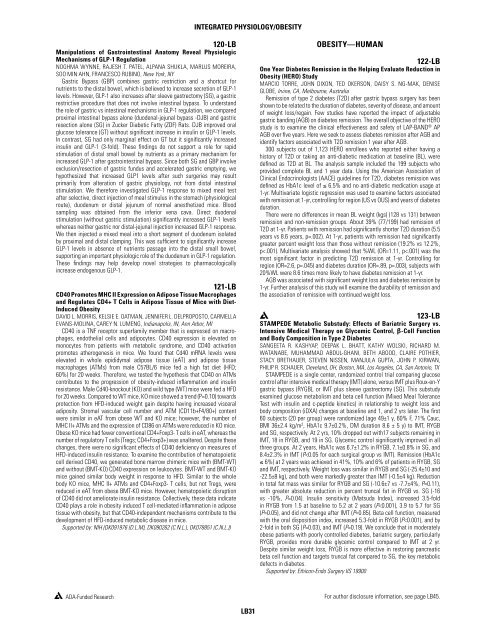
Integrated Physiology/Obesity120-LBManipulations <strong>of</strong> Gastrointestinal Anatomy Reveal PhysiologicMechanisms <strong>of</strong> GLP-1 RegulationNOGHMA WYNNE, RAJESH T. PATEL, ALPANA SHUKLA, MARLUS MOREIRA,SOO MIN AHN, FRANCESCO RUBINO, New York, NYGastric Bypass (GBP) combines gastric restriction and a shortcut fornutrients to <strong>the</strong> distal bowel, which is believed to increase secretion <strong>of</strong> GLP-1levels. However, GLP-1 also increases after sleeve gastrectomy (SG), a gastricrestrictive procedure that does not involve intestinal bypass. To understand<strong>the</strong> role <strong>of</strong> gastric vs intestinal mechanisms in GLP-1 regulation, we comparedproximal intestinal bypass alone (duodenal-jejunal bypass -DJB) and gastricresection alone (SG) in Zucker Diabetic Fatty (ZDF) Rats. DJB improved oralglucose tolerance (GT) without significant increase in insulin or GLP-1 levels.In contrast, SG had only marginal effect on GT but it significantly increasedinsulin and GLP-1 (3-fold). These findings do not support a role for rapidstimulation <strong>of</strong> distal small bowel by nutrients as a primary mechanism forincreased GLP-1 after gastrointestinal bypass. Since both SG and GBP involveexclusion/resection <strong>of</strong> gastric fundus and accelerated gastric emptying, wehypo<strong>the</strong>sized that increased GLP1 levels after such surgeries may resultprimarily from alteration <strong>of</strong> gastric physiology, not from distal intestinalstimulation. We <strong>the</strong>refore investigated GLP-1 response to mixed meal testafter selective, direct injection <strong>of</strong> meal stimulus in <strong>the</strong> stomach (physiologicalroute), duodenum or distal jejunum <strong>of</strong> normal anes<strong>the</strong>tized mice. Bloodsampling was obtained from <strong>the</strong> inferior vena cava. Direct duodenalstimulation (without gastric stimulation) significantly increased GLP-1 levelswhereas nei<strong>the</strong>r gastric nor distal-jejunal injection increased GLP-1 response.We <strong>the</strong>n injected a mixed meal into a short segment <strong>of</strong> duodenum isolatedby proximal and distal clamping. This was sufficient to significantly increaseGLP-1 levels in absence <strong>of</strong> nutrients passage into <strong>the</strong> distal small bowel,supporting an important physiologic role <strong>of</strong> <strong>the</strong> duodenum in GLP-1 regulation.These findings may help develop novel strategies to pharmacologicallyincrease endogenous GLP-1.121-LBCD40 Promotes MHC II Expression on Adipose Tissue Macrophagesand Regulates CD4+ T Cells in Adipose Tissue <strong>of</strong> Mice with Diet-Induced ObesityDAVID L. MORRIS, KELSIE E. OATMAN, JENNIFER L. DELPROPOSTO, CARMELLAEVANS-MOLINA, CAREY N. LUMENG, Indianapolis, IN, Ann Arbor, MICD40 is a TNF receptor superfamily member that is expressed on macrophages,endo<strong>the</strong>lial cells and adipocytes. CD40 expression is elevated onmonocytes from patients with metabolic syndrome, and CD40 activationpromotes a<strong>the</strong>rogenesis in mice. We found that Cd40 mRNA levels wereelevated in whole epididymal adipose tissue (eAT) and adipose tissuemacrophages (ATMs) from male C57BL/6 mice fed a high fat diet (HFD;60%) for 20 weeks. Therefore, we tested <strong>the</strong> hypo<strong>the</strong>sis that CD40 on ATMscontributes to <strong>the</strong> progression <strong>of</strong> obesity-induced inflammation and insulinresistance. Male Cd40-knockout (KO) and wild type (WT) mice were fed a HFDfor 20 weeks. Compared to WT mice, KO mice showed a trend (P=0.10) towardsprotection from HFD-induced weight gain despite having increased visceraladiposity. Stromal vascular cell number and ATM (CD11b+F4/80+) contentwere similar in eAT from obese WT and KO mice; however, <strong>the</strong> number <strong>of</strong>MHC II+ ATMs and <strong>the</strong> expression <strong>of</strong> CD86 on ATMs were reduced in KO mice.Obese KO mice had fewer conventional CD4+Foxp3- T cells in eAT, whereas <strong>the</strong>number <strong>of</strong> regulatory T cells (Tregs; CD4+Foxp3+) was unaltered. Despite <strong>the</strong>sechanges, <strong>the</strong>re were no significant effects <strong>of</strong> CD40 deficiency on measures <strong>of</strong>HFD-induced insulin resistance. To examine <strong>the</strong> contribution <strong>of</strong> hematopoieticcell derived CD40, we generated bone marrow chimeric mice with (BMT-WT)and without (BMT-KO) CD40 expression on leukocytes. BMT-WT and BMT-KOmice gained similar body weight in response to HFD. Similar to <strong>the</strong> wholebody KO mice, MHC II+ ATMs and CD4+Foxp3- T cells, but not Tregs, werereduced in eAT from obese BMT-KO mice. However, hematopoietic disruption<strong>of</strong> CD40 did not ameliorate insulin resistance. Collectively, <strong>the</strong>se data indicateCD40 plays a role in obesity induced T cell-mediated inflammation in adiposetissue with obesity, but that CD40-independent mechanisms contribute to <strong>the</strong>development <strong>of</strong> HFD-induced metabolic disease in mice.Supported by: NIH (DK091976 (D.L.M), DK090262 (C.N.L.), DK078851 (C.N.L.))Obesity—Human122-LBOne Year Diabetes Remission in <strong>the</strong> Helping Evaluate Reduction inObesity (HERO) StudyMARCIO TORRE, JOHN DIXON, TED OKERSON, DAISY S. NG-MAK, DENISEGLOBE, Irvine, CA, Melbourne, AustraliaRemission <strong>of</strong> type 2 diabetes (T2D) after gastric bypass surgery has beenshown to be related to <strong>the</strong> duration <strong>of</strong> diabetes, severity <strong>of</strong> disease, and amount<strong>of</strong> weight loss/regain. Few studies have reported <strong>the</strong> impact <strong>of</strong> adjustablegastric banding (AGB) on diabetes remission. The overall objective <strong>of</strong> <strong>the</strong> HEROstudy is to examine <strong>the</strong> clinical effectiveness and safety <strong>of</strong> LAP-BAND ® APAGB over five years. Here we seek to assess diabetes remission after AGB andidentify factors associated with T2D remission 1 year after AGB.300 subjects out <strong>of</strong> 1,123 HERO enrollees who reported ei<strong>the</strong>r having ahistory <strong>of</strong> T2D or taking an anti-diabetic medication at baseline (BL), weredefined as T2D at BL. The analysis sample included <strong>the</strong> 199 subjects whoprovided complete BL and 1 year data. Using <strong>the</strong> American Association <strong>of</strong>Clinical Endocrinologists (AACE) guidelines for T2D, diabetes remission wasdefined as HbA1c level <strong>of</strong> ≤ 6.5% and no anti-diabetic medication usage at1-yr. Multivariate logistic regression was used to examine factors associatedwith remission at 1-yr, controlling for region (US vs OUS) and years <strong>of</strong> diabetesduration.There were no differences in mean BL weight (kgs) (128 vs 131) betweenremission and non-remission groups. About 39% (77/199) had remission <strong>of</strong>T2D at 1-yr. Patients with remission had significantly shorter T2D duration (5.5years vs 8.6 years, p=.002). At 1-yr, patients with remission had significantlygreater percent weight loss than those without remission (19.2% vs 12.2%,p


