A JournAl of the AmericAn DiAbetes AssociAtion
A JournAl of the AmericAn DiAbetes AssociAtion
A JournAl of the AmericAn DiAbetes AssociAtion
- No tags were found...
Create successful ePaper yourself
Turn your PDF publications into a flip-book with our unique Google optimized e-Paper software.
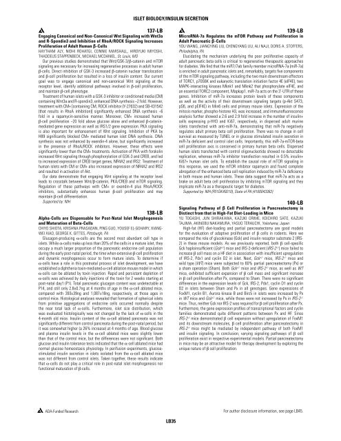
Islet Biology/Insulin Secretion137-LBEngaging Canonical and Non-Canonical Wnt Signaling with Wnt3aand R-Spondin3 and Inhibition <strong>of</strong> RhoA/ROCK Signaling IncreasesProliferation <strong>of</strong> Adult Human β-CellsHAYTHAM ALY, NIDHI ROHATGI, CONNIE MARSHALL, HIROYUKI MIYOSHI,THADDEUS STAPPENBECK, MICHAEL MCDANIEL, St. Louis, MOOur previous studies demonstrated that Wnt/GSK-3/β-catenin and mTORsignaling are necessary for increasing regenerative processes in adult humanβ-cells. Direct inhibition <strong>of</strong> GSK-3 increased β-catenin nuclear translocationand β-cell proliferation but resulted in a loss <strong>of</strong> insulin content. Our currentgoal was to engage canonical and non-canonical Wnt signaling at <strong>the</strong>receptor level, identify additional pathways involved in β-cell proliferation,and maintain β-cell phenotype.Treatment <strong>of</strong> human islets with a GSK-3 inhibitor or conditioned media (CM)containing Wnt3a and R-spondin3, enhanced DNA syn<strong>the</strong>sis ~2 fold. However,treatment with CM+ [containing CM, ROCK inhibitor (Y-27632) and SB-431542(that results in RhoA inhibition)] significantly enhanced DNA syn<strong>the</strong>sis ~6fold in a rapamycin-sensitive manner. Moreover, CM+ increased humanβ-cell proliferation ~20 fold above glucose alone and enhanced β-cateninmediatedgene expression as well as IRS1/2 gene expression. PKA signalingis also important for enhancement <strong>of</strong> Wnt signaling. Inhibition <strong>of</strong> PKA byH89 significantly blocked CM+ mediated human islet DNA syn<strong>the</strong>sis. DNAsyn<strong>the</strong>sis was not enhanced by exendin-4 alone, but significantly increasedin <strong>the</strong> presence <strong>of</strong> RhoA/ROCK inhibitors. However, <strong>the</strong>se effects weresignificantly lower than <strong>the</strong> CM+ treatments. Activation <strong>of</strong> PKA with forskolinincreased Wnt signaling through phosphorylation <strong>of</strong> GSK-3 and CREB, and ledto increased expression <strong>of</strong> CREB target genes, NR4A2 and IRS2. Treatment <strong>of</strong>human islets with CM or CM+ also increased expression <strong>of</strong> NR4A2 and IRS2and resulted in activation <strong>of</strong> Akt.Our data demonstrate that engaging Wnt signaling at <strong>the</strong> receptor levelleads to crosstalk between Wnt/β-catenin, PKA/CREB and mTOR signaling.Regulation <strong>of</strong> <strong>the</strong>se pathways with CM+ or exendin-4 plus RhoA/ROCKinhibitors, substantially enhances human β-cell proliferation and maymaintain β-cell differentiation.Supported by: NIH138-LBAlpha-Cells are Dispensable for Post-Natal Islet Morphogenesisand Maturation <strong>of</strong> Beta-CellsCHIYO SHIOTA, KRISHNA PRASADAN, PING GUO, YOUSEF EL-GOHARY, XIANG-WEI XIAO, GEORGE K. GITTES, Pittsburgh, PAGlucagon-producing α-cells are <strong>the</strong> second most abundant cell type inislets. While α-cells make up less than 20% <strong>of</strong> <strong>the</strong> cells in a mature islet, <strong>the</strong>yoccupy a much larger proportion <strong>of</strong> <strong>the</strong> pancreatic endocrine cell populationduring <strong>the</strong> early post-natal period, <strong>the</strong> time when extensive β-cell proliferationand dynamic morphogenesis occur to form mature islets. To determine ifα-cells have a role in this postnatal process <strong>of</strong> islet development, we haveestablished a diph<strong>the</strong>ria toxin-mediated α-cell ablation mouse model in whichα-cells can be ablated by toxin injection. Rapid and persistant depletion <strong>of</strong>α-cells was achieved by daily injections <strong>of</strong> <strong>the</strong> toxin for 2 weeks starting atpost-natal day1 (P1). Total pancreatic glucagon content was undetectable atP14, and still only 2.8±0.7ng at 4 months <strong>of</strong> age in <strong>the</strong> α-cell ablated mice,compared with 393±39ng and 1,087±18ng, respectively, at those ages incontrol mice. Histological analyses revealed that formation <strong>of</strong> spherical isletsfrom primitive aggregations <strong>of</strong> endocrine cells occurred normally despite<strong>the</strong> near total lack <strong>of</strong> α-cells. Fur<strong>the</strong>rmore, islet size distribution, whichwas evaluated histologically was not changed by <strong>the</strong> lack <strong>of</strong> α-cells in <strong>the</strong>4-month old mice. Insulin content <strong>of</strong> <strong>the</strong> α-cell ablated pancreata was notsignificantly different from control pancreata during <strong>the</strong> post-natal period, butit was somewhat higher (a 26% increase) at 4 months <strong>of</strong> age. Blood glucoseand plasma insulin levels in <strong>the</strong> α-cell ablated mice were slightly lowerthan that <strong>of</strong> <strong>the</strong> control mice, but <strong>the</strong> differences were not significant. Bothglucose and insulin tolerance tests indicated that <strong>the</strong> α-cell ablated mice hadnormal glucose homeostasis physiology. In perifusion experiments, glucosestimulatedinsulin secretion in islets isolated from <strong>the</strong> α-cell ablated micewas not different from control islets. Taken toge<strong>the</strong>r, <strong>the</strong>se results indicatethat α-cells do not play a critical role in post-natal islet morphogenesis norfunctional maturation <strong>of</strong> β-cells.139-LBMicroRNA-7a Regulates <strong>the</strong> mTOR Pathway and Proliferation inAdult Pancreatic β-CellsYOU WANG, JIANGYING LIU, CHENGYANG LIU, ALI NAJI, DORIS A. STOFFERS,Philadelphia, PAElucidating <strong>the</strong> mechanism underlying <strong>the</strong> poor proliferative capacity <strong>of</strong>adult pancreatic beta cells is critical to regenerative <strong>the</strong>rapeutic approachesfor diabetes. We find that <strong>the</strong> miR7/7ab family member microRNA-7a (miR-7a)is enriched in adult pancreatic islets and, remarkably, targets five components<strong>of</strong> <strong>the</strong> mTOR signaling pathway, including <strong>the</strong> two main downstream effectors<strong>of</strong> TORC1, p70S6K and eukaryotic translation initiation factor 4E (eIF4E), twoMAPK-interacting kinases Mknk1 and Mknk2 that phosphorylate eIF4E, andan essential TORC2 component, Mapkap1. miR-7a acts on <strong>the</strong> 3’-UTR <strong>of</strong> <strong>the</strong>segenes. Inhibition <strong>of</strong> miR-7a increases protein levels <strong>of</strong> <strong>the</strong>se componentsas well as <strong>the</strong> activity <strong>of</strong> <strong>the</strong>ir downstream signaling targets (p-Akt S473,pS6, and pEIF4E) in Min6 cells and primary mouse islets. Expression <strong>of</strong> <strong>the</strong>mitosis marker, phospho-histone H3, was increased, and immun<strong>of</strong>luorescenceanalysis fur<strong>the</strong>r showed a 2.6 and 2.9 fold increase in <strong>the</strong> number <strong>of</strong> insulin+cells expressing p-HH3 and Ki67, respectively, in dispersed adult murineislets transfected with anti-miR-7a, demonstrating that miR-7a negativelyregulates adult primary beta cell proliferation. There was no change in cellsurvival as measured by TUNEL or in glucose stimulated insulin secretion inmiR-7a deficient and control islet cells. Importantly, this miR-7a-mTOR-betacell proliferation axis is conserved in primary human beta cells. Dispersedhuman islets transfected with control oligonucleotide showed no detectablereplication, whereas miR-7a inhibitor transfection resulted in 0.5% insulin+Ki67+ human islet cells. To establish <strong>the</strong> causal role <strong>of</strong> mTOR signaling inthis response, we used <strong>the</strong> mTOR inhibitor rapamycin and found completeabrogation <strong>of</strong> <strong>the</strong> enhanced beta cell replication induced by miR-7a deficiencyin both mouse and human islets. These data suggest that miR-7a acts as abrake on adult beta cell proliferation by inhibiting mTOR signaling and <strong>the</strong>yimplicate miR-7a as a <strong>the</strong>rapeutic target for diabetes.Supported by: NIH (P01DK49210), State <strong>of</strong> PA (4100043362)140-LBSignaling Pathway <strong>of</strong> β Cell Proliferation in Pancreatectomy isDistinct from that in High-Fat Diet-Loading in MiceYU TOGASHI, JUN SHIRAKAWA, KAZUKI ORIME, KOICHIRO SATO, KAZUKITAJIMA, AKINOBU NAKAMURA, YASUO TERAUCHI, Yokohama, JapanHigh-fat (HF) diet-loading and partial pancreatectomy are good modelsfor <strong>the</strong> evaluation <strong>of</strong> adaptive proliferation <strong>of</strong> β cells in rodents. Here wecompared <strong>the</strong> role <strong>of</strong> glucokinase (Gck) and insulin receptor substrate 2 (IRS-2) in <strong>the</strong>se mouse models. As we previously reported, both β cell-specificGck haploinsufficient (Gck +/- ) mice and IRS-2-deficient (IRS-2 -/- ) mice failed toincrease β cell mass on a HF diet in association with insufficient upregulation<strong>of</strong> IRS-2, Pdx1 and cyclin D2 in islet. Next, Gck +/- mice, IRS-2 -/- mice andwild type (WT) mice were subjected to 60% partial pancreatectomy (Px) ora sham operation (Sham). Both Gck +/- mice and IRS-2 -/- mice, as well as WTmice, exhibited sufficient expansion <strong>of</strong> β cell mass and significant increasein β cell proliferation after Px, compared to Sham. There were no significantdifferences in <strong>the</strong> expression levels <strong>of</strong> Gck, IRS-2, Pdx1, cyclin D1 and cyclinD2 in islets between Sham and Px in all genotypes. Gene expressions <strong>of</strong>FoxM1, cyclin B1, Aurora kinase B and Birc5 in islets were increased by Pxin WT mice and Gck +/- mice, while those were not increased by Px in IRS-2 -/-mice. Thus, nei<strong>the</strong>r Gck nor IRS-2 was required for β cell proliferation after Px.Fur<strong>the</strong>rmore, <strong>the</strong> gene expression pr<strong>of</strong>iles <strong>of</strong> transcriptional factors and cyclinfamilies demonstrated quite different patterns between Px and HF. SinceIRS-2 -/- mice demonstrated β cell expansion without upregulation <strong>of</strong> FoxM1and its downstream molecules, β cell proliferation after pancreatectomy inIRS-2 -/- mice might be mediated by independent pathway <strong>of</strong> both FoxM1and insulin signaling. In conclusion, varying signaling pathways <strong>of</strong> β cellproliferation exist in respective experimental models. Partial pancreatectomyin mice may be an attractive model for <strong>the</strong>rapy development by exploring <strong>the</strong>unique nature <strong>of</strong> β cell proliferation.ADA-Funded ResearchFor author disclosure information, see page LB45.LB35


