TREATMENT OF DENTAL FLUOROSIS - Journal of IMAB
TREATMENT OF DENTAL FLUOROSIS - Journal of IMAB
TREATMENT OF DENTAL FLUOROSIS - Journal of IMAB

You also want an ePaper? Increase the reach of your titles
YUMPU automatically turns print PDFs into web optimized ePapers that Google loves.
<strong>Journal</strong> <strong>of</strong> <strong>IMAB</strong> - Annual Proceeding (Scientific Papers) 2008, book 2<strong>TREATMENT</strong> <strong>OF</strong> <strong>DENTAL</strong> <strong>FLUOROSIS</strong>Milena PenevaDepartment <strong>of</strong> Children’s dental medicineFaculty <strong>of</strong> Dental medicine, Medical university - S<strong>of</strong>ia, BulgariaSUMMARYDental fluorosis is the result <strong>of</strong> chronic endogenicintake <strong>of</strong> fluorides in amounts exceeding the optimal dailydose <strong>of</strong> 1 ppmIn our country different mineral waters with highcontent <strong>of</strong> fluorine are on the market. As a result innumerous regions and settlements the cases <strong>of</strong> fluorosiswith different degrees <strong>of</strong> seriousness have become morefrequent in recent years.The aim <strong>of</strong> this study is to test the application <strong>of</strong>hydrochloric acid-pumice abrasion for the removal <strong>of</strong>fluorosis stains and remineralisation <strong>of</strong> the enamel.At the Department <strong>of</strong> Children’s dental medicine tothe Medical university - S<strong>of</strong>ia 18 children aged 4 - 15 weretreated for different degrees <strong>of</strong> fluorosis. The method <strong>of</strong>microabrasion was applied employing the ready-madeproduct „Opalustre”, and the remineralisation - employingTooth Mousse. In the case <strong>of</strong> all chidren individualprophylactic programs were implemented, includingmotivation and training in oral hygiene, a food regimen andrules <strong>of</strong> carbohydrate intake, avoiding fluorine water, control<strong>of</strong> the local fluorine toothpastes used, etc.The treatment with microabrasion conducted and thesubsequent remineralisation give very good results. Theseresults include a recovery <strong>of</strong> the aesthetics <strong>of</strong> the teeth,remineralisation <strong>of</strong> the enamel, full disappearance <strong>of</strong> theenamel erosions and a dimimution <strong>of</strong> the dentine erosions.Dental fluorosis is the result <strong>of</strong> chronic endogenicintake <strong>of</strong> fluorides in amounts exceeding the optimal dailydose <strong>of</strong> 1 ppm (8, 13). Depending on the quantity <strong>of</strong> theintake different degrees <strong>of</strong> changes in the enamel areobserved. These changes can be slight, namely involvingthe emergence <strong>of</strong> white lines or stains on the enamel, thelines in question being vertical or horozontal. The mediumdegree <strong>of</strong> changes involves yellow to brown stains, the harddegree <strong>of</strong> changes involving an overall affection <strong>of</strong> theenamel with chalk-white stains, as well as with numerousdark-coloured stains and enamel losses (8, 11, 12, 13). Themicroscopic changes come down to a disturbed mineralisation<strong>of</strong> the enamel, the process involving different degrees<strong>of</strong> increased porosity <strong>of</strong> the subsurface layers (6, 13).Dental fluorosis is observed in regions with excessiveoccurrence <strong>of</strong> fluorine in the drinking water or is the result<strong>of</strong> overdoses <strong>of</strong> fluorine within an endogenic fluorineprophylaxis. In children the reason can even be the dailyintake <strong>of</strong> the fluorine tooth paste. In our country differentmineral waters with high content <strong>of</strong> fluorine are on themarket. For instance the Devin water has 4 milligrams <strong>of</strong>fluorine per litre, the Hissar water has 4.8 milligrams <strong>of</strong>fluorine per litre, etc. The absence <strong>of</strong> public programs forprophylaxis <strong>of</strong> the dental caries brings about a chaotic intake<strong>of</strong> fluorine <strong>of</strong> different origin, with no control on the part <strong>of</strong>competent and knowledgeable personnel. As a result innumerous regions and settlements the cases <strong>of</strong> fluorosiswith different degrees <strong>of</strong> seriousness have become morefrequent in recent years. Part <strong>of</strong> these cases bring onlyabout a cosmetic defect, while others represent a seriousmedical problem requiring medical treatment.It was in the 80-ties that the successful removal <strong>of</strong>the stains on the enamel surface caused by fluorine startedas a medical procedure. In the beginning an 18% solution<strong>of</strong> hydrochoric acid was used for treatment <strong>of</strong> the surface<strong>of</strong> the enamel (10). Later Croll and Cavanaugh introduced amodified procedure called „change <strong>of</strong> the enamel colourthrough controlled abrasion by means <strong>of</strong> hydrochloric acidand pumice” (1, 2, 3, 4). After that different schemes <strong>of</strong>treatment through different concentration <strong>of</strong> hydrochloricacid and different durations <strong>of</strong> the procedure wereimplemented, the term for the technique applied remaining„microabrasion” (5, 6, 9, 13, 14).It is the aim <strong>of</strong> this study to test the application <strong>of</strong>hydrochloric acid-pumice abrasion for the removal <strong>of</strong>fluorosis stains and remineralisation <strong>of</strong> the enamel.MATERIAL AND METHODSAt the Department <strong>of</strong> Children’s dental medicine tothe Medical university - S<strong>of</strong>ia 18 children aged 4 - 15 weretreated for different degrees <strong>of</strong> fluorosis. Through theanamnesis <strong>of</strong> all children’s condition an excessive and <strong>of</strong>many years intake <strong>of</strong> fluorine was ascertained. Most caseshad to do with the consumption <strong>of</strong> mineral water “Devin”as drinking water in the family. Part <strong>of</strong> the mothers hadconsumed this mineral water during their pregnancy. In othercases “Devin” was given to the child immediately afterborth. One <strong>of</strong> the cases with very serious fluorosis the71
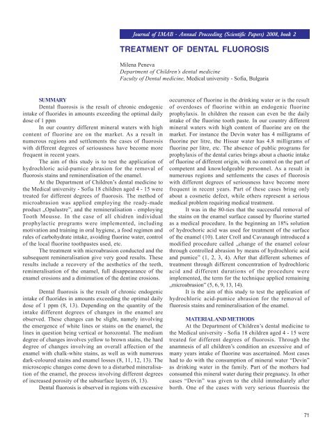
condition was caused by the intake <strong>of</strong> “Hissar” mineralwater. In the case <strong>of</strong> two children the mothers hadundergone a multiple fluorine prophylactic programinvolving the combined endogenic and local application <strong>of</strong>fluorine. The participants in the program had not beeninformed that consumption <strong>of</strong> water with high content <strong>of</strong>fluorine should be avoided. Yet another 6 children fromdifferent towns in the country had participated inprophylactic anti-caries programs and had simultaneouslydrunk “Devin” mineral water.The method <strong>of</strong> microabrasion was applied employingthe ready-made product „Opalustre”, and the remineralisation- employing Tooth Mousse.The scheme <strong>of</strong> application <strong>of</strong> the method involvedthree procedures within a period <strong>of</strong> one month. The teethwere cleaned pr<strong>of</strong>essionally and were isolated by means <strong>of</strong>K<strong>of</strong>edram. Under the K<strong>of</strong>edram, the teeth and gingivae wereoiled with Vaseline. The cervixes <strong>of</strong> the teeth were pastedwith a mixture <strong>of</strong> baking soda and water. The treatment <strong>of</strong>the enamel surface was conducted by for 5 secondsemploying Opalustre and a rubber from a prophylactic set.The procedure was repeated until a reduction <strong>of</strong> thepigmentation <strong>of</strong> achieved, but not more than 5Õ5 secondsfor a dental surface per visit. After that the dental surfaceswere washed abundantly with water later drained <strong>of</strong>f bymeans <strong>of</strong> saliva-sucking instrument, dried up and coveredamply with Tooth Mousse paste. The child was left in thatposition for five minutes and then asked not to consumeanything for 2-3 hours. The daily use <strong>of</strong> the Tooth Moussepaste at home was prescribed for brushing the teeth afterdinner for the periods between the separate visits to thedentist. Once an improvement <strong>of</strong> the colour <strong>of</strong> the teeth hasbeen achieved the domestic use <strong>of</strong> the paste was preservedfor a period <strong>of</strong> one year.For control <strong>of</strong> the degree <strong>of</strong> mineralisation <strong>of</strong> theenamel affected measurement by means <strong>of</strong> Kavo’sDIAGNOdent pen was employed. All surfaces affected wereexamined and recorded in the child’s card. At each next visit<strong>of</strong> the child the degree <strong>of</strong> mineralisation <strong>of</strong> the enamel waschecked up.In the case <strong>of</strong> all chidren individual prophylacticprograms were implemented, including motivation andtraining in oral hygiene, a food regimen and rules <strong>of</strong>carbohydrate intake, avoiding fluorine water, control <strong>of</strong> thelocal fluorine toothpastes used, etc.RESULTS AND DISCUSSIONThe original values arrived at by means <strong>of</strong> examiningthe affected dental surfaces employing the method <strong>of</strong> laserfluoerscence varied pretty much in accordance with thedegree <strong>of</strong> fluorosis. In the case <strong>of</strong> slight whitening <strong>of</strong> theenamel involving the occurrence <strong>of</strong> separate stains and linesand snow-whitening <strong>of</strong> the cutting edges <strong>of</strong> the teethingpermanent teeth, the values varied between 3 and 6. Theseare values corresponding to a minimum disturbance <strong>of</strong> themineralisation <strong>of</strong> the enamel. In the more serious cases thevalues reach a value <strong>of</strong> up to 10, and when brown stainswere observed – values <strong>of</strong> up to 15 - 20. When an overallwhitening <strong>of</strong> the enamel resembling the colour <strong>of</strong> chalk wasobserved the values were between 10 and 20, whichcorresponded to a condition when a major part <strong>of</strong> the enamelhas been affected.Fig. 1. Fluorosis stains on the enamel surface <strong>of</strong> a 9-year old child.On the enamel surface white perikimatic lines wereobserved. In some places the whitening formed verticalzones with a value - 7. Over the cutting edge <strong>of</strong> the righcentral incisor enamel erosion can be observed with a value<strong>of</strong> 19.Immediately after the application <strong>of</strong> the firstprocedure a shrinking <strong>of</strong> the white stains and noticeablewhitening <strong>of</strong> the brown and black stains was onserved. Thebest results were obtained in the case <strong>of</strong> dark-colouredstains, and especially if the treatment had started soon –up to a year - after the teeth had started to teethe. In thecase <strong>of</strong> the white stains, especially if the treatment hadstarted as late as 5-6 years after the teeth had started toteethe, the improvement was not that noticeable.A month after the first treatment <strong>of</strong> the dentalsurfaces, the Tooth Mousse paste being daily used, theimprovement was even more evident, including to theparents and children. Examination by means <strong>of</strong>DIAGNOdent showed a reduction <strong>of</strong> the value measured.In the case <strong>of</strong> the white stains the values are around 1-2.The dark stains, which now look yellowish, have valuesaround 6-7. The entirely affected chalk enamel too reactedto values between 8 - 15. A remineralisation <strong>of</strong> the enamellesions with affected surface layer is observed. These72
lesions have shrunk in size and have become shallower indepth and react with lower values than originally. With eachnext procedure the visible results become more evident. Theenamel loses its chalk tinge and begins to recover its slightlyyellowish colour. The enamel erosions become shallower andtheir edges lose their chalk colour. After the secondprocedure the effect improves and the improvementbecomes bigger in the next month. After the third month themilder forms <strong>of</strong> fluorosis reach a condition <strong>of</strong> visibly normalenamel with good mineralisation and restored values <strong>of</strong> laserfluorescence <strong>of</strong> 0 or 1. There were three cases in whichcertain whitening remained in the zones <strong>of</strong> the approximalsurfaces <strong>of</strong> the upper central incisors.The improvement is most noticeable in the case <strong>of</strong>the lower incisors, and then – in the case <strong>of</strong> the upperincisors. Particularly good are the results in the case <strong>of</strong>multiple point-like enamel erosions, which were left with theabove treatment, the recovery by means <strong>of</strong> composite beingplanned after the third month. Decrease <strong>of</strong> the size,improvement <strong>of</strong> the mineralisation and creation <strong>of</strong> goodconditions for the retention <strong>of</strong> the composite onto theenamel edges <strong>of</strong> the lesions was expected. In most caseswith purely enamel lesions a disappearance <strong>of</strong> the lesionsand a remineralisation <strong>of</strong> the enamel surface was observedwith values between 0 and 1.Fig. 2. A disappearance <strong>of</strong> the enamel erosion and arecovery <strong>of</strong> the values to 2 after the remineralisationFig. 3. The enamel surgace after 18 monthsParticularly valuable was this effect in the case <strong>of</strong> thefirst permanent molars, <strong>of</strong>ten with multiple erosions, whichcannot be efficiently obturated. After the third month none<strong>of</strong> the molars was in a condition requiring operativetreatment. The enamel erosions on the upper incisorsdisappeared too and no operative treatment was necessaryeither.A sharp decrease in the size and depth <strong>of</strong> the dentinelesions was observed. The degree <strong>of</strong> mineralisation <strong>of</strong> thedentine had improved too. Operative treatment provednecessary only in the cases <strong>of</strong> massive dentine lesions,which had shrunk in size, nevertheless, and had fullyremineralised surrounding enamel.The treatment with microabrasion conducted and thesubsequent remineralisation give very good results. Theseresults include a recovery <strong>of</strong> the aesthetics <strong>of</strong> the teeth,remineralisation <strong>of</strong> the enamel, full disappearance <strong>of</strong> theenamel erosions and a dimimution <strong>of</strong> the dentine erosions.The results from the treatment are corroborated by means<strong>of</strong> laser fluorescence that confirms the fact <strong>of</strong>remineralisation <strong>of</strong> the enamel. Moreover, the results arehighly appreciated by the parents and improve the children’sself-confidence. This kind <strong>of</strong> treatment is the onlyappropriate one since it aims at the maximum preservation<strong>of</strong> the dental structures and avoids the damages that areinflicted during the operative recovery <strong>of</strong> the dental surfacesaffected with fluorosis. According to data from the relevantliterature, by means <strong>of</strong> microabrasion an enamel layer73
etween 100 and 200µm is removed. The activeremineralisation using amorphous calcium phosphate andcasein phosphopeptids ensures the resistance <strong>of</strong> the enamellayer only to a minimum degree reduced by microabrasion.The method is not time-consuming and provides a solutionfor the complex situation created by dental fluorosis.CONCLUSIONThe method <strong>of</strong> acidic microabrasion, followed byremineralisation procedures, assures a considerablerecovery <strong>of</strong> the dental surfaces affected by fluorosis.REFERENCES1. Croll TP, Cavanaugh RR. Enamelcollor modification by controlled hydrochloricacid-pumice abrasion. I. Techniqueand examples, Quintesseence Int, 1986, 17,81-86.2. Croll TP, Cavanaugh RR. Enamelcollor modification by controlled hydrochloricacid-pumice abrasion. II. Fartherexamples, Quintessence Int, 1986, 17, 157-164.3. Croll TP. Enamel microabrasion forremoval <strong>of</strong> superficial dysmineralizationand decalcification defects. J Am DentAssoc 1990; 120: 411–415.4. Croll TP, Segura A. Tooth colorimprovement for children and teens: enamelmicroabrasion and dental bleaching. ASDCJ Dent Child 1996; 63: 17–22.5. Croll TP, Helpin ML. Enamelmicroabrasion: a new approach. J EsthetDent 2000; 1 M2: 64–71.6. Dalzell DP, Howes RI, Hubler PM.Microabrasion: effect <strong>of</strong> time, number <strong>of</strong>applications, and pressure on enamel loss,Pediatr Dent, 1995, 17, 207-211.7. Derry C, MT Hosey, P Waterhouse.Paediatric Cariology, Quintessentials, 2004,45-53.8. Kidd E. Essentials <strong>of</strong> Dental Caries,Oxford, 2005, 109-125.9. Kilpatrick NM, Welbury RR.Advanced restorative dentistry, in WelburyRR, M Duggal, Hosey. PaediatricDentistry, Oxford, 2005, 205-207.10. McCloskey R. A technique forremoval <strong>of</strong> fluorosis stains, J Am DentAssoc, 1984, 109, 63-64.11. McCloskey R. Et al. Dentalfluorosis: variability among different intakeduring the first year <strong>of</strong> life, CommunityDent Oral Epidemiol, 2002, 30, 286-295.12. Mathewson RJ, R Primosch.Fundamentals <strong>of</strong> Pediatric Dentistry,Quintessence, 1995, 307-309.13. Messer LB, K Mekertichian.Fluorid modalities. In Cameron AC, RWidmer “Handbook <strong>of</strong> PeediatricDentistry”, Mosby, Elsevier, 2008, 53-70.14. Millett D, R Welbury. Orthodonticsand Paediatric Dentistry, ChurchillLivingstone, 2000, 93-96.74Adress for corespondense:Milena PenevaDepartment <strong>of</strong> Pediatric Dentistry, Faculty <strong>of</strong> Dental Medicine,1, Georgi S<strong>of</strong>iiski str., 1431 S<strong>of</strong>ia, BulgariaE-mail: milenapeneva@mail.bg