An interactive flower image recognition system
An interactive flower image recognition system
An interactive flower image recognition system

Create successful ePaper yourself
Turn your PDF publications into a flip-book with our unique Google optimized e-Paper software.
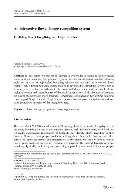
Multimed Tools Appl (2011) 53:53–73DOI 10.1007/s11042-010-0490-6<strong>An</strong> <strong>interactive</strong> <strong>flower</strong> <strong>image</strong> <strong>recognition</strong> <strong>system</strong>Tzu-Hsiang Hsu & Chang-Hsing Lee & Ling-Hwei ChenPublished online: 6 March 2010# Springer Science+Business Media, LLC 2010Abstract In this paper, we present an <strong>interactive</strong> <strong>system</strong> for recognizing <strong>flower</strong> <strong>image</strong>staken by digital cameras. The proposed <strong>system</strong> provides an <strong>interactive</strong> interface allowingeach user to draw an appropriate bounding window that contains the interested <strong>flower</strong>region. Then, a <strong>flower</strong> boundary tracing method is developed to extract the <strong>flower</strong> region asaccurately as possible. In addition to the color and shape features of the whole <strong>flower</strong>region, the color and shape features of the pistil/stamen area will also be used to representthe <strong>flower</strong> characteristics more precisely. Experiments conducted on two distinct databasesconsisting of 24 species and 102 species have shown that our proposed <strong>system</strong> outperformsother approaches in terms of the <strong>recognition</strong> rate.Keywords Flower <strong>image</strong> <strong>recognition</strong> . Image segmentation1 IntroductionThere are about 250,000 named species of <strong>flower</strong>ing plants in the world. Everyday, we cansee many blooming <strong>flower</strong>s in the roadside, garden, park, mountain path, wild field, etc.Generally, experienced taxonomists or botanists can identify plants according to their<strong>flower</strong>s. However, most people do know nothing about these wild <strong>flower</strong>s, even theirnames. To know the names or characteristics of the plants, we usually have to consult<strong>flower</strong> guide books or browse any relevant web pages on the Internet through keywordssearching. Typically, such a keyword searching approach is not practical for most people.This research was supported in part by the National Science Council of R.O.C. under contract NSC-97-2221-E-009-137.T.-H. Hsu : L.-H. Chen (*)Institute of Multimedia and Engineering, National Chiao Tung University, 1001 University Road,Hsinchu, Taiwan 300, Republic of Chinae-mail: lhchen@cc.nctu.edu.twC.-H. LeeDepartment of Computer Science and Information Engineering, Chung Hua University, Hsinchu,Taiwan 300, Republic of China
Multimed Tools Appl (2011) 53:53–73 55<strong>recognition</strong> results or tries to <strong>interactive</strong>ly adjust the parameters of the rose-curve modelwith mouse operations. According to the adjustment, the <strong>system</strong> will re-compute the modelparameters and re-rank the <strong>recognition</strong> results. Such an <strong>interactive</strong> process will repeat untilthe user accepts the <strong>recognition</strong> result. One major problem of this <strong>system</strong> is that too manyuser interactions have to be conducted to get high <strong>recognition</strong> accuracy.Nilsback and Zisserman [10] developed a visual vocabulary that explicitly describes thevarious characteristics (color, shape, and texture) of <strong>flower</strong>s. First, each <strong>image</strong> isautomatically segmented into foreground region (<strong>flower</strong> part) and background region usingthe contrast dependent prior Markov random field (MRF) cost function [1] and optimizedusing graph cuts. The HSV color values of all pixels in the training <strong>image</strong>s were thendivided into V c clusters using k-means clustering algorithm. The number of clusters V c isoptimized on the dataset. Then, a color vocabulary is constructed by the set of clustercenters (visual words). As a result, each <strong>image</strong> is represented by a V c -dimensionalnormalized frequency histogram of the set of visual words. To describe the shape of eachpetal, a rotation invariant descriptor called scale-invariant feature transform (SIFT)descriptor [8] was computed on a regular grid and optimized over three parameters: thegrid spacing M, the radius R of the support region for SIFT computation, and the number ofclusters. Vector quantization was then applied to get the visual words representing the petalshapes. The frequency histogram corresponding to the shape visual words was calculated todescribe the shape characteristic. To model the characteristic patterns on different petals,texture features were computed by convolving the <strong>image</strong> with maximum response 8 (MR8)filter bank [15]. The performance was optimized over the size of the square support regionsof the MR8 filters. A vocabulary was created by clustering the texture descriptors of alltraining <strong>image</strong>s and the frequency histogram was obtained for each <strong>image</strong>. For eachcharacteristic (color, shape, or texture), the distance between two <strong>image</strong>s is evaluated by theχ 2 measure of their frequency histograms. To get better performance, they combined thesethree vocabularies into a joint <strong>flower</strong> vocabulary and obtained a joint frequency histogram.A weight vector associated with the joint frequency histogram was introduced to optimizethe performance. Experimental results on a dataset of 1360 <strong>image</strong>s from 17 <strong>flower</strong> specieshave shown that the combined vocabulary outperforms each of the individual ones.Typically, there are too many parameters need to be optimized to get high <strong>recognition</strong> rate.Saitoh et al. [13] extended the route tracing method [12] to automatically extract the<strong>flower</strong> boundary under the assumption that the <strong>flower</strong> region is focused and the backgroundis out of focus. The extended route tracing method is based on the Intelligent Scissor (IS)approach [9] which searches a route that minimizes the sum of local costs according to anumber of manually selected points on the visually identified <strong>flower</strong> boundary. Instead ofminimizing the sum of local costs, the extended route tracing method tried to minimize theaverage cost defined as the sum of local costs divided by the route length. Four shapefeatures (the ratio of the route length to the sum of distances between the gravity center andall boundary points, the number of petals, the central moment, and the roundness) as well assix color features (the x and y coordinates and the proportions of <strong>flower</strong> pixels accumulatedin the two largest color cells in the HS color space) were extracted to recognize <strong>flower</strong><strong>image</strong>s. Experiments were conducted on 600 <strong>image</strong>s from 30 species with 20 <strong>image</strong>s perspecies. The <strong>recognition</strong> rates were 90.7%, 97.7%, and 99.0% if the correct one is includedin the top one, top two, and top three candidates, respectively. It is worth to note that thenumber of petals will change if some petals fell off or were occluded by others.Cho and Chi [2] proposed a structure-based <strong>flower</strong> <strong>image</strong> <strong>recognition</strong> method. Thegenetic evolution algorithm with adaptive crossover and mutation operations was employedto tune the learning parameters of the Backpropagation Through Structures algorithm [5]. A
56 Multimed Tools Appl (2011) 53:53–73region-based binary tree representation whose nodes correspond to the regions of the <strong>flower</strong><strong>image</strong> and links represent the relationships among regions was constructed to represent the<strong>flower</strong> <strong>image</strong> content. Experimental results showed that the structural representation of<strong>flower</strong> <strong>image</strong>s can produce a promising performance for <strong>flower</strong> <strong>image</strong> <strong>recognition</strong> in termsof generalization and noise robustness. In fact, the classification accuracy of the <strong>system</strong>depends on the selection of the feature values.Fukuda et al. [4] developed a <strong>flower</strong> <strong>image</strong> retrieval <strong>system</strong> by combining multipleclassifiers using fuzzy c-means clustering algorithm. In their <strong>system</strong>, <strong>flower</strong>s were classifiedinto three categories of different structures: gamopetalous <strong>flower</strong>s, many-petaled <strong>flower</strong>s, andsingle-petaled <strong>flower</strong>s. For each structure, a classifier with specific feature set wasconstructed. Fuzzy c-means clustering algorithm was then used to determine the degree ofmembership of each <strong>image</strong> to each structure. The overall similarity is a linear combinationof each individual similarity computed for each classifier with the weight being the degree ofmembership. The test database consists of 448 <strong>image</strong>s from 112 species with 4 <strong>image</strong>s perspecies. Experimental results have shown that the multiple-classifier approach outperformsany single-classifier approach. However, it is too rough a classification mechanism to classify<strong>flower</strong>s into three different categories according to the number of petals.Note that the previous researchers extracted color and shape features from the whole<strong>image</strong> region or <strong>flower</strong> boundary, without specifically treating the color and shapecharacteristics of the pistil/stamen area. Thus, an <strong>interactive</strong> <strong>flower</strong> <strong>image</strong> <strong>recognition</strong><strong>system</strong>, which extracts the color and shape features not only from the whole <strong>flower</strong> regionbut also from the pistil/stamen area, will be proposed to describe the characteristics of the<strong>flower</strong> <strong>image</strong>s more precisely. First, a <strong>flower</strong> segmentation method is developed to segmentthe <strong>flower</strong> boundary with as fewer user interactions as possible. Further, a simplenormalization procedure is employed to make the extracted features more robust to shapedeformations, including the number of petals, the relative positions of petals, the poses ofpetals taken from different directions, <strong>flower</strong> sizes, etc. The rest of this paper is organized asfollows. Section 2 describes the proposed <strong>flower</strong> <strong>image</strong> <strong>recognition</strong> <strong>system</strong>. Someexperimental results will be given in Section 3. Conclusions will be given in Section 4.2 The proposed <strong>flower</strong> <strong>image</strong> <strong>recognition</strong> <strong>system</strong>The proposed <strong>flower</strong> <strong>image</strong> <strong>recognition</strong> <strong>system</strong> consists of three major phases: <strong>flower</strong>region segmentation, feature extraction, and <strong>recognition</strong>, as shown in Fig. 1. In thesegmentation phase, the proposed <strong>system</strong> provides an interface allowing a user to draw arectangular window which circumscribes the <strong>flower</strong> region. A segmentation algorithmsimilar to that proposed by Saitoh et al. [13] is then developed to extract the <strong>flower</strong> regionwithin the rectangular window. In the feature extraction phase, the shape and color featuresof the whole <strong>flower</strong> region as well as the pistil/stamen area are extracted to measure thesimilarity between two <strong>flower</strong> <strong>image</strong>s. In the <strong>recognition</strong> phase, the <strong>flower</strong> <strong>image</strong> in thedatabase that is most similar to the input <strong>image</strong> will be found using the extractedfeatures.Fig. 1 Flow diagram of the proposed <strong>flower</strong> <strong>image</strong> <strong>recognition</strong> <strong>system</strong>
Multimed Tools Appl (2011) 53:53–73 572.1 Flower region segmentationIn order to extract the <strong>flower</strong> boundary as correctly as possible, the proposed <strong>system</strong>provides a simple <strong>interactive</strong> interface which allows the user to select the interested <strong>flower</strong>for <strong>recognition</strong>. Figure 2 illustrates the steps of the <strong>interactive</strong> <strong>flower</strong> region segmentationphase. First, the user can draw a rectangular window which circumscribes the interested<strong>flower</strong> by using mouse click and drag operations. Let P 0 denote the center point of therectangular window, P 1 , P 2 , P 3 , and P 4 denote the middle points on each of the fourboundary lines of the rectangular window as shown in Fig. 3. For each scan line startingfrom any P i (i=1,2,3,4)toP 0 , the edge point locating at the <strong>flower</strong> boundary will bedetected. These four edge points will then be regarded as the starting/ending points forboundary tracing. Since the proposed <strong>flower</strong> edge detection method used the “local cost”value associated with every pixel on each scan line, we will define the local cost first.2.1.1 Definition of local costThe local cost (LC) of a pixel on a scan line is defined as follows:LC ¼ 1 þ MG G; ð1Þwhere G denotes the gradient magnitude for the pixel and MG denotes the maximumgradient magnitude of all pixels in the <strong>image</strong>. According to the definition of local cost, wecan see that a pixel with strong edge gradient will have a small local cost. In this paper, theSobel operators (see Fig. 4) are employed to compute the horizontal and vertical gradientmagnitudes of a pixel. Let I R (x, y), I G (x, y), and I B (x, y) denote respectively the R, G, and Bcolor values of a pixel locating at (x, y). For R color value, the corresponding gradientmagnitude, notated G R (x, y), is defined as follows:G R ðx; yÞ ¼qffiffiffiffiffiffiffiffiffiffiffiffiffiffiffiffiffiffiffiffiffiffiffiffiffiffiffiffiffiffiffiffiffiffiffiffiffiffiffiffiffiffiffiffiG 2 R;H ðx;yÞþG2R;V ðx;yÞ; ð2Þwhere G R,H (x, y) and G R,V (x, y) denote respectively the horizontal and vertical gradientsderived by convolving the horizontal and vertical Sobel operators with the 3×3 <strong>image</strong>blocks centered at (x, y) and can be described by the following equations:G R;H ðx; yÞ ¼ ½I R ðx þ 1; y 1Þþ2I R ðx þ 1; yÞþI R ðx þ 1; y þ 1ÞŠ½I R ðx 1; y 1Þþ2I R ðx 1; yÞþI R ðx 1; y þ 1ÞŠ ð3ÞFig. 2 Block diagram for <strong>flower</strong> region segmentation
58 Multimed Tools Appl (2011) 53:53–73Fig. 3 Four boundary lines onthe <strong>flower</strong> bounding rectangularwindowandG R;V ðx; yÞ ¼ ½I R ðx 1; y þ 1Þþ2I R ðx; y þ 1ÞþI R ðx þ 1; y þ 1ÞŠ½I R ðx 1; y 1Þþ 2I R ðx; y 1Þþ I R ðx þ 1; y 1ÞŠ ð4ÞThe gradient magnitudes for G and B color values, notated G G (x, y) and G B (x, y), can becomputed in a similar manner. The overall gradient is then defined as the maximum valueamong G R (x, y), G G (x, y), and G B (x, y):Gx; ð yÞ ¼ max fG R ðx; yÞ; G G ðx; yÞ; G B ðx; yÞg ð5Þ2.1.2 Detection of <strong>flower</strong> edge pointsAccording the computed local cost associated with each pixel, four profiles of local costsalong the lines starting from every P i (i=1,2,3,4)toP 0 are generated. In this study, theestimated stamen region will be excluded on each profile (see P 3 →P 5 in Fig. 5). Theestimated stamen region is defined as the rectangular window with its center locating at P 0and its area being 1/9 of the <strong>flower</strong> bounding window. For each profile, the 5th percentile,P LC (5), of local costs is evaluated. The threshold value, T LC , used to find edge points oneach profile is defined as the average of the local costs smaller than P LC (5). If the local costvalue of a point is smaller than the threshold T LC , it will be considered as a candidate edgepoint. The one closest to the border of the <strong>flower</strong> bounding window is regarded as theFig. 4 The Sobel operators avertical Sobel operator b horizontalSobel operator
Multimed Tools Appl (2011) 53:53–73 59Fig. 5 <strong>An</strong> example for the detection of <strong>flower</strong> edge point on each profile<strong>flower</strong> edge point (see e 1 ,e 2 ,e 3 and e 4 in Fig. 5). These four <strong>flower</strong> edge points will betaken as the starting/ending points of the <strong>flower</strong> boundary tracing algorithm. In ourexperiments, about 14.4% (50/348) among the 348 <strong>image</strong>s in our database contain at leastone wrongly detected edge points. Figure 6 gives some examples of wrongly detected edgepoints. The main reasons for producing these wrongly detected edge points are: 1) thereexist strong edge points within the <strong>flower</strong> region (see Fig. 6(a)); 2) the contrast between the<strong>flower</strong> region and the background is not sharp enough (see Fig. 6(b)); 3) overlappingbetween neighboring <strong>flower</strong>s (see Fig. 6(c)); 4) no <strong>flower</strong> edge point survives in the profilewhen the stamen region is excluded (see Fig. 6(d)), etc. For these <strong>image</strong>s, we provide an<strong>interactive</strong> interface which allows the user to use the mouse to select the correct edge point(see Fig. 7).Fig. 6 Some example <strong>image</strong>s showing that the edge points detected are incorrect
60 Multimed Tools Appl (2011) 53:53–732.1.3 Flower boundary tracingLet e 1 ,e 2 ,e 3 , and e 4 denote the detected <strong>flower</strong> edge points. The two lines which connectP 1 and P 2 as well as P 3 and P 4 will divide the <strong>flower</strong> bounding window into four subregions(see R 1 , R 2 , R 3 , and R 4 in Fig. 8). The <strong>flower</strong> boundary of each sub-region will beindependently traced. Each pair of the four sets of edge points (e 1 ,e 4 ), (e 4 ,e 2 ), (e 2 ,e 3 ) and(e 3 ,e 1 ) will serve as the starting and ending tracing points of each sub-region, respectively.The proposed <strong>flower</strong> boundary tracing algorithm starts from the starting point and stopswhen the ending point is reached. These four partial <strong>flower</strong> boundaries will then becombined to form the whole <strong>flower</strong> boundary (see the yellow curve in Fig. 8).The proposed <strong>flower</strong> boundary tracing algorithm modifies the 2-D dynamic programminggraph search algorithm developed by Mortensen et al. [9]. It treats each pixel withinthe <strong>flower</strong> bounding window a vertex in a graph. <strong>An</strong> edge in the graph will connect a pixelto one of its 8-connected neighboring pixels. The cost associated with an edge is defined asthe local cost evaluated on the neighboring pixel. The concept of average path cost, whichis defined as the partial average cost computed from the previous pixel to the next pixel, isemployed to decide which direction to move. The partial average cost is updated by addingthe average of the previous pixel cost and the next pixel cost. The detailed algorithm of themodified <strong>flower</strong> boundary tracing algorithm is described as follows.Algorithm: <strong>flower</strong> boundary tracing algorithmInput:s Starting pixele Ending pixelc(q, r) Cost function for the edge between pixels q and rOutput:p Pointers from each pixel to its parent pixel with the minimum cost pathData Structures:L List of active pixels (i.e. pixels that are not determined with the minimum average cost yetand will be chosen as candidates to expand at next step) sorted by average cost (initiallyempty)N(q) 8-neighboring pixels of qB(q) Boolean function indicating if q has been expanded/processedG(q) Cost function from the starting pixel s to qAlgorithm:G(s) = 0; L = L + {s};//Initialize the active list and the cost functionwhile (L φ and not B(e)) do begin //While there exits pixels to expandq min(L); //Find the pixel from L with minimum costB(q) = TRUE;//Set q as expanded (i.e. processed)for each r ∈ N(q) with not B(r) do begintemp = (G(q)+c(q, r))/2; //Compute average cost to rif r ∈ L and temp < G(r) thenL = L – {r};//Remove higher cost pixel r from Lif r ∉ L then begin//If r is not in LG(r) = temp;//update average cost for rp(r) = q;//set the parent pixel of rL = L + {r};//and place r in Lendendend
Multimed Tools Appl (2011) 53:53–73 61Fig. 7 Correction of a wrongly detected edge point a a wrong edge point, e 3 b The corrected edge pointthrough user interaction2.2 Feature extractionThe most widely used features for describing <strong>flower</strong>s are color and shape descriptors. Inthis paper, the color and shape features of the whole <strong>flower</strong> region and the pistil/stamen areawill be extracted in an attempt to describe the characteristics of the <strong>flower</strong> <strong>image</strong>s moreprecisely.2.2.1 Features of the whole <strong>flower</strong> regionFirst, we define the <strong>flower</strong> region as the internal region within the segmented <strong>flower</strong>boundary. In this paper, nine color features in which the first six color features wereproposed by Saitoh et al. [13] and three shape features are extracted from the whole <strong>flower</strong>region for <strong>recognition</strong> purpose.Color features of <strong>flower</strong> region Since the <strong>flower</strong> <strong>image</strong>s were taken in differentenvironmental conditions, the variation in illumination will greatly affect the <strong>recognition</strong>result. To deal with such a problem, we convert each pixel from the RGB color space toHSV (hue, saturation, and value) space [6] and discard the illumination (V) component. Thecolor features are derived from the primary, secondary, and thirdly <strong>flower</strong> colors appearingin the whole <strong>flower</strong> region. First, the HS space is divided into 12×6 color cells representedby C i ,1≤ i ≤ 72 (please see Fig. 9). The color coordinate of each cell, which is defined asthe coordinate of the center point of each cell, can be represented by a pair of H and Svalues, (H i , S i ), 1 ≤ i ≤ 72. For each <strong>flower</strong> region, a color histogram (notated CH(i), 1 ≤ i ≤72), which describes the probability associated with each color cell C i , will be generated.Let DC(1), DC(2), and DC(3) denote respectively the first three dominant color cellsFig. 8 The <strong>flower</strong> boundingwindow is divided intofour sub-regions
62 Multimed Tools Appl (2011) 53:53–73Fig. 9 The HS color spaceis divided into 12×6 color cellsappearing in the <strong>flower</strong> region. The color coordinates of these three dominant color cellsand their corresponding probabilities are taken as the color features of the <strong>flower</strong> region.Let (dx i , dy i ) and p i denote the coordinate vector and the corresponding probability of DC(i), 1 ≤ i ≤ 3, where dx i = S DC(i) cos(H DC(i) ) and dy i = S DC(i) sin(H DC(i) ). These color featurescan be summarized as follows.CF 1 : x-coordinate value of DC 1 , dx 1CF 2 : y-coordinate value of DC 1 , dy 1CF 3 : the probability of DC 1 , p 1CF 4 : x-coordinate value of DC 2 , dx 2CF 5 : y-coordinate value of DC 2 , dy 2CF 6 : the probability of DC 2 , p 2CF 7 : x-coordinate value of DC 3 , dx 3CF 8 : y-coordinate value of DC 3 , dy 3CF 9 : the probability of DC 3 , p 3Shape features of <strong>flower</strong> region To get the shape features, we first define the centroid (g x ,g y ) of the <strong>flower</strong> region as the <strong>flower</strong> center, which is computed as follows:g x ¼ 1 NX Ni¼1x i ;ð6Þg y ¼ 1 Nwhere N is the number of pixels on the <strong>flower</strong> boundary, x i and y i are respectively the x andy coordinates of the i-th boundary pixel. The distance between the <strong>flower</strong> center and eachboundary pixel is then computed as follows:qffiffiffiffiffiffiffiffiffiffiffiffiffiffiffiffiffiffiffiffiffiffiffiffiffiffiffiffiffiffiffiffiffiffiffiffiffiffiffiffiffiffiffiffid i ¼ ð Þ 2 2þ y i g y ; 1 i N: ð8Þx ig xX Ni¼1y i ;ð7Þ
Multimed Tools Appl (2011) 53:53–73 63Without loss of generality, let the distances be sorted in an increasing order. That is, d i ≤d i+1 , for 1 ≤ i ≤ N-1. The three shape features (notated SF 1 , SF 2 , and SF 3 ) used to representthe shape characteristics of the <strong>flower</strong> region will be described as follows.1) SF 1 : A ratio which indicates the relevant sharpness of the petals and is computed fromthe distances between the <strong>flower</strong> boundary points to the <strong>flower</strong> center defined as follows:SF 1 ¼ R 10;ð9ÞR 90where R 10 and R 90 are respectively the average distances among all d i ’s which aresmaller than 10th percentile and larger than 90th percentile of all d i ’s:R 10 ¼10:1 N0:1N Xi¼1d i ;ð10ÞR 90 ¼10:1 N0:1N Xi¼1d N i : ð11ÞNote that the computed feature value SF 1 defined as a ratio between R 10 and R 90 will notchange greatly when the <strong>flower</strong> region is broken or captured from different directions.2) SF 2 : The average of normalized distances computed from every <strong>flower</strong> boundary pointto the <strong>flower</strong> center defined as follows:SF 2 ¼ 1 D i ;ð12ÞNi¼1where D i is the normalized distance defined as follows:8< 1; d i R 90dD i ¼ i R 10R 90 R 10; R 10 < d i < R 90 : ð13Þ:0; d i R 10Note that the definition of the feature value SF 2 using the averaged normalized valuesD i ’s will make it invariant to the size of the <strong>flower</strong> region.3) SF 3 : Roundness measure which indicates how much the shape of the <strong>flower</strong> petal iscloser to a circle and is defined as follows:X NSF 3 ¼ 4pSL 2 ; ð14Þwhere L is the length of the <strong>flower</strong> boundary and S is the area of the <strong>flower</strong> regiondefined as the total number of pixels in the <strong>flower</strong> region. When the <strong>flower</strong> shape isclose to a circle, SF 3 will be close to 1. Note that this feature value is robust to rotation,translation, and scaling of <strong>flower</strong> objects.In summary, the 12-dimensional feature vector used to represent the <strong>flower</strong> region can bedescribed as follows:f F ¼ ½CF 1 ; CF 2 ; ; CF 9 ; SF 1 ; SF 2 ; SF 3 Š T : ð15Þ
64 Multimed Tools Appl (2011) 53:53–732.2.2 Features of the pistil/stamen areaFirst, we define an initial estimate of the pistil/stamen area as the square area with its centerlocating at the <strong>flower</strong> center and its width 2/3 of the petal length, where the petal length isdefined as R 90 in (11). Let PDC(1) denote the dominant color cell in this estimated pistil/stamen area. Note that PDC(1) is found by excluding the primary color appearing in the<strong>flower</strong> region, DC(1). Then, all <strong>image</strong> pixels within the square area of width 4/3 of the petallength having color values identical to that of PDC(1) will constitute the pistil/stamen area.Since the color and shape of the pistil/stamen area also exhibit some discriminatinginformation for <strong>flower</strong> <strong>image</strong> <strong>recognition</strong>, the dominant color and its correspondingprobability will be taken as the color features of the pistil/stamen area. In addition, themean, standard deviation, and the third central moment of the normalized distance fromeach pixel in the pistil/stamen area to the center of the pistil/stamen area will be computedas the shape features of the pistil/stamen area.Color features of pistil/stamen area For most <strong>flower</strong>s, the dominant color of the pistil/stamen area is often different from that of the <strong>flower</strong> region. Thus, the color characteristicof the pistil/stamen area provides some discriminating information. In this study, thecoordinate vector (pdx 1 , pdy 1 ) and the corresponding probability pp 1 of PDC(1) will betaken as the color features of the pistil/stamen area, where pdx 1 = S PDC(1) cos(H PDC(1) ) andpdy 1 = S PDC(1) sin(H PDC(1) ). These color features can be summarized as follows.PCF 1 : x-coordinate value of PDC 1 , pdx 1PCF 2 : y-coordinate value of PDC 1 , pdy 1PCF 3 : the probability of PDC 1 , pp 1Shape features of pistil/stamen area Let the pistil/stamen area consist of M pixels and thecoordinates of these M pixels be notated as (px i , py i ), 1 ≤ i ≤ M. Next, the centroid (g px , g py )of the pistil/stamen area is computed as follows:g px ¼ 1 MX Mi¼1px i ;ð16Þg py ¼ 1 MThe distance between the centroid and every pixel of the pistil/stamen area is thencomputed as follows:qffiffiffiffiffiffiffiffiffiffiffiffiffiffiffiffiffiffiffiffiffiffiffiffiffiffiffiffiffiffiffiffiffiffiffiffiffiffiffiffiffiffiffiffiffiffiffiffiffiffiffiffiffi 2 2pd i ¼ px i g px þ pyi g py ; 1 i M: ð18ÞX Mi¼1py i :ð17ÞWithout loss of generality, let the distances be sorted in an increasing order. That is, pd i≤ pd i+1 ,1≤ i ≤ M-1. These distances are then normalized using the following equation:8< 1; pd i PR 90pdPD i ¼ i PR 10PR 90 PR 10; PR 10 < pd i < PR 90 ; ð19Þ:0; pd i PR 10
Multimed Tools Appl (2011) 53:53–73 65where PR 10 and PR 90 are respectively the average distances among all pd i ’s which aresmaller than 10th percentile and larger than 90th percentile of all pd i ’s:PR 10 ¼10:1 M0:1M Xi¼1pd i ;ð20ÞPR 90 ¼10:1 M0:1M Xi¼1pd M i : ð21ÞNote that the normalized distance values PD i ’s will make the extracted shape features ofthe pistil/stamen area invariant to the size of the <strong>flower</strong> <strong>image</strong>. The three shape features(notated PSF 1 , PSF 2 , and PSF 3 ) used to represent the shape characteristics of the pistil/stamen area are defined as follows.1) PSF 1 : The mean of the normalized distance values PD i ’s, defined as follows:PSF 1 ¼ m PD ¼ 1 MX Mi¼1PD i :2) PSF 2 : The standard deviation of the normalized distance values PD i ’s, defined as follows:PSF 2 ¼ s PD ¼1 MX Mi¼1ð22Þ! 1=2ðPD i s PD Þ 2 : ð23Þ3) PSF 3 : The third central moment of the normalized distance values PD i ’s, defined as follows:PSF 3 ¼ m 3 ¼1 MX Mi¼1! 1=3ðPD i s PD Þ 3 : ð24ÞNote that these three shape features are typically robust to rotation, translation, andscaling of <strong>flower</strong> <strong>image</strong>s. In summary, the 6-dimensional feature vector used to representthe pistil/stamen area can be described as follows:f P ¼ ½PCF 1 ; PCF 2 ; PCF 3 ; PSF 1 ; PSF 2 ; PSF 3 Š T : ð25ÞThe feature descriptor used to represent a <strong>flower</strong> <strong>image</strong> consists of all 18 featuresextracted from the <strong>flower</strong> region as well as the pistil/stamen area:hf ¼ ½f ð1Þ; f ð2Þ; ; f ð18ÞŠ T ¼ ðf F Þ T ðf Pi TÞ T¼ ½CF 1 ; ; CF 9 ; SF 1 ; ; SF 3 ; PCF 1 ; ; PCF 3 ; PSF 1 ; ; PSF 3 Š T :ð26Þ2.3 Flower <strong>image</strong> <strong>recognition</strong>In the <strong>recognition</strong> phase, the distances between the input <strong>image</strong> and all <strong>flower</strong> <strong>image</strong>sin the database are calculated. The distance between the input <strong>image</strong> and the i-th <strong>image</strong>
66 Multimed Tools Appl (2011) 53:53–73in the database, notated dist i , is measured by the weighted Euclidean distance defined asfollows:dist i ¼P NfwðkÞjf i ðkÞk¼1P Nfk¼1wðkÞf ðkÞj; ð27Þwhere f i (k) isthek-th feature value of the i-th <strong>image</strong>, f(k) isthek-th feature value of the input<strong>image</strong>, and w(k) is the weight associated with the k-th feature value. The variable N f determineswhich type of features is used for <strong>flower</strong> <strong>image</strong> <strong>recognition</strong>. If only the features extracted fromthe <strong>flower</strong> region are used for <strong>recognition</strong> purpose, set N f =12; if features extracted from boththe <strong>flower</strong> region and the pistil/stamen area are used for <strong>recognition</strong> purpose, set N f =18. In thispaper, the normalized top five <strong>recognition</strong> rate, NAR5(k), is used as the weight associated withthe k-th feature value (1 ≤ k ≤ 18). That is, w(k) =NAR5(k). NAR5(k) is derived by normalizingthe top five <strong>recognition</strong> rate associated with the k-th feature value, AR5(k):wðkÞ ¼NAR5ðkÞ ¼P N fAR5ðkÞj¼1AR5ðjÞ 100;ð28Þwhere AR5(k) denotes the <strong>recognition</strong> rate when the k-th feature value is individually used for<strong>flower</strong> <strong>recognition</strong> and the <strong>recognition</strong> result will be regarded as accurate if at least one of thespecies in the top five candidates is identical to the input one. Table 1 shows the top five<strong>recognition</strong> rate (AR5(k)) and the weight (w(k)) associated with each feature value for the first<strong>flower</strong> <strong>image</strong> database. Note that these initial weights are distinct for different databases. BasedTable 1 Top five <strong>recognition</strong>rate AR5(k) and weight w(k)associated with each feature valuefor the first <strong>flower</strong> <strong>image</strong>databaseFeature AR5(k) (%) w(k)f(1) : CF 1 66.67 6.91f(2) : CF 2 68.39 7.08f(3) : CF 3 58.62 6.07f(4) : CF 4 63.51 6.58f(5) : CF 5 66.95 6.93f(6) : CF 6 43.97 4.55f(7) : CF 7 61.49 6.37f(8) : CF 8 57.47 5.95f(9) : CF 9 45.11 4.67f(10) : SF 1 58.05 6.01f(11) : SF 2 39.94 4.14f(12) : SF 3 60.63 6.28f(13) : PCF 1 59.77 6.19f(14) : PCF 2 59.77 6.19f(15) : PCF 3 54.31 5.63f(16) : PSF 1 34.48 3.57f(17) : PSF 2 29.60 3.07f(18) : PSF 3 36.78 3.81
Multimed Tools Appl (2011) 53:53–73 67on the computation of all weighted distances, the top K candidate <strong>image</strong>s with minimumdistances to the input <strong>image</strong> will be returned to the user. In this study, the top K <strong>recognition</strong> ratewill be employed to evaluate the <strong>recognition</strong> performance.3 Experimental results3.1 Flower <strong>image</strong> databasesIn this paper, two <strong>flower</strong> <strong>image</strong> databases are used to evaluate the performance of theproposed method. The first <strong>flower</strong> <strong>image</strong> database constructed by us consists of 348 <strong>flower</strong><strong>image</strong>s from 24 species. A summary of these 24 plants is shown in Table 2 and someexample <strong>image</strong>s are shown in Fig. 10. The second database constructed by Zou and Nagy[16] consists of 612 <strong>flower</strong> <strong>image</strong>s from 102 species.The <strong>flower</strong> <strong>image</strong>s in the first database were taken in the fields by using digital camerasequipped with macro lens. The range of the lens aperture specified as an F-number was setbetween F2.8 and F5.6. To ensure the robustness of the proposed <strong>system</strong>, a number ofdifferent cameras, including SONY T9, Canon IS 860, and NIKON S1, were used to takeTable 2 Common names and Latin names of the plant species and their corresponding <strong>image</strong> numbers in thefirst databaseSC Common names Latin names Image numbers1 Yellow dots Wedelia trilobata (L.) Hitchc. 142 Globe amaranth Gomphrena globosa L. 83 African Marigold Tagetes erecta Linn. 64 Zinnia Zinnia elegans Jacq. 165 French Marigold Tagetes patula L. 46 Sun<strong>flower</strong> Helianthus annuus Linn. 67 Golden trumpet Allamanda cathartica L. 128 Common Cosmos Cosmos bipinnatus Cav. 99 Chinese hibiscus Hibiscus rosa-sinensis L. 1710 Purple Glory Tree Tibouchina semidecandra 1911 Bojers Spurge Euphorbia milii Ch. des Moulins 912 Pilose Beggarticks Bidens pilosa L. var. radiata Sch. 2713 Pericuinkle1 Catharathus roseus (L.) Don 1014 Phalaenopsis Phalaenopsis amabilis 815 Marguerite Chrysanthemum frutescens 1716 Pericuinkle2 Catharathus roseus (L.) Don 617 Gloxinia Gloxinia × hybrida Hort. 1118 <strong>An</strong>thurium <strong>An</strong>thurium scherzerianum Schott 1319 Gardenia Gardenia jasminoides Ellis 1020 Primrose Jasmine Jasminum mesnyi Hance 3321 Lavender Sorrel Oxalis martiana Zucc. 2822 Putchock Fringed iris Crested iris Iris japonica 1823 Azalea Rhododendron spp. 1524 African Touch-me-not Impatiens walleriana 32
68 Multimed Tools Appl (2011) 53:53–73Fig. 10 Some example <strong>image</strong>s in the first databasethese <strong>flower</strong> <strong>image</strong>s. In addition, the <strong>image</strong>s of the same species were taken from different<strong>flower</strong>s with different poses. The number of <strong>image</strong>s in each species is different and rangesfrom 4 to 33. Several <strong>image</strong>s contain multiple, tiny, overlapping <strong>flower</strong>s as shown in Fig. 10.Before <strong>recognition</strong>, all <strong>flower</strong> <strong>image</strong>s were re-scaled to be the same size of 400×300 pixels.The second database, which was constructed by Zou and Nagy, consists of 612 <strong>flower</strong> <strong>image</strong>sfrom 102 species collected from (http://www.ecse.rpi.edu/doclab/<strong>flower</strong>s). Each species consistsof six <strong>image</strong>s. The size of these <strong>image</strong>s are identical, 300×240 pixels. Some pictures are quiteout of focus, and several pictures contain multiple, tiny, overlapping <strong>flower</strong>s.3.2 Recognition resultsTo show the effectiveness of the proposed approach, we will compare the <strong>recognition</strong>results of the proposed approach with the feature sets proposed by Hong et al. [7], Zou andTable 3 Comparison of <strong>recognition</strong> rate on the first <strong>flower</strong> <strong>image</strong> databaseMethod Recognition rate (%)Top 1 Top 2 Top 3 Top 4 Top 5Hong et al. [7] 51.4 64.4 76.2 81.3 85.6Saitoh et al. [13] 83.6 92.0 95.1 97.7 99.1Our proposed approach 1 (using only shape features of <strong>flower</strong> region) 47.1 64.9 78.2 82.5 87.4Our proposed approach 2 (using only color features of <strong>flower</strong> region) 77.9 88.8 92.5 94.8 96.3Our proposed approach 3 (using color and shape features of 86.2 93.4 96.3 97.4 98.3<strong>flower</strong> region)Our proposed approach 4 (using color and shape features of <strong>flower</strong> 87.1 94.0 96.6 97.4 98.3region and shape features of pistil/stamen area)Our proposed approach 5 (using color and shape features of <strong>flower</strong> 88.5 95.1 98.6 99.7 99.7region and color features of pistil/stamen area)Our proposed approach 6 (using color and shape features of <strong>flower</strong>region and pistil/stamen area)89.1 95.1 98.6 99.7 99.7
Multimed Tools Appl (2011) 53:53–73 69Fig. 11 The 3 different specieswith similar appearance aZinnia b Pilose Beggarticks cMargueriteNagy [16], and Saitoh et al. [13]. The overall <strong>recognition</strong> accuracy is evaluated by takingeach <strong>flower</strong> <strong>image</strong> in the database as an input <strong>image</strong> and the remaining <strong>flower</strong> <strong>image</strong>s areconsidered as the training set.For the first <strong>flower</strong> <strong>image</strong> database, the comparison of the <strong>recognition</strong> rate of theproposed approach with those proposed by Hong et al. [7] and Saitoh et al. [13] is shown inTable 3. We can see that the color features give more discriminating ability than the shapefeatures. The proposed approach using both shape and color features of the <strong>flower</strong> regioncan achieve a very high <strong>recognition</strong> rate. Furthermore, the best performance is obtained bycombining the shape and color features of both the <strong>flower</strong> region as well as the pistil/stamenarea, which outperforms the approaches proposed by Hong et al. [7] and Saitoh et al. [13]interms of the <strong>recognition</strong> rate. Figure 11 shows in the first <strong>flower</strong> <strong>image</strong> database the threedifferent species (Zinnia, Pilose Beggarticks, and Marguerite) with similar appearance. Thecomparison of the <strong>recognition</strong> rate on these species is shown in Table 4. From this table, wecan see that our proposed approach also outperforms the other two approaches. Specifically,Fig. 12 shows the 27 <strong>image</strong>s of Pilose Beggarticks. We can see that these <strong>image</strong>s exhibitdifferent characteristics, including the number of petals, the relative positions of petals, theshape of petals, etc. Furthermore, for some <strong>image</strong>s there are some bugs appearing in thedifferent part of the <strong>flower</strong> region. From Table 4, we can see the comparison of <strong>recognition</strong>rates on this species. It is clear that the proposed approach yields the best <strong>recognition</strong> rate.This result illustrates that the proposed approach is more robust to noises and shapevariations.For the second database, we have also compared the <strong>recognition</strong> rate of the proposedapproach with those proposed by Hong et al. [7], Zou and Nagy [16], and Saitoh et al. [13].Table 4 Comparison of <strong>recognition</strong> rate on the three species (Zinnia, Pilose Beggarticks, and Marguerite)Plant species Method Recognition rate (%)Top 1 Top 2 Top 3 Top 4 Top 5Zinnia Hong et al. [7] 50 50 50 50 50Saitoh et al. [13] 50 50 50 75 100Our proposed approach 6 100 100 100 100 100Pilose Beggarticks Hong et al. [7] 18.5 44.4 85.2 85.2 92.6Saitoh et al. [13] 77.8 92.6 96.3 100 100Our proposed approach 6 88.9 100 100 100 100Marguerite Hong et al. [7] 35.3 52.9 70.6 82.4 88.2Saitoh et al. [13] 70.6 70.6 88.2 94.1 100Our proposed approach 6 82.4 94.1 100 100 100
70 Multimed Tools Appl (2011) 53:53–73Fig. 12 The 27 <strong>image</strong>s of Pilose Beggarticks in the first <strong>image</strong> databaseTable 5 compares the <strong>recognition</strong> rates of these approaches. From the table, we can seethat our proposed approach using shape and color features of the <strong>flower</strong> region and thepistil/stamen area always yields a higher <strong>recognition</strong> rate than the other approaches.Note that the method proposed by Zou and Nagy achieved a <strong>recognition</strong> rate of 93%when a number of user interactions were repeatedly conducted until the user acceptedthe <strong>recognition</strong> result, which takes a longer computation time (10.7 s) than ourproposed approach (4.2 s). Figure 13 shows the <strong>image</strong>s of two <strong>flower</strong> species(FlowerBridge1 and RosaArkansana) in the second database. We can see that the <strong>image</strong>sof the same species were taken from different directions and distances and thus these<strong>image</strong>s reveal different shapes and sizes. The comparison of the <strong>recognition</strong> rate on thesetwospeciesisshowninTable6. From this table, we can see that our proposed approachalso outperforms the other methods.Table 5 Comparison of <strong>recognition</strong> rate on the second <strong>flower</strong> <strong>image</strong> databaseMethod Recognition rate (%) Average <strong>recognition</strong>time (s)Top 1 Top 2 Top 3 Top 4 Top 5Hong et al. [7] 39.5 50.8 57.0 61.1 64.4 4.2Zou-Nagy et al. [16] (without user interaction) 52 – 79 – – 8.5Zou-Nagy et al. [16] (with user interaction) 93 10.7Saitoh et al. [13] 65.5 81.9 87.9 91.5 93.8 4.2Our proposed approach 1 12.8 22.6 28.4 36.1 43.5 4.2Our proposed approach 2 56.7 72.9 80.2 85.3 88.1 4.2Our proposed approach 3 69.3 84.0 89.1 92.7 94.8 4.2Our proposed approach 4 71.3 84.6 89.9 92.8 95.1 4.2Our proposed approach 5 76.1 88.7 95.9 97.2 98.2 4.2Our proposed approach 6 77.8 89.4 94.3 97.7 98.5 4.2
Multimed Tools Appl (2011) 53:53–73 71Fig. 13 The <strong>image</strong>s of the twospecies in the second database aFlowerBridge1 b RosaArkansana4 ConclusionsIn this paper, we have presented an <strong>interactive</strong> <strong>flower</strong> <strong>recognition</strong> <strong>system</strong>. First, the <strong>system</strong>provides a simple user interface which allows each user to draw a rectangular boundingwindow containing the interested <strong>flower</strong> region. Then, a boundary tracing algorithm isdeveloped to find the <strong>flower</strong> boundary as accurately as possible. Besides the color andshape features of the whole <strong>flower</strong> region, the color and shape features of the pistil/stamenarea are also extracted to represent the <strong>flower</strong> characteristics in a more precise fashion.Experiments conducted on two different <strong>flower</strong> <strong>image</strong> databases consisting of 24 speciesand 102 species have shown that our proposed approach achieves a higher <strong>recognition</strong> ratethan the methods proposed by Hong et al. [7], Zou and Nagy [16], and Saitoh et al. [13].Table 6 Comparison of <strong>recognition</strong> rate on FlowerBridge1 and RosaArkansana in the second databasePlant species Method Recognition rate (%)Top 1 Top 2 Top 3 Top 4 Top 5FlowerBridge1 Hong et al. [7] 33.3 33.3 33.3 33.3 33.3Saitoh et al. [13] 33.3 50.0 50.0 83.3 100Our proposed approach 6 66.7 83.3 100 100 100RosaArkansana Hong et al. [7] 0.0 33.3 50.0 50.0 50.0Saitoh et al. [13] 33.3 66.7 66.7 66.7 66.7Our proposed approach 6 100 100 100 100 100
72 Multimed Tools Appl (2011) 53:53–73References1. Boykov YY, Jolly MP (2001) Interactive graph cuts for optimal boundary and region segmentation ofobjects in N-D <strong>image</strong>s. Proc Int Conf Comput Vis 2:105–1122. Cho SY, Chi Z (2005) Generic evolution processing of data structures for <strong>image</strong> classification. IEEETrans Knowl Data Eng 17(2):216–2313. Das M, Manmatha R, Riseman EM (1999) Indexing <strong>flower</strong> patent <strong>image</strong>s using domain knowledge.IEEE Intell Syst 14:24–334. Fukuda K, Takiguchi T, Ariki Y (2008) Multiple classifier based on fuzzy c-means for a <strong>flower</strong> <strong>image</strong>retrieval. In: Proc. Int. Workshop on Nonlinear Circuits and Signal Processing (NCSP’2008): 76–795. Goller C, Kuchler A (1996) Learning task-dependent distributed representations by back-propagationthrough structure. Proc IEEE Int Conf Neural Networks 1:347–3526. Gonzalez RC, Woods RE (2002) Digital <strong>image</strong> processing, 2nd edn. Prentice-Hall, Upper Saddle River7. Hong A, Chen G, Li J, Chi Z, Zhang D (2004) A <strong>flower</strong> <strong>image</strong> retrieval method based on ROI feature.Journal of Zhejiang University (Science) 5(7):764–7728. Lowe D (2004) Distinctive <strong>image</strong> features from scale-invariant keypoints. Int J Comput Vis 60(2):91–1109. Mortensen EN, Barrett WA (1995) Intelligent scissors for <strong>image</strong> composition. In: Proc. ComputerGraphics (SIGGRAPH’95): 191–19810. Nilsback ME, Zisserman A (2006) A visual vocabulary for <strong>flower</strong> classification. Proc Comput VisPattern Recogn 2:1447–145411. Saitoh T, Kaneko T (2000) Automatic <strong>recognition</strong> of wild <strong>flower</strong>s. Proc Int Conf Pattern Recogn 2:507–51012. Saitoh T, Aoki K, Kaneko T (2003) Automatic extraction of object region from photographs. In: Proc.Scandinavian Conf. on Image <strong>An</strong>alysis: 1130–113713. Saitoh T, Aoki K, Kaneko T (2004) Automatic <strong>recognition</strong> of blooming <strong>flower</strong>s. Proc Int Conf PatternRecogn 1:27–3014. Swain MJ, Ballard DH (1991) Color indexing. Int J Comput Vis 7(1):11–3215. Varma M, Zisserman A (2002) Classifying <strong>image</strong>s of materials: achieving viewpoint and illuminationindependence. Proc Europ Conf Comput Vis 3:255–27116. Zou J, Nagy G (2004) Evaluation of model-based <strong>interactive</strong> <strong>flower</strong> <strong>recognition</strong>. Proc Int Conf PatternRecogn 2:311–314Tzu-Hsiang Hsu was born on Feb 7, 1984 in Taoyuan, Taiwan. She received the M.S. degree in MultimediaEngineering from National Chiao Tung University, Hsinchu, Taiwan in 2008. She is currently an engineer ofthe software development department of ZyXEL, a network terminal product company. Her research interestsinclude <strong>image</strong> processing, pattern <strong>recognition</strong> and segmentation.
Multimed Tools Appl (2011) 53:53–73 73Chang-Hsing Lee was born on July 24, 1968 in Tainan, Taiwan. He received the B.S. and Ph.D. degrees inComputer and Information Science from National Chiao Tung University, Hsinchu, Taiwan in 1991 and1995, respectively. He is currently an Associate Professor in the Department of Computer Science andInformation Engineering, Chung Hua University, Hsinchu, Taiwan. His main research interests includeaudio/sound classification, multimedia information retrieval, and <strong>image</strong> processing.Ling-Hwei Chen received the B.S. degree in mathematics and the M.S. degree in applied mathematics fromNational Tsing Hua University, Hsinchu, Taiwan in 1975 and 1977, respectively, and the Ph.D. in computerengineering from National Chiao Tung University, Hsinchu, Taiwan in 1987