B-cell depletion reveals a role for antibodies in the control of chronic ...
B-cell depletion reveals a role for antibodies in the control of chronic ...
B-cell depletion reveals a role for antibodies in the control of chronic ...
Create successful ePaper yourself
Turn your PDF publications into a flip-book with our unique Google optimized e-Paper software.
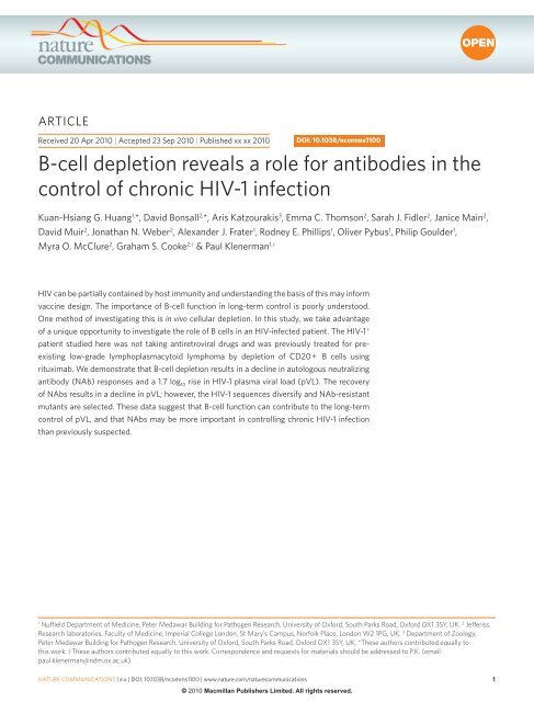
ARTICLEnature communications | DOI: 10.1038/ncomms1100HIV-1 is readily able to evade host defence and establish persistent<strong>in</strong>fection. The virus may, however, be partially conta<strong>in</strong>edby host adaptive immunity, <strong>in</strong>clud<strong>in</strong>g both B- andT-<strong>cell</strong> responses. There is good evidence that CD8 + T-<strong>cell</strong> responseshave a <strong>role</strong> <strong>in</strong> viral suppression. For example, <strong>the</strong>re are strong HLAclass I associations with cl<strong>in</strong>ical outcome 1 , and <strong>in</strong> <strong>the</strong> SIV model,macaques <strong>in</strong> whom CD8 + T <strong>cell</strong>s are depleted show significant<strong>in</strong>creases <strong>in</strong> <strong>the</strong>ir viral load 2 . These f<strong>in</strong>d<strong>in</strong>gs have led to a major <strong>in</strong>itiativeto develop T-<strong>cell</strong>-based vacc<strong>in</strong>es, and much work to def<strong>in</strong>e<strong>the</strong> correlates <strong>of</strong> protection based on assays <strong>of</strong> T-<strong>cell</strong> function 3,4 .However, as a lead<strong>in</strong>g HIV vacc<strong>in</strong>e candidate failed to <strong>in</strong>duce protectiveT-<strong>cell</strong> responses 5 and recent trial data suggest partial protectionfrom a vacc<strong>in</strong>e, <strong>in</strong>clud<strong>in</strong>g a gp120 component 6 , <strong>the</strong> <strong>in</strong>centive tounderstand <strong>the</strong> <strong>role</strong> <strong>of</strong> neutraliz<strong>in</strong>g <strong>antibodies</strong> (NAbs) and <strong>of</strong> B <strong>cell</strong>s<strong>in</strong> HIV <strong>in</strong>fection has <strong>in</strong>creased considerably 7–9 .B <strong>cell</strong>s have a multifaceted <strong>role</strong> <strong>in</strong> humoral and <strong>cell</strong>ular responsesto HIV-1, yet <strong>the</strong>ir impact <strong>in</strong> conta<strong>in</strong><strong>in</strong>g <strong>the</strong> virus rema<strong>in</strong>s unclear.This is partly because <strong>of</strong> <strong>the</strong> complex nature <strong>of</strong> <strong>the</strong> antigenic target(gp120) 10 . Key obstacles to <strong>the</strong> development <strong>of</strong> both T- and B-<strong>cell</strong>defence are <strong>the</strong> abilities <strong>of</strong> <strong>the</strong> HIV-1 genome to <strong>in</strong>tegrate and alsoevolve rapidly <strong>in</strong> <strong>the</strong> face <strong>of</strong> immune selection pressure: <strong>the</strong> latterfeature results from both <strong>the</strong> high replication rate <strong>of</strong> <strong>the</strong> virus and<strong>the</strong> error-prone nature <strong>of</strong> <strong>the</strong> HIV-1 reverse transcriptase, whichlacks pro<strong>of</strong>read<strong>in</strong>g capacity 11 . These mutations are readily accommodatedwith<strong>in</strong> gp120 because <strong>of</strong> plasticity <strong>of</strong> <strong>the</strong> structure and<strong>the</strong> capacity to acquire glycosylation sequons that provide a ‘glycanshield’ 12 . There<strong>for</strong>e, emerg<strong>in</strong>g NAbs repeatedly select mutant virusesthat are resistant to neutralization 13 . The NAb response also evolvesthrough somatic recomb<strong>in</strong>ation and aff<strong>in</strong>ity maturation <strong>of</strong> proliferat<strong>in</strong>gB <strong>cell</strong>s, but is only partially effective aga<strong>in</strong>st concurrentlycirculat<strong>in</strong>g viruses, while reta<strong>in</strong><strong>in</strong>g potent activity aga<strong>in</strong>st stra<strong>in</strong>spreviously circulat<strong>in</strong>g with<strong>in</strong> <strong>the</strong> patient 13–15 . Such data have castdoubt on <strong>the</strong> contribution <strong>of</strong> NAbs to <strong>the</strong> <strong>control</strong> <strong>of</strong> virus load,despite <strong>the</strong> counterargument that NAbs must <strong>in</strong>hibit virus replicationto drive <strong>the</strong> selection <strong>of</strong> resistant variants.Monoclonal <strong>antibodies</strong> have been identified that both crossrecognizeand potently neutralize a variety <strong>of</strong> stra<strong>in</strong>s 16–19 . The targets <strong>for</strong><strong>the</strong>se are well def<strong>in</strong>ed and <strong>in</strong>clude, among o<strong>the</strong>rs, CD4 and co-receptorb<strong>in</strong>d<strong>in</strong>g sites and epitopes with<strong>in</strong> <strong>the</strong> gp41 fusion subunit 20 . These areasare under some structural constra<strong>in</strong>t compared with o<strong>the</strong>r epitopes,such as <strong>in</strong> <strong>the</strong> exposed variable loops. Few potent monoclonal <strong>antibodies</strong>have been isolated, yet <strong>the</strong> proportion <strong>of</strong> gp120-specific <strong>antibodies</strong>capable <strong>of</strong> neutralization may be greater than once believed 8,21 . Recentstudies <strong>in</strong>dicate that some <strong>in</strong>dividuals possess NAbs that exhibit broadand potent neutralization capacity 18,21,22 , but functional data to causallyl<strong>in</strong>k such <strong>antibodies</strong> to <strong>the</strong> <strong>control</strong> <strong>of</strong> viremia are lack<strong>in</strong>g. Fur<strong>the</strong>rmore,<strong>the</strong> contribution <strong>of</strong> B-<strong>cell</strong> responses to <strong>the</strong> conta<strong>in</strong>ment <strong>of</strong> plasma viralload (pVL) has been difficult to determ<strong>in</strong>e, because <strong>of</strong> o<strong>the</strong>r importantfactors that limit virus replication, such as T-<strong>cell</strong> response, virus fitnessand host genetics 23 .Historically, functional importance has been confidently attributedto specific arms <strong>of</strong> <strong>the</strong> immune response by means <strong>of</strong> experimentsthat deplete or transfer that effector arm. Although such dataare readily available <strong>in</strong> animal models, <strong>the</strong>y are rarely available <strong>in</strong>humans. As recent vacc<strong>in</strong>e studies have highlighted substantial differencesbetween SIV models and HIV, it is important to seek suchevidence <strong>in</strong> human studies.Rituximab (Rituxan, MabThera; Roche) is a monoclonal antibodytarget<strong>in</strong>g CD20 antigen expressed on most B <strong>cell</strong>s, with <strong>the</strong>exception <strong>of</strong> term<strong>in</strong>ally differentiated plasma <strong>cell</strong>s. It has becomewidely used <strong>for</strong> treatment <strong>of</strong> lymphoma and also <strong>of</strong> antibodymediatedautoimmune diseases 24 . We report a unique case <strong>in</strong> whichrituximab mono<strong>the</strong>rapy was used <strong>in</strong> a patient with stable viraemia,<strong>in</strong> <strong>the</strong> absence <strong>of</strong> antiretroviral <strong>the</strong>rapy (ART).By comb<strong>in</strong>ed analyses <strong>of</strong> viral dynamics, viral evolution andautologous NAb responses, we show reversible loss <strong>of</strong> HIV-1<strong>control</strong> follow<strong>in</strong>g rituximab mono<strong>the</strong>rapy. Loss <strong>of</strong> <strong>control</strong> was associatedwith reduction <strong>in</strong> titres <strong>of</strong> NAbs target<strong>in</strong>g <strong>the</strong> CD4 b<strong>in</strong>d<strong>in</strong>g site, aswell as with an <strong>in</strong>crease <strong>in</strong> genetic diversity and transient reversion toNab-sensitive virus. As NAb levels later <strong>in</strong>creased, pVL was once more<strong>control</strong>led and this was accompanied by fur<strong>the</strong>r immune selection <strong>of</strong>NAb-resistant viruses. These data suggest that despite ongo<strong>in</strong>g immuneescape, NAbs secreted by CD20 + B <strong>cell</strong>s may have a significant <strong>role</strong><strong>in</strong> <strong>control</strong> <strong>of</strong> pVL dur<strong>in</strong>g <strong>chronic</strong> HIV-1 <strong>in</strong>fection, a f<strong>in</strong>d<strong>in</strong>g <strong>of</strong> significance<strong>for</strong> HIV-1 immuno<strong>the</strong>rapies and vacc<strong>in</strong>es.ResultsCl<strong>in</strong>ical course <strong>of</strong> <strong>the</strong> study subject. The patient, a Caucasianman, was diagnosed with HIV seroconversion illness at <strong>the</strong> age<strong>of</strong> 58 years. Three years earlier, he was diagnosed with low-gradelymphoplasmacytoid lymphoma, <strong>for</strong> which he did not receivetreatment 25 . Histologically, this was def<strong>in</strong>ed as a small lymphocyticB-<strong>cell</strong> lymphoma with plasmacytoid differentiation mostlyexpress<strong>in</strong>g CD20 and CD79. The tumour was IgM secret<strong>in</strong>g. Hisonly o<strong>the</strong>r medical history <strong>of</strong> note was an acute hepatitis B <strong>in</strong>fectionapproximately 30 years earlier. Follow<strong>in</strong>g HIV-1 seroconversion,<strong>the</strong> patient was recruited <strong>in</strong>to a prospective, non-randomizedobservational study <strong>of</strong> early treatment and received 3 months <strong>of</strong>highly active antiretroviral <strong>the</strong>rapy 26 . After 30 months, he developeda ris<strong>in</strong>g paraprote<strong>in</strong>aemia, attributed to his lymphoma, and receivedthalidomide treatment without cl<strong>in</strong>ical effect. He <strong>in</strong>itiated rituximab<strong>the</strong>rapy 1,075 days after HIV seroconversion and received fourweekly doses (375 mg m − 2 ). He felt unwell 20 weeks after <strong>the</strong> first dose<strong>of</strong> rituximab with malaise and fever associated with a marked rise<strong>in</strong> HIV pVL (<strong>in</strong>crease <strong>of</strong> 1.7 log 10 ; Fig. 1). In addition, he developedpVL (RNA copies/ml)CD4 T <strong>cell</strong> count106 <strong>cell</strong>s/mlARTRituximab1E+06100901E+05801E+0470601E+0350401E+02301E+0120101E+0000 200 400 600 800 1,000 1,200 1,400Days after seroconversion80060040020000 200 400 600 800 1,000 1,200 1,400Days after seroconversionFigure 1 | Autologous neutraliz<strong>in</strong>g antibody titres over time. (a)Pseudotyped virus particles were constructed us<strong>in</strong>g full-length envelopeclones that represent majority quasi-species at 336 or 1,042 days afterseroconversion. Plasma VL over time is <strong>in</strong>dicated (red l<strong>in</strong>e). The brief dip<strong>in</strong> viral load over <strong>the</strong> first 100 days represents <strong>the</strong> period <strong>of</strong> <strong>in</strong>itial ARTas part <strong>of</strong> <strong>the</strong> short-course ART trial (shaded grey). The neutraliz<strong>in</strong>geffects (%; right y axis) <strong>of</strong> patient sera are shown <strong>for</strong> autologous virusesconstructed with cloned env genes derived 336 days (black squares) and1,042 days (crosses) after seroconversion. Results from a s<strong>in</strong>gle clone(tested <strong>in</strong> triplicate) representative <strong>of</strong> three clones are shown. Serum wassampled sequentially be<strong>for</strong>e and after rituximab <strong>the</strong>rapy (1,075–1,106 days,shaded grey), and assayed at concentrations rang<strong>in</strong>g from 1:20 to 1:4,370;<strong>the</strong> neutraliz<strong>in</strong>g effects <strong>of</strong> sera diluted 1:180 are shown here. Error barsshow standard deviations <strong>of</strong> triplicate tests. (b) CD4 T lymphocyte count(dashed l<strong>in</strong>e) is shown over time.% Neutralization (1:180 dilution) nature communications | x:x | DOI: 10.1038/ncomms1100 | www.nature.com/naturecommunications© 2010 Macmillan Publishers Limited. All rights reserved.
nature communications | DOI: 10.1038/ncomms1100ARTICLEbiochemical hepatitis (attributed to reactivation <strong>of</strong> hepatitis B)that resolved with only supportive treatment. Antiretrovirals were<strong>in</strong>itiated 1,392 days after seroconversion.Impact <strong>of</strong> B-<strong>cell</strong> <strong>depletion</strong> on NAb titres. Follow<strong>in</strong>g rituximab <strong>the</strong>rapy,<strong>the</strong> subject’s HIV-1 pVL, previously stable, <strong>in</strong>creased by more than1.7 log 10 , peak<strong>in</strong>g at 737,400 copies per ml 4 months after <strong>the</strong> f<strong>in</strong>al dose<strong>of</strong> rituximab (Fig. 1). The pVL subsequently returned to basel<strong>in</strong>e levels<strong>in</strong> <strong>the</strong> absence <strong>of</strong> ART. We <strong>the</strong>re<strong>for</strong>e analysed <strong>the</strong> impact <strong>of</strong> rituximab<strong>the</strong>rapy on NAb titres <strong>in</strong> relation to viral dynamics.Rituximab <strong>the</strong>rapy led to a 30% reduction <strong>in</strong> total IgG levels,which reached <strong>the</strong>ir lowest po<strong>in</strong>t at <strong>the</strong> peak <strong>of</strong> virus load (10.8 g l − 1 ;normal range 8–16 g l − 1 ) and returned to pre-rituximab levels <strong>the</strong>reafter(Supplementary Table S1). To evaluate NAb activity throughoutthis period, we generated pseudoviruses derived from autologousenvelope (env) sequences. Serum obta<strong>in</strong>ed be<strong>for</strong>e <strong>the</strong> first dose <strong>of</strong>rituximab potently neutralized virus pseudotyped with autologousenvelope derived from plasma acquired 2 years previously (day 336after seroconversion) (Table 1). The same serum neutralized contemporaneousautologous Env pseudotypes, although to a lesserextent, with 1:60 serum dilutions effectively block<strong>in</strong>g more than90% <strong>in</strong>fection. Heterologous NAb activity was also observed aga<strong>in</strong>stHXB2 and 8/12 Tier 2/3 clade-B reference stra<strong>in</strong>s (NIH, AIDS)(Table 2). Follow<strong>in</strong>g rituximab <strong>the</strong>rapy, we observed a markeddecrease <strong>in</strong> neutraliz<strong>in</strong>g activity characterized by a drop <strong>of</strong> morethan threefold <strong>in</strong> IC 50 serum titres aga<strong>in</strong>st day 336 Env pseudotypes,which recovered over a 4-month period. Strik<strong>in</strong>gly, <strong>the</strong>se changesco<strong>in</strong>cided with <strong>the</strong> <strong>in</strong>crease <strong>in</strong> pVL. Recovery <strong>of</strong> NAb responses correlatedwith <strong>control</strong> <strong>of</strong> viraemia (Fig. 1).Analysis <strong>of</strong> sequence evolution <strong>in</strong> HIV-1 envelope over time.To evaluate sequence evolution over this period, env productswere generated by reverse transcription (RT)–PCR from plasmaviral RNA, be<strong>for</strong>e and after rituximab <strong>the</strong>rapy (days 1,075–1,392;Fig. 2a). We identified clear sequence diversification as <strong>the</strong> viruspeaked dur<strong>in</strong>g rituximab treatment (mean genetic distance = 0.02versus 0.004; P < 0.0001). This diversification manifested as an<strong>in</strong>crease <strong>in</strong> effective population size as measured us<strong>in</strong>g BEAST 27(fivefold change; Fig. 2b). To assess whe<strong>the</strong>r <strong>the</strong>se diverse stra<strong>in</strong>srepresented a re-emergence <strong>of</strong> archived provirus, we used an identicalsequenc<strong>in</strong>g strategy to analyse stored peripheral blood mononuclear<strong>cell</strong> (PBMC) samples available from <strong>the</strong> time <strong>of</strong> seroconversionup to day 1,042; <strong>the</strong>se sequences were placed on a time-structuredtree (Supplementary Fig. S1). Viruses detected after rituximabtreatment did not represent re-emergence <strong>of</strong> early archived stra<strong>in</strong>s;those stra<strong>in</strong>s present as pVL peaked were most closely related tostra<strong>in</strong>s present <strong>in</strong> plasma at day 1,042, with a subset related to thosearchived <strong>in</strong> PBMC at day 1,042.Immune selection and neutralization <strong>of</strong> HIV-1 envelope. The apparent<strong>in</strong> vivo impact <strong>of</strong> <strong>the</strong> NAbs <strong>in</strong> this <strong>in</strong>dividual prompted a search<strong>for</strong> <strong>the</strong> targets <strong>of</strong> b<strong>in</strong>d<strong>in</strong>g. To identify potential NAb targets, we firstanalysed immune selection us<strong>in</strong>g CODEML, analys<strong>in</strong>g <strong>the</strong> ratio <strong>of</strong>non-synonymous to synonymous mutations (dN/dS) at each site. Weobserved two sites under selection (Fig. 3; Supplementary Fig. S1)—position 339 (dN/dS = 7.6, P < 0.0001) and position 363 (dN/dS = 6.977,P = 0.03). Residue 363 <strong>in</strong>tersperses <strong>the</strong> β14 and β15 anti-parallel strands<strong>of</strong> gp120 and is exposed on <strong>the</strong> surface <strong>of</strong> <strong>the</strong> outer doma<strong>in</strong> <strong>in</strong> closeproximity to <strong>the</strong> CD4 b<strong>in</strong>d<strong>in</strong>g loop 28 . Computational modell<strong>in</strong>g <strong>of</strong>atomic level envelope structures has shown that mutations proximalto <strong>the</strong> CD4-b<strong>in</strong>d<strong>in</strong>g loop are critical <strong>in</strong> conferr<strong>in</strong>g resistance toCDbs <strong>antibodies</strong> 29 . Fur<strong>the</strong>rmore, site-directed mutagenesis <strong>of</strong> site 363has previously been shown to affect neutralization <strong>of</strong> <strong>the</strong> CD4bs <strong>antibodies</strong>M-14 and b12 30,31 . Be<strong>for</strong>e <strong>the</strong> first dose <strong>of</strong> rituximab (day 1,042after seroconversion), position 363 was occupied by an arg<strong>in</strong><strong>in</strong>e <strong>in</strong> allviral RNA sequences, whereas all proviral DNA clones sampled up toand <strong>in</strong>clud<strong>in</strong>g day 1,042 had a glutam<strong>in</strong>e at <strong>the</strong> same position. Follow<strong>in</strong>grituximab <strong>the</strong>rapy, R363 variants were temporarily replacedwith Q363 variants, which reverted to arg<strong>in</strong><strong>in</strong>e residues as NAb titresrebounded and viraemia was <strong>control</strong>led.Be<strong>for</strong>e rituximab <strong>the</strong>rapy, an asparag<strong>in</strong>e at site 339 <strong>for</strong>med part<strong>of</strong> an N-l<strong>in</strong>ked glycosylation site. This residue is located with<strong>in</strong> <strong>the</strong>α3 helix, which is exposed on <strong>the</strong> surface <strong>of</strong> <strong>the</strong> prote<strong>in</strong> accord<strong>in</strong>gto atomic level structures. A previous study showed that site 339 is<strong>in</strong>volved <strong>in</strong> recognition by 2G12, a monoclonal antibody <strong>the</strong> b<strong>in</strong>d<strong>in</strong>g<strong>of</strong> which is carbohydrate dependent 32 . However, <strong>in</strong> addition tothis, mutations at site 339 have been reported to affect b<strong>in</strong>d<strong>in</strong>g bymonoclonal antibody b12: this effect is observed despite site 339be<strong>in</strong>g some distance away from <strong>the</strong> CD4 b<strong>in</strong>d<strong>in</strong>g site, and potentiallyoccurs through con<strong>for</strong>mational perturbation 32 . No o<strong>the</strong>r sites<strong>in</strong> <strong>the</strong> envelope were found to be under positive selection, <strong>in</strong>dicat<strong>in</strong>gthat mutations at <strong>the</strong>se positions affected NAb b<strong>in</strong>d<strong>in</strong>g.To exam<strong>in</strong>e <strong>the</strong> effect <strong>of</strong> sequence changes on neutralizationsusceptibility, pseudoviruses mimick<strong>in</strong>g Q363R and R339N/E wereconstructed by site-directed mutagenesis. The selected Q363/339Emutation reduced <strong>the</strong> IC 50 titres <strong>of</strong> day 1,042 serum by more thantw<strong>of</strong>old (IC 50 from > 775 to 300). Confirm<strong>in</strong>g previous data, <strong>the</strong>Q363R mutation was also shown to <strong>in</strong>hibit neutralization by fivefoldby antibody b12 31, whereas N339E <strong>in</strong>hibited neutralization susceptibilityby fourfold to <strong>the</strong> carbohydrate-specific antibody 2G12 12,32 .To fur<strong>the</strong>r def<strong>in</strong>e <strong>the</strong> site <strong>of</strong> b<strong>in</strong>d<strong>in</strong>g <strong>of</strong> <strong>the</strong> patient’s NAbs, weper<strong>for</strong>med a direct competition experiment us<strong>in</strong>g well-def<strong>in</strong>edmonoclonal <strong>antibodies</strong> and recomb<strong>in</strong>ant clade B envelope as <strong>the</strong>antigenic target (Fig. 4). This experiment showed strong b<strong>in</strong>d<strong>in</strong>gcompetition between patient sera and <strong>antibodies</strong> b12, 4.8d (whichtargets an epitope overlapp<strong>in</strong>g with <strong>the</strong> co-receptor b<strong>in</strong>d<strong>in</strong>g site) and447-52D (which targets <strong>the</strong> V3 loop). No competition was observedaga<strong>in</strong>st 2G12. The experiment also revealed a clear decl<strong>in</strong>e <strong>in</strong> competition<strong>for</strong> b<strong>in</strong>d<strong>in</strong>g <strong>in</strong> samples taken dur<strong>in</strong>g <strong>the</strong> peak <strong>of</strong> viraemia,which recovered as pVL decl<strong>in</strong>ed, <strong>in</strong> good accordance with <strong>the</strong> dataobta<strong>in</strong>ed from <strong>the</strong> neutralization experiments, but us<strong>in</strong>g an <strong>in</strong>dependentsystem <strong>of</strong> evaluation (Fig. 4b).Table 1 | IC 50 neutraliz<strong>in</strong>g titres aga<strong>in</strong>st autologous.Serum (day) D336 pseudovirus D1042 pseudovirus HXB2 VSV-G1,042 > 775 150 850 < 201,135 > 775 225 1000 N/T1,239 220 < 120* 875 201,295 250 150 325 201,308 360 375 1000 < 201,353 600 425 3950 N/TNegative serum < 20 < 20 < 20 N/TAbbreviations: N/T, virus-serum comb<strong>in</strong>ations that were not tested because <strong>of</strong> limited availability <strong>of</strong> sera; VSV-G, vesicular stomatitis virus <strong>control</strong>.The mean half maximal <strong>in</strong>hibitory concentrations <strong>of</strong> sera (IC 50 , reciprocal dilutions) are given <strong>for</strong> three viruses pseudotyped with representative Env clones derived from d336 and d1042 plasma (autologous)or pseudoviruses constructed from a standardized reference panel (NIAID, NIH) (heterologous).*1:120 was <strong>the</strong> highest concentration <strong>of</strong> day 1,239 serum that could be tested aga<strong>in</strong>st day 1,042 virus due to limited serum availability.nature communications | x:x | DOI: 10.1038/ncomms1100 | www.nature.com/naturecommunications© 2010 Macmillan Publishers Limited. All rights reserved.
ARTICLEnature communications | DOI: 10.1038/ncomms1100Table 2 | IC 50 neutraliz<strong>in</strong>g titres aga<strong>in</strong>st heterologous viruses.Serum (day) HXB2 6535 QH0692 PVO TRO AC10.0 pREJO 4541 pTRJO 45511,042 850 220 220 90 120 90 140 50 501,353 3950 300 170 90 130 160 220 140 120Negative < 20 20* < 20 < 20 < 20 < 20 20* 20* < 20Reductions <strong>in</strong> <strong>in</strong>fected TZM-bl <strong>cell</strong>s by pooled HIV-negative sera (*) were attributed to <strong>cell</strong>ular toxicity which was not observed at concentrations below 1:20.pRHPA4259Q2Effective populationsize (N eτ )500400300200100Sample time po<strong>in</strong>td 1,042d 1,135d 1,270d 1,295d 1,353d 1,392Rituximab900 1,000 1,100 1,200 1,300 1,4000900 1,000 1,100 1,200 1,300 1,400Days after seroconversionFigure 2 | Diversification <strong>of</strong> HIV-1 env sequences after rituximab<strong>the</strong>rapy. (a) A time-structured tree, show<strong>in</strong>g divergence-time estimatesamong <strong>in</strong>tra-host plasma HIV-1 clonal sequences be<strong>for</strong>e and afterrituximab <strong>the</strong>rapy (shaded grey), based on a Bayesian relaxed molecularclock applied to 969 nucleotides 58 . The external nodes representsequences taken from time po<strong>in</strong>ts <strong>in</strong>dicated by <strong>the</strong> colours given <strong>in</strong> <strong>the</strong>figure key (<strong>in</strong>set). The topology is <strong>of</strong> <strong>the</strong> highest likelihood calculatedus<strong>in</strong>g BEAST, with branch lengths proportional to time estimates <strong>for</strong> <strong>the</strong>subtend<strong>in</strong>g nodes (summariz<strong>in</strong>g 60 million Markov cha<strong>in</strong> Monte Carlo(MCMC) simulations, generated after discard<strong>in</strong>g 6 million as burn-<strong>in</strong>).All <strong>the</strong> parameters <strong>in</strong> <strong>the</strong> MCMC started from random values, except <strong>for</strong>substitution rates and population size, where <strong>the</strong> priors were importedfrom <strong>the</strong> comb<strong>in</strong>ed proviral and plasma viral sequence data <strong>of</strong> <strong>the</strong> samehost calculated by BEAST s<strong>of</strong>tware. (b) The effective population sizes (N eτ )were calculated us<strong>in</strong>g BEAST. The x axis (days after seroconversion) is <strong>the</strong>same as <strong>for</strong> Figure 2a 59 .Analysis <strong>of</strong> o<strong>the</strong>r potential effects <strong>of</strong> rituximab. We <strong>in</strong>vestigatedo<strong>the</strong>r possible causes <strong>for</strong> <strong>the</strong> observations made, particularly <strong>the</strong>possibility <strong>of</strong> an <strong>in</strong>direct impact on <strong>cell</strong>ular immunity 33 . We analysed<strong>the</strong> sequence <strong>of</strong> key CTL epitopes over time, and carried out<strong>in</strong>terferon-γ (IFN-γ) ELISpot analyses. Although some mutations <strong>in</strong>pre-rituximab sequences were observed, <strong>the</strong> majority <strong>of</strong> <strong>the</strong> epitopesrema<strong>in</strong>ed stable (Table 3). It is <strong>of</strong> importance that <strong>control</strong> over pVLwas rega<strong>in</strong>ed without fur<strong>the</strong>r escape/reversion at <strong>the</strong>se sites.Second, immune activation through recrudescence <strong>of</strong> o<strong>the</strong>r<strong>in</strong>fectious agents may have a <strong>role</strong> <strong>in</strong> this case. Concurrent with <strong>the</strong>pVL <strong>in</strong>crease, <strong>the</strong> patient also developed elevated liver transam<strong>in</strong>ases,associated with reactivation <strong>of</strong> previously <strong>control</strong>led hepatitisB virus (HBV) <strong>in</strong>fection 34 . However, <strong>control</strong> <strong>of</strong> HIV was observedat a time when HBV load was still high, suggest<strong>in</strong>g that HBV replicationper se was not driv<strong>in</strong>g HIV replication (up to day 1,389;Supplementary Fig. S2) <strong>in</strong> this patient. In <strong>the</strong> sett<strong>in</strong>g <strong>of</strong> acute illness,<strong>the</strong>re was no evidence <strong>of</strong> EBV, HHV-6, HHV-8 or HCV <strong>in</strong>Proportion <strong>of</strong> polymorphismProportion <strong>of</strong> polymorphism100%Proviral sequenceRituximab50% K0%100%50%0%20 391 602 1,042 1,042 1,135 1,270 1,295 1,353 1,392Days after seroconversionProviral sequenceRituximabPlasma-viral sequencePlasma-viral sequence20 391 602 1,042 1,042 1,135 1,270 1,295 1,353 1,392Days after seroconversionFigure 3 | Track<strong>in</strong>g <strong>of</strong> selected envelope mutants. This figure shows<strong>the</strong> relative frequencies <strong>of</strong> polymorphisms at sites (a) 339, dN/dS = 7.61P < 0.0001 and (b) 363 dN/dS = 6.98 P < 0.031. The <strong>in</strong>dividual bars along<strong>the</strong> x axis represent <strong>the</strong> different time po<strong>in</strong>ts <strong>of</strong> sequenc<strong>in</strong>g (comparableto Fig. 2; Supplementary Fig. S1): <strong>the</strong>se are taken ei<strong>the</strong>r from proviral DNAsamples (days 20–1,042) or from plasma samples (days 1,042–1,392).The time <strong>of</strong> rituximab <strong>the</strong>rapy is <strong>in</strong>dicated. Each bar shows <strong>the</strong> frequency<strong>of</strong> mutations at <strong>the</strong>se positions, <strong>the</strong> key depicts <strong>the</strong> different am<strong>in</strong>o-acidresidues detected.blood. Cytomegalovirus was present only at low copy number(50 copies per ml). A bronchoalveolar lavage was tested <strong>for</strong> a broadpanel <strong>of</strong> relevant viruses, all <strong>of</strong> which were negative, <strong>in</strong>clud<strong>in</strong>g VZVand HSV1/2. Clearly, <strong>the</strong>se data do not exclude reactivation <strong>of</strong> afur<strong>the</strong>r latent <strong>in</strong>fection, or concurrent super<strong>in</strong>fection, although<strong>the</strong> major herpesvirus <strong>in</strong>fections appear unlikely to be contribut<strong>in</strong>gsignificantly.DiscussionIn this study, B-<strong>cell</strong> <strong>depletion</strong> led to a decl<strong>in</strong>e <strong>in</strong> NAb titres aga<strong>in</strong>stHIV, concurrent with ris<strong>in</strong>g pVL. Recovery <strong>of</strong> NAb titres co<strong>in</strong>cidedwith <strong>control</strong> over viral load. Multiple analytical techniques wereused to longitud<strong>in</strong>ally characterize <strong>the</strong> dist<strong>in</strong>ct virus population thatemerged as NAbs returned. The common ancestor <strong>of</strong> this new viruspopulation was def<strong>in</strong>ed by polymorphisms at two residues exposedon <strong>the</strong> surface <strong>of</strong> <strong>the</strong> virus envelope, both <strong>of</strong> which showed evidence<strong>of</strong> positive selection, with mutations affect<strong>in</strong>g b<strong>in</strong>d<strong>in</strong>g <strong>of</strong> NAbs.There are no published studies on <strong>the</strong> impact <strong>of</strong> rituximab aloneon HIV viraemia <strong>in</strong> humans. Studies on acute SIV mac <strong>in</strong>fection <strong>in</strong>macaques treated with rituximab <strong>in</strong>dicate a <strong>role</strong> <strong>for</strong> NAb responses<strong>in</strong> conta<strong>in</strong><strong>in</strong>g post-acute viral load 35,36 . In contrast, a recent studyENRRQ nature communications | x:x | DOI: 10.1038/ncomms1100 | www.nature.com/naturecommunications© 2010 Macmillan Publishers Limited. All rights reserved.
nature communications | DOI: 10.1038/ncomms1100ARTICLEus<strong>in</strong>g African Green monkeys did not reveal any effect <strong>of</strong> rituximabtreatment dur<strong>in</strong>g acute SIV agm <strong>in</strong>fection 37 ; thus, data from humanstudies are important to resolve this issue.Absorbance (A 450 )Absorbance (A 450 )Virus load (RNA copies/ml)0.70.60.50.40.30.20.1b12 447-52D0.4Absorbance (A 450 )000 1 2 3 4 5 0 1 2 3 4 5Serum dilution (log –1 ) Serum dilution (log –1 )0.30.20.10.30.20.14.8d + M33 0.72G120.1000 1 2 3 4 5 0 1 2 3 4 5Serum dilution (log –1 ) Serum dilution (log –1 )8E+57E+56E+55E+54E+53E+52E+51E+5Absorbance (A 450 )0.60.50.40.30.2Days after seroconversionRituximab0E+010 200 400 600 800 1,000 1,200 1,400100,00010,0001,00010010Bufferd1042d1295d1353d1270Figure 4 | Characterization <strong>of</strong> patient <strong>antibodies</strong> by competition ELISA.(a) Serial dilutions <strong>of</strong> patient serum (x axes) collected on days 1,042 (filleddiamonds), 1,270 (filled circles), 1,295 (filled triangles) and 1,353 (filledsquares) after seroconversion reduced gp120 b<strong>in</strong>d<strong>in</strong>g with NAbs b12,447-52D and 4.8d, <strong>the</strong>reby reduc<strong>in</strong>g colorimetric change and lightabsorption (A 450 nm , y axes). Pooled serum collected from HIV-un<strong>in</strong>fectedsubjects (star) did not compete with any NAb <strong>for</strong> gp120 b<strong>in</strong>d<strong>in</strong>g. Allsera failed to compete with 2G12 <strong>for</strong> gp120 b<strong>in</strong>d<strong>in</strong>g. (b) The reciprocaldilutions <strong>of</strong> sera, sampled be<strong>for</strong>e and after rituximab <strong>the</strong>rapy, which reducegp120-NAb b<strong>in</strong>d<strong>in</strong>g by 50% (IC 50 , right y axis). Hundred percent b<strong>in</strong>d<strong>in</strong>gwas taken as that observed <strong>in</strong> microtitre wells conta<strong>in</strong><strong>in</strong>g IgGb12 (emptysquares), 4.8d (empty triangles) or 447D (empty circles) <strong>in</strong> <strong>the</strong> absence <strong>of</strong>patient sera. Correspond<strong>in</strong>g VLs are shown by <strong>the</strong> red l<strong>in</strong>e (left y axis).IC 50 serum dilution (log –1 )It is possible that <strong>depletion</strong> <strong>of</strong> B <strong>cell</strong>s had an <strong>in</strong>direct impact onviral load through modulation <strong>of</strong> CD4 + or CD8 + T-<strong>cell</strong> subsets.Although we cannot exclude this, <strong>the</strong> mechanism <strong>for</strong> such an <strong>in</strong>directeffect is not clear and, importantly, no impact on CD8 + T-<strong>cell</strong>responses has been observed <strong>in</strong> <strong>the</strong> SIV models quoted above 36 . Thedelay <strong>of</strong> several weeks that is observed between rituximab treatmentand <strong>the</strong> change <strong>in</strong> viral load is much more consistent with<strong>the</strong> observed decl<strong>in</strong>e <strong>in</strong> antibody titres than a direct B-<strong>cell</strong>/T-<strong>cell</strong><strong>in</strong>teraction, which would be expected to take effect immediately.In addition, <strong>the</strong> changes <strong>in</strong> <strong>the</strong> prevail<strong>in</strong>g sequence (reversion andsubsequent re-selection <strong>of</strong> a mutated NAb epitope; Figs 2, 3; SupplementaryFig. S1) are consistent with relaxation and re-application <strong>of</strong>NAb-mediated selection pressure.The impact <strong>of</strong> o<strong>the</strong>r reactivat<strong>in</strong>g viruses on HIV load is complex.As <strong>in</strong> this case, rituximab treatment is known to have <strong>the</strong> potential toreactivate HBV 38 , even <strong>in</strong> those with previously <strong>control</strong>led <strong>in</strong>fection(HbsAg − , HBcoreAb + ). This po<strong>in</strong>ts to an important <strong>role</strong> <strong>for</strong> B <strong>cell</strong>s<strong>in</strong> long-term <strong>control</strong> <strong>of</strong> HBV—a virus <strong>in</strong> which CD8 + T <strong>cell</strong>s havean important <strong>role</strong> <strong>in</strong> acute disease 39 . The relationship between HBVpVL and HIV pVL is not fully understood, but overall <strong>the</strong>se appearto be <strong>in</strong>dependent 40,41 . In this case, HBV and HIV pVL <strong>in</strong>creasedwith<strong>in</strong> a similar timeframe; however, <strong>control</strong> over HIV occurredspontaneously, whereas HBV load rema<strong>in</strong>ed high, <strong>in</strong>dicat<strong>in</strong>g that<strong>the</strong>y were differentially regulated <strong>in</strong> this patient (SupplementaryFig. S2). However, this does not exclude a secondary impact <strong>of</strong> HBVthrough <strong>in</strong>duction <strong>of</strong> liver <strong>in</strong>flammation or o<strong>the</strong>r consequences <strong>of</strong>immune activation. In addition, although we did not f<strong>in</strong>d o<strong>the</strong>r evidence<strong>of</strong> systemic reactivation <strong>of</strong> o<strong>the</strong>r major pathogens <strong>in</strong> this <strong>in</strong>dividual,it rema<strong>in</strong>s possible that local or low-level reactivation couldalso affect HIV pVL.Overall, our f<strong>in</strong>d<strong>in</strong>gs support previous suggestions <strong>of</strong> an evolutionarybalance between virus and NAbs 12,42 . At first sight, <strong>the</strong>sef<strong>in</strong>d<strong>in</strong>gs appear at odds with previous studies that fail to confirman association between long-term virus <strong>control</strong> and NAb response,especially <strong>in</strong> elite <strong>control</strong>lers 43 . However, as shown here, <strong>the</strong> dynamics<strong>of</strong> immune responses are critical to <strong>the</strong>ir <strong>in</strong>terpretation; <strong>the</strong>re<strong>for</strong>ecomplex relationships could easily be missed <strong>in</strong> cross-sectionalstudies, especially analyses <strong>of</strong> those with favourable CD8 + T-<strong>cell</strong>responses 1,43,44 .There is a clear relationship between <strong>the</strong> changes <strong>in</strong> NAb titreand pVL demonstrated <strong>in</strong> this study, although <strong>the</strong> mechanism <strong>of</strong>action <strong>of</strong> such NAbs <strong>in</strong> vivo may be complex. Studies us<strong>in</strong>g eng<strong>in</strong>eered<strong>antibodies</strong> <strong>in</strong> which Fc b<strong>in</strong>d<strong>in</strong>g was disrupted have shownthat <strong>in</strong>teractions <strong>of</strong> NAbs with <strong>in</strong>fected <strong>cell</strong>s, lead<strong>in</strong>g to engagement<strong>of</strong> effector <strong>cell</strong>s, may provide an important component <strong>of</strong> <strong>the</strong> protectivecapacity <strong>in</strong> vivo 45,46 . It is also possible that o<strong>the</strong>r non-NAbspecificities not def<strong>in</strong>ed us<strong>in</strong>g <strong>in</strong> vitro NAb assays might have a <strong>role</strong>Table 3 | Analysis <strong>of</strong> CD8 + T <strong>cell</strong> responses and epitope evolution over time.EpitopeAutologous sequenceResponsepre-rituximab Year 1Responsepre-rituximab Year 3Mutationpre-rituximabMutationpost-rituximabA11 gag TK8 TLYCVHQK 1,200 — K8N Q7AA11 pol AK11 ACQGVGGPGHK 300 — — G9AA11 nef QK10 QVPLRPMTYK — — — —A11 pol AK9 AIFQCSMTK — — — —B8 gag p17 GK9 GGSKKYQLK — — K3R/K7Q K3R/K7QB8 gag p17 EV9 ELKSLYNTV 160 200 — —B8 gag p24 DI8 DIYKRWII 640 600 — —B8 gag p24 DL9 DCKTILKAL — — — —B8 nef WM9 WPKVRERM — — K3G/E6D K3RB8 nef FL8 FLKEKGGL — — K5Q K5QB57 gag TW10* TSNLQEQIGW* — — N3T (242)* N3T (242)*CD8 + T <strong>cell</strong> responses were analysed us<strong>in</strong>g ex vivo IFN-γ-ELISpot analysis (spot <strong>for</strong>m<strong>in</strong>g units/million PBMCs are shown). The HLA Class IA and Class IB genotypes are A0101, A1101 and B0801, B0801.Sequenc<strong>in</strong>g <strong>of</strong> <strong>the</strong> correspond<strong>in</strong>g peptides was carried out at <strong>the</strong> time po<strong>in</strong>ts shown, and mutat<strong>in</strong>g residues have been underl<strong>in</strong>ed.*This epitope is a B57 escape mutant, transmitted to, but not recognized by <strong>the</strong> recipient, and which shows reversion pre-rituximab <strong>the</strong>rapy.nature communications | x:x | DOI: 10.1038/ncomms1100 | www.nature.com/naturecommunications© 2010 Macmillan Publishers Limited. All rights reserved.
ARTICLEnature communications | DOI: 10.1038/ncomms1100<strong>in</strong> vivo 47 . It is noteworthy that as B-<strong>cell</strong> responses to HIV envelopemay be poorly susta<strong>in</strong>ed, it has been proposed that long-termplasma <strong>cell</strong> pools may not be established (reviewed <strong>in</strong> refs 48,49).As plasma <strong>cell</strong>s are spared <strong>the</strong> effects <strong>of</strong> rituximab 50 (and total IgGlevels <strong>the</strong>re<strong>for</strong>e rema<strong>in</strong> relatively less affected), it is suggested that<strong>the</strong> antiviral NAbs identified here, and potentially elsewhere, arema<strong>in</strong>ta<strong>in</strong>ed by B-<strong>cell</strong> populations that express CD20.In conclusion, this unique study suggests that B <strong>cell</strong>s, and <strong>the</strong>irsecreted Nabs, can affect HIV viral load <strong>in</strong> <strong>chronic</strong> <strong>in</strong>fection. Thisevidence, derived directly from observations <strong>in</strong> man, may <strong>in</strong><strong>for</strong>m<strong>the</strong> rational design <strong>of</strong> future immuno<strong>the</strong>rapies and HIV vacc<strong>in</strong>es.MethodsConstruction <strong>of</strong> HIV-1 env expression cassettes. Full-length virus env was PCRamplified from cDNA obta<strong>in</strong>ed by RT <strong>of</strong> plasma-derived viral RNA collected 336and 1,042 days after seroconversion <strong>for</strong> construction <strong>of</strong> pre-rituximab Env-pseudotypedvirus. The latter time po<strong>in</strong>t represented virus present around <strong>the</strong> time<strong>of</strong> rituximab treatment. The primers used were env1A (outer sense, 5′-CACCG-GCTTAGGCATCTCCTATGGCAGGAAGAA3′), env1M (outer antisense andRT, 5′-TAGCCCTTCCAGTCCCCCCTTTTCTTTTA-3′) 51 , envF1/2 (<strong>in</strong>ner sense,5′-GGGCTCGAGACCGGTGAGCAGAAGACAGTGGCAATG( ± A)-3′) andenvR1/2 (<strong>in</strong>ner antisense, 5′-AAATCTAGAGGGCCCCCATTGCCACCCATBTTA( ± TAGC)-3′). Bulk PCR products were restriction-cloned <strong>in</strong>to pcDNA3.1 (Invitrogen)us<strong>in</strong>g sites ApaI and XhoI to result <strong>in</strong> pcDNA3.1.env, followed by sequenc<strong>in</strong>g.Of 20 genetically dist<strong>in</strong>ct clones that were most representative <strong>of</strong> <strong>the</strong> consensussequence, 3 were selected <strong>for</strong> neutralization experiments.Site-directed mutagenesis. Where stated, 100 ng pcDNA3.1.env was used as atemplate <strong>for</strong> PCR amplification us<strong>in</strong>g PfuUltraII Fusion HS (Stratagene) <strong>in</strong> <strong>the</strong>supplied buffer supplemented with 250 µM dNTPs and 3% dimethylsulphoxide.Mutagenesis primers conta<strong>in</strong>ed restriction sites <strong>in</strong>serted by synonymousmutation (underl<strong>in</strong>ed): R339Nf (5′-ATTGTAACATTTCTCGAGTAGAATGGAATGACACCTTAA-3′), R339Nr (5′-TTAAGGTGTCATTCCATTCTACTCGAGAAATGTTACAAT-3′), R339Ef (5′-ATTGTAACATTTCTAGAGTAGAATGGGAAGACACCTTAA-3′), R339Er (5′-TTAAGGTGTCTTCCCATTCTACTCTAGAAATGTTACAAT-3′), Q363Rf (5′-TAAAACAATAATCTTTAATCGATCCTCAGGA-3′)and Q363Rr (5′-TCCTGAGGATCGATTAAAGATTATTGTTTTA-3′). After 18amplification cycles (95 °C <strong>for</strong> 50 s, 60 °C <strong>for</strong> 50 s and 68 °C <strong>for</strong> 9 m<strong>in</strong>), dam-methylatedPCR templates were elim<strong>in</strong>ated by DpnI restriction digestion. Mutant cloneswere identified by restriction analysis and confirmed by nucleotide sequenc<strong>in</strong>g.Double mutants were constructed by sequential PCR amplifications.Construction and neutralization <strong>of</strong> Env-pseudotyped virus particles. Autologousenv clones and a heterologous panel <strong>of</strong> subtype-B reference stra<strong>in</strong>s 52 wereco-transfected with <strong>the</strong> HIV-1 backbone nl4.3∆env 53 <strong>in</strong>to 293T-17 human epi<strong>the</strong>lialkidney <strong>cell</strong>s us<strong>in</strong>g Lip<strong>of</strong>ectam<strong>in</strong>e 2000 (Invitrogen). At 48 h follow<strong>in</strong>g transfection,supernatants were collected by 0.45-µm filtration and stored at − 150 °C be<strong>for</strong>e titrationonto <strong>the</strong> HIV-permissive reporter <strong>cell</strong> l<strong>in</strong>e, TZM-bl, which stably expressesEscherichia coli β-galactosidase under regulatory <strong>control</strong> <strong>of</strong> a Tat-responsive HIV-1long term<strong>in</strong>al repeat. Cells were fixed with glutaraldehyde (0.2%) and sta<strong>in</strong>ed withX-gal substrate 48 h after <strong>in</strong>fection. After sta<strong>in</strong><strong>in</strong>g, <strong>in</strong>fected <strong>cell</strong>s <strong>for</strong>med ‘blue’focus-<strong>for</strong>m<strong>in</strong>g units, which were counted under light microscopy to determ<strong>in</strong>e <strong>the</strong>virus titre. Autologous Env-pseudotyped virus was confirmed to be R5 tropic bytitration onto CD4 + CCR5 + and CD4 + CXCR4 + GHOST <strong>cell</strong> l<strong>in</strong>es, which expressgreen fluorescence prote<strong>in</strong> <strong>in</strong> response to viral Tat.For neutralization experiments, pseudotyped virus was <strong>in</strong>cubated <strong>for</strong> 1 h witha threefold dilution series <strong>of</strong> heat-<strong>in</strong>activated serum, or with broadly specific NAbs(b12 or 2G12) 12 . After 48 h, fixed and sta<strong>in</strong>ed <strong>cell</strong>s were manually counted andneutralization was measured as <strong>the</strong> percentage reduction <strong>of</strong> focus-<strong>for</strong>m<strong>in</strong>g unitsrelative to non-neutralized virus <strong>control</strong>s. Samples were prepared <strong>in</strong> triplicatemicrotitre wells and counts were verified by separate <strong>in</strong>vestigators (DB and ET).Competitive b<strong>in</strong>d<strong>in</strong>g assays. Antibody specificity was mapped us<strong>in</strong>g a competitive-b<strong>in</strong>d<strong>in</strong>genzyme-l<strong>in</strong>ked immunosorbent assay (ELISA). High-b<strong>in</strong>d<strong>in</strong>gmicrotitre plates (Gre<strong>in</strong>er) were coated overnight with recomb<strong>in</strong>ant Bal-stra<strong>in</strong>gp120 and blocked with 2% milk powder to limit non-specific antibody b<strong>in</strong>d<strong>in</strong>g.Each plate received threefold dilution series <strong>of</strong> patient sera and was <strong>in</strong>cubated with50% saturat<strong>in</strong>g titres <strong>of</strong> biot<strong>in</strong>ylated NAbs (b12, 2G12, 447-52D). An additionalplate was pre-<strong>in</strong>cubated with <strong>the</strong> CD4 peptide mimic M33 be<strong>for</strong>e <strong>in</strong>cubation withbiot<strong>in</strong>ylated NAb 4.8d 54 . Bound NAb was detected with streptavid<strong>in</strong> horseradishperoxidase and TMB chromogen substrate. Serum-mediated <strong>in</strong>hibition <strong>of</strong> NAbb<strong>in</strong>d<strong>in</strong>g was determ<strong>in</strong>ed as <strong>the</strong> percentage reduction <strong>in</strong> light absorbance (A 450 nm )compared with wells that received dilution buffer (PBS 0.05% Tween 20, 1% bov<strong>in</strong>eserum album<strong>in</strong>) <strong>in</strong>stead <strong>of</strong> serum.ELISpot <strong>for</strong> <strong>in</strong>terferon-γ secret<strong>in</strong>g CD8 + T <strong>cell</strong>s. This was per<strong>for</strong>med us<strong>in</strong>gpanels conta<strong>in</strong><strong>in</strong>g optimized HLA-matched peptides 55 . Fresh PBMCs were tested<strong>for</strong> T-<strong>cell</strong> responses us<strong>in</strong>g ex vivo Interferon-γ ELISpot assays. 96-well nitro<strong>cell</strong>uloseplates (Millipore) were coated with 0.05 µg ml − 1 recomb<strong>in</strong>ant anti-IFN-γ(Mabtech) <strong>in</strong> PBS overnight at 4 °C. Plates were <strong>the</strong>n washed seven times with PBSand blocked with R10 <strong>for</strong> 2 h at 37 °C. PBMCs (2 × 10 5 ) were added to each well and<strong>the</strong>n stimulated with peptide. The f<strong>in</strong>al concentration <strong>for</strong> peptide stimulation was10 µg ml − 1 and each peptide was tested <strong>in</strong> duplicate. Medium alone was used as anegative <strong>control</strong> and 1 µg ml − 1 phytohemagglut<strong>in</strong><strong>in</strong> was used as a positive <strong>control</strong><strong>for</strong> all assays. The assay plates were <strong>in</strong>cubated <strong>for</strong> 18 h at 37 °C and, after extensivewash<strong>in</strong>g with PBS, a biot<strong>in</strong>ylated secondary mouse anti-human IFN-γ monoclonalantibody (0.05 µg ml − 1 ; Mabtech) was added. The plates were developed us<strong>in</strong>g analkal<strong>in</strong>e phosphatase-conjugat<strong>in</strong>g substrate AP colour reagent A + B (BioRad). After15 m<strong>in</strong>, <strong>the</strong> colorimetric reaction was stopped by wash<strong>in</strong>g with tap water. Plateswere air dried and spots were counted us<strong>in</strong>g an automated ELISpot reader (AIDELISpot Reader System). IFN-γ-produc<strong>in</strong>g T <strong>cell</strong>s were expressed as spot-<strong>for</strong>m<strong>in</strong>gunits per 1×10 6 <strong>cell</strong>s. The number <strong>of</strong> specific IFN-γ-secret<strong>in</strong>g <strong>cell</strong>s was calculatedby subtract<strong>in</strong>g <strong>the</strong> unstimulated <strong>control</strong> value from <strong>the</strong> stimulated sample.Sequence evolution and bio<strong>in</strong><strong>for</strong>matics s<strong>of</strong>tware. For sequence evaluation <strong>in</strong>evolutionary studies, PCR amplification <strong>of</strong> HIV env, followed by clon<strong>in</strong>g andsequenc<strong>in</strong>g, was per<strong>for</strong>med as previously described 56 . Analyses <strong>of</strong> evolution andsite-specific selection were per<strong>for</strong>med us<strong>in</strong>g BEAST 27 and CODEML 57 . CODEMLwas used to identify sites that have been subjected to positive selection us<strong>in</strong>g sitespecificcodon models. These models allow dN/dS to vary among sites, with valuessignificantly above 1 <strong>in</strong>dicat<strong>in</strong>g sites that had been under positive selection. Wecompared <strong>the</strong> fit <strong>of</strong> models M7 (which does not allow any sites to have a dN/dS <strong>of</strong>above 1) with M8 (which fits a class <strong>of</strong> sites with a dN/dS <strong>of</strong> above 1), us<strong>in</strong>g a likelihoodratio rest. The empirical Bayesian approach implemented by CODEML wasused to identify codons under positive selection, us<strong>in</strong>g a cut-<strong>of</strong>f <strong>of</strong> > 90%. BEASTwas used to model <strong>the</strong> evolution <strong>of</strong> <strong>the</strong> viral sequences through time, us<strong>in</strong>g <strong>the</strong> lognormalrelaxed clock distribution to accommodate rate variation among branches.The multiple alignments conta<strong>in</strong>ed 969 nucleotides. BEAST was run <strong>for</strong> 60 millionsteps, with <strong>the</strong> first 10% discarded as burn-<strong>in</strong>, and convergence was checked us<strong>in</strong>gTracer. All analyses were repeated to ensure convergence. A prelim<strong>in</strong>ary BEASTanalysis was per<strong>for</strong>med that <strong>in</strong>cluded <strong>the</strong> proviral sequence data, as <strong>the</strong>se samplesspanned a longer period <strong>of</strong> time than just <strong>the</strong> sequences obta<strong>in</strong>ed from plasmasamples. The rate <strong>of</strong> evolution and effective population size were estimated from<strong>the</strong>se time-structured sequences, and used a priori <strong>in</strong> an analysis <strong>in</strong>clud<strong>in</strong>g onlysequences obta<strong>in</strong>ed from plasma samples.References1. Goulder, P. J. & Watk<strong>in</strong>s, D. I. Impact <strong>of</strong> MHC class I diversity on immune<strong>control</strong> <strong>of</strong> immunodeficiency virus replication. Nat. Rev. Immunol. 8, 619–30(2008).2. Schmitz, J. E. et al. Control <strong>of</strong> viremia <strong>in</strong> simian immunodeficiency virus<strong>in</strong>fection by CD8+ lymphocytes. Science 283, 857–860 (1999).3. Hersperger, A. R. et al. Per<strong>for</strong><strong>in</strong> expression directly ex vivo by HIV-specificCD8 T-<strong>cell</strong>s is a correlate <strong>of</strong> HIV elite <strong>control</strong>. PLoS Pathog. 6, e1000917(2010).4. McMichael, A. & Hanke, T. The quest <strong>for</strong> an AIDS vacc<strong>in</strong>e: is <strong>the</strong> CD8+ T-<strong>cell</strong>approach feasible? Nat. Rev. Immunol. 2, 283–291 (2002).5. Buchb<strong>in</strong>der, S. P. et al. Efficacy assessment <strong>of</strong> a <strong>cell</strong>-mediated immunity HIV-1vacc<strong>in</strong>e (<strong>the</strong> Step Study): a double-bl<strong>in</strong>d, randomised, placebo-<strong>control</strong>led, test<strong>of</strong>-concepttrial. Lancet 372, 1881–1893 (2008).6. Rerks-Ngarm, S. et al. Vacc<strong>in</strong>ation with ALVAC and AIDSVAX to prevent HIV-1 <strong>in</strong>fection <strong>in</strong> Thailand. N. Engl. J. Med. 361, 2209–2220 (2009).7. Sattentau, Q. Correlates <strong>of</strong> antibody-mediated protection aga<strong>in</strong>st HIV<strong>in</strong>fection. Curr. Op<strong>in</strong>. Immunol. 3, 368–374 (2008).8. Scheid, J. F. et al. Broad diversity <strong>of</strong> neutraliz<strong>in</strong>g <strong>antibodies</strong> isolated frommemory B <strong>cell</strong>s <strong>in</strong> HIV-<strong>in</strong>fected <strong>in</strong>dividuals. Nature 458, 636–640 (2009).9. Moir, S. & Fauci, A. S. B <strong>cell</strong>s <strong>in</strong> HIV <strong>in</strong>fection and disease. Nat. Rev. Immunol.9, 235–245 (2009).10. Pantophlet, R. & Burton, D. R. GP120: target <strong>for</strong> neutraliz<strong>in</strong>g HIV-1 <strong>antibodies</strong>.Annu. Rev. Immunol. 24, 739–769 (2006).11. Phillips, R. E. et al. Human immunodeficiency virus genetic variation that canescape cytotoxic T <strong>cell</strong> recognition. Nature 354, 453–459 (1991).12. Wei, X. et al. Antibody neutralization and escape by HIV-1. Nature 422,307–312 (2003).13. Frost, S. D. et al. Neutraliz<strong>in</strong>g antibody responses drive <strong>the</strong> evolution <strong>of</strong> humanimmunodeficiency virus type 1 envelope dur<strong>in</strong>g recent HIV <strong>in</strong>fection. Proc.Natl Acad. Sci. USA 102, 18514–18519 (2005).14. Richman, D. D., Wr<strong>in</strong>, T., Little, S. J. & Petropoulos, C. J. Rapid evolution <strong>of</strong> <strong>the</strong>neutraliz<strong>in</strong>g antibody response to HIV type 1 <strong>in</strong>fection. Proc. Natl Acad. Sci.USA 100, 4144–4149 (2003).15. Mahalanabis, M. et al. Cont<strong>in</strong>uous viral escape and selection by autologousneutraliz<strong>in</strong>g <strong>antibodies</strong> <strong>in</strong> drug-naive human immunodeficiency virus<strong>control</strong>lers. J. Virol. 83, 662–672 (2009).16. Burton, D. R. et al. Efficient neutralization <strong>of</strong> primary isolates <strong>of</strong> HIV-1 by arecomb<strong>in</strong>ant human monoclonal antibody. Science 266, 1024–1027 (1994).Q1 nature communications | x:x | DOI: 10.1038/ncomms1100 | www.nature.com/naturecommunications© 2010 Macmillan Publishers Limited. All rights reserved.
nature communications | DOI: 10.1038/ncomms1100ARTICLE17. Buchacher, A. et al. Generation <strong>of</strong> human monoclonal <strong>antibodies</strong> aga<strong>in</strong>st HIV-1 prote<strong>in</strong>s; electr<strong>of</strong>usion and Epste<strong>in</strong>–Barr virus trans<strong>for</strong>mation <strong>for</strong> peripheralblood lymphocyte immortalization. AIDS Res. Hum. Retroviruses 10, 359–369(1994).18. Walker, L. M. et al. Broad and potent neutraliz<strong>in</strong>g <strong>antibodies</strong> from an Africandonor reveal a new HIV-1 vacc<strong>in</strong>e target. Science 326, 285–289 (2009).19. Stiegler, G. et al. A potent cross-clade neutraliz<strong>in</strong>g human monoclonalantibody aga<strong>in</strong>st a novel epitope on gp41 <strong>of</strong> human immunodeficiency virustype 1. AIDS Res. Hum. Retroviruses 17, 1757–1765 (2001).20. Zhou, T. et al. Structural def<strong>in</strong>ition <strong>of</strong> a conserved neutralization epitope onHIV-1 gp120. Nature 445, 732–737 (2007).21. Braibant, M. et al. Antibodies to conserved epitopes <strong>of</strong> <strong>the</strong> HIV-1 envelope<strong>in</strong> sera from long-term non-progressors: prevalence and association withneutraliz<strong>in</strong>g activity. AIDS 20, 1923–1930 (2006).22. Simek, M. D. et al. Human immunodeficiency virus type 1 elite neutralizers:<strong>in</strong>dividuals with broad and potent neutraliz<strong>in</strong>g activity identified by us<strong>in</strong>g ahigh-throughput neutralization assay toge<strong>the</strong>r with an analytical selectionalgorithm. J. Virol. 83, 7337–7348 (2009).23. Virg<strong>in</strong>, H. W. & Walker, B. D. Immunology and <strong>the</strong> elusive AIDS vacc<strong>in</strong>e.Nature 464, 224–231.24. Mease, P. J. B <strong>cell</strong>-targeted <strong>the</strong>rapy <strong>in</strong> autoimmune disease: rationale,mechanisms, and cl<strong>in</strong>ical application. J. Rheumatol. 35, 1245 (2008).25. Lim, F. & Abdalla, S. H. Interactions between HIV <strong>in</strong>fection andlymphoplasmacytoid lymphoma. Leuk. Lymphoma 47, 163–166 (2006).26. Fidler, S. et al. Virological and immunological effects <strong>of</strong> short-courseantiretroviral <strong>the</strong>rapy <strong>in</strong> primary HIV <strong>in</strong>fection. AIDS 16, 2049–2054 (2002).27. Drummond, A. J. & Rambaut, A. BEAST: Bayesian evolutionary analysis bysampl<strong>in</strong>g trees. BMC Evol. Biol. 7, 214 (2007).28. Kwong, P. D. et al. Structure <strong>of</strong> an HIV gp120 envelope glycoprote<strong>in</strong> <strong>in</strong> complexwith <strong>the</strong> CD4 receptor and a neutraliz<strong>in</strong>g human antibody. Nature 393,648–659 (1998).29. Wu, X. et al. Mechanism <strong>of</strong> human immunodeficiency virus type 1 resistance tomonoclonal antibody B12 that effectively targets <strong>the</strong> site <strong>of</strong> CD4 attachment.J. Virol. 83, 10892–10907 (2009).30. Zhang, M. Y. et al. Identification and characterization <strong>of</strong> a new cross-reactivehuman immunodeficiency virus type 1-neutraliz<strong>in</strong>g human monoclonalantibody. J. Virol. 78, 9233–9242 (2004).31. Duenas-Decamp, M. J., Peters, P. J., Burton, D. & Clapham, P. R. Determ<strong>in</strong>antsflank<strong>in</strong>g <strong>the</strong> CD4 b<strong>in</strong>d<strong>in</strong>g loop modulate macrophage tropism <strong>of</strong> humanimmunodeficiency virus type 1 R5 envelopes. J. Virol. 83, 2575–2583 (2009).32. Scanlan, C. N. et al. The broadly neutraliz<strong>in</strong>g anti-human immunodeficiencyvirus type 1 antibody 2G12 recognizes a cluster <strong>of</strong> alpha1→2 mannose residueson <strong>the</strong> outer face <strong>of</strong> gp120. J. Virol. 76, 7306–7321 (2002).33. Liossis, S. N. & Sfikakis, P. P. Rituximab-<strong>in</strong>duced B <strong>cell</strong> <strong>depletion</strong> <strong>in</strong> autoimmunediseases: potential effects on T <strong>cell</strong>s. Cl<strong>in</strong>. Immunol. 127, 280–285 (2008).34. Garcia-Rodriguez, M. J., Canales, M. A., Hernandez-Maraver, D. & Hernandez-Navarro, F. Late reactivation <strong>of</strong> resolved hepatitis B virus <strong>in</strong>fection: an<strong>in</strong>creas<strong>in</strong>g complication post rituximab-based regimens treatment? Am. J.Hematol. 83, 673–675 (2008).35. Schmitz, J. E. et al. Effect <strong>of</strong> humoral immune responses on <strong>control</strong>l<strong>in</strong>g viremiadur<strong>in</strong>g primary <strong>in</strong>fection <strong>of</strong> rhesus monkeys with simian immunodeficiencyvirus. J. Virol. 77, 2165–2173 (2003).36. Miller, C. J. et al. Antiviral <strong>antibodies</strong> are necessary <strong>for</strong> <strong>control</strong> <strong>of</strong> simianimmunodeficiency virus replication. J. Virol. 81, 5024–5035 (2007).37. Gauf<strong>in</strong>, T. et al. Effect <strong>of</strong> B-<strong>cell</strong> <strong>depletion</strong> on viral replication and cl<strong>in</strong>icaloutcome <strong>of</strong> simian immunodeficiency virus <strong>in</strong>fection <strong>in</strong> a natural host. J. Virol.83, 10347–10357 (2009).38. Aksoy, S. et al. Rituximab-related viral <strong>in</strong>fections <strong>in</strong> lymphoma patients. Leuk.Lymphoma 48, 1307–1312 (2007).39. Ma<strong>in</strong>i, M. K. et al. Direct ex vivo analysis <strong>of</strong> hepatitis B virus-specific CD8(+) T<strong>cell</strong>s associated with <strong>the</strong> <strong>control</strong> <strong>of</strong> <strong>in</strong>fection. Gastroenterology 117, 1386–1396(1999).40. S<strong>in</strong>icco, A. et al. Co<strong>in</strong>fection and super<strong>in</strong>fection <strong>of</strong> hepatitis B virus <strong>in</strong>patients <strong>in</strong>fected with human immunodeficiency virus: no evidence <strong>of</strong> fasterprogression to AIDS. Scand. J. Infect. Dis. 29, 111–115 (1997).41. Rockstroh, J. K. Influence <strong>of</strong> viral hepatitis on HIV <strong>in</strong>fection. J. Hepatol. 44,S25–27 (2006).42. Burton, D. R., Stanfield, R. L. & Wilson, I. A. Antibody versus HIV <strong>in</strong> a clash <strong>of</strong>evolutionary titans. Proc. Natl Acad. Sci. USA 102, 14943–14948 (2005).43. Bailey, J. R. et al. Neutraliz<strong>in</strong>g <strong>antibodies</strong> do not mediate suppression <strong>of</strong> humanimmunodeficiency virus type 1 <strong>in</strong> elite suppressors or selection <strong>of</strong> plasmavirus variants <strong>in</strong> patients on highly active antiretroviral <strong>the</strong>rapy. J. Virol. 80,4758–4770 (2006).44. Harrer, T. et al. Strong cytotoxic T <strong>cell</strong> and weak neutraliz<strong>in</strong>g antibodyresponses <strong>in</strong> a subset <strong>of</strong> persons with stable nonprogress<strong>in</strong>g HIV type 1<strong>in</strong>fection. AIDS Res. Hum. Retroviruses 12, 585–592 (1996).45. Hessell, A. J. et al. Fc receptor but not complement b<strong>in</strong>d<strong>in</strong>g is important <strong>in</strong>antibody protection aga<strong>in</strong>st HIV. Nature 449, 101–104 (2007).46. Hessell, A. J. et al. Effective, low-titer antibody protection aga<strong>in</strong>st low-doserepeated mucosal SHIV challenge <strong>in</strong> macaques. Nat. Med. 15, 951–954 (2009).47. Xiao, P. et al. Multiple vacc<strong>in</strong>e-elicited nonneutraliz<strong>in</strong>g antienvelope antibodyactivities contribute to protective efficacy by reduc<strong>in</strong>g both acute and <strong>chronic</strong>viremia follow<strong>in</strong>g simian/human immunodeficiency virus SHIV89.6Pchallenge <strong>in</strong> rhesus macaques. J. Virol. 84, 7161–7173.48. Mascola, J. R. & Montefiori, D. C. The <strong>role</strong> <strong>of</strong> <strong>antibodies</strong> <strong>in</strong> HIV vacc<strong>in</strong>es.Annu. Rev. Immunol. 28, 413–444.49. Lewis, G. K. Challenges <strong>of</strong> antibody-mediated protection aga<strong>in</strong>st HIV-1. ExpertRev. Vacc<strong>in</strong>es 9, 683–687.50. Ahuja, A., Anderson, S. M., Khalil, A. & Shlomchik, M. J. Ma<strong>in</strong>tenance <strong>of</strong> <strong>the</strong>plasma <strong>cell</strong> pool is <strong>in</strong>dependent <strong>of</strong> memory B <strong>cell</strong>s. Proc. Natl Acad. Sci. USA105, 4802–4807 (2008).51. Derdeyn, C. A. et al. Envelope-constra<strong>in</strong>ed neutralization-sensitive HIV-1 afterheterosexual transmission. Science 303, 2019–2022 (2004).52. Li, M. et al. Human immunodeficiency virus type 1 env clones from acute andearly subtype B <strong>in</strong>fections <strong>for</strong> standardized assessments <strong>of</strong> vacc<strong>in</strong>e-elicitedneutraliz<strong>in</strong>g <strong>antibodies</strong>. J. Virol. 79, 10108–10125 (2005).53. Dorfman, T., Popova, E., Pizzato, M. & Gottl<strong>in</strong>ger, H. G. Nef enhances humanimmunodeficiency virus type 1 <strong>in</strong>fectivity <strong>in</strong> <strong>the</strong> absence <strong>of</strong> matrix. J. Virol. 76,6857–6862 (2002).54. Mart<strong>in</strong>, L. et al. Rational design <strong>of</strong> a CD4 mimic that <strong>in</strong>hibits HIV-1 entry andexposes cryptic neutralization epitopes. Nat. Biotechnol. 21, 71–76 (2003).55. Frater, A. J. et al. Effective T-<strong>cell</strong> responses select human immunodeficiencyvirus mutants and slow disease progression. J. Virol. 81, 6742–6751 (2007).56. Shankarappa, R. et al. Consistent viral evolutionary changes associated with<strong>the</strong> progression <strong>of</strong> human immunodeficiency virus type 1 <strong>in</strong>fection. J. Virol. 73,10489–10502 (1999).57. Yang, Z. & Nielsen, R. Synonymous and nonsynonymous rate variation <strong>in</strong>nuclear genes <strong>of</strong> mammals. J. Mol. Evol. 46, 409–418 (1998).58. Drummond, A. J., Ho, S. Y., Phillips, M. J. & Rambaut, A. Relaxedphylogenetics and dat<strong>in</strong>g with confidence. PLoS Biol. 4, e88 (2006).59. Drummond, A. J., Rambaut, A., Shapiro, B. & Pybus, O. G. Bayesian coalescent<strong>in</strong>ference <strong>of</strong> past population dynamics from molecular sequences. Mol. Biol.Evol. 22, 1185–1192 (2005).AcknowledgmentsWe thank <strong>the</strong> Wellcome Trust, NIHR Biomedical Research Programme (Ox<strong>for</strong>dand Imperial College NHS Trust) and <strong>the</strong> James Mart<strong>in</strong> School <strong>for</strong> 21st Century<strong>for</strong> support<strong>in</strong>g this work. We thank Quent<strong>in</strong> Sattentau <strong>for</strong> his advice and support.Antibodies b12 and 4.8d were gifts from Dennis Burton (Scripps research <strong>in</strong>stitute) andJames Rob<strong>in</strong>son (Tulane University), respectively.Author contributionsK.H.G.H., A.J.F. and P.J.G. per<strong>for</strong>med <strong>the</strong> sequence and T-<strong>cell</strong> analysis. D.B., E.C.T.and M.O.M. per<strong>for</strong>med and <strong>in</strong>terpreted <strong>the</strong> NAb analysis. A.K. and O.P. per<strong>for</strong>med <strong>the</strong>bio<strong>in</strong><strong>for</strong>matics analyses. S.J.F., D.M. and J.N.W. per<strong>for</strong>med <strong>the</strong> cl<strong>in</strong>ical studies. G.S.C. andP.K. coord<strong>in</strong>ated <strong>the</strong> studies <strong>in</strong> Ox<strong>for</strong>d and London and all authors contributed to <strong>the</strong>article preparation.Additional <strong>in</strong><strong>for</strong>mationSupplementary In<strong>for</strong>mation accompanies this paper on http://www.nature.com/naturecommunicationsCompet<strong>in</strong>g f<strong>in</strong>ancial <strong>in</strong>terests: The authors declare no compet<strong>in</strong>g f<strong>in</strong>ancial <strong>in</strong>terests.Repr<strong>in</strong>ts and permission <strong>in</strong><strong>for</strong>mation is available onl<strong>in</strong>e at http://npg.nature.com/repr<strong>in</strong>tsandpermissions/How to cite this article: Huang, K.-H.G. et al. B-<strong>cell</strong> <strong>depletion</strong> <strong>reveals</strong> a <strong>role</strong><strong>for</strong> <strong>antibodies</strong> <strong>in</strong> <strong>the</strong> <strong>control</strong> <strong>of</strong> <strong>chronic</strong> HIV-1 <strong>in</strong>fection. Nat. Commun. x:xdoi: 10.1038/ncomms1100 (2010).License: This work is licensed under a Creative Commons Attribution-NonCommercial-Share Alike 3.0 Unported License. To view a copy <strong>of</strong> this license, visit http://creativecommons.org/licenses/by-nc-sa/3.0/nature communications | x:x | DOI: 10.1038/ncomms1100 | www.nature.com/naturecommunications© 2010 Macmillan Publishers Limited. All rights reserved.


