Gamma Spectroscopy, Attenuation and the Compton Effect. A.
Gamma Spectroscopy, Attenuation and the Compton Effect. A.
Gamma Spectroscopy, Attenuation and the Compton Effect. A.

You also want an ePaper? Increase the reach of your titles
YUMPU automatically turns print PDFs into web optimized ePapers that Google loves.
<strong>Gamma</strong> <strong>Spectroscopy</strong>,<strong>Attenuation</strong> <strong>and</strong> <strong>the</strong> <strong>Compton</strong> <strong>Effect</strong>.Goals <strong>and</strong> Tasks (A <strong>and</strong> B are required, but with <strong>the</strong>advice <strong>and</strong> consent of <strong>the</strong> instructor, pick amongst <strong>the</strong>o<strong>the</strong>r task.)A. Calibrate <strong>the</strong> gamma-ray spectroscopy system interms of gamma energy.B. Underst<strong>and</strong> <strong>the</strong> gamma-ray spectroscopy systemincluding <strong>the</strong> NaI detector, <strong>the</strong> photomultiplier tube(PMT), <strong>the</strong> amplifier, <strong>the</strong> multichannel analyzer (MCA),<strong>and</strong> <strong>the</strong> associated software.C. Calibrate <strong>the</strong> gamma-ray spectroscopy system interms of detector efficiency.D. Study <strong>the</strong> effect of varying <strong>the</strong> distance between<strong>the</strong> source <strong>and</strong> <strong>the</strong> detector on <strong>the</strong> received signal.(Basically this is to look for an r -2 dependance.)E. Study <strong>the</strong> effect on <strong>the</strong> received signal ofintroducing material of various thicknesses between <strong>the</strong>source <strong>and</strong> <strong>the</strong> detector. (Basically look for <strong>the</strong>exponential attenuation or absorption of <strong>the</strong> gammas bydifferent material.)F. Study <strong>the</strong> <strong>Compton</strong> effect in terms of <strong>the</strong> energyloss by <strong>the</strong> gamma’s as a function of scattering angleG. Study <strong>the</strong> <strong>Compton</strong> effect in terms of <strong>the</strong>probability of scattering at different angles (Klein-Nashima equation ).Readings:A.C.Melissinos, Experiments in Modern Physics,Radiation safety, p.143-147.Nuclear line spectra, p 52-56Related equipment, p. 194-208<strong>Compton</strong> Scattering, p 252-265W.R.Leo, Techniques for Nuclea4 <strong>and</strong> ParticlePhysics Experiments, A How-to Approach, 2 nd , Springer-Verlag, Berlin, 1994 (QC793.46.L46)Experiments in Nuclear Science AN34Laboratory Manual 3rd, published by EG&G OrtecWall Chart of <strong>the</strong> Nuclei<strong>Compton</strong> bookletManual for <strong>the</strong> MCA hardware <strong>and</strong> softwareAdditional h<strong>and</strong>outs to receive:Nuclear Instruments h<strong>and</strong>outsMCA, what to know h<strong>and</strong>outRadiation Safety <strong>and</strong> Units h<strong>and</strong>outOfficial <strong>Compton</strong> Scattering ProtocolProcedure:During <strong>the</strong> semester leave <strong>the</strong> NIM BIN turnedON <strong>and</strong> leave high voltage turned ON. so that everythingis in <strong>the</strong>rmal equilibrium.Before making a serious calibration, observeeffect on line position, of changing high voltage (HV) by1, 10, 25, 50 Volts up <strong>and</strong> downBefore making a serious calibration, observe <strong>the</strong>effect on line position of slightly changing gain of <strong>the</strong>amplifier.Place a poker chip source on <strong>the</strong> plastic holderdirectly in front of NaI detector. ( You should try an ironholder <strong>and</strong> see how does <strong>the</strong> signal changes.)Toggle <strong>the</strong> MCA into <strong>the</strong> acquire-ON mode.(F1 key)Save each good run.( make your own privatedirectory) <strong>and</strong> convert it to spreadsheet formatEnergy Calibration procedure:This is very similar to <strong>the</strong> method used for <strong>the</strong>Rowl<strong>and</strong> grating experiment.Determine your best method of locating <strong>the</strong> peaklocations. Estimate <strong>the</strong> uncertainties.Use <strong>the</strong> structure in <strong>the</strong> spectra to get more thanone calibration pair from one spectral line.Efficiency Calibration ProcedureAssume known activity of Cs-137 poker chipsource. Calculate <strong>the</strong> number of gammas per second.Reduce to correct for estimated geometric effects.Compare actual measured counts per second withcalculation.<strong>Attenuation</strong> with Distance Procedure1. Vary <strong>the</strong> distance from <strong>the</strong> source to <strong>the</strong> detector.2. What distance do you use because <strong>the</strong> detector is3-D?3. Which measure of <strong>the</strong> intensity should one use:Counts in peak channel;Counts in full peak;Counts in full peak minus background; or Countsin entire spectra minus a no source run.
<strong>Attenuation</strong> with Shielding Procedure1. Test if gammas scattered back into detector2. Measure <strong>the</strong> effect on count rate on aperture size3. Fix detector <strong>and</strong> source distance but varyplacement of shielding4. Vary size of shielding<strong>Compton</strong> Scattering ProcedureThe basic procedure is that gamma radiationfrom a CS-137 source is directed at an aluminum target.A NaI detector can be rotated about this target, <strong>and</strong>spectra is collected at various scattering angles. The shiftin <strong>the</strong> energy of <strong>the</strong> peak <strong>and</strong> changes in intensity aremeasured from <strong>the</strong> zero angle measurements.In this part of <strong>the</strong> experiment, a higher activitysource is used, which your instructor will set in <strong>the</strong> pig.Before this source is brought out, you should check <strong>the</strong>operation <strong>and</strong> calibration of <strong>the</strong> system with a Cs-137check (pokerchip) source, <strong>and</strong> align lead bricks tocollimate <strong>the</strong> radiation.Background subtraction will be important. Thisis done by recording a second spectra at each angle butwith <strong>the</strong> target removed. The data collection time for <strong>the</strong>two spectra at a given angle should be <strong>the</strong> same.Which measure of <strong>the</strong> intensity should one::Counts in peak channel;Counts in full peak;Counts in full peak minus background; or Countsin entire spectra minus a no source run.Going Fur<strong>the</strong>r<strong>Gamma</strong> spectroscopy of common items: lanternmantels, fiesta ware, wipe of computer screenQuestions:Calibration:1. Even from a radioactive source that producesgamma rays of a single energy, <strong>the</strong> observedspectra has a number of features. How can <strong>the</strong>sefeatures be used to provide additional calibrationinformation?2. In what situations is it better to use <strong>the</strong> number ofcounts in <strong>the</strong> peak channel, <strong>the</strong> number of countsin <strong>the</strong> entire peak, <strong>and</strong> <strong>the</strong> total number of countsreceived (after correcting for background)?3. If you know <strong>the</strong> current activity of a radioactivesource how can you calibrate <strong>the</strong> detectionefficiency of <strong>the</strong> detector?<strong>Gamma</strong>-ray spectroscopy system:4. How does a PMT work?5. How does <strong>the</strong> NaI detector work?6. Compare <strong>the</strong> signal from <strong>the</strong> PMT before <strong>and</strong>after <strong>the</strong> amplifier in terms of size <strong>and</strong> shape.7. While <strong>the</strong> computer is being used as an MCA<strong>the</strong>re is a plot of <strong>the</strong> screen. On this plot, whatare <strong>the</strong> horizontal <strong>and</strong> vertical axes? Or what isan MCA?8. Explain <strong>the</strong> source of <strong>the</strong> various features in <strong>the</strong>gamma spectra as observed by this detectionsystem?9. How does this NaI detector-PMT create a signalproportional to <strong>the</strong> energy of <strong>the</strong> incidentgamma?10. What does “dead time,” “live time” <strong>and</strong> “realtime” mean? And why are <strong>the</strong>se conceptsimportant?Nuclear physics11. For each of <strong>the</strong> gamma source, what reaction(s)produce <strong>the</strong> observed gamma rays.<strong>Attenuation</strong>:12. Can gamma rays that are scattered in <strong>the</strong>attenuating material reach <strong>the</strong> detector? If so bywhat mechanism? How can <strong>the</strong>se scatteredgammas be discriminated against?
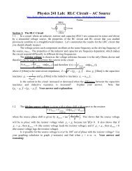
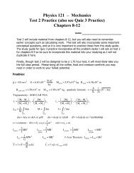
Figure 1Block diagram of <strong>the</strong> gamma spectroscopy systemFigure 2 A typical gamma spectrum taken with a detector using NaI (Sodium Iodine) crystal.All of <strong>the</strong> structure in <strong>the</strong> spectrum is associated with gamma ray that when initially emitted allhad <strong>the</strong> nearly same energy.
What to Learn about <strong>the</strong> MCA Program Used in<strong>Gamma</strong> SpectraI. Getting StartedA. To start <strong>the</strong> program at: c:\> type mcaB. Use <strong>the</strong> key associated with highlighted letter to select a menu item.C. Use <strong>the</strong> ESCAPE key to move up a menu tree.D. On <strong>the</strong> plot, <strong>the</strong> horizontal scale is channels. Each channel is associated with a smallrange of heights of <strong>the</strong> incoming pulse <strong>and</strong> for an NaI detector this pulse height isproportional to <strong>the</strong> energy deposited in <strong>the</strong> NaI crystal by <strong>the</strong> incident gamma. Thevertical scale is number of event in a given channel.E. Move markers with left <strong>and</strong> right arrow keys, Faster movement with CTRL <strong>and</strong> arrows,<strong>and</strong> read marker position in upper right. Counts at marker position see Status Page 3( Change status page using page up/down)F. Use <strong>the</strong> function key overlay chart to learn what shortcuts are available from <strong>the</strong> functionkeys.F1 to toggle Start <strong>and</strong> Stop of acquiring dataShift F2 to erase current dataF4 to cycle through vertical scaling method.G. To quit program from menu select EXIT.H. The input range is 0 to +8 volts.I. To change <strong>the</strong> directory into which data is saved use:Util Comm<strong>and</strong> cd “directory path”Please make your own directory under c:\mca\students using md “ me”II.Important parts of <strong>the</strong> manual to read.A. Chapter 3 Introduction ALL OF ITB. Chapter 4 Getting Acquainted4-4 Display spectrum from file4-5 Status pages4-16 Controlling <strong>the</strong> Display4-17 Changing <strong>the</strong> vertical scaleC. Chapter 7 is a break down of <strong>the</strong> functions available from <strong>the</strong> menu tree.7-1 Acquire On-OFF7-2,3 Presets Live, Real Total, Markers7-10 Calibrate\Manual\Energy7-15 to26 Display7-17 overlap7-17 vscale7-20 exp<strong>and</strong>7-24 cursor, Markers7-25 ROI7-27 Move\Data VERY IMPORTANT for saving data7-54 to 58 Util\ Dir, Print,7-58 MENU VERY IMPORTANT because this is <strong>the</strong> access key to <strong>the</strong> datamanipulation <strong>and</strong> analysis programs described in Chapters 10 to 19
D. Chapters 10 to 19 Basic <strong>Spectroscopy</strong> Programs10-1 to 5 Introduction19-1 to 13 LOTCNV ( convert data files from binary to text form)VERY IMPORTANT for converting saved data files intospreadsheet readable form.IIINeed to be able to:A. Manual calibration of energy scale. From menu Calibrate \ Manual \ Energy SeeManual page 7-10.B. Set Preset to stop data collection for a set Real Time, Live Time or Number of Counts.From menu Acquire \ Preset Set possible presets that are not used ones to zero. SeeManual pages 7-2,3.D..Save <strong>and</strong> retrieve data. From Menu: Move \ Data \. Use From RRH which is <strong>the</strong>display To filename.dat use defaults for header <strong>and</strong> eff files. See Manual page 7-27.E. Convert saved ( binary) datafiles to something a spreadsheet can read From MenuUTIL \ Menu <strong>the</strong>n Reports from groups ( left column, use page up/down to select) <strong>the</strong>nConvert Data to Lotus Format ( center column, use arrows). Use F2 to execute <strong>the</strong>program, <strong>the</strong>n give name of binary <strong>and</strong> name of new file to create. To return to MCAscreen use F10. See Manual pages 19-1 to 13.G. To get DOS Prompt from menu Util \ System Then can issue DOS comm<strong>and</strong>s like: dirto see list of filescdto change directoriesdel filename.extto delete filecopy old_name.ext new_name.ext to copy filesNote data acquisition continues while you are at <strong>the</strong> DOS prompt.Type EXIT to return to MCA program.H. To adjust <strong>the</strong> Lower Level Discriminator from <strong>the</strong> menu use Setup \ ADC \ CoarseLLD or Fine LLD . Also of use Setup \ ADC \ Zero <strong>and</strong> Setup \ ADC \ ULD
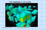
Discriminator, Single Analyzers <strong>and</strong> Multichannel AnalyzersIn nuclear <strong>and</strong> high energy physics most experiments depend on its associated <strong>the</strong> electronics.Usually, an electrical pulse is created by a detector when a particle is detected. These pulses may containinformation about <strong>the</strong> detection event such as when it occurred, where on <strong>the</strong> detector <strong>the</strong> event occurred, or<strong>the</strong> energy of <strong>the</strong> detected particle. Small signals are made larger with an amplifier, The shape, <strong>and</strong> durationof a signal pulse may also be changed by special amplifiers ( by integration or differentiation of <strong>the</strong> signal).In most cases, <strong>the</strong>re is some level of electrical noise on <strong>the</strong> signal wires <strong>and</strong> <strong>the</strong> real signals must be sort outfrom <strong>the</strong> noise. Frequently a discriminator, or a single channel analyzer (SCA) is used for separating <strong>the</strong>signal from <strong>the</strong> noise.If after an amplifier, pulses associated with real events are about 2 volts in peak amplitude, <strong>and</strong> <strong>the</strong>noise is 0.07 volts in amplitude, <strong>the</strong>n separating <strong>the</strong> signal from <strong>the</strong> noise seems simple enough-- just pay noattention to pulses less than a volt high. This is basically what a discriminator or single channel analyzerdoes. One defines what a valid input is <strong>and</strong> every time <strong>the</strong> input pulse meets this selection criteria, an outputpulse is produced. This output pulse carries only <strong>the</strong> information: “A valid event just happened.” Thisoutput pulse is often a + 5 Volt pulse similar to a digital logic TTL pulse. In addition to <strong>the</strong> existence of avalid pulse, <strong>the</strong>re is <strong>the</strong> information as to when <strong>the</strong> pulse occurred.. There are several ways of synchronizing<strong>the</strong> output to <strong>the</strong> input so that <strong>the</strong> relative timing remains <strong>the</strong> same.When using a discriminator, one defines <strong>the</strong> minimum voltage level for a valid pulse. That is youset <strong>the</strong> discriminator level to 1.4 volts, <strong>and</strong> any pulse with a peak height of 1.4 volts or greater results in anoutput pulse. Many discriminators delay <strong>the</strong> output signal slightly so that is set out a set delay after <strong>the</strong> pulsereaches a set fraction of its peak height ( NOT <strong>the</strong> discriminator level). These are called constant fractiondiscriminators ( CFD’s).A single channel analyzer is like a discriminator with added features. One can set <strong>the</strong> SCA to <strong>the</strong>Integrate mode (INT) where it will operate like a discriminator. When using <strong>the</strong> Window mode one has <strong>the</strong>added feature of defining both a maximum acceptable peak height as well as <strong>the</strong> minimum. This would beuseful if <strong>the</strong> detector was detecting two types of events with different pulse peak heights <strong>and</strong> you wereinterested in counting <strong>the</strong> number of events associated with <strong>the</strong> smaller pulses. You would <strong>the</strong>n put <strong>the</strong> SCAwindow around <strong>the</strong> signal of interest. That is you would set <strong>the</strong> lower level (LL)of window above <strong>the</strong> noisebut below <strong>the</strong> height of <strong>the</strong> signal of interest. Next you would set <strong>the</strong> upper level (UL) of <strong>the</strong> windowbetween <strong>the</strong> two pulse heights ( often using a knob labeled Window or UL).Often on an SCA, one actually specifies a lower level <strong>and</strong> <strong>the</strong> height of <strong>the</strong> upper level above <strong>the</strong>lower level. This is useful in analyzing a complex spectrum of signal pulse heights. For example, if youbreak <strong>the</strong> spectrum form 1 to 10 volts into a number of “channel” each 0.2 volts wide by set <strong>the</strong> height of <strong>the</strong>window at 0.5 volts, <strong>and</strong> <strong>the</strong>n counting for a minute for each of <strong>the</strong> following lower level settings: 1.0, 1.5,2.0, 2.5, . . . 9.5 volts. Plotting <strong>the</strong> number of counts recorded as a function of <strong>the</strong> lower level setting willyield a histogram of <strong>the</strong> incoming signals pulse height.If you need to do a lot of this, <strong>the</strong> counting gets ra<strong>the</strong>r tedious, especially if <strong>the</strong> window height issomething like only 0.05 Volts. Fortunately, someone invented <strong>the</strong> multichannel analyzer (MCA). Thisdevice looks at all of <strong>the</strong> “channels” at once. That is it break its input range into a number of pulse peakheight ranges (channels) <strong>and</strong> counts <strong>the</strong> number of pulses falling into each range. Usually an MCA willdisplay <strong>the</strong> resulting histogram <strong>and</strong> allow some numerical manipulation of this data. Note that often <strong>the</strong> pulsepeak height is associated with <strong>the</strong> energy of a single quanta of some incident radiation <strong>and</strong> thus <strong>the</strong> histogramis an energy spectra of <strong>the</strong> incident radiation.
General Goals:To provide a sufficient information aboutradiation safety to allow students to safely work inPAS 284.Reading:See A.C.Melissinos, Experiments inModern Physics, p.143-149.Introduction to Radiation Safety, by <strong>the</strong>University of Arizona Radiation Control Office(attached to this document)Units used when discussing radiationWhen discussing radiation safety, it isnecessary to know how radiation is measured.That is basically a discussion of units.The curie ( abbreviated Ci) is a measureof <strong>the</strong> activity of <strong>the</strong> sample that is <strong>the</strong> number ofnuclear disintegrations in a given time.Specifically <strong>the</strong> curie is 3.7 X 10 10 disintegrationsper second, which is <strong>the</strong> activity of a gram ofradium. In <strong>the</strong> SI system of units, <strong>the</strong> curie hasbeen replaced by <strong>the</strong> becquerel (Bq) with 1 curie= 37 X 10 9 Bq, so a Bq is one disintegration persecond.The activity of a source ( in Ci or Bq) isone way to describe <strong>the</strong> amount of radiationpresent. An alternative method is to give <strong>the</strong> fluxof radiation. That is <strong>the</strong> number of quanta (alpha, beta, gamma, protons, chickens . . . orwhatever) incident on a unit area per unit of time (for example <strong>the</strong> number of betas incident on 1cm 2 /s ).There is ano<strong>the</strong>r type of radiation unit thatis related to <strong>the</strong> interaction of radiation with livingtissue. As radiation passes through matter, itinteracts <strong>and</strong> in general deposits energy in <strong>the</strong>matter. A rad of radiation will deposit 100 ergs ofenergy for each gram of tissue it passes through.Starting from <strong>the</strong> activity of a source or <strong>the</strong> flux of<strong>the</strong> radiation <strong>and</strong> converting to rads is not asimple process. This involves details of <strong>the</strong>interaction between <strong>the</strong> specific type of tissue <strong>and</strong><strong>the</strong> specific type <strong>and</strong> energy of <strong>the</strong> radiation.Recently SI replaced <strong>the</strong> rad with <strong>the</strong> gray (Gy).A gray of radiation will deposit 10,000 ergs ofenergy for each gram ( or 1 J/kg) of tissue itpasses through ( 100 rad = 1 Gy).Even when losing <strong>the</strong> same amount ofenergy in a given bit of living tissue, differentkinds of radiation cause different degrees ofdamage. This is because for some type ofNuclear Safety <strong>and</strong> Unitsradiation a given particle may interact relativelyinfrequently, but deposits a relatively largeamount of energy during each interaction, while aparticle of different type of radiation, may interactfrequently, but lose only a small amount of energyin each interaction. The energy deposited pergram of tissue may be <strong>the</strong> same but <strong>the</strong> damageis different. Thus a fur<strong>the</strong>r set of units is needed.The rem ( roentgen equivalent unit ( aroentgen is basically 1 rad of gamma’s)) is <strong>the</strong>st<strong>and</strong>ard old unit for radiation damage <strong>and</strong> <strong>the</strong>sievert (Sv) is <strong>the</strong> new SI unit. 100 rem = 1 Sv.The amount of radiation present in Gy, ismultiplied by an RBE (relative biologicaleffectiveness) factor to yield <strong>the</strong> amount ofradiation damage in sieverts. The RBE for x rays,gammas <strong>and</strong> betas is 1, protons it is 10 <strong>and</strong> foralpha it is 20. Thus <strong>the</strong> same number of rads orgrays of alphas in much more damaging <strong>the</strong>equivalent amount of gammas.Note <strong>the</strong> rem or <strong>the</strong> sievert do not indicatehow quickly <strong>the</strong> damage was done. So often <strong>the</strong>radiation field is reported in rem/hr or mrem/ yearor Sv/year. Acceptable limits of radiationexposure have decreased over <strong>the</strong> years as moreinformation on <strong>the</strong> long term effects of radiationhave become available.Typical ExposuresTypical exposures per year to a member of <strong>the</strong>public may include: 35-60 mrem ( 0.0005 mrem/hr) fromcosmic rays ( strong altitude dependance) 35-70 mrem ( 0.0005 mrem/hr) fromnatural radioactive sources 30-350 mrem ( 0.0005 - 0.005 mrem/hr)from all natural radioactivity that includessources within <strong>the</strong> body• ( 25 mrem from K 40 ,• 1 mrem from C 14 ,• remainder from Ra 226 )100 mrem from medical x-rays.4 to 8 mrem (0.0005 to 0.001 mrem / hr)estimate of exposure in <strong>the</strong> Physics <strong>and</strong>Atomospheric Science Building at <strong>the</strong>University of Arizona.0.001 mrem/hr typical poker chip sourceat one meter.Legal Limits of Exposure
These limits are set by various governmentagencies <strong>and</strong> are subject to change.For <strong>the</strong> general public (Or Students) <strong>the</strong>acceptable whole body exposure is:100 mrem/yr = 1 mSv/yr,For radiation workers <strong>the</strong> limit is5000 mrem / yr.If <strong>the</strong> radiation exposure is limited to <strong>the</strong> skin orextremities, <strong>the</strong> limit is even higher.One of our typical “poker chip” sources willproduce a radiation field a one meter of about0.001 mrem/hr. A student working with one of <strong>the</strong>“poker chip” sealed source would reach <strong>the</strong>irannual full body radiation exposure in about 100hours. This assumes a distance of one meter<strong>and</strong> no shielding. Note that <strong>the</strong>re are about 75hours of advanced lab per semester. Double <strong>the</strong>distance to two meters <strong>and</strong> <strong>the</strong> time increases to400 hour.Official Information:Arizona Regulations dealing with radiation safety <strong>and</strong> radiation workers’ rights can be found on <strong>the</strong>table near <strong>the</strong> radioactive source cabinet.On <strong>the</strong> door of <strong>the</strong> radioactive source cabinet are:Notice to EmployeesRadiation Emergency ProcedureBasic Laboratory Procedure for Radioactive MaterialsRules for Package Radioactive WasteRadioactive Materials Approval List ( Basically an inventory of sources requiring approval)Sources <strong>and</strong> Location:The radioactive source cabinet is in <strong>the</strong> North East ( After entering <strong>the</strong> room this is <strong>the</strong> far left)corner of PAS 284. This cabinet should be locked unless PAS 284 is occupied.In PAS 284 <strong>the</strong>re are three sources that require approval. 15 mCurie Cs-137 is a beta <strong>and</strong> gamma source used for <strong>Compton</strong> scattering experiments.This sealed source stored in a lead pig inside <strong>the</strong> radioactive source cabinet. This source isquite hot, <strong>and</strong> SHOULD BE HANDLED ONLY BE THE LAB MANAGER or o<strong>the</strong>r trainindividuals <strong>and</strong> <strong>the</strong>n only at arms length <strong>and</strong> for as short of time as possible. The lead pigused for storage <strong>and</strong> <strong>the</strong> lead shielding used in <strong>the</strong> <strong>Compton</strong> experiment reduce <strong>the</strong> emittedgamma radiation to a level similar to a “poker chip” source. 0.001Curie Am-241 is an alpha source used for studying <strong>the</strong> range <strong>and</strong> energy loss ofalphas in gases. This source is a foil implanted with Americium <strong>and</strong> is mounted in a 9 by 5inch vacuum chamber. It is stored in <strong>the</strong> radioactive source cabinet. Am-241 is an alphaemitter <strong>and</strong> all alpha emitters are potentially dangerous because alphas can cause seriouscell damage. This damage usually only occurs if <strong>the</strong> alpha emitter is ingested becausealphas have a very limited range. Virtually all alphas emitted by this source all absorbed by<strong>the</strong> vacuum chamber housing. Unless <strong>the</strong> housing is opened this source is quite safe. NONOT OPEN THE VACUUM HOUSING 16 gram of Pu-239 mixed with beryllium is a neutron source used in neutron activationexperiments. This source is in a large brown drum ( a neutron howitzer) stored near <strong>the</strong>radioactive source cabinet. This drum is filled with paraffin that is an excellent shieldingmaterial for neutronsThe cabinet also contains a number of low activity test sources <strong>the</strong> size of a poker chip, lanternmantles containing thorium <strong>and</strong> o<strong>the</strong>r low level radioactive materials.WasteIn <strong>the</strong>se experiments no waste should be generated <strong>and</strong> neutron activated material are stored in<strong>the</strong> neutron howitzer after <strong>the</strong> experiment. ,
Accidents /emergenciesIf you believe, a source ( including an exempt “poker source” ) is damaged contact <strong>the</strong> lab manageror your TA immediately.If <strong>the</strong> radioactive sources are out of <strong>the</strong> cabinet <strong>and</strong> <strong>the</strong>re is a fire in <strong>the</strong> building, please inform <strong>the</strong>police, or fire departments or <strong>the</strong> building monitor.Working procedures:An official protocol is describing <strong>the</strong> use of each of <strong>the</strong> radioactive sources requiring approval. Thisis a legal document outlining <strong>the</strong> operational procedures. If you are preforming an experiment using one of<strong>the</strong>se sources, you should get a copy of <strong>the</strong> associated protocol <strong>and</strong> follow <strong>the</strong> protocol.The basic rule of thumb when working with radioactive material is to take all reasonable steps toreduce exposure. The rule is often referred to as ALARA, As Low As Reasonably Achievable. The threebasic methods of reducing exposure are: minimizing exposure time, maximizing distance ( Intensity 1/r 2 ), <strong>and</strong> using shieldingLead for most forms of radiationParaffin for neutronsApplying this rule requires that one stop <strong>and</strong> think before using a radioactive source. Think through whatyou are about to do.Lab Rules:11. No eating or drink in PAS 284! No open food or beverage in PAS 284. Any open food or beveragebrought into PAS 284 must be destroyed.12. The lab PAS 284 must ei<strong>the</strong>r be occupied or locked at all times.13. Do not remove any radioactive source including an exempt “poker source” from PAS 28414. Only Lab manager or specially trained TA or Instructor may move <strong>the</strong> 15 mCurie Cs -137 source,open <strong>the</strong> neutron howitzers, or open <strong>the</strong> Range of Alpha chamber. Please move at least 2 metersfrom <strong>the</strong> area when <strong>the</strong>se processes are occurring.15. Store radioactive sources in <strong>the</strong> cabinet when not in use.16. At <strong>the</strong> end of lab check <strong>the</strong> inventory to verify all sources are stored.17. Do not eat a source18. Do not put a source in your pocket19. Do not hold a source any longer than necessaryAdditional Concerns:If you are concerned about some aspect of <strong>the</strong> radiation safety in PAS 284 please discuss it with <strong>the</strong>Lab Manager or Someone at <strong>the</strong> Radiation Control Office 626-6850 or rcohelp@u.arizona.edu orhttp://www.radcon.arizona.edu/.Pregnancy:Anyone who is pregnant, thinks <strong>the</strong>y may be pregnant, is engaged in activities to become pregnantshould take special steps to limit exposure, <strong>and</strong> should (but are not required to ) discuss <strong>the</strong> situation with<strong>the</strong> Lab Manager or someone at <strong>the</strong> Radiation Control Office 626-6850 or rcohelp@u.arizona.edu orhttp://www.radcon.arizona.edu/.Additional Training:Additional radiation safety training is available through <strong>the</strong> Radiation Control Office 626-6850 orrcohelp@u.arizona.edu or http://www.radcon.arizona.edu/.