Visual Acuity Assessment with Snellen and ETDRS Charts
Visual Acuity Assessment with Snellen and ETDRS Charts
Visual Acuity Assessment with Snellen and ETDRS Charts
- No tags were found...

Create successful ePaper yourself
Turn your PDF publications into a flip-book with our unique Google optimized e-Paper software.
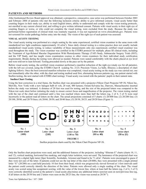
PATIENTS AND METHODS<strong>Visual</strong> <strong>Acuity</strong> <strong>Assessment</strong> <strong>with</strong> <strong>Snellen</strong> <strong>and</strong> <strong>ETDRS</strong> <strong>Charts</strong>After Institutional Review Board approval was obtained, a prospective, consecutive, case series was performed between October 2003<strong>and</strong> February 2004 of patients who met the following inclusion criteria: ability to give informed consent, visual acuity better thancounting fingers in their study eye, ability to read English letters, ability to underst<strong>and</strong> <strong>and</strong> comply <strong>with</strong> the vision testing protocols,not enrolled in any current clinical trial, <strong>and</strong> willing to give written informed consent. Patients <strong>with</strong> visual acuity in their right eye ofcounting fingers or worse, or who could not complete the visual testing, were excluded from the study. Because the study wasperformed before registration of clinical trials was routinely required, it was not registered on www.clinicaltrials.gov. Patients werenot screened for ocular pathology before entry into the study. The vision of the right eye of each patient was assessed.VISUAL ACUITY TESTINGThe visual acuity testing was performed at a single seating by the same experienced, certified vision examiner in the same room <strong>with</strong>st<strong>and</strong>ardized low light conditions (approximately 10 cd/m 2 ). Since daily clinical testing in a retina practice does not usually includest<strong>and</strong>ardized visual acuity testing, to reduce variability of these measurements only one experienced, certified visual examiner wasused throughout the study. The vision examiner was certified in the <strong>ETDRS</strong> protocol for multiple, phase III clinical trials, includingthe Treatment of Age-Related Macular Degeneration With Photodynamic Therapy (TAP) Study, Submacular Surgery Trials (SST),<strong>and</strong> the VIP Study, <strong>and</strong> has taught vision examination courses to other vision examiners for their individual study certificationrequirements. Breaks during the testing were allowed as needed. Patients were seated comfortably <strong>with</strong> the charts placed at eye level<strong>and</strong> were told not to lean forward. Testing proceeded slowly at the pace set by the patient.Before visual acuity testing, the certified vision examiner performed a manifest refraction of the right eye (study eye for all patients),<strong>with</strong> the left eye covered, using the <strong>ETDRS</strong> Chart R (catalog No. 2123; Precision Vision, La Salle, Illinois); a description of chartlighting follows. Once the best-corrected manifest refraction was completed, visual acuity testing on the study eye was carried out, onetest immediately after the other, <strong>with</strong> the chart <strong>and</strong> testing method used first, alternating between patients (eg, one patient started <strong>with</strong><strong>Snellen</strong> testing, the next started <strong>with</strong> <strong>ETDRS</strong> chart testing). <strong>Visual</strong> acuity was tested <strong>with</strong> the patients’ pupils in their natural state.<strong>Snellen</strong> Chart TestingUsing the best correction in a trial frame, the <strong>Snellen</strong> chart was presented <strong>with</strong> a projector (Nikon Chart Projector NP-3S; Nikon Inc,Melville, New York) <strong>with</strong> a new halogen bulb (6 volt, 20 watt, 480 lumens; Osram-Sylvania Inc, Danvers, Massachusetts) insertedbefore the study was initiated. A distance of 20 feet was used for testing, <strong>and</strong> the size of the projected letters was compared to theNikon test scale sheet before initiating the study to ensure correct focus <strong>and</strong> magnification of the projector. The vision testing started<strong>with</strong> the top of the chart <strong>and</strong> continued until a line was reached where more than half the letters (eg, 2 of 4, 3 of 5) were readincorrectly or the patient read all letters on the chart. The actual projections consisted of 5 charts: (1) 20/400 line; (2) 20/200 line; (3)20/100, 20/80, <strong>and</strong> 20/70 lines; (4) 20/60, 20/50, <strong>and</strong> 20/40 lines; (5) 20/30, 20/25, <strong>and</strong> 20/20 lines (Figure 1).FIGURE 1<strong>Snellen</strong> projection charts used by the Nikon Chart Projector NP-3S.Only the <strong>Snellen</strong> chart projections were used, <strong>and</strong> the additional features of the projector, including “illiterate E” charts (charts 10, 11,<strong>and</strong> 12), vertical masking, horizontal line masking, <strong>and</strong> single letter isolation, were not used. The luminance of the projected chart wasmeasured by a digital light meter (Sper Scientific, Scottsdale, Arizona) <strong>and</strong> found to be 71 cd/m 2 . Patients were encouraged to guess ifthey were not sure of the letter. If patients could not see the top letter of the chart (20/400), they were presented <strong>with</strong> a single printedletter “E” (20/200) on a card held precisely 10 feet from their head, which was progressively moved forward at 1-foot intervals untilthe patient could correctly identify the direction of the letter. Patients were allowed only a single reading of the chart. The visualacuity was scored using line assignment scoring, <strong>with</strong> the value of the lowest line, where at least half the letters (eg, 2 of 4, 3 of 5)were correctly identified scored as the patient’s visual acuity plus/minus any additional letters seen/not seen on next/previous line (eg,if all letters on the 20/30 line <strong>and</strong> 2 letters were seen on the 20/25 line, the vision was scored as 20/30 +2 ).Trans Am Ophthalmol Soc / 107 / 2009 314