1-TEP-2014-RESUMEN
1-TEP-2014-RESUMEN
1-TEP-2014-RESUMEN

You also want an ePaper? Increase the reach of your titles
YUMPU automatically turns print PDFs into web optimized ePapers that Google loves.
A.T. Cohen et al. / Thrombosis Research 133 (<strong>2014</strong>) 139–148141Table 1European Society of Cardiology risk stratification for patients with pulmonary embolism [10].Parameter High risk IntermediateriskPresence of shock a Yes No Noor hypotension bRight ventriculardysfunction c andmyocardial injury dDoes not need to bedetermined if patienthas shock or hypotensionEither or bothpresentLow risk30-day mortality rate N15% 3–15% b1%Clinical management Emergency intervention Admit to Early dischargehospital or care at homeabcBlood pressure b90 mm Hg.Blood pressure drop of 40 mm Hg below usual level.Right ventricular dilation; hypokinesis or pressure overload on echocardiography;right ventricular dilatation on spiral computed tomography; brain natriuretic peptide orN-terminal pro-brain natriuretic peptide elevation, elevated right heart pressure.d Positive cardiac troponin T or I.performed in acute situations to confirm a suspected high-risk PE.If CTPA is unavailable or the patient’s condition allows only bedsideinvestigations, echocardiography may be used (Fig. 1). In unstablepatients, evidence of acute pulmonary hypertension and RV overloadon echocardiography can justify thrombolysis or embolectomy. RVoverload on echocardiography may also be sufficient for consideringtreatment when other tests, such as transoesophageal echocardiographyfor PE or compression ultrasonography (CUS) for DVT, are unavailable[10]. ThrombosisseenontransoesophagealechocardiographyandCUSmay help with decision-making. An RV/left ventricular hazard ratio(HR) of N0.9, and elevated markers of RV dysfunction/dilation andmyocardial injury (brain natriuretic peptide and standard troponin Tor I, respectively), are of prognostic significance [10].If PE is confirmed, the Pulmonary Embolism Severity Index (PESI)score can help to estimate the 30-day mortality risk [24–26]. A valueis ascribed to each of the 11 variables present, and a total is thendetermined by summing the points and adding the patient’s ageinyears. In the simplified PESI (sPESI), which takes into account onlyage, history of cancer, chronic cardiopulmonary disease, elevatedpulse, low blood pressure and low oxygen saturation, any patient withat least one risk factor is considered at high risk of death (Table 2)[27,28].NoTable 2Simplified PESI score for assessing the likelihood of PE-related death [28].ParameterAge N80 years 1History of cancer 1Chronic cardiopulmonary disease 1Pulse ≥110 beats/min 1Systolic blood pressure b100 mm Hg 1Arterial oxyhaemoglobin saturation level b90% 1ScoreAny patient scoring 1 point or more is considered to be at high risk of early death.Abbreviations: PE, pulmonary embolism; PESI, Pulmonary Embolism Severity Index.Further Stratification and Confirmatory Diagnostic Testing in Non-high-riskPatientsPatients without shock and/or hypotension are considered nonhigh-riskand require appropriate diagnosis, further risk stratificationand treatment [10]. The presence of RV dysfunction or signs ofmyocardial injury suggests an intermediate (3–15%) risk of earlydeath, whereas the absence of all these signs correlates with a low(b1%) mortality risk (Table 1) [10].Determining Clinical Probability of Non-high-risk PE using Clinical RiskScoresClinical judgement or validated prediction rules (e.g. the Wells score)are beneficial in categorising a patient’spre-testprobabilityofPEbasedon initial findings and predisposing risk factors for PE. Although 30%of PE cases occur without any known cause [10], patient-andsettingrelatedfactors can help to categorise patients based on clinicalprobabilities. Subsequent investigations and examinations to excludePE can then be viewed in light of the clinical risk score and the resultsof initial investigations [10].Other clinical risk scores have been developed based on clinicalobservations that correlate with a positive diagnosis. These include theWells, Geneva and Miniati scores, and the Pulmonary Embolism Rule-Out Criteria [29–34]. The Wells score is the most extensively validatedand commonly used of these [29,35–37]. Using the Wells score, theclinical probability of PE is assessed as low (b2.0), moderate (2.0–6.0)Shock and/or hypotension:High-risk PE suspected>15% chance of early deathInitial risk stratification(Table 1)No shock/hypotensionNon-high-risk PE suspected≤15% chance of early deathCT availableimmediatelyCT not availableimmediatelyHigh probability ofPE by clinical riskscoreLow/moderateprobability of PE byclinical risk scoreCTPAEchocardiographyMulti-detectorCTPAPositiveD-dimerNegativePositiveYesRVoverloadNo RVoverloadPositiveNegativeNegativeSearch forother causesPatient stable andCT/other tests nowavailable?Search forother causesNo PENoHigh-risk PEThrombolysis/pulmonary endarterectomyNon-high-risk PETreat with anticoagulantsFig. 1. Diagnostic pathway recommended by the European Society of Cardiology for risk stratification of patients with PE [10]. Abbreviations: CT, computed tomography; CTPA, computedtomography pulmonary angiography; PE, pulmonary embolism; RV, right ventricular.
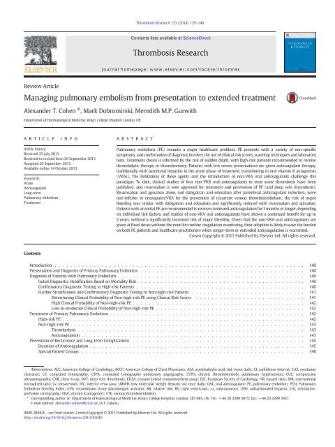
142 A.T. Cohen et al. / Thrombosis Research 133 (<strong>2014</strong>) 139–148or high (N6.0) by adding up values given to common clinical signsand symptoms of PE and clinical judgement (Table 3) [29]. Clinicalsigns and symptoms of DVT (3.0) and the clinician’s judgement thatan alternative diagnosis is less likely than PE (3.0) carry the mostweight [29]. Simplified versions of both the Wells and Geneva scoreshave been created and have a similar predictive value to the originalscores [38].High Clinical Probability of Non-high-risk PEFurther investigations, such as imaging, are required in thesepatients, and CTPA should be carried out in all patients with a moderateor high clinical probability of PE. A positive CTPA (Fig. 2) confirms a PEthat should be treated, typically with low molecular weight heparin(LMWH), whereas a negative multi-detector CTPA safely excludes PE[39–42]. By contrast, single-detector CTPA cannot exclude PE [43,44].Equivocal CTPA results should prompt further testing, possibly includingchest X-ray (CXR), D-dimer testing, repeat CTPA, lower-limb Dopplerscanning or a conventional pulmonary angiogram.A V/Q scan is an alternative to CTPA if a patient has a moderate orhigh pre-test probability of PE, despite normal CXR and D-dimer results(for example, if CTPA is contraindicated owing to renal impairmentor allergy to the contrast agent). V/Q scan results are usually dividedinto the five categories established by the PIOPED trial: normal, nearnormal-,low-, intermediate- (non-diagnostic), and high-probabilityof PE [45]. Ahigh-probability(twoormoremismatchedsegmentalperfusion defects) V/Q scan confirms PE, and the patient should bestarted on treatment. A normal- or high-probability V/Q scan excludesor confirms a suspected PE in most patients [45,46], andnon-diagnostic,low-probability results combined with low clinical probability show avery low prevalence of PE [45,47].Thecombinationofclinicalprobability,D-dimer, CUS and lung scanning confirms or excludes PE in 94% ofpatients [48]. However,consideredalone,intermediate-probabilityV/Qscan results or a low- or normal-probability scan in a patient with ahigh pre-test probability of PE require CTPA to rule out PE. The highfrequency of these intermediate scans has led critics to question theusefulness of V/Q scanning in excluding PE [10].Lower-limb CUS can also provide alternate rationale for therapyeither in patients with a high clinical probability of PE with a suspectedfalse-negative CTPA result or when CTPA is contraindicated [40,49].Anticoagulant treatment can be justified when a proximal DVT ispresent, because this is found in 30–50% of patients with PE [50,51].DVT can be diagnosed using CUS with a 90% sensitivity and 95%specificity for proximal DVT. In patients with suspected PE, proximalDVT warrants anticoagulation without further testing [52]. CUS canalso reduce the chance of a false-negative result with single-detectorCTPA.Pulmonary angiography was formerly the ‘gold standard’ forPE diagnosis. Non-invasive diagnostic investigations such as CTPA,D-dimer assays, lower-limb CUS and V/Q exams have largely removedthe need for this more invasive and costly technique [10,53,54], whichis now mostly used only when other test results are unclear [10].Table 3Wells score a for prediction of PE [29].ParameterScoreClinically suspected DVT 3Alternative diagnosis less likely than PE 3Rapid heart rate 1.5Immobilisation within past 4 weeks 1.5History of DVT 1.5Haemoptysis 1Malignancy 1Abbreviations: DVT, deep vein thrombosis; PE, pulmonary embolism.a A total score N6 indicates a high probability of a PE, 2–6 moderateprobability and b2 low probability.Fig. 2. Computed tomography pulmonary angiography scan of a bilateral pulmonaryembolism.Low-to-moderate Clinical Probability of Non-high-risk PEQuantitative enzyme-linked immunosorbent assay (ELISA) Ddimerassays and other ELISA-derived tests have a high sensitivity (N95%)and a specificity of approximately 40% [10]. Patients with a normalhigh-specificity D-dimer assay result and a low pre-test probabilitycan safely be excluded from having PE. Only low pre-test probabilitypatients can be excluded with a moderately sensitive assay. Assayspecificity decreases with age, active malignancy and pregnancy [10].Ddimer is also often elevated in hospitalised patients and is, therefore,of limited use in this setting. Recently, it was found that doubling theconcentration threshold that conventionally denotes a positive D-dimer result (from b500 to b1000 ng/ml) in elderly (age N70 years)or low-clinical-risk patients reduced the need for subsequent CTPAwithout a significantly increased risk of missing PE or pneumonia [55].Other common laboratory tests and investigations may excludeother causes for PE-like signs and symptoms but lack sensitivity orspecificity for PE. A CXR may help to exclude other causes for dyspnoeaand chest pain [10]. However, the most frequently observed abnormalitiesin CXRs are also non-specific [56]. Levelsofarterialbloodgases can remain normal in up to 21% of patients with PE who haveno history of prior cardiopulmonary disease [57], and may be abnormalin patients both with and without PE [58]. Recent changes inelectrocardiography indicative of RV strain (inversion of T waves inleads V1–V4, a QR pattern in lead V1, S1Q3T3 type incomplete orcomplete right bundle-branch block) may be helpful, but these changesare also found in RV strain of any cause [10,59]. A normal CXR andD-dimer assay with a low pre-test probability rules out PE. PE can alsobe excluded in patients with an abnormal CXR, normal D-dimer assayand low pre-test probability.Although at least 25% of patients with PE show RV dilation on anechocardiogram or CTPA [10], the low sensitivity (60–70%) of thetest in non-high-risk patients means an echocardiogram cannot excludePE [47,60–64]. However, echocardiography can help to risk-stratifynon-critical patients and the absence of signs of RV overload onan echocardiogram can exclude PE as a reason for haemodynamicinstability.Treatment of Primary Pulmonary EmbolismHigh-risk PEHigh-risk PE is severe and life-threatening, and restoration ofpulmonary arterial flow is a priority to prevent RV failure. In suchcases, immediate anticoagulation with intravenous unfractionatedheparin (UFH) and thrombolytic therapy are recommended, with
A.T. Cohen et al. / Thrombosis Research 133 (<strong>2014</strong>) 139–148143surgical intervention as a back-up in case of a contraindication to, orfailure of, thrombolysis [10,65]. In the latter case, both sets of guidelinesrecommend surgical or catheter embolectomy. Streptokinase, urokinaseor recombinant tissue plasminogen activator (rtPA) are therecommended thrombolytic drugs [10,65] and should preferably begiven over a short 2-hour infusion period, which is associated withmore rapid clot lysis and a reduced risk of bleeding than prolongedinfusion [65]. For this reason, rtPA, which can be infused over ashort duration, is commonly used. Thrombolysis significantly reducesthe incidence of recurrent PE and death compared with heparin[10,65,66], and was effective in 92% of patients in one study [67].Non-high-risk PEThrombolysisFour recent randomised trials have investigated thrombolysisin intermediate-risk PE. The first of these to be published was theMOderate Pulmonary Embolism Treated with Thrombolysis (MOPETT)study, in which 121 patients with intermediate-risk PE were randomisedto a moderate dose of rtPA plus anticoagulation or anticoagulation aloneover 22 months [68]. Thrombolysisledtoasignificant reduction inpulmonary hypertension and recurrent PE at 28 months (16% vs. 57%;P b 0.001), duration of hospitalisation (2.2 days vs. 4.9 days; P b 0.001)and death or recurrent PE (1.6% vs. 10%; P =0.0489),withnobleedingin either group. At the 2013 annual meeting of the American College ofCardiology (ACC), several further studies were presented. A small,randomised study (ULTrasound Accelerated ThrombolysIs of PulMonAryEmbolism [ULTIMA]; n = 57) of endovascular catheter-directed rtPA10 mg infused over 15 hours with UFH, or UFH alone, in patients withintermediate-risk PE found a significant 23% improvement in right heartfunction among patients given endovascular thrombolysis, as measuredby the RV/left ventricular ratio, compared with anticoagulation alone(3% improvement; P b 0.0001) [69,70]. Thisimprovementcontinuedto be statistically significant at 90 days (P b 0.0001). Another small,randomised study (Tenecteplase or Placebo: Cardiopulmonary OutcomesAt Three Months [TOPCOAT]; n = 83) found that thrombolysiswith tenecteplase plus LMWH was superior to LMWH alone interms of 5-day survival to hospital discharge without shock,intubation or major haemorrhage in patients with intermediateriskPE (65% vs. 40%; P =0.02),whilefeweradditionalpatientstreated with tenecteplase/LMWH compared with LMWH alone hadnon-favourable outcomes at 3 months (13 vs. 23) [71].A much larger study, the Pulmonary Embolism THrOmbolysis trial(PEITHO), was also presented at ACC 2013. In this trial, 1006 patientsaged ≥18 years with intermediate-risk PE were randomised to receivethrombolysis with weight-adjusted intravenous tenecteplase or placebo,both with standard heparin anticoagulation [72].Thrombolysisreducedthe risk of mortality from any cause and haemodynamic collapse within7days of randomisation (2.6% vs. 5.6%; P=0.015),mainlyasaresultofasignificant reduction in haemodynamic collapse (P=0.002),butatacostof a significant increase in non-intracranial major bleeding (6.3% vs.1.5%; P b 0.001) and stroke (2.4% vs. 0.2%; P =0.003)[73].AnticoagulationThe current guidelines recommend anticoagulation rather thanthrombolysis for non-high-risk presentations of PE [10,65]. TheACCPrecommendations for the treatment of PE categorise anticoagulationinto three phases: initial (up to approximately 7 days); long-term(approximately 7 days to approximately 3 months) and extended(approximately 3 months to indefinite) [65]. Recommendations issuedby the ESC [10] describe initial and long-term anticoagulation for PE.Although the terminology used by these guidelines differs, they broadlyconcur on most key points (Table 4) [10,65]. Notably,themorerecentACCP guidelines now provide preliminary advice on the non-VKAOACs, which were not available when the ESC issued its guidance.For initial anticoagulation, LMWH or fondaparinux are preferred overUFH (unless, according to the ESC, the patient has a high risk of bleedingor has severe renal dysfunction) [10,65]. Parenteral anticoagulationTable 4Summary and comparison of recommendations for the treatment of PE: ESC 2008 and ACCP 2012 [10,65]. Notable differences are highlighted in bold.Setting ESC 2008 ACCP 2012Acute PE (ACCP)/low-tointermediateriskPE (ESC)PE associated with hypotension(ACCP and ESC) and/or shock;other high-risk PE (ESC)Initial treatment with UFH, LMWH or fondaparinux initiated whilethe diagnostic work-up is still ongoing (LMWH/fondaparinuxpreferred over UFH except in patients with high bleeding risk orsevere renal impairment) for:• 5 days followed by transition to VKA once INR has remained inthe target range for at least 2consecutivedays, continuing for:−3 months if PE is secondary to a transient risk factor−3ormoremonthsforafirst or subsequent unprovoked PE witha low bleeding riskVKA dose should be adjusted to maintain an INR of 2.5 (range 2.0–3.0)• Anticoagulation with UFH initiated immediately• Thrombolytic therapy, haemodynamic and respiratory support• If thrombolysis is absolutely contraindicated or has failed, options are:−Surgical embolectomy (preferred)−Catheter embolectomy• Fragmentation of proximal pulmonary arterial clots withcatheters where appropriateInitial treatment with parenteral anticoagulant (LMWH/fondaparinuxpreferred over UFH) or rivaroxaban for:• 5 days followed by transition to VKA once INR has remained in thetarget range of 2.0–3.0 (target 2.5) for at least 24 hours, continuingfor:−3 months if provoked by surgery or a non-surgical risk factor, or ifbleeding risk is high−More than 3 months for a first or subsequent unprovoked PE withlow/moderate bleeding riskVKA dose should be adjusted to maintain an INR of 2.5 (range 2.0–3.0)• Initial anticoagulation with i.v. UFH rather than s.c. therapies• Thrombolytic therapy• If thrombolysis is absolutely contraindicated or has failed, options are:−Catheter embolectomy (preferred)−Pulmonary embolectomyPE associated with activecancerInitial treatment with LMWH for 3–6 months, after which LMWH orVKA should be continued indefinitely or until the cancer is curedInitial treatment with LMWH preferred over VKA, continuing for morethan 3 monthsPE in pregnancyAfter confirmatory diagnosis, anticoagulation is recommended(LMWH is preferred; VKAs may be used with caution in theLMWH is preferred to UFH and should continue for at least 6 weekspost-partum (minimum duration of therapy of 3 months)second trimester only) and should be continued for at least3 months after deliveryPatients with CTPH Pulmonary endarterectomy Extended anticoagulation; pulmonary thromboendarterectomy ifappropriate expertise is availablePE in patients with severe renalinsufficiency (CrCl b30 ml/min)UFH preferred to LMWH owing to lack of renal clearanceUFH preferred to LMWH, rivaroxaban and dabigatran owing to lackof renal clearancePE in children No specific recommendations provided Initial UFH/LMWH, with LMWH or VKA preferred long termAbbreviations: ACCP, American College of Chest Physicians; CrCl, creatinine clearance; CTPH, chronic thromboembolic pulmonary hypertension; ESC, European Society of Cardiology;INR, international normalised ratio; i.v., intravenous; LMWH, low molecular weight heparin; PE, pulmonary embolism; s.c., subcutaneous; UFH, unfractionated heparin; VKA, vitamin Kantagonist.
144 A.T. Cohen et al. / Thrombosis Research 133 (<strong>2014</strong>) 139–148should be started in cases of strongly suspected PE, even if diagnosticwork-up has not been completed [10,65]. VKAtherapyshouldalsobeinitiated as soon as possible and parenteral anticoagulation discontinuedafter 5 days or when the international normalised ratio (INR) stabiliseswithin the target range of 2.0–3.0 (target 2.5) for at least 24 hours(ACCP) [65] or 2 consecutive days (ESC) [10]. TheACCPguidelineshave a weak recommendation for traditional parenteral agents overoral rivaroxaban (Xarelto ® , Bayer HealthCare AG, Berlin, Germany)or dabigatran (Pradaxa ® , Boehringer Ingelheim GmbH, Ingelheimam Rhein, Germany) but they do acknowledge that emerging datawith non-VKA OACs means that this is likely to change [65]. Indeed,regulatory authorities have given approval for rivaroxaban for thetreatment of DVT and PE and prevention of recurrence [74,75].Guidelines also discuss the use of IVC filters in patients with PEand recommend insertion of an IVC filter only when anticoagulationis contraindicated, and the ESC adds ‘when the risk of recurrent VTEis high’ [10,65]. There are no data on the use of non-VKA OACs withthrombolytic therapy. In theory, it might be possible after lysis totreat with UFH/LMWH for 2 days and then consider switching to anon-VKA OAC rather than starting a VKA.Of the phase III clinical trials of non-VKA OACs for the treatment ofPE, the EINSTEIN PE trial of rivaroxaban is the only published studyevaluating a single-agent approach specifically for the treatment ofsymptomatic PE [76]. In this multicentre, randomised, open-label,non-inferiority study, 4832 patients with confirmed acute symptomaticPE with or without symptomatic DVT were recruited, but the mostseverely ill patients were excluded, as per the guidelines [76]. Asingle pre-study dose of parenteral anticoagulant was permittedand received by 92% of patients. Patients were treated for 3, 6 or12 months (based on a clinical evaluation of individual risk forrecurrent thrombosis and bleeding) with either rivaroxaban 15 mgtwice daily (bid) for the first 3 weeks followed by 20 mg once daily(od) thereafter, or the standard approach of enoxaparin 1 mg/kgbid transitioning to a VKA started no later than 48 hours afterrandomisation [76]. VKAdoseswereadjustedtomaintainatargetINR of 2.0–3.0 [76].Rivaroxaban was non-inferior to enoxaparin/VKA for the primaryefficacy endpoint of recurrent symptomatic VTE (fatal plus non-fatalPE and/or DVT; P=0.003fornon-inferiority;Table 5) [76].Interestingly,rates of recurrent VTE were similar regardless of the anatomical extentof initial PE (1.6% vs. 1.3% for limited lung involvement, 2.5% vs.2.2% for intermediate involvement and 1.7% vs. 1.4% for extensiveinvolvement for rivaroxaban and enoxaparin/VKA, respectively). Theprincipal safety outcome of major plus non-major clinically relevantbleeding occurred with a similar incidence in both study arms(Table 5), but major bleeding occurred significantly less frequentlyamong patients treated with rivaroxaban (P = 0.003; Table 5) [76].These outcomes reflected those of the parallel EINSTEIN DVT trial,which included patients with DVT but not PE [77].Other non-VKA OACs have been assessed in randomised, phase IIItrials for the treatment of acute PE with or without DVT. Althoughthere were no specifically PE-focused studies among these, severalincluded substantial numbers of patients with PE. Like rivaroxaban,apixaban (Eliquis ® , Bristol-Myers Squibb Inc., New York, NY, USA)was evaluated as a single-drug treatment against standard therapy,but in a mixed population of patients with acute VTE, in the recentlypublished randomised, double-blind phase III, AMPLIFY study [78]. Inthe apixaban arm, 87% of patients received a parenteral agent at thestart of treatment. Approximately one-third of patients (n = 1836)had PE with or without DVT and a high proportion had unprovokedVTE. Apixaban (10 mg bid for 7 days, then 5 mg bid for a fixed durationof 6 months) was non-inferior to standard enoxaparin/warfarin for theincidence of recurrent VTE or VTE-related death (P b 0.001 for noninferiority;Table 5) and was associated with a significant reduction inthe incidence of major bleeding and major plus non-major clinicallyrelevant bleeding (P b 0.001 for both; Table 5) [78].Table 5Overview of study design and principal efficacy and safety results from phase III studies of non-VKA oral anticoagulants for the treatment of acute PE (rivaroxaban) and acute VTE (apixaban, dabigatran and edoxaban) [76–80].Study name Design Patient diagnosis Study arms Treatment duration Primary efficacy outcome Bleeding outcomesFirst major or non-major clinically relevant bleeding:10.3% vs. 11.4% (HR = 0.90; 95% CI 0.76–1.07; P =0.23)Major bleeding: 1.1% vs. 2.2% (HR = 0.49; 95% CI 0.31–0.79; P =0.003)Recurrent symptomatic VTE: 2.1% vs.1.8% (HR = 1.12; 95% CI 0.75–1.68;P =0.003fornon-inferiority)3, 6 or 12 months(investigator discretion)Single-drug rivaroxabanvs. standard therapyAcute symptomatic PE withor without DVT (n = 4832)EINSTEIN PE Multicentre, randomised,open-label, noninferiorityMajor bleeding: 0.6% vs. 1.8% (RR = 0.31; 95% CI 0.17–0.55; P b 0.001)First major or non-major clinically relevant bleeding:4.3% vs. 9.7% (RR = 0.44; 95% CI 0.36–0.55; P b 0.001)6months(fixed) Recurrent VTE or related death(first episode): 2.3% vs. 2.7%(HR = 0.84; 95% CI 0.60–1.18;P b 0.001 for non-inferiority)Single-drug apixabanvs. standard therapyAcute, symptomatic proximalDVT and/or PE (n = 5400)AMPLIFY Multicentre, randomised,double-blind, non-inferiorityMajor bleeding: 1.6% vs. 1.9% (HR = 0.82; 95% CI 0.45–1.48)Major or non-major clinically relevant bleeding:5.6% vs. 8.8% (HR = 0.63; 95% CI 0.47–0.84)6months(fixed) Recurrent VTE or related death: 2.4% vs.2.1% (HR = 1.10; 95% CI 0.65–1.84;Parenteral anticoagulant/dabigatran vs. standardtherapyAcute, symptomatic proximalDVT and/or PE (n = 2564)RE-COVER Multicentre, randomised,double-blind, non-inferiorityP b 0.001 for non-inferiorityFirst major or non-major clinically relevant bleeding:8.5% vs. 10.3% (HR = 0.81; 95% CI 0.71–0.94; P =0.004)Major bleeding: 1.4% vs. 1.6% (HR = 0.84;95% CI 0.59–1.21; P =0.35)Recurrent VTE or related death 12 monthsafter treatment start: 3.2% vs. 3.5%(HR = 0.89; 95% CI 0.70–1.13;P b 0.001 for non-inferiority)3–12 months(investigator discretion)Parenteral anticoagulant/edoxaban vs. standardtherapyAcute, symptomatic DVTand/or PE (n = 8292)Hokusai-VTE Multicentre, randomised,double-blind, non-inferiorityAbbreviations: CI, confidence interval; DVT, deep vein thrombosis; HR, hazard ratio; PE, pulmonary embolism; RR, relative risk; VTE, venous thromboembolism.
A.T. Cohen et al. / Thrombosis Research 133 (<strong>2014</strong>) 139–148145Two phase III, randomised, double-blind, non-inferiority trials ofnon-VKA OACs given after standard parenteral induction have alsobeen published. In RECOVER, the efficacy and safety of dabigatranetexilate (150 mg bid) versus warfarin (target INR range 2.0–3.0)was evaluated for the treatment of patients with acute, symptomaticVTE [79]. Approximately one-third of patients in this study had PE(n = 786). Unlike in EINSTEIN PE and AMPLIFY, all patients, notjust those in the standard therapy arm, received induction with anapproved parenteral anticoagulant for 5–11 days, and the totaltreatment duration was 6 months. The parenteral agent/dabigatranarm was non-inferior to the parenteral agent/warfarin arm forthe primary efficacy endpoint, recurrent VTE or VTE-related deathduring the study period (P b 0.001 for non-inferiority; Table 5)[79]. Symptomatic, non-fatal PE, which was a secondary endpointin RECOVER, also occurred with a similar incidence (1.0% vs. 0.6%;HR = 1.85 [95% CI 0.74–4.64]) [79]. Rates of major bleeding, theprincipal safety outcome, were similar between the treatment arms(Table 5) [79]. Thefindings were consistent for patients with DVT andthose with PE in this study.Edoxaban (Lixiana ® , Daiichi Sankyo Co. Ltd, Tokyo, Japan) was alsoassessed for the treatment of VTE in the randomised, double-blind,phase III Hokusai-VTE study [80]. This study included a higher numberof patients with PE than the other mixed VTE studies (n = 3319)[78,79], and a high proportion of these (approximately 46%) hadanatomically extensive disease. As with dabigatran, patients in bothstudy arms received a parenteral anticoagulant (LMWH or UFH) fora minimum of 5 days, followed by edoxaban 60 mg od (30 mg od inpatients with creatinine clearance [CrCl] 30–50 ml/min, body weight≤60 kg or receiving potent P-glycoprotein inhibitors) or overlappingtransition to warfarin (INR 2.0–3.0) for up to 12 months, at thediscretion of the treating physician. Unlike other studies, the primaryefficacy endpoint (recurrent VTE or VTE-related death) was assessed12months after treatment initiation regardless of the time on treatment.Heparin/edoxaban was found to be non-inferior to heparin/warfarinaccording to this analysis and also during the treatment period(P b 0.001 for non-inferiority; Table 5) [80]. Efficacy was similar to theoverall population in the subgroups of PE and DVT patients. There wasasignificantly lower incidence of a first major plus non-major clinicallyrelevant bleeding event in the heparin/edoxaban arm (P = 0.004), butthe incidence of major bleeding was not significantly different tostandard therapy (Table 5) [80].Prevention of Recurrence and Long-term ComplicationsAfter an initial PE, patients are at high risk of recurrence withoutcontinued anticoagulation [81,82]. Recurrence seems more frequentafter unprovoked PE, and is more common in patients with persistentrisk factors (such as active cancer and elevated antiphospholipidantibodies and/or D-dimer concentrations) and CTPH than in those forwhom factors contributing to the initial event may no longer be present(e.g. surgery) [4,65]. Although one meta-analysis suggested a 3.1foldgreater risk of recurrent VTE with initial symptomatic PE than for initialproximal DVT [83], this finding has not been consistently demonstratedin all studies [84]. Indeed, in EINSTEIN DVT and EINSTEIN PE, the rateof VTE recurrence was 2.5% after initial DVT (with just over halfof recurrences being PE) [77] compared with 1.9% after initial PE (witha new PE approximately 1.5 times more common than a new DVT)[76]. Mortality as a result of recurrent VTE is higher in patients withPE than in those with DVT [82] and may be increased in patients withCTPH [65].Duration of AnticoagulationFor the above reasons, patients who receive successful treatment forinitial PE with acute and prolonged therapy require close follow-upafter discharge from hospital and, in many cases, will require extendedanticoagulation (longer than 3 months). The period of anticoagulationrecommended by the guidelines varies according to a patient’s riskstatus (Table 4). If the risk of bleeding is high and/or factors potentiallycontributing to the initial PE are no longer present, 3months’ treatmentis proposed [10,65]. Conversely, if the risk of bleeding is lower and/orrisk factors remain, or the PE was apparently unprovoked, anticoagulationbeyond 3 months is encouraged [10,65]. Patients with CTPHare recommended to undergo pulmonary endarterectomy. However,in the absence of suitable expertise or if the lesion is not surgicallyremediable (e.g. a distal obstruction), the ACCP guidelines recommendextended anticoagulant therapy [10,65].To investigate long-term rivaroxaban for secondary VTE prevention,an extension study was conducted. In EINSTEIN EXT, rivaroxaban(20 mg od) or placebo was given for 6 or 12 months to patientswho had completed 6–12 months of VKA or rivaroxaban treatmentfor initial symptomatic DVT or PE [77]. Approximately 19% of patientsfrom EINSTEIN PE entered EINSTEIN EXT. In the overall population,rivaroxaban was superior to placebo for the incidence of recurrentDVT, non-fatal PE, fatal PE or unexplained death possibly related to PE(1.3% vs. 7.1%; HR = 0.18 [95% CI 0.09–0.39]; P b 0.001 for superiority),with major bleeding seen in only 4/602 (0.7%) patients receivingrivaroxaban and none of the 594 placebo recipients; there were nofatal bleeding events and no major bleeding events at a critical site[77]. In the RE-MEDY trial, extended dabigatran treatment was noninferiorto warfarin for the prevention of recurrent VTE and VTErelateddeath when given after successful anticoagulant treatment ofinitial VTE (1.8% vs. 1.3%; HR = 1.44 [95% CI 0.78–2.64]; P =0.03fornon-inferiority) [85]. In addition, in the RE-SONATE study, dabigatranwas superior to placebo for long-term prevention of recurrentsymptomatic VTE in patients who had received 6–18 months of VKAtreatment for initial symptomatic DVT or PE (0.4% vs. 5.6%; HR = 0.08[95% CI 0.02–0.25]; P b 0.0001 for superiority) [86]. However,inRE-MEDY, although rates of major bleeding were similar (0.9% vs. 1.8%;HR=0.52 [95% CI 0.27–1.01]), acute coronary syndrome events occurredwith a significantly higher incidence in the dabigatran arm (0.9% vs. 0.2%,respectively; P =0.02)[85]. InRE-SONATE,majorbleedingoccurredintwo patients (0.4%) receiving dabigatran and none in the placebo group(P = 0.5), and, as expected for a placebo comparison, clinicallyrelevant bleeding occurred significantly more frequently with activetreatment (P =0.001)[86]. In the AMPLIFY-EXT study of apixaban(2.5 or 5 mg bid) versus placebo given for 12 months in patientswho had completed 6–12 months of anticoagulation treatment,there was a significant reduction in recurrent VTE or VTE-relateddeath with apixaban compared with placebo (1.7% for both apixabandoses vs. 8.8%; P b 0.001) with minimal (≤0.5%) major bleeding inboth the treatment and placebo arms [87].Recently, there has been renewed interest in the potential benefitsof acetylsalicylic acid (ASA) in thromboembolic disorders. In themulticentre, randomised, double-blind WARFASA study, patients whohad experienced an initial VTE and completed 6–18 months of OACtherapy were randomly assigned to ASA 100 mg od or placebo fora further 2 years, with the option of extension [88]. The incidenceof VTE recurrence was lower in patients receiving ASA (28/205 vs.43/197 with placebo; 6.6% vs. 11.2% per year; HR = 0.58 [95% CI0.36–0.93]). Only one patient in each arm had a major bleedingevent, and adverse event profiles were similar. The positive effect ofASA remained regardless of whether the initial event was PE or DVT,and irrespective of age, sex and duration of initial anticoagulation[88]. Another similar study, ASPIRE, failed to demonstrate statisticalsuperiority for ASA over placebo for prevention of recurrent VTE inpatients who had previously been treated for VTE with anticoagulants(rates of 4.8% vs. 6.5% per year; P = 0.09), but ASA did show acardioprotective effect, reducing the rate of VTE, myocardial infarction,stroke or cardiovascular death by 34% (P = 0.01), without an increasein major bleeding compared with placebo (1.1% vs. 0.6% per year;P =0.22) [89].
146 A.T. Cohen et al. / Thrombosis Research 133 (<strong>2014</strong>) 139–148Special Patient GroupsAs mentioned previously, guidelines recommend extended anticoagulationwhile the benefit–risk balance remains favourable (Table 4),and this needs to be considered in patients at high risk of bleeding.This also applies to patients with active cancer, in whom studies haveshown a preference for long-term LMWH rather than short-termLMWH followed by VKA. Non-VKA OACs are not recommended forpregnant women, who should receive LMWH [10,65]. Patientswithsevere renal impairment (CrCl b30 ml/min), who are at increased riskof bleeding, are recommended to receive UFH and to be carefullyassessed after the initial 3-month treatment window for the benefitof continued therapy [10,65]. Minimal data exist regarding anticoagulationin paediatric patients, but the ACCP recommends initialUFH or LMWH, with LMWH or VKA preferred in the long term [90].DiscussionThe risk of early death from PE necessitates rapid treatmentdecisions. Although clinical diagnosis is difficult, increasingly sophisticatedscanning techniques and risk scores allow stratification ofpatients by early mortality risk. However, some of these tests taketime, and guidelines recommend initiation of parenteral anticoagulationwhen PE is clinically suspected, regardless of whether or not a definitediagnosis has been established [10,65].Thrombolysis accompanied by UFH is the established treatmentapproach to high-risk PE and has been shown to be effective in thesepatients who require urgent clot lysis [10,65,66]. Forintermediate-riskPE, the picture is less clear. Data from small studies suggest a possiblerole for thrombolysis at a reduced dose [68–71]. However, the resultsof the much larger, randomised PEITHO study of tenecteplase plusanticoagulation versus anticoagulation alone in intermediate-riskpatients with PE indicated an excess of major bleeding and strokewith thrombolysis [73], suggesting that a benefit isbynomeansclearcut.It should also be noted that tenecteplase is not currentlyrecommended for thrombolysis, and that current guidelines do notsupport thrombolysis in non-high-risk patients [10,65].For PE classified as non-high risk, the traditional approach toanticoagulant treatment – a parenteral agent overlapping with andtransitioning to an oral VKA – is based on the need for a rapidanticoagulant effect (an advantage of subcutaneous or intravenousformulations) balanced with the practical advantages of oral treatmentduring the secondary prevention phase. However, the transitionrequires close monitoring of the INR, which must then also be tightlycontrolled for the entire period of VKA therapy. In practical terms,this is difficult. Two large cohort studies using healthcare insurancedatabases indicated that patients treated for VTE with warfarinremained in the therapeutic range only 38% of the time [91,92]. Non-VKA OACs offer a potential alternative by providing a fast-actinganticoagulant effect with fixed oral dosing and without the need forroutine coagulation monitoring. Rivaroxaban and apixaban alone, anddabigatran and edoxaban after parenteral anticoagulant induction,were non-inferior to standard therapy for the prevention of recurrentVTE [76–80]. The risk of major bleeding was similar after parenteralinduction with dabigatran and edoxaban and significantly reducedwith single-drug rivaroxaban or apixaban [76–80].After an initial PE, patients should continue to receive anticoagulantsto prevent recurrence [10,65]. Treatment usually continues for atleast 3 months and often longer, or even indefinitely, provided thatthe risk of bleeding continues to be outweighed by the potentialtherapeutic benefits (Table 4). Although initial management may occurin the inpatient or outpatient setting, depending on the severity ofPE (Table 1) and local practice, most patients will self-administeranticoagulant treatment at home at some point, supported by generalpractitioner or nurse visits, or sessions at an anticoagulation clinic orpharmacy. In this context, patients may prefer oral agents over parenteraldrugs that require injection by the patient, a family member, carer, nurseor doctor, with potential consequences for adherence. Meta-analysis datainvolving approximately 6500 patients have indicated that patientscan safely self-manage their anticoagulation with no detrimental effecton efficacy and safety [93], andseveralcountrieshaveadoptedorareconsidering this approach. Nevertheless, unlike VKAs, non-VKA OACsare given at fixed doses without the need for routine monitoring andcould, therefore, further ease the burden on both patients and healthcarepractitioners.Guidelines tend to view any therapy after 3–6monthsasextendedtreatment; studies such as EINSTEIN EXT [77], RE-MEDY [85] andAMPLIFY-EXT [87] have shown a continued benefitwithanticoagulationfor between 12 months and 3 years, albeit with a small increase in therisk of bleeding and possibly, in the case of dabigatran, acute coronarysyndrome events [77,85]. ASAalsoappearstohaveabenefit in thisscenario [88,89], althoughitshouldbenotedthatthereductioninrecurrent VTE was much greater in the studies of non-VKA OACs,without a large difference in rates of major bleeding. Guidelines providerecommendations for balancing benefits and risks but, ultimately, theclinician will decide the type and length of treatment on a case-bycasebasis.As scanning techniques become more sensitive, asymptomatic PEis increasingly being detected [16]. Most of these emboli are smalland located in segmental and subsegmental vessels [16]. Detectedinisolation and without clinical symptoms, these emboli probably donot require anticoagulant treatment because evidence suggests thatpatients are not at an increased risk of adverse outcomes or death[94,95]. However, anticoagulation would be mandated if the patient’scardiopulmonary function is compromised or a concomitant proximalDVT is present [95].Non-VKA OACs have predictable pharmacokinetic and pharmacodynamiccharacteristics that make them effective, safe and convenientfor a wide range of patients. In particular, and in contrast to VKAs,demographic characteristics such as sex and ethnicity, advancedage, mild or moderate renal impairment, and low and high bodyweight do not necessitate dose adjustments [96]. However, there arecircumstances in which non-VKA OACs may not be appropriate fortreating PE and preventing recurrence, such as in critically ill patientswho require surgery or thrombolysis; during pregnancy; in children;and in patients with severe renal impairment (CrCl b30 ml/min).The EINSTEIN studies, for example, recruited relatively few patientswith renal impairment, and those with a clinically relevant bleedingrisk were excluded [76,77]; therefore, there is limited informationabout the use of rivaroxaban in these patients, and caution shouldbe applied. There is currently no specific reversal agent for rivaroxabanor any non-VKA OAC although, given their short half-lives, standardmanagement strategies may be sufficient to control mild or moderatebleeding. Use of prothrombin complex concentrates, active prothrombincomplex concentrates or recombinant Factor VIIa may beconsidered to attempt to reverse the action of these agents in anemergency [97,98].In conclusion, the treatment of PE requires both a short-term anda long-term view. Clinical risk scores and diagnostic tests provide ameans to stratify patients by risk of early death and to treat themaccordingly. Patients may need to continue anticoagulation for severalmonths or even years. Although traditional agents are effective, thenon-VKA OACs are likely to confer practical advantages in terms ofregular, fixed, oral dosing that will benefit both patients and healthcarepractitioners.Conflict of Interest StatementATC has received consultancy fees and clinical trial funding frompharmaceutical companies, including Bayer, Boehringer-Ingelheim,BMS, Daiichi, GSK, Johnson and Johnson, Mitsubishi Pharma, Pfizer,Portola, Sanofi-Aventis, Schering Plough and Takeda. MD and MMPG
A.T. Cohen et al. / Thrombosis Research 133 (<strong>2014</strong>) 139–148147have no conflicts of interest to declare. All authors confirm that theyhad full access to data and contributed to the drafting of the paper.AcknowledgementsThe authors would like to acknowledge Stephen Purver, whoprovided editorial support, including partial development of the firstdraft under the direction of the authors, with funding from BayerHealthCare Pharmaceuticals and Janssen Scientific Affairs, LLC. Theauthors were responsible for the manuscript concept, the collection,analysis and interpretation of content, critically reviewing and revisingthe manuscript drafts, and the decision to submit the manuscript forpublication.References[1] Cohen AT, Agnelli G, Anderson FA, Arcelus JI, Bergqvist D, Brecht JG, et al. Venousthromboembolism (VTE) in Europe. The number of VTE events and associatedmorbidity and mortality. Thromb Haemost 2007;98:756–64.[2] Becattini C, Agnelli G, Pesavento R, Silingardi M, Poggio R, Taliani MR, et al. Incidenceof chronic thromboembolic pulmonary hypertension after a first episode ofpulmonary embolism. Chest 2006;130:172–5.[3] Miniati M, Monti S, Bottai M, Scoscia E, Bauleo C, Tonelli L, et al. Survival andrestoration of pulmonary perfusion in a long-term follow-up of patients afteracute pulmonary embolism. Medicine (Baltimore) 2006;85:253–62.[4] Poli D, Miniati M. The incidence of recurrent venous thromboembolism and chronicthromboembolic pulmonary hypertension following a first episode of pulmonaryembolism. Curr Opin Pulm Med 2011;17:392–7.[5] Pengo V, Lensing AW, Prins MH, Marchiori A, Davidson BL, Tiozzo F, et al. Incidenceof chronic thromboembolic pulmonary hypertension after pulmonary embolism.NEnglJMed2004;350:2257–64.[6] Mohr DN, Silverstein MD, Heit JA, Petterson TM, O'Fallon WM, Melton LJ. Thevenous stasis syndrome after deep venous thrombosis or pulmonary embolism:a population-based study. Mayo Clin Proc 2000;75:1249–56.[7] Kahn SR, Ginsberg JS. Relationship between deep venous thrombosis and thepostthrombotic syndrome. Arch Intern Med 2004;164:17–26.[8] Guyatt GH, Akl EA, Crowther M, Gutterman DD, Schünemann HJ, American Collegeof Chest Physicians Antithrombotic Therapy and Prevention of Thrombosis Panel.Executive summary: antithrombotic therapy and prevention, 9th ed: AmericanCollege of Chest Physicians evidence-based clinical practice guidelines. Chest2012;141:7S–47S.[9] Heit JA, Silverstein MD, Mohr DN, Petterson TM, Lohse CM, O'Fallon WM, et al. Theepidemiology of venous thromboembolism in the community. Thromb Haemost2001;86:452–63.[10] Torbicki A, Perrier A, Konstantinides S, Agnelli G, Galiè N, Pruszczyk P, et al.Guidelines on the diagnosis and management of acute pulmonary embolism:the Task Force for the Diagnosis and Management of Acute PulmonaryEmbolism of the European Society of Cardiology (ESC). Eur Heart J 2008;29:2276–315.[11] Sandler DA, Martin JF. Autopsy proven pulmonary embolism in hospital patients: arewe detecting enough deep vein thrombosis? J R Soc Med 1989;82:203–5.[12] Stein PD, Saltzman HA, Weg JG. Clinical characteristics of patients with acutepulmonary embolism. Am J Cardiol 1991;68:1723–4.[13] Miniati M, Prediletto R, Formichi B, Marini C, Di Ricco G, Tonelli L, et al. Accuracy ofclinical assessment in the diagnosis of pulmonary embolism. Am J Respir Crit CareMed 1999;159:864–71.[14] Miniati M, Cenci C, Monti S, Poli D. Clinical presentation of acute pulmonaryembolism: survey of 800 cases. PLoS One 2012;7:e30891.[15] Stein PD, Beemath A, Matta F, Weg JG, Yusen RD, Hales CA, et al. Clinicalcharacteristics of patients with acute pulmonary embolism: data from PIOPED II.Am J Med 2007;120:871–9.[16] Desai SR. Unsuspected pulmonary embolism on CT scanning: yet another headachefor clinicians? Thorax 2007;62:470–2.[17] Soler S, Delgado C, Ballaz A, Cisneros E, Maly R, Bablis D, et al. Unsuspected pulmonaryembolism in patients with cancer. Thromb Res 2012;129:S16–9 [Suppl.].[18] Cronin CG, Lohan DG, Keane M, Roche C, Murphy JM. Prevalence and significance ofasymptomatic venous thromboembolic disease found on oncologic staging CT. AJRAm J Roentgenol 2007;189:162–70.[19] Douma RA, Kok MG, Verberne LM, Kamphuisen PW, Büller HR. Incidental venousthromboembolism in cancer patients: prevalence and consequence. Thromb Res2010;125:e306–9.[20] Huisman MV, Büller HR, ten Cate JW, van Royen EA, Vreeken J, Kersten MJ, et al.Unexpected high prevalence of silent pulmonary embolism in patients with deepvenous thrombosis. Chest 1989;95:498–502.[21] Tzoran I, Saharov G, Brenner B, Delsart D, Román P, Visoná A, et al. Silent pulmonaryembolism in patients with proximal deep vein thrombosis in the lower limbs.J Thromb Haemost 2012;10:564–71.[22] Wood KE. Major pulmonary embolism: review of a pathophysiologic approachto the golden hour of hemodynamically significant pulmonary embolism. Chest2002;121:877–905.[23] Matthews JC, McLaughlin V. Acute right ventricular failure in the setting ofacute pulmonary embolism or chronic pulmonary hypertension: a detailedreview of the pathophysiology, diagnosis, and management. Curr Cardiol Rev2008;4:49–59.[24] Aujesky D, Obrosky DS, Stone RA, Auble TE, Perrier A, Cornuz J, et al. Derivation andvalidation of a prognostic model for pulmonary embolism. Am J Respir Crit Care Med2005;172:1041–6.[25] Aujesky D, Roy PM, Le Manach CP, Verschuren F, Meyer G, Obrosky DS, et al.Validation of a model to predict adverse outcomes in patients with pulmonaryembolism. Eur Heart J 2006;27:476–81.[26] Aujesky D, Perrier A, Roy PM, Stone RA, Cornuz J, Meyer G, et al. Validation of aclinical prognostic model to identify low-risk patients with pulmonary embolism.J Intern Med 2007;261:597–604.[27] Jiménez D, Aujesky D, Moores L, Gómez V, Lobo JL, Uresandi F, et al.Simplification of the Pulmonary Embolism Severity Index for prognosticationin patients with acute symptomatic pulmonary embolism. Arch Intern Med2010;170:1383–9.[28] Jiménez D, Aujesky D, Díaz G, Monreal M, Otero R, Martí D, et al. Prognosticsignificance of deep vein thrombosis in patients presenting with acute symptomaticpulmonary embolism. Am J Respir Crit Care Med 2010;181:983–91.[29] Wells PS, Anderson DR, Rodger M, Ginsberg JS, Kearon C, Gent M, et al. Derivation ofa simple clinical model to categorize patients probability of pulmonary embolism:increasing the models utility with the SimpliRED D-dimer. Thromb Haemost2000;83:416–20.[30] Wicki J, Perneger TV, Junod AF, Bounameaux H, Perrier A. Assessing clinicalprobability of pulmonary embolism in the emergency ward: a simple score. ArchIntern Med 2001;161:92–7.[31] Klok FA, Mos IC, Nijkeuter M, Righini M, Perrier A, Le Gal G, et al. Simplification of therevised Geneva score for assessing clinical probability of pulmonary embolism. ArchIntern Med 2008;168:2131–6.[32] Miniati M, Bottai M, Monti S, Salvadori M, Serasini L, Passera M. Simple and accurateprediction of the clinical probability of pulmonary embolism. Am J Respir Crit CareMed 2008;178:290–4.[33] Kline JA, Mitchell AM, Kabrhel C, Richman PB, Courtney DM. Clinical criteria toprevent unnecessary diagnostic testing in emergency department patients withsuspected pulmonary embolism. J Thromb Haemost 2004;2:1247–55.[34] Le Gal G, Righini M, Roy PM, Sanchez O, Aujesky D, Bounameaux H, et al. Predictionof pulmonary embolism in the emergency department: the revised Geneva score.Ann Intern Med 2006;144:165–71.[35] Wolf SJ, McCubbin TR, Feldhaus KM, Faragher JP, Adcock DM. Prospective validationof Wells Criteria in the evaluation of patients with suspected pulmonary embolism.Ann Emerg Med 2004;44:503–10.[36] Wells PS, Anderson DR, Rodger M, Stiell I, Dreyer JF, Barnes D, et al. Excludingpulmonary embolism at the bedside without diagnostic imaging: management ofpatients with suspected pulmonary embolism presenting to the emergencydepartment by using a simple clinical model and D-dimer. Ann Intern Med2001;135:98–107.[37] Yap KS, Kalff V, Turlakow A, Kelly MJ. A prospective reassessment of the utility of theWells score in identifying pulmonary embolism. Med J Aust 2007;187:333–6.[38] Douma RA, Mos IC, Erkens PM, Nizet TA, Durian MF, Hovens MM, et al. Performanceof 4 clinical decision rules in the diagnostic management of acute pulmonaryembolism: a prospective cohort study. Ann Intern Med 2011;154:709–18.[39] Elias A, Cazanave A, Elias M, Chabbert V, Juchet H, Paradis H, et al. Diagnosticmanagement of pulmonary embolism using clinical assessment, plasma D-dimerassay, complete lower limb venous ultrasound and helical computed tomographyof pulmonary arteries. A multicentre clinical outcome study. Thromb Haemost2005;93:982–8.[40] Perrier A, Roy PM, Sanchez O, Le Gal G, Meyer G, Gourdier AL, et al. Multidetectorrowcomputed tomography in suspected pulmonary embolism. N Engl J Med2005;352:1760–8.[41] van Belle A, Büller HR, Huisman MV, Huisman PM, Kaasjager K, Kamphuisen PW,et al. Effectiveness of managing suspected pulmonary embolism using an algorithmcombining clinical probability, D-dimer testing, and computed tomography. JAMA2006;295:172–9.[42] Ghanima W, Almaas V, Aballi S, Dörje C, Nielssen BE, Holmen LO, et al.Management of suspected pulmonary embolism (PE) by D-dimer and multislicecomputed tomography in outpatients: an outcome study. J ThrombHaemost 2005;3:1926–32.[43] Perrier A, Howarth N, Didier D, Loubeyre P, Unger PF, de Moerloose P, et al.Performance of helical computed tomography in unselected outpatients withsuspected pulmonary embolism. Ann Intern Med 2001;135:88–97.[44] Van Strijen MJ, De Monye W, Kieft GJ, Pattynama PM, Prins MH, Huisman MV.Accuracy of single-detector spiral CT in the diagnosis of pulmonary embolism:a prospective multicenter cohort study of consecutive patients with abnormalperfusion scintigraphy. J Thromb Haemost 2005;3:17–25.[45] The PIOPED Investigators. Value of the ventilation/perfusion scan in acute pulmonaryembolism. Results of the Prospective Investigation of Pulmonary EmbolismDdiagnosis (PIOPED). JAMA 1990;263:2753–9.[46] Wells PS, Ginsberg JS, Anderson DR, Kearon C, Gent M, Turpie AG, et al. Use of aclinical model for safe management of patients with suspected pulmonary embolism.Ann Intern Med 1998;129:997–1005.[47] Roy PM, Colombet I, Durieux P, Chatellier G, Sors H, Meyer G. Systematic review andmeta-analysis of strategies for the diagnosis of suspected pulmonary embolism. BMJ2005;331:259.[48] Perrier A, Desmarais S, Miron MJ, de Moerloose P, Lepage R, Slosman D, et al. Noninvasivediagnosis of venous thromboembolism in outpatients. Lancet 1999;353:190–5.
148 A.T. Cohen et al. / Thrombosis Research 133 (<strong>2014</strong>) 139–148[49] Elias A, Colombier D, Victor G, Elias M, Arnaud C, Juchet H, et al. Diagnosticperformance of complete lower limb venous ultrasound in patients with clinicallysuspected acute pulmonary embolism. Thromb Haemost 2004;91:187–95.[50] Kearon C, Ginsberg JS, Hirsh J. The role of venous ultrasonography in the diagnosis ofsuspected deep venous thrombosis and pulmonary embolism. Ann Intern Med1998;129:1044–9.[51] Perrier A, Bounameaux H. Ultrasonography of leg veins in patients suspected ofhaving pulmonary embolism. Ann Intern Med 1998;128:243–5.[52] Le Gal G, Righini M, Sanchez O, Roy PM, Baba-Ahmed M, Perrier A, et al. A positivecompression ultrasonography of the lower limb veins is highly predictive ofpulmonary embolism on computed tomography in suspected patients. ThrombHaemost 2006;95:963–6.[53] Stein PD, Athanasoulis C, Alavi A, Greenspan RH, Hales CA, Saltzman HA, et al.Complications and validity of pulmonary angiography in acute pulmonary embolism.Circulation 1992;85:462–8.[54] van Beek EJ, Brouwers EM, Song B, Bongaerts AH, Oudkerk M. Lung scintigraphy andhelical computed tomography for the diagnosis of pulmonary embolism: a metaanalysis.Clin Appl Thromb Hemost 2001;7:87–92.[55] Kline JA, Hogg MM, Courtney DM, Miller CD, Jones AE, Smithline HA. D-dimerthreshold increase with pretest probability unlikely for pulmonary embolism todecrease unnecessary computerized tomographic pulmonary angiography. J ThrombHaemost 2012;10:572–81.[56] Elliott CG, Goldhaber SZ, Visani L, DeRosa M. Chest radiographs in acute pulmonaryembolism. Results from the International Cooperative Pulmonary EmbolismRegistry. Chest 2000;118:33–8.[57] Stein PD, Goldhaber SZ, Henry JW, Miller AC. Arterial blood gas analysis in theassessment of suspected acute pulmonary embolism. Chest 1996;109:78–81.[58] Rodger MA, Carrier M, Jones GN, Rasuli P, Raymond F, Djunaedi H, et al. Diagnosticvalue of arterial blood gas measurement in suspected pulmonary embolism. Am JRespir Crit Care Med 2000;162:2105–8.[59] Geibel A, Zehender M, Kasper W, Olschewski M, Klima C, Konstantinides SV.Prognostic value of the ECG on admission in patients with acute major pulmonaryembolism. Eur Respir J 2005;25:843–8.[60] Kasper W, Konstantinides S, Geibel A, Tiede N, Krause T, Just H. Prognosticsignificance of right ventricular afterload stress detected by echocardiography inpatients with clinically suspected pulmonary embolism. Heart 1997;77:346–9.[61] Grifoni S, Olivotto I, Cecchini P, Pieralli F, Camaiti A, Santoro G, et al. Short-termclinical outcome of patients with acute pulmonary embolism, normal blood pressure,and echocardiographic right ventricular dysfunction. Circulation 2000;101:2817–22.[62] Miniati M, Monti S, Pratali L, Di Ricco G, Marini C, Formichi B, et al. Value oftransthoracic echocardiography in the diagnosis of pulmonary embolism: resultsof a prospective study in unselected patients. Am J Med 2001;110:528–35.[63] Perrier A, Tamm C, Unger PF, Lerch R, Sztajzel J. Diagnostic accuracy of Dopplerechocardiographyin unselected patients with suspected pulmonary embolism. IntJ Cardiol 1998;65:101–9.[64] Rudoni RR, Jackson RE, Godfrey GW, Bonfiglio AX, Hussey ME, Hauser AM. Useof two-dimensional echocardiography for the diagnosis of pulmonary embolus.J Emerg Med 1998;16:5–8.[65] Kearon C, Akl EA, Comerota AJ, Prandoni P, Bounameaux H, Goldhaber SZ, et al.Antithrombotic therapy for VTE disease: antithrombotic therapy and prevention ofthrombosis, 9th ed: American College of Chest Physicians evidence-based clinicalpractice guidelines. Chest 2012;141:e419S–94S.[66] Wan S, Quinlan DJ, Agnelli G, Eikelboom JW. Thrombolysis compared with heparinfor the initial treatment of pulmonary embolism: a meta-analysis of the randomizedcontrolled trials. Circulation 2004;110:744–9.[67] Meneveau N, Seronde MF, Blonde MC, Legalery P, Didier-Petit K, Briand F, et al.Management of unsuccessful thrombolysis in acute massive pulmonary embolism.Chest 2006;129:1043–50.[68] Sharifi M, Bay C, Skrocki L, Rahimi F, Mehdipour M. Moderate pulmonary embolismtreated with thrombolysis (from the "MOPETT" Trial). Am J Cardiol 2013;111:273–7.[69] Kucher N, Boekstegers P, Blessing E, Kupatt C, Beyer-Westendorf J, Heitzer T, et al.The ULTIMA trial: a prospective, randomized, controlled study of ultrasoundaccelerated thrombolysis for the treatment of acute pulmonary embolism. J AmColl Cardiol 2013;61: Abstract 2667–8.[70] World's first pulmonary embolism trial comparing endovascular therapy tostandard of care. Available at: http://www.tctmd.com/show.aspx?id=118137&AspxAutoDetectCookieSupport=1; 2013.[71] Kline J, Hernandez J, Kabrhel C, Courtney DM, Jones AE, Nordenholtz K, et al.Randomized trial of tenecteplase or placebo with low molecular weight heparin foracute submassive pulmonary embolism: assessment of patient-oriented cardiopulmonaryoutcomes at three months. J Am Coll Cardiol 2013;61: Abstract 932–8.[72] The Steering Committee. Single-bolus tenecteplase plus heparin compared withheparin alone for normotensive patients with acute pulmonary embolism whohave evidence of right ventricular dysfunction and myocardial injury: rationaleand design of the Pulmonary Embolism Thrombolysis (PEITHO) trial. Am Heart J2012;163:33–8.[73] Konstantinides S, Meyer G, Danays T, Vicaut E, The PEITHO Investigators. Singlebolustenecteplase plus heparin compared with heparin alone for normotensivepatients with acute pulmonary embolism who have evidence of right ventriculardysfunction and myocardial injury: the Pulmonary Embolism Thrombolysis (PEITHO)study. J Am Coll Cardiol 2013;61: Abstract 304–15.[74] Bayer Pharma AG. Xarelto ® Summary of Product Characteristics. Available at:http://www.ema.europa.eu/docs/en_GB/document_library/EPAR_-_Product_Information/human/000944/WC500057108.pdf; 2013.[75] Janssen Pharmaceuticals Inc. Xarelto ® Prescribing Information. Available at: http://www.accessdata.fda.gov/drugsatfda_docs/label/2013/202439s008lbl.pdf; 2013.[76] The EINSTEIN–PE Investigators. Oral rivaroxaban for the treatment of symptomaticpulmonary embolism. N Engl J Med 2012;366:1287–97.[77] The EINSTEIN Investigators. Oral rivaroxaban for symptomatic venous thromboembolism.N Engl J Med 2010;363:2499–510.[78] Agnelli G, Buller HR, Cohen A, Curto M, Gallus AS, Johnson M, et al. Oral apixaban forthe treatment of acute venous thromboembolism. N Engl J Med 2013;369:799–808.[79] Schulman S, Kearon C, Kakkar AK, Mismetti P, Schellong S, Eriksson H, et al.Dabigatran versus warfarin in the treatment of acute venous thromboembolism.N Engl J Med 2009;361:2342–52.[80] The Hokusai-VTE Investigators. Edoxaban versus warfarin for the treatmentof symptomatic venous thromboembolism. N Engl J Med 2013;369:1406–15.[81] Eichinger S, Weltermann A, Minar E, Stain M, Schonauer V, Schneider B, et al.Symptomatic pulmonary embolism and the risk of recurrent venous thromboembolism.Arch Intern Med 2004;164:92–6.[82] Douketis JD, Kearon C, Bates S, Duku EK, Ginsberg JS. Risk of fatal pulmonaryembolism in patients with treated venous thromboembolism. JAMA 1998;279:458–62.[83] Baglin T, Douketis J, Tosetto A, Marcucci M, Cushman M, Kyrle P, et al. Doesthe clinical presentation and extent of venous thrombosis predict likelihoodand type of recurrence? A patient level meta-analysis. J Thromb Haemost 2010;8:2436–42.[84] Heit JA. Predicting the risk of venous thromboembolism recurrence. Am J Hematol2012;87(Suppl. 1):S63–7.[85] Schulman S, Eriksson H, Goldhaber SZ, Kakkar AK, Kearon C, Kvamme M, et al.Dabigatran or warfarin for extended maintenance therapy of venous thromboembolism.J Thromb Haemost 2011;9(Suppl. 2):733. Abstract O-TH-033.[86] Schulman S, Baanstra D, Eriksson H, Goldhaber SZ, Kakkar AK, Kearon C,et al. Dabigatran versus placebo for extended maintenance therapy of venousthromboembolism. J Thromb Haemost 2011;9(Suppl. 2):22. Abstract O-MO-037.[87] Agnelli G, Buller HR, Cohen A, Curto M, Gallus AS, Johnson M, et al. Apixaban forextended treatment of venous thromboembolism. N Engl J Med 2013;368:699–708.[88] Becattini C, Agnelli G, Schenone A, Eichinger S, Bucherini E, Silingardi M, et al. Aspirinfor preventing the recurrence of venous thromboembolism. N Engl J Med 2012;366:1959–67.[89] Brighton TA, Eikelboom JW, Mann K, Mister R, Gallus A, Ockelford P, et al. Low-doseaspirin for preventing recurrent venous thromboembolism. N Engl J Med 2012;367:1979–87.[90] Monagle P, Chan AK, Goldenberg NA, Ichord RN, Journeycake JM, Nowak-Gottl U,et al. Antithrombotic therapy in neonates and children: antithrombotic therapyand prevention of thrombosis, 9th ed: American College of Chest Physiciansevidence-based clinical practice guidelines. Chest 2012;141:e737S–801S.[91] Willey VJ, Bullano MF, Hauch O, Reynolds M, Wygant G, Hoffman L, et al. Managementpatterns and outcomes of patients with venous thromboembolism in the usualcommunity practice setting. Clin Ther 2004;26:1149–59.[92] Deitelzweig SB, Lin J, Kreilick C, Hussein M, Battleman D. Warfarin therapy inpatients with venous thromboembolism: patterns of use and predictors of clinicaloutcomes. Adv Ther 2010;27:623–33.[93] Heneghan C, Ward A, Perera R, Bankhead C, Fuller A, Stevens R, et al. Self-monitoringof oral anticoagulation: systematic review and meta-analysis of individual patientdata. Lancet 2012;379:322–34.[94] Goodman LR. Small pulmonary emboli: what do we know? Radiology 2005;234:654–8.[95] Stein PD, Goodman LR, Hull RD, Dalen JE, Matta F. Diagnosis and management ofisolated subsegmental pulmonary embolism: review and assessment of the options.Clin Appl Thromb Hemost 2012;18:20–6.[96] Eriksson BI, Quinlan DJ, Eikelboom JW. Novel oral Factor Xa and thrombin inhibitorsin the management of thromboembolism. Annu Rev Med 2011;62:41–57.[97] Siegal DM, Crowther MA. Acute management of bleeding in patients on novel oralanticoagulants. Eur Heart J 2013;34:489–98.[98] Levy JH, Faraoni D, Spring JL, Douketis JD, Samama CM. Managing new oralanticoagulants in the perioperative and intensive care unit setting. Anesthesiology2013;118:1466–74.