The serologic diagnosis of celiac The serologic ... - UZ Leuven
The serologic diagnosis of celiac The serologic ... - UZ Leuven
The serologic diagnosis of celiac The serologic ... - UZ Leuven
You also want an ePaper? Increase the reach of your titles
YUMPU automatically turns print PDFs into web optimized ePapers that Google loves.
Critically Appraised Topic<strong>The</strong> <strong>serologic</strong> <strong>diagnosis</strong> <strong>of</strong> <strong>celiac</strong>disease in adultsDr. P. VermeerschDr. G. Mariën and Pr<strong>of</strong>. Dr. X. Bossuyt
Introduction- Celiac disease is an autoimmune disorder characterized by animmunologic responsiveness to ingested gluten- Diagnosis is based on intestinal biopsy (Marsh classification)- Genetic susceptibility (HLA DQ2 and DQ8)- Prevalence in the general population estimated at 0.5-1%- Most patients are diagnosed as adults and rarely present with overt<strong>celiac</strong> disease
Clinical PresentationHopper, A. D et al. BMJ 2007;335:558-562
PathogenesisLumenPartially digested gluten peptidesEpithelial barrierIncreased permeability <strong>of</strong> mucosaSubepithelial region Gln GlutTGDQ2 / DQ8Antibodies to- GliadinAPC- Deamidated gliadin- tTG- Neo-epitopeB-cellIFN-γT-cell
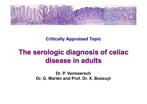
Marsh ClassificationMarsh 1 Intra-epithelialMarsh 2lymphocytosisCrypthyperplasiaMarsh 3bSubtotal villous atrophyMarsh3cTotal villous atrophy
Serology- Serologic testing is traditionally performed as a screening assay inpatients suspected <strong>of</strong> <strong>celiac</strong> disease- Current methods include the detection <strong>of</strong> antibodies direct against:1. Endomysium (indirect immun<strong>of</strong>luorescence)2. Tissue transglutaminase (ELISA)3. Gliadin (ELISA)- Detection <strong>of</strong> IgA antibodies is considered the most sensitive andspecific- IgA anti-endomysial antibodies and IgA anti-tTG tTG antibodies areconsidered the best screening assay in adults (sens. and spec. >90%).
Meta-analysis (2005, Rostom et al)Studies in Sensitivity (95% Specificity (95%Nadults CI) CI)Prev PPVIgA-AGA 11 0.75–0.90 (H) 0.80–0.90 (H) 0.36 H HIgG-AGA 7 0.17-1.00 (H) 0.70-0.80 (H) 0.37 H HNPVIgA-EMA (ME) 11 0.974 (0.957-0.985) 0.996 (0.988-0.999) 0.40 0.974 0.996IgA-EMA (HUC) 6 0.902 (0.859-0.934) 1.000 (0.991-1.000) 0.33 0.902 1.000IgA-tTG (GP) 5 0.859 (0.808-0.898) 0.953 (0.930-0.969) 0.31 0.859 0.953IgA-tTG (HR) 3 0.0.981 (0.901-0.997) 0.997) 0.981 (0.958-0.991) 0.991) 0.16 0.981 0.981H: Heterogeneous; ME: monkey oesophagus; HUC: human umbilical cord; GP: guinea pig;HR: human recombinantRostom et al. Gastroenterology 2005;128:S38-S46
Meta-analysis (2005, Rostom et al)Studies in Sensitivity (95% Specificity (95%NAdults CI) CI)Prev PPVIgA-AGA 11 0.75–0.90 (H) 0.80–0.90 (H) 0.36 H HIgG-AGA 7 0.17-1.00 (H) 0.70-0.80 (H) 0.37 H HNPVIgA-EMA (ME) 11 0.974 (0.957-0.985) 0.996 (0.988-0.999) 0.40 0.974 0.996IgA-EMA (HUC) 6 0.902 (0.859-0.934) 1.000 (0.991-1.000) 0.33 0.902 1.000IgA-tTG (GP) 5 0.859 (0.808-0.898) 0.953 (0.930-0.969) 0.31 0.859 0.953IgA-tTG (HR) 3 0.0.981 (0.901-0.997) 0.997) 0.981 (0.958-0.991) 0.991) 0.16 0.981 0.981H: Heterogenous; ME: monkey oesophagus; HUC: human umbilical cord; GP: ginea pig;HR: human recombinant1) Performance <strong>of</strong> anti-gliadin antibodies is inferior to anti-endomysiumor anti-tTG
Meta-analysis (2005, Rostom et al)Studies in Sensitivity (95% Specificity (95%NAdults CI) CI)Prev PPVIgA-AGA 11 0.75–0.90 (H) 0.80–0.90 (H) 0.36 H HIgG-AGA 7 0.17-1.00 (H) 0.70-0.80 (H) 0.37 H HNPVIgA-EMA (ME) 11 0.974 (0.957-0.985) 0.996 (0.988-0.999) 0.40 0.974 0.996IgA-EMA (HUC) 6 0.902 (0.859-0.934) 1.000 (0.991-1.000) 0.33 0.902 1.000IgA-tTG (GP) 5 0.859 (0.808-0.898) 0.953 (0.930-0.969) 0.31 0.859 0.953IgA-tTG (HR) 3 0.0.981 (0.901-0.997) 0.997) 0.981 (0.958-0.991) 0.991) 0.16 0.981 0.981H: Heterogenous; ME: monkey oesophagus; HUC: human umbilical cord; GP: ginea pig;HR: human recombinant1) Performance <strong>of</strong> anti-gliadin antibodies is inferior to anti-endomysiumor anti-tTG2) Sensitivity appears lower when more patients are included with lowgrade histologic damage
Meta-analysis (2005, Rostom et al)Studies in Sensitivity (95% Specificity (95%NAdults CI) CI)Prev PPVIgA-AGA 11 0.75–0.90 (H) 0.80–0.90 (H) 0.36 H HIgG-AGA 7 0.17-1.00 (H) 0.70-0.80 (H) 0.37 H HNPVIgA-EMA (ME) 11 0.974 (0.957-0.985) 0.996 (0.988-0.999) 0.40 0.974 0.996IgA-EMA (HUC) 6 0.902 (0.859-0.934) 1.000 (0.991-1.000) 0.33 0.902 1.000IgA-tTG (GP) 5 0.859 (0.808-0.898) 0.953 (0.930-0.969) 0.31 0.859 0.953IgA-tTG (HR) 3 0.0.981 (0.901-0.997) 0.997) 0.981 (0.958-0.991) 0.991) 0.16 0.981 0.981H: Heterogenous; ME: monkey oesophagus; HUC: human umbilical cord; GP: ginea pig;HR: human recombinant1) Performance <strong>of</strong> anti-gliadin antibodies is inferior to anti-endomysiumor anti-tTG2) Sensitivity appears lower when more patients are included with lowgrade histologic damage3) PPV is likely lower when applied to a low prevalence population
IgA deficiency- Selective IgA deficiency: primary immunodeficiency characterized bya selective deficiency <strong>of</strong> IgA in patients with normal serum levels <strong>of</strong>IgG and IgM in whom other causes <strong>of</strong> hypogammaglobulinemia havebeen excluded.- <strong>The</strong> frequency in Western Europe is estimated at 1/400-1/900.- When IgA antibodies are determined, it is important to rule outselective IgA deficiency since CD occurs 10-15 times more <strong>of</strong>ten inthese patients than in the general population.=> Determine serum IgA in all patients who have a IgA anti-tTG
Current AlgorythmRequest IgA anti-tTGSerum IgADecreased(Adults
Critical Appraisal- Questions have been raised regarding the diagnostic performance <strong>of</strong>IgA anti-tTG testing in routine clinical practice.- <strong>The</strong> reported highh sensitivities and specificities iti might be related tothe use <strong>of</strong> pre-selected groups <strong>of</strong> <strong>celiac</strong> disease patients (e.g. severehistological changes <strong>of</strong> the small bowel) and/or controls.- <strong>The</strong> specificity <strong>of</strong> the IgG anti-gliadin assay is not good.- <strong>The</strong>re are reports that increased serum IgA can cause false-positiveIgA anti-tTG results, but this has not systematically been studied.
Questions1) What is the diagnostic performance <strong>of</strong> IgA anti-tTG in routine clinicalpractice?2) Does taking into account IgA anti-tTG titer and IgA concentrationimprove clinical interpretation?3) Is the detection <strong>of</strong> IgG antibodies against deamidated gliadinpeptides (IgG DGP-AGA) a better alternative than IgG anti-gliadinantibodies (IgG AGA) in patients t with aselective IgAdeficiency?i
Questions1) What is the diagnostic performance <strong>of</strong> IgA anti-tTG in routine clinicalpractice?2) Does taking into account IgA anti-tTG titer and IgA concentrationimprove clinical interpretation?3) Is the detection <strong>of</strong> IgG antibodies against deamidated gliadinpeptides (IgG DGP-AGA) a better alternative than IgG anti-gliadinantibodies (IgG AGA) in patients t with aselective IgAdeficiency?i
Methods- Retrospective analysis over a 42-month period to determine theperformance <strong>of</strong> IgA anti-tTG- Consecutive non-IgA deficient i (≥0.82 g/L) patients t aged 16 years orolder who had a IgA anti-tTG and for whom biopsy results wereavailable were identified (558/2050).- Patients who were previously diagnosed with <strong>celiac</strong> disease ordermatitis herpetiformis, a severe skin manifestation <strong>of</strong> glutensensitivity associated with <strong>celiac</strong> disease, or that were on a glutenfreediet were excluded.
MethodsDiagnosis <strong>of</strong> <strong>celiac</strong> disease by the clinician was consideredconfirmed when:- Duodenal biopsy showed Marsh 3- Duodenal biopsy showed Marsh 1 or 2 and the patient responded to a glutenfree diet clinically or on duodenal biopsy- Skin biopsy indicated d dermatitis i herpetiformis i and the lesions disappeareddwith a gluten free dietPatient was diagnosed as non-<strong>celiac</strong> disease when:- Duodenal biopsy showed Marsh 0 and the clinician did not consider thebiopsy to be false-negative
ResultsCeliac diseaseNon-<strong>celiac</strong> diseaseDemographic dataNumber <strong>of</strong> patients 48 558Male/Female l 15/33 207/351Duodenal biopsyMarsh 0 0 522Marsh 1 7 35Marsh 2 3 1 #Marsh 3 34 0Dermatitis herpetiformis i 4 0#P Patient twas diagnosed dwith giardiasisi
ResultsPatient resultsIgA anti-tTG CD Non CD
ResultsPatient resultsIgA anti-tTG CD Non CD
ResultsLikelihood ratioIgA anti-tTG CD Non CD LR10
ResultsLikelihood ratioIgA anti-tTG CD Non CD LR
ResultsLikelihood ratioIgA anti-tTG CD Non CD LR
Summary (1)1. Overall sensitivity (95.8%) and specificity (91.9%) <strong>of</strong> our secondgeneration IgA anti-tTG assay were good and comparable to otherstudies.2. Given a prevalence <strong>of</strong> 7.9% in patients who had a IgA anti-tTG tTG testand a biopsy, the positive predictive value (PPV) <strong>of</strong> a positive IgAanti-tTG result was only 50.5%.
Summary (1)1. Overall sensitivity (95.8%) and specificity (91.9%) <strong>of</strong> our secondgeneration IgA anti-tTG assay were good and comparable to otherstudies.2. Given a prevalence <strong>of</strong> 7.9% in patients who had a IgA anti-tTG tTG testand a biopsy, the positive predictive value (PPV) <strong>of</strong> a positive IgAanti-tTG result was only 50.5%.Of note: Since only patients with biopsy results were included, specificityis most likely underestimated and the PPV overestimated dueto a selection bias.
Selection Bias- Clinicians are more likely to propose an intestinal biopsy in patientswith a high likelihood <strong>of</strong> <strong>celiac</strong> disease or who test false-positive.- When IgA anti-tTG+ patients who were clinically diagnosed as non-CD and IgA anti-tTG- tTGpatients who did not require a biopsy wereincluded as true negative:- prevalence decreases from 7.9% to 2.3%- PPV decreases from 50.5% to 40.9%- Specificity increases from 91.9% 9% to 97.8%
Questions1) What is the diagnostic performance <strong>of</strong> IgA anti-tTG in routine clinicalpractice?2) Does taking into account IgA anti-tTG titer and IgA concentrationimprove clinical interpretation?3) Is the detection <strong>of</strong> IgG antibodies against deamidated gliadinpeptides (IgG DGP-AGA) a better alternative than IgG anti-gliadinantibodies (IgG AGA) in patients t with aselective IgAdeficiency?i
ResultsIgA anti-tTG100806040200CD CD + DHNon no CD
IgA anti-tTG concentration
IgA anti-tTG concentrationAll patientsPatientswith biopsy
IgA anti-tTG concentrationMarsh 1 Marsh 2 Marsh 3Patientswith biopsy
ResultsLikelihood ratioIgA patients LH+ CD LH+ non CD LR0.82-2.00 g/L 247 0.929 0.034 27.32.00-4.53 g/L 325 0.960 0.097 9.9>4.53 g/L 34 1.000 0.32 3.1- IgA anti-tTG only modestly increases pretest-posttest probability in patientswith an increased IgA concentration- Increased IgA is associated with significantly higher percentage <strong>of</strong> false-positive results (32% vs. 6.9%)- BUT: prevalence <strong>of</strong> <strong>celiac</strong> disease was also higher in patients with anincreased IgA concentration (26% versus 6.8%)
Likelihood ratioLikelihood ratios
Summary (2)Taking into account IgA anti-tTG concentration and serum IgAconcentration improves clinical interpretation.
Questions1) What is the diagnostic performance <strong>of</strong> IgA anti-tTG in routine clinicalpractice?2) Does taking into account IgA anti-tTG titer and IgA concentrationimprove clinical interpretation?3) Is the detection <strong>of</strong> IgG antibodies against deamidated gliadinpeptides (IgG DGP-AGA) a better alternative than IgG anti-gliadinantibodies (IgG AGA) in patients t with aselective IgAdeficiency?i
IgG anti-deamidated gliadinPreva alenceTestMetho odSensi itivitySpeci ificityVolta et al., 2008Selected CD (M3) and diseasedcontrol patientsVillalta et al., 2007Consecutive patients with completeIgA deficiency and 113 controlsNiveloni i et al., 2007Unselected consecutive patientsattending small bowel clinicAnkelo et al., 2007Selected CD patients and healthycontrolsNANAIgA anti-tTGIgA DGP-AGAIgG AGAIgG DGP-AGAIgG anti-tTGIgG AGAIgG DGP-AGA43% IgA anti-tTGIgA DGP-AGAIgG DGP-AGANAIgA anti-tTGIgA DGP-AGAIgG AGAIgG DGP-AGAEurospitalInovaEurospitalInovaInovaRadimInovaInovaInovaInovaBi<strong>of</strong>ileIn-houseBi<strong>of</strong>ileIn-house96.8%83.6%73.4%84.4%95%40%80%95.0%98.3%96.7%90%92%78%75%91.0%90.3%76.9%98.5%99%87%98%97.5%93.8%100%90%90%64%98%IgG DGP AGA In house 75% 98%
IgG anti-deamidated gliadinPreva alenceTestMetho odSensi itivitySpeci ificityVolta et al., 2008Selected CD (M3) and diseasedcontrol patientsVillalta et al., 2007Consecutive patients with completeIgA deficiency and 113 controlsNiveloni i et al., 2007Unselected consecutive patientsattending small bowel clinicAnkelo et al., 2007Selected CD patients and healthycontrolsNANAIgA anti-tTGIgA DGP-AGAIgG AGAIgG DGP-AGAIgG anti-tTGIgG AGAIgG DGP-AGA43% IgA anti-tTGIgA DGP-AGAIgG DGP-AGANAIgA anti-tTGIgA DGP-AGAIgG AGAIgG DGP-AGAEurospitalInovaEurospitalInovaInovaRadimInovaInovaInovaInovaBi<strong>of</strong>ileIn-houseBi<strong>of</strong>ileIn-house96.8%83.6%73.4%84.4%95%40%80%95.0%98.3%96.7%90%92%78%75%91.0%90.3%76.9%98.5%99%87%98%97.5%93.8%100%90%90%64%98%IgG DGP AGA In house 75% 98%
IgG anti-deamidated gliadinPreva alenceTestMetho odSensi itivitySpeci ificityVolta et al., 2008Selected CD (M3) and diseasedcontrol patientsVillalta et al., 2007Consecutive patients with completeIgA deficiency and 113 controlsNiveloni i et al., 2007Unselected consecutive patientsattending small bowel clinicAnkelo et al., 2007Selected CD patients and healthycontrolsNANAIgA anti-tTGIgA DGP-AGAIgG AGAIgG DGP-AGAIgG anti-tTGIgG AGAIgG DGP-AGA43% IgA anti-tTGIgA DGP-AGAIgG DGP-AGANAEurospitalInovaEurospitalInovaInovaRadimInovaInovaInovaInova96.8%83.6%73.4%84.4%95%40%80%95.0%98.3%96.7%91.0%90.3%76.9%98.5%99%87%98%97.5%93.8%100%IgA anti-tTGIgA DGP-AGAIgG AGAIgG DGP-AGABi<strong>of</strong>ileIn-houseBi<strong>of</strong>ileIn-house90%92%78%75%90%90%64%98%=> Detection <strong>of</strong> IgG DGP-AGA is both more sensitive and more specificthan detection <strong>of</strong> IgG AGA
IgG anti-deamidated gliadinPreva alenceTestMetho odSensi itivitySpeci ificityVolta et al., 2008Selected CD (M3) and diseasedcontrol patientsVillalta et al., 2007Consecutive patients with completeIgA deficiency and 113 controlsNiveloni i et al., 2007Unselected consecutive patientsattending small bowel clinicAnkelo et al., 2007Selected CD patients and healthycontrolsNANAIgA anti-tTGIgA DGP-AGAIgG AGAIgG DGP-AGAIgG anti-tTGIgG AGAIgG DGP-AGA43% IgA anti-tTGIgA DGP-AGAIgG DGP-AGANAEurospitalInovaEurospitalInovaInovaRadimInovaInovaInovaInova96.8%83.6%73.4%84.4%95%40%80%95.0%98.3%96.7%91.0%90.3%76.9%98.5%99%87%98%97.5%93.8%100%IgA anti-tTGIgA DGP-AGAIgG AGAIgG DGP-AGABi<strong>of</strong>ileIn-houseBi<strong>of</strong>ileIn-house90%92%78%75%90%90%64%98%=> <strong>The</strong> sensitivity and specificity <strong>of</strong> IgG DGP-AGA was not superior tothe much better characterized IgA anti-tTG
Summary (3)1. IgG DGP-AGA appears to be a better alternative than IgG AGA inpatients with selective IgA deficiency, but cannot replace IgA antitTGas routine screening assay in non-IgA deficient patients.2. Further research is needed to determine the diagnosticperformance, especially the false-positive rate, <strong>of</strong> IgG DGP-AGA inroutine clinical practice.
To Do’s1. Inform clinicians that the performance <strong>of</strong> the current IgA anti-tTGassay is good.2. To report the likelihood ratio <strong>of</strong> the relevant interval to clinicians(including confidence intervals).3. Evaluate the 2 currently available IgG DGP-AGA assays (Inova andEuroimmune) on the consecutive non-IgA deficient patients.4. Replace IgG AGA with IgG DGP-AGA in patients with selective IgAdeficiency. <strong>The</strong> cost <strong>of</strong> both assays is comparable.
AcknowledgementsDep. <strong>of</strong> Laboratory Medicine (<strong>UZ</strong> <strong>Leuven</strong>)Pr<strong>of</strong>. Dr. X. BossuytDr. G. MariënApr. D. CoenenDep. <strong>of</strong> Gastroenterology (<strong>UZ</strong> <strong>Leuven</strong>)Pr<strong>of</strong>. Dr. M. HieleDep. <strong>of</strong> Pathology (<strong>UZ</strong> <strong>Leuven</strong>)Pr<strong>of</strong>. Dr. K. Geboes


