The Physiology of Flowering Plants - KHAM PHA MOI
The Physiology of Flowering Plants - KHAM PHA MOI
The Physiology of Flowering Plants - KHAM PHA MOI
- No tags were found...

Create successful ePaper yourself
Turn your PDF publications into a flip-book with our unique Google optimized e-Paper software.
This page intentionally left blank
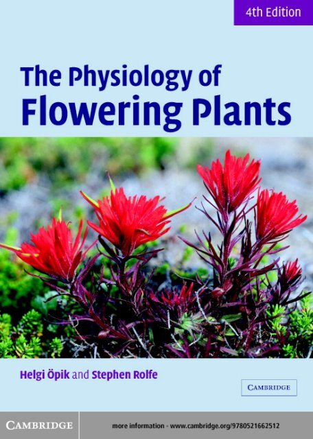
<strong>The</strong> <strong>Physiology</strong> <strong>of</strong> <strong>Flowering</strong> <strong>Plants</strong>Fourth EditionThis latest edition <strong>of</strong> <strong>The</strong> <strong>Physiology</strong> <strong>of</strong> <strong>Flowering</strong> <strong>Plants</strong> has been completelyupdated to cover the explosion <strong>of</strong> interest in plant biology. A whole-plantapproach has been used to produce an integrated view <strong>of</strong> plant function,covering both the fundamentals <strong>of</strong> whole plant physiology and the latestdevelopments in molecular biology. New developments in molecular techniquesare explained within practical applications such as geneticallymodified plants. <strong>The</strong> book further examines:* photosynthesis, respiration, plant growth and development;* nutrition, water relations, photomorphogenesis and stress physiology;* function, with particular attention to adaptations to different habitats.Each chapter is fully referenced with suggestions for complementary readingincluding references to original research papers.<strong>The</strong> <strong>Physiology</strong> <strong>of</strong> <strong>Flowering</strong> <strong>Plants</strong> is valuable to both undergraduate andpostgraduate students studying plant biology.HELGI ÖPIK was Senior Lecturer in the School <strong>of</strong> Biological Sciences at theUniversity <strong>of</strong> Wales, Swansea until her retirement. Throughout her careershe has taught plant physiology at all undergraduate levels, and sinceretiring has lectured in plant physiology for adult education. Her researchinterests have included plant respiration and ultrastructure, always aimingat integration <strong>of</strong> structure and physiological function.STEPHEN ROLFE was awarded a European Molecular Biology Fellowship andundertook postdoctoral research on the phytochrome regulation <strong>of</strong> geneexpression at the University <strong>of</strong> California, Los Angeles. He took up a post atthe Department <strong>of</strong> Animal and Plant Sciences, University <strong>of</strong> Sheffield in1991. His research interests include the study <strong>of</strong> photosynthesis and primaryplant metabolism, with a special interest in non-invasive imagingtechniques.
<strong>The</strong> <strong>Physiology</strong> <strong>of</strong> <strong>Flowering</strong> <strong>Plants</strong>Fourth EditionHelgi ÖpikFormerly Senior Lecturer,School <strong>of</strong> Biological Sciences,University <strong>of</strong> Wales,SwanseaStephen A. RolfeSenior Lecturer,Department <strong>of</strong> Animal and Plant Sciences,University <strong>of</strong> SheffieldAcademic Consultant EditorArthur J. WillisEmeritus Pr<strong>of</strong>essor,University <strong>of</strong> Sheffield
CAMBRIDGE UNIVERSITY PRESSCambridge, New York, Melbourne, Madrid, Cape Town, Singapore, São PauloCambridge University Press<strong>The</strong> Edinburgh Building, Cambridge CB2 2RU, UKPublished in the United States <strong>of</strong> America by Cambridge University Press, New Yorkwww.cambridge.orgInformation on this title: www.cambridge.org/9780521662512© H . Ö p i k & S . R o l f e 2 0 0 5This book is in copyright. Subject to statutory exception and to the provision <strong>of</strong>relevant collective licensing agreements, no reproduction <strong>of</strong> any part may take placewithout the written permission <strong>of</strong> Cambridge University Press.First published in print format2005ISBN-13 978-0-511-11323-9 eBook (NetLibrary)ISBN-10 0-511-11323-4 eBook (NetLibrary)ISBN-13 978-0-521-66251-2 hardbackISBN-10 0-521-66251-6 hardbackISBN-13 978-0-521-66485-1 paperbackISBN-10 0-521-66485-3 paperbackCambridge University Press has no responsibility for the persistence or accuracy <strong>of</strong>URLs for external or third-party internet websites referred to in this book, and does notguarantee that any content on such websites is, or will remain, accurate or appropriate.
ContentsPrefacepage ixChapter 1 Introduction 11.1 Appreciating plants 11.2 What kind <strong>of</strong> plant physiology? 21.3 Molecular biology and plant physiology: the integration<strong>of</strong> disciplines 31.4 Outline <strong>of</strong> the text 5Part INutrition and transportChapter 2Flow <strong>of</strong> energy and carbon through the plant:photosynthesis and respiration 92.1 Introduction 92.2 Energy flow and carbon turnover in the biosphere 92.3 Photosynthesis: light absorption and utilization 122.4 <strong>The</strong> fixation <strong>of</strong> carbon dioxide 182.5 Limiting factors for photosynthesis 302.6 <strong>The</strong> efficiency <strong>of</strong> energy conversion in photosynthesis 322.7 Photosynthesis and the increase in atmosphericcarbon dioxide 362.8 Respiration: the oxidative breakdown <strong>of</strong> organic compounds 382.9 Terminal oxidation and oxidative phosphorylation 462.10 Anaerobic respiration 492.11 Respiration and plant activity 53Chapter 3 Water relations 603.1 Introduction 603.2 Water movement and energy: the concept <strong>of</strong>water potential 613.3 Water potentials <strong>of</strong> plant cells and tissues 613.4 Water relations <strong>of</strong> whole plants and organs 683.5 <strong>The</strong> transport <strong>of</strong> solutes in the xylem 853.6 Water uptake and loss: control by environmental andplant factors 863.7 Water conservation: xerophytes and xeromorphic characters 95Chapter 4 Mineral nutrition 1004.1 Introduction 1004.2 Essential elements 100
VICONTENTS4.3 Ion uptake and transport in the plant 1064.4 Nitrogen assimilation, fixation and cycling 1224.5 Problems with mineral elements: deficiencyand toxicity 128Chapter 5 Translocation <strong>of</strong> organic compounds 1335.1 Introduction 1335.2 Phloem as the channel for organic translocation 1335.3 <strong>The</strong> rate and direction <strong>of</strong> translocation 1395.4 Phloem loading and unloading 1425.5 Partitioning <strong>of</strong> translocate between sinks: integrationat the whole-plant level 1465.6 <strong>The</strong> mechanism <strong>of</strong> phloem translocation 148Part IIGrowth and developmentChapter 6 Growth as a quantitative process 1616.1 Introduction 1616.2 <strong>The</strong> measurement <strong>of</strong> plant growth 1626.3 Growth, development and differentiation 1636.4 Localization <strong>of</strong> growth in space and time 1646.5 Conditions necessary for growth 1656.6 Growth rates 167Chapter 7 Plant growth hormones 1777.1 Introduction 1777.2 Plant growth hormones 1787.3 Detection and quantification <strong>of</strong> hormones in plants 1917.4 How do plant hormones cause responses? 194Chapter 8 Cell growth and differentiation 2058.1 Introduction 2058.2 Meristems and cell division 2058.3 Mitochondrial and plastid division 2118.4 Cell expansion: mechanism and control 2138.5 Cell differentiation 218Chapter 9 Vegetative development 2219.1 Introduction 2219.2 <strong>The</strong> structure and activity <strong>of</strong> the shoot apical meristem 2219.3 Organ formation 2259.4 Secondary growth 2279.5 Development <strong>of</strong> the leaf 2289.6 <strong>The</strong> structure and activity <strong>of</strong> the root apical meristem 239
CONTENTSVIIChapter 10 Photomorphogenesis 24610.1 Introduction 24610.2 <strong>The</strong> switch from etiolated to de-etiolated growth 24710.3 Phytochrome and photomorphogenesis 24810.4 UV-A/blue light photoreceptors (cryptochrome) 25510.5 Genes controlling etiolated growth 25610.6 Unravelling photomorphogenesis 25710.7 Phytochrome signal transduction 263Chapter 11 Reproductive development 27011.1 Introduction 27011.2 Juvenility and ‘ripeness to flower’ 27011.3 <strong>The</strong> control <strong>of</strong> flowering by daylength andtemperature 27111.4 Plant size and flowering 27711.5 <strong>The</strong> regulation <strong>of</strong> floral induction is amultifactorial process 27911.6 Floral development 28111.7 Pattern development in flowers 28711.8 <strong>The</strong> formation <strong>of</strong> pollen 29111.9 <strong>The</strong> formation <strong>of</strong> the embryo sac 29311.10 Pollination 29511.11 Embryo formation 30111.12 Seeds and nutrition 30311.13 Fruit development 30811.14 Seed dormancy 31011.15 Germination and the resumption <strong>of</strong> growth 315Chapter 12 Growth movements 31812.1 Introduction 31812.2 Nastic responses 31812.3 Tropisms 320Chapter 13 Resistance to stress 34413.1 Introduction 34413.2 Terminology and concepts 34413.3 Water-deficit stress 34613.4 Low-temperature stress 35413.5 High-temperature stress 36213.6 Relationships between different types <strong>of</strong> stressresistance: cross-tolerance 36613.7 Development <strong>of</strong> stress-resistant crop plants 368
VIIICONTENTSAppendix 373A.1 Naming genes, proteins and mutations 373A.2 Units <strong>of</strong> measurement 373A.3 Prefixes for units 375Index 376
Preface<strong>The</strong> history <strong>of</strong> this book dates back to the late 1960s, when thepublishers Edward Arnold launched a series <strong>of</strong> student textbooks asthe Contemporary Biology series, designed to provide up-to-datetexts at elementary university and final-year school level. One <strong>of</strong>the first authors who was asked to contribute, on the topic <strong>of</strong>flowering plant physiology, was Pr<strong>of</strong>essor H. E. Street, thenPr<strong>of</strong>essor <strong>of</strong> Botany at the University <strong>of</strong> Wales, Swansea. He askedone <strong>of</strong> us (H.Ö.) to collaborate, and the first edition was dulypublished by Edward Arnold in 1970 under the authorship <strong>of</strong>H. E. Street and Helgi Öpik, and entitled <strong>The</strong> <strong>Physiology</strong> <strong>of</strong> <strong>Flowering</strong><strong>Plants</strong>: <strong>The</strong>ir Growth and Development. <strong>The</strong> emphasis <strong>of</strong> the text was onthe ‘whole plant’ aspects <strong>of</strong> physiology. <strong>The</strong> second edition followedin 1976 and the third in 1984, although Pr<strong>of</strong>essor Street sadlydeceased in 1977.While the second and third editions were still very much revisions<strong>of</strong> the original text, the longer time interval since the last edition, andthe rapid pace at which biological knowledge has grown in the lastfew decades, have now necessitated a very thorough rewriting <strong>of</strong>large sections <strong>of</strong> the book, and the task has been quite challengingin the face <strong>of</strong> an accumulation <strong>of</strong> facts that on occasion has seemedquite overwhelming. It is not possible now to interpret many aspects<strong>of</strong> plant physiology without reference to molecular biology, evenwhen one is basically interested in functioning at the organismallevel. This applies particularly to the developmental aspects <strong>of</strong> physiology.Some reorganization <strong>of</strong> the text and shift <strong>of</strong> emphasis hasaccordingly been necessitated, though we have tried to retain theoverall spirit <strong>of</strong> the original book.One thing has remained unchanged during the preparation <strong>of</strong> thisbook from the first edition to the fourth: the unfailing encouragementand help from our editor, Pr<strong>of</strong>essor A. J. Willis. Without him,the present text would not have been written. We are also grateful forthe support <strong>of</strong> Dr Ward Cooper, Commissioning Editor, and Dr AlanCrowden, Editorial Director, <strong>of</strong> Cambridge University Press. Thanksare due for reading, and advising on, parts <strong>of</strong> the manuscript, toPr<strong>of</strong>essor Richard C. Leegood, Pr<strong>of</strong>essor David Read and Dr JulieGray <strong>of</strong> the University <strong>of</strong> Sheffield.H.Ö. would like to acknowledge the generosity <strong>of</strong> Pr<strong>of</strong>essor RayWaters, Head <strong>of</strong> the School <strong>of</strong> Biological Sciences at the University <strong>of</strong>Wales, Swansea, for use <strong>of</strong> departmental facilities in preparing illustrations.H.Ö. also would like to thank Ken Jones <strong>of</strong> the School <strong>of</strong>Biological Sciences, Swansea, for printing figures; my nephew KevinMiller and my niece, Heather Nagey, for help with word processing;and Pr<strong>of</strong>essor Kevin Flynn and Dr Charles Hipkin <strong>of</strong> theUniversity <strong>of</strong> Wales, Swansea, for helpful discussions.
XPREFACEWe are grateful to all the people who have permitted us to reproducetheir published data, and have provided material and helpfuladvice for figures; particular thanks are due to Pr<strong>of</strong>essor Jane Sprentand Dr Euan James <strong>of</strong> the University <strong>of</strong> Dundee for supplying theoriginal micrograph <strong>of</strong> bacteroids (Fig. 4.7).
Chapter 1Introduction1.1 Appreciating plantsDuring a public open day at a university, a child trying to look at abotanical exhibit was dragged away by an impatient parent with thewords ‘Come on – we can’t spend all day looking at dull green things!’<strong>The</strong>re is a tendency to consider plants as somewhat dull, passiveand inactive. Yet plants face and overcome the same problems asanimals: how to obtain nutrients and water, how to survive extremeenvironmental conditions, how to ensurereproductionandthesurvival<strong>of</strong> the next generation. <strong>The</strong> photosynthetic mode <strong>of</strong> life has conditionedplants to evolve as sessile organisms; their basic necessities – light,carbon dioxide, water and mineral ions – are ubiquitous and therehas therefore been no selection pressure for mobility. An animal mayobtain its nutrients and water by skilfully stalking its prey, and learningthe path to a pool; this catches human attention as interesting behaviour.A flowering plant obtains nutrients and water by millions <strong>of</strong>minute root tips constantly growing through the soil, and by pumpingions across root cell plasma membranes with molecular-sized pumps.This is plant behaviour: plant physiology is plant behaviour.Itneednot be considered dull because it is less spectacular to the eye than whatis called animal behaviour. <strong>The</strong> subsequent hauling up <strong>of</strong> the absorbedwater and minerals to the top <strong>of</strong> a tree, 100 metres high, might indeedbe considered a quite spectacular feat (imagine doing it with a bucket!).Without stirring from the spot, flowering plants are unceasinglymonitoring their environment and responding to environmentalsignals. For them as for us, light is an information medium; theycontain optical sensors (pigments) with which they perceive andrespond to light direction, wavelength composition (i.e. colour) andthe duration <strong>of</strong> daylight. <strong>The</strong>y are sensitive to touch, with someresponses as fast as animal movement: a Venus flytrap leafsnaps shut in a second or two to catch (and digest!) insects asbig as wasps or small moths. Pea tendrils are a bit slower toreact, but can be seen curling around a support within minutes<strong>of</strong> making contact. <strong>Plants</strong> respond also to the direction <strong>of</strong> gravity;it is the continuous responses to gravity and light that are largely
2 INTRODUCTIONresponsible for plants growing ‘the right way up’, and with roots,branches and leaves orientated at various angles. Temperaturechanges are sensed. Signals with a regular annual variation –such as the changes in daylength – enable plants to synchronizetheir life cycles with the seasonal cycles in their environment.Even the embryo in a dormant seed, apparently quite inactive inits coat, is able to receive specific signals that stimulate it tocommence germination at the appropriate season. <strong>Flowering</strong>plants, when looked at from the physiologist’s point <strong>of</strong> view,are not merely alive, they are very lively. We hope that thereaders <strong>of</strong> our book will be led to appreciate how marvellousand varied in their activities flowering plants are.<strong>The</strong>re are other reasons, too, for studying flowering plants. We areutterly dependent on them. Being the dominant plants in present-dayterrestrial (land) vegetation, they are the primary producers, byphotosynthesis, <strong>of</strong> organic material – food – on which all otherterrestrial organisms including ourselves rely. We moreover needplants for wood, textiles, drugs, and hundreds <strong>of</strong> household chemicals;their very presence gives us joy, even comfort. Gardens are amain source <strong>of</strong> pleasure for many individuals; windowsills get filledwith house plants, floral baskets are hung from balconies <strong>of</strong> high-riseflats. Animals not only feed on plants, but live in them, on them andunder them. <strong>The</strong> oxygen we breathe is formed as a by-product <strong>of</strong>photosynthesis. Soil and climate, too, are influenced by plants; vegetationmay for instance stabilize soil against physical erosion. Thusthe importance <strong>of</strong> understanding the activities <strong>of</strong> flowering plantscannot be overemphasized. With the changes currently being imposedon the biosphere <strong>of</strong> this planet by human activities, it has become moreimperative than ever to study the physiology <strong>of</strong> plants, if there is to beany hope <strong>of</strong> predicting how vegetation, all-important for life on earth,might respond to such changes in the environment. An understanding<strong>of</strong> flowering plant physiology is also vital for any attempts to improveplant productivity.1.2 What kind <strong>of</strong> plant physiology?Plant physiology is the functioning <strong>of</strong> plants. This can be studied atseveral levels <strong>of</strong> complexity and organization, as indicated by suchterms as metabolic physiology, cellular physiology,orwhole-plant physiology.<strong>The</strong> aim <strong>of</strong> the current text is to give an account <strong>of</strong> the physiology <strong>of</strong>flowering plants mainly from the whole plant or organismal point<strong>of</strong> view. Moreover, since an organism functions within its environment,and by virtue <strong>of</strong> its physiological activities continuously interactswith its environment, plant–environment interactions areemphasized throughout the book. But many plant life processes arecarried out at the level <strong>of</strong> the individual cell, even <strong>of</strong> the individualorganelle. Physiological processes <strong>of</strong> plants can therefore bedescribed only to a limited degree without reference to activities at
MOLECULAR BIOLOGY AND PLANT PHYSIOLOGY 3the cellular or subcellular level. Water movement through an intactplant is a whole-plant level process; for some specified ecophysiologicalproject, it may be adequate to discuss it, say, in terms <strong>of</strong>quantities <strong>of</strong> water absorbed by the roots, and the amounts lost bytranspiration from the leaves. If, however, one wishes to understandthe control exerted on the process by the plant, one is soon involvedwith the cellular physiology <strong>of</strong> the stomatal guard cells, which moveto open and close the stomatal pores through which most <strong>of</strong> thetranspiration occurs. Equally relevant to an understanding <strong>of</strong> physiologicalprocesses is a knowledge <strong>of</strong> cellular structure. To continuewith the example <strong>of</strong> stomata, the opening and closing movementsare also dependent on the shapes <strong>of</strong> the guard cells and on the pattern<strong>of</strong> their cell wall thickenings, right down to the precise arrangement<strong>of</strong> the cellulose micr<strong>of</strong>ibrils. Whilst the goal <strong>of</strong> this book is to promotethe understanding <strong>of</strong> flowering plants as organisms, one cannotescape consideration <strong>of</strong> information from the realms <strong>of</strong> cellularphysiology, biochemistry, cell structure and ultrastructure, andmolecular biology. In this respect we have had to exercise our judgementabout the depth to which such information should be presented,and the extent to which it might be taken for granted. Infact an elementary knowledge <strong>of</strong> basic metabolism, biochemistry,plant anatomy and plant cell structure has been assumed. Textbookslisted at the end <strong>of</strong> this chapter as Complementary reading willserve to fill in the background for readers who find this necessary,and will enable all interested readers to extend their knowledge <strong>of</strong>the more cellular aspects <strong>of</strong> plant physiology, and <strong>of</strong> plant structure,beyond this text.1.3 Molecular biology and plant physiology: theintegration <strong>of</strong> disciplinesReferences to molecular biology will be found to figure liberally inparts <strong>of</strong> the text. <strong>The</strong> discipline <strong>of</strong> molecular biology embraces thestudy <strong>of</strong> genes: their isolation and identification, analysis (molecularsequencing) and their manipulation, i.e. modification and introduction<strong>of</strong> selected genes into cells at will. Gene expression, their‘switching on and <strong>of</strong>f ’, can also be manipulated. This is a branch <strong>of</strong>science that is enabling biologists to identify and study the roles <strong>of</strong>specific genes in specified activities.Molecular biology techniques have led to considerable advancesin the understanding <strong>of</strong> plant physiology, particularly <strong>of</strong> developmentand differentiation. Every activity <strong>of</strong> a living organism canultimately be traced to control by some gene(s) at some stage <strong>of</strong> itslife history; the entire blueprint for everything that a flowering plantis capable <strong>of</strong> performing is inscribed in the DNA <strong>of</strong> the single-celledzygote from which it develops. Some physiological functions can bediscussed with minimal reference to genetic activity. <strong>The</strong>se include
4 INTRODUCTIONnumerous aspects <strong>of</strong> the processes <strong>of</strong> nutrition and transportdescribed in the first part <strong>of</strong> this book. Such processes may alsoshow some short-term, quantitative, reversible responses to environmentalfactors, which are not mediated at the genetic level. Whenone comes to the physiology <strong>of</strong> growth and differentiation, however,as covered in the second part <strong>of</strong> the book, the need arises to referconstantly to genetic control. One tends to think <strong>of</strong> growth as aquantitative increase in mass. Growth is, however, almost alwaysaccompanied by differentiation, a change <strong>of</strong> form and/or physiologicalactivity, and this results in the process termed development:development ¼ growth þ differentiation<strong>The</strong> growing seedling, for instance, does not become an enlargedversion <strong>of</strong> the embryo in the seed, it develops into the adult form <strong>of</strong>the plant. Differentiation means differential activity <strong>of</strong> genes, i.e.activation and suppression <strong>of</strong> specific (sets <strong>of</strong>) genes at particulardevelopmental stages. Developmental processes all involve qualitativeas well as quantitative changes in response to environmentalconditions. Environmental factors act as specific stimuli calling forthspecific and generally irreversible changes in physiological activitiesand morphology. This applies to such events as the onset <strong>of</strong> reproductivegrowth, and reactions to environmental stresses. Such processesare the result <strong>of</strong> interactions between the external factors andthe genome <strong>of</strong> the plant, and our understanding <strong>of</strong> such processes isdependent on studies <strong>of</strong> gene activity. Paradoxical as it may seem atfirst sight, it is the most conspicuously organism-oriented activities,such as the onset <strong>of</strong> flowering – which entails pr<strong>of</strong>ound changes inthe physiology <strong>of</strong> the whole plant – that need the molecular biologyapproach to the greatest extent. <strong>The</strong>se are the processes which aremost strongly under control at the genetic level.We started this chapter with a very wide view, considering theoverall role <strong>of</strong> flowering plants in the biosphere. We now havereached consideration <strong>of</strong> the finest, ultimate level <strong>of</strong> biological analysis– the individual gene. <strong>Flowering</strong> plants, like any other organisms,are complex entities and understanding <strong>of</strong> them as organisms can beachieved only by studying them at various levels <strong>of</strong> organization andintegrating knowledge obtained by numerous biological disciplines,from molecular biology to ecology. <strong>The</strong> division <strong>of</strong> biology intovarious disciplines is an artificial one, made for the convenience <strong>of</strong>handling a vast field <strong>of</strong> knowledge; no such division exists in theplant. We hope that we have been able to keep the organism, the‘whole’ plant, in sight throughout this text, even when interpretingevents in terms <strong>of</strong> molecular biology.Plant physiology also has something to <strong>of</strong>fer to molecular biologists.<strong>The</strong> science <strong>of</strong> molecular biology has led to applications knownunder the general term <strong>of</strong> genetic engineering, i.e. the production <strong>of</strong>transgenic (transformed) organisms, containing selected genes whichmay come from other species. Basic gene structure being identical in
OUTLINE OF THE TEXT 5all organisms on planet earth, genes from animals and even fromprokaryotes can be inserted into the flowering-plant genome.<strong>Flowering</strong> plants possess great regenerative powers; single cells fromvegetative organs are able to grow into fully formed plants. This makesthem ideal subjects for genetic transformations. Some transgenic cropplants are already being grown commercially and many more potentialtransformations are being investigated. While some scientists seegenetic engineering as a wonderful tool for improving plants as economicresources, many people regard transgenic operations withstrong reservations, fearing that, accidentally or deliberately, newand dangerous organisms might be let loose upon the world. <strong>The</strong>recan be no scientific safeguards against the deliberate misuse <strong>of</strong> geneticengineering. <strong>The</strong> defence against accidental disasters lies in knowledgeand understanding. For successful engineering <strong>of</strong> ‘plants for thefuture’, it is imperative that the physiology <strong>of</strong> plants should be understood,and moreover understood at the organismal level. A particulartransformation may be undertaken with one single activity in mind;but that activity needs to be studied in the context <strong>of</strong> the plant as awhole if one is to have a reasonable chance <strong>of</strong> predicting the result <strong>of</strong>the operation.1.4 Outline <strong>of</strong> the text<strong>The</strong> book falls essentially into two parts: Nutrition and transport(Chapters 2 to 5) and Growth and development (Chapters 6 to 13).<strong>Flowering</strong> plants are essentially autotrophic, photosyntheticorganisms, with basic requirements <strong>of</strong> light, CO 2 , water and some17 elements as inorganic mineral ions. <strong>The</strong> survey <strong>of</strong> plant physiologyaccordingly begins with photosynthesis (Chapter 2), detailinghow the flowering plants obtain and process the first two necessitieslisted above, light and CO 2 . This is followed by respiration, theprocess providing energy and intermediates for metabolism. Next itis considered how they obtain and transport water (Chapter 3), andthen how mineral ions are absorbed and assimilated (Chapter 4).<strong>The</strong> translocation <strong>of</strong> organic materials, the products <strong>of</strong> photosynthesis,is covered in Chapter 5, completing the first part <strong>of</strong>the book.<strong>The</strong> second part <strong>of</strong> the text starts with treatment <strong>of</strong> growth as aquantitative process and covers some simple mathematical analysis<strong>of</strong> growth (Chapter 6). <strong>The</strong>n we proceed to discussions <strong>of</strong> thephysiology <strong>of</strong> growth and development, beginning with surveys<strong>of</strong> plant growth hormones (Chapter 7) and cell growth anddifferentiation (Chapter 8). <strong>The</strong>se are followed by consideration<strong>of</strong> vegetative development, photomorphogenesis, reproductivedevelopment, and growth movements (Chapters 9 to 12).Finally, in Chapter 13, the reactions <strong>of</strong> flowering plants to someenvironmental stresses are discussed.
6 INTRODUCTIONComplementary readingAnderson, J. W. & Beardall, J. Molecular Activities <strong>of</strong> Plant Cells. Oxford:Blackwell, 1991.Dennis, D. T., Turpin, D. H., Lefebvre, D. D. & Layzell, D. B., eds. PlantMetabolism, 2nd edn. Harlow: Addison Wesley Longman, 1997.Gunning, B. E. S. & Steer, M. W. Plant Cell Biology, Structure and Function.Sudbury, MA: Jones & Bartlett, 1996.Lea, P. J. & Leegood, R. C., eds. Plant Biochemistry and Molecular Biology, 2nd edn.Chichester: Wiley, 1999.Mauseth, J. D. Plant Anatomy. Menlo Park, CA: Benjamin/Cummings, 1988.Mauseth, J. D. Botany: an Introduction to Plant Biology, 2nd edn. Sudbury,MA: Jones & Bartlett, 1998.Taiz, L. & Zeiger, E. Plant <strong>Physiology</strong>, 3rd edn. Sunderland, MA: Sinauer, 2003.Troughton, J. & Donaldson, L. A. Probing Plant Structure. London: Chapman &Hall, 1972.
Part INutrition and transport
Chapter 2Flow <strong>of</strong> energy and carbonthrough the plant: photosynthesisand respiration2.1 IntroductionAll living organisms need a supply <strong>of</strong> raw materials from which theirbodies can be constructed, and a supply <strong>of</strong> energy. This energy isneeded for growth, i.e. for the formation <strong>of</strong> their bodies, also for themaintenance <strong>of</strong> their bodies, and for all the various types <strong>of</strong> work,chemical and mechanical, that are carried out by living systems.Life as we know it is based on organic compounds <strong>of</strong> carbon (C).This element accordingly occupies a central place among the rawmaterials and it is found on earth abundantly in its inorganic formsas carbon dioxide (CO 2 ), carbonate (CO 3 2– ) and bicarbonate (HCO 3 – ).<strong>The</strong>ultimateenergysourceformostlifeformsonearthisthethermonuclearenergy <strong>of</strong> the sun, transmitted to earth as electromagneticradiation, light. Photosynthesis is the process by which the solarlight energy is transformed into the chemical bond energy <strong>of</strong> organiccarbon compounds. Photosynthesis is thus simultaneously a process<strong>of</strong> energy transduction, and a process by which inorganic carbon isconverted to organic form and incorporated into living organisms.Chemically it is a reductive process. Respiration is the process <strong>of</strong>oxidative breakdown by which the energy stored in the organicproducts <strong>of</strong> photosynthesis is tapped for driving metabolism, forgrowth, for movements, and by which the C is returned to inorganicform again as CO 2 .2.2 Energy flow and carbon turnover in the biosphere2.2.1 <strong>Plants</strong> and the biosphere<strong>The</strong> flow <strong>of</strong> energy and the turnover <strong>of</strong> C in the biosphereare illustrated in Fig. 2.1. Photosynthesis utilizes relatively highenergy quanta, ‘light’, from the electromagnetic spectrum andthese quanta energize the combination <strong>of</strong> hydrogen from waterwith CO 2 to organic compounds. In flowering plants, the first stablephotosynthetic products are predominantly sugars. <strong>The</strong>se sugarsform the starting point <strong>of</strong> other plant constituents, including the
10 FLOW OF ENERGY AND CARBON THROUGH THE PLANTFig: 2:1 <strong>The</strong> flow <strong>of</strong> carbon andenergy through the biosphere. <strong>The</strong>term ‘plant’ is used here to denoteall photosynthetic autotrophs and‘protoplasm’ for all parts <strong>of</strong> anorganism. <strong>The</strong> energy that is lost asheat is irradiated into space asinfrared (long wavelength) radiationand cannot be recycled, but the Cand other elements recycle asindicated.macromolecules which are the units <strong>of</strong> cellular architecture. <strong>The</strong>sugars and polysaccharides (sugar polymers), sometimes also lipids,form a store <strong>of</strong> potential energy for plant cells. This store is continuouslybeing drawn upon during respiration. <strong>The</strong> substrates are oxidizedto CO 2 and water again, while some <strong>of</strong> the potential energy <strong>of</strong>the sugar molecules is transferred to molecules <strong>of</strong> ATP (adenosinetriphosphate). This extremely reactive compound has been termedthe energy currency molecule <strong>of</strong> living cells and its potential energycan be harnessed for cellular work – biosynthesis, growth, membranetransport, movement. During biosynthesis, other elements (Chapter 4)may be incorporated into organic combination. In the course <strong>of</strong> theseactivities the ATP is broken down to ADP or AMP (adenosinedi- or monophosphate) respectively and it must be unceasinglyresynthesized. In photosynthetic cells in the light, some cellularworkmayalsobedrivenbyphotosyntheticallyformedATP(seeSection 2.3.2) without the intervention <strong>of</strong> respiration.
ENERGY FLOW AND CARBON TURNOVER IN THE BIOSPHERE 11Photosynthesis is carried out by virtually all land plants. <strong>The</strong> onlyexceptions are some non-green parasitic or saprophytic species. <strong>The</strong>algae are also photosynthetic and land plants plus algae are sometimescollectively referred to as ‘the green plants’. <strong>The</strong> green plantsare photoautotrophic (literally ‘light-self-feeding’), needing onlylight and inorganic compounds: CO 2 , water and mineral ions. <strong>The</strong>reare also photosynthetic bacteria (although some <strong>of</strong> these are photoheterotrophic,i.e. they need a supply <strong>of</strong> organic C compounds). <strong>The</strong>photoautotrophs are the primary producers and they form thebasis <strong>of</strong> food chains. Plant organic matter is ingested as food byanimals, fungi and other microorganisms. Any organic materialsynthesized by these non-photosynthetic organisms is regarded assecondary production, being derived from the photosynthetic products.As indicated in the lower part <strong>of</strong> Fig. 2.1, the cycles <strong>of</strong> biosynthesisand respiration are repeated in the secondary producers.Secondary producers in turn feed on each other and the cycles aremultiplied, until ultimately microorganism action, decay, returnsthe last <strong>of</strong> the elements again to their inorganic forms <strong>of</strong> CO 2 ,water and mineral ions. <strong>The</strong> chemicals are recycled, although therecycling may take a long time. A living tree may retain organicmaterials in its body for thousands <strong>of</strong> years. Carbon fixed by theCarboniferous forests over 300 million years ago is only now beingreturned to the atmosphere by the burning <strong>of</strong> coal. <strong>The</strong> energy, however,cannot be recycled. In every process illustrated in Fig. 2.1, there occurssome energy loss as heat; this is in due course irradiated back intospace as infrared radiation, low-energy quanta which can never drivephotosynthesis again. <strong>The</strong> laws <strong>of</strong> thermodynamics make this energyloss inevitable: reactions are possible only if they proceed with anoverall free energy loss.Photosynthesis means a steady input <strong>of</strong> energy into the biosphere,vital for the continuance <strong>of</strong> most life. Hence photosynthesis occupiesa central position, not only in the life <strong>of</strong> the green organisms whichcarry it out, but for life on earth in general. (An exception is providedby chemosynthetic bacteria.) On dry land, flowering plants areresponsible for the major proportion <strong>of</strong> photosynthesis.<strong>The</strong> global turnover <strong>of</strong> carbonIt is estimated that around 10 11 tons <strong>of</strong> C are fixed annually inphotosynthesis, representing a primary production <strong>of</strong> some 1.7 10 11tons <strong>of</strong> dry matter, while the total C held in the earth’s biomass at anyone time amounts to about 5.6 10 11 tons. This, however, is a minutefraction <strong>of</strong> the C present on earth. Much <strong>of</strong> this C is present ascarbonates in rocks and as sedimentary organic material in the deepoceans, unavailable for photosynthesis. But even the available storesare enormous: atmospheric CO 2 holds 7.5 10 11 tons <strong>of</strong> C, and theoceans, which equilibrate with the atmosphere, contain in theirupper, accessible, layers some 420 10 11 tons as dissolved CO 2 ,carbonateand bicarbonate.
12 FLOW OF ENERGY AND CARBON THROUGH THE PLANT<strong>The</strong> site <strong>of</strong> photosynthesisAny part <strong>of</strong> the shoot can be green and photosynthetic to some degree –stem, leaf, flowerbud, or young fruit; but the photosynthetic organpar excellence is the leaf, an outgrowth from the stem; by far thegreater part <strong>of</strong> land plant photosynthesis is achieved in the leaves.<strong>The</strong> variety <strong>of</strong> leaf shapes and sizes may seem bewildering, but theymostly share a common basic structure which is adapted to obtainlight and CO 2 . Sunlight is diffuse and atmospheric CO 2 is present at alow concentration; a large aerial surface is required to collect thesenecessities efficiently. Essentially, the typical leaf is a thin, flat structure,<strong>of</strong>ten less than a millimetre thick. <strong>The</strong> photosynthetic cellsmake up the mesophyll (‘mid-leaf’) enclosed by an epidermis,usually non-photosynthetic, on both surfaces. Where the leaf hasdistinct upper and lower surfaces (a dorsiventral leaf ), the mesophyllis generally differentiated into an upper palisade layer <strong>of</strong> elongate,cylindrical cells and a lower spongy mesophyll <strong>of</strong> more rounded,loosely packed cells. A transparent cuticle, relatively impervious towater, water vapour and other gases, covers the epidermis. <strong>The</strong>vascular bundles (veins) run through the mesophyll, supplyingwater and minerals, and translocating away the photosynthate.Even superficially such a leaf presents a large surface : volumeratio. From the point <strong>of</strong> view <strong>of</strong> cell surface, the ratio is even greater,for most <strong>of</strong> the leaf surface is inside, the mesophyll containing muchair space, which communicates with the external air via stomatalpores in the epidermis. This structural arrangement enables thephotosynthetic cells to function in an internal environment protectedagainst excessive water loss. <strong>The</strong> physiological efficiency <strong>of</strong>the leaf may be inferred from the fact that it has arisen independentlyon numerous occasions during the evolution <strong>of</strong> land plants, and innearly all except the most primitive groups.Not all leaves conform to the ‘typical’ pattern. <strong>The</strong>re are succulentleaves where the thickness may exceed a centimetre; there are plantswith vestigial leaves, the stems taking over the function <strong>of</strong> photosynthesis.<strong>The</strong>se variations represent adaptations to extreme environmentalconditions, and are considered in connection with waterconservation in Chapters 3 and 13.Illustrated accounts <strong>of</strong> leaf structure may be found in the textsby Mauseth listed under Complementary reading at the end <strong>of</strong> thischapter.2.3 Photosynthesis: light absorption and utilization2.3.1 <strong>The</strong> capture <strong>of</strong> light<strong>The</strong> photosynthesis <strong>of</strong> green plants requires light in the visible range<strong>of</strong> the spectrum, with wavelengths approximately in the range <strong>of</strong>400–700 nm; this range is called the photosynthetically activeradiation (PAR). <strong>The</strong> photosynthetic pigments have their absorption
PHOTOSYNTHESIS: LIGHT ABSORPTION AND UTILIZATION 13peaks in the blue and red wavelengths (chlorophylls a and b) or in theblue (carotenoids, comprising carotenes and carotenols), i.e. at thetwo ends <strong>of</strong> the visible spectrum (Fig. 2.2). Since sunlight that haspenetrated the atmosphere has its maximum energy output almostin the middle <strong>of</strong> the visible spectrum, in the green and blue-greenrange (Fig. 2.2; see also Fig. 10.1), the photosynthetic pigments mayseem at first sight to be somewhat poorly adapted to capture solarenergy. When, however, the action spectrum <strong>of</strong> photosynthesis for awhole leaf is considered, i.e. the rate <strong>of</strong> photosynthesis for the samenumber <strong>of</strong> incident photons (quanta) is plotted against the wavelength,only a moderate dip in the rate is seen in the green region<strong>of</strong> the spectrum (Fig. 2.3). A photon <strong>of</strong> green light which is notabsorbed immediately is likely to be reflected and refracted betweenmany internal leaf surfaces. If it spends long enough inside the leaf itmay eventually be absorbed by a photosynthetic pigment molecule inspite <strong>of</strong> the low absorptivity <strong>of</strong> pigments in the green region.Carotenoid absorption is shifted towards green wavelengths throughtheir association with chloroplast membranes, and this improvesabsorption in the green wavelengths. With high irradiance, it maybe advantageous for pigments not to absorb too strongly in the green,for excessive energy absorption can damage the photosyntheticsystem.2.3.2 <strong>The</strong> utilization <strong>of</strong> lightIt is convenient to divide photosynthesis into two stages: (1) thereactions which achieve the transduction <strong>of</strong> light energy to chemicalbond energy; and (2) the reactions in which the chemical energy isutilized – in flowering plants mainly for CO 2 fixation. In the livingplant these stages proceed simultaneously, but they can be separatedexperimentally.Fig: 2:2 Absorption spectra <strong>of</strong>chlorophyll a, chlorophyll b,b-carotene and fucoxanthin(a carotenol/xanthophyll). Maximalenergy <strong>of</strong> sunlight lies in the greenand yellow regions <strong>of</strong> the spectrum,the minimum region <strong>of</strong> absorptionby chlorophylls and carotenoids.Adapted from Goodwin & Mercer(1972).
14 FLOW OF ENERGY AND CARBON THROUGH THE PLANTFig: 2:3 <strong>The</strong> efficiency <strong>of</strong> energyutilization at different wavelengthsin leaves <strong>of</strong> 8 species <strong>of</strong> crop plantsgrown in the field (A), and 20species grown in a growth chamber(B). <strong>The</strong> efficiency is expressed asquantum yield, i.e. amount <strong>of</strong> Cfixed for the same number <strong>of</strong>quanta, setting the maximum yieldat unity. From McCree (1972).ª Elsevier Science. Reprinted withpermission.<strong>The</strong> entire process <strong>of</strong> photosynthesis is carried out within thechloroplasts. <strong>The</strong> light-driven reactions occur in the thylakoid membranes(Fig. 2.4) which provide a very large surface area for lightabsorption and associated reactions. <strong>The</strong> thylakoid membranes containa number <strong>of</strong> multimolecular protein complexes including thephotosystems I and II (PSI, PSII), each consisting <strong>of</strong> several hundredpigment molecules and a number <strong>of</strong> specific proteins. One ‘average’chloroplast may contain some 2 million photosystems. Each photosystemhas a reaction centre containing a pair <strong>of</strong> chlorophyll amolecules in a special position. Only the reaction centre chlorophylla molecules can undergo the photochemical reactions which are atthe heart <strong>of</strong> photosynthesis. <strong>The</strong> remaining pigment molecules makeup the light-harvesting pigment complexes, LHPC (also known asantenna pigments) and their function is to absorb photons and tochannel the energy to the reaction centres. <strong>The</strong> LHP include carotenoidsas well as most <strong>of</strong> the chlorophyll. <strong>The</strong> presence <strong>of</strong> LHPCenhances the efficiency <strong>of</strong> light capture very greatly over what itwould be if the reaction centres had to rely on direct hits.<strong>The</strong> energizing <strong>of</strong> the reaction centres by quanta <strong>of</strong> light energyresults in a flow <strong>of</strong> electrons from water, the source, to the coenzyme
PHOTOSYNTHESIS: LIGHT ABSORPTION AND UTILIZATION 15ABGIGG1 µ m 0.5 µ mNADP + (nicotinamide adenine dinucleotide phosphate) along a precisepath via the multimolecular complexes in the thylakoidmembranes:water-splitting complex ! PSII ! PSI ! NADP-reducing system<strong>The</strong> electrons together with the protons from the water reduce theNADP + to NADPH, while the oxygen (O 2 ) <strong>of</strong> the water is released as aby-product. In both PSI and PSII the electron receives a boost <strong>of</strong>energy. This means that two quanta <strong>of</strong> energy are used per electron,enabling green plants to use water as a reductant. Photosyntheticbacteria (cyanobacteria excepted) possess only PSI, use one quantumper electron, and utilize reductants which require a lower energyinput than water. Concurrent with electron flow there is a synthesis<strong>of</strong> ATP from ADP and inorganic phosphate (Pi), the process <strong>of</strong> photophosphorylation,by a mechanism known as chemiosmosis,which can be summarized as follows:(1) Inside the thylakoid lumen, hydrogen ions (H + ) accumulatefrom water splitting and from a coupling <strong>of</strong> the electron flow with aninward transfer <strong>of</strong> H + from the stroma. <strong>The</strong> H + being positivelycharged, this also makes the lumen more electropositive and thestroma more electronegative.(2) <strong>The</strong> combined concentration gradient and electric potentialgradient make a free energy gradient for the H + , favouring theiroutward movement from the lumen into the stroma.(3) <strong>The</strong> thylakoid membrane is highly impermeable to H + . But itcontains ATP-synthase enzyme complexes, with a proton channelFig: 2:4 (A) Low-power and (B)high-power electron micrographs<strong>of</strong> chloroplasts from the grassAgrostis stolonifera sectioned mainlyat right angles to the thylakoidmembranes. In three dimensions,the thylakoids are flattenedmembrane-bound sacs enclosing anarrow lumen. In a granum, G, thethylakoids are like stacked hollowdisks; grana are joined by largerintergrana thylakoids, IG, so thatthe lumen within the thylakoids isprobably a continuous if tortuouscompartment through the wholechloroplast. <strong>The</strong> ground material orstroma contains some very denselystained lipid globules (plastoglobuli).
16 FLOW OF ENERGY AND CARBON THROUGH THE PLANTthrough which the H + move to the stroma, and this movement, downtheir free energy gradient, is coupled with ATP synthesis.<strong>The</strong> net result <strong>of</strong> the light reactions is thus the synthesis <strong>of</strong> ATPand NADPH, which can be utilized in CO 2 fixation (but can also bechannelled into other processes, e.g. nitrate reduction). Normally allphotosynthetic reactions occur simultaneously: ATP and NADPHcannot be stored and the cessation <strong>of</strong> illumination results in a stoppage<strong>of</strong> CO 2 fixation within a second or two. Several enzymes <strong>of</strong> CO 2metabolism need light activation.2.3.3 Levels <strong>of</strong> irradiance and rates <strong>of</strong> photosynthesisBox 2.1ROS, reactive oxygen species(alternative: AOS, active oxygenspecies) are extremely unstable,reactive and potentially destructive;they attack membranes bylipid peroxidation, and degradeDNA, RNA and proteins. From thesuperoxide radical O2 *– , reactionswith cellular protons and electronsproduce further ROS: the perhydroxylradical HO2 * , the hydroxylradical OH * and hydrogen peroxide,H2O2. <strong>The</strong> symbol*denotesan unpaired electron. Smallamounts <strong>of</strong> ROS are inevitablyproduced during photosyntheticand respiratory electron transport,and continually removed.Superoxide is broken down by theenzyme superoxide dismutase,SOD, and hydrogen peroxideby catalase, two very fastactingenzymes. SOD exists inseveral forms with different metalc<strong>of</strong>actors, FeSOD, MnSOD andCu-ZnSOD, specific to subcellularlocations. Cells also producereductive antioxidants which reactwith ROS, including ascorbic acid(vitamin C) and glutathione.Numerous stresses stimulate theformation <strong>of</strong> ROS to levels whichcan be dangerous (Chapter 13).Photometric unitsIn view <strong>of</strong> the basic role <strong>of</strong> light in photosynthesis, the rate <strong>of</strong> photosynthesiswould be expected to vary with the amount <strong>of</strong> light available.Here it is appropriate to consider what exactly is meant by the‘amount’ <strong>of</strong> light.Since light is the energy source for photosynthesis, one’s firstinstinct might be to measure it in energy units, say J m –2 s –1 (energyper unit area, as joules per square metre per second). For energybalance sheets this may be appropriate. However, photochemicalreactions are energized by individual quanta, the units <strong>of</strong> lightenergy carried by individual photons <strong>of</strong> light. One pigment moleculeabsorbs one quantum <strong>of</strong> energy at a time, to undergo one photochemicalreaction. Hence, for many studies, the most meaningfulmeasure <strong>of</strong> the ‘amount’ <strong>of</strong> light is the number <strong>of</strong> photons (orquanta), this number being given in moles. One mole <strong>of</strong> quantacan energize one mole <strong>of</strong> pigment molecules. <strong>The</strong> number <strong>of</strong> moles<strong>of</strong> photons <strong>of</strong> PAR, per unit area and unit time, is called the photonflux density or PFD. It is the PFD that exhibits the most directrelationship with the rate <strong>of</strong> photosynthesis. Bright sunlight has aPFD <strong>of</strong> 2000–2300 mmol m –2 s –1 .Effects <strong>of</strong> varying the PFD: reactions <strong>of</strong> sun and shadeplantsAs the level <strong>of</strong> irradiance on a photosynthetic organ is increased,the rate <strong>of</strong> photosynthesis at first rises linearly, then levels <strong>of</strong>f toa steady rate as light saturation is reached (Fig. 2.5). But theabsorption <strong>of</strong> light does not fall proportionately, so that atincreasing PFD, less CO 2 fixation takes place per photon absorbed:photoinhibition occurs. This was originally interpreted as dueto photochemical damage. Excess light-excited chlorophyll canenergize the formation <strong>of</strong> reactive oxygen species, ROS, fromO 2 : singlet oxygen 1 O 2 *andthesuperoxideradicalO 2 *– (oxygenwith an extra unpaired electron). <strong>The</strong>se chemicals and their derivatives(Box 2.1) can destroy components <strong>of</strong> the photosystems.<strong>The</strong>re is good evidence now that photoinhibition is in fact aprotective process, during which excess energy is dissipated
PHOTOSYNTHESIS: LIGHT ABSORPTION AND UTILIZATION 17CO 2 uptake (µmol h –1 dm –2 )15001000500CO 2 h –1 mg –1 Chl'Light plants'CO 2 h –1 dm –2CO 2 h –1 mg –1 Chl'Shade plants'CO 2 h –1 dm –2400300200100CO 2 uptake (µmol h –1 mg –1 chlorophyll)Fig: 2:5 Light saturation curves <strong>of</strong>photosynthesis for plants <strong>of</strong> Sinapisalba grown either under strongillumination, ‘light (sun) plants’(dashed lines), or weak illumination,‘shade plants’ (solid lines). <strong>The</strong> rate<strong>of</strong> photosynthesis changes similarlywith change <strong>of</strong> irradiance whetherexpressed per unit <strong>of</strong> leaf area orunit <strong>of</strong> chlorophyll, showing that thedifferences in rates between sunand shade plants do not just resultfrom a difference in totalchlorophyll per unit area <strong>of</strong> leaf.Where the curves cut the x-axis isthe light compensation point, belowwhich respiration exceedsphotosynthesis (negative CO 2uptake = CO 2 output); this pointlies at a lower irradiance for theshade plants. From Grahl & Wild(1972).0–20075 150 225 300Irradiance (10 –4 J cm –2 s –1 )harmlessly (ultimately to heat) by reactions involving carotenoidsin the thylakoids and preventing the buildup <strong>of</strong> ROS (Bartley &Scolnick 1995). When the PFD is too high to be counteracted bythis process, then damage does occur, though it may still berepairable if not too extreme.<strong>The</strong> irradiance level at which saturation occurs depends ona number <strong>of</strong> factors. If the temperature is very low, for instance,light saturation is reached at a low PFD: the rate <strong>of</strong> thermochemicalreactions soon becomes limiting. Similarly, at low CO 2 concentrations,light saturation is reached once CO 2 has become limiting.Conversely, at higher temperatures and higher levels <strong>of</strong> CO 2, lightsaturation is reached at a higher PFD. However, other conditionsbeing equal, significant differences in light saturation valuesare shown by individual photosynthesizing systems. Some species,e.g. plants <strong>of</strong> forest floors, are obligate shade plants, able to liveonly at low irradiance levels; examples include dog’s mercury(Mercurialis perennis) and the enchanter’s nightshade (Circaea lutetiana).<strong>The</strong>re are also obligate sun plants, such as the aptly namedsunflower (Helianthus annuus) and the daisy (Bellis perennis), plants <strong>of</strong>open habitats. Such species are genetically adapted for extremes <strong>of</strong>sun or shade. But for many species individuals can adjust appreciablyto the light levels to which they are exposed during growth, as
18 FLOW OF ENERGY AND CARBON THROUGH THE PLANTshown in Fig. 2.5. Trees commonly produce ‘sun leaves’ on theoutside <strong>of</strong> the canopy, and ‘shade leaves’ within it. Shade plants(or leaves) become light-saturated at a much lower PFD than sunplants (or leaves). At low levels <strong>of</strong> light flux they have higher rates<strong>of</strong> photosynthesis than sun plants/leaves, whether the rate is measuredper unit leaf area or per unit weight <strong>of</strong> chlorophyll. Shadeplantshavealowlight compensation point, the value <strong>of</strong> irradiance atwhich photosynthesis exactly equals respiration, and below whichrespiration exceeds photosynthesis, leading to a net loss <strong>of</strong> organicmatter. Numerical values <strong>of</strong> PFD at the light compensation pointhave been quoted as 20 mmol m –2 s –1 for shade plants, 80 mmol m –2s –1 for sun plants. <strong>The</strong> shade plants can therefore survive at levels<strong>of</strong> light too low to support the growth <strong>of</strong> sun plants. In deep shadethe PFD can fall below 50 mmol m –2 s –1 . Adaptations to growth inthe shade include thin leaves (see Fig. 9.8) and very pigment-richchloroplasts; there is a high proportion <strong>of</strong> LHPC to reaction centres,which increases the efficiency <strong>of</strong> light capture. In bright light theshade plants are relatively inefficient with respect to photosynthesis(Fig. 2.5) because <strong>of</strong> their low density <strong>of</strong> reaction centres, and thereforethey are likely to be outcompeted by sun species. <strong>The</strong> shadeplants are also highly susceptible to photochemical damage bybright light. <strong>The</strong> capacity for energy dissipation in shade plants islimited, whereas adaptable plants grown at a high PFD showincreased levels <strong>of</strong> carotenoid pigments.For whole plants, light saturation requires much higher levels <strong>of</strong>irradiance than for single leaves, because in an intact plant outerand upper leaves shade inner and lower ones. This shading is kept toa minimum by the arrangement <strong>of</strong> leaves in ‘leaf mosaics’, leavesarranging themselves so as to shade each other minimally (seeneasily by looking up through the foliage <strong>of</strong> a tree!). Nevertheless,whereas a single leaf may be light-saturated with c. 25% <strong>of</strong> fullsunlight, an entire plant may not reach light saturation even withthe PFD <strong>of</strong> the full midsummer sun. Heavy clouding may bring aplant as a whole to its light compensation point.2.4 <strong>The</strong> fixation <strong>of</strong> carbon dioxide2.4.1 <strong>The</strong> absorption <strong>of</strong> carbon dioxideGaseous diffusionIn the atmosphere CO 2 is present at an average concentration <strong>of</strong>about 370 mmol mol –1 (see Box 2.2). <strong>The</strong> leaf provides a large absorbingsurface, and in this surface the stomata provide pores for entry.Within the leaf, the abundant air spaces permit gaseous diffusionbetween the cells and the large internal surface <strong>of</strong> the leaf is themain area for absorption <strong>of</strong> the gas into cells. <strong>The</strong> internal CO 2concentration is kept below the atmospheric by photosynthesis.
THE FIXATION OF CARBON DIOXIDE 19<strong>The</strong> driving force for the inward movement <strong>of</strong> CO 2 is the concentrationgradient, CO 2 , between the sites <strong>of</strong> fixation and the externalatmosphere:CO 2 ¼½CO 2 Š external½CO 2 Š internal(2:1)<strong>The</strong> CO 2 is equivalent to a gradient <strong>of</strong> free energy: higher concentrationis equivalent to higher free energy. <strong>The</strong> steepness <strong>of</strong> thegradient depends on both the external and internal concentrations.Under field conditions, photosynthesizing plants do not deplete theCO 2 supply in their vicinity greatly, for air mixes rapidly (though seebelow on boundary layers); however, CO 2 concentrations <strong>of</strong> about270 mmol mol –1 have been measured within a crop.During diffusion to the photosynthetic sites, the CO 2 moleculesencounter resistances at various points: at the boundary air layer justoutside the leaf; at the cuticle; at the stomata; and in the mesophyll.Since CO 2 concentration is <strong>of</strong>ten the limiting factor in photosynthesis,these resistances can determine the photosynthetic rate.<strong>The</strong> boundary-layer resistance is the result <strong>of</strong> a layer <strong>of</strong> relativelystill air, also known as the unstirred layer, immediately adjacentto the outside <strong>of</strong> the leaf. In this layer the CO 2 concentration islower than in the bulk atmosphere owing to depletion by the leaf. Itspresence has the effect <strong>of</strong> decreasing the effective CO 2 . In still air, arelatively thick boundary layer builds up over a plant surface andthis slows down the rate <strong>of</strong> diffusion <strong>of</strong> CO 2 into the leaf; but usuallythere is sufficient air movement to keep the boundary-layer resistancelow.<strong>The</strong> cuticle, which forms a continuous layer over the epidermis,presents a very high resistance to CO 2 diffusion. As long as thestomata are open at all, the proportion <strong>of</strong> CO 2 entering through thecuticle is very small.<strong>The</strong> mesophyll resistance is a combination <strong>of</strong> all the resistancesthat a CO 2 molecule meets while diffusing through the mesophyllair spaces, the cell walls, the plasma membrane, the cytosol andthe chloroplast envelope, until it finally reaches the carboxylationsites within the chloroplast. This resistance accordingly dependson leaf structure and is more or less fixed once growth <strong>of</strong> the leafhas ceased.<strong>The</strong> stomatal resistance depends on stomatal density (numberper unit area) and the size <strong>of</strong> the stomatal pores. <strong>The</strong> stomataldensity, like mesophyll structure, is fixed during leaf development.But the size <strong>of</strong> the pore is variable: stomata respond to several stimuliby ‘stomatal movements’, i.e. by opening or closing (partly or fully).<strong>The</strong> stomatal resistance is under physiological control.Box 2.2<strong>The</strong>re are several ways <strong>of</strong> expressingthe atmospheric concentration<strong>of</strong> CO2 or other gases. <strong>The</strong> SI unit isused here, mmol mol –1 ,micromolesper mole. Another unit in frequentuse is ppm, parts per million,numerically equal to mmol mol –1(since 1 mmol = 1 millionth <strong>of</strong> amole). Other alternative units are %(per cent, parts per 100), or partialpressure as Pa, Pascals. Thus 370mmol mol –1 = 37 Pa = 370 ppm =0.0370%.Box 2.3Diffusion <strong>of</strong> CO 2 and resistances<strong>The</strong> rate <strong>of</strong> diffusion is inversely proportional to the resistance, R. If we denote therate <strong>of</strong> entry <strong>of</strong> CO 2 , which equals the rate <strong>of</strong> photosynthesis, by P, then
20 FLOW OF ENERGY AND CARBON THROUGH THE PLANTP ¼ CO 2R(2:2)R comprises components contributed respectively by the stomata, R s ; the cuticle,R c ; the boundary air layer, R a; and by the mesophyll tissue, R m . (Some authors referto conductances rather than resistances; conductance is the reciprocal <strong>of</strong> resistance,1/R.) Because the stomatal and cuticular resistances act in parallel rather than inseries, the mathematical relationship between them is1R ðsþcÞ¼ 1 R sþ 1 R c(2:3)But since values <strong>of</strong> R c are 500–1000 times higher than values <strong>of</strong> R s, 1/R c is negligiblecompared with 1/R s and is consequently <strong>of</strong>ten omitted in calculations. <strong>The</strong> boundarylayer, stomatal and mesophyll resistances all act in series and consequently can beaddeduptomakeR. If cuticular resistance is ignored, we can now expand Equation 2.2 :P ¼ CO 2R a þ R s þ R m(2:4)Usually there is sufficient air movement to keep R a low relative to R s and R m , andvariation in wind speed, once above a minimum, does not have much effect on CO 2uptake. As stated in the text, mesophyll resistance R m does not vary once growthhas ceased. Hence R s , the stomatal resistance, becomes the critical one.StomataMost <strong>of</strong> the entry <strong>of</strong> CO 2 into photosynthetic tissues occurs throughthe stomata (singular: stoma). <strong>The</strong>se are minute structures in theepidermis, consisting <strong>of</strong> two highly specialized elongate guard cellsenclosing a pore between them (Fig. 2.6). <strong>The</strong> guard cells are <strong>of</strong>tenflanked by a few subsidiary (accessory) cells differing morphologicallyfrom the remaining epidermal cells. <strong>The</strong> shape <strong>of</strong> the guard cellsand the arrangement <strong>of</strong> their cell-wall thickenings ensure that whenthe guard cells are more turgid than the subsidiary cells, the guardcells bulge outwards into the subsidiary cells and separate in themiddle, opening the pore. When the guard cell turgor equals or isless than that <strong>of</strong> the adjacent cells, the guard cells shrink togetherand the pore closes. All intermediate stages between maximal opening,as permitted by the elasticity <strong>of</strong> the walls, and complete closureare possible. At full opening, the stomatal apertures <strong>of</strong> Phaseolusvulgaris measure only 37 mm, while fully open stomata <strong>of</strong> Zebrinapendula reach pore sizes <strong>of</strong> 1231 mm. In the grass family, Poaceae,stomata are very elongate; a fully open stomatal pore <strong>of</strong> Avena sativa(oat) measures 838 mm. <strong>The</strong> stomatal frequency per cm 2 <strong>of</strong> leafsurface usually ranges from c. 1000 to 200 000. <strong>The</strong> apertures are sosmall that at the most 3% <strong>of</strong> the total leaf surface is occupied by thepores. Yet an illuminated leaf absorbs CO 2 from the atmosphere withgreat efficiency. A leaf can maintain a steep diffusion gradient for thegas, and many small pores have a large amount <strong>of</strong> edge in relation totheir surface area. Gas diffusion through a hole is more rapid round
22 FLOW OF ENERGY AND CARBON THROUGH THE PLANTComplex physiological mechanisms underlie the changesin guard cell turgor. <strong>The</strong> current view is that, basically, increases inguard cell turgidity follow from an active pumping <strong>of</strong> K + (potassium)ions into the guard cells, water then entering by osmosis. Decreasesin guard cell turgor are attributed to an outward leakage <strong>of</strong> K +because <strong>of</strong> an opening <strong>of</strong> K + channels (Chapter 4) in the plasmamembranes. This opening is promoted by increases in cellular Ca 2+concentrations caused by the closing stimulus. Chloride and malateanions accompany the K + cations so that ionic balance is maintained.2.4.2 <strong>The</strong> pathways <strong>of</strong> carbon dioxide fixation<strong>The</strong> mechanisms <strong>of</strong> energy transduction and the synthesis <strong>of</strong> ATP andNADPH are essentially the same not only in all flowering plants, butin all land plants, algae and cyanobacteria. <strong>The</strong> mechanism <strong>of</strong> CO 2fixation, however, shows variations. <strong>The</strong> flowering plants can bedivided into three categories according to their strategies for CO 2fixation: the C 3 plants, the C 4 plants and the CAM (crassulacean acidmetabolism) plants.<strong>The</strong> C 3 cycle and C 3 plants<strong>The</strong> C 3 cycle is the universal CO 2 -fixing cycle present in all CO 2 -fixingphotosynthetic organisms (Fig. 2.7). It is located in the chloroplaststroma. <strong>The</strong> CO 2 -fixing enzyme <strong>of</strong> this cycle is Rubisco (ribulose-1,5-bisphosphate carboxylase-oxygenase). Rubisco catalyses the reaction<strong>of</strong> CO 2 with the 5-carbon (5-C) phosphorylated sugar, ribulose-1,5-bisphosphate (RuBP), the CO 2 acceptor. <strong>The</strong> resulting 6-C compoundimmediately splits into two molecules <strong>of</strong> phosphoglycerate, PGA (seeBox 2.4) and the PGA is reduced to triose (3-C, or C 3 ) sugar in reactionsutilizing the products <strong>of</strong> the light reactions, ATP and NADPH:CO 2 þ RuBP ! 2PGA (2:5)Box 2.4and2PGA þ 2ATP þ 2NADPH ! 2C 3 þ 2ADP þ 2NADP þ (2:6)‘Phosphoglycerate’ is the anion <strong>of</strong>phosphoglyceric acid, which candissociate to phosphoglycerateions and H + . <strong>The</strong> organic acidsfound in cells are weak acids and atthe cytosolic pH they are largelydissociated. Hence they are <strong>of</strong>tenreferred to by their anionic names– phosphoglycerate, pyruvate, glutamate,etc., although in formulaethe acid form is commonly given;this terminology is followed in thepresent text.<strong>The</strong> name <strong>of</strong> the cycle derives from the fact that the first stableproducts are three-carbon compounds. Some <strong>of</strong> the sugar formed iswithdrawn as C gain from the cycle and may be further processed tostarch; the rest is recycled to replenish the acceptor RuBP, using onemore molecule <strong>of</strong> ATP for every molecule <strong>of</strong> CO 2 fixed. <strong>The</strong> overallstoichiometry is therefore1CO 2 : 3 ATP : 2 NAD<strong>PHA</strong> plant which fixes CO 2 exclusively by the C 3 cycle is known as a C 3plant. <strong>The</strong> great majority <strong>of</strong> flowering plant species are C 3 plants.Export <strong>of</strong> carbohydrate from the chloroplast is mainly as triosephosphate, which is exchanged across the inner envelope membrane
THE FIXATION OF CARBON DIOXIDE 23Fig: 2:7 Summary <strong>of</strong> the C 3cycle <strong>of</strong> photosynthetic CO 2fixation. <strong>The</strong> enzymes catalysingthe numbered steps are:ffi Rubisco; ffl phosphoglycerokinase;ƒ glyceraldehydephosphate dehydrogenase;Ð phosphoribulokinase.Ł ¼ phosphate group.for Pi from the cytosol by a specific transport protein, the phosphatetranslocator.Whilst carbohydrate is the main photosynthetic product in floweringplants, a proportion <strong>of</strong> the C 3 cycle intermediates is channelledinto the synthesis <strong>of</strong> amino acids and lipids.Photorespiration<strong>The</strong> net rate or amount <strong>of</strong> CO 2 fixation is given by the differencebetween photosynthetic fixation and respiratory loss:net photosynthesis ¼ total ðgrossÞ photosynthesisrespirationWhen leaf respiration rates are measured in the dark, they are lowcompared with the net rate <strong>of</strong> photosynthesis under favourable conditions<strong>of</strong> PFD, CO 2 concentration and temperature. Accurate valuesfor the respiration rate <strong>of</strong> a photosynthesizing organ in the light arevery difficult to obtain, for while respiration utilizes O 2 and evolvesCO 2 , photosynthesis utilizes CO 2 and evolves O 2 . Estimates can be
24 FLOW OF ENERGY AND CARBON THROUGH THE PLANTmade by the use <strong>of</strong> isotopes; for instance, radioactive 14 CO 2 can besupplied for photosynthesis, while respiration is evolving unlabelledCO 2 . By such means it has been found that in photosynthetic tissues<strong>of</strong> C 3 plants, respiration is strongly stimulated by light. <strong>The</strong> lightstimulatedrespiration does not follow the same biochemical pathwaysas the ‘dark’ respiration proceeding in all organs in darknessand light. It is a distinct photorespiration, involving a specialorganelle, the peroxisome, in addition to chloroplasts and mitochondria(Fig. 2.8). <strong>The</strong> substrate is the newly fixed organic carbon,and a considerable loss <strong>of</strong> photosynthate can occur. Photorespirationcan reduce net photosynthesis by up to 50%; 15–25% reductions arecommonly quoted.Photorespiration results from the potential for dual action byRubisco. In addition to carboxylating RuBP with CO 2 , the enzymecan also oxygenate RuBP with O 2 to give one molecule each <strong>of</strong> PGAand phosphoglycolate, a two-C compound (Fig. 2.9). <strong>The</strong> phosphoglycolatebecomes the substrate for photorespiration, with eventualloss <strong>of</strong> CO 2 , the intermediates reacting in turn with enzymes inchloroplasts, peroxisomes and mitochondria. <strong>The</strong> two gases CO 2and O 2 compete in the reaction catalysed by Rubisco. Hencehigh levels <strong>of</strong> O 2 and low levels <strong>of</strong> CO 2 favour photorespiration,while high CO 2 concentrations suppress it. Photorespiration is stimulatedby high irradiance, which may create a CO 2 shortage bypromoting rapid assimilation. High temperatures also promoteFig: 2:8 Electron micrographfrom leaf <strong>of</strong> Agrostis stolonifera,showing a ‘photorespiratoryassembly’ <strong>of</strong> closely adpressedorganelles: chloroplast, C;mitochondrion, M; and peroxisome,P. <strong>The</strong> crystal in the peroxisome iscatalase, an enzyme involved inphotorespiration.CMP0.5 µ m
THE FIXATION OF CARBON DIOXIDE 25Fig: 2:9 Photorespiration: initialreactions and summary.Ł = phosphate group;PGA = phosphoglycerate. <strong>The</strong>oxygenation and carboxylationreactions are both catalysed byRubisco.photorespiration through rapid assimilation <strong>of</strong> CO 2 . Additionally,with rising temperature the affinity <strong>of</strong> Rubisco for O 2 relative toCO 2 becomes greater, whilst the solubility <strong>of</strong> CO 2 relative to O 2falls; this is relevant, for both gases must dissolve in cell water toreach the reaction sites.Photorespiration seems to be a wasteful process, dissipatingnewly fixed C to CO 2 again and although some ATP is produced,there is no net ATP gain. One view regards photorespiration as anevolutionary accident. We know that photosynthesis originallyevolved in an atmosphere rich in CO 2 but devoid <strong>of</strong> O 2 . IfRubisco by chance evolved with an oxygenase activity, thiswould have been latent, with no evolutionary pressure againstit. <strong>The</strong>n, when O 2 accumulated in the atmosphere – as a result <strong>of</strong>photosynthesis – the series <strong>of</strong> reactions we term photorespirationevolved in response to the need to metabolize the phosphoglycolate,which must be removed or it accumulates indefinitely. Amutant <strong>of</strong> Arabidopsis thaliana is unable to metabolize the phosphoglycolateand in normal air it dies; it can survive in an atmosphere<strong>of</strong> 1% O 2 , which suppresses the oxygenase activity <strong>of</strong>Rubisco. Alternatively, photorespiration has been assigned thefunction <strong>of</strong> protecting the photosynthetic apparatus from damageunder conditions <strong>of</strong> high PFD and high temperature. Under theseconditions, the pigments may absorb more energy than can beutilized in CO 2 reduction. As stated earlier (Section 2.3.3), ifphotoexcited chlorophyll is unable to pass on electrons to normalacceptors, ROS such as the superoxide radical are formed andreact destructively with photosynthetic pigments and other
26 FLOW OF ENERGY AND CARBON THROUGH THE PLANTBox 2.5Strictly speaking, the substrate forPEP carboxylase is bicarbonate,HCO3 – , derived from CO2 by theaction <strong>of</strong> carbonic anhydrase(Section 4.2.2, p. 105). <strong>The</strong> cellularconcentration <strong>of</strong> bicarbonatedepends on that <strong>of</strong> CO2 and it iscustomary to give the Km value <strong>of</strong>the enzyme in terms <strong>of</strong> CO2concentration.Fig: 2:10 (A) C 3 plant (barley,Hordeum vulgare) and (B) C 4 plant(maize, Zea mays) leaf cross-section,both to same scale. <strong>The</strong> C 4 plant isdistinguished by large bundle sheathcells, B, rich in chloroplasts. E =epidermis; chloroplasts shownblack.components <strong>of</strong> thylakoid membranes. Photorespiration can dissipatesurplus energy by recycling the CO 2 .Photorespiration also results in a considerable cycling <strong>of</strong> nitrogenouscompounds; one can talk <strong>of</strong> the photorespiratory nitrogencycle. <strong>The</strong> processing <strong>of</strong> the glycolate involves amino acids as intermediates,and for every molecule <strong>of</strong> CO 2 released, a molecule <strong>of</strong>ammonia (NH 3 ) is also produced. This is assimilated to glutamatein the usual way (see Chapter 4), and the glutamate then returns tothe cycle.<strong>The</strong> C 4 cycle and C 4 plantsRubisco is not the only plant enzyme capable <strong>of</strong> fixing CO 2 . In somespecies application <strong>of</strong> radioactive 14 C-labelled CO 2 to photosynthesizingleaves results in the tracer appearing first in the 4-C acids malateand/or aspartate. Only after a time-lag does the radioactivity appearin PGA and other C 3 cycle intermediates. As these experiments indicate,these species run an additional reaction series, the C 4 cycle,preceding the C 3 cycle in which the final CO 2 fixation takes place. <strong>The</strong>C 4 cycle by itself cannot achieve a net fixation <strong>of</strong> CO 2. <strong>The</strong>se plants areknown as the C 4 plants. In such plants there is typically a division <strong>of</strong>labour between two types <strong>of</strong> photosynthetic cells, mesophyll andbundle sheath. Both cell types are concentrically arranged roundthe vascular bundles. In the mesophyll, which contacts the epidermis,the cells are loosely packed with air spaces into which thestomata open. <strong>The</strong> bundle sheath cells are tightly packed aroundthe vascular bundles and have large chloroplasts (Fig. 2.10). Thisleaf structure is known as ‘Kranz’ anatomy, from the German wordfor wreath; the appearance <strong>of</strong> the bundle sheaths as seen in crosssectionis reminiscent <strong>of</strong> wreaths. <strong>The</strong> primary CO 2 -fixing enzyme <strong>of</strong>the C 4 cycle is PEP carboxylase (phosphoenolpyruvate carboxylase)which catalyses the reaction <strong>of</strong> CO 2 (see Box 2.5) with a 3-carbonacceptor, phosphoenol pyruvate, to produce the 4-C acid oxaloacetate(Fig. 2.11). <strong>The</strong> PEP carboxylase is confined to, or mainlyconcentrated in, the mesophyll cells and is present in their cytosol.<strong>The</strong> oxaloacetate does not accumulate but is immediately eitherreduced to malate or transaminated to aspartate, according to species.<strong>The</strong>se 4-C acids then travel to the bundle sheath cells where theyare decarboxylated to yield CO 2 again, which is fixed by Rubisco andthe C 3 cycle proceeds in the normal way. <strong>The</strong> Rubisco is confined to,or concentrated in, the bundle sheath cells. <strong>The</strong> 3-C fragment leftafter the decarboxylation <strong>of</strong> malate or aspartate returns to the mesophyllwhere it is reconverted to PEP, ready for another carboxylation.<strong>The</strong>re is much chemical traffic between the mesophyll and bundlesheath cells, believed to occur by diffusion through plasmodesmata.<strong>The</strong>re is no evidence for active transport; the plasmodesmata are veryabundant and calculations indicate that, over the distances involved,diffusion should be adequate.<strong>The</strong> above may seem to be a roundabout way <strong>of</strong> fixing CO 2 . ExtraATP, too, is needed; there are three biochemical variants <strong>of</strong> the C 4
THE FIXATION OF CARBON DIOXIDE 27Fig: 2:11 Summary <strong>of</strong> the C 4cycle <strong>of</strong> photosynthetic CO 2fixation with particular reference tothe most common malate pathway.Black arrows denote chemicalreactions, grey arrows movement <strong>of</strong>chemicals between cells andsubcellular compartments. <strong>The</strong>enzymes catalysing the numberedsteps are: ffi PEP carboxylase;ffl glutamate, 2-oxoglutarateaminotransferase; ƒmalatedehydrogenase; Ðmalicenzyme;ðpyruvate, inorganic phosphatedikinase.Ł = phosphategroup;Pi = inorganicphosphate;PiPi = inorganic pyrophosphate.Details <strong>of</strong> reactions involved in thedecarboxylation and recycling <strong>of</strong> themalate to pyruvate have beenomitted. Aspartate, if formed, isdecarboxylated in thebundlesheathcytosol and also eventually yieldspyruvate.cycle, requiring, according to the variant, four or five molecules <strong>of</strong>ATP for every molecule <strong>of</strong> CO 2 finally fixed, compared with the threeATP per CO 2 as needed for the C 3 cycle by itself. Actually the C 4 plantsare very efficient at fixing CO 2 . Firstly, PEP carboxylase has a highersubstrate affinity than Rubisco and, being in the outer cells, PEPcarboxylase captures the gas efficiently from its low concentrationin the air. But even more importantly, the C 4 cycle acts as a mechanismfor concentrating CO 2 at the site <strong>of</strong> Rubisco; the cycle has beencalled a CO 2 pump. <strong>The</strong> decarboxylation <strong>of</strong> the 4-C acids in the bundlesheath cells results in a localized concentration <strong>of</strong> CO 2 10–20 timeshigher than the CO 2 concentration in photosynthetic cells <strong>of</strong> C 3
28 FLOW OF ENERGY AND CARBON THROUGH THE PLANTTable 2.1 Maximum rates <strong>of</strong> dry matter production by C 3 and C 4 cropand pasture plants in the field, per unit leaf area. Energy conversionefficiency is given as the percentage <strong>of</strong> intercepted light energy utilized.From Hatch (1992).PlanttypeDry matter production(g m –2 day –1 )Energy conversionefficiency (%)Range Average AverageC 4 37–57 49 4.1C 3 13–43 26 2.6plants. Back-diffusion <strong>of</strong> the CO 2 is prevented by a very low permeabilityto the gas at the bundle sheath/mesophyll interface. <strong>The</strong> highconcentration <strong>of</strong> CO 2 suppresses the oxygenase activity <strong>of</strong> Rubiscoand consequently suppresses photorespiration in C 4 plants, in whichphotorespiration is virtually undetectable. <strong>The</strong> photorespiratoryenzymes are present in small amounts, and probably a low level <strong>of</strong>photorespiration does occur, but the CO 2 thus released would becaptured by the PEP carboxylase in the mesophyll cells before itcould diffuse outside and be detected. Thus C 4 plants can synthesizeefficiently at high PFD and high temperature; the optimum temperaturefor net photosynthesis in normal air is 30–40 8C for C 4 plants,against 10–25 8C for many C 3 species. Under optimal conditions <strong>of</strong>light and temperature, C 4 plants can attain, in the field, rates <strong>of</strong> netphotosynthesis averaging about twice those <strong>of</strong> C 3 plants (Table 2.1).Sometimes the C 4 species have been termed ‘high photosynthesisplants’ contrasted with the ‘low photosynthesis’ C 3 species, but this istoo sweeping a statement. When photorespiration is suppressed in C 3plants by high external levels <strong>of</strong> CO 2 , they can reach rates <strong>of</strong> netphotosynthesis as high as C 4 plants at high irradiances and hightemperatures. At lower levels <strong>of</strong> irradiance and temperature, whenCO 2 concentration is not limiting, C 4 plants do not have an advantage.It might be more appropriate to term C 4 species ‘low photorespirationplants’ than ‘high photosynthesis plants’.C 4 plants also are more efficient at using water; they can photosynthesizeefficiently without full opening <strong>of</strong> the stomata, andthereby suffer less water loss per unit <strong>of</strong> C fixed.Crassulacean acid metabolism (CAM) plants<strong>The</strong>re is a third strategy <strong>of</strong> fixing CO 2 , crassulacean acid metabolismor CAM. <strong>The</strong> name is derived from the family Crassulaceae inwhich CAM was first observed, although it is by no means confined tothis family. In CAM plants the stomata typically open at night andclose by day. During darkness, CO 2 is fixed by PEP carboxylase t<strong>of</strong>orm malate, as in C 4 plants, and the malate is stored in vacuoles. <strong>The</strong>PEP is derived from stored carbohydrate. <strong>The</strong> photosynthetic tissues<strong>of</strong> CAM plants are succulent, with very large vacuoles, and the
THE FIXATION OF CARBON DIOXIDE 29vacuolar pH falls sharply at night (dark acidification). <strong>The</strong> nightlyfixation is promoted by low temperatures. During the day, the PEPcarboxylase is inactivated by light, and the malate is transported tothe cytosol and decarboxylated to provide CO 2 for Rubisco and the C 3cycle. Biochemically CAM is very similar to C 4 photosynthesis, butwhereas in C 4 plants CO 2 fixation by PEP carboxylase and by Rubiscooccur simultaneously but separated in space, in different cells, inCAM plants the two enzymes act in the same cells but sequentially,separated in time. CAM metabolism enables plants to conserve waterby closing the stomata in the heat <strong>of</strong> the day, when transpirationwould be most rapid. <strong>The</strong> large vacuoles also serve for water storage.CAM plants are extremely economical on water. But the advantagehas its price. To provide a supply <strong>of</strong> PEP during the night requiresboth substrate and energy; in CAM plants the ATP requirement is5.5–6.5 ATP molecules per molecule <strong>of</strong> CO 2 fixed, and substrateequalling 20% leaf dry weight can be respired nightly. <strong>The</strong> smallsurface : volume ratio <strong>of</strong> succulent organs is moreover unfavourablefor gas exchange, and the rate <strong>of</strong> net photosynthesis <strong>of</strong> CAM plants islow, as is their growth rate.<strong>The</strong> taxonomy, ecology and evolution <strong>of</strong> C 3 ,C 4 and CAM plants<strong>The</strong> majority <strong>of</strong> flowering plant species are C 3 species, with C 4 plantsaccounting for only about 1%, while species exhibiting CAM havebeen estimated at 5–10%. In assessing their importance, however,their overall biomass as well as species numbers must be considered,and in fact C 4 and CAM plants are <strong>of</strong> considerable importance ecologicallyand to some extent also agriculturally.Both groups <strong>of</strong> plants are confined to a limited number <strong>of</strong> familiesand also have characteristic ecological distributions. C 4 photosynthesisis particularly associated with tropical and semitropical grasses.Sixty per cent <strong>of</strong> all C 4 species belong to the Poaceae (grasses includingcereals); this includes the two very important and highly productivecrops, maize (Zea mays) and sugarcane (Saccharum <strong>of</strong>ficinarum). It alsoincludes eight grasses considered at least by some authors to be amongthe world’s most troublesome weeds. Another 32% <strong>of</strong> C 4 speciescan be found in five other families: Amaranthaceae, Asteraceae,Chenopodiaceae, Cyperaceae and Euphorbiaceae, with the remainderscattered as odd genera or species amongst another dozen families. N<strong>of</strong>lowering plant family is known to consist exclusively <strong>of</strong> C 4 species.Almost all C 4 species are natives <strong>of</strong> warm habitats; few are found intemperate zones and none in arctic regions. C 4 plants are, as statedearlier, adapted to take advantage <strong>of</strong> high temperatures and highirradiance and to conserve water, which also can run short in warmclimates.CAM plants are found in about 30 flowering plant families,although 80% <strong>of</strong> the species belong to five families: Cactaceae andCrassulaceae (consisting exclusively <strong>of</strong> CAM species), Aizoaceae,Bromeliaceae, and Orchidaceae. Some families include examples<strong>of</strong> both CAM and C 4 plants, e.g. Asteraceae and Euphorbiaceae.
30 FLOW OF ENERGY AND CARBON THROUGH THE PLANTEconomically CAM plants do not make a great contribution; thepineapple (Ananas comosus), the vanilla orchid (Vanilla fragrans) andagave (Agave americana) are CAM crops. <strong>The</strong> significant feature <strong>of</strong> CAMplants is water conservation, and they are found in arid regions. <strong>The</strong>succulent flora <strong>of</strong> a hot desert may consist nearly entirely <strong>of</strong> CAMspecies. But CAM plants are found also in water-poor niches in temperateclimates; such are, for example, the stonecrops (Sedum spp.)which grow on stony ground. Another large group <strong>of</strong> CAM plantsembraces the bromeliads and orchids growing epiphytically on treesin tropical rainforests. One thinks <strong>of</strong> rainforests as very wet areas; butepiphytes, which have no access to soil water, may be water-limitedeven in a humid climate and at the top <strong>of</strong> the canopy, humidity levelsduring the day can be very low.<strong>The</strong>re are facultative CAM plants (C 3 –CAM intermediates), whichcan switch from C 3 metabolism to CAM in response to water stress;an example is the ice plant (Mesembryanthemum crystallinum) from theMediterranean region. <strong>The</strong> aquatic plant Hydrilla verticillata on theother hand develops C 4 characteristics in response to a low level <strong>of</strong>CO 2 . In some species the induction <strong>of</strong> CAM is rapidly reversible, inothers it is permanent. Under certain environmental conditions,some CAM intermediates do not close stomata fully by day and thusfix CO 2 by day and night.It is believed that both C 4 photosynthesis and CAM arose severaltimes independently in evolution, since both types <strong>of</strong> metabolism arefound in totally unrelated families, including monocotyledons anddicotyledons, which diverged very long ago; CAM is exhibited also bysome spore-bearing plants. One genus can contain representatives <strong>of</strong>all three photosynthetic types: Euphorbia corcollata –C 3 ; E. maculata –C 4 ; E. grandidens – CAM. <strong>The</strong> selection pressure for CAM has obviouslybeen water stress. It has been suggested that the evolution <strong>of</strong> C 4photosynthesis has been stimulated by the decline in atmosphericCO 2 levels which occurred from 120 to 30 million years ago.Evolution is considered further in Section 2.7.1.2.5 Limiting factors for photosynthesisA leaf is photosynthesizing in a garden. What determines its rate <strong>of</strong>photosynthesis at a given moment? This is not an easy question toanswer.2.5.1 Limitation in the short term<strong>The</strong> rate <strong>of</strong> photosynthesis is affected by the PFD, CO 2 concentration,temperature, wind speed, the plant’s water status and the degree <strong>of</strong>stomatal opening. <strong>The</strong> levels <strong>of</strong> some inorganic nutrients, too, mayaffect the rate; e.g. an adequate phosphate supply is critical formaintaining the levels <strong>of</strong> phosphorylated intermediates, and fornormal opening <strong>of</strong> stomata. <strong>The</strong> effect <strong>of</strong> varying any one <strong>of</strong> thesefactors depends on the value <strong>of</strong> the others so that, in a sense, the rate
LIMITING FACTORS FOR PHOTOSYNTHESIS 31is determined by all these factors acting together. <strong>The</strong> rate alsodepends on the intrinsic capacity <strong>of</strong> the particular system beingobserved; the leaves <strong>of</strong> two different species side by side in the gardenwill probably not have the same photosynthetic rates. It is, however,possible that one particular factor is limiting the process at a particulartime. If photosynthesis is light-limited, then only an increasein the PFD will achieve an increase in the rate (and <strong>of</strong> course adecrease in PFD will cause a fall in the rate). But an increase in theirradiance level may bring the plant to a state where the CO 2 concentrationbecomes limiting, or temperature. At dawn on a warmsummer morning, a well-watered plant would be light-limited; bymidday the CO 2 concentration is most likely to have become thelimiting factor. A final maximal limit to the rate <strong>of</strong> photosynthesiswould be set by the plant’s photosynthetic system. In the field, aplant would very seldom be functioning at its theoretical maximalphotosynthetic capacity, i.e. with all the external controlling factorsat their optimum levels. Photosynthesis is also subject to controlsfrom the rate <strong>of</strong> utilization <strong>of</strong> photosynthate: accumulation <strong>of</strong> endproducts in leaves is inhibitory, utilization by growing areas (‘sinks’)is stimulatory.<strong>The</strong> effects <strong>of</strong> temperature on photosynthesis are complex.Temperature has no effect on light absorption and on the primaryphotochemical reactions, which proceed with a Q 10 <strong>of</strong> 1. (<strong>The</strong> Q 10 isthe ratio <strong>of</strong> the rate <strong>of</strong> a process at [x + 10] 8C to its rate at x 8C.) Exceptfor the primary energizing <strong>of</strong> the reaction centres, photosynthesisinvolves thermochemical reactions and the overall process shows aQ 10 <strong>of</strong> 2–3 (except at a very low PFD when the photochemical reactionsare limiting), up to the temperature optimum <strong>of</strong> the particularplant. But temperature has indirect effects on photosynthesis, hightemperatures promoting water loss, with decrease or even completeclosure <strong>of</strong> stomatal apertures.2.5.2 Long-term effects; global limitation<strong>The</strong> photosynthetic capacity <strong>of</strong> a plant, or plant organ, depends onthe conditions under which it develops. A deficiency <strong>of</strong> inorganicnutrients will result in poor development <strong>of</strong> the photosyntheticapparatus. In particular, nitrogen is needed in relatively highamounts; Rubisco normally forms 20–25% <strong>of</strong> total leaf protein andthe thylakoid membranes have a high protein content; chlorophyllsare nitrogenous compounds. Leaves grown under N-deficient conditionswill therefore have lowered rates <strong>of</strong> photosynthesis. <strong>The</strong>temperature at which a plant develops affects the amount <strong>of</strong> photosyntheticapparatus formed per unit leaf area, lower temperaturesresulting in higher levels <strong>of</strong> chloroplast components. <strong>The</strong> effect <strong>of</strong>irradiance levels on the development <strong>of</strong> the photosynthetic apparatushas already been discussed (sun and shade plants, Section 2.3.3).Thus while the rate <strong>of</strong> photosynthesis <strong>of</strong> the leaf photosynthesizingin the garden may at a particular moment be limited by one
32 FLOW OF ENERGY AND CARBON THROUGH THE PLANTparticular factor, it is also limited by the capacity <strong>of</strong> its photosyntheticsystem, which during leaf growth will have been subject tovarious influences.To take a global and long-term view, the availability <strong>of</strong> water isthought to be the most critical limiting factor for photosynthesison dry land, and hence for agricultural production; famine causedby drought has scourged humanity down the ages. Not only iswater one <strong>of</strong> the raw materials for photosynthesis, but water stresscauses stomatal closure and has deleterious effects on numerousplant processes, which can impinge on photosynthesis. <strong>The</strong> mostproductive plant communities are the ones best supplied withwater.So here is the paradox. <strong>The</strong> basic requirements for photosynthesisare light, CO 2 and water. Of the PAR reaching the earth’s surface,perhaps some 0.15% is used in photosynthesis; <strong>of</strong> the available CO 2 ,only a fraction is fixed. Nearly four-fifths <strong>of</strong> the globe is covered bywater. Yet shortages <strong>of</strong> light, CO 2 and water keep limiting photosynthesis.One aspect <strong>of</strong> the problem is that all the necessities are notalways plentiful in the same place simultaneously, and combined with asuitable temperature and an adequate nutrient supply. A brightsunny day may be too dry; a rainy day may have a limiting level <strong>of</strong>irradiance. <strong>The</strong>re is an inexhaustible supply <strong>of</strong> water in the oceans,but a cactus in the Arizona desert is in a decidedly dry place. Light isbound to become limiting at dawn and dusk. As regards CO 2 ,although the total global store is very large, its concentration in theair (and water in equilibrium with the air) is low, and reaction ratesare determined by concentrations.2.6 <strong>The</strong> efficiency <strong>of</strong> energy conversion inphotosynthesisAs discussed at the beginning <strong>of</strong> this chapter, photosynthesis isessentially a process <strong>of</strong> energy conversion. <strong>The</strong> efficiency <strong>of</strong> thisconversion can be considered at a range <strong>of</strong> levels, from the quantumefficiency (thermodynamic efficiency) <strong>of</strong> the biochemical energyconversion process, to the efficiency <strong>of</strong> biomass production on theecological scale, or even ultimately the efficiency <strong>of</strong> yield <strong>of</strong> anagricultural product. <strong>The</strong>se levels are treated in turn.2.6.1 Quantum efficiency<strong>The</strong> overall process <strong>of</strong> photosynthesis can be expressed by the vanNiel equation:CO 2 þ 2H 2 O !½CH 2 OŠþH 2 O þ O 2 (2:7)where [CH 2 O] represents one-sixth <strong>of</strong> a carbohydrate molecule.However, the radiant energy is used in the first instance to synthesizeATP and NADPH, <strong>of</strong> which minimally three and two molecules
THE EFFICIENCY OF ENERGY CONVERSION IN PHOTOSYNTHESIS 33respectively are needed to reduce one molecule <strong>of</strong> CO 2 ; hence thegain in free energy, G, per mole <strong>of</strong> CO 2 fixed into carbohydrate,must be calculated as follows (Pi = inorganic phosphate):2H 2 O þ 3ADP þ 3Pi þ 2NADP þ ! 3ATP þ 2NADPH þ 2H þG ¼ 565 kJ(2:8)<strong>The</strong> positive value <strong>of</strong> the free energy, G, indicates an energy gain, orinput. For C 4 and CAM plants the extra synthesis <strong>of</strong> ATP must betaken into account. <strong>The</strong> energy input <strong>of</strong> course comes from the light.Light has a dual nature. Although it travels as a continuous wavemotion, it can also be regarded as composed <strong>of</strong> particles, photons,each associated with one quantum <strong>of</strong> energy. Quanta are indivisibleand as explained in Section 2.3.3, one quantum at a time is absorbedby a molecule and energizes one photochemical reaction. <strong>The</strong> energyper quantum is not a constant but it varies inversely with the wavelength,i.e. light with twice the wavelength has half the energy perquantum. Taking red light at 670 nm as an example, it can becalculated that if one quantum at 670 nm is allowed per everymolecule <strong>of</strong> CO 2 , the energy provided per one mole <strong>of</strong> CO 2 is179 kJ. This is well short <strong>of</strong> the theoretical minimum <strong>of</strong> 565 kJ neededper mole CO 2 ; obviously more than one quantum is used for everymolecule <strong>of</strong> CO 2 . Many experimental measurements have givenvalues <strong>of</strong> 8–12 quanta per molecule <strong>of</strong> CO 2 and the most generallyaccepted scheme <strong>of</strong> the light-driven reactions is compatible with avalue <strong>of</strong> 8. This corresponds to an efficiency <strong>of</strong> energy conversion <strong>of</strong>about 30%. Some reports claim a higher efficiency with only 5–6moles quanta required per mole CO 2 , which would raise the efficiency<strong>of</strong> energy conversion to 40–48%. On the other hand, for C 4and CAM plants the quantum efficiency is lower because <strong>of</strong> theirneed for more ATP per mole CO 2 .Quanta at the blue end <strong>of</strong> the spectrum, with shorter wavelengths,have a higher energy value per quantum. However, thequantum requirement per molecule <strong>of</strong> CO 2 is the same throughoutthe PAR range: the higher energy <strong>of</strong> the shorter-wavelength quanta islost as heat because, as a first approximation, the energy level <strong>of</strong>light-excited chlorophyll molecules corresponds to the quantumenergy <strong>of</strong> red light. In terms <strong>of</strong> joules <strong>of</strong> energy conserved, red lightcan be said to be most efficient. According to the wavelength, theefficiency <strong>of</strong> energy conversion (taking the quantum requirement as 8quanta per 1 molecule <strong>of</strong> CO 2 ) varies over the spectrum from about 22to 35%, whilst the quantum requirement does not vary throughout thePAR. (Some workers prefer to use the quantum yield, molecules <strong>of</strong> CO 2fixed per quantum, instead <strong>of</strong> the quantum requirement; e.g. a quantumrequirement <strong>of</strong> 8 corresponds to a quantum yield <strong>of</strong> 1/8 = 0.125.)2.6.2 Efficiency in the field; crop efficiency<strong>The</strong> efficiency <strong>of</strong> energy conversion as calculated above applies onlyfor the quanta actually utilized by the photosynthetic pigments. Of the
34 FLOW OF ENERGY AND CARBON THROUGH THE PLANTradiation absorbed by a photosynthetic organ, some will also beabsorbed by other molecules and will be transformed to kineticenergy, heat, to be ultimately re-emitted as infrared radiation. <strong>The</strong>quantum efficiency calculated for the total light absorbed by a leafwill therefore be less than the values presented in Section 2.6.1. <strong>The</strong>efficiency in terms <strong>of</strong> total incident radiation falling on an area <strong>of</strong>vegetation will be much lower still, for some <strong>of</strong> the light will betransmitted or reflected and some will fall on barren ground. Thusthe proportion <strong>of</strong> incident light energy utilized for photosynthesis inthe field is much lower than the 22–35% efficiency <strong>of</strong> energy conversioncalculated in the preceding section. Under the best fieldconditions, the maximum gross photosynthesis may achieve a utilization<strong>of</strong> the order <strong>of</strong> 10% <strong>of</strong> the incident PAR. <strong>The</strong> efficiencybecomes much lower if averaged over an extended timescale includingunfavourable seasons and periods <strong>of</strong> dormancy. Water stress,limiting availability <strong>of</strong> CO 2 , and disease can further decrease theefficiency. Moreover, a considerable proportion <strong>of</strong> the photosynthateis respired by the plant. When all these losses are taken into account,even under conditions <strong>of</strong> intensive agriculture, dry matter productionby a crop over a growing season works out at equivalent to a conservation<strong>of</strong> less than 1% <strong>of</strong> the solar energy available. <strong>The</strong> energy conservedin the utilizable harvest product (seeds, tubers) will be less still.2.6.3 Prospects for improving crop efficiencyWe have just seen that less than 1% <strong>of</strong> the available PAR is realized asharvestable product. Earlier (Section 2.2.1) we also saw that globalphotosynthesis utilizes only a fraction <strong>of</strong> the atmospheric and oceanicC stores per annum. <strong>The</strong> potential for improved photosynthetic efficiency<strong>of</strong> crops is theoretically present as far as the supplies <strong>of</strong> lightand CO 2 go.<strong>The</strong> quantum efficiency <strong>of</strong> energy conversion, equivalent to autilization <strong>of</strong> some 22–35% <strong>of</strong> the available energy, is much higherthan the overall storage <strong>of</strong> less than 1% <strong>of</strong> the available incidentenergy in organic material, over a growing season. <strong>The</strong> efficiency <strong>of</strong>energy conversion, once a quantum has been absorbed by a pigmentmolecule, is thus not the limiting factor. Since an appreciable fraction<strong>of</strong> the incident PAR fails to be absorbed by plants, one approachmight be to try to increase the absorption <strong>of</strong> light by crops. <strong>The</strong> ratio<strong>of</strong> leaf area to ground area is called the leaf area index (LAI), and anincreased LAI might lead to increased light absorption; on the otherhand, an excessive LAI would lead to severe mutual shading <strong>of</strong> leavesand defeat the object by decreasing light absorption by the lowerleaves. <strong>The</strong> ideal values can be determined by computer modelling.<strong>Plants</strong> with roughly vertical leaves, such as cereals, are the bestcandidates for benefiting from a high LAI, since with vertical leavesmutual shading is minimal. Photosynthetic yields have beenimproved in wheat (Triticum aestivum) and maize by breeding for anincreased LAI.
THE EFFICIENCY OF ENERGY CONVERSION IN PHOTOSYNTHESIS 35C 4 plants can attain, in the field, rates <strong>of</strong> net photosynthesis averagingtwice those <strong>of</strong> C 3 plants (Table 2.1). Nearly all important cropplants <strong>of</strong> the world are C 3 species, but the C 4 crops maize and sugarcaneare very productive. Ever since the C 4 pathway was discovered,scientists have been mooting the idea <strong>of</strong> trying to convert C 3 crops toC 4 metabolism, with the hope <strong>of</strong> boosting net photosynthesis andharvest yield. Techniques <strong>of</strong> genetic engineering now permit us totransfer genes between species, but C 4 photosynthesis does notdepend on a single extra gene or even on a few defined extra genes.<strong>The</strong> enzymes <strong>of</strong> the C 4 cycle are in fact not unique to C 4 plants; nearlyall are present in all flowering plants, though amounts may be low.What is unique to C 4 plants is a combination <strong>of</strong> properties: the quantityin which the enzymes are found and the way in which they arepartitioned between the mesophyll and bundle sheath cells; themorphology <strong>of</strong> the leaves; and the transport and permeability properties<strong>of</strong> the mesophyll/bundle sheath interface. To transfer the entire‘C 4 syndrome’ is a pretty tall order. Many genes – including some as yetunidentified – must be involved. Any genetic engineering not onlyneeds to achieve transfer <strong>of</strong> genes, but must also ensure that particulargenes are expressed in the appropriate cells only. Nevertheless, progressin understanding genetic control <strong>of</strong> the cycle is being made. Evensingle-gene transfer has potential: the transfer <strong>of</strong> multiple copies <strong>of</strong>the maize PEP carboxylase gene into the C 3 cereal rice has resulted inhigh levels <strong>of</strong> the enzyme and a reduction in photorespiration (Ku et al.1999). But overexpression <strong>of</strong> C 4 enzyme genes has also led to metabolicdisturbances and stunted growth.It should also be remembered that C 4 photosynthesis is nota universal prescription for high photosynthetic rates, but confersan advantage only under appropriate environmental conditions,i.e. high temperature and high PFD, when the CO 2 concentrationbecomes limiting. Hence the value <strong>of</strong> converting temperate-zonecrop plants is doubtful, especially since now the atmospheric CO 2concentration is increasing and the CO 2 limitation <strong>of</strong> C 3 plantsshould become less marked (Section 2.7).<strong>The</strong> high net rates <strong>of</strong> photosynthesis <strong>of</strong> C 4 plants are in the lastanalysis largely the result <strong>of</strong> their lack <strong>of</strong> photorespiration. Perhapsthen one can improve the photosynthetic performance <strong>of</strong> C 3 plantsby the simpler expedient <strong>of</strong> suppressing photorespiration? Sincephosphoglycolate cannot be permitted to accumulate, suppressionmust be achieved at its source: one needs to eliminate the oxygenaseactivity <strong>of</strong> Rubisco, or at least to lower it substantially. Attemptsin this direction by modifying the protein’s structure are, however,being frustrated by the fact that the two gases react at thesame catalytic site. To date, modifications to the active site havechanged the reactivity <strong>of</strong> the enzyme more or less equally towardsboth substrates (e.g. Whitney et al. 1999, Spreitzer & Salvucci2002). <strong>The</strong>re are naturally occurring variations in the carboxylase/oxygenase ratio <strong>of</strong> the enzyme, but a reduced oxygenase activity isassociated with an overall lowering <strong>of</strong> the catalytic rate. That is
36 FLOW OF ENERGY AND CARBON THROUGH THE PLANTundesirable: Rubisco already has a low turnover number, presumablycompensated for by its concentration in chloroplasts, which isunusually high for an enzyme. Whether suppression <strong>of</strong> the oxygenaseactivity is a viable proposition depends also on whether photorespirationcan be eliminated without inducing photooxidation; butthis can be tested only on a plant where the suppression has beenachieved.<strong>The</strong> introduction <strong>of</strong> C 4 photosynthesis, or changing the molecularstructure <strong>of</strong> Rubisco, would be direct ‘improvements’ <strong>of</strong> the photosyntheticapparatus. One can also try to improve yield by meanswhich do not target the photosynthetic apparatus directly. One possibilityis the introduction <strong>of</strong> genes for drought resistance, for photosynthesisis very sensitive to water stress (Chapter 13). Similarly,since pests and diseases account for enormous agricultural losses,yields would be increased by engineering pest-resistant and diseaseresistantcrop plants. In many situations, such indirect approachesindeed seem more promising, for water stress, disease, and damageby animals, especially insects, frequently limit the harvest yield to agreater extent than the capacity <strong>of</strong> the photosynthetic apparatus.Some progress has already been made along these lines. <strong>The</strong> genesfor the toxin synthesized by Bacillus thuringiensis have been insertedinto plant genomes and have conferred protection against insectattack. Another approach has been to breed for an increased harvestindex, that is for more partitioning <strong>of</strong> the photosynthate to theharvestable product – seeds, fruits – compared with the rest <strong>of</strong> theplant body. Seed weights <strong>of</strong> up to 50% <strong>of</strong> the above-ground biomasshave been obtained in wheat, rice (Oryza sativa), oats (Avena sativa) andbarley (Hordeum vulgare) in this way. Before any attempts to improveproductivity, one needs to understand what is limiting the productivity,so that this limitation can be targeted.2.7 Photosynthesis and the increase in atmosphericcarbon dioxide<strong>The</strong> concentration <strong>of</strong> CO 2 in the earth’s atmosphere is increasingrapidly. After remaining essentially steady at 280 mmol mol –1 for thelast 50 000–60 000 years, as indicated by air bubble analysis fromAntarctic ice, it started rising in the early 1800s. By now the value isaround 370 mmol mol –1 and it is rising at a rate <strong>of</strong> about 1.8 mmol mol –1per year. <strong>The</strong> rise is accelerating, so that by the end <strong>of</strong> the twenty-firstcentury the concentration may be expected to have doubled to about700 mmol mol –1 . This rise is the result <strong>of</strong> human activity – the burning<strong>of</strong> coal, oil, gas and wood. <strong>The</strong> destruction <strong>of</strong> forests, both tropical andnorth temperate/boreal, is moreover decreasing the potential forphotosynthesis. Fear <strong>of</strong> the ‘greenhouse effect’ and rising <strong>of</strong> globaltemperatures as a result <strong>of</strong> the accumulation <strong>of</strong> CO 2 is spurring the
PHOTOSYNTHESIS AND THE INCREASE IN ATMOSPHERIC CO 2 37world’s governments to contemplate the curbing <strong>of</strong> CO 2 emissions, butaction has been minimal and it is obvious that no curbs will make asignificant impact before global CO 2 levels are much higher than now.Too many people derive an economic advantage from activities thatcontribute to rising CO 2 levels.Atmospheric CO 2 levels have been very far from constant duringthe history <strong>of</strong> the earth. <strong>The</strong> recent level <strong>of</strong> 280 mmol mol –1 wasprobably the lowest since the dawn <strong>of</strong> life some 3500 million yearsago. Analysis <strong>of</strong> rocks has suggested a value <strong>of</strong> around 5600 mmol mol –1for the Upper Ordovician, shortly before the first land plants emerged,and 1500–3000 mmol mol –1 for the Mid-Cretaceous period, when theflowering plants began to spread and evolve rapidly. Since then thelevels have been falling (with some fluctuations) until the current risebegan. <strong>Flowering</strong> plant evolution has proceeded during a period <strong>of</strong>(mostly) falling CO 2 concentration and, as indicated previously(Section 2.4.2, p.30),thisdropinCO 2 levels has been suggested asthe evolutionary pressure responsible for the emergence <strong>of</strong> C 4 photosynthesis.<strong>The</strong> present-day existence <strong>of</strong> C 3 –C 4 intermediate plants,with partial C 4 characteristics, has been interpreted as evidence thatthe evolutionary pressure is still acting on plants. One possibleresponse <strong>of</strong> flowering plants to rising CO 2 levels could be that suchevolution is slowed, if not stopped. This, however, is a long-term effect;most interest is concentrating on the direct effect <strong>of</strong> the rise in CO 2concentration on the flora as it currently exists.Experiments have <strong>of</strong> course been carried out on the effects <strong>of</strong>exposing plants to raised levels <strong>of</strong> CO 2 , usually up to 700–1000 mmolmol –1 . <strong>The</strong> immediate response <strong>of</strong> C 3 plants is an increased rate <strong>of</strong>photosynthesis per unit leaf area. In longer-term studies, theenhancement <strong>of</strong> photosynthesis is usually accompanied by increasedgrowth and greater accumulation <strong>of</strong> biomass. But stomatal conductancedecreases: CO 2 stimulates (partial) stomatal closure and, inplants grown at elevated CO 2 levels, stomatal density per unitleaf area is reduced. In C 4 plants the stimulatory effect <strong>of</strong> higherCO 2 concentration is less; one review quotes growth enhancements<strong>of</strong> 40–44% for C 3 plants and 22–23% for C 4 species. For CAMplants, data are fewer but dry mass increases <strong>of</strong> 17–51% have beenreported.Numerous earlier studies on plants exposed to high CO 2 concentrationsshowed what has been termed acclimation: the initialstimulation <strong>of</strong> rates <strong>of</strong> photosynthesis and growth failed to bemaintained. But this applied to plants grown in containers,where nutrient shortage was possible. Acclimation has not beendetected over several years in field experiments where trees growingin a forest have been treated in situ by pumping CO 2 -enrichedair around them (this technique is known as FACE, Free Air CO 2Enrichment). It has, however, become clear that for increased cropyields under increased CO 2 levels, maintenance <strong>of</strong> adequate levels<strong>of</strong> nutrients, especially nitrogen, is vital, otherwise nutrient limitationsets in.
38 FLOW OF ENERGY AND CARBON THROUGH THE PLANTPredictions <strong>of</strong> the effect <strong>of</strong> atmospheric CO 2 enrichment are hamperedby the complexity <strong>of</strong> interaction between all the factors thatinfluence photosynthetic rate (Section 2.5). Nevertheless the generalconsensus <strong>of</strong> opinion, based on experimental studies complementedwith mathematical modelling, is that the rising CO 2 level will resultin a rise in the overall global rate <strong>of</strong> photosynthesis: plants wouldbenefit (Kirschbaum 1994). Photosynthesis being more sensitive toCO 2 concentrations at higher temperatures, the stimulation wouldbe more pronounced in warmer regions. <strong>The</strong> stimulation would alsobe particularly important under conditions <strong>of</strong> water shortage, whenstomatal apertures are small; a higher CO 2 concentration increasesits rate <strong>of</strong> diffusion. C 3 plants would gain more than C 4 plants andbecome more competitive in the warmer and drier regions <strong>of</strong> theworld. Over the long term, species composition <strong>of</strong> plant communitieswould change. <strong>The</strong>re is already evidence that in the Great Plains <strong>of</strong>the USA, C 3 plants have gained in dominance since the end <strong>of</strong> thenineteenth century.<strong>Plants</strong> are not passively at the mercy <strong>of</strong> ambient CO 2 levels. Aselevated CO 2 levels stimulate the rate <strong>of</strong> photosynthesis, this willremove the gas at a faster rate. <strong>The</strong> fall in atmospheric CO 2 levelsthrough the geological ages has been attributed at least partly tophotosynthesis. Possible effects on plant respiration, too, have to beremembered, but the data on this are unfortunately very contradictory.On average, there is some evidence for a lowering <strong>of</strong> plantrespiration rates by increased CO 2 concentrations, <strong>of</strong> the order <strong>of</strong>10%, but doubt is being cast on earlier reports on the grounds <strong>of</strong>methodology. <strong>The</strong>re are altogether still too many unknowns topredictwhenoratwhatconcentration<strong>of</strong>CO 2 an equilibrium mightbe re-established. Our understanding <strong>of</strong> the effects <strong>of</strong> CO 2 concentrationson plants in the long term is still very modest, especially when itcomes to mixed natural communities. <strong>The</strong> effect would dependstrongly on temperature. <strong>The</strong> majority opinion associates the rise inatmospheric CO 2 levels with the ‘greenhouse effect’, a rise in temperature.An opposing view is that there will be increased cloud coverwhich will reflect so much <strong>of</strong> the incoming solar radiation back intospace that the net effect will be a cooling – and increased cloudinesswill decrease photosynthesis. Finally, the potential for photosynthesiswill depend on the amount <strong>of</strong> plant biomass, and this is being diminishedby deforestation, by desertification following agricultural mismanagement,and by urban development.2.8 Respiration: the oxidative breakdown <strong>of</strong>organic compounds2.8.1 <strong>The</strong> overall process and respiratory substratesEarlier in this chapter (Section 2.2.1, Fig2.1) it was discussed howrespiration counterbalances photosynthesis in the biosphere,
RESPIRATION: THE OXIDATIVE BREAKDOWN OF ORGANIC COMPOUNDS 39oxidizing the C fixed in photosynthesis to CO 2 and water again.Most <strong>of</strong> the respiration is aerobic and utilizes the O 2 produced inphotosynthesis, so that the O 2 is recycled as well. <strong>The</strong> primaryfunction <strong>of</strong> respiration is to provide for the energy needs <strong>of</strong> livingcells: some <strong>of</strong> the potential energy <strong>of</strong> the oxidizable substrates isconserved in ATP (Fig. 2.1). Respiration is sometimes described asbeing ‘the reverse <strong>of</strong> photosynthesis’. Chemically, the overall result<strong>of</strong> respiration is the reverse <strong>of</strong> that <strong>of</strong> photosynthesis; taking carbohydrateas the end product <strong>of</strong> photosynthesis or the substrate <strong>of</strong>respiration, one can write½CH 2 OŠ xþ x O 2respiration!photosynthesisx CO 2 þ x H 2 O (2:9)With respect to the reaction mechanism, however, respiration as awhole is not the exact reverse <strong>of</strong> photosynthesis, although there aremany common intermediates, and some reactions do run in thereverse direction in the two processes. Quantitatively, the amount<strong>of</strong> photosynthesis by a plant must exceed its respiration or a positivemass balance would be impossible. It has been estimated that aflowering plant respires daily 30 to 70% <strong>of</strong> its photosynthate, notcounting any photorespiratory losses that may have occurred(Lambers 1997).In most plant tissues, carbohydrate is the main respiratory substrate,entering the oxidative pathways as hexose sugars. Such sugarsare metabolically reactive and are not stored in cells in largeamounts. Carbohydrate is stored as polysaccharides <strong>of</strong> which starch,a glucose polymer, is the most common; it is insoluble and formsstarch grains in plastids. Some species store fructosans, soluble polymers<strong>of</strong> fructose, in vacuoles. <strong>The</strong> disaccharide sucrose is the mainvacuolar store in yet other plants, and sucrose is also the mostcommon form in which carbohydrate is transported in plants. Allthese more complex carbohydrates have to be hydrolysed to theirhexose monomers for respiration. Many seeds store lipids as oil andthis can serve as respiratory substrate, although most <strong>of</strong> the lipidstore is converted to carbohydrate before being respired. Protein isnot commonly utilized as a respiratory substrate, but C skeletonsfrom amino acids can enter the respiratory pathways. During starvation,or senescence, and during reserve mobilization in seed storagetissues, there is large-scale hydrolysis <strong>of</strong> proteins and respiration <strong>of</strong> atleast part <strong>of</strong> the amino acid pool.Much <strong>of</strong> the basic biochemistry <strong>of</strong> respiration is common toorganisms from all the living kingdoms, though some details maybe specific to plants. With reference to photosynthetic organisms,the respiration which passes through the universal respiratory pathways,as described below, is <strong>of</strong>ten termed dark respiration, to distinguishit from the unique, photosynthesis-linked photorespiration(Section 2.4.2). <strong>The</strong> so-called dark respiration still proceeds in photosynthetictissues in the light.
40 FLOW OF ENERGY AND CARBON THROUGH THE PLANTAs in the case <strong>of</strong> photosynthesis, which proceeds in two stages,one can distinguish two stages in respiration: the stage <strong>of</strong> breakdown<strong>of</strong> substrate, which yields CO 2 and reduced coenzymes, NAD(P)H; andthe stage <strong>of</strong> terminal oxidation, which achieves oxidation <strong>of</strong> thereduced coenzymes. Both <strong>of</strong> these stages yield ATP, but by far thegreater proportion is produced during terminal oxidation.2.8.2 Pathways <strong>of</strong> substrate breakdownGlycolysis<strong>The</strong> word glycolysis means ‘sugar breakdown’; it brings about theoxidative breakdown <strong>of</strong> glucose to pyruvate, as illustrated in Fig. 2.12.<strong>The</strong> glucose is first converted to fructose-1,6-bisphosphate byFig: 2:12 Summary <strong>of</strong>glycolytic pathway <strong>of</strong> respiration.<strong>The</strong> enzymes catalysingthe numbered steps are:ffi hexokinase; ffl glucosephosphate isomerase;ƒ 6-phosph<strong>of</strong>ructokinase;Ð aldolase; ð triosephosphateisomerase; Þ glyceraldehydephosphate dehydrogenase;þ phosphoglycerate kinase;¼ phosphoglyceromutase;½ enolase; Š pyruvate kinase.Ł = phosphate group; Pi = inorganicphosphate. Reactions 2 and 5 areisomerization reactions. Note thatall reactions from 6 onwards occurtwice for every molecule <strong>of</strong> hexoseentering the pathway.
RESPIRATION: THE OXIDATIVE BREAKDOWN OF ORGANIC COMPOUNDS 41phosphorylation at the expense <strong>of</strong> ATP, catalysed by hexokinase,followed by isomerization and a second phosphorylation byphosph<strong>of</strong>ructokinase. <strong>The</strong> fructose-1,6-bisphosphate is cleaved byaldolase to the two triose sugar phosphates, 3-phosphoglyceraldehydeand dihydroxyacetone-3-phosphate. Now comes the oxidative reaction:the 3-phosphoglyceraldehyde is oxidized by glyceraldehyde-phosphatedehydrogenase to 1,3-phosphoglycerate (PGA) and the coenzyme NAD +is reduced. <strong>The</strong> 1, 3-PGA donates a phosphate group to ADP to synthesizeATP, catalysed by PGA kinase, a process known as substrate level phosphorylation.After some molecular rearrangements, phosphoenolpyruvate,PEP, is formed and pyruvate kinase catalyses a second substratelevel phosphorylation to give the end product <strong>of</strong> glycolysis, pyruvate.Since all the reactions from the oxidation step onwards proceed twiceper molecule <strong>of</strong> glucose, 4 ATP per 1 glucose can be formed; but 2 ATPare used to prime the system, so that the net gain is 2 ATP per 1 glucose:C 6 H 12 O 6 þ 2 ATP þ 2 NAD þ! 2C 3 H 4 O 3 þ 4 ATP þ 2 NADH þ 2H þ(2:10)All the C <strong>of</strong> the glucose is still in organic combination, i.e. no CO 2 hasbeen evolved and most <strong>of</strong> the potential energy <strong>of</strong> the glucose is stillpresent in the pyruvate and the NADH; the net gain <strong>of</strong> 2 ATP representsa very small percentage <strong>of</strong> the potential total energy. It may alsobe noted that no O 2 has been used: glycolysis is an anaerobicpathway.Substrates can enter the glycolytic pathway at several points.Fructose-6-phosphate may come from the hydrolysis <strong>of</strong> fructans orsucrose, or from the PPP (see below). Triose sugar phosphates aretransported out <strong>of</strong> chloroplasts in the light and may also be derivedfrom the PPP. Starch hydrolysis by the starch phosphorylase enzymeproduces glucose-1-phosphate, which is easily isomerized to glucose-6-phosphate. When such phosphorylated sugars enter glycolysis, oneor both <strong>of</strong> the priming reactions with ATP is/are bypassed and the ATPgain is correspondingly greater. Intermediates can also be withdrawninto other metabolic sequences from the glycolytic pathway at variouspoints before pyruvate is produced.<strong>The</strong> complete glycolytic sequence is located in the cytosol. Inplant cells, however, isozymes <strong>of</strong> all the glycolytic enzymes areadditionally found in plastids. (Isozymes are variants <strong>of</strong> anenzyme, catalysing the same reaction, but differing slightly instructure and properties such as substrate affinity; isozymes maybe coded by different genes, or their differences may result frompost-transcriptional or post-translational modifications.) <strong>The</strong> function<strong>of</strong> glycolysis in plastids appears to be the production <strong>of</strong>pyruvate for fatty acid biosynthesis, a process confined to plastidsin plant cells and particularly active in seed tissues synthesizingoil as nutrient store. Photosynthetic CO 2 metabolism involvesseveral reactions catalysed by glycolytic isozymes, but in theopposite direction to respiratory reactions. For instance, in theC 3 cycle (Fig. 2.7) chloroplast aldolase catalyses the condensation <strong>of</strong>
42 FLOW OF ENERGY AND CARBON THROUGH THE PLANTFig: 2:13 Summary <strong>of</strong> thepentose phosphate pathway (PPP)<strong>of</strong> respiration. <strong>The</strong> enzymescatalysing the numbered steps are:ffi glucose-6-phosphatedehydrogenase and ffl 6-phosphogluconate dehydrogenase.Ł = phosphate group. One turn <strong>of</strong>the cycle effects the oxidation <strong>of</strong>one-sixth <strong>of</strong> an individual molecule<strong>of</strong> glucose; or, as shown, ifsix molecules <strong>of</strong> glucose enter thecycle, the equivalent <strong>of</strong> onemolecule <strong>of</strong> glucose is lost as CO 2 ,and the equivalent <strong>of</strong> five moleculesis recycled.triose phosphates to fructose-1,6-bisphosphate; in glycolysis, cytosolicaldolase cleaves the sugar.<strong>The</strong> pentose phosphate pathway, PPPThis reaction series is also known as the hexose monophosphateshunt, or as the oxidative pentose pathway, OPP, to distinguish itfrom an alternative name for the C 3 cycle, which is sometimestermed the reductive pentose phosphate pathway. <strong>The</strong> PPP islocated in the cytosol. It is outlined in Fig. 2.13.<strong>The</strong> starting substrate for the PPP is glucose-6-phosphate (or glucosefollowed by the hexokinase reaction). <strong>The</strong> PPP commences withthe oxidation <strong>of</strong> glucose-6-phosphate to 6-phosphogluconate, followedby an oxidative decarboxylation <strong>of</strong> the gluconate to ribulose-5-phosphate with the release <strong>of</strong> CO 2 . <strong>The</strong> respective enzymes forthese two reactions are glucose-6-phosphate dehydrogenase andphosphogluconate dehydrogenase; the coenzyme which receivestheHequivalentsfrombothreactionsisNADP + .<strong>The</strong>ribulose-5-phosphate is recycled to glucose-6-phosphate:6 ribulose-5-phosphate ! 5 glucose-6-phosphate (2:11)<strong>The</strong> recycling reactions here are largely a reversal <strong>of</strong> the C 3 cyclereactions which regenerate the CO 2 acceptor ribulose-1,5-bisphosphate,but no ATP is expended and the chloroplast and the cytosol each has adistinctive set <strong>of</strong> isozymes, just as for glycolytic ones.<strong>The</strong> balance sheet for the PPP is shown in Equations 2.12 and 2.13;to be able to work with whole molecules, one must start with 6hexose (C 6 ) molecules. Phosphates have been omitted for simplicity.6C 6 þ 12 NADP þ ! 6CO 2 þ 5C 6 þ 12 NADPH þ 12 H þ (2:12)
RESPIRATION: THE OXIDATIVE BREAKDOWN OF ORGANIC COMPOUNDS 43This reduces to:C 6 þ 12 NADP þ ! 6CO 2 þ 12 NADPH þ 12 H þ (2:13)<strong>The</strong> PPP thus does achieve the complete oxidation <strong>of</strong> glucose to CO 2,but without ATP formation; there is no substrate-level phosphorylationinvolved. <strong>The</strong> major function <strong>of</strong> the PPP is thought to be theprovision <strong>of</strong> NADPH for reductive biosyntheses, e.g. lipid formation,and for the production <strong>of</strong> metabolic intermediates; the pentosesugars can be utilized for nucleotide synthesis. (Some <strong>of</strong> the NADPHmay be oxidized by mitochondria with ATP formation: see Section 2.9,terminal oxidation.)<strong>The</strong> Krebs cycle<strong>The</strong> Krebs cycle is the reaction series which achieves the completeoxidation <strong>of</strong> pyruvate (coming mainly from glycolysis) to CO 2. It islocated entirely and exclusively in the mitochondria. <strong>The</strong> reactionsare summarized in Fig. 2.14.<strong>The</strong> pyruvate (with three C atoms) first loses CO 2 by oxidativedecarboxylation catalysed by the enzyme pyruvate dehydrogenase,which also links the remaining 2-C fragment, an acetyl moiety,to coenzyme A producing acetyl-CoA. This condenses with the 4-Cacid oxaloacetate to form the 6-C acid citrate. <strong>The</strong>n, as indicated inFig. 2.14, there follows a series <strong>of</strong> molecular rearrangements, oxidationsteps and oxidative decarboxylation steps, until the equivalent<strong>of</strong> the pyruvate has been converted to CO 2 and the oxaloacetate hasbeen regenerated. <strong>The</strong>re is one substrate-level phosphorylation producingATP, associated with the oxidation <strong>of</strong> 2-oxoglutarate. At threesteps, a molecule <strong>of</strong> water is added to the reactants. <strong>The</strong> overall finalbalance sheet for respiration shows water as a product (Equation 2.9),but water is also a substrate in respiration. (Compare with photosynthesis,where water is not only consumed as indicated by theoverall equation, but is also a product.) <strong>The</strong> 5 pairs <strong>of</strong> H equivalentsremoved from the substrates in the Krebs cycle reduce NAD + , exceptfor the succinate oxidation step, where no coenzyme is involved. <strong>The</strong>enzymes are present in the mitochondrial matrix, but succinatedehydrogenase is again an exception, being bound to the cristamembrane <strong>of</strong> the mitochondrion.Between them the Krebs cycle and glycolysis can carry out thecomplete oxidation <strong>of</strong> glucose. With 2[H] representing the pairs <strong>of</strong> Hequivalents removed from substrates, one can writeGlycolysis C 6 H 12 O 6 ! C 3 H 4 O 3 þ 2 2½HŠ (2:14)Krebs cycle 2C 3 H 4 O 3 þ 6H 2 O ! CO 2 þ 10 2½HŠ (2:15)Sum C 6 H 12 O 6 þ 6H 2 O ! CO 2 þ 12 2½HŠ (2:16)
44 FLOW OF ENERGY AND CARBON THROUGH THE PLANTFig: 2:14 An outline <strong>of</strong> the Krebscycle. CoA = coenzyme A. <strong>The</strong>numbers <strong>of</strong> C atoms in theintermediates are given as 2C, etc.<strong>The</strong> enzymes catalysing thenumbered steps are: ffi pyruvatedehydrogenase; ffl citrate synthase;ƒ and Ð aconitase (aconitatehydratase); ð isocitratedehydrogenase; Þ 2-oxoglutaratedehydrogenase; þ succinyl CoAligase; ¼ succinate dehydrogenase;½ fumarase (fumarate hydratase);Š malate dehydrogenase. All thereducing equivalents, 2[H], arepassed to NAD except from thesuccinate dehydrogenase reaction(which enter the electron transportchain as indicated in Fig. 2.17). Forevery molecule <strong>of</strong> glucose oxidized,two molecules <strong>of</strong> pyruvate enterthe cycle. Substrate levelphosphorylation is associated withreaction 7. Adapted from Öpik(1980).<strong>The</strong> H equivalents can be oxidized in the terminal oxidationreactions using molecular O 2 , and producing 12 H 2 O per 1 moleculeglucose.Like glycolysis and the PPP, the Krebs cycle can oxidize intermediatesfed in at any place in the sequence; its role in thisrespect is discussed later. <strong>The</strong> Krebs cycle is also an importantsource for metabolic intermediates. Malate, oxaloacetate and 2-oxoglutarate are C skeletons for amino acids. Running down <strong>of</strong>the cycle by removal <strong>of</strong> intermediates is prevented by the occurrence<strong>of</strong> anaplerotic reactions which replenish it. One such reaction isthe carboxylation <strong>of</strong> PEP by PEP carboxylase to give oxaloacetate;this is <strong>of</strong> course the initial reaction in C 4 and CAM photosynthesis,but it takes place also in non-photosynthetic cells, at a lower rate.Malate can be produced by malic enzyme from pyruvate, CO 2 andNADPH.
RESPIRATION: THE OXIDATIVE BREAKDOWN OF ORGANIC COMPOUNDS 45Lipid oxidationLipids are frequently stored in seeds as oil bodies (oleosomes,sphaerosomes), where they may account for over 50% <strong>of</strong> the tissuemass. <strong>The</strong> storage oils are triglycerides consisting <strong>of</strong> a glycerol moleculeesterified with three long-chain fatty acids, mostly with 16 or 18C atoms per chain. <strong>The</strong> first step in lipid oxidation is hydrolysis bylipase enzymes within the oil body to glycerol and fatty acids.Glycerol, a 3-C sugar alcohol, is closely related to triose sugars andis converted to dihydroxyacetone-3-phosphate, which can then enterthe glycolytic pathway. <strong>The</strong> fatty acids are activated by linkage tocoenzyme A and are metabolized in the glyoxysomes (specializedmicrobodies <strong>of</strong> oily seeds) by two metabolic sequences. Firstly, theprocess <strong>of</strong> b-oxidation results in the cleavage <strong>of</strong> the fatty acid chaininto 2-C fragments, acetyl-CoA. Secondly, the glyoxysomes processthe acetyl-CoA via the glyoxylate cycle to succinate and this can,through a series <strong>of</strong> reactions in the cytosol and the mitochondria,finally be synthesized to sucrose. Most <strong>of</strong> the lipid stored in seeds isconverted to sucrose and transported to the growing parts <strong>of</strong> theseedling. Some <strong>of</strong> the acetyl-CoA is utilized in the mitochondria <strong>of</strong>the storage cells as respiratory substrate in the Krebs cycle.2.8.3 Interactions <strong>of</strong> pathwaysIn the cell, none <strong>of</strong> the pathways <strong>of</strong> respiratory substrate oxidationfunctions in isolation. A summary overview <strong>of</strong> all the pathways, andtheir interrelationships, is presented in Fig. 2.15. <strong>The</strong> first stage,‘hydrolysis’ in Fig. 2.15, does not involve oxidations and is not specificto respiration: the monomers produced from the polymers serve assubstrates not only for respiration but for numerous other metabolicsequences. <strong>The</strong> free energy change associated with the hydrolyticreactions is minimal, less than 1% <strong>of</strong> the total available, and cannotsupport ATP synthesis. <strong>The</strong> ‘incomplete oxidation’ stage produces asmall number <strong>of</strong> fairly simple organic acids and the free energy content<strong>of</strong> the substrate falls by about 33%; some <strong>of</strong> the energy is conservedin ATP during substrate level phosphorylations, and also inNADH. <strong>The</strong> organic acids feed into the ‘complete oxidation’ stage,the Krebs cycle, which in association with the mitochondrial electrontransport chain forms the final stage for most aerobic respiratoryactivity and where most <strong>of</strong> the ATP synthesis takes place. <strong>The</strong> PPPdoes not appear to fit the overall pattern in that it is a direct oxidativepathway; sugars metabolized exclusively by the PPP do not pass throughthe Krebs cycle. However, the PPP and glycolysis share common intermediates,fructose-6-phosphate and triose phosphates. Since bothreaction series take place in the cytosol, it is almost inevitable thatsome exchange <strong>of</strong> metabolites between the two pathways shouldoccur. If, say, there is a high demand for NADPH, the flow <strong>of</strong>material through the PPP might be boosted by intermediates fromthe glycolytic sequence. A large demand for pyruvate, on the otherhand, could result in the channelling <strong>of</strong> PPP intermediates into
46 FLOW OF ENERGY AND CARBON THROUGH THE PLANTFig: 2:15 Interactions <strong>of</strong>pathways <strong>of</strong> substrate breakdown inplant cells. <strong>The</strong> Krebs cycle andterminal oxidation via themitochondrial electron transportchain are so closely integrated thatthey are here considered together,although strictly speaking theelectron transport chain is not part<strong>of</strong> the substrate breakdown system.glycolysis. <strong>The</strong> PPP accordingly should be considered an integralpart <strong>of</strong> a glycolysis – Krebs cycle – PPP network. This network inturn interacts with the rest <strong>of</strong> cellular metabolism via interchange<strong>of</strong> metabolites, at many steps. <strong>The</strong> provision <strong>of</strong> intermediates andreduced coenzymes is just as much a function <strong>of</strong> respiration asprovision <strong>of</strong> ATP; respiration, defined as a substrate breakdown process,is also the starting point <strong>of</strong> many biosyntheses.2.9 Terminal oxidation and oxidativephosphorylation2.9.1 <strong>The</strong> mitochondrial electron transport chain<strong>The</strong> pathways <strong>of</strong> substrate breakdown considered in the precedingsection are all concerned with the oxidation <strong>of</strong> substrate to CO 2; theoxidative reactions throughout these pathways have consisted <strong>of</strong> theremoval <strong>of</strong> pairs <strong>of</strong> H atoms from the substrates, producing reducedcoenzymes, NADH in glycolysis and the Krebs cycle and NADPH inthe PPP. Terminal oxidation is the oxidation <strong>of</strong> the reduced coenzymesto water, with molecular O 2 . Most <strong>of</strong> the respiratory ATP is
TERMINAL OXIDATION AND OXIDATIVE PHOSPHORYLATION 47produced during the terminal oxidation stage, by the process <strong>of</strong>oxidative phosphorylation.<strong>The</strong> mitochondria are the main site for terminal oxidation and thesole site for oxidative phosphorylation. <strong>The</strong> reactions take place inthecrista membranes <strong>of</strong> the mitochondrion. <strong>The</strong> cristae, infoldings <strong>of</strong>the inner envelope membrane, give the mitochondrion a large internalarea for the accommodation <strong>of</strong> the enzyme systems (Fig. 2.16). Electrontransfer occurs, in principle similar to the electron transfer occurringin the chloroplast thylakoid membranes during photosynthesis(Section 2.3.2). In photosynthesis, light energy was harnessed to splitwater and to produce NADPH using the resulting electrons and protons.Now, however, the electrons and protons are removed fromNADH and combined with O 2 so that water is the product <strong>of</strong> theelectron transfer reactions and energy becomes available for ATPsynthesis. <strong>The</strong> mitochondrial electron transport chain is shown inFig. 2.17. Electrons are fed in at separate points from NADH andsuccinate respectively and passed to molecular O 2 ; the enzymewhich achieves the reduction <strong>of</strong> O 2 is cytochrome oxidase, theterminal oxidase <strong>of</strong> the system. <strong>The</strong> H + from the coenzyme and succinatereact with O 2 to form water. <strong>The</strong> flow <strong>of</strong> electrons and protonsdrives oxidative phosphorylation as during photophosphorylation. H +and positive charge accumulate in the intracristal spaces and move outto the matrix through the proton channel <strong>of</strong> the mitochondrial ATPsynthase complex, driving ATP synthesis. Respiratory electron transportvia the cytochrome chain and oxidative phosphorylation areessentially the same in flowering plants and in other organisms includingprokaryotes. <strong>The</strong> ATP is transported out <strong>of</strong> mitochondria by atranslocator in the mitochondrial inner membrane, which exchangesit for ADP produced in the cytosol from ATP utilization.2.9.2 <strong>The</strong> alternative oxidaseOne peculiarity <strong>of</strong> higher plant respiration is that it is not fullyinhibited by cyanide, a very potent inhibitor <strong>of</strong> cytochrome oxidase(hence the great toxicity <strong>of</strong> cyanide to animals). Plant mitochondriacontain an additional terminal oxidase, known as the alternativeoxidase or cyanide-resistant oxidase. This siphons <strong>of</strong>f electrons asshown in Fig. 2.17 and passes them to O 2 so that cytochrome oxidaseis bypassed. When the cytochrome oxidase pathway is blocked bycyanide, the rate <strong>of</strong> respiration may be 50% <strong>of</strong> the original rate,but growth and other metabolic activities are inhibited: terminaloxidation exclusively through the alternative oxidase cannot supportnormal physiological function. Electron transport through the0.5 µmCFig: 2:16 Electron micrograph <strong>of</strong>mitochondria from a leaf <strong>of</strong> mungbean (Vigna radiata). <strong>The</strong> cristae, C,are infoldings from the innermitochondrial membrane and give alarge membrane area. Junctions <strong>of</strong>the inner membrane with thecristae are visible only when caughtat the correct angle by the section(arrow).Fig: 2:17 <strong>The</strong> electron transportchain <strong>of</strong> plant mitochondria. <strong>The</strong>bold arrows denote the direction <strong>of</strong>electron flow through thecytochrome chain, terminating incytochrome oxidase; ATPformation is associated with theactivities <strong>of</strong> Complexes I, III and IV,but the actual synthesis <strong>of</strong> ATP ismediated by a separate system,Complex V (ATP synthase), notillustrated. <strong>The</strong> alternative oxidasebypasses the cytochrome system(fine arrows) and is not associatedwith ATP synthesis.
48 FLOW OF ENERGY AND CARBON THROUGH THE PLANTTable 2.2 Respiration rates <strong>of</strong> a variety <strong>of</strong> plant tissues at 25 8C. Where therate has been measured at some other temperature, the rate for 25 8C has beencalculated assuming a Q 10 <strong>of</strong> 2 (i.e. a 2-fold change per 10 8C). Rates are given asO 2 uptake per unit dry weight; the Arum spadix exhibits thermogenicrespiration. From Öpik (1980).TissueRespiration rate(mL O 2 h –1 (mg dry weight) –1 )Resting dry seeds 0.0005Resting tubers, storage roots 0.23–2.6Leaves 0.80–5.9Young growing root tips up to 15Flower spadix <strong>of</strong> Arum maculatum 600alternative oxidase is not coupled to ATP synthesis and the energy isliberated as heat. One ATP per NADH can be formed before theelectrons enter the alternative pathway but presumably this doesnot suffice to cover the cell’s energy needs. <strong>The</strong> physiological significance<strong>of</strong> the alternative pathway is therefore much disputed. <strong>The</strong>re isone situation in which a clear physiological role can be ascribed tothe alternative oxidase, namely in the floral organs <strong>of</strong> certain species<strong>of</strong> the Araceae, the arum family. In these plants, when the flowers areready for pollination, a sterile part (spadix) <strong>of</strong> the inflorescenceundergoes extremely rapid thermogenic respiration (Table 2.2)which causes the tissue to heat up by 10–20 8C above the ambient.This thermogenic respiration utilizes the alternative oxidase; it canproceed so fast because the alternative oxidase is not subject to thefeedback controls associated with ATP-producing electron transport(see below, Section 2.11.2). <strong>The</strong> heat volatilizes chemicals whichattract pollinating flies (although to the human nose the smell ishorrible!). In some Araceae, the inflorescence melts its way upthrough snow. But the aroid inflorescence is a special case; the alternativeoxidase is found in many species and in all types <strong>of</strong> plantorgan. Attempts have been made to explain the alternative oxidaseas an ‘overflow system’ enabling plants to dispose <strong>of</strong> surplus reducingpower when a large amount <strong>of</strong> C skeletons is withdrawn fromrespiratory pathways while there is a low demand for ATP. <strong>The</strong>weakness <strong>of</strong> this argument is that a process requiring large amounts<strong>of</strong> metabolic intermediates (e.g. growth) is also likely to require alarge supply <strong>of</strong> ATP and/or reductant. Another suggestion is that thealternative oxidase prevents the build-up <strong>of</strong> damaging superoxideradicals during electron transport via the cytochrome system.2.9.3 <strong>The</strong> ATP balance sheet and energy-conversionefficiency <strong>of</strong> respiration<strong>The</strong> theoretical balance sheet for respiratory ATP production is easilydrawn up. If glucose is completely oxidized via glycolysis and
ANAEROBIC RESPIRATION 49Table 2.3 <strong>The</strong> balance sheet for respiratory production <strong>of</strong> molecules <strong>of</strong> ATPper 1 molecule <strong>of</strong> glucose, assuming complete coupling and complete oxidationvia glycolysis, Krebs cycle and cytochrome oxidase system.Substrate level oxidation in glycolysis 4Substrate level oxidation in Krebs cycle 2Terminal oxidation, 2 NADH from glycolysis 4Terminal oxidation, 8 NADH from Krebs cycle 24Terminal oxidation, 2 succinate 4Total produced 38Used up in glycolysis, priming reactions 2Net gain 36the Krebs cycle, with terminal oxidation through cytochrome oxidase,one can add up the ATP molecules per 1 molecule glucose(Table 2.3).<strong>The</strong> NADH from glycolysis, reacting with mitochondria from theoutside, is oxidized with the production <strong>of</strong> only 2 ATP per NADH,whilst mitochondrially produced coenzyme oxidation yields 3 ATPper 1 NADH. <strong>The</strong> free energy <strong>of</strong> complete oxidation <strong>of</strong> glucose is2880 kJ mol –1 . <strong>The</strong> free energy <strong>of</strong> hydrolysis <strong>of</strong> ATP is highly dependenton factors such as ATP concentration and pH, but under cellularconditions is at least 42 kJ mol –1 . Assuming this value, a gain <strong>of</strong>36 ATP per molecule <strong>of</strong> glucose is equivalent to an energy conservation<strong>of</strong> [36 42], 1512 kJ mol –1 , or 52% <strong>of</strong> the total available, a veryhigh degree <strong>of</strong> energy conservation. (<strong>The</strong> value would be pushedeven higher if phosphorylated sugars enter the glycolytic pathway,Section 2.8.2.)<strong>The</strong> calculation in Table 2.3 is, however, based on an assumption<strong>of</strong> complete coupling <strong>of</strong> substrate breakdown and terminal oxidation toATP synthesis. This is unlikely to be the situation in a cell, at least notin all circumstances. Any NADH which is used in reductive reactionsdoes not yield ATP, and terminal oxidation by the alternative oxidaseproduces only 1 ATP per molecule <strong>of</strong> NADH oxidized. <strong>The</strong> 12 NADPHformed per molecule <strong>of</strong> glucose in the PPP could theoretically beoxidized by the mitochondria with the production <strong>of</strong> 24 ATP, but,as stated, the PPP probably provides NADPH mainly for reductivereactions rather than for terminal oxidation. It is therefore not possibleto say exactly how much ATP is produced per glucose moleculein a particular situation.2.10 Anaerobic respiration2.10.1 Occurrence and endurance <strong>of</strong> anaerobiosis in plants<strong>Flowering</strong> plants are obligate aerobes: no flowering plant can completeits life cycle without O 2 . Most flowering plant organs, except dormant
50 FLOW OF ENERGY AND CARBON THROUGH THE PLANTseeds, are killed by anoxia (complete lack <strong>of</strong> O 2 )withinhoursorafewdays at most. Nevertheless there are also many situations in whichplant tissues regularly survive at least hypoxia, O 2 concentrations toolow to support a normal level <strong>of</strong> aerobic respiration. Anaerobic respiration(fermentation) is therefore not unusual in plants. In hypoxia,both aerobic and anaerobic pathways operate simultaneously.Germinating seeds <strong>of</strong>ten undergo a period <strong>of</strong> O 2 shortage duringimbibition, because the testa can be highly impermeable towards O 2 ;also before the cells are fully hydrated and expanded there are hardlyany air spaces between them. Respiration during this period has ahigh RQ, respiratory quotient, i.e. the ratio CO 2 evolution : O 2 uptake.For aerobic respiration this ratio is unity (Equation 2.9); in anaerobicrespiration, CO 2 is evolved without any O 2 uptake, raising the RQ.Analysis shows an accumulation <strong>of</strong> end products <strong>of</strong> fermentation,such as ethanol, in the seed tissues. When the emerging radicle splitsthe testa, the RQ falls. Complete anoxia is, however, tolerated by seeds<strong>of</strong> very few species.Meristematic tissues carry out partly anaerobic respiration. Cellsin meristematic regions are closely packed, without air spaces, makingdiffusion <strong>of</strong> O 2 a problem. <strong>The</strong> vascular cambium lies quite deeplywithin plant organs and outside it lies the phloem, a living tissuewithout intercellular air spaces and with a high demand for O 2 .Inapical meristems the mitochondria are immature, with few cristaeper unit volume, so that their capacity for terminal oxidation is low.All these factors favour anaerobic respiration.Inside bulky organs – large fruits, tubers, thick stems and thickroots – the level <strong>of</strong> O 2 may be under 2.5% and partially anaerobicrespiration would be expected; ethanol accumulation has beendetected. However, some bulky organs are well supplied with airspaces and then the problem <strong>of</strong> O 2 diffusion is much reduced.In seeds, large organs and meristems, hypoxia is the result <strong>of</strong> thestructure <strong>of</strong> the plant itself. In aquatic plants hypoxia is imposed by theenvironment. <strong>The</strong> solubility <strong>of</strong> O 2 in water is low. Water at 10 8C inequilibrium with normal air, which is 21% O 2 by volume, contains onlyabout 0.80% O 2 and the solubility falls with rising temperature. Rootsand rhizomes growing in mud at the bottom <strong>of</strong> a body <strong>of</strong> water, or inboggy ground, will be in an essentially anoxic environment. By daytime,green parts <strong>of</strong> submerged aquatics can respire aerobically on theO 2 evolved in photosynthesis and O 2 is then also conducted to the rootsthrough the extensive air spaces <strong>of</strong> these plants. By night the O 2 levelsin and around the plants will fall very low, especially for the parts inthe muddy substratum. <strong>The</strong>se parts are highly tolerant <strong>of</strong> anoxia. <strong>The</strong>marsh plant Acorus calamus grows from a rhizome and normally overwintersin a submerged resting state, with a low metabolic rate.Explants from the resting plants, consisting <strong>of</strong> segments <strong>of</strong> rhizomewith attached roots and small leaves, have survived laboratory incubationunder total anoxia, in the dark, for two months.Seeds <strong>of</strong> a limited number <strong>of</strong> aquatic species can germinate in atotally anaerobic environment. Examples are rice, a few species <strong>of</strong> the
ANAEROBIC RESPIRATION 51grass Echinochloa found as a weed in rice fields, and the yellow water lily(Nuphar luteum). This is obviously an adaptation to their natural environment,where these seeds germinate at the bottom <strong>of</strong> the water. Riceand Echinochloa seedlings can survive for at least four days anaerobically.As the above examples demonstrate, varying degrees <strong>of</strong> anaerobiosisare normal for, and tolerated by, numerous plant species ortissues. On the other hand, many species are intermittently damagedby anaerobiosis owing to flooding. Once the air spaces in the soil arefilled with water, the limited amount <strong>of</strong> O 2 that is dissolved is rapidlyused up by the respiration <strong>of</strong> plant roots and soil microorganismsand the roots then suffer an O 2 shortage; this can have seriousconsequences for an agricultural crop. An understanding <strong>of</strong>anaerobic respiration is therefore <strong>of</strong> considerable practical interest.Knowledge <strong>of</strong> how e.g. aquatic plants endure anaerobiosis might helpthe development <strong>of</strong> flooding-tolerant crop plants.2.10.2 Respiratory metabolism under anaerobiosis<strong>The</strong> respiratory pathway which is functional in anaerobic plant tissues isglycolysis, just as it is in anaerobic microorganisms; this pathway isessentially an anaerobic reaction sequence. On transfer to anoxic orhypoxic conditions, the transcription and translation <strong>of</strong> most plantgenes is inhibited, but there is enhancedtranscriptionandtranslation<strong>of</strong> some 20 genes, a major part <strong>of</strong> which code for glycolytic enzymes:aldolase, glucose-phosphate isomerase, enolase and glyceraldehydephosphate dehydrogenase, including some isozymes not synthesizedaerobically. <strong>The</strong> pyruvate produced in glycolysis in the absence <strong>of</strong> O 2becomes the oxidant which receives the H equivalents from glycolyticNADH and recycles it to NAD + , which is vital for the continuance <strong>of</strong>glycolysis; cells contain only very small amounts <strong>of</strong> the coenzyme. Threepathways <strong>of</strong> fermentation have been demonstrated in plant tissues:(1) Lactic fermentation: the enzyme lactate dehydrogenase catalysesthe reaction:pyruvate þ NADH ! lactate þ NAD þ (2:17)(2) Alcoholic fermentation: firstly pyruvate decarboxylase decarboxylatesthe pyruvate to acetaldehyde, then this is reduced withNADH by alcohol dehydrogenase (ADH):pyruvate ! CO 2 þ acetaldehyde (2:18)acetaldehyde þ NADH ! ethanol (2:19)(3) Malic fermentation: malic enzyme carboxylates the pyruvateto malate:pyruvate þ CO 2 þ NADH ! malate þ NAD þ (2:20)Increased transcription <strong>of</strong> mRNA for lactate dehydrogenase, pyruvatedecarboxylase and ADH occurs as a response to anaerobiosis.
52 FLOW OF ENERGY AND CARBON THROUGH THE PLANTWhy should anaerobiosis be so harmful to plants? <strong>The</strong>re is still noclear answer to this, nor to what confers tolerance on such plants asshow it. One serious drawback <strong>of</strong> anaerobic metabolism is theextremely low yield <strong>of</strong> ATP per unit <strong>of</strong> substrate respired. A switchfrom complete aerobic oxidation to complete anaerobiosis couldmean a fall from 36 to 2 ATP produced per molecule <strong>of</strong> glucoseoxidized, a decrease to 5.5%, insufficient to keep a cell alive(Section 2.11). Even if aerobic respiration were producing onlyhalf the theoretical maximal ATP, the decrease induced by anaerobiosiswould still be drastic. <strong>The</strong> low ATP yield can be <strong>of</strong>fset to somedegree by the increases in the rate <strong>of</strong> glycolysis which typicallyoccur under anaerobiosis (the Pasteur effect). But increases in therate <strong>of</strong> glycolysis induced in plant tissues by anaerobiosis are only1.5- to 6-fold. With high rates <strong>of</strong> glycolysis there is a danger <strong>of</strong>substrate exhaustion and starvation, and also much accumulation<strong>of</strong> potentially harmful products <strong>of</strong> fermentation. Lactate and malateare acids and acidification <strong>of</strong> the cytoplasm has been cited as themain cause <strong>of</strong> cell death in some instances. Acetaldehyde, the intermediatein alcoholic fermentation, is highly toxic; ethanol is, however,tolerated comparatively well. Maize mutants deficient in anADH gene succumb rapidly to anaerobiosis, presumably becausethey cannot remove the acetaldehyde. <strong>The</strong>re is also, strange as itmay seem, evidence <strong>of</strong> oxidative stress, with formation <strong>of</strong> hydrogenperoxide, a dangerous oxidant, from trace amounts <strong>of</strong> O 2 . Tissuedamage and death could result from substrate exhaustion, acidification,lipid peroxidation and toxin accumulation, singly or incombination.In plant tissues tolerant to anoxia or hypoxia, no basic differencesin anaerobic metabolism have been detected, in comparison withsensitive material. Differences seem to be quantitative rather thanqualitative; alcoholic fermentation predominates. In submergedplants, at least a sizeable proportion <strong>of</strong> the ethanol leaches out intothe surrounding water, the permeability <strong>of</strong> cellular membranestowards ethanol being high. This helps to keep down the concentration<strong>of</strong> alcohol in the tissues. <strong>The</strong> Acorus calamus rhizome whichoverwinters in mud (Section 2.10.1), lays down an abundant store<strong>of</strong> starch during the summer. Rice grains, which are very tolerant toanaerobiosis, are able to synthesize a-amylase in anoxic conditionsand hence can mobilize their starch reserves. <strong>The</strong> sensitive wheatgrain is unable to synthesize the enzyme and cannot mobilize starchin the absence <strong>of</strong> O 2 , but can be induced to germinate anaerobicallyby feeding with sucrose or glucose. <strong>The</strong>se examples show that anability to maintain an adequate supply <strong>of</strong> respiratory substrate can bea factor in survival under anaerobiosis.Tolerance towards anaerobiosis may need induction. Rice seedlings(as distinct from the ungerminated grains), which are killedwithin 24 hours when suddenly exposed to complete anoxia, cansurvive anoxia for several days when first pretreated at low O 2 levels.During the pretreatment period, there are increases in the activities
RESPIRATION AND PLANT ACTIVITY 53<strong>of</strong> ADH and pyruvate decarboxylase, giving the plants a greaterpotential for alcoholicfermentation. In maize, development <strong>of</strong> floodingtolerance involves increases in activity <strong>of</strong> several genes codingfor ADH and other enzymes <strong>of</strong> anaerobic respiration (Sachs et al.1996).2.11 Respiration and plant activity2.11.1 Correlation <strong>of</strong> respiration rate with physiologicalactivityRespiration proceeds unceasingly in all active (i.e. non-dormant) livingcells. Even when a cell is not performing any net metabolic workand is simply subsisting unchanged – say a mature pith parenchymacell – it still requires repair and resynthesis <strong>of</strong> protoplasmic components,which are labile and in a constant state <strong>of</strong> turnover. Membranepotentials are sustained only by continued pumping <strong>of</strong> ions acrossmembranes, requiring ATP. This aspect <strong>of</strong> cellular activity has led tothe concept <strong>of</strong> maintenance respiration, required for such processes.<strong>The</strong> proportion <strong>of</strong> respiration that supports net cellular workis then termed growth respiration (synthetic respiration). Moreprecise definitions <strong>of</strong> the terms have been attempted, while someplant physiologists have disputed the validity <strong>of</strong> any division <strong>of</strong>respiration into these components. <strong>The</strong> general concept <strong>of</strong> maintenancerespiration is, however, useful as a reminder that cells mustspend energy for their survival, without cessation. It is not impliedthat there is a biochemical distinction between growth respirationand maintenance respiration. Measurement <strong>of</strong> the proportion <strong>of</strong>maintenance respiration is even more controversial than its definition,but for plant cells, its magnitude has been estimated at up to50% <strong>of</strong> the total.Respiration rates are positively correlated with physiologicalactivity. On a unit mass basis (fresh or dry mass), high rates arefound in young, actively growing regions, such as growing apices,or in tissues performing metabolic work at a high rate, such as glands(Table 2.2). To some extent this is the result <strong>of</strong> the higher ratio <strong>of</strong>living protoplasm per unit mass in such tissues; mature and metabolicallymore inert tissues have larger proportions <strong>of</strong> cell wall and/orvacuoles and storage materials per unit mass, and these tissue compartmentsdo not contribute to respiratory activity. On a unitnitrogen basis, which reflects more truly the ‘living’ mass, differencesbetween tissues <strong>of</strong> varying maturity and metabolic activitybecome less marked. But in the same tissue, respiration rate can beshown to increase with increasing activity. For instance, when rootsare washed in distilled water and then transferred to a nutrientsolution from which they proceed to take up ions, their respirationrate increases concomitantly with ion absorption. In nectaries, theperiod <strong>of</strong> rapid sugar secretion coincides with a period <strong>of</strong> rapidrespiration.
54 FLOW OF ENERGY AND CARBON THROUGH THE PLANT<strong>The</strong> relationship between respiration and growth has been thesubject <strong>of</strong> much study and speculation. As already noted, growingtissues are characterized by high rates <strong>of</strong> respiration. <strong>The</strong> quantitativerelationship between growth rate and respiration rate is, however,not a simple one. On the whole, plants with higher growth ratestend to have the higher respiration rates, although there are alsoreports to the contrary, e.g. <strong>of</strong> faster-growing genotypes <strong>of</strong> a grass(Lolium perenne) having lower respiration rates. When plant systemsare compared, the differences in their rates <strong>of</strong> respiration are typicallyless than the differences in their growth rates. In a study withnine species <strong>of</strong> grasses, for a 2–3-fold higher relative growth rate, RGR(for definition <strong>of</strong> RGR see Section 6.6.1), the increase in respirationwas only 1.4–1.7-fold (Scheurwater et al. 1998). Explanations must besought in differences in metabolism, resulting in a greater efficiency<strong>of</strong> respiratory energy production (or utilization) in the faster-growingspecies. One such difference might be in the degree to which thealternative oxidase (Section 2.9.2) participates in the respiration <strong>of</strong>different tissues. In a study <strong>of</strong> root respiration and growth in fourinbred lines <strong>of</strong> the greater plantain (Plantago major) it was found thathigher RGR were associated with a higher proportion <strong>of</strong> respirationpassing through the more energy-efficient cytochrome pathway asopposed to the alternative oxidase.An understanding <strong>of</strong> relationships between growth and respirationis <strong>of</strong> great relevance to considerations <strong>of</strong> plant productivity.Respiration results in a loss <strong>of</strong> biomass, even while it isnecessarytosupportgrowth.<strong>The</strong>rehavebeenargumentsastowhether it is more favourable to breed a crop with a low respirationrate, for minimal loss <strong>of</strong> biomass, or with a high rate, associatedwith a high RGR. It appears now that it is not just overall respirationrate that is relevant to productivity. To maximize productivity, onewould need to maximize the efficiency with which the potentialenergy <strong>of</strong> the respiratory substrates is converted to usable form –ATP and reduced coenzymes. We are not yet certain wherein thisefficiency lies.2.11.2 Metabolic control <strong>of</strong> rates: feedback mechanismsRespiration rate is geared to the requirements <strong>of</strong> tissues by numerousmechanisms. One <strong>of</strong> these is the cellular concentration <strong>of</strong> ADP, andthe ATP : ADP ratio. Respiration produces ATP, growth and maintenanceprocesses consume it and produce ADP, the total cellularamount <strong>of</strong> [ATP + ADP] remaining constant for prolonged periods.ADP is an essential reactant in respiration. It is directly used insubstrate-level phosphorylations, and mitochondrial electron transportthrough cytochrome oxidase is normally coupled to oxidativephosphorylation, which requires ADP. When there is an increase inan ATP-utilizing cellular activity, more ATP per unit time is convertedto ADP. <strong>The</strong> higher ADP concentration stimulates a faster rate <strong>of</strong>substrate breakdown and mitochondrial electron transport – with a
higher rate <strong>of</strong> ATP synthesis. <strong>The</strong> result is a higher rate <strong>of</strong> respirationand a higher turnover <strong>of</strong> ATP and ADP. <strong>The</strong> ratio ATP : ADP seems tobe fairly constant in cells.<strong>The</strong> effects <strong>of</strong> ATP and ADP are not only simple concentrationeffects. <strong>The</strong> glycolytic enzyme phosph<strong>of</strong>ructokinase is inhibited byATP, which is one <strong>of</strong> its substrates; pyruvate kinase is also inhibitedby ATP. On the other hand, ADP activates pyruvate kinase. <strong>The</strong>seeffects would result in slowing glycolysis by an increase in ATPconcentration and a speeding up by a rise in the concentration<strong>of</strong> ADP.Accumulation <strong>of</strong> intermediates <strong>of</strong> the respiratory pathwayscauses inhibition <strong>of</strong> enzymes acting earlier in the pathways – thewell-known phenomenon <strong>of</strong> feedback inhibition. Phosph<strong>of</strong>ructokinase,one <strong>of</strong>the first enzymes <strong>of</strong> the glycolytic pathway, is inhibitedby phosphoglycerate and by phosphoenolpyruvate. Citrate (fromthe Krebs cycle) inhibits both phosph<strong>of</strong>ructokinase and pyruvatekinase. A fall in the demand for ATP would slow down the mitochondrialelectron transport; citrate metabolism would slow down correspondingly,being dependent on the mitochondrial terminaloxidation. <strong>The</strong> accumulating citrate would cause slowing down <strong>of</strong>glycolysis. Similarly, a fall in the demand for biosynthetic intermediateswould result in a build-up <strong>of</strong> these intermediates, and in aninhibition <strong>of</strong> preceding steps. Any change in requirement for ATPor/and metabolic intermediates would result in an adjustment inrespiration rate, until a new equilibrium between supply anddemand was established. For the PPP, the ratio <strong>of</strong> NADP + : NADPHis a regulatory factor.When metabolic pathways were first elucidated, rate control waspostulated to be concentrated at a few key steps catalysed by ‘pacemaker’enzymes. For glycolysis, phosph<strong>of</strong>ructokinase and pyruvatekinase were considered to be pacemakers, these being the two glycolyticenzymes for which the reactions are irreversible underphysiological conditions. Several metabolites are moreover knownto regulate the activities <strong>of</strong> these enzymes, as noted in the previousparagraph. But changing the amounts <strong>of</strong> individual enzymes in cellsby genetic manipulation has shown that large changes in the activities<strong>of</strong> phosph<strong>of</strong>ructokinase and pyruvate kinase have little effect onrespiration rate. It now appears that all steps in glycolysis and theKrebs cycle contribute to regulation <strong>of</strong> respiration rate, though notall to the same degree. From the viewpoint <strong>of</strong> the cell, the larger thenumber <strong>of</strong> control points, the more opportunities there are for finetuningand interactions between pathways. From the viewpoint <strong>of</strong>the investigator, it makes the study <strong>of</strong> rate control exceedingly complex.<strong>The</strong>re is nevertheless no doubt regarding the basic principle:respiration rate is integrated with cellular activity so that a depletion<strong>of</strong> respiratory products (ATP and metabolites) leads to an increase inthe rate <strong>of</strong> respiration. An accumulation <strong>of</strong> the same leads to adecrease in the respiration rate and in each case a new steady stateis achieved.RESPIRATION AND PLANT ACTIVITY 55
56 FLOW OF ENERGY AND CARBON THROUGH THE PLANT2.11.3 <strong>Plants</strong> at work: energy, ATP and heat productionIn Table 2.3 it is shown that the respiration <strong>of</strong> 1 molecule <strong>of</strong> glucosecan result in the synthesis <strong>of</strong> up to 36 molecules <strong>of</strong> ATP, equivalent toa conservation <strong>of</strong> about 52% <strong>of</strong> the potential energy <strong>of</strong> the glucose.One can compare this value with the actual amounts <strong>of</strong> energyconserved as measured experimentally. If E represents the totalamount <strong>of</strong> energy produced by the complete oxidation <strong>of</strong> glucose(i.e. 2880 kJ mol –1 ), calculated for a tissue from its measured respirationrate, then some <strong>of</strong> this energy will be released as heat, H;some will be expended on physical work, W (e.g. movement), andsome on synthetic work, S:E ¼ H þ W þ S (2:21)<strong>The</strong> energy used for W and S would be in the first instance conservedin ATP. <strong>The</strong> value <strong>of</strong> H can be measured directly by calorimetry. Whensuch measurements are carried out on plant tissues, it is found that90–99% <strong>of</strong> the potential energy <strong>of</strong> the oxidation <strong>of</strong> glucose is actuallyreleased as heat, leaving only 1–10% to cover work, W + S. This is avery low value compared with the maximum possible conservation<strong>of</strong> 52% in ATP. What has happened to all the ATP ?This loss <strong>of</strong> nearly all the respiratory energy, as heat, has causedsome plant scientists to wonder whether most <strong>of</strong> plant respirationmight be a wasteful process. If, however, the way in which ATP isutilized in biosyntheses is considered, it is seen that energy releasedas heat during metabolism does not necessarily represent a waste.<strong>The</strong> high reactivity <strong>of</strong> ATP is utilized to make phosphorylated ornucleotide-linked derivatives <strong>of</strong> metabolites such as sugars oramino acids. This synthesis entails the loss <strong>of</strong> some energy as heat.<strong>The</strong> ‘activated’ derivatives are themselves now highly reactive andable to participate in reactions not possible with the parent compounds– these reactions again releasing energy as heat. Whenmacromolecules are synthesized from monomers, in many instancesfor each bond made one phosphorylated (or nucleotide-linked) reactantmolecule is needed and one ATP molecule is used up while most<strong>of</strong> the energy <strong>of</strong> the ATP is released as heat. It is this loss <strong>of</strong> heat energythat makes the reactions possible, according to the laws <strong>of</strong> thermodynamics. Inthe case <strong>of</strong> protein synthesis, there is an expenditure <strong>of</strong> at least fivemolecules <strong>of</strong> ATP for every peptide bond formed, equivalent to aconservation <strong>of</strong> ATP energy <strong>of</strong> no more than 1%. Yet the 99% <strong>of</strong> energyreleased as heat is not wasted: it is needed for the informationtransfer, for the placing <strong>of</strong> each amino acid in its correct location.<strong>The</strong> electrical energy used in transmitting a telephone call is notconserved in the transmitted message, but it is not wasted. Most <strong>of</strong>the energy conserved in cellular macromolecules is expended in theoriginal synthesis <strong>of</strong> the monomers, and most <strong>of</strong> this energy inputhas already occurred during photosynthesis. <strong>The</strong> transport <strong>of</strong> manychemicals across cellular membranes is linked to the hydrolysis <strong>of</strong>ATP (Chapters 4 and 5); again the energy <strong>of</strong> hydrolysis is released as
RESPIRATION AND PLANT ACTIVITY 57heat. <strong>The</strong> energy output <strong>of</strong> maintenance respiration must appearentirely as heat, there being no net work or synthesis accomplished.Nevertheless it cannot be assumed that the coupling <strong>of</strong> respirationto ATP synthesis is 100% efficient, so some <strong>of</strong> the observed heatrelease can represent a waste in the sense <strong>of</strong> failure to synthesize ATP.<strong>The</strong> problem <strong>of</strong> the alternative oxidase has already been discussed(Section 2.9.2). But respiration which is (partially) uncoupled couldstill be fulfilling a vital function in producing metabolites.According to the laws <strong>of</strong> thermodynamics, any system shouldassume the state <strong>of</strong> maximum entropy, i.e. maximum randomness,disorganization, and minimum free energy. A growing plant increasesin complexity and organization both at the chemical and at the structurallevel. This is possible only at the expense <strong>of</strong> entropy increasingsomewhere else, in the plant’s environment. <strong>The</strong> high energy quantafrom the sun are transformed during photosynthesis to chemical bondenergy, but always with some energy loss as heat, low energy quanta.<strong>The</strong>n, as the products <strong>of</strong> photosynthesis are incorporated into cells,more heat energy is released during respiration and the subsequentutilization <strong>of</strong> the respiratory ATP. All this heat loss represents anincrease in entropy and dissipation <strong>of</strong> energy, which compensatesfor the increased complexity and free energy content <strong>of</strong> the livingplant. In this sense again, the loss, the ‘waste’ <strong>of</strong> energy as heat, isnecessary to keep living systems functional.Complementary readingAinsworth, E. A., Davey. P. A., Hymus, G. J. et al. Is stimulation <strong>of</strong> leafphotosynthesis by elevated carbon dioxide concentrations maintainedin the long term? A test with Lolium perenne for 10 years at two nitrogenfertilization levels under Free Air CO 2 Enrichment (FACE). Plant, Cell andEnvironment, 26 (2003), 705–14.Bowes, G. Facing the inevitable: plants and increasing atmosphericCO 2 . Annual Review <strong>of</strong> Plant <strong>Physiology</strong> and Plant Molecular Biology, 44 (1993),309–32.Bryce, J. H. & Hill, S. A. Energy production in plant cells. In Plant Biochemistryand Molecular Biology, 2nd edn, ed. P. J. Lea & R. C. Leegood. Chichester:Wiley, 1999, pp. 1–28.Bunce, J. A. Responses <strong>of</strong> respiration to increasing atmospheric carbondioxide concentrations. Physiologia Plantarum, 90 (1994), 427–30.Dennis, D. T., Huang, Y. & Negm, F. B. Glycolysis, the pentose pathway andanaerobic respiration. In Plant Metabolism, 2nd edn, ed. D. T. Dennis,D. H. Turpin, D. D. Lefebvre & D. B. Layzell. Harlow: Addison WesleyLongman, 1997, pp. 105–23.Dodd, A. N., Borland, A. M., Haslam, R. P., Griffiths, H. & Maxwell, K.Crassulacean acid metabolism: plastic, fantastic. Journal <strong>of</strong> ExperimentalBotany, 53 (2002), 569–80.Drake, B. G., Gonzàlez-Meler, M. A. & Long, S. P. More efficient plants: aconsequence <strong>of</strong> rising atmospheric CO 2 . Annual Review <strong>of</strong> Plant <strong>Physiology</strong>and Plant Molecular Biology, 48 (1997), 609–39.
58 FLOW OF ENERGY AND CARBON THROUGH THE PLANTDrennan, P. M. & Nobel, P. S. Responses <strong>of</strong> CAM species to increasing atmosphericCO 2 concentrations. Plant, Cell and Environment, 23 (2000), 767–81.Ehleringer, J. R., Sage, R. F., Flanagan, L. B. & Pearcy, R. W. Climate change andthe evolution <strong>of</strong> C 4 photosynthesis. Trends in Ecology and Evolution, 6(1991), 95–9.Hall, D. O. & Rao, K. K. Photosynthesis, 5th edn. Cambridge: CambridgeUniversity Press, 1994.Hamilton, J. G., Thomas, R. B. & DeLucia, E. H.. Direct and indirect effects <strong>of</strong>elevated CO 2 on leaf respiration in a forest ecosystem. Plant, Cell andEnvironment, 24 (2001) 975–82.Lawlor, D. W. Photosynthesis, 2nd edn. Harlow: Longman, 1993.Leegood, R. C., Sharkey, T. D. & Von Caemmerer, S. Photosynthesis, <strong>Physiology</strong>and Metabolism. Dordrecht: Kluwer, 2000.Mauseth, J. D. Plant Anatomy. Menlo Park, Benjamin/Cummings, 1988.Mauseth, J. D. Botany: an Introduction to Plant Biology, 2nd edn. Sudbury, MA:Jones & Bartlett, 1998.Nishio, J. N. Why are higher plants green? Evolution <strong>of</strong> the higher plantphotosynthetic pigment complement. Plant, Cell and Environment, 23(2000), 539–48.Norby, R. J., Kobayashi, K. & Kimball, B. A. Rising CO 2 – future ecosystems. NewPhytologist, 150 (2001), 215–21.Perata, P. & Alpi, A. Plant responses to anaerobiosis. Plant Science, 93 (1993),1–17.Reiskind, J. B., Madsen, T. V., Van Ginkel, L. C. & Bowes, G. Evidence thatinducible C 4 photosynthesis is a chloroplastic CO 2 -concentratingmechanism in Hydrilla, a submerged monocot. Plant, Cell and Environment,20 (1997), 211–20.Rolletschek, H., Borisjuk, L., Koschurreck, M., Wobus, U. & Weber, H. Legumeembryos develop in a hypoxic environment. Journal <strong>of</strong> Experimental Botany,53 (2002), 1099–107.ReferencesBartley, G. E. & Scolnick, P.A. (1995). Plant carotenoids: pigments for photoprotection,visual attraction and human health. <strong>The</strong> Plant Cell, 7, 1027–38.Goodwin, T.W. & Mercer, E. I. (1972). Introduction to Plant Biochemistry. Oxford:Pergamon Press.Grahl, A. & Wild, A. (1972). Die Variabilität der Gröbe derPhotosyntheseeinheit bei Licht und Schattenpflanzen. Zeitschrift fürPflanzenphysiologie, 67, 443–53.Hatch, M. D. (1992). C 4 photosynthesis: an unlikely process full <strong>of</strong> surprises.Plant and Cell <strong>Physiology</strong>, 33, 333–42.Kirschbaum, M. U. F. (1994). <strong>The</strong> sensitivity <strong>of</strong> C 3 photosynthesis to increasingCO 2 concentration: a theoretical analysis <strong>of</strong> its dependence on temperatureand background CO 2 concentration. Plant, Cell and Environment, 17, 747–54.Ku, M. S. B., Agarie, S., Nomura, M. et al. (1999). High-level expression <strong>of</strong> maizephosphoenol pyruvate carboxylase in transgenic rice plants. NatureBiotechnology, 17, 76–80.Lambers, H. (1997). Oxidation <strong>of</strong> mitochondrial NADH and the synthesis <strong>of</strong>ATP. In Plant Metabolism, 2nd edn, ed. D. T. Dennis, D. H. Turpin,
D. D. Lefebvre & D.B. Layzell. Harlow: Addison Wesley Longman, pp.200–19.McCree, K. J. (1972). <strong>The</strong> action spectrum, absorptance and quantum yield <strong>of</strong>photosynthesis in crop plants. Agricultural Meteorology, 9, 191–216.Öpik, H. (1980). <strong>The</strong> Respiration <strong>of</strong> Higher <strong>Plants</strong>. Studies in Biology, 120.London: Edward Arnold.Sachs, M. M., Subbaiah, C. C. & Saab, I. N. (1996). Anaerobic gene expressionand flooding tolerance in maize. Journal <strong>of</strong> Experimental Botany, 47, 1–15.Scheurwater, I., Cornelissen, C., Dictus, F., Welschen, R. & Lambers, H. (1998).Why do fast- and slow-growing grass species differ so little in their rate <strong>of</strong>root respiration, considering the large differences in rate <strong>of</strong> growth and ionuptake? Plant, Cell and Environment, 21, 995–1005.Spreitzer, R. J. & Salvucci, M. E. (2002). Rubisco: structure, regulatory interactionsand possibilities for a better enzyme. Annual Review <strong>of</strong> Plant Biology,53, 449–75.Whitney, S. M., von Caemmerer, S., Hudson, G. S. & Andrews, T. J. (1999).Directed mutation <strong>of</strong> the Rubisco large subunit <strong>of</strong> tobacco influencesphotorespiration and growth. Plant <strong>Physiology</strong>, 121, 579–88.REFERENCES 59
Chapter 3Water relations3.1 IntroductionLiquid water is absolutely necessary for life as we know it. Firstlyit is the solvent and reaction medium <strong>of</strong> all living cells, whichcontain some 75–90% water by weight; secondly it is a reactant inmany metabolic processes; and thirdly, as the hydration water<strong>of</strong> macromolecules, it forms part <strong>of</strong> the structure <strong>of</strong> protoplasm,existing as ‘liquid ice’ in a labile but ordered structure. <strong>The</strong>physicochemical properties <strong>of</strong> water (H 2 O) are unique; heavywater (D 2 O or DHO), containing deuterium, the heavy isotope <strong>of</strong>hydrogen, differs sufficiently to be toxic. In multicellular organisms,water provides the transport medium. Additionally, forplants, water is one <strong>of</strong> the raw materials for photosynthesis andproduces the turgor pressure <strong>of</strong> water-filled vacuoles which givesmechanical rigidity to thin-walled tissues, while some movements<strong>of</strong> plant organs occur as a result <strong>of</strong> turgor pressure changes. Plantcell expansion is driven by turgor pressure and hence growth ratesdepend on hydration levels.On ‘dry’ land, the highly hydrated body <strong>of</strong> a terrestrial plant inmany situations tends to lose water to the environment, especially tothe atmosphere, in accordance with gradients <strong>of</strong> free energy <strong>of</strong> water.<strong>The</strong>re are few habitats where plants do not suffer some water shortageat least intermittently. <strong>The</strong> necessity for maintaining an adequateinternal water content has been a major factor in the evolution <strong>of</strong>land plants with respect to structure and numerous aspects <strong>of</strong> physiology.It is not an exaggeration to say that the colonization <strong>of</strong> land byplants has depended upon the evolution <strong>of</strong> systems for the absorptionand conservation <strong>of</strong> water. Another difficulty for land plants isthe transport <strong>of</strong> water from the underground supply tapped by theroots to the aerial shoots. For the tallest flowering plants this maymean moving water some 100 m against gravity.This chapter deals with the forces and factors involved in wateruptake and loss in flowering plants, the mechanisms <strong>of</strong> water movementwithin the plants, and the controls exerted on water exchangeand water transport by the plant and the environment respectively.
WATER POTENTIALS OF PLANT CELLS AND TISSUES 613.2 Water movement and energy: the concept <strong>of</strong>water potentialWater moves into plants, in the case <strong>of</strong> terrestrial plants mainly fromthe soil; and water moves out <strong>of</strong> plants, mainly into the atmosphere.<strong>The</strong>re is also much movement <strong>of</strong> water within plants. Movementimplies the involvement <strong>of</strong> energy. It was noted at the end <strong>of</strong> thelast chapter that metabolism is driven by changes in free energy.Water movement, too, is driven by energy levels. Water will movefrom a system or area where it is at a higher free energy, to a systemwhere it is at a lower free energy. If we consider a plant cell and itsenvironment, we can therefore say:When the free energy <strong>of</strong> cell water is lower than the free energy<strong>of</strong> external water, net flux will be into the cell.When the free energy <strong>of</strong> cell water is higher than the free energy<strong>of</strong> external water, net flux will be out <strong>of</strong> the cell.When the free energy <strong>of</strong> water is equal inside and outside, therewill be no net movement in or out, and if water movement results inequalization <strong>of</strong> the energy levels, net movement will cease. Inboth cases, however, a flux (exchange) <strong>of</strong> water may still proceed,equal quantities moving in each direction per unit time. (This canbe demonstrated by applying radiolabelling to the water.)In order to predict the direction <strong>of</strong> movement <strong>of</strong> water into/out <strong>of</strong>plants, plant cells or tissues, we therefore need a measure for the freeenergy <strong>of</strong> water. This measure is the water potential,denoted by theGreek letter C (psi), or C w . Water moves along gradients <strong>of</strong> waterpotential, from higher to lower water potential. Although C w isbasically a measure <strong>of</strong> free energy, for plant physiology it is most<strong>of</strong>ten expressed in pressure units, since hydrostatic pressures andtensions (negative pressures) contribute to water potential and play avery important part in the water relations <strong>of</strong> plants. <strong>The</strong> pressure unitis the pascal Pa and its multiples, the pascal being rather small.Throughout this text, the megapascal MPa (10 6 Pa) is used. For thederivation <strong>of</strong> water potential from basic principles, see note at end<strong>of</strong> chapter; for the relationship <strong>of</strong> MPa to the older pressure units,atmospheres and bars, see Appendix.It should be noted that water potential applies to water in anysituation, and in any form, liquid, ice, or water vapour. Whereverthere is water, it has a water potential.3.3 Water potentials <strong>of</strong> plant cells and tissues3.3.1 Forces determining cellular water potential<strong>The</strong> water potential <strong>of</strong> a plant cell is determined by three kinds <strong>of</strong>forces which affect the free energy <strong>of</strong> the cellular water.
62 WATER RELATIONS(1) In plant cells, the cell wall exerts a hydrostatic pressure, theturgor pressure (wall pressure) on the protoplast; a cell within acompact tissue may also be under pressure from surrounding cells.Hydrostatic pressure in excess <strong>of</strong> atmospheric increases the freeenergy and raises water potential; thus the pressure potential C p isa positive value.(2) Plant cells contain low-molecular-weight solutes, mainlyvacuolar in a vacuolated cell. <strong>The</strong>se exert osmotic forces, whichdecrease the free energy and lower the water potential; the osmoticpotential C p is therefore a negative value. (Osmotic pressure isnumerically equal to osmotic potential, but has a positive sign.)(3) Plant cells contain high-molecular-weight colloids, in thecytoplasm and the cell wall. Matric forces exerted by colloidsdecrease the free energy <strong>of</strong> water and lower the water potential;their effect is represented by the matric potential C m . Surfacetension forces at air/water interfaces in cell wall capillary spacesalso contribute to C m .<strong>The</strong> overall water potential <strong>of</strong> a plant cell is the sum <strong>of</strong> these threequantities:C ¼ C p þ C p þ C m (3:1)One can think in terms <strong>of</strong> the wall pressure tending to squeeze thewater out, while osmotic and matric forces tend to draw the water in.In vacuolated cells <strong>of</strong> high water content, the matric potential isthought to make a relatively minor contribution and, for such cells,the water potential is <strong>of</strong>ten given simply asC ¼ C p þ C p (3:2)But matric potential is important in controlling water uptake andretention by tissues <strong>of</strong> low water content, such as ‘dry’ and partlyimbibed seeds and in the soil. In the soil, colloid content also canbe high.Osmosis <strong>of</strong> water is water movement driven by an osmotic potentialgradient through a semipermeable membrane, i.e. a membranepermeable to water but impermeable, or very much less permeable,to solutes that are present. Osmosis drives water movement from alower to a higher solute concentration: a high solute concentrationgives a low osmotic potential, hence a low C. All membranes <strong>of</strong> livingcells are semipermeable towards nearly all the solutes within the cell,or encountered in the environment, hence osmosis is important inplant water relations. However, as the preceding discussion shows,osmosis is not the only process involved in plant water relations.Hydrostatic pressure (or tension) can overrule osmotic forces.A cell with a positive C p is said to be turgid, though the degree <strong>of</strong>turgidity varies according to water content. As a cell takes up water,its C rises and its volume expands as the wall is stretched (Fig. 3.1).When a cell is fully water-saturated, i.e. the wall can yield no more,the cell can take up no more water even from pure water. Its C is zero
WATER POTENTIALS OF PLANT CELLS AND TISSUES 63(= that <strong>of</strong> pure water, with which it is in equilibrium), the osmoticpotential <strong>of</strong> the cell contents being balanced by the pressurepotential:C ¼ 0; and C p ¼ C p (3:3)This cell is at full turgidity, at maximal volume, maximal wall pressureand maximal water potential – <strong>of</strong> 0 MPa! <strong>The</strong> values <strong>of</strong> plant cellC usually vary from 0 down, i.e. are negative, and negative values areless than zero.On the other hand, as water is progressively lost from a cell, its Cdecreases, both as a result <strong>of</strong> the reduction <strong>of</strong> the pressure, as theFig: 3:1 Effect <strong>of</strong> waterabsorption and water removal on aplant cell. Starting with a turgid cell(second from top), absorption <strong>of</strong>water leads to increase in turgidityand C increases up to a maximum<strong>of</strong> 0. Water loss beyond a certainlevel in a solution <strong>of</strong> low C results inplasmolysis, shrinkage <strong>of</strong> cellcontents from the wall. Water lossin dry air results in wilting and thepull <strong>of</strong> the shrinking contents on thewall can lead to a wall tensioninstead <strong>of</strong> a pressure. V = vacuole.
64 WATER RELATIONSshrinking cell contents press less strongly on the wall, and as aconsequence <strong>of</strong> the solutes being concentrated into a smaller volumeand lowering the C p component. When the stage is reached wherethe protoplast no longer presses against the wall, the cell is said to beflaccid; nowC p ¼ 0; and C ¼ C p (3:4)Box 3.1<strong>The</strong>re is some variation in notationregarding the components <strong>of</strong>water potential. Osmotic potential,which here is represented by Cp,has by various workers been calledCs (for solute), Co (for osmotic),or just plain p. It is also quite commonto find subscripts such asCplant or Csoil used to denote thewater potentials <strong>of</strong> varioussystems.If still more water is removed, the effect depends on the mode <strong>of</strong>removal (Fig. 3.1). If the water is removed by evaporation, drying outin air, the cell shrivels in size and the wall caves in or folds as theshrinking protoplast pulls on it. <strong>The</strong> degree <strong>of</strong> flexibility <strong>of</strong> the wallmay determine how much water can be removed. <strong>The</strong> tissue becomeswilted. If, however, the water is removed by immersion <strong>of</strong> the cell in asolution <strong>of</strong> low C p , plasmolysis results: the protoplast shrinks awayfrom the wall and the external solution fills the space between theplasma membrane and the wall; there is no further decrease in overallcell size. <strong>The</strong> relationships between C, C p and C p are showngraphically in Fig. 3.2. Under field conditions, wilting is more usualthan plasmolysis. Loss <strong>of</strong> water beyond a limit, which varies with thetissue, is fatal; this is discussed in Chapter 13 under desiccationstress.Actual values <strong>of</strong> cell C <strong>of</strong> plants growing in the field in a temperateclimate and with a fairly adequate supply <strong>of</strong> water fall mostly inthe range <strong>of</strong> –0.1 to –2.0 MPa, but may fall well below this in times <strong>of</strong>water shortage, and in extreme climates or habitats such as deserts orsalt marshes; in the latter the apparently generous external watersupply is at a low C owing to the osmotic effect <strong>of</strong> the salt. Plant Cvalues below –10 MPa have been recorded.Quantitative consideration <strong>of</strong> plant water potentials needs clearthinking. Because values <strong>of</strong> C <strong>of</strong> plants and in the natural environmentare negative, one must be careful to keep in mind thate.g. –2 MPa is lower than –1 MPa. Sometimes the terms ‘higher’ and‘lower’ are avoided by referring to ‘less negative’ and ‘more negative’values. One must also accustom oneself to thinking <strong>of</strong> zero as a highvalue, the highest that the C <strong>of</strong> plant or soil in most cases can attain.(Plant tissues under high pressure, such as squirting glands or squirtingfruits, which can eject their contents to considerable distances,may have positive C values. But these play no significant part inoverall plant water relations.)3.3.2 Measurement <strong>of</strong> water potential and its components inplant cells and tissues<strong>The</strong> overall C <strong>of</strong> a cell or tissue can be measured by exposing replicatesamples <strong>of</strong> the tissue to a graded series <strong>of</strong> C, either by immersing thesamples in solutions <strong>of</strong> known C, or by enclosing them in atmospheres<strong>of</strong> known C (vapour pressure). Changes in water content <strong>of</strong>the samples are detected by weighing the samples before and after
WATER POTENTIALS OF PLANT CELLS AND TISSUES 65Fig: 3:2 Generalized quantitativerelationship between overall cell C,C p , C p and cell volume. Note thatthe x-axis scale runs from amaximum value for the volume onthe left, at full turgor, to a minimumvalue at the point <strong>of</strong> limitingplasmolysis/ wilting. <strong>The</strong> extent towhich the cell volume variesbetween these points dependsgreatly on the extensibility <strong>of</strong> thecell wall; hence no numerical valuesare given, but if the volume at fullturgidity is taken as 100%, theminimum values quoted fordifferent cells vary from about 95%to 70%. <strong>The</strong> dashed lines indicatewhat happens if the cell wall caves inunder tension after limiting wilting:the volume decreases still further,C p becomes negative (top section<strong>of</strong> graph), and the cell C falls belowC p (bottom section <strong>of</strong> graph). If thewall does not cave in, but the cellwrinkles, C continues to equal C pwith further water loss.the incubation period; the tissue C equals that <strong>of</strong> the environment inwhich it neither gains nor loses water.Another method for measuring tissue water potential is thethermocouple psychrometer. <strong>The</strong> tissue is allowed to equilibratewith the atmosphere in a small chamber which houses a thermocouplejunction and which is incubated at constant temperature. <strong>The</strong>C <strong>of</strong> the chamber atmosphere will become equal to that <strong>of</strong> the tissue.
66 WATER RELATIONSA small drop <strong>of</strong> pure water is then introduced on to the thermocouplejunction. As this water evaporates, it causes cooling <strong>of</strong> the thermocouple,causing a current to flow. <strong>The</strong> current is proportional to therate <strong>of</strong> cooling; the rate <strong>of</strong> cooling depends on the rate <strong>of</strong> evaporation,which in turn depends on the C <strong>of</strong> the vapour in the chamber – andthis equals the C <strong>of</strong> the tissue. <strong>The</strong> instrument is calibrated withmaterial <strong>of</strong> known C, so that the tissue C can be obtained from thecurrent reading.<strong>The</strong> pressure bomb method for obtaining water potentials,which is more controversial, is discussed later in connection withxylem transport (Section 3.4.3, p. 80).It may also be relevant to measure the individual components <strong>of</strong>C. <strong>The</strong> osmotic potential can be obtained by finding the point <strong>of</strong> limitingplasmolysis, when the C p has been just reduced to 0 and the cell’sC equals its C p (Equation 3.4 and Fig. 3.2). This is done by immersingthe plant material samples in a series <strong>of</strong> graded C. Since the precisepoint at which a cell just becomes flaccid is in practice almostimpossible to see, the point is found at which 50% <strong>of</strong> the cells canbe seen to be plasmolysed, i.e. have gone beyond the limiting point,while the remainder still appear turgid. This point is taken as equivalentto an average limiting plasmolysis value for all the cells. <strong>The</strong>method does have an inherent error. By the time the point <strong>of</strong> limitingplasmolysis is reached, the cell volume has shrunk somewhatbecause <strong>of</strong> the loss <strong>of</strong> water and cellular solutes have become moreconcentrated, lowering the value <strong>of</strong> the C p . Fortunately the volumechange is small and the error is estimated at less than 10%. Analternative method for obtaining the C p is to express the cell sapand to measure its C p by physicochemical methods applicable to anysolution. Errors can be introduced here through dilution <strong>of</strong> the sapwith water from the cell walls, and by chemical changes resultingfrom the mixing <strong>of</strong> the vacuolar contents with the cytoplasm.<strong>The</strong> pressure potential is more difficult to measure and is <strong>of</strong>tentaken as the difference between the overall C and the C p . However,there is an instrument, the pressure probe, which does measurepressures directly. It consists <strong>of</strong> a very fine hollow glass capillaryneedle, oil- or water-filled, which is inserted into the cell and at theother end joined to a pressure sensor. <strong>The</strong> operation is followedunder a microscope, and requires skill. It may be difficult to seeprecisely where the probe tip is positioned and not all types <strong>of</strong> cellare amenable to this method.3.3.3 Water permeability <strong>of</strong> plant membranesGradients <strong>of</strong> C determine the direction <strong>of</strong> water movement betweencell and environment, and between cell and cell. <strong>The</strong> speed <strong>of</strong> watermovement into or out <strong>of</strong> living cells is controlled by membranepermeability. In plant cells, two membranes need to be considered:the plasma membrane (plasmalemma) which surrounds the cell; andthe tonoplast, the membrane surrounding the vacuole, which containsthe bulk <strong>of</strong> the water in most mature plant cells.
WATER POTENTIALS OF PLANT CELLS AND TISSUES 67<strong>The</strong> permeability <strong>of</strong> biological membranes towards water is generallyhigh. <strong>The</strong> permeability <strong>of</strong> plant cell plasma membranestowards water has been estimated to be from several hundred toseveral thousand times higher than for such small uncharged organiccompounds as urea, glycerol or sugars, and for charged ions thedifference is greater still. Nevertheless, the resistance <strong>of</strong> biologicalmembranes towards water is not negligible. <strong>The</strong> quantitative measure<strong>of</strong> the permeability <strong>of</strong> a membrane towards a chemical is thepermeability coefficient, the rate <strong>of</strong> diffusion per unit area, unittime and unit concentration gradient. <strong>The</strong> permeability coefficients<strong>of</strong> biological membranes towards water are so high that they aredifficult to measure; the concentration gradients are small and equilibrationbetween external and internal water is reached rapidly. Veryvariable values have been reported for the permeability coefficients<strong>of</strong> plant cell plasma membranes. Because <strong>of</strong> the practical problems inmeasuring the permeability coefficient for water, the hydraulicconductivity is <strong>of</strong>ten measured instead, the rate <strong>of</strong> diffusion <strong>of</strong>water per unit area and unit time per unit hydrostatic pressuregradient. This can, however, be used only to compare water permeabilities<strong>of</strong> various systems, not for comparing the permeabilities <strong>of</strong>water and other chemicals.<strong>The</strong> ease with which water, and other small hydrophilic moleculeswith masses not above 46 Da, traverse biological membraneswas first attributed to minute pores opening up transiently in thebimolecular lipid leaflet <strong>of</strong> the membrane by the random thermalvibrations <strong>of</strong> the lipid molecules. Subsequently membrane proteinsnamed aquaporins, which make water-specific channels throughbiological membranes, have been identified in all types <strong>of</strong> organisms(Box 3.2; Tyerman et al. 2002). <strong>Plants</strong> contain a greater variety <strong>of</strong>aquaporins than organisms from other kingdoms; over 30 aquaporinsare known from maize, for instance. Aquaporins show specificitywith respect to organ and membrane, i.e. plasma membrane ortonoplast. Evidence is accumulating that the variability <strong>of</strong> thereported values <strong>of</strong> the water permeability coefficients <strong>of</strong> differentplant cells results at least partly from differences in the concentrations(and maybe types) <strong>of</strong> aquaporins. Development and differentiationare associated with changes in aquaporin density; e.g. during thecell elongation stage which is accompanied by rapid water uptake,aquaporins are particularly abundant. A diurnal rhythm in expression<strong>of</strong> aquaporin genes has been correlated with leaf movementswhich depend on turgor changes in their motor cells, mediated bywater movements in and out <strong>of</strong> the cells.It must be emphasized that all water movement into or out <strong>of</strong>plant cells is along the C gradient. <strong>The</strong>re is no active pumping <strong>of</strong>water against its free energy gradient, at the expense <strong>of</strong> metabolicenergy, as occurs with many other metabolites. <strong>The</strong> permeabilities <strong>of</strong>the plasma membrane and tonoplast towards water are so high thatthe water would leak out very fast; the amount <strong>of</strong> metabolic energyneeded to pump it against the leakage would be unrealistic.Box 3.2<strong>The</strong> plasma membrane aquaporinsare collectively termed the PIPs(plasma membrane intrinsicproteins) and the tonoplast-locatedones, TIPs (tonoplast intrinsicproteins). <strong>The</strong>re exist alsoaquaglyceroporins, which permitpassage <strong>of</strong> small non-electrolytemolecules as well as water. All theabove-mentioned are classedtogether as MIP, major intrinsicprotein family, found in organisms<strong>of</strong> all kingdoms. Each aquaporinmolecule has six transmembrane-helices embedded in themembrane, and forms onetransmembrane water channel; themolecules are commonly groupedin the membrane as tetramers.
68 WATER RELATIONS3.4 Water relations <strong>of</strong> whole plants and organs<strong>The</strong> water relations <strong>of</strong> a whole plant, or even an organ such as a leaf,are much more complex than those <strong>of</strong> individual cells. <strong>The</strong> formulaegiven in the preceding section, relating water potential to pressureand osmotic (and matric) potentials are applicable only at the cellularlevel. <strong>The</strong>re is no such thing as the C p <strong>of</strong> a whole plant; the value willvary between different tissues. <strong>The</strong> overall C, too, usually variesbetween different parts <strong>of</strong> a plant. Most flowering plants are landplants and, in the terrestrial environment, the C <strong>of</strong> the atmosphere isnearly always much lower than that <strong>of</strong> plant tissues, <strong>of</strong>ten by tens <strong>of</strong>MPa, and hence there is a great tendency for water loss from theplant. <strong>The</strong> large surface area necessitated by the photosyntheticmode <strong>of</strong> life provides a large surface for evaporation <strong>of</strong> water, transpiration.This loss must be made good from the soil, which isgenerally at a much higher C than the atmosphere or the plant.Hence for most <strong>of</strong> the time there is a flow <strong>of</strong> water through theplant, along a C gradient, as below:soil ! root ! stem ! leaf ! airThis flow is frequently termed the transpiration stream. Only in times<strong>of</strong> a water-saturated atmosphere, or in times <strong>of</strong> extreme drought,may there be equilibrium, more or less, between plants and theenvironment, with gradients within the plant eliminated and watermovement nearly at a standstill. Of the water absorbed by the roots,only a very small fraction is retained by the plant in temperatehabitats. For maize, an annual, this fraction has been estimated atless than 1% <strong>of</strong> the water absorbed during its growing season. Duringone bright sunny day leaves may transpire several times their ownweight <strong>of</strong> water; a leaf <strong>of</strong> Senecio jacobaea growing on a sand dune cantranspire its own weight in water in 45 minutes. <strong>The</strong> water content <strong>of</strong>aerial organs <strong>of</strong> a plant is generally lower in the daytime, when therate <strong>of</strong> transpiration is high, than during the night, when the transpirationrate is much lower (owing to the lower temperature andclosure <strong>of</strong> stomata) and the deficit is made good. In the roots, whichexperience much less temperature change over the diurnal cycle andwhich have no stomata, the water content fluctuates much less.3.4.1 Absorption <strong>of</strong> water by roots<strong>The</strong> root systems <strong>of</strong> plants are <strong>of</strong>ten very extensive. Roots <strong>of</strong> someplants extend much further underground than the shoot rises intothe air (Fig. 3.3). <strong>The</strong> roots <strong>of</strong> apple tree (Malus domestica) may go downto about 10 m, and even in herbaceous plants such depths can bereached, e.g. in alfalfa (Medicago sativa). <strong>The</strong> actual area <strong>of</strong> a rootsystem is formidable. It has been reported that a rye (Secale cereale)plant had a root surface area <strong>of</strong> over 600 m 2 , <strong>of</strong> which two-thirds was
WATER RELATIONS OF WHOLE PLANTS AND ORGANS 69Fig: 3:3 Semi-diagrammaticsketch <strong>of</strong> the root system <strong>of</strong> aCalifornian woody shrub, thechamise (Adenostoma fasciculatum).From Hellmers et al. (1955).1 mroot hair area, and the total length <strong>of</strong> the root system was over 11 000km, including 10 000 km <strong>of</strong> root hairs. <strong>The</strong> total area <strong>of</strong> the shoot(including areas <strong>of</strong> cells bordering leaf air spaces) was only 28 m 2 .Most <strong>of</strong> the water absorption takes place near the root tips, wherethere is a thin epidermis with root hairs (Fig. 3.4). Not only do the roothairs provide a large area, but they make intimate contact with thesoil, bending around soil particles and penetrating into tiny crevices.As the root tissues mature, the epidermis with its hairs is replaced bya more impermeable suberized periderm. For efficient water uptake,root growth must continuously regenerate the absorbing zonebehind each growing apex. Continuous growth is also necessary toinvade new areas <strong>of</strong> soil, for there is little lateral movement <strong>of</strong> waterin soil compared with downward drainage directly after water addition.Water will not move to the roots, so roots must grow to thewater. Positive hydrotropism (Chapter 12) may direct root growthtowards water. Roots can grow rapidly; a rate <strong>of</strong> 10 mm per day iscommon for grasses; maize (Zea mays) roots can extend by as much as50–60 mm per day. <strong>The</strong> average daily increase in length <strong>of</strong> the total
70 WATER RELATIONSFig: 3:4 Scanning electronmicrograph <strong>of</strong> root hairs on a radish(Raphanus sativus) root tip. <strong>The</strong> roothairs appear at a distance <strong>of</strong> about1.8 mm from the apex. FromTroughton & Donaldson (1972).1 mmroot system <strong>of</strong> the rye plant discussed above (hairs excluded) wasalmost 5 km.Though the root hair zones provide the most efficient waterabsorbingsurfaces, uptake in older regions is still appreciable,particularly during conditions <strong>of</strong> water shortage and at times whenroot growth is slow, such as in winter. Points <strong>of</strong> emergence <strong>of</strong> lateralroots break the suberized layers and enable the entry <strong>of</strong> water.3.4.2 <strong>The</strong> route <strong>of</strong> water movement through the plant<strong>The</strong> xylem as the water-transporting system<strong>The</strong> main channel for upward/long-distance movement <strong>of</strong> water inthe plant is the xylem, the wood. When the tissues outside the xylemare peeled <strong>of</strong>f over a short length <strong>of</strong> woody stem where the xylem iscentral, the conduction <strong>of</strong> water beyond the stripped region continuesunimpeded. <strong>The</strong> non-living cells <strong>of</strong> the xylem are filled witha watery sap, at least in young wood, and dyes and Indian ink can beseen to move in the xylem. Toxic solutions have been shown to passfrom roots to leaves, indicating that the route does not involve livingcells; heat-killed stems, too, can conduct water. Chilling does not stopwater movement as long as no freezing occurs. All this points to nonlivingcells <strong>of</strong> the xylem as the water-conducting cells. When the
lumina <strong>of</strong> these cells are blocked with mercury or cocoa butter, watermovement is inhibited.<strong>The</strong> xylem is a complex tissue. In addition to water-conductingcells and lignified fibres which all are dead at maturity, it containsliving parenchyma cells and sometimes also living transfer cells (fortransfer cells, see Section 5.4.1). Functional xylem is accordingly not adead tissue, though it contains a large proportion <strong>of</strong> dead cells. Inflowering plants there are two kinds <strong>of</strong> conducting cells, the tracheidsand the vessel elements. <strong>The</strong>y have lignified secondary wallswith the secondary thickening laid down in distinctive patternsleaving areas <strong>of</strong> primary wall as pits; these facilitate the passage <strong>of</strong>water from cell to cell (Fig. 3.5). Somewhat confusingly, the thinprimary wall across the pit is <strong>of</strong>ten called the pit membrane.<strong>The</strong> tracheids function as single cells, but vessel elements arejoined to make elongate vessels by the perforation or partial breakdown<strong>of</strong> end walls in files <strong>of</strong> cells; these end walls are then known asperforation plates (Fig. 3.5). <strong>The</strong>ir possession is the distinguishingfeature <strong>of</strong> the vessel element. <strong>The</strong> diameters <strong>of</strong> vessel units rangefrom below 10 mm to several hundred mm, even 1000 mm in somelianas. Tracheid diameters overlap with those <strong>of</strong> the narrower vesselelements. Any one piece <strong>of</strong> xylem has conducting elements <strong>of</strong> variedwidth; this may be <strong>of</strong> functional importance (Section 3.4.4). <strong>The</strong>lengths <strong>of</strong> vessels range from under 1 cm to 10 m or more, and arevery variable even within the same plant. In some trees some continuousvessels run right from the crown to the roots, but most vessels areshorter than the height <strong>of</strong> the plant, and even in trees many vesselsmeasure only a few centimetres. <strong>The</strong> possession <strong>of</strong> vessels makinglong continuous channels for water movement is considered to beone <strong>of</strong> the advanced features <strong>of</strong> flowering plants; the earliest landplants had only tracheids, and while the evolution <strong>of</strong> vessels hasoccurred in several divisions <strong>of</strong> land plants they are lacking in theconifers. Tracheids <strong>of</strong>fer much more resistance than vessels to watermovement.In woody perennials, new layers <strong>of</strong> xylem known as annual ringsare produced each year during the growing season. <strong>The</strong> older regions<strong>of</strong> xylem eventually lose their water-conducting capability andbecome air-filled or blocked by ingrowths (tyloses) from adjacentliving parenchyma cells, or by gums, resins and tannins. <strong>The</strong> waterthen moves only through the young, outer xylem, the sapwood,which may comprise only the current year’s growth or include afew youngest annual rings. <strong>The</strong> inner non-conducting xylem isknown as the heartwood.Living parenchyma cells make up rays in secondary wood, runningradially through the xylem from the pith towards the cortex(Fig. 3.6) and lying also among the conducting cells. <strong>The</strong>y storeorganic nutrients, and some botanists have assigned a role in watertransport to them (Section 3.4.4). Transfer cells are not always presentin xylem, but may occur next to conducting cells especially inleaf veins; they have highly involuted cell walls, which gives them aWATER RELATIONS OF WHOLE PLANTS AND ORGANS 71
72 WATER RELATIONSFig: 3:5 <strong>The</strong> structure <strong>of</strong> xylemconducting cells as seen by lightmicroscopy, all to the same scale.(A)Tracheid with helical wallthickening from beech (Fagussylvatica); (B) pitted tracheid fromlime (Tilia sp.); (C) vessel element,sparsely pitted, with simpleperforation plates seen in side view,from oak (Quercus robur); (D) vesseltip showing slanted perforationplate in face view, from oak; (E) twoends <strong>of</strong> vessel element with barredperforation plates, also borderedpits, from oak; (F) very wide pittedvessel element from a vine (liana,Aristolochia brasiliensis); (G) and (H)diagrams <strong>of</strong> longitudinal sections <strong>of</strong>vessels; in (G) the perforation platesare simple and at right angles to thewalls; in (H) the plates are barredand oblique, the oblique orientationgiving a larger total plate surfacearea, which compensates for theloss <strong>of</strong> open area owing to thebarring. (F) from Esau (1965).ª John Wiley & Sons. Reprinted bypermission.large surface area <strong>of</strong> plasma membrane, and they function in thetransfer <strong>of</strong> solutes into and out <strong>of</strong> the conducting cells.<strong>The</strong> movement <strong>of</strong> water into and out <strong>of</strong> the xylemIn the root, the xylem is the central tissue; to reach it, water mustpass radially through the epidermis, cortex, endodermis and pericycle(Fig. 3.7). <strong>The</strong> precise radial movement pathway is still underdiscussion. <strong>The</strong>re are three possible routes for the water: the apoplasticroute, the symplastic route and the transcellular route(Fig. 3.8). <strong>The</strong> apoplast is the collective term for all the non-livingparts <strong>of</strong> the plant body: cell walls, intercellular spaces and xylemconducting cells. Apoplastic water movement would occur in thecapillary spaces <strong>of</strong> the root cell walls and perhaps in the intercellular
WATER RELATIONS OF WHOLE PLANTS AND ORGANS 73RRFig: 3:6 Scanning electronmicrograph <strong>of</strong> wood from Knightiaexcelsa showing mainly a transversesection, with a small area <strong>of</strong>tangential longitudinal sectionvisible at bottom left. <strong>The</strong> vesselelements are grouped into bands,with associated bands <strong>of</strong>parenchyma cells (thin-walled),separated by wide bands <strong>of</strong> thickwalledfibres. <strong>The</strong> three bands <strong>of</strong>cells, R, running diagonally are theparenchymatous rays. From Meylan& Butterfield (1972).R250 µmApprox.300 µ mApprox.200 µ mEpidermis Fig: 3:7 Transverse section <strong>of</strong> aportion <strong>of</strong> a young maize root (Zeamays), showing the tissues throughwhich water must pass on its wayCortexfrom the soil to the xylem. <strong>The</strong>epidermis, endodermis andpericycle (to the inside <strong>of</strong> theendodermis) are single cell layers,but the number <strong>of</strong> cell layers inthe cortex varies widely accordingto species and type <strong>of</strong> root. FromFrensch & Hsiao (1993).ª Springer-Verlag GmbH &Co. KG.Xylem vesselsEndodermis
74 WATER RELATIONSEPIDERMISCORTEXENDODERMISXYLEMPARENCHYMAVESSELVessel lumenAPOPLASTIC?TRANSCELLULARSYMPLASTICVacuoleVacuoleVacuoleVacuoleFig: 3:8 Diagram <strong>of</strong> the postulated apoplastic, symplastic and transcellular routes <strong>of</strong> water movement through the root to thexylem. <strong>The</strong> arrowed lines represent the pathway <strong>of</strong> water; the symplast (cytoplasm and plasmodesmata) is stippled; the apoplast iswhite and the Casparian strips <strong>of</strong> the endodermal wall are shown in black. <strong>The</strong> xylem vessel wall and lumen are both parts <strong>of</strong>the apoplast, but their outlines are shown with dashed lines. For simplicity, all but one cortical cell layer and the pericycle(a parenchymatous layer inside the endodermis) are omitted. <strong>The</strong> transcellular route is shown in two variations; in the strictest senseit crosses every plasma membrane and tonoplast (upper arrows) but could also combine passage through membranes with passagethrough plasmodesmata (lower arrows). It is not certain to what extent the water in the apoplastic route has to bypass the Casparianstrips as indicated by the curved arrows: see text for discussion.spaces; there are reports <strong>of</strong> these spaces in roots containing fluid. <strong>The</strong>symplast is the collective living part <strong>of</strong> the plant; nearly all the livingcells <strong>of</strong> the plant body are joined by plasmodesmata, submicroscopicprotoplasmic connections <strong>of</strong> diameters around 50 nm. In the symplasticroute, water has to cross a plasma membrane to enter thecytoplasm <strong>of</strong> an outer root cell; it would then move in the cells withinthe cytoplasm, around the vacuoles, and from cell to cell through theplasmodesmata without the necessity to cross more membranes, tillit exited into a xylem conducting cell. <strong>The</strong> transcellular route envisagesmovement ‘straight’ through the vacuoles, crossing the tonoplasts<strong>of</strong> each cell; cell-to-cell movement could be via plasmodesmataor crossing the plasma membranes.<strong>The</strong> apoplast route has been supported on the grounds that it isthe path <strong>of</strong> least resistance, with minimal traversing <strong>of</strong> membranes.However, the radial walls <strong>of</strong> endodermal cells at the level <strong>of</strong> mostactive water absorption develop strips <strong>of</strong> wall thickening, theCasparian strips, chemically resembling the water-impermeablesuberin (Fig. 3.9). In older parts <strong>of</strong> the root, all endodermal wallsexcept the outer tangential ones become heavily thickened. It istherefore frequently suggested that at least at the endodermiswater must pass through living protoplasts, and that this layer regulateswater movement to the root xylem. <strong>The</strong>re is some evidence tosupport this idea, but also data to the contrary, probably reflecting
WATER RELATIONS OF WHOLE PLANTS AND ORGANS 75CORTEXABFig: 3:9 <strong>The</strong> structure <strong>of</strong> youngendodermal cells <strong>of</strong> a root, showingthe Casparian strips in the radialwalls. (A) transverse section,Casparian strip black; (B)3-dimensional diagram with thecortex-facing wall cut away,Casparian strip shaded. Any watermoving in the cell walls from thecortex towards the stele (arrows)will encounter the Casparian strip,which runs like a girdle around allthe radial walls. Cell contents notshown. In older endodermal cells,all the walls except the cortexfacingones may get heavilythickened.the extent <strong>of</strong> wall thickening in the material studied. Decreasedaquaporin content in plasma membranes <strong>of</strong> tobacco roots (Nicotianatabacum), achieved by antisense repression <strong>of</strong> an aquaporin mRNAsynthesis, has decreased greatly the roots’ hydraulic conductivity.This indicates that there is movement through the plasma membranesand certainly supports the symplast route. But the data donot exclude the transcellular route. This route has been criticized asbeing the path <strong>of</strong> greatest resistance in view <strong>of</strong> the large number <strong>of</strong>membranes traversed. However, the density <strong>of</strong> aquaporins in tonoplastsis very high (up to 40% <strong>of</strong> total tonoplast protein), which maygive them a much higher permeability towards water than is shownby the plasma membranes, and crossing the vacuole may <strong>of</strong>fer lessresistance than originally supposed. <strong>The</strong> three routes are notmutually exclusive and it is quite possible that all three contributeto water movement in proportions varying according to circumstances.When the rate <strong>of</strong> water movement is slow, most <strong>of</strong> themovement might be along the low-resistance apoplastic route, thehigher resistance pathways beginning to contribute when demandincreases. But the opposite has also been suggested, with a strongtranspiration pull (Section 3.4.3) increasing the apoplastic flow.In a herbaceous plant, where the vascular strands <strong>of</strong> the stem havenot been joined to a continuous vascular cylinder by secondarygrowth, any part <strong>of</strong> the root system normally supplies those parts<strong>of</strong> the shoot which are directly above it, these being the parts withwhich it is in direct vascular connection; lateral movement does notoccur or is very restricted. But if a part <strong>of</strong> the root system is deprived<strong>of</strong> water, lateral movement is activated and the overlying aerial partsreceive a water supply from other root sectors. In trees with a
76 WATER RELATIONScontinuous cylinder <strong>of</strong> wood, dye injection has shown that the path<strong>of</strong> water movement frequently spirals round the stem, following ahelical arrangement <strong>of</strong> the conducting cells around the trunk.<strong>The</strong> route <strong>of</strong> water movement through stem and leaf tissue afterits exit from the xylem is also problematic. Fluorescent dyes introducedinto the xylem to act as tracers for water movement move fromthe conducting cells into cell walls or crystallize out in intercellularspaces in the shoot. Such observations have been interpreted asindicating an apoplastic route for water passing out <strong>of</strong> the xylem.However, careful analysis <strong>of</strong> the data points to the opposite view: thedye becomes concentrated in the apoplast precisely because thewater passes into the living cells near the xylem, leaving behindthe dye to which the plasma membranes are impermeable (Canny1990). It is therefore likely that water in leaves and stems first movessymplastically from the xylem, before it finally passes into cell wallsagain and evaporates.3.4.3 <strong>The</strong> motive forces for water movement: root pressureand transpiration pull<strong>The</strong> rate <strong>of</strong> water movement through plants is very variable; Table 3.1gives examples <strong>of</strong> maximum rates attained in a number <strong>of</strong> types <strong>of</strong>flowering plants, and conifers for comparison. In any individualplant the rate is <strong>of</strong> course also highly variable, depending on environmentalconditions (Section 3.6.2). <strong>The</strong> order <strong>of</strong> increasing maximalspeed <strong>of</strong> water movement is the order <strong>of</strong> anatomical developmenttowards wider and more numerous vessels in the xylem. Conifers,with the lowest maximal speeds, lack vessels, having only tracheids,while lianas may have very wide vessel elements (Fig. 3.5).Water moves through the xylem along a C gradient, from roots toall parts <strong>of</strong> the shoot, most <strong>of</strong> the flow terminating in the leaves.<strong>The</strong>re must be some motive force, some energy input, which maintainsthe C gradient and overcomes the frictional and gravitationalresistances along the way. <strong>The</strong>re are two possibilities: the xylem sapTable 3.1 <strong>The</strong> midday maximum speeds <strong>of</strong> the transpiration stream,measured by observation <strong>of</strong> dye movement or heat conduction. Sclerophyllsare plants with tough, woody leaves; diffuse-porous wood has large vesselsevenly scattered throughout the secondary xylem; in ring-porous wood thelarge vessels are concentrated into bands. After Huber (1956). ª Springer-Verlag GmbH & Co. KG.Plant type Speed (m h –1 )Evergreen conifers 1.2Mediterranean sclerophylls 0.4–1.5Deciduous diffuse-porous trees 1–6Deciduous ring-porous trees 4–44Herbaceous plants 10–60Lianas 150
WATER RELATIONS OF WHOLE PLANTS AND ORGANS 77could be pumped up under pressure, or it could be pulled up undertension, negative pressure. Capillary rise (a surface tension phenomenon)could not account for a rise <strong>of</strong> more than 1 m in the finestconducting elements; many plants are much taller than this, treesreaching 90–100 m.Root pressureIn certain circumstances, the xylem sap is under positive hydrostaticpressure; when the plant is decapitated just above root level, thestump exudes sap, a phenomenon <strong>of</strong>ten called ‘bleeding’. A manometerfitted over such a bleeding stump registers a pressure knownas the root pressure, usually in the range <strong>of</strong> 0.1 to 0.2 MPa, exceptionallyreaching 0.5 to 0.6 MPa. <strong>The</strong> development <strong>of</strong> this pressure isdependent on the metabolic activity <strong>of</strong> the roots. No positive rootpressure is found when the roots are subjected to treatments inhibitingmetabolic activity, such as lack <strong>of</strong> oxygen, application <strong>of</strong> respiratoryinhibitors, low temperature, or starvation. <strong>The</strong> mechanism isthought to be osmotic. <strong>The</strong> exuded sap has a C value below that <strong>of</strong> thesoil because <strong>of</strong> a higher concentration <strong>of</strong> solutes, mainly inorganicions, but sometimes including organic solutes, too. It is postulatedthat the living parenchyma and transfer cells <strong>of</strong> the xylem secrete thesolutes into the conducting cells using respiratory energy. <strong>The</strong> lowering<strong>of</strong> C causes water to follow the ions into the xylem, building upa pressure – the classical osmotic pressure – which pushes up the sap.<strong>The</strong> xylem C does not equilibrate with that <strong>of</strong> the soil, since the ionsare continuously swept away with the water movement, and the rootcells continuously secrete more. In favour <strong>of</strong> this hypothesis is thefact that when the C <strong>of</strong> the medium around the roots is suddenlylowered, reversing the gradient, exudation rate falls and may evenbecome negative, so that externally applied liquid is sucked in at thecut stump. Root pressure can result in guttation, drops <strong>of</strong> liquidappearing at leaf tips and edges where the xylem sap is forced outthrough pores overlying vein endings. This liquid is much moredilute than bleeding sap at a stump, solutes having been absorbedby leaf cells. <strong>The</strong> manifestation <strong>of</strong> root pressures in temperate climatesis most frequent during warm humid weather and the pressureshows a diurnal rhythm with maxima at nights, <strong>of</strong>ten dropping tonear zero by day. <strong>The</strong> drops <strong>of</strong> guttation fluid are easily mistaken fordew in the morning, but an attentive examination shows that thedrops are arranged regularly, corresponding to the positions <strong>of</strong> thepores. In a strawberry (Fragaria ananassa) leaf, for instance, there is aneat droplet at the tip <strong>of</strong> every tooth <strong>of</strong> the leaf edge. In tropicalrainforests, where it is warm and humid all the time, guttation fluiddrips from shrubs and small trees, mimicking rain.Although the development <strong>of</strong> root pressures is well authenticated,in the majority <strong>of</strong> cases root pressure cannot account forwater movement. Some species apparently never develop root pressure.<strong>The</strong> observed pressures are too low to raise water to therequired level in tall plants. A pressure <strong>of</strong> 0.2 MPa can raise water
78 WATER RELATIONSmaximally to 10 m, so root pressures in the usual range could raisewater no higher than 10 m. <strong>The</strong> maximal values <strong>of</strong> 0.6 MPa could raisewater to 30 m, still far short <strong>of</strong> the height <strong>of</strong> many trees. <strong>The</strong> quantity<strong>of</strong> water that can be moved by root pressure is small: e.g. wheatseedlings (Triticum aestivum), which transpired about 3 mL water perhour, exuded only 0.5 mL per hour by root pressure. In manyinstances maximum bleeding rates are only 1–2% <strong>of</strong> the water lossoccurring by transpiration. Root pressure persists only as long as thewater-yielding capacity <strong>of</strong> the soil is high, but plants can still extractand transport water after root pressure becomes inactivated througha lowering <strong>of</strong> the soil C. As stated, root pressures in temperateclimates are most frequently developed during warm nights; most<strong>of</strong> water transport occurs during daytime. It is moreover mostlyherbaceous species that develop root pressures. In deciduous treesroot pressures are demonstrable in the spring before the buds open,but once the leaves have expanded and rapid water movementthrough the plant begins, root pressures can no longer be detected.During the periods <strong>of</strong> rapid water movement associated withrapid transpiration, the vast majority <strong>of</strong> evidence in fact indicatesthat the water is not under positive pressure but under tension. Insuch circumstances, a cut in the xylem does not result in sap exudation,but if the cut is made under water, the water is sucked in (as seenif a dye is added to the water). Transpiring twigs can pull wateragainst an artificial resistance more effectively than a vacuumpump; leafy twigs can raise a column <strong>of</strong> mercury to heights greaterthan can be supported by atmospheric pressure. On the basis <strong>of</strong> suchevidence it is generally accepted that water movement in plants,particularly in woody species, is the result <strong>of</strong> water being pulled upto replace that lost by transpiration. We therefore need to explainhow water can be pulled up under tension to the topmost leaves <strong>of</strong>the tallest trees.<strong>The</strong> cohesion–tension theory (transpiration–cohesion theory)for the ascent <strong>of</strong> xylem sapA theory <strong>of</strong> the ascent <strong>of</strong> sap based on the cohesive properties <strong>of</strong>water was advanced independently in 1894 by Dixon and Joly, and in1895 by Askenasy. This theory, as described below, is still supportedin its essentials by most plant physiologists, although alternativeshave also been postulated (Section 3.4.4).<strong>The</strong> motive force for root pressure is generated at the root end <strong>of</strong>the plant. <strong>The</strong> generation <strong>of</strong> tension takes place at the leaf end. <strong>The</strong>living cells <strong>of</strong> the leaf contain solutes and commonly have a C wellbelow 0, say down to –2 MPa under ‘average’ conditions in a temperateclimate. But they are still relatively water-saturated comparedwith the atmosphere for most <strong>of</strong> the time in most climates. <strong>The</strong> C <strong>of</strong>the atmosphere is usually very much lower than that <strong>of</strong> the leaf cells,say –10 to –50 MPa. To put it another way, the atmospheric vapourpressure is usually very much lower than would be in equilibriumwith the leaf cells. <strong>The</strong>re are <strong>of</strong> course occasions when the
atmosphere is very humid and water may condense out as mist ordew. But generally, there being a tremendous drop in C between leafand atmosphere, amounting to tens <strong>of</strong> megapascals, there is a greattendency for water to evaporate, or transpire from the leaf. <strong>The</strong> cells incontact with air lose water, mainly into the intercellular air spaces: itshould be remembered that most <strong>of</strong> the leaf surface is inside the leaf,as discussed in the preceding chapter. Moreover the external surfacesare covered by a cuticle which strongly impedes the passage <strong>of</strong> watervapour. As the cells bordering the air spaces lose water, their C dropsand water moves into them by osmosis from the deeper-seated cellswith which they are in contact. <strong>The</strong>se in turn replenish their lossfrom cells still deeper in the tissue, until water is extracted from thexylem conducting cells, especially at the veinlet endings. <strong>The</strong> ‘pull’on the water is transmitted right down the xylem and the water lossis made good by further uptake <strong>of</strong> water from the soil by the roots.<strong>The</strong>re is thus envisaged an uninterrupted column <strong>of</strong> water beingpulled under tension from the roots to the leaves. <strong>The</strong> energy forthe movement comes from solar heat, which provides the latent heat<strong>of</strong> evaporation: transpiration is the one plant process which usessolar energy directly without the intervention <strong>of</strong> photosynthesis.Lignification gives the xylem conducting cells the strength to endurethe tensions without collapsing inwards. This briefly summarizesthe transpiration–cohesion theory for the movement <strong>of</strong> water in aqualitative way. <strong>The</strong> quantitative aspects are now considered.It was stated (p. 77) that the positive pressure needed to pump waterup the xylem by 10 m is 0.2 MPa; the negative tension needed to pull itup by 10 m is numerically equal but opposite in sign, –0.2 MPa. Ofthis, –0.1 MPa is needed to counteract the force <strong>of</strong> gravity and afurther –0.1 MPa is needed to overcome frictional forces opposingthe flow. If 100 m is taken as the height <strong>of</strong> the tallest tree, themechanism for xylem transport under tension requires that tensions<strong>of</strong> at least –2 MPa must be generated by transpiration. That is perfectlyfeasible. <strong>The</strong> final evaporation <strong>of</strong> water takes place from thecapillary spaces between cell wall fibrils; these spaces may be asnarrow as 5 nm and the tensions generated at the tiny menisci (thecurved air–water interfaces) can be calculated to reach –29 MPa(Canny 1995). <strong>The</strong>re is accordingly no problem with the generation<strong>of</strong> tensions.A second requirement for this mechanism <strong>of</strong> water movement isthat the water in the xylem must be able to withstand the necessarytensions without the columns breaking. Here the situation is morecomplicated. <strong>The</strong> cohesive power <strong>of</strong> water is great, resulting fromhydrogen bonding between the molecules: theoretical calculations <strong>of</strong>the tensile strength <strong>of</strong> pure water have given values as low as –1400MPa. Tensions <strong>of</strong> –20 to –30 MPa are claimed to have been withstoodexperimentally. But xylem sap is not pure water: it contains dissolvedmaterials. Taking into account the presence <strong>of</strong> these, together withthe diameters <strong>of</strong> xylem cells and the adhesive properties <strong>of</strong> theirwalls, the expected tensile strength <strong>of</strong> xylem sap in situ has beenWATER RELATIONS OF WHOLE PLANTS AND ORGANS 79
80 WATER RELATIONSput at about only –3 MPa. This, however, would still be enough toraise the sap to 150 m, higher than the tallest trees.<strong>The</strong> experimental measurement <strong>of</strong> tensions in the xylem is noteasy and most workers have relied on indirect estimates. One techniqueis known as the pressure bomb method <strong>of</strong> Scholander, whointroduced it in the 1960s. Branches or leaves are cut from transpiringplants, where, if the cohesion theory is correct, the xylem fluidshould be under tension. <strong>The</strong> leaf/branch is enclosed in a pressurechamber with the cut end protruding and nitrogen gas at increasingpressures is applied inside the chamber; eventually a liquid droplet isseen appearing at the cut end. <strong>The</strong> xylem tension before cutting istaken to equal the pressure at this point, only with a minus sign. <strong>The</strong>argument is that, when the cut is made, the water column in thexylem snaps and the water retreats deep into the xylem strand; t<strong>of</strong>orce it back to its original position, a pressure is needed numericallyequal to the original tension. With this technique, tensions <strong>of</strong> – 0.5 to–8.0 MPa have been recorded, the highest values occurring in halophytesand desert plants, which must extract their water against avery low C in the soil. Twigs <strong>of</strong> trees have been centrifuged, thecentrifugal force exerting a tension on the xylem water until thewater columns broke; according to species, tensions from –0.4 tobelow –3.5 MPa were endured (Pockman et al. 1995). Another indirectmethod for estimating the xylem tension is to measure the waterpotentials <strong>of</strong> living leaf cells in contact with the xylem. In wiltedplants <strong>of</strong> tomato (Lycopersicon esculentum), privet (Ligustrum lucidum)and cotton (Gossypium barbadense), leaf C values were found to reach–4.1, –7.0 and –14.3 MPa respectively. It was then assumed (withoutdirect pro<strong>of</strong>) that the xylem sap was under a tension <strong>of</strong> the samemagnitude, since the tension must be balanced by the C <strong>of</strong> the leafcells if any water is to move into the leaves. If numerical values forxylem tensions <strong>of</strong> magnitudes as quoted above are accepted, thesesupport the cohesion theory. <strong>The</strong>re have also been measurementsshowing gradients <strong>of</strong> tension or leaf water potential, with the valuesbecoming progressively more negative higher up the plant – as is <strong>of</strong>course required for the flow.<strong>The</strong>re is nevertheless a serious problem which has led to reservationsin accepting the transpiration–cohesion theory. Water columnsunder tension are liable to cavitate (embolize) onmechanical disturbance, breaking up into droplets <strong>of</strong> water andwater vapour. When water is sealed into a glass capillary undertension, the slightest tapping or shaking will bring about cavitation.<strong>Plants</strong> are buffeted by the wind, sometimes very violently. Anyintroduction <strong>of</strong> an air bubble would similarly break up the column.Twigs, even large branches, are broken <strong>of</strong>f by the wind; animals biteaway pieces; any such damage might be expected to let air spreadthrough large expanses <strong>of</strong> xylem, if not the whole plant. Even withoutexternal damage, air seeding, i.e. the sucking in <strong>of</strong> air throughthe cell walls, is very likely when the xylem water is under hightension. Yet the plants go on conducting water. <strong>The</strong> actual great
WATER RELATIONS OF WHOLE PLANTS AND ORGANS 81stability <strong>of</strong> the water-conducting system under natural conditionsseems at variance with the metastable state <strong>of</strong> water columns undertension. Twigs which are cut so as to allow air into the vessels willresume water uptake when the severed end is placed in water. In thewinter xylem sap may freeze; the dissolved air trapped as bubbles inthe ice should remain as bubbles when the sap thaws – yet sap flowis resumed in the spring. On the other hand, cavitation does certainlyoccur in plants. Each cavitation event in the xylem makes aminute noise that can be amplified by means <strong>of</strong> suitable electronicequipment to an audible ‘click’. <strong>The</strong>se clicks can then be countedand recorded over time. During rapid drying out one can countseveral hundred clicks per minute in a wilting leaf. When thewater supply is restored, cavitation ceases and water uptake isresumed. Another method for detecting cavitation is to freezeplant material very rapidly with liquid nitrogen and then to examinethe xylem in the frozen state by cryoscanning electron microscopy.Empty (i.e. embolized) and ice-filled vessels are easilydistinguished. Cavitation has turned out to be frequent in the fieldunder natural conditions. <strong>The</strong>se observations are difficult to reconcilewith the cohesion theory and some plant physiologists postulatealternative mechanisms.3.4.4 Validity <strong>of</strong> the cohesion–tension theoryAlternative hypothesesAll the experimental values for xylem tensions quoted above insupport <strong>of</strong> the cohesion–tension theory have been obtained by indirectmeans. It was considered for many years that the direct measurement<strong>of</strong> tensions in the xylem was not practicable. Since 1990,however, a group <strong>of</strong> workers led by U. Zimmermann has publishedmeasurements which are claimed to be direct readings <strong>of</strong> pressureswithin intact, water-conducting xylem using a pressure probe(Section 3.3.2). It is not feasible to insert this delicate glass needle,10 mm in diameter, into thick wood, but fine veins such as in leafmidribs were probed and the probe failed to register tensions asstrong as obtained by the indirect methods, –0.4 MPa being theusual limit (Zimmermann et al. 1994). Occasionally down to –0.6MPa was registered, but then the xylem vessels cavitated withinminutes <strong>of</strong> probe insertion. Moreover, many <strong>of</strong> the probe readingsfailed to register any tensions at all but recorded weak positive pressures,up to 0.1 MPa.On the basis <strong>of</strong> the pressure probe readings, alternative mechanismsfor xylem transport have been put forward. Zimmermann andcoworkers suggest that the mass flow <strong>of</strong> sap in the xylem is aidedosmotically (in a manner similar to the building up <strong>of</strong> root pressures),by the secretion <strong>of</strong> solutes into the conducting cells, from the livingcells <strong>of</strong> the xylem. <strong>The</strong>re would still occur a transpiration pull, but themagnitude <strong>of</strong> the tension would be reduced and hence there wouldbe no high tensions and less risk <strong>of</strong> cavitation. Zimmermann claims
82 WATER RELATIONSthat xylem sap is quite rich in solutes, contrary to the generallyaccepted view that it is extremely dilute (except in early spring,before transpiration commences). Another idea (Canny 1995) isthat, whilst transpiration provides the driving force, there is nobuilding up <strong>of</strong> large tensions because the living turgid cells <strong>of</strong> thexylem exert a positive pressure on the vessels and tracheids, as doesthe phloem, where cells are under a high positive pressure.In defence <strong>of</strong> the cohesion–tension theory<strong>The</strong> majority <strong>of</strong> plant physiologists nevertheless support the cohesion–tensiontheory. <strong>The</strong> new postulates <strong>of</strong> Zimmermann and Cannyrely on tension measurements from one single type <strong>of</strong> instrument,the pressure probe, whereas evidence for strong tensions has beenobtained with several methods, with consistent results. <strong>The</strong> discrepancymay be due to problems with handling the pressure probe(Milburn 1996). Its insertion could cause cracks in the xylem cellwalls, letting air in and causing cavitation. <strong>The</strong> probe diameter <strong>of</strong>10 mm is not negligible compared with the diameters <strong>of</strong> the xylemvessels probed, 50–90mm; the insertion <strong>of</strong> the probe could do appreciabledamage. With living cells, the plastic cell contents pressingagainst a pierced wall could seal up cracks, but in the xylem conductingcells there is nothing to protect against air entry. Claims for ahigh solute content could derive from the probe sampling preferentiallythe young xylem (which is outermost), where immature cellsmight still contain solutes derived from the breakdown <strong>of</strong> cell contents(Milburn 1996); such cells might also be under more positivepressures. Later experiments by other workers have succeeded inmeasuring stronger tensions in the xylem with the probe, down to–1 MPa, and have shown agreement between pressure-bomb andpressure-probe data. <strong>The</strong> osmotic theory implies a high energyexpenditure by the living xylem parenchyma cells for pumpingsolutes into the xylem, but the low O 2 level in the xylem <strong>of</strong> woodyaxes precludes high aerobic respiration rates and the limited volume<strong>of</strong> living cells would moreover have to control a large volume <strong>of</strong> deadconducting cells (Richter 1997). <strong>The</strong>re would also have to be amechanism for recycling the solutes. Mercury is pulled up by aleafy, transpiring twig inserted into the top <strong>of</strong> a water-filled glasstubing, which has its bottom end in a reservoir <strong>of</strong> mercury. <strong>The</strong> twigsucks the mercury into the xylem and right into very narrow taperingcell tips, which would require a tension <strong>of</strong> –2 MPa, and there is <strong>of</strong>course no question <strong>of</strong> osmotic forces acting on the mercury. Strongpositive pressures, postulated to be exerted by living xylem parenchymaand by the living phloem, would probably have little effect onthe conducting cells with their rigid walls.At present, the bulk <strong>of</strong> the evidence seems to be in favour <strong>of</strong> thecohesion–tension theory; but then one has to account for the factthat airlocks can be introduced into the xylem in nature by cavitationowing to water stress, freezing, mechanical damage – or experimentally,without permanently or even temporarily stopping the overall
flow. How is this to be reconciled with the need <strong>of</strong> continuous watercolumns for the movement under cohesion?To account for the resumption <strong>of</strong> sap flow in the spring afterfreezing, the suggestion has been made that the air bubbles releasedfrom thawing ice might be forced into solution again by root pressures,which are highest in early spring, although root pressures tendto be low in trees. Another mechanism by which cavitated vesselscould be refilled at any season is by capillary forces. Perhaps refilling<strong>of</strong> cavitated cells is a situation where the activities <strong>of</strong> the living cells <strong>of</strong>the xylem and <strong>of</strong> the phloem, emphasized by Canny and Zimmermann,do come into play in building up pressures and forcing fluid into theempty vessels. Counts <strong>of</strong> embolized vessels in sunflower leaves atdifferent times <strong>of</strong> day, and at various stages <strong>of</strong> drying out, have revealedfar fewer empty vessels than expected from the rate <strong>of</strong> cavitation. Fromthis it was deduced that the embolized vessels were refilled withinminutes by pressure from living cells (Canny 1997). A similar conclusionwas reached from scoring maize roots from dawn to dusk forembolized vessels (McCully et al. 1998). It is also possible that manyairlocked conduits never resume water conduction, since in perennialsa new ring <strong>of</strong> fresh sapwood is produced annually; in some species, onlythe current year’s growth is active in transport. Redundancy <strong>of</strong> xylemdoes occur. <strong>The</strong> total amount <strong>of</strong> xylem in a plant is generally much inexcess over that needed to satisfy the plant’s water requirements, asshown when large amounts <strong>of</strong> wood are removed. <strong>The</strong> presence <strong>of</strong> quitelarge numbers <strong>of</strong> cavitated vessels can therefore be tolerated withouttoo much ill effect.<strong>The</strong>re is also evidence that airlocks produced by cavitation do notnecessarily spread from their origins over large volumes <strong>of</strong> xylem.Vessels are formed by perforation <strong>of</strong> end walls between individualvessel element cells, but very <strong>of</strong>ten a perforation plate is crossed bybars <strong>of</strong> cell wall material (Fig. 3.5 E, H) which break up the openinginto numerous fine slits. It is suggested that in such vessels an airlockmay remain confined to the single vessel element in which it forms,unable to pass the perforation plate due to water menisci holdingfirm in the narrow openings because <strong>of</strong> surface tension forces. Even ifa whole vessel cavitates, its length may be limited compared with theheight <strong>of</strong> the plant. In trees, some vessels may extend to over 10 m,but most are much shorter; e.g. in a holly (Ilex verticillata) the maximumvessel length was 1.3 m, but 99.5% <strong>of</strong> the vessels were under5 cm long. Similar data can be quoted from other species. Cavitations<strong>of</strong> limited extent can be bypassed by lateral flow through the cellwalls (Fig. 3.10), mainly through pits. Water movement can continueeven in the presence <strong>of</strong> two overlapping cuts from two sides <strong>of</strong> astem, as long as the distance between the two cuts is great enough toleave some intact vessels within this region. An increase in theresistance to flow can be detected when such cuts are made, aswould be expected: cell walls <strong>of</strong>fer more resistance to flow than thelumina <strong>of</strong> vessel elements. But the cohesive system as a whole is notseriously reduced.WATER RELATIONS OF WHOLE PLANTS AND ORGANS 83
84 WATER RELATIONSFig: 3:10 Bypassing an airlock(embolism) in the xylem following acut at the right. <strong>The</strong> slanted dashedlines represent vessel end wallperforation plates; the arrows showthe pathways <strong>of</strong> water flow.Air-filled vessel elements areshaded. <strong>The</strong> necessity to movesideways through cell walls (via pits)increases the resistance to watermovement in the damaged area.<strong>The</strong> necessity to protect the xylem transport system against theeffects <strong>of</strong> cavitation and airlocks may explain why tracheids havepersisted in the flowering plants alongside vessels. <strong>The</strong> vessels, withno barriers to water flow between cell and cell, <strong>of</strong>fer much lessresistance to water flow than tracheids, where water must passfrom tracheid to tracheid through the cell wall, mainly throughpits. Moreover some vessel elements are extremely narrow, therebeing normally a range <strong>of</strong> widths <strong>of</strong> vessel elements in the sameplant organ. Volume flow through a tube is proportional to the fourthpower <strong>of</strong> the radius <strong>of</strong> the tube. Hence a two-fold increase in the radius<strong>of</strong> a vessel from say, 5 mm to10mm would increase volume flow (forthe same tension) 16-fold (2 4 = 16). Since wide vessels are so muchmore efficient for a bulk flow <strong>of</strong> water, why should a vascular bundlecontain, in addition to wide vessels, narrow vessels and tracheids?<strong>The</strong> most probable answer is that this gives the plant the flexibility toreact efficiently to varying environmental water status. When thesoil C is high the plant does not require very high tensions in thexylem to extract the water; in such a situation, most <strong>of</strong> the transpirationstream would pass through the widest vessels, which <strong>of</strong>fer the
THE TRANSPORT OF SOLUTES IN THE XYLEM 85least resistance. But when water stress sets in and xylem tensionincreases, it will be the widest vessels that are the most vulnerableto cavitation. <strong>The</strong> narrower vessels and the tracheids then can takeover the function <strong>of</strong> water conduction; the same high tensions whichcause vessels to cavitate also overcome the resistance in the narrowerconducting cells. In tracheids, any cavitation event is confined to onesingle cell, and tracheids are also relatively narrow, so they are theleast vulnerable to water stress.<strong>The</strong> movement <strong>of</strong> cohesive water columns in the xylem undertranspiration pull may thus be buffered against serious disruptionfrom cavitation by excess capacity; by the regular annual replacement<strong>of</strong> old xylem by new in perennials; by refilling <strong>of</strong> air-filledvessels by root pressures, by capillarity and by pressure from adjacentliving cells; by the bypassing <strong>of</strong> airlocks in cell walls; and by thepresence in xylem <strong>of</strong> tracheids and narrow vessels, which are lesssusceptible to cavitation. <strong>The</strong>re is not enough evidence for acceptingalternative theories for the ascent <strong>of</strong> sap which deny the existence <strong>of</strong>high tensions in the xylem. Nevertheless, data from the pressureprobemeasurements, and other apparently anomalous observations,are drawing attention to the possible functions <strong>of</strong> the living cells <strong>of</strong>the xylem, and <strong>of</strong> the neighbouring phloem, in water movement.3.5 <strong>The</strong> transport <strong>of</strong> solutes in the xylemXylem transport is <strong>of</strong>ten considered primarily in terms <strong>of</strong> watermovement, but the xylem also has an important function in thetransport <strong>of</strong> solutes. Samples <strong>of</strong> xylem sap can be obtained for analysisby collecting the exudate when it is under pressure (the bleedingsap); or it can be sucked out under vacuum, pressed out or centrifugedout, from pieces <strong>of</strong> wood. <strong>The</strong> concentration <strong>of</strong> dry matter inxylem sap is low, commonly 0.1 to 0.5%, but the total volume <strong>of</strong> sapmoved in the xylem is high, so the amount <strong>of</strong> solute that is carried inthe xylem is significant. <strong>The</strong> mineral ions absorbed by the roots aredistributed around the plant in the xylem; this can be shown bytracing the pathways <strong>of</strong> the ions with radioactive labelling, as wellas from sap analysis. From two-thirds to three-quarters <strong>of</strong> the solidsare, however, organic, including amino acids, amides and carboxylicacids, giving the sap a pH <strong>of</strong> about 5. <strong>The</strong> xylem sap composition inany individual plant varies with the environmental conditions; duringrapid transpiration, when large amounts <strong>of</strong> water are passingthrough the xylem, the concentration <strong>of</strong> solids may fall very low.<strong>The</strong>re are also more regular seasonal variations correlated with plantdevelopment. In woody perennials, the mineral content <strong>of</strong> xylem sapis highest in the spring, when active growth is resumed.<strong>The</strong> carbohydrate content <strong>of</strong> xylem sap is usually below 0.05% andmay be undetectable. But in woody species in the early spring, beforeleaf expansion and the onset <strong>of</strong> transpiration, there may be a period
86 WATER RELATIONS<strong>of</strong> high sugar content in the xylem sap, up to 8%. This sugar is derivedfrom reserve starch, stored in the woody stem over the winter.During this period <strong>of</strong> sugar mobilization, the xylem sap acquires apositive pressure. <strong>The</strong> sugary sap flows out in quantity from cutsmade into the wood, and the tapping <strong>of</strong> birch (Betula spp.) and sugarmaple (Acer saccharum) in the spring has been a tradition in northernregions <strong>of</strong> Europe and America probably for millennia. Maple syrup iscommercially produced from sugar maple sap.Plant hormones can also move in the xylem; e.g. the sensing <strong>of</strong>water stress in roots stimulates the synthesis <strong>of</strong> abscisic acid, which istransported in the transpiration stream to leaves, where it inducesstomatal closure. Other hormones transported up in the xylem areimportant in growth correlations between root and shoot.3.6 Water uptake and loss: control byenvironmental and plant factors<strong>The</strong> rates <strong>of</strong> water absorption and water loss, and consequently <strong>of</strong>water movement through the plant, are determined by an interactionbetween plant and environmental factors. <strong>The</strong> environmental factorscan be classified as soil (edaphic) and atmospheric. With regard to thesoil, important considerations are the amount and availability <strong>of</strong> soilwater, soil temperature and soil aeration. Above ground, the relevantfactors are atmospheric humidity, temperature, wind speed andlight. <strong>The</strong> plant factors are the area and water permeability <strong>of</strong>the absorbing surface in the roots; the area and water permeability<strong>of</strong> the evaporating surfaces <strong>of</strong> the shoot; the frequency <strong>of</strong> stomataand the degree <strong>of</strong> their opening.3.6.1 Soil water and uptake by the roots<strong>The</strong> soil is a complex system. Physically, it consists <strong>of</strong> particles withsizes ranging from large stones to submicroscopic colloidal material,and it contains pores <strong>of</strong> varied dimensions. Chemically the particlesare <strong>of</strong> various composition, organic and inorganic, and there aremany solutes in the soil water. <strong>The</strong> high colloidal content <strong>of</strong> mostsoils (coarse sand is an exception) gives it a significant matricpotential; solutes such as mineral ions give it an osmotic potential.<strong>The</strong> pressure potential is represented by tensions (negative), i.e. thesurface-tension forces at water menisci in small pores. Electrostaticforces around particles and capillary forces in pores also decreasefree energy and help to retain water in the soil.When a soil is saturated with water, all the pores are filled, but awell-drained soil does not remain water-saturated for long. Waterdrains away quickly under gravity from the larger spaces, but some isretained in the smaller pores by the colloidal, surface-tension andcapillary forces, and as adsorbed surface films around soil particles.
WATER UPTAKE AND LOSS 87When a soil contains as much water as it can hold against gravity, it issaid to be at field capacity. <strong>The</strong> amount <strong>of</strong> water present at fieldcapacity depends on the soil type. Soils with fine particles have manysmall pores and much total particle surface area, and can hold morewater than coarse soils (Fig. 3.11). A clay soil at field capacity mayhold 55% water on a dry-weight basis (i.e. 55 g water per 100 g drysoil), while a coarse sand may hold only 17%. Once the water contenthas fallen to field capacity, there is almost no movement <strong>of</strong> liquidwater in the soil, though water evaporates to the atmosphere.<strong>The</strong> C <strong>of</strong> a soil at field capacity is very high, just below zero (unlessthe soil is highly saline) and uptake by plants can proceed freely. Asthe water content <strong>of</strong> a soil falls, its C decreases progressively. <strong>The</strong>concentration <strong>of</strong> solutes rises and the C p falls and the smallervolumes <strong>of</strong> water between soil particles have more curved menisci;this increases the surface tension forces and lowers the C. Also, as theouter layers <strong>of</strong> water are removed from the surface films, the innerlayers are held more strongly by electric charges and van der Waalsforces. At first the lowering <strong>of</strong> the soil C is matched by lowering <strong>of</strong>the C in the plant and water absorption continues. Eventually a stageis reached, however, when the soil C falls so low that the plant can nolonger obtain enough water to compensate for transpirational losses,and it wilts. At first this may be temporary, the plant wilting by daybut recovering at night, when the transpiration is low and wateruptake catches up with water loss. Eventually there comes the permanentwilting point (PWP), defined as the stage when the plantwill not recover from wilting unless more water is added to the soil.Numerically the PWP is expressed as the percentage <strong>of</strong> water left inFig: 3:11 <strong>The</strong> relation betweensoil moisture content andsoil C in asandy soil (Panoche loam) and a claysoil (Chino silty clay loam). <strong>The</strong>permanent wilting point (PWP)varies between plants, but manyspecies reach PWP at about –1.5MPa, as indicated in the graph.Adapted from Kramer (1949).
88 WATER RELATIONSthe soil. <strong>The</strong> value <strong>of</strong> the PWP depends on the species as well as on thesoil, different species reaching the PWP at different values <strong>of</strong> soil C.Water uptake does not totally cease at the PWP but leaf turgorpressure remains at zero. Once the PWP has been reached, removals<strong>of</strong> very small amounts <strong>of</strong> soil water cause very large decreases in thesoil C: the relationship between soil water content and soil C is not alinear one (Fig. 3.11). Whether the plant can survive wilting dependson the species, on the degree <strong>of</strong> water loss and on the length <strong>of</strong> timein the wilted state.Most <strong>of</strong> the water uptake by a plant takes place when the soilmoisture is between field capacity and PWP. As the soil dries out, theforces opposing plant water uptake increase; the rate <strong>of</strong> wateruptake, plant hydration and plant growth rate can be impaired bywater stress even if wilting is not reached. This reduction <strong>of</strong> growthrate can aggravate the effect <strong>of</strong> water shortage, for the slowing down<strong>of</strong> root growth decreases the rate at which new areas <strong>of</strong> the soil aretapped by the roots.Soil aeration affects water uptake. Field capacity is the ideal state<strong>of</strong> soil for plants since it has a high C but also has air-filled spaces.A fully water-saturated soil has an even higher C, but it is waterlogged,without air spaces. An adequate O 2 supply is necessary forroot growth; a lack <strong>of</strong> O 2 and a high concentration <strong>of</strong> CO 2 (accumulatingfrom anaerobic respiration <strong>of</strong> roots and soil microorganisms) aremoreover reported to decrease the permeability <strong>of</strong> roots to water. Soiltemperature affects root growth and root permeability, both beingdecreased at low temperatures. <strong>The</strong> viscosity <strong>of</strong> water on the otherhand increases as the temperature falls; low soil temperature maythus considerably reduce the uptake <strong>of</strong> water by roots under transpirationpull, leading to ‘physiological drought’ when there is wateravailable in the soil, yet the plants suffer water stress. Root pressuresdevelop only in warm, well-aerated soils <strong>of</strong> favourable moisturecontent.3.6.2 <strong>The</strong> atmosphere and transpiration<strong>The</strong> daily course <strong>of</strong> water absorption by plants closely follows, with atime lag, the course <strong>of</strong> transpiration (Fig. 3.12). Thus the atmosphericfactors which determine the rate <strong>of</strong> transpiration also largely determinethe rate <strong>of</strong> water uptake.<strong>The</strong> rate <strong>of</strong> transpiration, the outward diffusion <strong>of</strong> water vapourfrom plants, is subject to the same physical laws as the inwarddiffusion <strong>of</strong> CO 2 , as discussed in Chapter 2. Transpiration is directlyproportional to the water potential gradient C between the leafand the air. Or, since atmospheric water status is <strong>of</strong>ten expressed interms <strong>of</strong> vapour pressure e, which is proportional to C (Equation 3.7,page 97), we can substitute e, the water vapour pressure gradient,for C. Transpiration, on the other hand, is inversely proportional toR a (boundary layer resistance) and R s (stomatal resistance). Denotingthe rate <strong>of</strong> transpiration by T, we have:
WATER UPTAKE AND LOSS 89Fig:3:12 <strong>The</strong> daily course <strong>of</strong> rate<strong>of</strong> transpiration and rate <strong>of</strong> waterabsorption by some plants. FromKramer (1937).T ¼C ¼e(3:5)R a þ R s R a þ R s<strong>The</strong>re is no term in this equation to correspond with the mesophyllresistance which is involved in CO 2 diffusion (Section 2.4.1), for watervapour does not pass through cells on its way to the outside.Transpiration rate increases with increasing temperature, a risein temperature resulting in a steeper concentration gradient <strong>of</strong> watervapour out <strong>of</strong> the leaf, i.e. e increases. <strong>The</strong> air spaces within the leafare normally at near saturation vapour pressure, c. 100% RH (relativehumidity, the ratio <strong>of</strong> actual vapour pressure to saturation vapourpressure as a percentage). <strong>The</strong> absolute concentration <strong>of</strong> watervapour at a given RH increases with increasing temperature, i.e. airat 100% RH at 20 8C will contain more water vapour than air at 100%RH at 10 8C. A rise in leaf temperature therefore increases the vapourpressure in the leaf without a corresponding rise in the external air.<strong>The</strong> gradient is approximately doubled for a 10 8C rise in temperature.Transpiration decreases with increasing atmospheric humidity,for this decreases the e. Wind stimulates transpiration, decreasingR a as it sweeps away the water vapour accumulating in the boundarylayer at the leaf surface. By causing bending <strong>of</strong> the leaf it may causemass flow <strong>of</strong> air into and out <strong>of</strong> the leaf, thereby enhancing waterloss. Light has no direct effect on water loss, but it does havea pr<strong>of</strong>ound effect on transpiration indirectly: it warms up thetissues, increasing transpiration, and it promotes stomatal opening(Section 2.4.1). <strong>The</strong> combined effects <strong>of</strong> light and temperature resultin the diurnal changes in the rates <strong>of</strong> transpiration (and hence <strong>of</strong>water uptake) illustrated in Fig. 3.12.
90 WATER RELATIONS3.6.3 Stomatal control <strong>of</strong> transpiration<strong>The</strong> features <strong>of</strong> the plant which control the rate <strong>of</strong> passage <strong>of</strong> waterthrough it act largely through controlling the rate <strong>of</strong> transpiration. Ifcomparison is made between plants <strong>of</strong> different species, or differentindividuals <strong>of</strong> the same species (which can show variation accordingto their growing conditions), the following shoot characters are seento favour rapid transpiration: a thin cuticle; lack <strong>of</strong> hairs; a highstomatal frequency per unit area; a large surface area and a largeratio <strong>of</strong> internal to external surface area. <strong>The</strong> water uptake capacity <strong>of</strong>the roots can also limit the rate; when half the leaves are removedfrom a plant, the remainder may transpire more rapidly per unit area<strong>of</strong> leaf, being now able to draw on the whole root system rather thanon half <strong>of</strong> it.However, when an individual plant is considered over a shortterm so that developmental changes in, say, the extent <strong>of</strong> the rootsystem do not come into play, the degree <strong>of</strong> opening <strong>of</strong> the stomata is<strong>of</strong>ten the most important single plant factor directly controlling therate <strong>of</strong> transpiration. By far the greater proportion <strong>of</strong> the water lostfrom the leaf comes from the leaf air spaces via the stomata, althoughthe stomatal pore area may be only 1–2% <strong>of</strong> the total leaf surface. <strong>The</strong>daily course <strong>of</strong> transpiration rate follows closely the course <strong>of</strong> stomatalopening (Fig. 3.13). Where there is a midday closure <strong>of</strong> stomata,this is always accompanied by a reduction <strong>of</strong> transpiration rate, asshown in Fig. 3.13. In plants with a thick cuticle, such as the baylaurel (Laurus nobilis) water loss through the epidermal surfaceFig: 3:13 <strong>The</strong> relationshipbetween stomatal aperturemeasured by stomatal conductance(A) and the rate <strong>of</strong> evaporation (B)in an attached leaf <strong>of</strong> the strawberrytree (Arbutus unedo) underconditions typical <strong>of</strong> aMediterranean day. Note themarked midday decrease instomatal aperture andtranspiration. Adapted fromRaschke & Resemann (1986). ªSpringer-Verlag GmbH & Co. KG.ABm mol m –2 s –1 mol m –2 s –10.150.100.0505432108 9 10 11 12 13 14 15 16 17 18 19Time <strong>of</strong> day, h
WATER UPTAKE AND LOSS 91excluding stomata, the ‘cuticular transpiration’, may be as low as 2%<strong>of</strong> the total transpiration. When the cuticle is thinner, cuticulartranspiration can constitute up to about 50% <strong>of</strong> the total; thecuticular transpiration <strong>of</strong> the ‘average’ mesophyte is 10–25% <strong>of</strong> thetotal. Closing <strong>of</strong> the stomata will therefore, according to species,reduce transpiration to some 2–50% <strong>of</strong> that occurring with fullyopen stomata.Transpiration is slowest when stomata are completely closed andincreases with increasing stomatal opening. If the atmospheric conditionsfavour rapid transpiration, the increase continues right up tomaximum opening, the stomatal aperture being the limiting factor.But if the atmospheric conditions do not favour rapid transpiration,maximal transpiration rate may be reached when the stomataare only partly open, the external conditions becoming limiting(Fig. 3.14). In the pathway <strong>of</strong> water movement from soil to atmosphere,the stomata are situated between the leaf air spaces and theatmosphere:soil ! root ! stem ! leaf ! stomata ! airStomatal transpiration6005004003002001000640370Fig: 3:14 <strong>The</strong> relationshipbetween transpiration rate andstomatal aperture in birch (Betulapubescens) under differentconditions <strong>of</strong> evaporation. <strong>The</strong>number by each curve equals therate <strong>of</strong> evaporation <strong>of</strong> water, in mgper hour, from 25 cm 2 wet blottingpaper surface under the sameconditions. At low rates <strong>of</strong>evaporation (and <strong>of</strong> transpiration),full transpiration rate is reached at astomatal aperture <strong>of</strong> only 2 mmsince the atmospheric conditionsare limiting. At high evaporationrates, maximal transpiration rate isscarcely reached with a stomatalaperture <strong>of</strong> 8 mm because now thestomata are limiting. From Stå lfelt(1956). ª Springer-Verlag GmbH &Co. KG.2301001100 2 4 6 8 10Stomatal aperture, µm
92 WATER RELATIONSA5 µmThat is the point where the drop in C is the greatest, and thereforethe stomata can exert very effective control over water movementwhen, in moving air, the boundary layer resistance is low. In still air,however, the boundary layer resistance may become the limitingfactor and be more important than the stomatal resistance. Partialstomatal closure cuts down the rate <strong>of</strong> transpiration more than therate <strong>of</strong> CO 2 diffusion; there being no mesophyll resistance for watervapour movement, the stomatal resistance assumes proportionallymore importance.As already mentioned in Chapter 2, stomata are highly sensitiveto water stress and some species react by (partial) closure atquite low levels <strong>of</strong> water deficit, protecting the plant againstfurther water loss. In wilted plants the stomata are shut. <strong>The</strong>early stages <strong>of</strong> wilting may, however, be accompanied by awidening <strong>of</strong> the stomatal aperture, the guard cells being pulledapart by the shrinking <strong>of</strong> surrounding epidermal cells, which losewater more rapidly. In extreme wilting, too, the protectivemechanism may break down as the epidermal cells shrink andagain pull the pores open.BCC5 µmFig: 3:15 Scanning electronmicrographs showing the cuticleridge C arching over stomata. (A)Open stoma in a leaf <strong>of</strong> mung bean(Vigna radiata); wax granules arevisible on nearby cells. (B) Closedstoma in wilting leaf <strong>of</strong> bean(Phaseolus vulgaris); the edges <strong>of</strong> theguard cells can be seen closelyadpressed. Photo (B) courtesy <strong>of</strong>Dr. J. M. Milton.3.6.4 Waterpro<strong>of</strong>ing the surface: cuticle and waxOne <strong>of</strong> the key features that arose during the evolution <strong>of</strong> terrestrialplants is the xylem, which makes possible the transport <strong>of</strong> water toparts <strong>of</strong> the organism not in contact with a water supply. Another keyfeature was the evolution <strong>of</strong> waterpro<strong>of</strong>ing chemicals which cutdown the rate <strong>of</strong> evaporation from plant surfaces. <strong>The</strong> control <strong>of</strong>water loss by stomata can be significant only if the rest <strong>of</strong> the surfaceis waterpro<strong>of</strong>ed to some extent. This is achieved by the presence <strong>of</strong>the cuticle and wax.<strong>The</strong> whole outer surface <strong>of</strong> all land plants (even the Bryophyta) iscovered by a cuticle. In the flowering plants, the cuticle covers notjust the external surface, but the walls <strong>of</strong> cells lining internal airspaces, although here it is extremely thin; there is a very thin cuticleon the root epidermis, too. A cuticular ridge commonly overarchesstomata (Fig. 3.15). <strong>The</strong> cuticle is a continuous skin over an organ, notseparate for each individual cell; it can be detached by chemicaltreatment in one piece from e.g. a leaf. <strong>The</strong> cuticle is made up <strong>of</strong>several layers which differ in chemical composition, but the maincomponent in all layers is cutin, a complex hydrophobic polymer <strong>of</strong>mainly hydroxy fatty acids. Some wax (see below) may be present inthe cuticle, and secondary compounds such as tannins. <strong>The</strong> innermostlayer <strong>of</strong> the cuticle is rich in pectins; when the pectins arehydrolysed, the cuticle is detached. <strong>The</strong> thickness <strong>of</strong> the cuticle variesfrom a fraction <strong>of</strong> a mm to over 10 mm, being thick in plants <strong>of</strong> dryhabitats.On top <strong>of</strong> the cuticle aerial organs have a layer <strong>of</strong> epicuticular wax.This is attached only loosely and can be wiped or rubbed <strong>of</strong>f. <strong>The</strong> wax
gives the ‘bloom’ to glaucous leaves and fruits; some dark plums andso-called black grapes look almost sky-blue when untouched, owing tothe wax, but much <strong>of</strong> that gets rubbed <strong>of</strong>f when the fruit is handled.<strong>The</strong> sheen and texture <strong>of</strong> many floral parts result from their wax layer.<strong>The</strong> wax is not a pure chemical, but a mixture <strong>of</strong> long-chain hydrocarbons,long-chain fatty acids, long-chain hydroxy acids, esters, alcohols,aldehydes and terpenoids. Each species produces its ownmixture; about 50 different chemicals have been detected in applewax. Even as for the cuticle, the thickness varies from a fraction <strong>of</strong> amm upwards. <strong>The</strong> wax palm (Klopstockia cerifera) native to the Andes, has5 mm <strong>of</strong> wax on its leaves; carnauba wax, from the leaves <strong>of</strong> Coperniciacerifera, is harvested commercially in Brazil.<strong>The</strong> thickness and chemical composition <strong>of</strong> the cuticle and waxdetermine the extent <strong>of</strong> transpiration with closed stomata, the ‘cuticulartranspiration’ discussed in Section 3.6.3. <strong>The</strong> more impermeablethese layers, the smaller is the cuticular transpiration (and themore complete is the control exerted by the stomata).<strong>The</strong> cuticle and wax also prevent the entry <strong>of</strong> water. It would not bea healthy situation if rain penetrated plants freely, flooded the airspaces, soaked the cell walls and leached out solutes from the apoplast.Raindrops largely roll <strong>of</strong>f owing to the hydrophobic nature <strong>of</strong>the surface. <strong>The</strong> wettability is determined mainly by the epicuticularwax, its thickness and even more by its structure. To the eye a waxyleaf or fruit looks smooth. Microscopy, especially scanning electronmicroscopy, reveals that the wax is present as plates, rods, granulesor tufts (Fig. 3.15, 3.16). <strong>The</strong>se formations increase the hydrophobicity;even smooth wax is water-repellent, but a surface bristling withsmall wax projections is almost unwettable. Agricultural and horticulturalsprays are mixed with detergent to lower the surface tensionand enable the fluid to wet plant surfaces. <strong>The</strong> cuticle and wax alsogive some protection against the penetration <strong>of</strong> pathogens, andagainst ultraviolet radiation.<strong>The</strong> mode <strong>of</strong> production <strong>of</strong> these extracellular layers by the plantis something <strong>of</strong> a puzzle. <strong>The</strong> components are generally assumed tobe secreted as liquid precursors by the epidermal cells and to solidifyon the outside, otherwise continuous layers cannot be produced, norcan solid wax rods be transported through a cell wall. <strong>The</strong> mostdifficult problem is the wax, which must move not only throughthe cell wall, but through the cuticle as well. It might be suggestedthat the wax layer is formed first and then the cuticle under it; butwhen the wax is wiped <strong>of</strong>f, leaving the cuticle intact, a new layer <strong>of</strong>wax is formed by young leaves (and in some species by mature leavesalso). Searches for channels in the epidermal cell walls have not givenclear-cut results. <strong>The</strong>re have been reports <strong>of</strong> channel-like structuresin cell walls seen by light microscopy, but these have not beenconfirmed by electron microscopy. Another idea is that the waxmolecules are carried along with water vapour molecules duringcuticular transpiration, a process that has been compared to steamdistillation.WATER UPTAKE AND LOSS 93
94 WATER RELATIONSFig: 3:16 Scanning electronmicrograph <strong>of</strong> the leaf surface <strong>of</strong>wheat (Triticum aestivum) showing ameshwork <strong>of</strong> wax rodlets. FromTroughton & Sampson (1973).5 µm<strong>The</strong> patterns <strong>of</strong> the wax can be produced physically, with no controlfrom the living cells. Waxes from various species dissolved inorganic solvents and allowed to crystallize out as the solvent evaporateshave been found to crystallize into the pattern originallyexhibited on the epidermis. However, in some cases the pattern hasdiffered from the original.3.6.5 Is transpiration really necessary?To put it another way, could water be moved to the tops <strong>of</strong> plants insufficient quantities by any other mechanism, root pressure havingbeen shown to be insufficient?Continuous columns <strong>of</strong> water under tension could in fact bemaintained as water is used up in photosynthesis, and in anyother chemical reactions where it is a reactant. As long as somethingremoves water at the leaf end, the water would be pulledup; it does not have to be removed by evaporation. Osmoticforces in the leaf cells could also maintain C gradients betweenthe leaf and the root (and soil). But the amount <strong>of</strong> water movedin such circumstances would be no more than is used up ingrowth, in chemical reactions, and in maintaining the hydrationlevels <strong>of</strong> the leaf cells. Xylem transport is important also indistributing mineral ions, organic nitrogenous compounds(Chapter 4) and hormones. <strong>The</strong> critical question is whether the
WATER CONSERVATION 95much slower water movement that would be maintained in theabsence <strong>of</strong> transpiration would suffice for the distribution <strong>of</strong>these solutes. Information on this is limited. One investigationwith sunflower plants (Tanner & Beevers 2001) indicated that thesuppression <strong>of</strong> transpiration had no adverse effect on mineral ionuptake and distribution over 30 days. But one wonders whetherrapid xylem transport might be advantageous for the transmission<strong>of</strong> hormonal stimuli in situations such as the sensing <strong>of</strong>drought by the roots.<strong>The</strong> cooling effect <strong>of</strong> transpiration is also very important in keepingdown the temperatures <strong>of</strong> plant organs, especially in hot weather.Throughout the day, a leaf absorbs radiant energy. <strong>The</strong> amount <strong>of</strong>energy stored in photosynthate is about 1% <strong>of</strong> that absorbed, maybeup to 4% in some C 4 species. <strong>The</strong> rest <strong>of</strong> the absorbed energy isconverted to heat. If the leaf is not to heat up steadily, this heatmust be dissipated, and it has been calculated that transpirationalcooling accounts for about half <strong>of</strong> this heat dissipation. (<strong>The</strong> remainderis lost through convection and conduction.) Hence transpirationis generally regarded as a ‘necessary evil’.3.7 Water conservation: xerophytes andxeromorphic characters<strong>Plants</strong> growing in extremely dry habitats (‘xeric habitats’) are termedxerophytes. <strong>The</strong>y exhibit a number <strong>of</strong> structural features which areregarded as potentially conserving water and are called xeromorphiccharacters. Some <strong>of</strong> these features may also be found inplants <strong>of</strong> habitats which are not particularly deficient in water. It iscommon to find xeromorphic characters in evergreen plants <strong>of</strong> temperateand cold climates, where the plants may suffer water shortageduring cold seasons owing to an inability to absorb water adequatelyby chilled roots, or even because <strong>of</strong> the soil water freezing. <strong>The</strong>character <strong>of</strong> succulence is typical <strong>of</strong> plants <strong>of</strong> saline habitats, wherewater is abundant, but the plants must absorb it from a medium <strong>of</strong>low C. Xeromorphic characters include:(1) Deep and/or extensive root systems. <strong>The</strong>se enable the plantto reach water at considerable depths. In the Mediterranean region,tree roots grow right into the porous limestone rock. In some species,the ratio <strong>of</strong> root weight to shoot weight is very high, as is root lengthto shoot height.(2) Water storage organs and tissues: succulence. Any part <strong>of</strong>the plant may store water – root, stem or leaf. <strong>The</strong> storage organs aresucculent; they contain large, highly vacuolate cells which swell upwhen water is available and gradually release water to growingregions when there is no external supply, shrinking in the process.Examples <strong>of</strong> stem succulents are cacti, whilst Aloe species and stonecrops(Sedum spp.) have succulent leaves.
96 WATER RELATIONS(3) Low surface : volume ratio <strong>of</strong> shoot organs. Leaves usuallyare the organs with the highest surface : volume ratios. Xeromorphicplants tend to have small leaves, or succulent leaves, succulenceleading to a decrease in the ratio and hence a low water loss bytranspiration. Sometimes the leaves lose photosynthetic activityaltogether and are replaced by scales or spines – the cacti are theclassic example <strong>of</strong> the latter. <strong>The</strong> stem then becomes the photosyntheticorgan. <strong>The</strong> possession <strong>of</strong> spines discourages animals fromusing the plants as their supply <strong>of</strong> water and food in an area whereboth are scarce.(4) Hidden stomata. <strong>The</strong> stomata may lie in deep depressionsor grooves, which trap a volume <strong>of</strong> still air and increase the boundarylayer resistance. In dry conditions, the leaves may roll up andprotect the stomata. Sometimes the surface cuticle and wax form adome over the stoma, with a small aperture, again enclosing a region<strong>of</strong> still air.(5) Thick cuticle and wax. <strong>The</strong>se clearly serve to conservewater by preventing evaporation through the outer epidermalsurface.(6) Hairiness. <strong>The</strong> hairs trap air and effectively increase theboundary layer. <strong>The</strong> hairs also help to keep the temperature downsince they are usually colourless or weakly pigmented, resulting in apale surface which reflects light and decreases heat absorption.(7) Lignified leaves. When the tissues do become dehydrated,lignification prevents collapse.Several <strong>of</strong> these features can be present simultaneously. It maybe noted that the xeromorphic features are incompatible with fastgrowth rates. A high root-to-shoot ratio puts a burden on the limitedamount <strong>of</strong> photosynthetic tissues. A low surface : volume ratioas in cacti (and other succulents) results in slow diffusion <strong>of</strong> CO 2into the photosynthetic organs and only the outermost cell layersare well illuminated, so that photosynthesis is confined to theselayers. In succulent leaves and stems, the ratio <strong>of</strong> photosyntheticcells to total mass is low. <strong>The</strong> net assimilation rate is consequentlylow and growth is slow; cacti are notorious for their low growthrates. <strong>The</strong> sinking or overarching <strong>of</strong> stomata, and hairy surfaces,increasing the boundary layer, slow down the inward diffusion <strong>of</strong>CO 2 even as these features protect against excessive outward diffusion<strong>of</strong> water. That is the price the plants pay for survival in xerichabitats.Box 3.2 Derivation <strong>of</strong> water potential and itsrelationship to vapour pressureWater potential is derived from basic principles as follows. <strong>The</strong> amount <strong>of</strong> freeenergy contributed per mole water in a system is called its chemical potential, m w .This quantity cannot be measured directly, but the chemical potentials <strong>of</strong> water in
WATER CONSERVATION 97different systems can be compared against m 0 w , the chemical potential <strong>of</strong> purewater at atmospheric pressure and the same temperature as the system beingstudied:C ¼ ðm wm 0 w ÞV w(3:6)<strong>The</strong> term V w is the partial molal volume <strong>of</strong> water, i.e. the volume <strong>of</strong> 1 mole water, 18mL. By dividing through by this value, the water potential is obtained in convenientunits:C ¼ðenergy per mole waterÞ=ðvolume per mole waterÞ¼ energy per unit volume <strong>of</strong> water<strong>The</strong> basic units <strong>of</strong> C are thus energy per unit volume, e.g. J m –3 . But energy per unitvolume can easily be converted to an equivalent force per unit area, i.e. pressure, inpascals Pa:1Jm 3 ¼ 1Pa; or 10 6 Jm 3 ¼ 1 MPaWater potential <strong>of</strong> the atmosphere is <strong>of</strong> great importance in plant waterrelations and can be calculated from its water vapour pressure according tothe formula:C ¼ RT lnðe=e0 Þ(3:7)V wwhere R is the gas constant, T the absolute temperature, e the vapour pressure<strong>of</strong> water in the air and e 0 the vapour pressure <strong>of</strong> pure water at the sametemperature.<strong>The</strong> water potential <strong>of</strong> pure distilled water at STP (standard temperatureand pressure, i.e. 0 8C and 1 atmosphere) is by definition zero. See Equation 3.6,above: for pure water, the right-hand side becomes (m 0 w m 0 wÞ=V w , which = 0.Complementary readingBalling, A. & Zimmermann, U. Comparative measurements <strong>of</strong> the xylempressure <strong>of</strong> Nicotiana plants by means <strong>of</strong> the pressure bomb and pressureprobe. Planta, 182 (1990), 325–38.Clearwater, M. J. & Clark, C. J. In vivo magnetic resonance imaging <strong>of</strong>xylem vessel contents in woody lianas. Plant, Cell and Environment, 26 (2003),1205–14.Cochard, H., Forestier, S. & Améglio, T. A new validation <strong>of</strong> the Scholanderpressure chamber technique based on stem diameter variations. Journal <strong>of</strong>Experimental Botany, 52 (2001), 1361–5.Cochard, H., Lemoine, D. & Dreyer, E. <strong>The</strong> effects <strong>of</strong> acclimation to sunlighton the xylem vulnerability to embolism in Fagus sylvatica L. Plant, Cell andEnvironment, 22 (1999), 101–8.Kramer, P. J. Plant and Soil Water Relationships: a Modern Synthesis. New York, NY:McGraw-Hill, 1969.
98 WATER RELATIONSMeylan, B. A. & Butterfield, B. G. Three-Dimensional Structure <strong>of</strong> Wood. London:Chapman & Hall, 1972.Milburn, J. A. Water Flow in <strong>Plants</strong>. London: Longman, 1979.Milburn, J. A.Sap ascent in vascular plants: challengers to thecohesion theory ignore the significance <strong>of</strong> immature xylem and therecycling <strong>of</strong> Münch water. Annals <strong>of</strong> Botany, 78 (1996), 399–407.Neinhuis, C., Koch, K. & Barthlott, W. Movement and regeneration <strong>of</strong>epicuticular waxes through plant cuticles. Planta, 213 (2001), 427–34.Richter, H. Water relations <strong>of</strong> plants in the field: some comments on themeasurement <strong>of</strong> selected parameters. Journal <strong>of</strong> Experimental Botany, 48 (1997),1–7.Salleo, S., Lo Gullo, M. A., Raimondo, F. & Nardini, A. Vulnerability tocavitation <strong>of</strong> leaf minor veins: any impact on leaf gas exchange? Plant, Celland Environment, 24 (2001), 851–9.Siefritz, F., Tyree, M. T., Lovisolo, C., Schubert, A. & Kaldenh<strong>of</strong>f, R. PIP1 plasmamembrane aquaporins in tobacco: from cellular effects to function inplants. <strong>The</strong> Plant Cell, 14 (2002), 869–76.Steudle, E. <strong>The</strong> cohesion–tension mechanism and the acquisition <strong>of</strong> water byplant roots. Annual Review <strong>of</strong> Plant <strong>Physiology</strong> and Plant Molecular Biology, 52(2001), 847–75.Stiller, V. & Sperry, J. S. Cavitation fatigue and its reversal in sunflower(Helianthus annuus L). Journal <strong>of</strong> Experimental Botany, 53 (2002), 1155–61.Tyree, M. T., Salleo, S., Nardini, A., Lo Gullo, M. A. & Mosca, R. Refilling <strong>of</strong>embolized vessels in young stems <strong>of</strong> laurel. Do we need a new paradigm?Plant <strong>Physiology</strong>, 120 (1999), 11–21.Tyree, M. T., Cochard, H. & Cruiziat, P. <strong>The</strong> water-filled versus air-filled status<strong>of</strong> vessels cut open in air: the ‘Scholander assumption’ revisited. Plant, Celland Environment, 26, (2003). 613–21.Xia, Y., Sarafis, V., Campbell, E. O. & Callaghan, P. T. Non-invasive imaging <strong>of</strong>water flow in plants by NMR microscopy. Protoplasma, 173 (1993), 170–6.Zimmermann, M. H. & Jeje, A. A. (1981). Vessel-length distribution in stems<strong>of</strong> some American woody plants. Canadian Journal <strong>of</strong> Botany, 59 (1981),1882–92.Zimmermann, U., Meinzer, F. C., Benkert, R. et al. Xylem water transport: isthe evidence consistent with the cohesion theory? Plant, Cell andEnvironment, 17 (1994), 1169–81.ReferencesCanny, M. J. (1990). What becomes <strong>of</strong> the transpiration stream? NewPhytologist, 114, 314–68.Canny, M. J. (1995). A new theory for the ascent <strong>of</strong> sap: cohesion supported bytissue pressure. Annals <strong>of</strong> Botany, 75, 343–57.Canny, M. J. (1997). Vessel contents during transpiration: embolisms andrefilling. American Journal <strong>of</strong> Botany, 84, 1223–30.Esau, K. (1965). Plant Anatomy, 2nd edn. New York and London: Wiley.Frensch, J. & Hsiao, T. C. (1993). Hydraulic propagation <strong>of</strong> pressure alongimmature and mature xylem vessels <strong>of</strong> Zea mays measured by pressureprobe techniques. Planta, 190, 263–70.Hellmers, H., Horton, J. S., Juhren, G. & O’Keefe, J. (1955). Root systems <strong>of</strong>some chaparral plants in southern California. Ecology, 36, 667–78.
Huber, B. (1956). Die Gefäßleitung. In Encyclopedia <strong>of</strong> Plant <strong>Physiology</strong>, vol. 3, ed.W. Ruhland. Berlin: Springer. pp. 541-82.Kramer, P. J. (1937). <strong>The</strong> relation between rate <strong>of</strong> transpiration and rate <strong>of</strong>absorption <strong>of</strong> water in plants. American Journal <strong>of</strong> Botany, 24, 10–15.Kramer, P. J. (1949). Plant and Soil Water Relationships. New York: McGraw-Hill.McCully, M. E., Huang, C. X. & Ling, L. E. C. (1998). Daily embolism andrefilling <strong>of</strong> xylem vessels in the roots <strong>of</strong> field-grown maize. New Phytologist,138, 327–42.Meylan, B. A. & Butterfield, B. G. (1972). Three-Dimensional Structure <strong>of</strong> Wood.London: Chapman & Hall.Pockman, W. T., Sperry, J. S. & O’Leary, J. W. (1995). Sustained and significantnegative water pressure in xylem. Nature, 378, 715–16.Raschke, K. & Resemann, A. (1986). <strong>The</strong> midday depression <strong>of</strong> CO 2assimilation in leaves <strong>of</strong> Arbutus unedo L.: diurnal changes in photosyntheticcapacity related to changes in temperature and humidity. Planta, 168,546–58.Sta lfelt, M. G. (1956). Die stomatäre Transpiration und die Physiologie derSpaltöffnungen. In Encyclopedia <strong>of</strong> Plant <strong>Physiology</strong>, vol. 3, ed. W. Ruhland.Berlin: Springer.Tanner, W. & Beevers, W. (2001). Transpiration, a prerequisite for long--distance transport <strong>of</strong> minerals in plants? Proceedings <strong>of</strong> the NationalAcademy <strong>of</strong> Sciences (USA), 98, 9943–7.Troughton, J. H. & Donaldson, L. A. (1972). Probing Plant Structure. London:Chapman & Hall.Troughton, J. H. & Sampson, F. B. (1973). <strong>Plants</strong>: a Scanning Electron MicroscopeSurvey. Sydney: Wiley.Tyerman, S. D., Niemietz, C. M. & Bramley, H. (2002). Plant aquaporins:multifunctional water and solute channels with expanding roles. Plant, Celland Environment, 25, 173–94.REFERENCES 99
Chapter 4Mineral nutrition4.1 IntroductionOf the naturally occurring 92 elements <strong>of</strong> the periodic table, about aquarter are essential to plants. Water and CO 2 provide the plant withthe elements C, H and O; the remaining necessary elements areobtained by flowering plants as inorganic mineral ions, mostlyfrom the soil solution. Water uptake and ion uptake are to someextent linked, e.g. water uptake mediated by root pressure dependson ion uptake, and the rate <strong>of</strong> ion uptake tends to increase withincreasing rate <strong>of</strong> transpiration. But the uptake <strong>of</strong> mineral ionsdiffers greatly from water uptake in that it proceeds against thefree energy gradient <strong>of</strong> the ions and is dependent on metabolicenergy. <strong>The</strong> transport <strong>of</strong> ions through cellular membranes ismediated by numerous membrane-bound transport proteins whichenable the plant to exert considerable control and selectivity over theprocess. This is vital if the nutritional needs <strong>of</strong> the plant are to besatisfied. Heterotrophic organisms obtain nearly all their essentialelements via plants and the element composition <strong>of</strong> plants is accordingly<strong>of</strong> major interest and importance also for human nutrition.4.2 Essential elements4.2.1 Definition: macronutrients and micronutrientsAn element is classed as essential to a plant if the plant cannotcomplete its life cycle without it and no other element can substitutefor it. <strong>The</strong> effect <strong>of</strong> the element must also be direct, i.e. it should notact by promoting the uptake <strong>of</strong> another essential element, or byretarding the absorption <strong>of</strong> a toxic one. To test for the essentiality<strong>of</strong> an element, the test plants must be placed in an environmenttotally free from that element. In practice this means growing theplants in a liquid culture medium <strong>of</strong> precisely known composition.Some elements are required in such minute amounts that they arevery difficult to eliminate from solutions to levels below thoserequired by plants. Even distilled water, glass <strong>of</strong> containers, gaseous
ESSENTIAL ELEMENTS 101pollution <strong>of</strong> the atmosphere and dust particles may provide enough<strong>of</strong> certain elements to support plant life. Specially purified water,spectroscopically pure chemicals and a filtered air supply must beused. As techniques have been refined, elements have been addedgradually to the list <strong>of</strong> those needed. Whilst the macronutrient elements(see below) were known by the second half <strong>of</strong> the nineteenthcentury, the essentiality <strong>of</strong> chlorine was not established until 1954.Nickel was added to the essential list even later, in 1987. <strong>The</strong>re is thefurther problem <strong>of</strong> the minerals already present in the propaguleused to start the culture; the supply in this may suffice for a considerableperiod <strong>of</strong> growth. More elements may possibly be added to thecurrent list, which stands at 18–21 (Table 4.1). <strong>The</strong> precise numberdepends on the species studied, some elements being apparentlyessential for certain species but not for others; it also depends onhow strictly the criteria for essentiality are applied. It may well bethat some elements, hitherto known to be required by only a fewplants, may eventually be found to be universally essential.As shown in Table 4.1, the essential elements are classified asmacronutrients and micronutrients. <strong>The</strong> macronutrients arerequired in large amounts relative to the micronutrients; in culturesolutions, macronutrients are supplied at 10 –3 to 10 –2 mol L –1 , whilstthe micronutrient concentrations may be as low as 10 –7 mol L –1 . Most<strong>of</strong> the micronutrients become toxic at quite moderate concentrations,say above 10 –4 mol L –1 .In addition to essential elements there are beneficial elements,as indicated in Table 4.1, which are not absolutely necessary forsurvival but promote the growth and vigour <strong>of</strong> plants. Non-essentialelements are also taken up by plants; any element present in theenvironment will be absorbed at least in small amounts. For plantsgrown in the soil, large amounts <strong>of</strong> Al and Na are frequently presentas these are common in soils. Though inessential, such elements arefar from being inert. <strong>The</strong>y influence the ionic balance and osmoticpotential <strong>of</strong> the cells and may affect the uptake <strong>of</strong> essential ions.Many non-essential elements are toxic in quite low amounts andtheir uptake is detrimental to the plants and to the animals whichfeed on them.4.2.2 <strong>The</strong> physiological functions <strong>of</strong> the elements in plants<strong>The</strong> roles <strong>of</strong> the essential elements in plants are at least partly known;the majority <strong>of</strong> the essential elements are indeed universal for allliving organisms and many <strong>of</strong> their functions are the same in plants,animals, fungi and prokaryotes. In discussing the functions <strong>of</strong> theelements, most emphasis is here put on those functions that arecharacteristic <strong>of</strong> flowering plants. <strong>The</strong> macronutrient elements areconstituents <strong>of</strong> cellular macromolecules, including all the majorbuilding blocks <strong>of</strong> protoplasm, whereas many micronutrients areenzyme c<strong>of</strong>actors or occur as parts <strong>of</strong> prosthetic groups <strong>of</strong> enzymes.Carbon and hydrogen are <strong>of</strong> course constituents <strong>of</strong> all organicmolecules and the majority <strong>of</strong> organic molecules <strong>of</strong> living cells
102 MINERAL NUTRITIONTable 4.1 Macronutrients, micronutrients and beneficial elements for flowering plants, and the main forms inwhich they are obtained. An asterisk* marks elements so far found to be essential only in some species; <strong>of</strong> these,silicon, chlorine and sodium are beneficial in numerous other species. Additional beneficial species are listed at theend <strong>of</strong> the table. Nitrate is the main N source. Of the three types <strong>of</strong> phosphate, the most important P source for plantsis H 2 PO – 4 ; all three phosphates can be referred to as Pi, inorganic phosphate, especially when it is uncertain whichion(s) is/are involved.Element Symbol Form absorbedEssential macronutrientsCarbon C CO 2 ,CO 2– 3 (carbonate), HCO – 3 (bicarbonate)Hydrogen H H 2 OOxygen O O 2 ,H 2 O, CO 2Nitrogen N NO – 3 (nitrate), NH + 4 (ammonium)Sulphur S SO 2– 4 (sulphate)Phosphorus P H 2 PO – 4 , HPO 2– 4 ,PO 3– 4 (phosphates)Calcium Ca Ca 2+Potassium K K +*Silicon Si H 4 SiO 4 (silicic acid)Essential micronutrientsIron Fe Fe 2+ (ferrous)Magnesium Mg Mg 2+Manganese Mn Mn 2+Copper Cu Cu 2+ (cupric)Zinc Zn Zn 2+Boron B H 3 BO 3 (boric acid)Nickel Ni Ni 2+*Cobalt Co Co 2+*Molybdenum Mo MoO 2– 4 (molybdate)*Chlorine Cl Cl – (chloride)*Sodium Na Na +Beneficial elementsSelenium Se SeO 2– 4 (selenate)Rubidium Rb Rb +Strontium Sr Sr 2+Aluminium Al Al 3+contain oxygen as well; these three elements are present in thegreatest amounts.Nitrogen, too, is a constituent <strong>of</strong> many cellular molecules, inparticular proteins and nucleic acids, the key macromolecules <strong>of</strong> lifeas we know it. <strong>The</strong>re are many lower molecular weight nitrogenousorganic compounds vital to cell metabolism – vitamins, c<strong>of</strong>actors,hormones, the chlorophyll pigments and the phytochrome photoreceptors.<strong>Flowering</strong> plants additionally contain an extraordinaryvariety <strong>of</strong> nitrogenous secondary compounds not involved in basicmetabolism. <strong>The</strong>se include alkaloids, among which are compounds
used as drugs, e.g. morphine, nicotine and quinine. <strong>Plants</strong> also containnumerous non-protein amino acids, which are not incorporatedinto normal proteins. <strong>The</strong>re has been much dispute about the possiblephysiological functions <strong>of</strong> the secondary chemicals. Both thealkaloids and the non-protein amino acids are toxic and <strong>of</strong>ten bittertasting; one possible function is protection against herbivores. Inseeds, non-protein amino acids, with a high proportion <strong>of</strong> N byweight, can act as N storage compounds. Some non-photosyntheticpigments contain N, e.g. betacyanin, the red pigment <strong>of</strong> beetroot(Beta vulgaris).Sulphur performs an important structural role in proteins wherethe disulphide bridges –S–S– stabilize tertiary protein structures.Sulphydryl groups, –SH, are found in the active sites <strong>of</strong> manyenzymes. <strong>The</strong>re are also –SH-containing coenzymes, e.g. coenzymeA, whilst glutathione, again with a –SH group, is an important antioxidant.Several iron–sulphur proteins, e.g. ferredoxins, occur in theelectron transfer systems <strong>of</strong> chloroplasts and mitochondria; theseproteins contain clusters <strong>of</strong> linked S and Fe atoms at their reactivesites. Membrane sulpholipids are structural molecules which containa sulphate group, found in chloroplast thylakoid membranes.Numerous flowering plants contain pungent secondary S-containingcompounds appreciated as flavours; these are very common in theBrassicaceae (cabbage family) which includes mustard (Sinapis alba).Onions (Allium cepa), garlic (Allium sativum) and related species are als<strong>of</strong>lavoured with S-containing chemicals. <strong>The</strong> presence <strong>of</strong> such compoundsmay deter some herbivores.Phosphorus is contained in nucleic acids and also in membranephospholipids which make up the bimolecular lipid leaflet<strong>of</strong> biological membranes. As a component <strong>of</strong> the adenosine phosphates(ATP, ADP and AMP) and related nucleotides, the phosphategroup is involved in ‘energy metabolism’ and intermediary metabolisminvolves many phosphorylated intermediates. In metabolicallyactive cells there is a continuous turnover <strong>of</strong> phosphate from organiccombination to Pi (inorganic phosphate) and back again.Calcium, as in cells <strong>of</strong> other kingdoms, contributes to membranestability in plant cells by its association with membrane phospholipids,and it is necessary for the maintenance <strong>of</strong> the normal permeability<strong>of</strong> the plasmalemma. In plants it also contributes to cell wallstructure as calcium pectate; this is a major component <strong>of</strong> the middlelamella which cements adjacent cell walls together. <strong>The</strong> Ca 2+ ion isextremely important in stimulus perception; one <strong>of</strong> the first effectsin the chain <strong>of</strong> reactions set <strong>of</strong>f by a stimulus, environmental orhormonal, is very <strong>of</strong>ten a change in the cellular concentration <strong>of</strong>Ca 2+ which is termed a ‘second messenger’ (see Section 7.4.4). Ca 2+further acts as activator to some enzymes – amylases, ATPases andphospholipases.Potassium is something <strong>of</strong> a mystery element. It is present incells as the free K + ion; it does not enter into organic combination. Itis known to be the activator <strong>of</strong> some enzymes, but other elementsESSENTIAL ELEMENTS 103
104 MINERAL NUTRITIONwhich act as enzyme activators are required only in micronutrientamounts. <strong>The</strong> affinity <strong>of</strong> proteins for K is, however, low and it maybe that fairly high concentrations are needed to make enzymepotassiumcomplexes. It is the chief cation <strong>of</strong> protoplasm and assuch it balances the charges on cytoplasmic anions, organic cationsbeing few. Chloroplasts have a high content <strong>of</strong> K + ; the movement <strong>of</strong>H + from the stroma into the thylakoid lumen during photosynthesis(Section 2.3.2) is electrically balanced by a movement <strong>of</strong> K + into thestroma from the cytosol and a shortage <strong>of</strong> the element leads to a lowrate <strong>of</strong> photosynthesis. <strong>The</strong> K + ion is very important in controlling C(the water potential) and hence the water content <strong>of</strong> plant cells. Cellexpansion is associated with the accumulation <strong>of</strong> K + in vacuoles,which induces water uptake into the vacuoles and an increase insize. In plant cells which function in movements involving turgorchanges, K + ions are concerned in turgor control; such cells arestomatal guard cells and the pulvinar cells (hinge cells) <strong>of</strong> leavesand petioles. In these cells, increases and decreases <strong>of</strong> turgor areachieved by K + moving in or out, water following according to theresulting C gradient. Since so many <strong>of</strong> the effects <strong>of</strong> K + are physicaleffects on C or electric potential, the question is why K + should be sospecifically required and why the very similar Na + can replace it toonly a limited extent.Silicon in the form <strong>of</strong> silica gel, a hydrated oxide <strong>of</strong> Si, gives thecell walls <strong>of</strong> grasses, including cereals, their characteristic rigidity;this is very conspicuous in the dried-out straw. Si is not known to takepart in any biochemical reactions within cells and Si-requiring plantscan be nursed to maturity in culture in the absence <strong>of</strong> the element.Lack <strong>of</strong> Si does, however, result in some wilting, withering andnecrosis, and under natural conditions such Si-deficient plantswould have little chance <strong>of</strong> survival; hence it is reasonable to considerSi as an essential element for species which normally havehighly silicified cell walls, e.g. wetland grasses. Many other speciescontain smaller amounts <strong>of</strong> Si in their walls and Si can be regardedgenerally as a beneficial element. It can ameliorate toxic effects <strong>of</strong> Aland Mn and can increase resistance to fungal disease.Many <strong>of</strong> the micronutrients have been identified as enzyme activatorsor as parts <strong>of</strong> the prosthetic groups <strong>of</strong> enzymes. Iron andcopper are present in the respiratory and photosynthetic electrontransfer chain cytochromes. <strong>The</strong>y are also needed for other oxidativeenzymes: Fe for catalase and peroxidase, Cu for ascorbic acid oxidaseand polyphenol oxidase; Fe is present in iron–sulphur proteins, asmentioned in connection with S (p. 103) and Fe is necessary forchlorophyll synthesis.Magnesium and manganese activate many dehydrogenasesand phosphate transfer enzymes and are also important in photosynthesis,a Mg atom being part <strong>of</strong> the chlorophyll molecule whereasMn is present in the O 2 -evolving complex. All the three elementsFe, Cu and Mn are transition metals, able to change valency asthey lose or gain an electron; hence their association with
ESSENTIAL ELEMENTS 105oxidoreduction activities, which involve the transfer <strong>of</strong> electronsbetween reactants.Zinc is again an activator for many enzymes. Particularly importantin plants are alcohol dehydrogenase, superoxide dismutase(which degrades the highly reactive and dangerous superoxide radicalsformed during certain oxidative and photosynthetic reactions),and carbonic anhydrase. <strong>The</strong> last-named catalyses the reactionCO 2 þ H 2 O! H2 CO 3 ðcarbonic acidÞ (4:1)In aquatic plants, where carbonate is the main C source for photosynthesis,this reaction produces CO 2 as substrate for Rubisco. In C 4plants the reverse reaction occurs, resulting in the formation <strong>of</strong>carbonic acid which dissociates to form bicarbonate, the substratefor PEP carboxylase.Nickel is a constituent <strong>of</strong> the enzyme urease, which hydrolysesurea; the enzyme is needed for N metabolism in plants.Molybdenum is present in the enzyme nitrate reductase, whichis needed to utilize nitrate, the major source <strong>of</strong> inorganic N for mostplants, and it is needed for symbiotic N 2 fixation. It is also part <strong>of</strong> thec<strong>of</strong>actor (Moco) for aldehyde oxidase, an enzyme involved in thesynthesis <strong>of</strong> ABA, and in a few other oxidases (Mendel & Hänsch2002). <strong>The</strong> amount required is extremely small.Cobalt also is needed in minute quantities only, and is known tobe needed for symbiotic N 2 fixation, which involves the Cocontainingvitamin B 12 . Since plants normally associated with symbioticN 2 fixers can survive without the symbionts, Co might beargued to be beneficial rather than essential. However, symbioticN 2 fixation is very important not only for the species in which itoccurs, but in the overall ecological context. Hence it seems appropriateto include Co in the essential list.Chlorine is required for the O 2 -evolving system <strong>of</strong> photosynthesis.For this it is needed in only micronutrient amounts.However, the element is taken up by cells in large quantities andthe chloride ion Cl – is the chief inorganic anion in cells, <strong>of</strong>ten accompanyingK + , e.g. during K + fluxes in stomatal guard cells, so that it isbeneficial in much larger amounts than required to fulfil its essentialbiochemical role.Sodium is required for C 4 photosynthesis in some C 4 specieswhere it seems to be involved with the conversion <strong>of</strong> pyruvate toPEP. It is present in cells as the free Na + ion and like Cl – is tolerated inrelatively high concentrations. Chemically it is very similar to K andto some extent it can interchange with that element; e.g. in Commelinabenghalensis Na can replace K in the control <strong>of</strong> turgor <strong>of</strong> the stomatalguard cells. In succulent halophytes, plants which live in salinehabitats, Na + acts as an osmoregulatory ion, with Cl – .Boron is the element for which the physiological role has provedmost difficult to elucidate. Much <strong>of</strong> the B in the plant is associatedwith cell walls where it cross-links cell wall polymers, such as pectins.
106 MINERAL NUTRITIONIt also is needed for normal membrane function. In B-deficient roots,ion uptake capacity deteriorates but when such roots are suppliedwith B, recovery is considerable by 20 minutes and complete withinan hour. Such fast action suggests a primary action at the membranelevel, B either affecting membrane permeability or acting on membrane-boundenzymes. <strong>The</strong>re is also some evidence for B affectingenzymes <strong>of</strong> auxin and ascorbate metabolism. <strong>The</strong> greatest demandfor B is during the reproductive phase; in the absence <strong>of</strong> B, pollengrain formation fails, and pollen tubes germinating in the absence <strong>of</strong>B swell and burst.Regarding the beneficial elements, individual species differ intheir requirements. Sodium benefits many species, being able tosubstitute to some extent for K. Rubidium and strontium alsoprobably owe their beneficial effect to an ability to replace some <strong>of</strong>a plant’s requirements for K and Ca respectively. Rubidium enhancesgrowth most markedly in K-deficient media. Aluminium is morelimited in its beneficial effects; it is beneficial to tea (Camellia sinensis),Fallopia sachalinensis, and a number <strong>of</strong> grasses. Selenium is accumulatedin large amounts by some ‘accumulator species’ e.g. certainspecies <strong>of</strong> Astragalus growing in Se-rich soils, also by Lupinus albus andPhleum pratense; it affords some protection against insect attack andprotects against toxicity from excess Pi. <strong>The</strong> beneficial effects <strong>of</strong>silicon have already been discussed in connection with its essentialityfor some species.Complex interactions take place between mineral elements andmetabolism over and above the primary roles <strong>of</strong> the minerals. Severalexamples <strong>of</strong> interactions between mineral supply and growth hormonemetabolism have been reported. In sunflower plants, deficiency<strong>of</strong> N, P or K in the rooting medium has been found to decrease the flow<strong>of</strong> cytokinin hormones from the roots to the shoots. Macronutrientdeficiency can thus act on plant development not only through directshortage <strong>of</strong> elements, but via the hormone supply. <strong>The</strong> biosynthesis <strong>of</strong>the gaseous hormone ethylene is promoted by Ca 2+ ions. Ethylene actsantagonistically to the hormone auxin in a number <strong>of</strong> effects. ThusCa 2+ antagonizes auxin by promoting ethylene biosynthesis. Cobaltinhibits the biosynthesis <strong>of</strong> ethylene and hence Co salts are used toprolong the life <strong>of</strong> cut flowers, ethylene being normally produced bythe flowers and promoting senescence. More such interactions havebeen reported and undoubtedly more still remain to be noted.4.3 Ion uptake and transport in the plant4.3.1 Ions in the soilWith the exception <strong>of</strong> C, H and O, which are derived from water andCO 2 and are incorporated by photosynthesis as described in Chapter 2,plants acquire all other elements as inorganic ions. Even C can beobtained as the carbonate or bicarbonate ion. Organic nitrogenouscompounds can act as the N source, but normally are available to
ION UPTAKE AND TRANSPORT IN THE PLANT 107plants in limited amounts if at all. <strong>The</strong> ions which serve as sources <strong>of</strong>the essential elements for flowering plants are listed in Table 4.1. Forterrestrial flowering plants the chief source <strong>of</strong> mineral ions is the soil.<strong>The</strong> mineral rock particles <strong>of</strong> the soil yield ions by weathering whichgradually brings them into solution; ions are also released by theaction <strong>of</strong> microorganisms on dead organic material. <strong>The</strong> ion concentration<strong>of</strong> the soil solution rises as the water content <strong>of</strong> the soil falls,but except under very dry conditions the solution is very dilute. It hasbeen shown that a solution corresponding in ionic composition tothat <strong>of</strong> a soil solution will support good growth <strong>of</strong> crop plants providedit is frequently renewed or applied as a flowing solution so thatit is not depleted. In a natural soil the ions in the solution areconstantly being replenished. <strong>The</strong> laws <strong>of</strong> physicochemical equilibriumensure that, as ions derived from soil solids are removed fromthe solution, more ions dissolve from the rock particles. Soil Piconcentration is always low, 1 mg L –1 or less; it has been calculatedthat the Pi <strong>of</strong> the soil solution may need to be renewed 10 or moretimes per day to meet the P demands <strong>of</strong> a growing crop. Nitrate alsoneeds rapid replenishment; it is absorbed rapidly by plants, N beingneeded in larger quantities than most other elements, and beingextremely soluble, nitrate is easily washed downwards in rainwater.Not all the ions in the soil are totally free in the soil solution. <strong>The</strong>colloidal matter in the soil, both inorganic clay particles and organicparticles, ‘humus’, which help to retain water in the soil (Chapter 3)also serve to retain ions by adsorption. <strong>The</strong> colloidal constituents <strong>of</strong>the soil usually carry a net negative charge; cations, being positivelycharged, are adsorbed to the negatively charged groups on the clayand on the organic particles. <strong>The</strong>se ions are held at the surface <strong>of</strong> thesoil particles by electrostatic attraction only loosely and can beexchanged for other cations; by washing a soil with a concentratedsolution <strong>of</strong> a salt such as NH 4 NO 3 the soil cations can be displacedinto the solution in exchange for the ammonium ions (NH 4 + ). Formost <strong>of</strong> the anions, there is little adsorption because <strong>of</strong> the lack <strong>of</strong>positive charges on the soil colloids. <strong>The</strong>re may be some adsorption <strong>of</strong>phosphate ions, especially <strong>of</strong> the trivalent PO 4 3– which has a highelectric charge density, and phosphate ions can also replace hydroxyland silicate anions in clays.4.3.2 Ion uptake by the rootAdsorption, absorption and accumulation<strong>The</strong> region <strong>of</strong> most active ion uptake by roots is the same as for wateruptake, i.e, the young region <strong>of</strong> the root behind the apical meristem,the root hair region. <strong>The</strong> uptake <strong>of</strong> cations begins with the adsorption<strong>of</strong> the cations to the cell walls where polysaccharides carrynegative charges, which attract H + ions moving out <strong>of</strong> cells by theaction <strong>of</strong> proton pumps (Section 4.3.4, p. 117). <strong>The</strong>se H + are thenaccessible to be exchanged for soil solution cations, which thusbecome electrostatically bound to the cell walls. <strong>The</strong> binding sites
108 MINERAL NUTRITIONlie not just on outer surfaces <strong>of</strong> walls, but inside the capillary spaceswithin the cell walls; a living cell’s wall may be thought <strong>of</strong> as ahydrated sponge, with much ‘wet space’ and much adsorptive surfaceinside it. <strong>The</strong>re is also the possibility <strong>of</strong> some cation exchange,termed contact exchange, between H + ions adsorbed on the rootsurface and the cations adsorbed onto solid colloidal soil particles.Anions have few adsorption sites in cell walls; they must simplydiffuse into the water-filled capillary spaces <strong>of</strong> the cell walls beforepassing through the plasmalemma. To some extent, they ‘follow’ thecations by electrostatic attraction.<strong>The</strong> adsorption <strong>of</strong> ions may then be succeeded by absorption,passage through the plasma membrane. <strong>The</strong>re is an important differencein meaning between these very similar words, adsorption forattraction outside the plasma membrane, absorption for actual entryinto the cell, to the inside <strong>of</strong> the plasma membrane. When the ionsare taken up to a greater concentration inside the plasma membranethan outside, this is termed accumulation (Fig. 4.1).Uptake <strong>of</strong> ions is by no means confined to root cells. Roots take upthe ions in the first instance, but these ions are distributed aroundthe whole plant in the xylem, where they are in the apoplast, outsideliving cells, and cells in various parts <strong>of</strong> the plant have to take up ionsfrom the supply carried up in the xylem sap. All living cells <strong>of</strong> theplant are capable <strong>of</strong> ion uptake from their environment; in theFig: 4:1 Processes involved in ionuptake by plant cells.
ION UPTAKE AND TRANSPORT IN THE PLANT 109natural state, for cells other than the outer root cells, the environmentfor ion uptake is the apoplast.Compartmentation <strong>of</strong> the plant cell and the concept <strong>of</strong> freespaceThroughout discussions <strong>of</strong> ion uptake reference is made to ‘inside’and ‘outside’. Plant cells are, however, more complicated than asac surrounded by a membrane. When plant tissue is thoroughlyprewashed and then immersed in a solution containing mineralions, there is first a very rapid uptake, followed by a much slowerrate (Fig. 4.2). When the immersion takes place at low temperature,around 4 8C, or under anaerobic conditions, only the initial rapiduptake occurs, suggesting that the initial uptake is a physicochemicalprocess not requiring metabolic energy. When the tissue is washed indistilled water after a period <strong>of</strong> uptake, some <strong>of</strong> the ions taken up arequickly washed out. <strong>The</strong>se ions are said to occupy the ‘water freespace’ which seems to correspond to the cell walls <strong>of</strong> the tissue. Somemore cations can be removed from the tissue by immersion in asolution <strong>of</strong> cations, e.g. K + might be washed out in exchange forexcess NH 4 + . <strong>The</strong> exchangeable cations are again present in the cellwalls, but they are associated with the negative charges on the cellwall polysaccharides and at the plasma membrane surface; these ionsare described as being in the ‘Donnan free space’, named afterDonnan, an investigator in this field. A certain fraction <strong>of</strong> ions is,however, retained firmly; these are the ions which have passedFig: 4:2 Diagrammaticrepresentation <strong>of</strong> a typical timecourse <strong>of</strong> ion uptake by plant tissue.Curve (A) under aerobic conditionsand at a relatively high temperature,say 15–25 8C; curve (B) underanaerobiosis, or in the presence <strong>of</strong>respiratory inhibitors, or at a lowtemperature, say 0–5 8C. <strong>The</strong> greycurves indicate what happens whenthe tissue is placed in distilled waterat the time indicated by the arrow.<strong>The</strong> initial rapid uptake lasts 10–20minutes and represents uptakeinto the free space; in water, thefree-space ions are washed out andx 1 = x 2 .
110 MINERAL NUTRITIONthrough the plasma membrane. Quantitatively, the fraction that iswashed or exchanged out with ease corresponds to the fractionacquired during the rapid uptake phase; the firmly held fractioncorresponds to what was taken up in the slow, energy-requiring process.Diffusion and adsorption into the free space or apoplast, beingphysicochemical processes, do not use metabolic energy and henceproceed even at low temperature or under anaerobic conditions, butpassage through the plasma membrane, into the symplast, requiresATP. Only what has passed through the plasma membrane is trulyinside the living cell, although free space ions are inside the plant ortissue as a whole.<strong>The</strong> cell wall is not a highly selective barrier to movement <strong>of</strong>solutes except in so far as, owing to negative charges on the wallpolysaccharides, it tends to attract cations. <strong>The</strong> larger pore spacesbetween the wall macromolecules reach dimensions up to about5 nm; the diameters <strong>of</strong> the ions, including water <strong>of</strong> hydration, arebelow 1 nm. <strong>The</strong> wall does slow down the diffusion <strong>of</strong> chemicals withmasses above some 200–300 Da, but usually transmits molecules upto several thousand Da.<strong>The</strong> plasma membrane is generally regarded as the primary barrierto ion movement. <strong>The</strong>re are, however, further barriers withinplant cells. In mature cells the vacuole forms a compartment occupying80–90% <strong>of</strong> the protoplast volume and the tonoplast (vacuolarmembrane) is an important barrier to ion movement, for a largeproportion <strong>of</strong> the free inorganic ions in a plant cell passes into thevacuole. <strong>The</strong> organelle membranes constitute further barriers limitingthe free diffusion <strong>of</strong> ions.4.3.3 <strong>The</strong> transport <strong>of</strong> ions within the plant<strong>The</strong> long-distance transport <strong>of</strong> ions takes place in the xylem concurrentlywith water transport (Chapter 3). Just as for water, it has beenquestioned whether the ions move through the outer root tissues byan apoplastic, symplastic or transcellular route (Fig. 3.8).Apoplastic ion movement could be partly by diffusion, partlyalong with the flow <strong>of</strong> water moving into the transpiration stream.<strong>The</strong> endodermal Casparian strips are believed to form a barrier to ionmovement, as for water. Indeed for ions there is more direct evidencefor this being so. Solutions <strong>of</strong> salts <strong>of</strong> the heavy elements lanthanum(La), lead (Pb) and uranium (U) have been used as tracers for ionmovement. <strong>The</strong>se elements can be located by electron microscopybecause <strong>of</strong> their high atomic masses (La = 139, Pb = 207, U = 238) andcan be seen in cell walls <strong>of</strong> the root cortex and in the endodermal cellwalls as far as the Casparian strips, but not beyond; La and U areapparently unable to cross the plasmalemma and are excluded fromthe stele; Pb, however, enters the endodermal cell cytoplasm and alsothe stele. <strong>The</strong> above observations strongly support the suppositionsthat (1) ions move in the apoplast; (2) the Casparian strip is aneffective barrier to ion movement; and (3) the endodermis can becrossed by ions only if they can enter the symplast. <strong>The</strong> weakness <strong>of</strong>
the argument lies in extrapolating from data obtained with theseions <strong>of</strong> high atomic weight to the behaviour <strong>of</strong> ions <strong>of</strong> essentialelements, with lower masses. <strong>The</strong>re is, however, indirect evidencefor similar behaviour by the nutrient ions. In barley (Hordeum vulgare)roots, Pi ions and K + , which readily enter the symplast, are translocatedto shoots even from the older parts <strong>of</strong> the roots, where theendodermal Casparian strips are fully developed. <strong>The</strong> ion Ca 2+ , whichpenetrates the symplast with difficulty, is translocated to the shootsmainly from apical regions where the endodermal cell walls are stillpermeable.When the cortex was stripped from a segment <strong>of</strong> barley roots (stillattached to the shoots) so as to break the endodermal cells across atthe Casparian strips, the concentrations <strong>of</strong> 32 Pi and 85 Sr 2+ in thetranspiration stream equalized with the external medium, whichwas not the case when the endodermis was intact. <strong>The</strong> endodermiscan thus regulate the entry <strong>of</strong> ions into the stele. <strong>The</strong> endodermis alsoacts as a barrier against the outward leakage <strong>of</strong> ions from the stele.Experiments with 45 Ca 2+ in barley roots have shown that over 60% <strong>of</strong>the radioactivity was exchanged out from the cortex for excessunlabelled Ca 2+ ions in 10 minutes; but from the pericycle (the celllayer just inside the endodermis), only 19% was lost.In summary, the mineral ions absorbed by roots can travel to thexylem in both the apoplast and the symplast. In the extreme apices <strong>of</strong>the roots, the apoplastic pathway is continuous, but once theCasparian strips have fully developed (this may occur within a fewmillimetres <strong>of</strong> the tip) the endodermis can be crossed only via thesymplastic route. In regions <strong>of</strong> the root where secondary rootletsemerge, the endodermis may be interrupted, giving freer access tothe stele again. Control <strong>of</strong> what passes into the xylem is possible bythe selective transfer <strong>of</strong> ions to the symplast anywhere between theepidermis and the endodermis, by the retention <strong>of</strong> ions by living rootcells, and perhaps by a filtering action in the endodermis. <strong>The</strong> sapcomposition is further modified during long-distance transport bythe absorption <strong>of</strong> ions from the flowing stream by living xylemparenchyma cells adjacent to the conducting cells.When the rate <strong>of</strong> water transport from the soil to the xylemis increased, particularly by increased transpiration, ion uptakeand transport to the shoots are also increased. It may be that theadditional ions entering the xylem represent a passive mass flow<strong>of</strong> ions carried along in the water current through the apoplast.Alternatively, it has been suggested that the dilution <strong>of</strong> the xylemsap by an increased rate <strong>of</strong> water uptake into the conducting cellsstimulates a higher rate <strong>of</strong> ion secretion into the sap from the symplast<strong>of</strong> living xylem cells. A still further possibility is that the tensionpull created by a high transpiration rate lowers the resistance to ion(and water) movement through membranes and thereby increasesthe rate <strong>of</strong> secretion or <strong>of</strong> passive leakage from the symplast. <strong>The</strong>re isevidence that tensions developed in the xylem vessels are transmittedacross the root diameter.ION UPTAKE AND TRANSPORT IN THE PLANT 111
112 MINERAL NUTRITIONMovement in the xylem is one-way traffic. In leafy plants, most <strong>of</strong>the solution in the xylem eventually arrives in the leaves, thoughsome solutes and water are <strong>of</strong> course absorbed on the way by olderroot segments, stems and petioles. For the ions moving away fromthe conducting cells, there are again the two possible pathways: viadiffusion through the apoplast, or by absorption into adjacent livingcells with subsequent distribution through the symplast.Some <strong>of</strong> the elements are retained in the tissues to which theywere first transported; these are known as immobile elements. Onesuch example is Ca. Crystals <strong>of</strong> calcium carbonate or calcium oxalateare <strong>of</strong>ten precipitated in vacuoles <strong>of</strong> older cells <strong>of</strong> leaves and stems.Silicon is deposited as the insoluble silica and is immobile. In manyspecies B also is not relocated; a common symptom <strong>of</strong> B deficiency isthe death <strong>of</strong> meristems: once the external supply fails, the newlyformed tissues immediately run short, while the older tissues stillhave the B originally deposited in them. <strong>The</strong>re are, however, specieswhich can translocate and redistribute B in the phloem; in thesespecies the phloem sap contains sugar alcohols, with which B canform complexes. Other ions, the mobile ones, can be transported out<strong>of</strong> old, senescing tissues and relocated to young, growing regions; K +is a mobile ion. This transport occurs in the phloem (Chapter 5). Someelements can be moved around the plant in organic combination: Ncan move as amides or amino acids, S as S-containing amino acids.Proteins in senescing tissues are largely hydrolysed and the solubleamino acids transported to regions <strong>of</strong> growth and storage, so thatmost <strong>of</strong> the N is conserved.<strong>The</strong> constant supply <strong>of</strong> minerals in the xylem stream leads to theaccumulation <strong>of</strong> ions in the leaves. It has been suggested that theshedding <strong>of</strong> leaves is equivalent to excretion, ridding the plant <strong>of</strong>waste minerals (and organic waste, too). <strong>The</strong>re occurs also someleaching <strong>of</strong> minerals by rain. Around trees one can sometimes distinguisha drip-zone flora, which thrives on the leachings from the tree.4.3.4 Ion transport across cellular membranesMethods <strong>of</strong> study <strong>of</strong> ion concentrations, and ion fluxesIn studies <strong>of</strong> ion uptake, it is <strong>of</strong>ten necessary to measure the cellularconcentration <strong>of</strong> an ion in order to determine concentration gradients,or to follow the increase in concentration, during an uptakeperiod. But the various cellular compartments all have their ownspecific ion concentrations, and an average concentration fromwhole tissue analysis may not be sufficient or meaningful for aparticular study. Obtaining separate analyses for individual subcellularcompartments is on the other hand difficult and may notbe feasible for all types <strong>of</strong> plant material. Much <strong>of</strong> the work on ionuptake by ‘plants’ has been carried out on algae with giant cellsallowing the separate sampling <strong>of</strong> cytosol, vacuole and sometimesalso chloroplasts. <strong>The</strong>se algae include Nitella translucens, Hydrodictyon
sp. and Valonia sp. One Nitella cell, 1 mm wide and up to 100 mm long,yields 40–60 mL <strong>of</strong> vacuolar sap, ample for analysis, and the cytoplasm,too, can be sampled separately. By contrast, the vacuolar volume <strong>of</strong> abarley root cortex cell is 710 –3 mL.X-ray microanalysis under the electron microscope has beenused to estimate the ion concentrations in individual cells and subcellularcompartments. <strong>The</strong> electron beam which produces theimage also causes the emission <strong>of</strong> X-rays from the atoms <strong>of</strong> thespecimen; each element emits X-rays at specific wavelengths. Bycollecting the X-rays the elements present can be identified andquantitative analysis can be carried out by measuring the intensity<strong>of</strong> X-ray emission. To avoid the loss <strong>of</strong> the water-soluble ions duringspecimen preparation, they may be immobilized by precipitation, or,better still, by very rapid freezing. <strong>The</strong> frozen but still fully hydratedcells can then be analysed in the deep-frozen state.Radioisotopes have proved invaluable in studies <strong>of</strong> ion movements.On the whole-plant scale, they can be used to trace the pathways<strong>of</strong> movement. At the cell or tissue level, radioisotopes are theroutine means for measuring rates <strong>of</strong> ion flux, in or out. For shorttermexperiments (minutes to hours) it would be impossible to detectreliably the changes in concentration <strong>of</strong> an ion within a tissue bychemical analysis, because the amounts would be too small. Butradioactivity can be detected accurately when the total amount <strong>of</strong>chemical is minute. Radioisotopes with a suitable half-life are notavailable for all elements <strong>of</strong> interest; thus K has no ‘good’ isotope.In such cases, it may be possible to substitute the radioisotope<strong>of</strong> another element with similar physicochemical properties as amarker. <strong>Plants</strong> may transport such markers by the same mechanismsas the ion <strong>of</strong> interest, even though the marker cannot substitutemetabolically for the essential element.For detailed studies on cellular physiology and molecular biology,the whole plant cell, with its numerous compartments and severalbarrier membranes, is too complex. Intact vacuoles can be isolatedfrom cells to investigate transport properties <strong>of</strong> the tonoplast. <strong>The</strong>plasma membrane breaks up during cell fractionation, but the vesiclesformed from it can be isolated and purified for experimentation.Small areas <strong>of</strong> plasma membrane or tonoplast can be sealed acrossthe tip <strong>of</strong> a glass microcapillary for study, a technique known aspatch-clamping, enabling the detection <strong>of</strong> movement <strong>of</strong> as few as60 ions (Tester 1997).Reference to the literature on ion transport at the cellular levelshows that much <strong>of</strong> the work has been carried out on the three ions K + ,Na + and Cl – . <strong>The</strong>re are two reasons for this. Firstly, these ions are takenup by cells in large amounts, and are tolerated in the external mediumin relatively high concentrations. This makes them convenient tohandle. Secondly, they remain in the cells as free ions. <strong>The</strong> ionsNO 3 – ,NH 4 + ,SO 4 2– and phosphates are also taken up by plants inlarge amounts, but are rapidly incorporated into organic combination.Analytical methods such as use <strong>of</strong> radioisotopes or X-ray analysisION UPTAKE AND TRANSPORT IN THE PLANT 113
114 MINERAL NUTRITIONdetect the element only, with no information as to the form in which itis present. If cellular Cl – concentration is estimated by X-ray analysis,one can be confident that a count <strong>of</strong> the Cl wavelength X-rays gives ameasure <strong>of</strong> ion concentration. But X-rays at the P wavelength wouldcome from organic compounds as well as from Pi and would beunreliable as an estimate <strong>of</strong> the concentration <strong>of</strong> phosphate ions.Active accumulation: the electrochemical potential gradientIon uptake usually is a process <strong>of</strong> ion accumulation, plant cellsacquiring a total ion content greater than that in their environment,and this applies for many individual ions as well. For some ions, theinternal concentration can be very much higher than the external;several hundred-fold accumulation is common. Phosphate, which ispresent in very low concentrations in natural soils, can be accumulatedseveral thousand-fold. This immediately suggests movementagainst a free energy gradient, for higher concentration <strong>of</strong> a solutemeans higher free energy. However, there is another component tothe free energy gradient <strong>of</strong> ions, namely the electric field. For electricallycharged particles, an electric potential gradient is a freeenergy gradient. A cation, being positively charged, will movetowards a more electronegative region. An anion, being negativelycharged, will move towards an electropositive region. For ions thereforethe free energy gradient is the combined electrochemicalpotential gradient to which both the concentration <strong>of</strong> the ion(determining its chemical potential) and the electric potential contribute.This is highly relevant to ion uptake by plant cells, becausethere does exist an electric charge difference across the membranes<strong>of</strong> cells. As mentioned earlier, positively charged protons are pumpedto the outside <strong>of</strong> plant cells, into the walls; this results in the inside <strong>of</strong>the plasma membrane being left electronegative with respect to theoutside. <strong>The</strong> potential difference is in the range <strong>of</strong> 100–250 millivolts(mV). Similarly, pumping <strong>of</strong> protons from the cytoplasm across thetonoplast into the vacuole makes the cytosol side <strong>of</strong> the tonoplastelectronegative with respect to the vacuolar side.<strong>The</strong> cytoplasm is accordingly more electronegative than the apoplastoutside it. <strong>The</strong>refore, when a cation, say K + , is found to be in ahigher concentration within a cell than outside it, the questionarises: has it been moving against the free energy gradient (as suggestedby the concentration gradient) or along the free energy gradient(as suggested by the electric potential gradient)? When the overallelectrochemical potential gradient is taken into account, it is foundthat the accumulation <strong>of</strong> cations, even allowing for the electronegativity<strong>of</strong> the cytoplasm, is very <strong>of</strong>ten, though not exclusively, againstthe electrochemical potential gradient. For anions, any accumulationinto the cytoplasm must be against the free energy gradient, foranions are negatively charged particles moving into a moreelectronegative area as well as against the concentration gradient.Movement against the free energy gradient is <strong>of</strong>ten termed active andrequires an input <strong>of</strong> energy by the cell. Where an ion is accumulated
ION UPTAKE AND TRANSPORT IN THE PLANT 115along the free energy gradient, the cell is still not getting somethingfor nothing, since maintenance <strong>of</strong> the potential difference acrossmembranes requires energy. As knowledge <strong>of</strong> the mechanisms <strong>of</strong>ion uptake has developed, the distinction between active and passiveaccumulation has become rather blurred. It is perhaps best to regardaccumulation generally as an active process, and then to consider, forindividual cases, precisely how the energy input is achieved, directlyor indirectly: see below on mechanisms <strong>of</strong> membrane transport.When concentrations <strong>of</strong> ions in the cytoplasm and vacuole areanalysed separately, and the electric charge difference across thetonoplast is measured, it is found that the movement <strong>of</strong> ions fromthe cytoplasm to the vacuole is also an active process. In maturevacuolated plant cells most <strong>of</strong> the ions <strong>of</strong> the cell are in fact transferredto the vacuole, which occupies much <strong>of</strong> the volume inside thecell wall. <strong>The</strong> vacuolar store is tapped as required. It is also found thations may be actively transported out <strong>of</strong> cells. This is commonly thesituation with Na + . Although the cytoplasmic concentration <strong>of</strong> Na + isgenerally higher than the external, it is usually lower than would bein electrochemical equilibrium with the external medium. Indeedthe active transport <strong>of</strong> ions into the vacuole can also be regarded asbeing out <strong>of</strong> the cytoplasm into a non-living space. Some cells arespecialized for the outward transport <strong>of</strong> ions: these include the cells<strong>of</strong> salt glands <strong>of</strong> halophytes, which secrete excess Na + and Cl – ionsfrom the plants; and xylem parenchyma and transfer cells, whichsecrete ions into the xylem conducting cells. Directions <strong>of</strong> active flux<strong>of</strong> some nutrient ions across the plasmalemma and the tonoplast <strong>of</strong>plant cells are shown in Fig. 4.3.<strong>The</strong> maintenance <strong>of</strong> physiological concentrations <strong>of</strong> ions in plantcells is thus an active process requiring an energy supply in the form<strong>of</strong> ATP. It is estimated that up to half <strong>of</strong> the energy from root respirationis expended on membrane transport <strong>of</strong> ions. In photosynthetictissues, ATP from the photochemical reactions can be utilized topower ion transport.Fig: 4:3 Directions <strong>of</strong> active flux<strong>of</strong> some important ions across theplasmalemma and the tonoplast <strong>of</strong>plant cells. Phosphate is recorded asPi since several ionic species may beinvolved and the (main) species maydiffer according to the membrane.<strong>The</strong> direction <strong>of</strong> movement <strong>of</strong> Pibetween cytosol and vacuoledepends on the Pi concentrations inthe two compartments: the vacuoleacts as a reservoir, releasing Pi tothe cytosol when the cytosolicconcentration falls low.
116 MINERAL NUTRITIONMechanisms <strong>of</strong> membrane transport<strong>The</strong> two main characteristics that determine the ease with which aparticle can diffuse through a biological membrane are the lipidsolubility<strong>of</strong> the particle and its molecular size.<strong>The</strong> more lipid-soluble(lipophilic) and the smaller the particle, the easier the penetration.<strong>The</strong> mineral ions are very hydrophilic, so they do not dissolve in thelipid bilayer. <strong>The</strong> atomic weights <strong>of</strong> some <strong>of</strong> the nutrient ions arequite low. However, the electric charge on the ions attracts hydrationshells <strong>of</strong> water molecules; e.g. K + (mass 39 Da) carries 4 molecules <strong>of</strong>water whereas the divalent Ca 2+ (mass 40 Da) has about 12 associatedwater molecules. <strong>The</strong>se hydration shells increase the effective size <strong>of</strong>the ion considerably. <strong>The</strong> permeability <strong>of</strong> biological membranestowards ions is therefore very low. <strong>The</strong> flux <strong>of</strong> ions across membranesis enabled by specific transport proteins in the membranes whichfacilitate the movement <strong>of</strong> ions and not only provide the physicalmeans <strong>of</strong> passage for the ions, but utilize the energy <strong>of</strong> ATP totransport the ions against their electrochemical potential gradients.<strong>The</strong> total number <strong>of</strong> ion transport proteins in plant membranes ismuch greater than the number <strong>of</strong> nutrient ions, there <strong>of</strong>ten beingmore than one transport protein for the same ion. In Arabidopsis, 16genes have been identified coding for proteins involved with nitrateuptake, and the same number for phosphate (Vance 2001). <strong>The</strong> specificity<strong>of</strong> transport proteins varies; some are highly specific to singleions, but <strong>of</strong>ten they can transport several related ions, i.e. ions <strong>of</strong>similar physicochemical properties such as valency and size. <strong>The</strong>rubidium ion, for instance, is transported by a number <strong>of</strong> cellularsystems for K + transport, and the radioactive 86 Rb is <strong>of</strong>ten used inexperiments as a substitute for K + , there being no convenient Kradioisotope available.<strong>The</strong> ion transport proteins can be divided into the pumps, theporters (carriers) and the channels; their main characteristics aresummarized in Table 4.2 and Fig. 4.4. <strong>The</strong> term ‘carrier’ was originallyused for all membrane transport proteins, before different typeswere distinguished.(1) Pumps.<strong>The</strong>se are transport proteins which hydrolyse ATPand simultaneously transfer an ion across the membrane. <strong>The</strong> energyis derived from the ATP hydrolysis, directly. Pumps are vectorial, i.e. aTable 4.2 Plant ion transport systems and their basic features. Porters and channels exist also for organicchemicals. All cellular membranes contain transport proteins, mostly specific for a particular membrane.Pump Porter ChannelEnergetics Uses ATP directly Uses H + gradient fromH + pumpMovement along freeenergy gradientIons moved s –1 up to 510 2 510 2 to 10 4 10 6 to 10 8Examples <strong>of</strong> ionstransportedH + ,Ca 2+ Most if not all Ca 2+ ,K + ,Cl – ,H +
ION UPTAKE AND TRANSPORT IN THE PLANT 117particular pump can move an ion only in one direction. <strong>The</strong> hydrolysis<strong>of</strong> the ATP results in a conformational change in the pumpprotein, which causes the transmembrane passage <strong>of</strong> the ion. Two<strong>of</strong> the most important ion pumps <strong>of</strong> plant cells are noted in Table 4.2.Ca 2+ pumps are situated in both the plasma membrane and thetonoplast and they pump Ca 2+ ions out <strong>of</strong> the cytosol (Fig. 4.3),keeping the cytosolic concentration very low, 1–510 –7 mol L –1(compare with c. 2.510 –2 mol L –1 for K + ), much lower than in thevacuole and the apoplast. Another supremely important ion pump isthe proton pump, also known as the proton ATPase. One might nottend to think <strong>of</strong> H + as an important metabolite. In fact the protonpumps constitute the metabolic machinery driving the porters and,in some instances, controlling the channels. Most <strong>of</strong> the energyutilized in membrane transport in plant cells is via the protonpumps. <strong>The</strong> previously mentioned movement <strong>of</strong> protons out <strong>of</strong> cytoplasmacross the plasmalemma and the tonoplast, building up thepotential differences across these membranes, is achieved by protonpumps. <strong>The</strong>ir activity <strong>of</strong> course builds up also a proton gradient. Incombination the electric gradient and proton concentration gradientmake up an electrochemical potential gradient for protons, favouringthe inward movement <strong>of</strong> the protons. This free energy gradient isharnessed by the porters.(2) Porters are mostly transport proteins which couple thetransport <strong>of</strong> an ion with the inward movement <strong>of</strong> a proton orprotons. <strong>The</strong> transport can be symport (cotransport), the protonand the ion moving in the same direction, or antiport (countertransport),the two moving in opposite directions (Fig. 4.4). <strong>The</strong>carrier in the plasma membrane for Cl – , for instance, is a symporter,transporting one Cl – in with 2 H + . <strong>The</strong> outward pumping<strong>of</strong> Na + is achieved by an antiporter, which exchanges one protongoing in for one Na + moved out. <strong>The</strong>re is also a H + /K + symporterfor the entry <strong>of</strong> K + ions into the cytoplasm, amongst the numeroustransport systems existing for this ion. <strong>The</strong> energy inputoccurs during the activity <strong>of</strong> the proton pumps which build upthe proton gradients.Fig: 4:4 Ion movement through amembrane via pumps, porters orchannels: highly diagrammatic. <strong>The</strong>plasma membrane is used as theexample, but similar systems exist inall cellular membranes. <strong>The</strong> circlesrepresent the transporters, whichare integral transmembraneproteins and may have multiplesubunits. Pumps split ATP directlyand undergo a conformationalchange which moves an ion, e.g. H + ,through the membrane. <strong>The</strong> porterproteins must simultaneously bind aH + ion and their specific substrateion, to move the substrate ionacross the membrane, eitherinwards with the H + ion(symport)or outwards in exchange for theH + ion(antiport). Someporters require several H + persubstrate ion.
118 MINERAL NUTRITION(3) Channels. As the name suggests, channels are formed byproteins with several subunits enclosing an aqueous pore. An openchannel is an open hole and movement through a channel is alongthe free energy gradient; it is very much faster than movementmediated by pumps and porters (Table 4.2); presumably because <strong>of</strong>that, channel proteins are present in low numbers per cell (except foraquaporins, Section 3.3.3 ). <strong>The</strong> channels nevertheless show specificity;some are extremely specific, for one single ion; others willpermit passage according to size. Some interaction is believed tooccur between the ion and the channel protein as the ion movesthrough: ‘Passage <strong>of</strong> an ion through a channel may be likened moreto a python swallowing its prey than to a ball rolling through adrainpipe’ (Tester 1990). Passage through a channel has been termeduniport since only one chemical moves.A very important feature <strong>of</strong> ion channels is that they open onlytransiently, in response to some stimulus. Any one channel is estimatedto be open over only a few per cent <strong>of</strong> the lifetime <strong>of</strong> a cell. Apermanently open ion channel would be fatal, the cell losing control<strong>of</strong> its ion concentration. Movement through a channel is along a freeenergy gradient, so active accumulation would become impossible.(Aquaporins are permanently open and active water accumulationdoes not occur.) <strong>The</strong> antibiotic gramicidin acts by inserting into theplasma membrane and forming permanently open channels for K +which leak out <strong>of</strong> the cells. Over 20 ion channels are known fromplants. <strong>The</strong> Ca 2+ pumps keep the cytosolic Ca 2+ concentration belowthe apoplastic and vacuolar levels. Numerous stimuli cause a transientopening <strong>of</strong> Ca 2+ channels and flooding <strong>of</strong> Ca 2+ ions from theapoplast or internal compartments into the cytosol, where a reactionchain is started. <strong>The</strong> Ca 2+ pumps then restore the cytosolic concentrationto its previous level. K + channels are very important in turgorcontrol <strong>of</strong> stomatal guard cells (Section 2.4.1). <strong>The</strong> rapid changes inturgor <strong>of</strong> motor cells also depend on K + movements through K +channels.4.3.5 Control <strong>of</strong> ion uptake by plant and environmentinteraction<strong>The</strong> ion content and the elemental composition <strong>of</strong> a plant reflect aninteraction between the plant and its environment. <strong>Plants</strong> show greatselectivity: ions are not taken up in the proportions in which they arepresent in the surroundings (Table 4.3). For example, most floweringplants show a strong preference for K + over Na + and maintain ahigher internal K + concentration irrespective <strong>of</strong> the external proportions<strong>of</strong> the two ions. <strong>The</strong>re are accumulator species which concentratesome element to a particularly high degree; the seleniumaccumulators mentioned earlier (Section 4.2.2, p. 106) accumulate Seto 200 times higher levels than non-accumulators in the same habitat.When several species are grown with an identical external ionsupply, each shows a different internal content <strong>of</strong> mineral elements
ION UPTAKE AND TRANSPORT IN THE PLANT 119Table 4.3 <strong>The</strong> differences between ion contents <strong>of</strong> plant tissues and theexternal medium, and between ion contents <strong>of</strong> different species. Bean andmaize plants were placed into a culture medium <strong>of</strong> identical initial compositionfor four days; the plant ion content was measured on sap pressed fromroots. <strong>The</strong> points to note are (1) the ions have been accumulated in the plantsto much higher concentrations than present in the original solution; (2) theions have been accumulated in proportions very different from those in theoriginal solution; (3) the two species have very different ionic contents. Datafrom Marschner (1995). # Reprinted by permission from Elsevier Science.IonInitial concentrationin medium (mM)Concentration in rootpress sap (mM)maizebeanK + 2.00 160 84Ca 2+ 1.00 3 10Na + 0.32 0.6 6Pi a 0.25 6 12–NO 3 2.00 38 352–SO 4 0.67 14 6a Pi = inorganic phosphate, which has several ionic forms – see Table 4.1.(Table 4.3). Sometimes a specific preference can be interpreted interms <strong>of</strong> function; in a pasture, the grasses contain much higherlevels <strong>of</strong> Si than other herbs, and this is correlated with the presence<strong>of</strong> silica in the grass cell walls, on which the grasses depend forrigidity. But the physiological significance <strong>of</strong>, say, maize sap havingdouble the K + content <strong>of</strong> bean sap in the experiment illustrated inTable 4.3 is unknown.<strong>The</strong> rate <strong>of</strong> uptake <strong>of</strong> an ion is dependent on the physiologicalrequirements <strong>of</strong> the plant. <strong>The</strong> rate <strong>of</strong> nitrate uptake by a grass hasbeen found to vary with the diurnal growth rhythm <strong>of</strong> the plants, thehighest uptake rates coinciding with the maxima <strong>of</strong> growth rate.<strong>The</strong>re are reports <strong>of</strong> nitrate uptake remaining approximately constantover a wide range <strong>of</strong> external concentrations, and <strong>of</strong> sometendency to maintain more or less constant internal concentrations<strong>of</strong> several ions – e.g. K + ,Cl – , phosphate and nitrate. <strong>The</strong>re is a negativefeedback between the tissue content <strong>of</strong> an ion and the rate <strong>of</strong> itsuptake: other things being equal, plants with a high content <strong>of</strong> an iontake it up at lower rates than plants with a low internal concentration(Table 4.4). Higher external concentrations do usually promotehigher ion uptake rates, and result in higher internal concentrations,but not in direct proportion to the external. In one experiment, as themedium Pi concentration was raised from 0.03 to 30 mmol L –1 ,athousand-fold increase, the shoot P content rose only four-fold from0.23 to 0.96% <strong>of</strong> dry weight.Physiological control <strong>of</strong> ion uptake is thus well documented, butthe selectivity <strong>of</strong> plants is far from absolute and the environment also
120 MINERAL NUTRITIONTable 4.4 <strong>The</strong> effect <strong>of</strong> the internal K + concentration on the rate <strong>of</strong> K +uptake by barley (Hordeum vulgare) roots. <strong>The</strong> roots were allowed to accumulateK + to the levels shown, then excised and transferred to 86 Rb-labelled KClsolution for measurement <strong>of</strong> the rate <strong>of</strong> uptake. <strong>The</strong> values are averagesfrom three replications. With increasing internal K + concentration, the rate<strong>of</strong> K + uptake falls. Data from Glass & Dunlop (1979). # Springer-VerlagGmbH & Co. KG.Root K + concentrationK + uptake(mmol (g fresh weight) –1 ) (mmol (g fresh weight) –1 h –1 )20.9 3.0532.1 2.7247.9 2.1657.8 1.61Table 4.5 Induction <strong>of</strong> an increased sulphate uptake capacity in the roots <strong>of</strong>a tropical legume, Macroptilium atropurpureum, by sulphate deprivation. <strong>Plants</strong>were preincubated in a medium containing 0.25 mM sulphate or withoutsulphate for times as shown, and then the capacity <strong>of</strong> the roots to take upsulphate from a 0.25 mM solution was measured. After six days, the roots’uptake capacity is more than 10 times higher in the sulphate-deprived plants.Data from Bell et al. 1995. By permission <strong>of</strong> Society for Experimental Biology.Days incubated Sulphate uptake (mmol h –1 (g fresh weight) –1 )with sulphatewithout sulphate0 0.306 0.3061 0.196 0.8116 0.254 2.78exerts a very considerable influence on a plant’s elemental composition.It was noted above that representatives <strong>of</strong> different species inthe same environment differ in their ion contents; it is equally truethat specimens <strong>of</strong> the same species in different ionic environmentsacquire distinctive elemental compositions. Inessential ions andtoxic ions are absorbed. Not only do these have direct effects, buttheir uptake may compete with that <strong>of</strong> essential elements; e.g. selenatecompetes with phosphate for transport proteins, and arsenatecompetes with sulphate. Any elements present in the environmentwill be found in plant tissues, even the artificially produced transuraniumelements such as plutonium.<strong>The</strong> uptake capacity <strong>of</strong> a plant adapts to the current environmentalconcentration <strong>of</strong> that ion. <strong>Plants</strong> grown at low concentrations<strong>of</strong> an ion develop an enhanced capacity for absorbing that ioncompared to plants grown with an abundant supply (Table 4.5). Forany one ion there may be several transport systems available,with different affinities for the ion. At low external concentrations,
ION UPTAKE AND TRANSPORT IN THE PLANT 121high-affinity systems are activated, which are efficient under theseconditions. For K + , the high-affinity system predominates at externalconcentrations below 0.5 mmol L –1 . <strong>The</strong> mobilization <strong>of</strong> high-affinitytransport systems involves transcription <strong>of</strong> genes for the highaffinitytransport proteins. <strong>The</strong>se can be numerous; in Arabidopsis,6 genes for high-affinity phosphate transporters have beenidentified. Other responses also enhance uptake in a nutrientdeficientenvironment, including increased root : shoot ratios,increases in number and length <strong>of</strong> root hairs and increased development<strong>of</strong> mycorrhiza.4.3.6 Mycorrhiza<strong>The</strong> word mycorrhiza means ‘fungus-root’. It is the name given to asymbiotic association between a plant root and a fungus, which inmost cases enhances the mineral nutrient supply <strong>of</strong> the plant, whilstthe fungus benefits from a supply <strong>of</strong> organic C from the plant.Mycorrhizal associations in some species have been known formany years; gradually it has become apparent that, far from beingan odd exception, the formation <strong>of</strong> mycorrhizal associations occursin most species <strong>of</strong> flowering plants in the field and it is highlybeneficial. Fossil evidence indicates the presence <strong>of</strong> mycorrhiza-likeassociations already in the primitive terrestrial plants <strong>of</strong> theDevonian era.In a mycorrhizal association, part <strong>of</strong> the fungal mycelium is freein the soil, part is closely associated with roots. In ectomycorrhizathere is a thick sheath <strong>of</strong> fungal mycelium around the outside <strong>of</strong>young roots, and from this sheath hyphae grow into the intercellularspaces <strong>of</strong> the root cortex. In endomycorrhiza, all the plant-associatedpart <strong>of</strong> the mycelium is inside the root. <strong>The</strong> most common type <strong>of</strong>endomycorrhiza is the vesicular–arbuscular (VA) type. <strong>The</strong> hyphaegrow into the cortex cells, penetrating the walls and branchinggreatly to form the arbuscules (‘little trees’, Fig. 4.5), but they donot penetrate the plasma membrane and the cells remain alive.<strong>The</strong> plasma membrane grows to surround the arbuscules andthere is a very large area <strong>of</strong> surface contact between the plantcell and the fungal arbuscule. Some <strong>of</strong> the fungal hyphae swellinto vesicles, hence the name <strong>of</strong> vesicular–arbuscular. It is estimatedthat about 80% <strong>of</strong> field-grown plants have VA mycorrhiza.<strong>The</strong>re is also ectendomycorrhiza, with limited cell penetration bythe fungus. Special associations occur in some plant groups, e.g.orchids. Exchange <strong>of</strong> nutrients takes place over the large area <strong>of</strong>contact between the fungus and the root cells. <strong>The</strong> surface area<strong>of</strong> fungus exposed to the soil is also very large, enabling efficientmineral absorption.Mushrooms (agarics) beneath trees are <strong>of</strong>ten the fruiting bodies <strong>of</strong>ectomycorrhizal fungi. Some fungal species are associated with particulargenera <strong>of</strong> trees: the brown birch bolete (Leccinum scabrum) isconfined to birch (Betula spp.) whereas others have a broad hostrange; e.g. the famous red-and-white patterned fly agaric mushroomFig: 4:5 Part <strong>of</strong> an arbuscule in acell; the entire arbuscule hashundreds <strong>of</strong> branches and spreadsout in three dimensions. <strong>The</strong>plasmalemma <strong>of</strong> the host cell isinvaginated around it so that thefungus is still physiologically outsidethe cell.
122 MINERAL NUTRITION(Amanita muscaria) is associated with birch, oak (Quercus spp.), pine(Pinus spp.) and other trees.Most flowering plants can survive without mycorrhiza, and inlaboratory culture with abundant nutrients they grow vigorously,although even in well-nourished cultures the presence <strong>of</strong> mycorrhizamay enhance growth. But in the field, especially if the soil is deficientin mineral nutrients, growth is very much poorer withoutmycorrhiza and the plants are less tolerant <strong>of</strong> stress. <strong>The</strong> VA associationis particularly important in transferring Pi to the plant,whereas ectomycorrhiza is known to provide increased access toboth N and P.<strong>The</strong> occurrence <strong>of</strong> mycorrhiza demonstrates how closely organismsin an ecosystem interact. <strong>Flowering</strong> plants are autotrophicorganisms and their basic needs are just light, CO 2 , water and some20 mineral ions. But as parts <strong>of</strong> natural communities, the majority <strong>of</strong>flowering plants are provided with an appreciable proportion <strong>of</strong> theirmineral requirements by mycorrhizal fungi.4.4 Nitrogen assimilation, fixation and cycling4.4.1 Nitrogen assimilationC, H and O enter organic combination by photosynthesis, on dry landin greatest amounts by the activity <strong>of</strong> flowering plants, and theseelements are returned to inorganic form again through the universalprocess <strong>of</strong> respiration; in Chapter 2 it was shown how these elements,especially C, are cycled globally. Next to C, H and O, nitrogenis the most abundant element in living organisms and N, too, isbrought into organic combination by flowering plants as well as byalgae and certain prokaryotes. <strong>The</strong> following paragraphs explain howN is incorporated into organic molecules in flowering plants, andhow N is globally recycled.<strong>The</strong> main store <strong>of</strong> N on this planet is in the atmosphere, where Nmakes up about 79% as nitrogen gas (N 2 ), but this gas is very unreactive(inert). It is totally unavailable to flowering plants, which obtaintheir N from the soil as the nitrate anion NO 3 – or the ammoniumcation NH 4 + ; one or other predominates at a particular site, but inmost soils nitrate is the main source or almost the sole one. Nitrate isa highly oxidized compound, but in all organic compounds <strong>of</strong> livingcells N is found in a highly reduced form and the nitrate must bereduced in plant cells to the ammonium level before it can be incorporatedinto organic molecules. This process <strong>of</strong> nitrate reduction iscarried out by two enzymes acting sequentially, nitrate reductasefollowed by nitrite reductase. Nitrate reductase contains molybdenum.It reduces nitrate (NO 3 – ) to nitrite (NO 2 – ); this then acts as thesubstrate for the second enzyme, nitrite reductase, which producesammonium as its end product. In summary the reactions can berepresented as follows:
NITROGEN ASSIMILATION, FIXATION AND CYCLING 123NO 3 þ 2H þ nitrate reductaseþ 2e !NO 2 þ H 2 O (4:2)NO 2 þ 8H þ nitrite reductaseþ 6e !NH 4 þ þ 2H 2 O (4:3)Sum NO 3 þ 10H þ þ 8e ! NH 4 þ þ 3H 2 O ð4:4Þ<strong>The</strong> reducing power (as protons and electrons) is supplied for nitratereductase by NADH whilst reduced ferredoxin is the reductant fornitrite reductase. As the equations (4.2) to(4.4) show, a total <strong>of</strong> 10reducing equivalents is needed for every molecule <strong>of</strong> NO 3 – fullyreduced to the ammonium level – a considerable energy input. <strong>The</strong>reactions can take place in the roots so that organic N is transportedto the shoot, or in the leaves. In roots, or in the dark, the reduction <strong>of</strong>nitrate uses reduced coenzyme from respiratory reactions andnitrate competes with O 2 and acts as the terminal H and electronacceptor, so that the RQ (respiratory quotient) rises when nitratereduction is stimulated. In photosynthetic tissues in the light, photosynthesissupplies the reductant and the reduction <strong>of</strong> nitrate competeswith CO 2 reduction so that the assimilatory quotient <strong>of</strong> CO 2 /O 2falls. Nitrate reductase is an inducible enzyme; its levels rapidlyincrease in response to an increase in the nitrate supply whereas inplants having ammonia as the N source activity is very low orundetectable.<strong>The</strong> ammonium is incorporated into organic molecules mainly byreacting with the amino acid glutamate to produce the amide glutamine,a molecule with two amino groups; the glutamine then donatesone amino group to the acid 2-oxoglutarate, resulting in two molecules<strong>of</strong> glutamate:2-oxoglutarate þ glutamine ! glutamate þ glutamate (4:5)Other amino acids can be produced from glutamate by aminotransferasereactions, and the amino acids form starting points for syntheses<strong>of</strong> all other nitrogenous compounds <strong>of</strong> the cells.4.4.2 Symbiotic nitrogen fixationWhilst the atmospheric N 2 is inert with respect to flowering plants,there are numerous prokaryotes that can utilize gaseous N 2 and fix itinto the form <strong>of</strong> ammonia. In a limited number <strong>of</strong> flowering plantspecies symbiotic relationships with N 2 -fixing prokaryotes haveevolved. <strong>The</strong>se symbioses mainly involve heterotrophic bacteria,but there are a few symbioses also with the photosynthetic cyanobacteria.<strong>The</strong> most widespread and most fully studied symbiosesoccur between legumes (i.e. plants <strong>of</strong> the family Fabaceae, the peaand bean family, formerly called the Leguminosae) and bacteria <strong>of</strong>the family Rhizobiaceae, especially the genera Rhizobium andBradyrhizobium. <strong>The</strong> bacteria reside in nodules on the plant roots but
124 MINERAL NUTRITIONare also able to live saprophytically in the soil. When living symbioticallythe bacterium receives organic nutrients from the plant whilethe plant gains ammonium ions from the symbiont.<strong>The</strong> symbiotic association begins with the attraction <strong>of</strong> the bacteriato young roots <strong>of</strong> the plant by flavonoids, iso-flavonoids andbetaines diffusing from the root; these activate bacterial genes andthe bacteria in turn produce chemicals which induce curling <strong>of</strong> roothairs and stimulate nodule formation (Fig. 4.6). <strong>The</strong> bacteria attach tothe root hairs, and invade the hair cells. Breakage <strong>of</strong> the hair cell’swall is the combined result <strong>of</strong> wall loosening due to auxin secretionby the bacteria and the action <strong>of</strong> wall-digesting enzymes <strong>of</strong> the plant.<strong>The</strong> bacteria become enclosed within an infection thread formed bythe host plant. <strong>The</strong> thread grows into the root and discharges theFig: 4:6 Infection <strong>of</strong> a legumeroot by Rhizobium. Each stage leadsto the next by a complex interactionbetween host plant and bacteria.Chemicals from the plant attract thebacteria and activate genes in thebacterium, which responds(1) (among other stimulatedactivities) by synthesizing chemicalsthat induce further gene activity inthe plant, leading to (2) curling <strong>of</strong>the root hair (3) forming <strong>of</strong> infectionthread (4) entry <strong>of</strong> bacteria intocortex, and eventually (5) noduleformation. <strong>The</strong> infection thread isan ingrowth <strong>of</strong> plasma membraneand cell wall material <strong>of</strong> the plantcell.Attractantsexcreted123Rhizobia attracted,secrete lipopolysaccharidesRoot hair curls,encloses bacteria4Cell wall degraded,infection thread growsinwards5Bacteria form bacteroidsCortical cells proliferateNODULE FORMEDBacteria entercortex cells
NITROGEN ASSIMILATION, FIXATION AND CYCLING 125bacteria into a cell <strong>of</strong> the root cortex. <strong>The</strong>re the bacteria grow andmultiply, taking on a form known as bacteroids, much larger thanthe free-living bacteria (Fig. 4.7). Each bacteroid or group <strong>of</strong> a fewbacteroids forms a symbiosome, surrounded by a peribacteroidmembrane produced by the plant cell, which separates the bacteriafrom the host cell. As the bacteroids multiply, the cortical cells alsoANFig: 4:7 (A) Low-powertransmission electron micrograph<strong>of</strong> bacteroids in a root nodule cell <strong>of</strong>cowpea (Vigna unguiculata);N = host cell nucleus; W = cell wall.(B) Higher–power view <strong>of</strong> a nodulecell <strong>of</strong> pea (Pisum sativum); theperibacteroid membrane can nowbe clearly seen (arrows). Scale line =1 mm in each case. Micrographskindly provided by Dr Euan James,CHIPs, University <strong>of</strong> Dundee,Scotland.WB
126 MINERAL NUTRITIONdivide and form the nodule, shape and size depending on the legumespecies. <strong>The</strong> infected cells fill the central part <strong>of</strong> the nodule and arecharacteristically tetraploid.<strong>The</strong> fixation <strong>of</strong> N 2 is catalysed in the bacteroids by the enzymenitrogenase, an enzyme present only in prokaryotes. <strong>The</strong>re areseveral forms <strong>of</strong> this enzyme; the type found in the symbionts containsMo, like nitrate reductase, and also Fe. It carries out a multistepprocess reducing the N 2 to ammonium:N 2 þ 10H þ þ 8e ! 2NH 4 þ þ H 2 (4:6)Sixteen ATP are also used per molecule <strong>of</strong> N 2 fixed. Thus, like nitratereduction, the process needs a large supply <strong>of</strong> reducing power (H + andelectrons delivered by coenzymes) and a large supply <strong>of</strong> ATP. Somebacterial strains are able to oxidize the H 2 produced as a by-product(Equation 4.6), yielding some ATP, but even then most <strong>of</strong> the ATP andall the reductant are produced by aerobic respiration. Nitrogenase is,however, irreversibly denatured by O 2 . <strong>The</strong> nodule must fulfil twocontradictory requirements: the enzyme nitrogenase must be protectedfrom O 2 while the high demand for ATP and reduced coenzymesrequires a high rate <strong>of</strong> aerobic respiration and by implicationahighO 2 flux into the nodule. <strong>The</strong> nodules contain leghaemoglobin,a nodule-specific protein resembling vertebrate haemoglobin and witha very high affinity for O 2 . <strong>The</strong> leghaemoglobin is found in the hostcell cytosol where it combines with O 2 coming from the outside sothat the concentration <strong>of</strong> free O 2 in the infected cells is kept low butO 2 is delivered to the bacteroid at the peribacteroid membrane inresponse to demand from respiration. <strong>The</strong> leghaemoglobin givesnodules a pink colour. <strong>The</strong> protein <strong>of</strong> the leghaemoglobin is codedfor by the host cells, but the gene is expressed only in the nodules andthe haem part <strong>of</strong> the molecule is synthesized by the bacterium.<strong>The</strong> outer layer <strong>of</strong> nodules is also such as to limit the diffusion <strong>of</strong>gaseous O 2 .<strong>The</strong> ammonium ions which diffuse from the bacterium to thehost cells are used to synthesize glutamine as described above(Section 4.4.1). In some species this reacts to transaminate aspartateto its amide asparagine, which is transported from the nodules. Inother species there is a more complex biochemical pathway producingureides such as allantoin for transport. <strong>The</strong>se compounds havehigh N : C atomic ratios, 1 : 2 for asparagine and 1 : 1 for allantoin;hence they are efficient means <strong>of</strong> transporting organic N.<strong>The</strong> legume–Rhizobium symbiosis is a highly developed system.Strains <strong>of</strong> Rhizobium are specific to legume species or even to cultivars<strong>of</strong> a species. <strong>The</strong> genetics <strong>of</strong> the symbiosis has been studied in detail.In the bacterium there are 20 nif genes involved in the N 2 fixationprocess which are also present in free-living N 2 fixers. In additionthere are nod genes essential for infection and nodule formation andfix genes needed for fixation specifically by the symbiotic fixers. Inflowering plant hosts about 60 genes involved with the symbiosis
NITROGEN ASSIMILATION, FIXATION AND CYCLING 127have been identified. <strong>The</strong> two partners <strong>of</strong> the symbiosis controlgenetic activity in each other, inducing the appropriate genes bychemical signals. For instance, plant mutants are found in whichnon-functional nodules are formed, the bacteria failing to synthesizenitrogenase: this indicates that a plant signal is needed for thesynthesis <strong>of</strong> the bacterial enzyme.While the legume–Rhizobium symbioses are the most intenselystudied, they are not the only N 2 -fixing symbioses known for floweringplants. <strong>The</strong> filamentous actinobacterium Frankia sp. formsN 2 -fixing nodules in the roots <strong>of</strong> numerous woody dicotyledons,e.g. alders (Alnus spp.) and the bog myrtle (Myrica gale).4.4.3 <strong>The</strong> nitrogen cycle<strong>The</strong> cycling <strong>of</strong> N in the biosphere is shown in Fig. 4.8. For floweringplants it is the supply <strong>of</strong> available N in the soil that is critical.Fig: 4:8 <strong>The</strong> nitrogen cycle. <strong>The</strong>main N source for flowering plantsis NO – 3 . <strong>The</strong> soil NO – 3 isdepleted by plant uptake, bydenitrification and by run<strong>of</strong>f. It isreplenished by regenerationfrom decaying organic matter viaammonium, by N fixation(by symbiotic and free-livingmicroorganisms) and by the action<strong>of</strong> lightning on atmospheric N 2 .Processes omitted from thediagram are (1) addition <strong>of</strong> oxides <strong>of</strong>N to the atmosphere by volcanicactivity and by industrial pollution,and (2) addition to the soil <strong>of</strong>nitrogen fertilizer derived fromindustrial N fixation fromatmospheric N 2 .
128 MINERAL NUTRITIONDeath and activity <strong>of</strong> microorganisms return the N <strong>of</strong> plants andother organisms to the soil as ammonia, which dissolving in soilwater yields ammonium ions. Some <strong>of</strong> the ammonia is convertedto nitrate by nitrifying bacteria, e.g. Nitrosomonas and Nitrobacter, inthe soil. But nitrate and ammonium ions are very soluble andeasily leached from the soil by rain into bodies <strong>of</strong> water; ammoniais volatile and escapes to the atmosphere. Moreover the soil containsalso denitrifying bacteria, e.g. Thiobacillus denitrificans, whichconvert nitrate to N 2 gas or to nitrous oxides. <strong>The</strong> N supply in thesoil, on which flowering plants depend, and hence on which mostother terrestrial organisms also depend, needs an input over andabove that which comes from the direct recycling <strong>of</strong> organicmaterial. <strong>The</strong>re are no nitrogenous minerals in the soil. <strong>The</strong> supplycomes from the atmospheric N 2 via N 2 -fixing microorganisms, <strong>of</strong>which the legume–Rhizobium symbioses account for 25–50%. <strong>The</strong>rest comes from the activities <strong>of</strong> symbioses involving non-leguminousplants, and from free-living N 2 -fixing bacteria, e.g. Azotobacter andClostridium. A non-biotic fixation is achieved by lightning: theelectric discharges convert N 2 to oxides <strong>of</strong> N which dissolve inrainwater to give nitrite and nitrate ions.4.5 Problems with mineral elements: deficiency andtoxicity4.5.1 Nutrient deficienciesWhen the supply <strong>of</strong> any <strong>of</strong> the essential elements falls below aminimal level, plants usually show visible deficiency symptomsand the yield <strong>of</strong> cultivated plants can be severely reduced; in extremecases the plants die. Many economically important and symptomcharacteristicplant diseases are recognized as resulting from mineralelement deficiencies. <strong>The</strong>se have been given names descriptive <strong>of</strong> thevisible symptoms <strong>of</strong> the deficiency. Examples are ‘tea-yellows’– Sdeficiency <strong>of</strong> tea; ‘grey-speck’ <strong>of</strong> oats (Avena sativa) – Mn deficiency;‘sickle leaf’ <strong>of</strong> cocoa (<strong>The</strong>obroma cacao) – Zn deficiency; ‘brown heart’<strong>of</strong> swede (Brassica napus) – B deficiency; ‘scald’ disease <strong>of</strong> beans – Modeficiency. Texts are available with such symptoms described andphotographed for a diagnosis to be made. Unfortunately, by the timethe symptoms are fully developed a crop may be beyond recovery.Earlier diagnosis can be achieved by leaf analysis, or by checkingfor curative effects <strong>of</strong> foliar sprays containing various elements,applied to crops suspected <strong>of</strong> incipient mineral deficiency. <strong>Plants</strong>pecies which rapidly develop characteristic deficiency symptomsfor particular elements are used as indicator species for testing suspectsoils.
In natural habitats, the elements are recycled and returned to thesoil by decomposition <strong>of</strong> dead organisms and by excretion fromanimals. Losses by leaching downwards with water drainage aremade good by fresh solution from mineral particles. Large-scaleharvesting <strong>of</strong> crops, however, removes minerals wholesale andnecessitates the application <strong>of</strong> fertilizers to avoid the development<strong>of</strong> deficiencies. Even when deficiencies are not severe enough toproduce disease symptoms, mineral supply may still limit growthand productivity. It is in the interests <strong>of</strong> producers to maximize theyield, and agriculture in the developed world makes extensive use <strong>of</strong>fertilizers to make good nutrient limitation as far as is feasible. <strong>The</strong>addition <strong>of</strong> fertilizers, however, creates its own problems. For maximalyield, fertilizer is added in excess <strong>of</strong> that absorbed by the plants;the surplus is leached by rainwater and is liable to find its way intorivers and lakes. <strong>The</strong> consequence can be eutrophication, givingexcessive growth <strong>of</strong> algae and other water plants. <strong>The</strong> dense mat <strong>of</strong>plant material can physically choke up shallow water. When microorganismsin turn grow on and decompose the organic materialproduced, they deplete the water <strong>of</strong> O 2 and aquatic animals may die.Nitrate which is added in large amounts as a fertilizer is extremelysoluble and, being an anion, is not retained by attraction to soilcolloids so that it is particularly easily washed into bodies <strong>of</strong> waterand has become a serious source <strong>of</strong> pollution directly endangeringhuman health. Nitrate levels in the drinking water <strong>of</strong> some areas havebecome so high that the water is not recommended for drinkingby babies.<strong>The</strong> molecular biology <strong>of</strong> mineral uptake is being elucidated, andthe genes for numerous transport proteins have been identified. Canthis knowledge be utilized to decrease the dependence <strong>of</strong> cultivatedcrops on fertilizer? In addition to the deleterious effects <strong>of</strong> large-scaleapplication <strong>of</strong> fertilizer, the supplies <strong>of</strong> minerals used for fertilizerare not inexhaustible. One promising line <strong>of</strong> investigation for geneticmodification is to encourage the expression <strong>of</strong> genes coding for thehigh-affinity transport proteins, which would enable lower amounts<strong>of</strong> fertilizer to be used. <strong>The</strong> overexpression <strong>of</strong> single genes for NO 3–and Pi uptake has been found to increase the uptake <strong>of</strong> N and Prespectively, with an increase in plant biomass (Vance 2001). <strong>The</strong>idea <strong>of</strong> engineering more plants able to support N 2 fixation, symbioticallyor even independently, is also attractive, but the complexity <strong>of</strong>the process and the large number <strong>of</strong> genes involved present majorproblems. Not only must all the biochemical reactions be coded, butnitrogenase must be protected from O 2 . Some tropical C4 grasseshave a loose association with a N 2 -fixing genus <strong>of</strong> soil bacteria,Azospirillum. <strong>The</strong> bacteria are attracted to the vicinity <strong>of</strong> the roots byexcreted organic compounds; nitrogenous compounds released bythe bacteria are absorbed by the plant. This type <strong>of</strong> association mightbe easier to engineer. Some <strong>of</strong> these problems are discussed in areview by Vance (2001).PROBLEMS WITH MINERAL ELEMENTS: DEFICIENCY AND TOXICITY 129
130 MINERAL NUTRITION4.5.2 Mineral element toxicityMost <strong>of</strong> the micronutrient elements can become toxic to plants inexcess, i.e. when soils contain more than micronutrient amounts – B,Co, Cu, Mo, Ni, Zn – and many non-essential elements are toxic,especially heavy metals – Ag (silver), As (arsenic), Cd (cadmium), Cr(chromium), Hg (mercury) and Pb (lead). At the cellular level, toxicmetals cause oxidation <strong>of</strong> protein –SH groups to –S––S– bridges,oxidation <strong>of</strong> lipids and damage to DNA. <strong>The</strong> same elements are alsotoxic to organisms from other kingdoms, so that plants growing onmetal-rich areas can cause poisoning when eaten. Metal-contaminatedareas occur naturally; e.g. serpentine soils are characterizedby high levels <strong>of</strong> Co, Cr, Fe, Mg, Mn and Ni whilst calamine soils areenriched with Cd, Zn and <strong>of</strong>ten Pb. Others are the result <strong>of</strong> mining,ore extraction, use <strong>of</strong> agricultural sprays, and contamination withlead from petrol. <strong>The</strong> main elements involved are Cu, Zn, Ni, and Pb.Species vary in their sensitivity to heavy metals; areas <strong>of</strong> high heavymetal content exhibit a characteristic flora <strong>of</strong> tolerant species. Somespecies are naturally confined to sites <strong>of</strong> unusually high heavy-metalcontent and may be resistant to more than one toxic element, e.g.Viola calaminaria, Thlaspi caerulescens and Minuartia verna. Such speciesare at a high competitive advantage in such sites, but the metals arenot directly beneficial to them. Many species <strong>of</strong> flowering plants havemetal-tolerant ecotypes growing in areas <strong>of</strong> high metal content. <strong>The</strong>evolution <strong>of</strong> such ecotypes can be rapid, over a few generations, andit has accordingly been possible to breed and select metal-tolerantraces <strong>of</strong> numerous species for the revegetation <strong>of</strong> many areas <strong>of</strong>industrial dereliction. <strong>The</strong> tolerance depends to a large extent onthe exclusion <strong>of</strong> metals from the cytosol by binding <strong>of</strong> the metals inthe cell walls, which possess binding sites specific to individualmetals, and by vacuolar storage <strong>of</strong> the metal ions chelated withvarious organic compounds. Since the metals are absorbed by theplants, the contaminated areas can be landscaped, but they are notsuitable for production <strong>of</strong> crops, even if tolerant strains could bedeveloped, for these would contain the elements at unacceptableconcentrations. However, there is the potential for using the plantsto ‘mine’ the metals out <strong>of</strong> contaminated soil, a process known asphytoextraction. Some <strong>of</strong> the tolerant species are hyperaccumulators,taking up large amounts: 20% <strong>of</strong> Ni by dry weight has beenfound in the latex <strong>of</strong> a tree, Sebertia acuminata! This <strong>of</strong> course leavesthe problem <strong>of</strong> disposing <strong>of</strong> the accumulator plants at the end <strong>of</strong> thegrowing season. It may be possible to recover and use the metal fromashed plant material.Complementary readingBeevers, L. Nitrogen Metabolism in <strong>Plants</strong>. New York, NY: Elsevier, 1976.Blevins, D. G. & Lukaszevski, K. M. Boron in plant structure and function.Annual Review <strong>of</strong> Plant <strong>Physiology</strong> and Plant Molecular Biology, 49 (1999), 481–500.
REFERENCES 131Brown, P. H., Bellaloui, N., Hu, H. & Dandekar, A. Transgenically enhancedsorbitol synthesis facilitates boron transport and increases tolerance <strong>of</strong>tobacco to boron deficiency. Plant <strong>Physiology</strong>, 119 (1999), 17–20.Crawford, N. M. Nitrate: nutrient and signal. <strong>The</strong> Plant Cell, 7 (1995), 859–868.Dupont, F. M. & Leonard, R. T. <strong>The</strong> use <strong>of</strong> lanthanum to study the functionaldevelopment <strong>of</strong> the Casparian strip in corn roots. Protoplasma, 91 (1977),315–23.Epstein, E. <strong>The</strong> anomaly <strong>of</strong> silicon in plant biology. Proceedings <strong>of</strong> the NationalAcademy <strong>of</strong> Sciences (USA), 91 (1994), 11–17.Flowers, T. J. & Yeo, A. R. Solute Transport in <strong>Plants</strong>. Glasgow: Blackie, 1992.Imsande, J. & Touraine, B. N demand and the regulation <strong>of</strong> nitrate uptake.Plant <strong>Physiology</strong>, 105 (1994), 3–7.Krämer, U. (2000). Cadmium for all meals – plants with an unusual appetite.New Phytologist, 145 (2000), 1–5.Raghothama, K. G. Phosphate acquisition. Annual Review <strong>of</strong> Plant <strong>Physiology</strong> andPlant Molecular Biology, 50 (1999), 665–93.Raskin, I. & Ensley, B. D. Phytoremediation <strong>of</strong> Toxic Metals: Using <strong>Plants</strong> to Clean Upthe Environment. New York, NY: Wiley, 2000.Read, D. J. & Perez-Moreno, J. Mycorrhizas and nutrient cycling in ecosystems –a journey towards relevance? New Phytologist, 157 (2003), 475–92.Smith, R. J., Lea, P. G. & Gallon, J. R. Nitrogen fixation. In Plant Biochemistry andMolecular Biology, 2nd edn, ed. P. J. Lea & R. C. Leegood. Chichester: Wiley,1999, pp. 137–62.Smith, S. E. & Read, D. J. Mycorrhizal Symbiosis, 2nd edn. London: AcademicPress, 1997.Terry, N., Zayed, A. M., de Souza, M. P. & Tarun, A. S. Selenium in higherplants. Annual Review <strong>of</strong> Plant <strong>Physiology</strong> and Plant Molecular Biology, 51 (2000),401–32.van der Heijden, M. G. A., Klironomos, J. N., Ursic, M. et al. Mycorrhizal fungaldiversity determines plant biodiversity, ecosystem variability andproductivity. Nature, 396 (1998), 69–72.Vance, C. P. <strong>The</strong> molecular biology <strong>of</strong> N metabolism. In Plant Metabolism, 2ndedn, ed. D. T. Dennis, D. H. Turpin, D. D. Lefebvre & D. B. Layzell. Harlow:Addison Wesley Longman, 1997, pp. 449–77.Zhao, F. J., Lombi, E., Breedon, T. & McGrath, S. P. Zinc hyperaccumulationand cellular distribution in Arabidopsis halleri. Plant, Cell and Environment, 23(2000), 507–14.ReferencesBell, C. I., Clarkson, D. T. & Cram, W. J. (1995). Sulphate supply and itsregulation <strong>of</strong> transport in roots <strong>of</strong> a tropical legume Macroptiliumatropurpureum cv. Siratro. Journal <strong>of</strong> Experimental Botany, 46, 65–71.Glass, A. D. M. & Dunlop, J. (1979). <strong>The</strong> regulation <strong>of</strong> K + influx in excised barleyroots. Planta, 145, 395–7.Marschner, H. (1995). Mineral Nutrition <strong>of</strong> Higher <strong>Plants</strong>, 2nd edn. London:Academic Press.Mendel, R. R. & Hänsch, R. (2002). Molybdoenzymes and molybdenumc<strong>of</strong>actors in plants. Journal <strong>of</strong> Experimental Botany, 53, 1689–98.Tester, M. (1990). Plant ion channels: whole-cell and single-channel studies.New Phytologist, 114, 305–40.
132 MINERAL NUTRITIONTester, M. (1997). Techniques for studying ion channels: an introduction.Journal <strong>of</strong> Experimental Botany, 48, 353–9.Vance, C. P. (2001). Symbiotic nitrogen fixation and phosphorus acquisition:plant nutrition in a world <strong>of</strong> declining renewable resources. Plant <strong>Physiology</strong>,127, 390–7.
Chapter 5Translocation <strong>of</strong> organiccompounds5.1 Introduction<strong>Flowering</strong> plants are described as being autotrophic, ‘self-feeding’,capable <strong>of</strong> synthesizing all their organic material via photosynthesis.But a flowering plant is a complex organism with cells and organsspecialized for diverse functions, and only the green photosyntheticcells are truly autotrophic; they must accordingly supply all the nonphotosyntheticparts with organic carbon. Over small distances, i.e.between individual cells and within small groups <strong>of</strong> cells, chemicalscan move by diffusion through plasmodesmata, or across plasmamembranes by diffusion and by active transport. But organic materialsmust move for long distances; the growing tips <strong>of</strong> the roots <strong>of</strong> atree are many metres away from the nearest photosynthetic leavesand even in a herbaceous plant diffusion would be too slow for thedistances involved. We have already seen (Chapter 3) how watermoves in plants over long distances in a specialized transport tissue,the xylem. <strong>The</strong> subject <strong>of</strong> this chapter is the long-distance, multidirectionalmovement or translocation <strong>of</strong> organic compoundswhich takes place in the phloem.5.2 Phloem as the channel for organic translocation5.2.1 Evidence for translocation in the phloemIn flowering plants, the xylem is regularly associated with thephloem, the two together making up the vascular tissues. In youngorgans the two tissues are in contact; when secondary growth occursthey become separated by the vascular cambium, the meristemwhich then adds xylem to one side and phloem to the other.In woody stems, where the vascular tissues form complete cylinders,it is fairly easy to cut through the outer stem tissues down to thevascular cambium and to remove the ‘bark’, which includes the
134 TRANSLOCATION OF ORGANIC COMPOUNDSphloem, leaving the central xylem intact. Regions <strong>of</strong> defoliated stemseparated from all leaves by such ‘bark rings’ become deficient incarbohydrates, but the transport <strong>of</strong> water and minerals continues,showing that xylem transport is still functional. This suggests thatthe transport <strong>of</strong> carbohydrates occurs in the phloem. Admittedly thecortex (if present) and periderm are also removed in the bark ring;but these tissues do not contain cells which are structurally suited forlong-distance transport. A variation on debarking is to kill the livingtissues in a length <strong>of</strong> woody stem by the application <strong>of</strong> hot steam. <strong>The</strong>heat-killed ring stops the translocation <strong>of</strong> organic substances whilststill permitting xylem transport. This type <strong>of</strong> experiment shows thatorganic material is translocated through living cells, as are found inthe phloem, in contrast to xylem, which functions with dead conductingcells. <strong>The</strong> high concentration <strong>of</strong> organic compounds inphloem sap also strongly supports the postulate <strong>of</strong> its function inthe transport <strong>of</strong> organic nutrients.<strong>The</strong> most direct evidence for phloem as the channel <strong>of</strong> organictranslocation comes from the use <strong>of</strong> tracers. Various fluorescent dyes,such as fluorescein and its derivatives, can be directly observed underthe microscope to move in the phloem. At first this observation wasregarded with caution, since these are artificial compounds andmight move along paths different from those <strong>of</strong> natural metabolites.Final confirmation has come from radioactive labelling. When radioactiveCO 2 is supplied to photosynthesizing leaves, the radioactivitysoon appears in the phloem <strong>of</strong> the petiole and the stem, as radioactivesugars. Here there has been no introduction <strong>of</strong> any foreignsubstance, nor any interference with the plant’s normal activity, andthe data prove that the products <strong>of</strong> photosynthesis move from theirsites <strong>of</strong> production in the phloem. Radiolabelling has shown that thephloem is also the pathway <strong>of</strong> translocation out <strong>of</strong> non-photosyntheticstorage organs. Since it has been established that naturallytransported metabolites and numerous fluorescent dyes move alongthe same pathway, the dyes are now frequently used as tracers forphloem transport.5.2.2 <strong>The</strong> structure <strong>of</strong> phloem<strong>The</strong> phloem <strong>of</strong> flowering plants consists <strong>of</strong> several types <strong>of</strong> cell.Tracer experiments have shown that, at the cellular level, translocationproceeds through the sieve tubes, built <strong>of</strong> longitudinal files <strong>of</strong>individual sieve tube elements (sieve tube cells). <strong>The</strong> sieve tube diameterusually lies between 10 and 50 mm and the length <strong>of</strong> individual cells is150–1000 mm, but in palms diameters <strong>of</strong> 400 mm and lengths <strong>of</strong>5000 mm have been reported; in minor veins, however, sieve tubescan be very narrow, below 2 mm. <strong>The</strong> transverse or oblique end wallsbetween the individual sieve tube cells, some 0.5–2 mm thick, arepierced by pores giving them a sieve-like appearance and areknown as the sieve plates, hence the cells’ name (Fig. 5.1). <strong>The</strong>diameter <strong>of</strong> the sieve plate pores is extremely variable betweenspecies and in different parts <strong>of</strong> a plant. <strong>The</strong> narrowest are only
PHLOEM AS THE CHANNEL FOR ORGANIC TRANSLOCATION 135ACCCCCCCSESEPaSESE0.5 µ mDSieve plateSECC2 µ mPaBVVPMSPP MNPdPPSPMERPERV1 µ mSieve plateFig: 5:1 <strong>The</strong> fine structure <strong>of</strong> phloem. (A), (B) and (C) are transmission electron micrographs. SE = sieve tube element;CC = companion cell; Pa = parenchyma; SP = sieve plate. Loading phloem is illustrated, hence the narrow diameter <strong>of</strong> the SE.(A) Transverse section <strong>of</strong> part <strong>of</strong> a vascular bundle in the cotyledon <strong>of</strong> a French bean seedling (Phaseolus vulgaris). All the SE are almostdevoid <strong>of</strong> contents except for some mitochondria and indistinct peripheral material. <strong>The</strong> CC are full <strong>of</strong> cytoplasm containingmitochondria, plastids and a high density <strong>of</strong> ribosomes. Inset: diagram <strong>of</strong> a wide-pored cucurbit sieve plate as seen in surface view.(B) Longitudinal section <strong>of</strong> sieve tubes in a leaf <strong>of</strong> mung bean (Vigna radiata ); two sieve plates, with open pores, are visible. At thismagnification, peripheral material is recognizable as endoplasmic reticulum, ER, and P-protein fibrils; there are a few mitochondria anda number <strong>of</strong> plastids with black-stained starch grains; some plastids have burst.(C) Branched plasmodesma between a SE and its CC. Three distinct branches and the suggestion <strong>of</strong> two more can be seen on the CCside, but the thinness <strong>of</strong> the section means that some branches may have been missed.(D) <strong>The</strong> SE–CC complex: composite diagram based on electron microscopy and confocal laser microscopy. M = mitochondrion;N = nucleus; P = plastid; Pd = plasmodesma; PP = P-protein; V = vacuole in CC.
136 TRANSLOCATION OF ORGANIC COMPOUNDS0.1 mm wide; 0.5–1.0 mm might be considered an ‘average’ value. Butin the Cucurbitaceae (marrow family) pore diameters up to 10 mm arefound, and 14 mm has been reported for Ailanthus altissima (tree <strong>of</strong>heaven). Associated with the sieve tube elements there <strong>of</strong>ten arecompanion cells, one or more to each sieve tube cell, lined uplongitudinally beside the conducting cells. <strong>The</strong> sieve tube elementshave a very unusual structure (Fig. 5.1 A, B, D). In the mature statethey lack nuclei and the only organelles identifiable are small plastids,sparse mitochondria and endoplasmic reticulum, but theseorganelles do not occupy much <strong>of</strong> the cellular volume, lying next tothe cell walls. Most <strong>of</strong> the lumen is filled with a kind <strong>of</strong> sap but thereis no tonoplast and no demarcation <strong>of</strong> a vacuole. <strong>The</strong> cells are still,however, bounded by a functional plasmalemma which also lines thesieve plate pores. <strong>The</strong> sap contains P-proteins (P for phloem), sometimesvisible by light microscopy as protein bodies or longitudinalstrands, though at least some <strong>of</strong> the reported strands are now thoughtto be damage artefacts. <strong>The</strong> companion cells on the other hand have afull complement <strong>of</strong> organelles (Fig. 5.1 A, D). <strong>The</strong>y are rich in cytoplasm,with small vacuoles and very numerous, highly cristate mitochondria,which gives them a high potential for metabolic energyproduction. <strong>The</strong> sieve tube cell and its adjacent companion cell arederived by longitudinal division <strong>of</strong> the same mother cell and the pairis <strong>of</strong>ten referred to as the sieve element–companion cell (SE–CC)complex. It is generally believed that the companion cell with itsnucleus exerts control over the enucleate sieve element. <strong>The</strong> companioncells are joined to sieve tube cells by abundant plasmodesmatathat are unusually large on the sieve element side and branched onthe companion cell side (Fig. 5.1 C). <strong>The</strong>se plasmodesmata also have avery high molecular exclusion limit, i.e. a high limit for the molecularmass that can pass through, much higher than in other plant tissues(see p. 138 and Section 5.4.2). <strong>The</strong> materials that are translocated arefound in the companion cells, too; when radioactive photosynthate isbeing translocated, the companion cells also become radioactive. Othercell types found in the phloem are phloem parenchyma cells and fibres.<strong>The</strong>re is some difference in the structure <strong>of</strong> phloem in various parts<strong>of</strong> the plant. Photosynthate is loaded into the phloem in the fine minorveins <strong>of</strong> leaves, and this loading (collecting) phloem has sieve tubes narrowerthan the companion cells. In the petioles, stems and older parts<strong>of</strong> roots, the transport phloem sieve tubes are wider than the companioncells. In the unloading (release) phloem, where the solutes leave thetransport system, the companion cells are very small and may beabsent altogether. <strong>The</strong>re are moreover two basic mechanisms <strong>of</strong>phloem loading, and phloem structure varies accordingly (Section 5.4).In most perennials with secondary growth, the sieve tubes andtheir companion cells die after one growing season and are replacedby the cambium in the next season. In perennial monocotyledons,such as the palms, which lack secondary growth, sieve tubes persistmany years; in palms 50-year-old sieve tubes have been identified. Ina few dicotyledons, too, e.g. lime (Tilia sp.) and grapevine (Vitis vinifera),
PHLOEM AS THE CHANNEL FOR ORGANIC TRANSLOCATION 137the phloemconductingcellssurviveforseveral seasons. Insuchspecies,the sieve plate pores may be blocked during the winter by a deposit <strong>of</strong>dormancy callose, a polysaccharide <strong>of</strong> glucose subunits.5.2.3 <strong>The</strong> composition <strong>of</strong> phloem sieve tube sapObtaining uncontaminated samples <strong>of</strong> phloem sieve tube sap inquantities adequate for analysis presents problems. Species differgreatly in the ease with which they yield phloem sap. Sometimesexudate is obtained by cutting into the phloem, but this poses the risk<strong>of</strong> contamination with the contents <strong>of</strong> other cells. Very <strong>of</strong>ten sap failsto exude at all from cut phloem, although exudation may be elicitedby mechanical stimulation prior to cutting. <strong>The</strong> most reliable method<strong>of</strong> obtaining sieve tube sap from numerous species is to use phloemfeedingaphids to tap the sieve tubes. <strong>The</strong>se insects feed by insertingtheir stylets into sieve tubes from the outside. When the insertion isaccomplished, the insect is cut away under anaesthesia. Sap will thencontinue to drip from the stylets for up to several days, at a rate <strong>of</strong>1–2 mL per hour. This liquid has been assumed to represent more-orlessunadulterated sieve tube sap. <strong>The</strong>re is evidence that the salivainitially released by the aphid exerts no digestive function on thephloem and the composition <strong>of</strong> the sap certainly remains unchangedover many hours <strong>of</strong> collection. <strong>The</strong>re still does remain the possibility<strong>of</strong> some continuous seepage <strong>of</strong> liquid from surrounding tissues intothe tapped sieve tube units.Sap analyses vary markedly from species to species. <strong>The</strong> sap isquite viscous, reflecting its high content <strong>of</strong> organic solutes; sugarsgenerally make up 90% <strong>of</strong> the solids and may be present at concentrations<strong>of</strong> 2–25% w/v; the water potential is correspondingly low,from –0.6 to –3.4 MPa. In the majority <strong>of</strong> species, sucrose is the mainsugar, <strong>of</strong>ten present at 0.4–0.5 mol L –1 , with traces <strong>of</strong> the oligosaccharidesraffinose (a trisaccharide), stachyose or verbascose (twotetrasaccharides); but in some species one <strong>of</strong> these oligosaccharidespredominates. In yet other species, the predominant carbohydrate isa polyol (sugar alcohol) such as sorbitol, dulcitol or mannitol. Glucoseon the other hand is found only in very low concentrations and maybe undetectable.Amino acids and amides are regularly present in phloem sap,amounting to 0.2–12% <strong>of</strong> the transported solutes, with glutamate,glutamine, aspartate and asparagine being the most abundant. In theCucurbitaceae soluble nitrogenous compounds make up a high percentage<strong>of</strong> the solids, and in perennials nitrogenous compounds areabundant at certain times (see below, seasonal patterns). Phloemwhich translocates materials out <strong>of</strong> seed storage tissues can havehigh levels <strong>of</strong> nitrogenous compounds; in Ricinus communis seedlings,a total amino acid/amide content <strong>of</strong> 0.16 mol L –1 has been found,more than half the sucrose content <strong>of</strong> 0.27 mol L –1 . In species wherenitrate reduction (Chapter 4) takes place mainly in the leaves, rootsare dependent for their N supply on amino acids and amides translocateddown in the phloem. Protein is detectable in the sap in
138 TRANSLOCATION OF ORGANIC COMPOUNDSvariable amounts and will be discussed separately. Sulphate reductionis located in leaves, roots receiving S-containing amino acidsvia the phloem. In some plants, different regions <strong>of</strong> the phloemtransport different materials; in cucurbits, phloem within minorleaf veins transports mainly carbohydrate, whereas phloem strandsoutside the veins carry mainly amino acids.Some mineral elements are found in the phloem sap; K + is thepredominant ion, reaching concentrations <strong>of</strong> 0.03–0.5 mol L –1 . ATP isa regular constituent. In small quantities, many other compoundshave been detected including organic acids, hormones and secondaryproducts; numerous plant viruses, too, spread in the phloem. <strong>The</strong> pHis usually alkaline at 7.5–8.6, although in perennials it may be faintlyacid in the spring. This contrasts with xylem sap and vacuolar saps,which are typically acid.<strong>The</strong> carbohydrate concentration <strong>of</strong> phloem sap derived fromphotosynthesizing leaves is strongly dependent on the rate <strong>of</strong> photosynthesis,and hence on weather. It also exhibits regular diurnalvariation. In the cotton plant (Gossypium barbadense), the highest concentrationin the stem is recorded in the latter part <strong>of</strong> the day. In anumber <strong>of</strong> trees, the highest concentration occurs at night and closeto the leaves, a concentration wave moving down the tree.In perennials there are seasonal patterns in phloem exudationand sap composition. In several tree species, abundant exudate isobtained in late summer but no flow occurs before about mid-June.Marked seasonal changes are found with respect to amino acid content;this is high in spring, drawing on N stored in woody tissues overwinter, falls in the summer, and rises to a second peak in the autumn,when leaf proteins break down prior to abscission. <strong>The</strong> N translocatedout <strong>of</strong> leaves at this time is deposited as organic nitrogenouscompounds in the stems, where it remains stored during dormancy.Carbohydrate, too, may be transported to woody stems for storage.Proteins and RNA in phloem sapPhloem sap contains proteins, mostly not more than 0.1 mg mL –1 , butin the Cucurbitaceae the values reach 10–60 mg mL –1 . Gel chromatographicmethods have revealed a large number <strong>of</strong> sap proteins, withmolecular masses from 9000 to 200 000 Da; over 200 protein specieshave been detected in wheat (Triticum aestivum) phloem sap, andindividual species exhibit specific patterns. RNA including mRNAcan also be detected. Since the sieve tube elements lack a systemfor transcription or translation, the proteins and RNA must besynthesized in the companion cells and enter through the connectingplasmodesmata. Such movement is vividly demonstrated intransgenic plants programmed to synthesize green fluorescent protein(GFP) in leaf companion cells; the fluorescence appears in thesieve tubes. A very interesting finding is that some proteins inphloem sap have the property <strong>of</strong> increasing the exclusion limits <strong>of</strong>plasmodesmata. Proteins from Cucurbita maxima phloem sap weremicroinjected into individual cells in Cucurbita cotyledon mesophyll,
THE RATE AND DIRECTION OF TRANSLOCATION 139along with fluorescently labelled dextrans, i.e. high molecular masscarbohydrates (Balachandaran et al. 1997). <strong>The</strong>se dextrans showed nocell-to-cell movement if injected by themselves. But in the presence<strong>of</strong> the phloem sap proteins, the spread <strong>of</strong> fluorescence indicated thatdextrans with masses <strong>of</strong> at least 20 000 Da moved through up to 20cells from the site <strong>of</strong> injection within 1–2 minutes. <strong>The</strong> accompanyingphloem proteins, with masses <strong>of</strong> up to 100 000 Da, must havemoved, too. <strong>The</strong> exclusion limit for numerous ‘ordinary’ plant tissuesis 800–1000 Da. Phloem proteins from Cucurbita and castor bean(Ricinus communis) also increased the exclusion limits <strong>of</strong> plasmodesmatain the leaf mesophyll <strong>of</strong> other species, e.g. Nicotiana tabacum. <strong>The</strong>presence <strong>of</strong> such proteins in the SE–CC complex presumably maintainsthe high exclusion limits <strong>of</strong> the plasmodesmata. Additionally,some proteins in the SE–CC complex may act as chaperonins andunfold large proteins for passage.Some <strong>of</strong> the proteins in phloem sap have been identified asenzymes, or as components <strong>of</strong> the P-protein filaments, and togetherwith others as yet unidentified are presumably necessary for themaintenance <strong>of</strong> the life functions <strong>of</strong> the sieve tube cells. <strong>The</strong> questionthen arises how such macromolecules avoid being swept with thetranslocation stream into the sinks: maybe by adsorption on theP-protein fibrils, or to the peripheral ER (endoplasmic reticulum)?But proteins do move in the phloem, even through graft unions.Evidence is mounting that some sieve tube sap proteins and RNAare information molecules destined for transport to the sinks,where they are unloaded (Oparka & Santa Cruz 2000). <strong>The</strong>seinformation molecules include, for instance, signals (believed tobe small RNA molecules) which can suppress the activity <strong>of</strong>specific genes in the sinks. <strong>The</strong> phloem is emerging as an importantcarrier <strong>of</strong> macromolecular information as well as hormonalsignals.5.3 <strong>The</strong> rate and direction <strong>of</strong> translocation5.3.1 <strong>The</strong> rate <strong>of</strong> translocation: velocity and mass transferOne measure <strong>of</strong> the rate <strong>of</strong> translocation is velocity, the distancemoved by the translocated material per unit time, expressed in, say,cm h –1 . This seems a simple value, but it is difficult to measure. Mostestimates to date have been made by introducing a marker into thephloem at a specified point and noting the time <strong>of</strong> its arrival atanother specified point. Fluorescent dyes have been timed, butthere is a time delay <strong>of</strong> unknown length between the external application<strong>of</strong> the dye and its entry into the sieve tubes, and the dye, as aforeign substance, might conceivably adversely affect the phloemand change the velocity <strong>of</strong> translocation. <strong>The</strong> timing <strong>of</strong> the movement<strong>of</strong> radioactivity from 14 C-labelled photosynthate avoids theintroduction <strong>of</strong> foreign material, but is not straightforward; amongother problems, 14 C is a weak b-emitter and to detect its presence,
140 TRANSLOCATION OF ORGANIC COMPOUNDSplant segments have to be extracted for assay; it is not feasible to holda detector to the plant. More recently the measurement <strong>of</strong> nuclearmagnetic resonance (NMR) has been utilized; NMR depends on themagnetic properties <strong>of</strong> atomic nuclei. This technique is used on livingplants and causes no damage. <strong>The</strong> specimen is placed within adetector which gives information about the presence, concentrationand movement <strong>of</strong> selected atoms in known locations in the plant – inthis case, the H atoms <strong>of</strong> the water molecules in the phloem. Actualvalues <strong>of</strong> the velocity <strong>of</strong> translocation, measured in different specimensand using a variety <strong>of</strong> methods, range from below 10 to 660 cmh –1 , with woody plants coming in the lower part <strong>of</strong> the range, at20–30 cm h –1 . NMR measurements have yielded a value <strong>of</strong> 200 cm h –1for castor bean (Ricinus communis) seedlings and 90 cm h –1 for the adultplants.A somewhat different measure <strong>of</strong> the rate <strong>of</strong> translocation is masstransfer, the total mass <strong>of</strong> translocate moved per unit time. If trafficis used as an analogy, the mass transfer would be equivalent tocounting the total number <strong>of</strong> cars passing an observation point in agiven time interval; the velocity is equivalent to the speed at whichthe cars pass the observation point. <strong>The</strong> units for mass transfer aregh –1 (cf. velocity, cm h –1 ). If the mass transfer is calculated per unitarea <strong>of</strong> phloem, it is termed specific mass transfer (SMT) andexpressed in g cm –2 h –1 . <strong>The</strong> (specific) mass transfer is measured byincreases in dry weight in growing organs. It also is not easy tomeasure accurately, for a correction must be applied for the amount<strong>of</strong> translocate respired away in the organ, and for SMT precise measurements<strong>of</strong> transporting cell cross-sectional areas are required.5.3.2 <strong>The</strong> direction <strong>of</strong> translocationIn contrast to movement in the xylem, which is a strictly one-waytraffic, the direction <strong>of</strong> translocation in the phloem is variable.Physiologically it is described as passing ‘from source to sink’, i.e.from organs <strong>of</strong> synthesis (or organs <strong>of</strong> storage) to organs <strong>of</strong> utilization.In a more-or-less mature leafy plant, the mature leaves aresources by virtue <strong>of</strong> their photosynthetic activity. <strong>The</strong> main sinksare the growing regions, which are numerous (Fig. 5.2). <strong>The</strong> rootsand underground perennating organs such as tubers, corms andrhizomes require downward translocation through the shoot axis.In a young vegetative plant, most <strong>of</strong> the total translocate goes tosupport the rapidly growing root system. <strong>The</strong> growing shoot tips,on the other hand, require upward translocation, as do young leavesbefore they have developed their full photosynthetic capacity. In thereproductive stage, flowers and fruits need upward translocation,fruits in their most rapid growth stage receiving nearly all the translocatednutrients. In the appropriate season, underground storageorgans mobilize their reserves and translocate the soluble productsupwards to growing shoot organs. Mature axial tissues in stems,petioles and roots <strong>of</strong> course also need organic nutrients for theirmaintenance. <strong>The</strong>y draw on the translocation flow as it passes
THE RATE AND DIRECTION OF TRANSLOCATION 141Fig: 5:2 Phloem translocation‘from source to sink’ in a floweringplant. <strong>The</strong> small curved arrowsindicate translocate moving into theaxial sinks. Lower mature leavesfeed (mainly) the roots, the highermature leaves feed (mainly) theyoung leaves and the shoot apex.<strong>The</strong>re are situations when the root,or some other undergroundstorage organ, becomes the mainsource and the direction <strong>of</strong>transport is upwards from it.through the axis and are termed axial sinks, to distinguish themfrom the terminal sinks in the growing regions. Generally, a sink issupplied by the nearest source; thus upper leaves tend to feed theshoot growing points, whilst the roots are fed mainly by the lowerleaves. If, however, leaves close to fruits are removed, the fruits startto receive translocate from further away. Tracer experiments haveshown that simultaneous upward and downward translocation canoccur in the same part <strong>of</strong> an axis.
142 TRANSLOCATION OF ORGANIC COMPOUNDSSince a source = organ <strong>of</strong> production where solutes are passedinto the phloem, and a sink = organ <strong>of</strong> utilization where solutesare removed from the phloem, it might be postulated that thesource-to-sink translocation represents movement along a concentrationgradient. <strong>The</strong>re is typically indeed a concentration gradient <strong>of</strong>translocated material, decreasing from source to sink. However, thesituation is more complicated. If a mature leaf is shaded so that it fallsbelow its light compensation point (Section 2.3.3) it might beexpected to act as a sink, but in fact shaded mature leaves starve,senesce and die. Sugar transport towards a sink is sometimes fasterthan its utilization in the storage or reproductive organ to which it ismoving and temporary accumulation <strong>of</strong> sugar occurs in petioles andfruit stalks without preventing or reversing the flow. It is obviousthat there are metabolic controls for the direction <strong>of</strong> flow over andabove simple concentration gradients. <strong>The</strong>re must also be control <strong>of</strong>the amount <strong>of</strong> mass transfer to various sinks.<strong>The</strong> overall direction <strong>of</strong> phloem translocation is under metaboliccontrol; the precise pattern <strong>of</strong> movement is determined by thearrangement <strong>of</strong> the vascular tissue. Leaves generally export photosyntheticproducts to young leaves or floral organs directly abovethem, and to parts <strong>of</strong> the root system directly below them. Thiscorresponds to the pattern <strong>of</strong> vascular connections. Lateral transportfrom one vascular strand to another hardly ever occurs in either rootor stem, and usually the removal <strong>of</strong> leaves from one side <strong>of</strong> a plantresults in asymmetric, lop-sided growth as a consequence <strong>of</strong> onesidedtransport. Lateral diversion is possible in species with a suitablyanastomosing vascular system, as occurs in e.g. the beetroot (Betavulgaris).5.4 Phloem loading and unloading5.4.1 Phloem loadingPhloem loading is the transfer <strong>of</strong> material into the phloem at thesource; unloading is the removal <strong>of</strong> this material from the phloemin the sink. Of the various organs that can act as sources, the minorveins <strong>of</strong> leaves, which collect the photosynthate from the mesophyllcells, have been studied in most detail. <strong>Flowering</strong> plant species can bebroadly divided into two groups according to their method <strong>of</strong> phloemloading in the minor veins: the apoplastic loaders and the symplasticloaders.In the apoplastic loaders, sucrose is discharged from the mesophyllcells into the apoplast (the cell walls), possibly via carriers. Subsequentuptake into the SE–CC complexes is mediated by sucrose-proton symportersin the plasma membranes (Fig. 5.3); the loading process is thusdriven by the proton pumps which pump protons into the apoplast atthe expense <strong>of</strong> ATP hydrolysis (Section 4.3.4, p. 117). <strong>The</strong> apoplasticloaders can be anatomically recognized by having very few plasmodesmatalconnections between the companion cells and phloem
PHLOEM LOADING AND UNLOADING 143Fig: 5:3 Apoplastic phloemloading. Sucrose moves (greyarrows) from a mesophyll cell intothe cell wall (apoplast) adjoining acompanion cell, with wallingrowths, and is transported by asucrose-proton symporter (circles)into the companion cell. <strong>The</strong>sucrose can then diffuse into thesieve tube cell via plasmodesmata,and there are also sucrose-protonsymporters in the sieve tube cellplasma membrane which canachieve sucrose transfer. <strong>The</strong>re isan energy input via ATP-splittingproton pumps which pump theprotons into the apoplast, and wallingrowths increase the cell surfacearea available for transport.parenchyma or mesophyll cells; their SE–CC complex is a highly isolatedunit, relying on membrane transport to move sucrose against aconcentration gradient into the phloem. <strong>The</strong> companion cells <strong>of</strong> theapoplastic loaders are typically <strong>of</strong> transfer cell structure, with wallinfoldings and hence a large surface (i.e. plasma membrane) area. Anumber <strong>of</strong> sucrose transport proteins and their genes has been identified.For example, in rice seedlings (Oryza sativa), the sucrose transporterSUT1 is confined to the companion cells, implying that (most <strong>of</strong>)the sucrose is taken up first by the companion cells and then passesinto the sieve tube elements via plasmodesmata (Matsukura et al.2000). In the potato (Solanum tuberosum), tomato (Lycopersicon esculentum)and tobacco (Nicotiana tabacum), however, SUT1 is distributed throughoutthe SE–CC complex, so that the sieve elements can take up sugardirectly. <strong>The</strong> expression <strong>of</strong> the sucrose porter genes occurs whenleaves mature and begin to export photosynthate. <strong>The</strong> repression <strong>of</strong>sucrose porter gene expression by transforming potato plantswith antisense DNA leads to inhibition <strong>of</strong> sucrose transport, a greataccumulation <strong>of</strong> starch and lipid in the leaves, and severe reduction<strong>of</strong> root and tuber growth.<strong>The</strong> loading <strong>of</strong> amino acids has been studied less intensely thanthat <strong>of</strong> sucrose, but a number <strong>of</strong> amino acid/H + symporters are foundin mature leaves. <strong>The</strong>y are not specific for single amino acids.In symplastic loaders (Fig. 5.4) the photosynthate moves to thephloem through plasmodesmata, and there are abundant plasmodesmatabetween companion cells and phloem parenchyma, andbetween companion cells and mesophyll cells if in contact; theBox 5.1Sugars are among the mosthydrophilic chemicals known, dueto the large number <strong>of</strong> OH groupsin the molecules, which H-bondwith the OH groups <strong>of</strong> water. <strong>The</strong>permeability coefficients <strong>of</strong> cellularmembranes towards sugars aretherefore extremely low and theirmovement through membranesdepends on transport proteins,mainly <strong>of</strong> the porter type. Forsucrose, SUT1 (SucroseTransporter No.1) is only one <strong>of</strong>several types <strong>of</strong> transporter knownfrom plants, some located in theplasmalemma, some in the tonoplast.Amino acid transporters arealso important; it is mainly thehydrophilic amino acids that aretransported in the phloem (andxylem), especially glutamate,aspartate and their amides glutamineand asparagine.
144 TRANSLOCATION OF ORGANIC COMPOUNDSFig: 5:4 Symplastic phloemloading by the polymer trapmechanism. Sucrose moves fromthe mesophyll cell to the companioncell (lacking wall ingrowths) throughplasmodesmata by diffusion andpossibly mass flow. In thecompanion cell the sucrose isconverted to oligosaccharide, whichdiffuses through branchedplasmodesmata into the sieve tube,but the plasmodesmata on themesophyll side do not permitback-flow.sieve elements must receive their supply via plasmodesmata fromthe companion cells since they do not have direct plasmodesmatalconnections to other cells. <strong>The</strong> companion cells lack transfer cellcharacteristics. <strong>The</strong> precise mode <strong>of</strong> movement <strong>of</strong> the solutesthrough the plasmodesmata is not certain. It has been noted thatsymplastic loaders translocate mainly galactose-containing oligosaccharidesrather than sucrose, predominantly raffinose and stachyose.Chemically, these are equivalent to sucrose with respectively one andtwo galactose residues added. It has therefore been suggested that, insymplastic loaders, sucrose diffusing into companion cells is thereconverted to oligosaccharides, and these are transferred to the sieveelement (Fig. 5.4). This would keep up a concentration gradient forsucrose from mesophyll to the companion cells. This ‘polymer trap’hypothesis implies that the oligosaccharides cannot back-flow intothe mesophyll through the plasmodesmata, which in the relevantcell walls must have rather low exclusion limits, permitting thepassage <strong>of</strong> sucrose (molecular mass 342 Da) but not <strong>of</strong> raffinose(504 Da) or stachyose (666 Da).In some symplastic loaders sucrose is the main carbohydratetranslocated. <strong>The</strong>re are not many data on such species, but in twocases at least, willow (Salix babylonica) and poplar (Populus deltoides)there is evidence that the mesophyll cytosol has a high sucroseconcentration and there is a diffusion gradient from mesophyll cytosolto the SE–CC complex.Apoplastic loading is found in many herbaceous plants and infamilies generally regarded as more highly evolved: Asteraceae(= Compositae), Brassicaceae (= Cruciferae), Chenopodiaceae, Fabaceae(= Leguminosae) and Solanaceae. Apoplastic loading is considered moreefficient. It achieves higher loading rates, is relatively insensitive to lowtemperaturesandis found in temperate-zone species.Symplasticloadingis characteristic <strong>of</strong> the more primitive families, including tropical andsubtropical families with many tree species; it is cold-sensitive.
PHLOEM LOADING AND UNLOADING 145Apoplastic loaders, transporting sucrose, can build up higher sugarconcentrations in the phloem sap since sucrose is highly soluble; concentrations<strong>of</strong> 0.5 mol L –1 are not rare in phloem sap and values up to0.9 mol L –1 have been reported, but raffinose saturates at 0.25 mol L –1 .Osmosis depends on the number <strong>of</strong> molecules per unit volume <strong>of</strong> solution,so that for the same weight, sucrose, with the smaller molecularmass, gives a lower osmotic potential and therefore a higher turgorpressure, which is postulated to drive translocation in the phloem.Table 5.1 summarizes the features <strong>of</strong> apoplastic and symplastic loaders.<strong>The</strong>twotypes<strong>of</strong>loadingarenotmutuallyfullyexclusive,acertainextent<strong>of</strong> apoplastic transfer occurring in symplastic loaders, and vice versa.5.4.2 Phloem unloading and post-phloem transport in thesinksUnloading <strong>of</strong> translocate from the SE–CC complex can also proceedeither symplastically or apoplastically, but in this respect no taxonomicdivision <strong>of</strong> species according to unloading type has becomeapparent; rather, the type <strong>of</strong> unloading depends on the plant organ,and sometimes also on physiological conditions. In apical meristemsand in fruits, unloading is mainly symplastic but with some apoplasticcontribution. Symplastic movement has been traced in root apiceswith fluorescent and radioactive labelling. In unloading phloem thecompanion cells are small and there is some direct contact betweensieve tube elements and other cells. Plasmodesmata pass from sievetube cells in the root apex protophloem to meristematic cells. Inmature stems, unloading from the transport phloem (feeding theTable 5.1 Characteristics <strong>of</strong> plants with predominantly apoplastic orsymplastic modes <strong>of</strong> phloem loading. CC = companion cells.ApoplasticSymplasticPlasmodesmata between FewManyCC and phloemparenchymaor mesophyllCC <strong>of</strong> transfer cell type Yes NoMain carbohydratestranslocatedSucroseOligosaccharides,polyolsLoading rates Higher LowerLoading sensitivity to low Moderate MarkedtemperatureGeographical distribution Mainly temperate Many tropical,subtropicalType <strong>of</strong> plant Many herbaceous Many treesTaxonomic distributionMostly more highlyevolved familiesMostly moreprimitive families
146 TRANSLOCATION OF ORGANIC COMPOUNDSaxial tissues) tends to be apoplastic. <strong>The</strong> movement <strong>of</strong> sugar out <strong>of</strong> theSE–CC complex into the apoplast is a simple leakage process, diffusion-drivenby the steep concentration gradient. This leakage would infact deplete the translocation stream, if it were not counteracted by acontinuous (partial) retrieval <strong>of</strong> the sugar by the sucrose-proton symportsystem. In the terminal sinks, the retrieval capacity <strong>of</strong> the phloemis low, favouring exit <strong>of</strong> solutes from the phloem.After unloading from the transporting cells has taken place, thenutrients must still move from cell to cell, for distances ranging froma few hundred micrometres (e.g. apical meristems) to several centimetres(e.g. some fruits). This post-phloem movement is mainlysymplastic. In immature sinks, the exclusion limit <strong>of</strong> plasmodesmatais very high, up to about 50 000 Da in young leaf tissue (cf. about1000 Da for mature leaf mesophyll) and 27 000 in developing wheatgrains. This enables the distribution <strong>of</strong> protein and small RNA moleculesfrom the sap (Section 5.2.3) through the sink; GFP can be seen tomove from the sieve tubes into all tissues <strong>of</strong> immature leaves. <strong>The</strong>remay be bulk flow <strong>of</strong> solution through the plasmodesmata, especiallyin apical meristematic regions, where there is as yet no functionalxylem and the phloem, which differentiates much closer to theapex, must supply water as well as nutrients. In root apices, thefirst sieve tubes differentiate within 250–750 mm <strong>of</strong> the tip <strong>of</strong> themeristem, but the first protoxylem matures at 400–8500 mm, oreven further away. <strong>The</strong> possibility <strong>of</strong> bulk flow in the apoplast, too,has been suggested. <strong>The</strong> driving force for solute movement would bethe diffusion gradient, from the high concentration in the sieve tubesto the sink cells, where the concentration is kept low by utilizationin growth and metabolism. Bulk flow could be maintained by theutilization <strong>of</strong> the water.5.5 Partitioning <strong>of</strong> translocate between sinks:integration at the whole-plant levelIn addition to control <strong>of</strong> the direction <strong>of</strong> translocation, there must becontrol <strong>of</strong> how much passes to different sinks at any particular time –i.e. how nutrients are partitioned. Balanced, integrated growth <strong>of</strong> aplant would otherwise be impossible. <strong>The</strong> photosynthetic potential<strong>of</strong> a plant depends on its leaf area; translocation <strong>of</strong> organic C to theleaf primordia enables leaf growth and increases the photosyntheticpotential <strong>of</strong> the plant. But the growth and subsequent maintenance<strong>of</strong> the leaves need mineral and water supplies from the roots: duringleaf growth there must be translocation <strong>of</strong> enough photosynthate tothe root meristems to support a concomitant increase in the mass(and maintenance cost) <strong>of</strong> the root system. <strong>The</strong> organic nutrientsavailable at any particular moment must be partitioned in appropriateproportion between the shoot and root sinks. In fact the growthrates <strong>of</strong> various parts <strong>of</strong> a plant can keep in proportion with
PARTITIONING OF TRANSLOCATE BETWEEN SINKS 147mathematical precision (see allometric growth, Chapter 6). Thisimplies that partitioning <strong>of</strong> organic nutrients, too, is precisely controlled.<strong>The</strong>re must be signalling between the sinks and the sources.Partitioning control is a complex process. Farrar (1996) lists thesteps involved in C flow to a sink: ‘phloem loading in a source leaf,phloem transport into the sink, unloading and short distance transportwithin the sink and then metabolism and storage in the sink.’Just about everything that affects the physiology <strong>of</strong> a plant has thepotential to affect partitioning, and control <strong>of</strong> partitioning is far frombeing fully understood. One important signalling molecule issucrose itself (Fig. 5.5). Sucrose acts in growing regions as a promoter<strong>of</strong> the expression <strong>of</strong> genes involved in growth and respiration (whichprovides the energy for growth); in the photosynthetic leaves,sucrose represses the expression <strong>of</strong> genes involved in photosynthesis,including the gene coding for the small subunit <strong>of</strong> Rubisco.Leaf growth leads to an increased production <strong>of</strong> sucrose and its translocationto the roots, where the sugar would stimulate the activity <strong>of</strong>genes which promote growth <strong>of</strong> more root biomass – corresponding tothe growth <strong>of</strong> the leaves. A fall in the sink demand has been shown todecreasetherate<strong>of</strong>loadingatthesource,althoughitisnotclearhowthemessageistransmitted.Turgorhasbeensuggested,achangeinturgoratFig: 5:5 Sucrose as aninformation molecule integratingactivity between source and sink.
148 TRANSLOCATION OF ORGANIC COMPOUNDSthe sink being transmitted to the source and suppressing loading activity.<strong>The</strong> consequent increase in sugar concentration in the mesophyllwould eventually repress the rate <strong>of</strong> photosynthesis. Some <strong>of</strong> the proteinsmobile in the phloem may also act as signals.<strong>The</strong> axial sinks can act as buffers between the sources and theterminal sinks: when demand by terminal sinks falls, more photosynthateis unloaded laterally from the transport phloem into thesurrounding tissues.<strong>The</strong> status <strong>of</strong> various nutrient elements affects the partitioning <strong>of</strong>photosynthate. When plant growth is limited by a low supply <strong>of</strong> theelements N, P, S or Fe, the ratio <strong>of</strong> root to shoot mass rises, moreorganic C being delivered to the roots, whereas limitation <strong>of</strong> thesupply <strong>of</strong> Mg, Mn or K leads to a decrease <strong>of</strong> the root : shoot massratio. Water stress increases the root : shoot ratio, root growth beingstimulated while shoot growth is reduced.Hormones are molecules that transmit various signals from onepart <strong>of</strong> the plant to another. Translocates can be attracted to a cutstem stump by applying the hormone auxin to it, or diverted awayfrom a growing wheat ear by the application <strong>of</strong> auxin to a pointremote from the ear. <strong>The</strong> hormone, however, appears to act bystimulating growth and/or metabolic activity at its point <strong>of</strong> application,creating a new sink. Cytokinins, too, can induce sink activity.<strong>The</strong>re seems to be no evidence <strong>of</strong> a hormonal signal specifically anddirectly controlling loading or unloading.5.6 <strong>The</strong> mechanism <strong>of</strong> phloem translocationFor the movement <strong>of</strong> any substance in solution, there are two possibilities.Either it is carried along by mass flow (bulk flow) <strong>of</strong> thesolution, or else only the solute moves, e.g. by diffusion, while thesolvent remains stationary. <strong>The</strong> rates <strong>of</strong> phloem translocation are <strong>of</strong>the order <strong>of</strong> 10 5 times faster than diffusion, so this mechanism can bediscounted: there must be either mass flow, or some special, rapidtransport <strong>of</strong> the solutes. Movement in the xylem is undisputedly amass flow <strong>of</strong> the whole solution, and the motive force is either thetension pull <strong>of</strong> transpiration, or root pressure. For phloem translocation,too, the current consensus is that mass flow occurs in the sievetubes, driven by an osmotic gradient. But the structure <strong>of</strong> sieve tubecells is less obviously suited for mass flow than that <strong>of</strong> the xylemconducting elements. <strong>The</strong> hypothesis <strong>of</strong> mass flow in the phloemneeds to be examined critically.5.6.1 <strong>The</strong> Münch hypothesis <strong>of</strong> mass flow driven by anosmotic gradientOne <strong>of</strong> the earliest theories for the mechanism <strong>of</strong> phloem translocationwas Münch’s 1930 hypothesis, which postulated a mass flow
THE MECHANISM OF PHLOEM TRANSLOCATION 149Fig: 5:6 <strong>The</strong> Münch mass-flowhypothesis. (A) Principledemonstrated by a simplelaboratory experiment. Two dialysissacs permeable to water but not tosucrose are filled respectively with aconcentrated sucrose solution (D 1 )and a dilute sucrose solution (D 2 ),then connected with glass tubing Tand immersed in beakers <strong>of</strong> distilledwater. <strong>The</strong> greater C gradientbetween water and D 1 results in agreater rate <strong>of</strong> water uptake intothis sac; a ‘turgor’ pressure buildsup, solution can be seen to passalong tube T into D 2 and pressure isregistered by manometer M. <strong>The</strong>flow stops when the sucroseconcentrations in the sacs haveequalized. (B) Operation in theplant. A continuous loading <strong>of</strong> thephloem at the source and unloadingat the sink can keep up the turgorgradient and the flow indefinitely.along a turgor pressure gradient, driven by a physiologically maintainedgradient <strong>of</strong> osmotic potential (Fig. 5.6). At the source end,sugars (or other solutes) are loaded into the sieve tubes; this lowerstheir water potential and induces an inflow <strong>of</strong> water. <strong>The</strong> turgorpressure in the sieve tubes rises and the solution is pushed alongthe phloem by the pressure. At the sink end the solutes are unloadedand water moves out with them, according to the water potentialgradient, so that the turgor pressure here is kept low. A lateral movement<strong>of</strong> solutes along the way into surrounding tissues <strong>of</strong> stem androot, which all need to be supplied, would also work to maintain asolute concentration gradient (and hence pressure gradient) fromsource to the final sink. Water would be absorbed into the phloemfrom the xylem at the source and released at the sink, returning into
150 TRANSLOCATION OF ORGANIC COMPOUNDSthe xylem, or the water might be utilized at the sink end, as notedearlier.Evidence in favour <strong>of</strong> the mass-flow hypothesisMany observations can be quoted in favour <strong>of</strong> a mass flow in thephloem. When one single sieve tube unit is pierced by an aphidstylet, the volume <strong>of</strong> sap exuded in an hour is around 1 mL, equivalentto the volume <strong>of</strong> some 2500 individual sieve tube cells; the flow cancontinue for days at this rate. On a macroscopic scale, the phloem sap<strong>of</strong> sugar palms and agaves is tapped for sugar production or fermentationinto an alcoholic drink. One tree can exude for several monthsand yield several thousand litres <strong>of</strong> sap during this period. This ismanifestly a mass flow <strong>of</strong> liquid! Moreover, the sugar concentrationremains steady during the period <strong>of</strong> exudation in both the aphidstylet exudate and in the palm sap, so that the flow <strong>of</strong> liquid cannotbe attributed to a leakage <strong>of</strong> water into punctured sieve tube cells fromthe nearby xylem. If mass flow is rejected, all the above-mentionedexudations <strong>of</strong> sap must be ascribed to an injury reaction unrelated tonormal translocation in the phloem. NMR techniques (Section 5.3.1)have demonstrated water flow in the phloem in intact plants.<strong>The</strong> Münch hypothesis requires that sieve tube contents should beunder a positive turgor pressure, and that there should be a gradient<strong>of</strong> turgor pressure (and <strong>of</strong> osmotically active solutes), decreasing fromsource to sink. <strong>The</strong> sieve tubes are certainly under positive turgorpressure; this is demonstrated even by the simple fact that sap canexude from cuts. Aphid stylets contain only a very narrow channel,<strong>of</strong>fering a considerable resistance to the flow <strong>of</strong> the viscous sap; apressure <strong>of</strong> 1–3 MPa is required to force the sap through the stylets.Direct measurements <strong>of</strong> the turgor <strong>of</strong> sieve tubes have been achieved,either by insertion <strong>of</strong> sensitive pressure probes, or by the externalapplication <strong>of</strong> pressure cuffs (analogous to the apparatus used formeasuring blood pressure). Such measurements are in practicefraught with great difficulty; nevertheless, not only have positivepressures been recorded, but some workers have succeeded indemonstrating gradients <strong>of</strong> decreasing turgor passing down trees,away from source leaves. Gradients <strong>of</strong> sugar concentration have beenobserved. In soybean (Glycine max(soja)) stalks, for example, the sugarconcentration in the sieve tubes <strong>of</strong> the leaflet stalk has been found tobe 10.5–12.5%, when the sieve tubes <strong>of</strong> the root contained only4.4–6.3%.All this is good evidence for the Münch mass-flow hypothesis. Butthe hypothesis cannot be accepted until it has been shown to befeasible quantitatively as well as qualitatively: until it is shown thatmass flow at the experimentally measured velocities can be driven by theactually existing pressure gradients, through channels <strong>of</strong> the dimensionspresent in the sieve tubes. It is difficult to prove this with completecertainty, the main problem being the interpretation <strong>of</strong> the finestructure <strong>of</strong> the sieve plate area.
<strong>The</strong> dimensions <strong>of</strong> transport channels in the sieve tube cellsMass flow requires continuous open channels, the wider the better,for the more narrow the diameter, the greater is the frictional resistanceto the flow, and the greater is the force that is required to drivethe liquid along. <strong>The</strong> diameter <strong>of</strong> the sieve tube cells at 10–50 mmwould pose no problems. But at each sieve plate the solution mustpass through the sieve plate pores. <strong>The</strong> crucial question for mass flowis: are these pores open channels <strong>of</strong> sufficient cross-section to permittransport at the observed rates?In mature sieve tubes the pore diameters, as stated, range from 0.1to over 10 mm. <strong>The</strong>se are the diameters <strong>of</strong> the holes in the wall. If it isassumed that the entire hole is open to flow, a hydrostatic pressuregradient <strong>of</strong> 0.06–0.10 MPa m –1 has been regarded as adequate to drivemass flow at the measured rates even for narrow pores. (That means,for every metre length along the phloem towards the sink, thepressure in the phloem must decrease by the above value.) Some workershave put the value higher, up to 0.5 MPa m –1 . Experimentallymeasured pressure gradients vary from 0.02 to 0.55 MPa m –1 ,whichlie within the required range. But if there is any cytoplasmic structurewithin the pores, much higher pressure gradients become necessary,<strong>of</strong> tens <strong>of</strong> MPa m –1 . It is clear that turgor-driven mass flow is possibleonly if the pores are more or less fully unobstructed, at least for specieswith narrow pores.Sieve plates and their pores are usually seen lined with a distinctivepolysaccharide, callose (a glucose polymer), which may narrowthe pore diameters to 0.1 mm even when the aperture in the wallproper is over 1 mm, but it is arguable how much <strong>of</strong> the callose seen inany preparation was there in the living state, for injury, e.g. resultingfrom excision, has been shown to induce callose deposition on sieveplates within seconds. Because <strong>of</strong> this, it can be practically impossiblein many instances to know the precise diameter <strong>of</strong> the pores in afunctional sieve plate.It is even more difficult to decide on the content <strong>of</strong> a sieve platepore. <strong>The</strong> pore diameters are <strong>of</strong>ten too close to the limit <strong>of</strong> resolution<strong>of</strong> the light microscope for a satisfactory in vivo examination. Sievetubes cannot be dissected out singly undamaged, and when wholevascular bundles are viewed by conventional light microscopy,clarity is lost. Confocal laser microscopy, however, enables the observerto focus on a narrow plane even in a thick specimen without interferencefrom surrounding material. This method has been used tostudy living phloem in a leaf still attached to the plant and activelytranslocating. As far as could be seen the pores were unobstructed,but submicroscopic contents cannot be ruled out.Electron microscopy overcomes the problems <strong>of</strong> resolution andinterference by surrounding structures, but the results remaindisputable. <strong>The</strong> sieve tubes are extremely delicate, being highlyhydrated and under high pressure; this makes them very susceptibleto fixation damage. In particular, as the pressure is releasedby cutting, the contents surge towards the cut. According to theTHE MECHANISM OF PHLOEM TRANSLOCATION 151
152 TRANSLOCATION OF ORGANIC COMPOUNDSABCDFig: 5:7 Diagrams <strong>of</strong> interpretations <strong>of</strong> sieve plate pore contents based on electronmicrographs <strong>of</strong> phloem from a number <strong>of</strong> plant species and organs, and prepared with arange <strong>of</strong> methods. <strong>The</strong> lines represent P-protein filaments. (A) Pores empty, P-proteinperipheral; (B) Sparse P-protein filaments pass through pores; (C) Compact P-proteinstrands pass through pores; (D) Dense plugging by P-protein, sometimes combined withER. Mass flow would be feasible with models (A) and (B), doubtful with (C) and not feasiblewith (D). Models with membrane-bounded strands or tubules traversing the pores are notillustrated, being based on limited observations not confirmed by subsequent work.method <strong>of</strong> preparation and the species studied, the appearance <strong>of</strong>the pores varies. At one extreme pores appear quite empty, orcontain some sparse P-protein filaments; at the other extreme,the pores are completely packed with plugs <strong>of</strong> P-protein andsometimes also with endoplasmic reticulum (Fig. 5.7).Proponents <strong>of</strong> the mass-flow theory can regard the empty poresas the natural state and dismiss the densely filled pores as artefacts,containing material swept in as turgor was released oncutting, and/or material coagulated on fixation. Opponents <strong>of</strong>the mass-flow theory are, however, equally free to argue thatempty-looking pores have had their natural contents destroyedby the fixative! Freeze-fracture electron microscopy, where thetissue is frozen extremely rapidly by being plunged into a liquidnitrogen-cooledbath at about –196 8C, avoids the artefacts causedby chemical fixatives. But in sieve tubes, ice crystal formation hasobscured the cell contents and, where structure could be discerned,it has been very variable. In the same sieve plate, somepores may look empty, others are traversed by various amounts<strong>of</strong> filaments, and some have filaments lying across them. Even‘instantaneous’ freezing may not be fast enough to show the truestructure for sieve tubes pores through which material is movingrapidly. A translocation velocity <strong>of</strong> 100 cm h –1 is slow on amacroscopic scale; a snail can crawl faster. But relative to cellulardimensions, translocation is very fast (see Sjölund 1997). A particlewould traverse a 1-mm-thick sieve plate in 0.0036 seconds(indeed probably less: the measured velocity is an average for thewhole sieve tubes and the rate should be even faster in thenarrow pores). A fraction <strong>of</strong> a second would permit structuraldisturbances to occur in the pores between the start <strong>of</strong> freezingand final total solidification.Current view inclines to accept the pores as more or lessunoccluded. When damage has been identified with certainty, it
THE MECHANISM OF PHLOEM TRANSLOCATION 153has resulted in formation <strong>of</strong> P-protein strands and deposition<strong>of</strong> P-protein material on sieve plates rather than loss <strong>of</strong> discretestructures. This was observed by confocal microscopy when sievetubes were deliberately wounded (Knoblauch & van Bel 1998). <strong>The</strong>blocking <strong>of</strong> pores on wounding confines leakage <strong>of</strong> sap to thedamaged cell.Metabolic activity in the phloemAccording to the original mass-flow hypothesis, the sieve tubesact as passive conducting channels, with no necessity for theexpenditure <strong>of</strong> metabolic energy on the way, for the energyinput would take place at the source during loading and possiblyalso at the sink during unloading. Normally the transport phloemhas a high rate <strong>of</strong> respiration; sieve tube sap contains ATP at anaverage concentration <strong>of</strong> 0.4 mmol L –1 and the turnover rate <strong>of</strong>ATP is high, suggesting an expenditure <strong>of</strong> ATP all along thepathway. This has been quoted as an argument against themass-flow hypothesis. It has become clear, however, that allalong the transport route there is leakage <strong>of</strong> translocate, andwhilst some <strong>of</strong> this goes to nourish the axial tissues, a majorproportion is retrieved by energy-requiring membrane pumps. Ahigh rate <strong>of</strong> turnover is claimed for phloem proteins and proteinsynthesis requires an energy supply. Perhaps the unusual state <strong>of</strong>the sieve tube cells, lacking nuclei, and traversed by a rapid flow<strong>of</strong> sap, requires a higher than usual rate <strong>of</strong> maintenance respiration.<strong>The</strong> high respiratory rate in the phloem therefore cannot betaken as a contraindication to mass flow driven by an osmoticgradient built up locally in the loading region.<strong>The</strong> function <strong>of</strong> the sieve platesFor mass flow, sieve plates appear to be obstructions and theirpresence has been interpreted as being against the mass-flowhypothesis. <strong>The</strong>y do nevertheless perform an important functionin preventing sap loss from damaged phloem: on injury, thedeposition <strong>of</strong> callose narrows the pore diameters, P-proteinsurges towards the cut because <strong>of</strong> release <strong>of</strong> turgor and thenpiles up against the sieve plates forming slime plugs over thealready narrowed pores, making them impassable. Plastids burst,releasing starch grains which help to plug the pores, and possiblythey also release chemicals which promote P-protein coagulation.As noted (Section 5.2.3), from most species there is little exudationfrom cut phloem in spite <strong>of</strong> the fact that the sieve tubecontents are under considerable hydrostatic pressure. <strong>The</strong> systemcan be desensitized by repeated rubbing or even beating in thecase <strong>of</strong> robust woody specimens so that cutting no longer inducesblockage, and this effect is utilized when sugar palms are tapped.<strong>The</strong> sieve plates may also give some mechanical support to the
154 TRANSLOCATION OF ORGANIC COMPOUNDSsieve tubes. Different functions for the sieve plates have beensuggestedinconnectionwithalternativeviewsonthemechanism<strong>of</strong>phloemtranslocation.Alternatives to the mass-flow hypothesisAlternatives to mass flow have from time to time been proposed. <strong>The</strong>concept <strong>of</strong> spreading along interfaces visualizes the translocated moleculesforming a monomolecular film on surfaces (P-protein fibrils?),like oil films at an air–water interface. This film would then remainintact, with molecules added at the source end and removed at thesink. <strong>The</strong> hypothesis does not account for the flow <strong>of</strong> water, whichdoes occur in the phloem. It is also difficult to conceive <strong>of</strong> surfaces ableto form films with all the variety <strong>of</strong> compounds that can be carried inthe phloem – sugars, amino acids, mineral ions, ATP, fluorescent dyes,hormones, etc. Some sieve tubes moreover contain very little P-protein.Protoplasmic streaming has been claimed to proceed in the sievetubes, with transcellular strands running from cell to cell through thesieve plate pores, but after the original observations were published inthe 1960s and 1970s, other workers have been unable to confirmthem. <strong>The</strong> apparent streaming rates <strong>of</strong> 3.5 cm h –1 are also far tooslow compared with the velocity <strong>of</strong> translocation which reaches severalm h –1 . Submicroscopic tubules passing through the sieve platepores, and pumping the tubule contents along by contractions (as inperistalsis) have been proposed without any solid evidence; the samegoes for the idea <strong>of</strong> lashing protein filaments, like miniature cilia,anchored to the outsides <strong>of</strong> the hypothetical tubules and to sieve tubewalls, wafting along a flow in the bulk <strong>of</strong> the cell. <strong>The</strong> theory <strong>of</strong>electroosmosis regards the sieve plates as pumping stations for ions: anelectric potential difference (PD) is built up across the plate, by pumping<strong>of</strong> H + ions from companion cells into the sieve tube upstream <strong>of</strong>the plate, and out <strong>of</strong> the sieve tube into the companion cells downstream<strong>of</strong> the plate. This would result in the movement <strong>of</strong> the positivelycharged K + ions in the phloem sap across the plate, towards thenegatively charged downstream side and the K + ion flow would carrywith it water and other solutes. <strong>The</strong> attractiveness <strong>of</strong> the electroosmotichypothesis is that it ascribes both a specific function to the high K +content <strong>of</strong> the phloem sap, and a physiological function to the sieveplates. But the idea has no experimental data to support it. <strong>The</strong>energy expenditure for the pumping would be high; there wouldhave to be localized differentiation <strong>of</strong> sieve elements and companioncells with the direction <strong>of</strong> H + ion movement between thecells reversed over a micrometre or two near sieve plates, and allother reported cases <strong>of</strong> H + pumping involve pumping <strong>of</strong> the ions out<strong>of</strong> cells. Quantitatively, too, with sieve tube cells <strong>of</strong>ten over 100 mmlong, it seems hardly credible that so much force could be built upby sieve plates so far apart. In summary, no viable alternative toturgor-driven mass flow in the phloem has been proposed. But it isconceivable that in species with very narrow sieve plate pores thereis facilitation <strong>of</strong> mass flow through the pores from some mechanism
THE MECHANISM OF PHLOEM TRANSLOCATION 155like electroosmosis. With sieve pore diameters in the floweringplants covering two orders <strong>of</strong> magnitude, from 0.1 mm to over10 mm, the forces required to drive mass flow through the narrowestpores must be very much larger than for the widest ones.5.6.2 <strong>The</strong> translocating system in the plant kingdom andmacroalgaeAll vascular terrestrial plants possess a phloem <strong>of</strong> elongated livingcells joined by some kind <strong>of</strong> ‘sieve areas’, i.e. fields <strong>of</strong> pores on lateralwalls, on end walls or on both. In the Psilotaceae, probably the mostprimitive group <strong>of</strong> living vascular plants, the sieve cells <strong>of</strong> the phloemare described as just elongated parenchyma cells with lateral sieveareas. True sieve tube cells, with transverse sieve plates and poresreaching 10-mm dimensions, are found only in the flowering plants.Highly differentiated companion cells are also confined to the floweringplants, although the sieve cells <strong>of</strong> conifers have associated cellsknown as albuminous cells. It is reasonable to suppose that themechanism <strong>of</strong> organic translocation is essentially the same throughoutthe kingdom Plantae.<strong>The</strong> brown algae (Phaeophyceae) include species with differentiatedbodies measurable in metres, and these macroalgae translocateorganic compounds through sieve elements, elongate cells joined bypores. In most <strong>of</strong> the algal species these cells have nuclei and are filledwith cytoplasm containing small vacuoles, small plastids and manymitochondria; the sieve plate pore diameters range from 0.03 to0.1mm. But in the giant kelp (Macrocystis pyrifera), which can grow to alength <strong>of</strong> 50 m, the cells are enucleate, with cytoplasm confined to aperipheral layer, and the pores are much larger, 2.4–6.0 mm wide; thepores are lined with callose and callose deposits block them on injury –features strikingly similar to those exhibited by flowering-plant sievetube cells. <strong>The</strong> compounds translocated are the algal photosyntheticproducts – mannitol (and amino acids) in Macrocystis. <strong>The</strong> direction <strong>of</strong>translocation is ‘source to sink’, from mature photosynthetic regionsto meristems, and the algal translocation velocities fall within therange measured in flowering plants, 10 cm h –1 in Laminaria, 70cmh –1 in Macrocystis. <strong>The</strong>re seems to have been a high degree <strong>of</strong> parallelevolution <strong>of</strong> the translocating systems in terrestrial plants and thesemacroalgae. Both groups share the photosynthetic mode <strong>of</strong> life, andhave a common fundamental structural feature in the possession <strong>of</strong>a cell wall. When walled cells form a multicellular body, the onlypossibility <strong>of</strong> direct intercellular communication is via pores in thecell wall. Once the basic structure <strong>of</strong> cells joined by communicatingpores was established, it is not surprising that these pores shouldhave evolved in several evolutionary lines into a means <strong>of</strong> largescaletransport <strong>of</strong> nutrients, when transport was necessitated bysize increase accompanied by functional differentiation <strong>of</strong> the body<strong>of</strong> the green terrestrial plant or alga.
156 TRANSLOCATION OF ORGANIC COMPOUNDSComplementary readingEsau, K. & Cheadle, V. I. Size <strong>of</strong> pores and their contents in sieve elements <strong>of</strong>dicotyledons. Proceedings <strong>of</strong> the National Academy <strong>of</strong> Sciences (USA), 45 (1959),156–62.Fisher, D. B. <strong>The</strong> estimation <strong>of</strong> sugar concentration in individual sieve-tubeelements by negative staining. Planta, 139 (1978), 19–24.Fisher, D. B. & Cash-Clark, C. E. Sieve tube unloading and post-phloemtransport <strong>of</strong> fluorescent tracers and proteins injected into sieve tubes viasevered aphid stylets. Plant <strong>Physiology</strong>, 123 (2000), 125–37.Flowers, T. J. & Yeo, A. R. Solute Transport in <strong>Plants</strong>. London: Blackie/Chapman &Hall, 1992.Golecki, B., Schulz, A., Carstens-Behrens, U. & Kollmann, R. Evidence <strong>of</strong> grafttransmission <strong>of</strong> structural phloem proteins or their precursors in heterografts<strong>of</strong> Cucurbitaceae. Planta, 206 (1998), 630–40.Haupt, S., Duncan, G. H., Holzberg, S. & Oparka, K. J. Evidence for symplasticunloading in sink leaves <strong>of</strong> barley. Plant <strong>Physiology</strong>, 125 (2001), 209–18.Köckenberger, W., Pope, J. M., Xia, Y., Jeffrey, K. R., Komor, E. & Callaghan, P.T. A non-invasive measurement <strong>of</strong> phloem and xylem water flow in castorbean seedlings by nuclear magnetic resonance microimaging. Planta, 201(1997), 53–63.Mauseth, J. D. Plant Anatomy. Menlo Park, CA: Benjamin/Cummings, 1988.Patrick, J. W. Phloem unloading: sieve element unloading and post-sieveelement transport. Annual Review <strong>of</strong> Plant <strong>Physiology</strong> and Plant Molecular Biology,48 (1997), 191–222.Pickard, B. G. & Beachy, R. N. Intercellular connections are developmentallycontrolled to help move molecules through the plant. Cell, 98 (1999), 5–8.Sasaki, T., Chino, M., Hayashi, H. & Fujiwara, T. Detection <strong>of</strong> several mRNAspecies in rice phloem sap. Plant and Cell <strong>Physiology</strong>, 39 (1998), 895–7.Sideman, E. J. & Scheirer, D. C. Some fine structural observations on developingand mature sieve elements in the brown alga Laminaria saccharina.American Journal <strong>of</strong> Botany, 64 (1977), 649–57.Sweetlove, L. J. & Hill, S. A. Source metabolism dominates the control <strong>of</strong>source to sink carbon flux in tuberizing potato plants throughout thediurnal cycle and under a range <strong>of</strong> environmental conditions. Plant, Cell andEnvironment, 23 (2000), 523–9.van Bel, A. J. E. <strong>The</strong> phloem, a miracle <strong>of</strong> ingenuity. Plant, Cell and Environment,26 (2003), 125–49.van Bel, A. J. E. & Gamalei, Y. V. Ecophysiology <strong>of</strong> phloem loading in sourceleaves. Plant, Cell and Environment, 15 (1992), 265–70.Versch, J., Kalusche, B., Köhler, J. et al. <strong>The</strong> kinetics <strong>of</strong> sucrose concentrationin the phloem <strong>of</strong> individual vascular bundles <strong>of</strong> the Ricinus communis seedlingmeasured by nuclear magnetic resonance. Planta, 205 (1998), 132–9.Wark,M.C.Finestructure<strong>of</strong>thephloem<strong>of</strong>Pisum sativum. II. <strong>The</strong> companion celland phloem parenchyma. Australian Journal <strong>of</strong> Botany, 13 (1965), 185–93.Wark, M. C. & Chambers, T. C. Fine structure <strong>of</strong> the phloem <strong>of</strong> Pisum sativum. I.<strong>The</strong> sieve element ontogeny. Australian Journal <strong>of</strong> Botany, 13 (1965), 171–83.
REFERENCES 157ReferencesBalachandaran, S., Xiang, Y., Schobert, C., Thompson, G. A. & Lucas, W. J.(1997). Phloem sap proteins from Cucurbita maxima and Ricinus communishave the capacity to traffic cell to cell through plasmodesmata. Proceedings<strong>of</strong> the National Academy <strong>of</strong> Sciences (USA), 94, 14150–5.Farrar, J. F. (1996). Sinks: integral parts <strong>of</strong> a whole plant. Journal <strong>of</strong> ExperimentalBotany, 47, 1273–9.Knoblauch, M. & van Bel, A. J. E. (1998). Sieve tubes in action. <strong>The</strong> Plant Cell, 10,35–50.Matsukura, C., Saitoh, T., Hirose, T., Ohsugi, R., Perata, P. & Yamaguchi,J. (2000). Sugar uptake and transport in rice embryo. Expression <strong>of</strong> companioncell-specific sucrose transporter (OsSUT1) induced by sugar andlight. Plant <strong>Physiology</strong>, 124, 85–93.Oparka, K. J. & Santa Cruz, S. (2000). <strong>The</strong> great escape: phloem transport andunloading <strong>of</strong> macromolecules. Annual Review <strong>of</strong> Plant <strong>Physiology</strong> and PlantMolecular Biology, 51, 323–47.Sjölund, R. D. (1997). <strong>The</strong> phloem sieve element: a river runs through it. <strong>The</strong>Plant Cell, 9, 1137–46.
Part IIGrowth and development
Chapter 6Growth as a quantitativeprocess6.1 IntroductionGrowth is one <strong>of</strong> the most fundamental and conspicuous characteristics<strong>of</strong> living organisms, being the consequence <strong>of</strong> increase in theamount <strong>of</strong> living protoplasm. Externally this is manifested by thegrowing system getting bigger, and growth is therefore <strong>of</strong>ten definedas an irreversible increase in the mass, weight or volume <strong>of</strong> a livingsystem. <strong>The</strong> size increase must be permanent; the swelling <strong>of</strong> a cell inwater is not growth, being easily reversed by returning the cell to asolution <strong>of</strong> lower C. It is, however, possible to consider as growthdevelopmental changes not immediately involving an increase insize. An amphibian embryo, or a Selaginella female gametophyte, fora long time utilizes the nutrient store with which it was released fromthe parent, to produce many new cells without any increase in overallsize, yet growing in the sense that living protoplasm is increasingat the expense <strong>of</strong> stored nutrients. Again, if dry mass is measured, aflowering plant seedling loses dry mass while utilizing reserves andgrowing.Growth is an exceedingly complex process. Every reaction associatedwith the synthesis and maintenance <strong>of</strong> living protoplasm isassociated with it, which makes it complicated enough at thecellular level. At the organismal level, it means the coordinatedmultiplication, size increase and specialization <strong>of</strong> millions <strong>of</strong> cells,all arranged in precise positions. Growth processes are also synchronizedwith seasonal changes, plants responding to appropriateenvironmental stimuli to achieve this synchronization. Thischapter is an introduction to growth and development <strong>of</strong> floweringplants, discussing the overall process, methods <strong>of</strong> measurement,elementary quantitative analysis <strong>of</strong> growth patterns, andgrowth rhythms. This is followed in the next chapters by a moredetailed discussion <strong>of</strong> events at the cellular level and then byan analysis – in so far as it is feasible – <strong>of</strong> the controlling andintegrating factors, hormonal, genetic and environmental, whichlead to the visible patterns <strong>of</strong> development at tissue and higherorganizational levels.
162 GROWTH AS A QUANTITATIVE PROCESS6.2 <strong>The</strong> measurement <strong>of</strong> plant growthGrowth can be measured in a variety <strong>of</strong> ways. Since increase in theamount <strong>of</strong> protoplasm, the fundamental growth process, is difficultto measure directly, generally some quantity is measured which ismore or less proportional to the increase in protoplasm. In higherplants or their organs the four most commonly employed measures(parameters) are:(1) Fresh weight. This measurement is technically easy. However,a plant organ must be detached from the plant to be weighed, killingthe organ in the process, and an entire plant which has been removedfrom its growth environment can rarely be replaced undisturbed.Thus growth measurements by weighing almost always necessitatethe taking <strong>of</strong> successive samples from a series <strong>of</strong> plants.(2) Dry weight. This is sometimes considered more meaningfulthan fresh weight, because the increase in fresh weight may belargely the result <strong>of</strong> water uptake, and fluctuations in fresh weightmay occur as a result <strong>of</strong> chance fluctuations in a plant’s water content.In a germinating seedling, living on seed reserves, the dryweight decreases until the seedling builds up an adequate photosyntheticcapacity, and in such a case the fresh weight increase is abetter indication <strong>of</strong> growth.(3) Linear dimensions. For an organ growing predominantly inone direction, such as a root tip or a pollen tube, length is a suitablemeasure. Indeed linear elongation can be used to assess the growth <strong>of</strong>an entire shoot. An increase in width (diameter) may be relevant inother cases, e.g. an expanding fruit or a thickening axis.(4) Area. This can be used to assess growth in a system extendingmainly in two dimensions, such as an expanding leaf.Measurements <strong>of</strong> length, width and area have the attraction that theycan be carried out on the same plant or organ over a period <strong>of</strong> timewithout destroying it. Growth in length can be measured with highlysophisticated auxanometers which detect or magnify size incrementsfar too small to be measured directly, and auxanometers record thedata automatically. Auxanometers can be sensitive enough to recordgrowth increments <strong>of</strong> less than 1 mm and hence can detect changes ingrowth rate within minutes <strong>of</strong> applying, say, a hormone to an organwith an overall growth rate <strong>of</strong> no more than 1–2 mm h –1 . At the sametime the apparatus can be robust enough to be used in the field. <strong>The</strong>drawback <strong>of</strong> auxanometers is that they involve some kind <strong>of</strong> attachment<strong>of</strong> the plant to the apparatus, although the force therebyexerted on the plant is generally considered to be too small to affectthe growth rate.Refined optical methods for measurement <strong>of</strong> length, width and areaare available. Beginning with simple time-lapse photography, wheregrowth is estimated from successive photographs with a camera
GROWTH, DEVELOPMENT AND DIFFERENTIATION 163electronically controlled to take exposures at set time intervals, thetechniques have progressed to the use <strong>of</strong> digital cameras interfacedwith computers to analyse the images. Great sensitivity can beattained both spatially and temporally, detecting small incrementsover short time periods (Schmundt et al. 1998). Photography involvesminimal interference with the growing system although somephysical fastening <strong>of</strong> the plant (organ) to a support may be necessary,for vibrations might be recorded as ‘growth movements’ by theanalyser. <strong>The</strong> system is illuminated with near-infrared radiation,since much <strong>of</strong> the visible spectrum has the potential to affect plantgrowth rates.For assessing the growth <strong>of</strong> plant cell cultures, the methods appliedare those used for estimating the growth <strong>of</strong> microorganisms. Volume<strong>of</strong> a cell culture can be determined by centrifuging the cells into agraduated centrifuge tube; for single cells, the volume can be calculatedfrom measurements made under the microscope, <strong>of</strong>ten utilizingconfocal microscopy. Increase in the turbidity <strong>of</strong> a cell suspension isproportional to the increase in cell number and this can also beobtained by direct counting in a special counting chamber on a slide,a haemocytometer. Automatic cell counters are available. Other criteriafor estimating the mass or number <strong>of</strong> cells in culture are analyses <strong>of</strong>protein nitrogen or rate <strong>of</strong> respiration.<strong>The</strong> units in which growth rate is expressed are as diverse as themethods <strong>of</strong> measurement and depend also on the overall size <strong>of</strong>the system under study. <strong>The</strong> length increase <strong>of</strong> a pollen tube mightbe measured in mm min –1 ; the length (height) growth <strong>of</strong> a tree mightbe expressed in m year –1 .6.3 Growth, development and differentiationGrowth is always accompanied by a change in form and in physiologicalactivity, by differentiation. <strong>The</strong> identical cells produced bycell division in a meristem (region <strong>of</strong> cell division) enlarge, i.e. grow,and at the same time become different from the meristematic cells,and from each other, forming for instance pith parenchyma, or xylemvessels, or companion cells. In their mature form these cells are <strong>of</strong> verydifferent structure and function. Nevertheless, certain growth processesare common to all plant cells and, during the initial stages <strong>of</strong>a cell’s development, these common processes predominate. In themeristem, the newly formed cells first grow by plasmatic growth, asynthesis <strong>of</strong> protoplasm including multiplication <strong>of</strong> organelles. Incells destined to remain meristematic, a doubling <strong>of</strong> cell mass isfollowed by division. In cells destined to undergo further growth,there follows a phase <strong>of</strong> expansion (elongation) growth, characterizedby a rapid volume increase, extensive water uptake accompanied byvacuolation, and by cell wall synthesis. <strong>The</strong>n, as expansion slowsdown, divergences in cell development become dominant. While
164 GROWTH AS A QUANTITATIVE PROCESSbasic protoplasmic components such as proteins and nucleic acidsincrease in quantity in all cells during growth, the proportionalincreases in particular proteins differ so that cells <strong>of</strong> varied structureand metabolism are formed. Cell wall growth occurs in all cells, but asdifferentiation proceeds cell-type-specific differences in the chemistryand structural arrangement <strong>of</strong> the cell wall components becomeapparent. <strong>The</strong>se processes are considered in more detail in subsequentchapters. <strong>The</strong> completion <strong>of</strong> differentiation leads to the formation <strong>of</strong>the mature tissue cells. Finally, mature cells ultimately age and die.Such a sequence <strong>of</strong> changes constitutes the overall sequence <strong>of</strong>development:development ¼ growth þ differentiationAlthough the growth and differentiation stages overlap in time atleast partly, it is possible to separate growth from differentiationconceptually and experimentally. Thus it is possible to suppressdifferentiation by inhibitory chemicals, while permitting growth tocontinue. Further, these two aspects <strong>of</strong> development seem in manydeveloping systems to be mutually competitive: conditions whichfavour rapid growth <strong>of</strong>ten suppress differentiation, and vice versa.For instance, plants whose growth is retarded by a deficient watersupply may show an enhanced degree <strong>of</strong> cellular differentiation.In the light <strong>of</strong> the above discussion it is possible to advance adefinition <strong>of</strong> growth as follows:Growth is a synthesis <strong>of</strong> protoplasm, usually accompanied by a change in form andan increase in mass <strong>of</strong> the growing system. <strong>The</strong> total mass increase may be manytimes that <strong>of</strong> the increase in the mass <strong>of</strong> the protoplasmic components proper.Levels <strong>of</strong> growthGrowth can be studied at several levels <strong>of</strong> structure. In order <strong>of</strong>increasing complexity, one can consider the physiology <strong>of</strong> growthat levels <strong>of</strong>cell > tissue > organ > organism<strong>The</strong> growth <strong>of</strong> a system above the cell level is brought about by acombination <strong>of</strong> cell multiplication and cell growth. <strong>The</strong> extent to whichthese two processes contribute to the growth <strong>of</strong> a tissue or organdepends on the particular system under study, and on its developmentalstage: it is common to find during the growth <strong>of</strong> a tissue ororgan a stage <strong>of</strong> cell division followed by cell expansion (Section 8.4).6.4 Localization <strong>of</strong> growth in space and time<strong>Flowering</strong> plants continue growth throughout their life history byvirtue <strong>of</strong> persistent localized growth regions. Cells are formed by celldivision in meristems in these growth regions, and size increase and
CONDITIONS NECESSARY FOR GROWTH 165organ formation result from the activities <strong>of</strong> these meristems; theassociated cell expansion, differentiation and maturation take placein close proximity to the meristems. Whilst still in the seedling stagea flowering plant already contains cells at all stages <strong>of</strong> development –meristematic, expanding, differentiating, mature and dead. <strong>The</strong> age<strong>of</strong> a plant and the age <strong>of</strong> its cells are consequently two quite differentthings. Trees live for hundreds and even thousands <strong>of</strong> years; ages <strong>of</strong>ancient bristlecone pines (Pinus longaeva), growing at high altitudes inCalifornia, have been estimated to exceed 5000 years. But few livingcells in a tree are more than a few years old. Leaves typically survivefor one growth season and even the so-called evergreen leaves areshed after a few years, or a few decades at the most. Tree pith cellsmay live to 100 years. This continuous replacement <strong>of</strong> the livingtissues <strong>of</strong> a perennial is probably one <strong>of</strong> the secrets <strong>of</strong> tree longevity.Another characteristic <strong>of</strong> plant growth is its indeterminacy: the totalsize and the size and number <strong>of</strong> organs are not precisely fixed. <strong>The</strong>plasticity that plants can exhibit in response to environmental conditionsis quite remarkable. A common garden weed, the groundsel(Senecio vulgaris), growing in a well-fertilized vegetable bed will growto a bushy plant some 30 cm high and bear over 50 flower heads.A groundsel established in a crack between paving stones producesa tiny plantlet, a few centimetres tall with a single flower head,scarcely recognizable as the same species. Continued growth enablesa plant to respond to environmental stimuli with growth reactions tothe end <strong>of</strong> its life. Individual organs – leaves, fruits, floral parts – on theother hand typically have determinate growth which ceases when themature size is reached, and their shapes, too, are fixed.Localization <strong>of</strong> growth in particular areas and along particulardirections achieves the shaping <strong>of</strong> plant organs, the process <strong>of</strong> morphogenesis,which is the subject <strong>of</strong> later chapters.For flowering plants, which are immobile, growth takes the place<strong>of</strong> movement in response to certain environmental stimuli, particularlydirectional ones. A motile unicellular alga might swimtowards a source <strong>of</strong> light (phototaxis); a flowering plant shoot willgrow towards the light (phototropism). Such responses are known asgrowth movements and are the subject <strong>of</strong> Chapter 12.6.5 Conditions necessary for growthTo grow, a plant tissue must be in a potentially growing state. A maturetissue is no longer capable <strong>of</strong> growth except in response to specialstimuli such as wounding or the application <strong>of</strong> growth hormones; suchstimuli restore it to a potentially growing state. <strong>The</strong> ‘potentially growingstate’ is an internal, physiological condition that must be satisfied.Additionally, environmental conditions and other factors within theplant must be favourable. Some <strong>of</strong> the requirements for plant growthhave already been mentioned in preceding chapters. <strong>The</strong> environment
166 GROWTH AS A QUANTITATIVE PROCESSTable 6.1 Temperature limits for the growth <strong>of</strong> some flowering plants. Datafrom Pfeffer (1903) and Stiles (1950).SpeciesMinimum (8C) Maximum (8C)Pisum sativum (garden pea) roots –2 44.5Sinapis alba (white mustard) 0 >37Triticum vulgare(= aestivum)(wheat) 0–5 42Lepidium sativum (cress) 2 28Acer platanoides (Norway maple) 7–8 26Zea mays (sweetcorn, maize) 9 46Cucurbita pepo (pumpkin) 14 46Cucumis sativus (cucumber) 15–18 44–50must provide a supply <strong>of</strong> water, O 2 and inorganic nutrients. <strong>Plants</strong> cangrow only when they are turgid and in nature, as stated, water supply<strong>of</strong>ten limits growth. Most flowering plants require O 2 for growth,although some aquatic plants can pass through the stages <strong>of</strong> seedgermination and early seedling growth under more-or-less anaerobicconditions (see Chapter 2, Section2.10.1). For some types <strong>of</strong> growth aspecific environmental stimulus may be required. <strong>The</strong> dividing andgrowing cells must also be nurtured by more mature parts <strong>of</strong> the plant,which supply organic nutrients and growth hormones or theirprecursors.<strong>The</strong> temperature range compatible with plant growth varies fromspecies to species (Table 6.1). Within this range there is an optimumtemperature whose value will depend also on factors such as previousgrowth conditions and irradiance levels. <strong>Plants</strong> native to warmhabitats require higher temperatures for growth than those <strong>of</strong> coolerregions. <strong>The</strong> optimum growth temperature for winter wheat (Triticumaestivum), a cereal <strong>of</strong> temperate climate, is 20–25 8C; for maize (Zeamays), a cereal from a warm climate, it is 30–35 8C.<strong>The</strong> effect <strong>of</strong> temperature on plant growth is complex. An alternation<strong>of</strong> lower temperature by night and higher temperature by dayis frequently better for growth than any one constant temperature.<strong>The</strong> optimum growth temperature varies not only between speciesbut between organs <strong>of</strong> a plant, and changes with age. What emergesas the optimum temperature also depends on whether one measuresgrowth over a short time interval or over a prolonged period. <strong>The</strong>temperaturelimitsforgrowtharegenerallynarrowerthanthetemperaturelimits for individual physiological processes, or for survival.Respirationforinstancecontinuesattemperaturesabovewhichgrowthis inhibited (Fig. 6.1). Growth requires a harmonious interaction <strong>of</strong> allphysiological processes. Individual chemical reactions in the cell do notall have quite the same values <strong>of</strong> Q 10 (temperature coefficient, measure<strong>of</strong>change<strong>of</strong>rateper10 8Ctemperaturedifference).Hencethebalance<strong>of</strong>reactions needed for growth can be disturbed at temperatures notinhibitory to individual reactions or reaction series.
GROWTH RATES 167Fig: 6:1 <strong>The</strong> effect <strong>of</strong>temperature on growth(elongation) and respiration rate inthe bean Phaseolus coccineus,measured over a 2-hour period.At extreme temperatures, growthceases before respiration stops.From Bünning (1953). # Springer-Verlag GmbH & Co. KG.Light is not an absolute requirement for growth. <strong>Flowering</strong> plantsare normally dependent on light for photosynthesis and will starve in itsabsence, but they can grow in the dark if a supply <strong>of</strong> organic nutrient isavailable, and may be able to complete their life history in darkness.Maize has been grown from seed to seed-setting in the dark on anartificial sugar supply and Arisaema triphyllum ( Jack-in-the-pulpit, familyAraceae) has been raised in the dark from one large original corm forfour successive growth seasons. But light does pr<strong>of</strong>oundly affect growthand differentiation; it is indeed supremely important as a morphogeneticfactor, and plants grown in the dark are highly abnormal (Chapter10). Light actually suppresses elongation growth while promoting differentiation.In the field plants <strong>of</strong>ten grow more in height during thenight than during the day provided that the night temperature is not toolow. <strong>The</strong> growth <strong>of</strong> the date palm (Phoenix dactylifera)isstoppedcompletelyby direct sunlight. However, the controlling factor in the day/nightgrowth cycle may be water supply rather than light; this is the case withbamboos (Bambusa spp.) in various tropical regions which can grow intotal height about two to three times more during the night than duringthe day. Many studies indicate that growth is greater in the day iftemperature is the limiting factor and in the night if water is limiting.<strong>The</strong> highest growth rates <strong>of</strong>ten occur in the early morning (temperaturerising and water content still high) and early evening (temperaturestill high and water content increasing).6.6 Growth rates6.6.1 Comparing growth ratesQuantitative comparisons between the growth rates <strong>of</strong> living systemscan be made from two viewpoints. One can measure and compare
168 GROWTH AS A QUANTITATIVE PROCESStheir absolute growth rates, i.e. the total growth per unit time; ortheir relative growth rates (RGR), the growth <strong>of</strong> each per unit timeexpressed on a common basis, e.g. per unit mass. To estimate plantyield the absolute amount <strong>of</strong> growth may be appropriate, but forcomparing the growth activities <strong>of</strong> two different systems values <strong>of</strong>RGR are more meaningful. If two leaves with respective leaf areas <strong>of</strong>5cm 2 and 50 cm 2 both expand by a further 2 cm 2 in a day, the absolutegrowth rates are the same, but the smaller leaf has a ten times higherRGR and generally would be considered as growing faster.<strong>The</strong> relative rates <strong>of</strong> linear elongation growth <strong>of</strong> a number <strong>of</strong>flowering plant organs, and two fungi, are compared in Table 6.2.<strong>The</strong> table is compiled from results obtained under varied experimentalconditions, but the differences in growth rates are far greater than canbe accounted for by differences in external conditions. Under noconditions will the growing zone <strong>of</strong> a Vicia root double its length perminute, as does that <strong>of</strong> the fungus Botrytis cinerea. Pollen tubes andstaminal filaments show rates equalling or approaching those <strong>of</strong>fungal growth, but these rates are short-lived; staminal filamentsexpand over a period <strong>of</strong> minutes, whereas the fungal growth is maintainedsteadily over long periods.Bacteria have the highest growth rates <strong>of</strong> all terrestrial livingorganisms; bacterial cells can double their mass and divide to formdaughter cells in 20–30 minutes. <strong>The</strong> duration <strong>of</strong> a cell cycle in plantsis much longer than this; in pea root tip meristems, mitosis takes onehour at 30 8C and the interval between divisions is much longer (seeChapter 8). <strong>The</strong> high growth rates <strong>of</strong> microorganisms reflect theirTable 6.2 Relative growth rates (RGR), using linear elongation as the growthmeasure, <strong>of</strong> some higher plant organs and, for comparison, hyphae <strong>of</strong> twospecies <strong>of</strong> fungi. Sources <strong>of</strong> data as for Table 6.1.Organ Species RGR aPollen tubesImpatiens hawkeri220(New Guinea balsam)Impatiens balsamina100(garden balsam)Staminal filamentsTriticum sp. (wheat);37.5Secale sp. (rye)Shoot growing zones Bambusa sp. (bamboo) 1.27Bryonia sp. (white bryony) 0.58Root, fastest growing zone Vicia faba (broad bean) 0.36Fungal hyphae Botrytis cinerea 83–200Mucor (= Rhizopus) stolonifer 118a RGR is expresed as percentage increase per unit length <strong>of</strong> growing zone per minute,i.e.length increase per min ðmmÞlength <strong>of</strong> the growing zone ðmmÞ 100
GROWTH RATES 169generally high rates <strong>of</strong> physiological activity and are believed to berelated to their small cell size, which allows rapid diffusion <strong>of</strong> metabolitesand gases into and out <strong>of</strong> the cells.Table 6.2 presents the RGR, growth per unit length <strong>of</strong> growingzone. <strong>The</strong> absolute growth rates depend on these values and on theactual lengths <strong>of</strong> the growing zones. In the hyphae <strong>of</strong> Botrytis thegrowing zone is only 0.018 mm long, and even with a 200% increasein length per minute the total extension growth made in 24 hours,the daily absolute growth rate, is about 5 cm. In the bamboo shoot,with a RGR <strong>of</strong> only 1.27% per minute, the growing zone is 5 cm longand hence the daily absolute growth rate is 90 cm.In the above examples, length has been used as the growth measure.Other bases can be used for calculating relative growth rates, e.g. massincrease per unit mass is very widely utilized. Once the basis <strong>of</strong> calculatingtheRGR hasbeenstated,the growthmeasureunit maybeomittedinquoting the RGR numerically, since it cancels out in the ratio:RGR ¼ 0:2 mm per mm per day ¼ 0:2mm 1mm day ¼ 0:2d 1 (6:1)<strong>The</strong> extent <strong>of</strong> total growth achieved during a given time periodvaries enormously between plant species. A marrow plant (Cucurbitasp.) or a hop (Humulus lupulus) grows from seed to a length <strong>of</strong> 12 m ina summer; an oak seedling may grow 12 cm in the same time, andsince mass varies as [length] 3 , the differences in mass are greater still.Marrow and hop are plants with large growing zones and producemuch s<strong>of</strong>t, thin-walled tissue; a large proportion <strong>of</strong> the plant mass isgreen photosynthetic tissue. <strong>The</strong> oak, a perennial, has a small growingzone and is woody; it can be regarded as showing a higher degree<strong>of</strong> differentiation, and growth and differentiation are to some degreemutually antagonistic. Generally, herbaceous species attain higherRGR at comparable developmental stages and under comparableexternal conditions (Table 6.3). Herbaceous plants <strong>of</strong> open, disturbedhabitats, including species regarded as weeds, have the highest RGR.Such differences are genetically programmed. Even individuals <strong>of</strong> thesame species can show genetically determined variations in growthrates. This has been found for example in a study <strong>of</strong> the RGR in 48Table 6.3 Relative growth rates (RGR), as dry weight increase per day inherbaceous and woody plants measured over 14 days (herbaceous) or 21 days(woody). Young but photosynthetic seedlings were studied, grown from 43herbaceous and 16 woody species derived from a wide range <strong>of</strong> naturalhabitats. Data from Hunt & Cornelissen (1997).RGR d –1HerbaceousWoodyAverage 0.20 0.09Range 0.10–0.31 0.01–0.14
170 GROWTH AS A QUANTITATIVE PROCESSclones <strong>of</strong> white clover (Trifolium repens) sampled from the same field:the RGR varied almost two-fold between individual clones.LL 0Fig:6:2 Constant linear growthin length (diagrammatic). A plot <strong>of</strong>total length L against time t gives astraight line. See text for furtherexplanation.bta6.6.2 Mathematical analysis <strong>of</strong> growthGrowth <strong>of</strong>ten follows regular patterns which lend themselves tomathematical analysis, and formulae can be used to express varioustypes <strong>of</strong> growth. <strong>The</strong> simplest case is that <strong>of</strong> constant linear orarithmetic growth, as shown for example by a root elongating ata constant rate. If one denotes the length at zero time by L 0 , the lengthat time t by L t and the growth rate (elongation per unit time) by r, thenL t ¼ L 0 þ rt (6:2)If L is plotted against time t, the graph is a straight line (Fig. 6.2), theintercept <strong>of</strong> the graph is L 0 and the slope a/b gives the growth rate r.Once r is known, the length L at any time can be calculated fromFormula 6.2.Constant linear growth is observed only in relatively few growingsystems. More frequently, the linear growth rate does not remain constant.In an entire root system, any one tip may be elongating at a steadylinear rate, but more and more tips are continuously formed and hencethe linear rate <strong>of</strong> elongation <strong>of</strong> the root system as a whole keepsincreasing. <strong>The</strong> relative growth rate per unit <strong>of</strong> growing mass maystill remain constant, and in that case there is constant exponential(or logarithmic)growth.Iftherelativegrowthrateisdenotedbyr 0 ,andthe other symbols are as for Equation 6.2,thenL t ¼ L 0 e r 0 t(6:3)where e is the base <strong>of</strong> natural logarithms. This is the formula forcontinuous compound interest. In this case a plot <strong>of</strong> L against timewill give a curve as shown in Fig. 6.3A. To obtain a straight line thelogarithm <strong>of</strong> L must be plotted against t (Fig. 6.3B). From Equation 6.3,taking logs to base e,ln L t ¼ ln L 0 þ r 0 t (6:4)or, converting to logs to base 10,log L t ¼ log L 0 þ log e r 0 t (6:5)<strong>The</strong>n in the graph, the intercept gives log L 0 and the slope a/b giveslog e r 0 = 0.43 r 0 .A period <strong>of</strong> such exponential growth is found in the development <strong>of</strong>many living systems but, since it implies growth at an ever-increasingrate, it obviously can proceed only for a limited period in the development<strong>of</strong> an organ or organism. If the mass <strong>of</strong> a plant organ (or a wholeplant) is plotted against time from the start to the cessation <strong>of</strong> growth,the graph is an S-shaped or sigmoid curve as illustrated in Fig. 6.4 for asingle cucumber leaf (Cucumis sativus). This curve shows that the growthis slow initially but then speeds up and for a time approximates toexponential, later slowing down and finally stopping. With the onset <strong>of</strong>
GROWTH RATES 171Area <strong>of</strong> leaf surface (cm 2 )1300120011001000900800700600500AFig:6:3 Constant exponentialgrowth as illustrated by the increasein total leaf area <strong>of</strong> a cucumber plant(Cucumis sativus). (A) A plot <strong>of</strong> theleaf area (equivalent to L in Equation6.3) against time gives a curve <strong>of</strong>ever-increasing steepness. (B) Aplot <strong>of</strong> the logarithm <strong>of</strong> the leaf areaagainst time gives a straight line.From Gregory (1921).40030020010004 6 8 10 12 14 16 18 20 22DaysB3.0Log leaf surface area2.0ba1.0log L 004 6 8 10 12 14 16 18 20 22 24Days
172 GROWTH AS A QUANTITATIVE PROCESSFig: 6:4 <strong>The</strong> grand period <strong>of</strong>growth <strong>of</strong> a single cucumber leaf(Cucumis sativus.) Curve for thegrowth <strong>of</strong> a whole plant would besimilar. From Gregory (1921).240220200180160Area <strong>of</strong> leaf (cm 2 )140120100806040200 2 4 6 8 10 12 14 16 18 20Dayssenescence there may occur a loss <strong>of</strong> mass. This sequence covers theso-called grandperiod<strong>of</strong>growth<strong>of</strong> an organ or organism. It is notdifficult to see how the period <strong>of</strong> exponential growth rate is limited. Inthe very young plant, mass increase means an increase in growingpoints, in photosynthetic area and in absorptive young roots.<strong>The</strong> growth potential increases and, as growth rate increases, thegrowth potential rises even more. However, not all the new tissueremains growing nor does it necessarily add to the synthetic andabsorptive capacity <strong>of</strong> the plant. <strong>The</strong> proportion <strong>of</strong> non-growing andnon-photosynthetic tissue soon increases and growth rate declines.During senescence there is finally a loss <strong>of</strong> mass resulting from anexcess <strong>of</strong> respiration over photosynthesis and from abscission <strong>of</strong>organs. A phase <strong>of</strong> vegetative growth is eventually followed by one <strong>of</strong>reproductive growth, marked by the shoot apex changing quiteabruptly from the production <strong>of</strong> vegetative organs to the production<strong>of</strong> flowers. This may be reflected in the growth curve <strong>of</strong> the plant as anew surge <strong>of</strong> growth after the grand period curve <strong>of</strong> vegetative growthhas levelled <strong>of</strong>f, while floral organs and fruits pass through their individualgrand periods. Reproductive growth is discussed in Chapter 11.
GROWTH RATES 173<strong>The</strong> smooth growth curves shown in Fig. 6.3 and 6.4 were obtainedwith plant material grown under more-or-less uniform external conditions.In the field, environmental conditions which influencegrowth fluctuate considerably, especially temperature and water supply.<strong>The</strong>se fluctuations are reflected in plant growth rates, but theunderlying pattern <strong>of</strong> the grand period can still be recognized.<strong>The</strong> growth analysis formulae and curves so far discussed areapplicable to living organisms in general, not just to floweringplants. <strong>The</strong>re is a special measure <strong>of</strong> RGR which is designed forleafy plants. Since the growth potential <strong>of</strong> a plant depends verymuch on its rate <strong>of</strong> photosynthesis, the growth rate may be calculatedper unit leaf area, i.e. per unit <strong>of</strong> photosynthetic area. This givesthe net assimilation rate (NAR), also called unit leaf rate, orspecific leaf rate:NAR ¼dry weight increase <strong>of</strong> whole plantunit time1leaf area(6:6)This way <strong>of</strong> calculating the RGR gives an estimate <strong>of</strong> the efficiency <strong>of</strong>the photosynthetic tissues in supporting growth.Growth ratios: allometric growthWhere a constant ratio is maintained between the growth rates <strong>of</strong>different parts <strong>of</strong> the plant these parts are said to show allometricgrowth (heterogonic growth). This relationship can be expressed bythe allometry formula. If x and y represent the sizes <strong>of</strong> two plant partsgrowing allometrically, and k equals the ratio <strong>of</strong> their growth rates,theny ¼ bx k (6:7)where b is a constant (b = the value <strong>of</strong> y when x is taken as unity).Taking logarithms,log y ¼ log b þ k log x (6:8)A plot <strong>of</strong> log x against log y gives a straight line and the slope <strong>of</strong> thegraph gives k. If both parts are growing at equal rates, the slope <strong>of</strong> thegraph is 458 and k = 1. Figure 6.5 shows an allometric relationshipexhibited by the growth <strong>of</strong> roots and shoots <strong>of</strong> some plant species. Anallometric relationship has also been observed in many casesbetween the growth rates <strong>of</strong> different parts <strong>of</strong> leaves, and betweenthe growth rates <strong>of</strong> the various organs in developing flowers. <strong>The</strong>functional significance <strong>of</strong> allometric growth is obvious: since all parts<strong>of</strong> a plant are physiologically interdependent, they need to grow incertain proportions so that e.g. the root system is <strong>of</strong> the ‘right’ mass tosupply the leaves with water and minerals. This implies signallingbetween the allometrically growing parts; both hormones and metabolitessuch as sucrose (Section 5.5) may be involved. <strong>The</strong> value k <strong>of</strong>changes in response to external conditions; e.g. when plants areplaced in conditions <strong>of</strong> low mineral nutrient supply, the ratio <strong>of</strong>root growth to shoot growth (on a dry-mass basis) increases.
174 GROWTH AS A QUANTITATIVE PROCESSFig: 6:5 Allometric growth inweight shown between stems androots <strong>of</strong> a number <strong>of</strong> species. <strong>The</strong>vertical scale is reduced to one-halfin the case <strong>of</strong> etiolated peas. FromPearsall (1927).6.6.3 Growth rhythmsDiurnal rhythmsIf measurements <strong>of</strong> growth are made at short time intervals, sayhourly, then rhythmic changes in growth rate become apparent.Growth has a diurnal rhythm, with maxima and minima occurringat definite times <strong>of</strong> day. This rhythm conforms to the general pattern<strong>of</strong> endogenous diurnal rhythms controlled by the ‘biological clock’mechanism (in conjunction with the regular diurnal alternation <strong>of</strong>light and darkness) and exhibited by all types <strong>of</strong> living organisms. Aplant grown from seed in darkness and under completely constantconditions does not show a diurnal growth rhythm; but once arhythm has been initiated – a single period <strong>of</strong> illumination maysuffice for this – the rhythm persists with an approximately24-hour periodicity in the darkness for several days. <strong>Plants</strong> maygrow better with an alternation <strong>of</strong> light and darkness than in continuouslight, the alternating regime harmonizing with their naturalrhythmicity. <strong>The</strong> alternation <strong>of</strong> light : dark must fit an approximately
GROWTH RATES 17524-hour cycle. <strong>The</strong> growth <strong>of</strong> tomato plants (Lycopersicon esculentum)is inhibited if the plants are subjected to light : dark cycles <strong>of</strong> 6 : 6 or24 : 24 hours; the endogenous rhythm <strong>of</strong> the plants apparentlycannot adjust to cycles so far removed from the natural. A diurnalgrowth cycle may show more than one maximum in 24 hours.Short-term rhythmsGrowth rhythms not related to the 24-hour period are also known.Growing shoot tips bend so as to rotate in a circle as viewed fromabove. This movement is known as circumnutation or nutationand is most pronounced in climbers, in which it aids the plant to findand to twine round a support. Tendrils also nutate till they contact asupport. Nutation involves a wave <strong>of</strong> cell extension growth movinground the axis tip; at any instant growth is most intense in onelimited region <strong>of</strong> the tip and the wave <strong>of</strong> activity completes a cycleround the tip in anything from 1.25 to 24 hours according to speciesand environmental conditions. Changes in cell turgor, however, alsocontribute to the bending. <strong>The</strong> nutational rhythm is not dependenton any rhythm in the external conditions, though the speed <strong>of</strong> rotationis affected by environmental factors such as temperature. <strong>The</strong>direction <strong>of</strong> nutation is fixed and in most species is anticlockwise asviewed from the top.Annual and other long-term rhythms<strong>The</strong> grand period <strong>of</strong> growth represents the entire life history <strong>of</strong>ephemerals and annuals; in perennials it corresponds to the course<strong>of</strong> growth during one growing season. In most temperate-zone perennialsgrowth has a regular annual rhythm, with shoot growthcompleted within a short period in the spring and early summer;some trees for example complete 90% <strong>of</strong> a year’s growth in a 30-dayperiod, starting 7–14 days after the commencement <strong>of</strong> growth. <strong>The</strong>first days represent the ‘slow phase’ <strong>of</strong> the grand period (Fig. 6.4).Such an early cessation <strong>of</strong> growth during prime climatic conditionsmust be the result <strong>of</strong> an internal control mechanism. Sometimes asecond flush <strong>of</strong> growth occurs later in the season. Root growth continueslonger into the summer: plants must tap fresh areas <strong>of</strong> the soilfor water and minerals even when the shoots are not growing.<strong>The</strong> annual growth cycle <strong>of</strong> plants living in a climate with distinctseasonal changes is synchronized with the climatic cycle so that thegrowth period coincides with the favourable season, environmentalsignals such as changes in daylength and temperature serving assynchronizing agents (Chapter 11). Nevertheless the endogenousrhythm also plays a part. Trees <strong>of</strong> the same species and age, growingside by side, may show slight differences in their times <strong>of</strong> e.g. leaf fall,which are consistent from year to year, with the same individualsbeing the earliest and the latest in each season.In tropical habitats, growth <strong>of</strong> a plant community as a wholecontinues all the year round with almost equal intensity, but in
176 GROWTH AS A QUANTITATIVE PROCESSindividual plants periods <strong>of</strong> high and low growth activity alternate,each lasting some months. Perennials seem to have an innate tendencyfor an alternation <strong>of</strong> growth and rest periods. In tropicalclimates where the environment does not exercise a synchronizinginfluence, each plant – sometimes even a branch <strong>of</strong> a plant – growsaccording to its own internal rhythm.Complementary readingBell, A. D. Plant Form: an Illustrated Guide to <strong>Flowering</strong> Plant Morphology. Oxford:Oxford University Press, 1991.Burdon, J. J. & Harper, J. L. Relative growth rates <strong>of</strong> individual members <strong>of</strong> aplant population. Journal <strong>of</strong> Ecology, 68 (1980), 953–7.Caré, A,-F., Nefed’ev, L., Bonnet, B., Millet, B. & Badot, P.-M. Cell elongationand revolving movement in Phaseolus vulgaris L. twining shoots. Plant and Cell<strong>Physiology</strong>, 39 (1998), 914–21.Groeneveld, H. W., Bergkotte, M. & Lambers, H. Leaf growth in the fast-growingHolcus lanatus and the slow-growing Deschampsia flexuosa: tissue maturation.Journal <strong>of</strong> Experimental Botany, 49 (1998), 1509–17.Hunt, R. Basic Growth Analysis. London: Unwin Hyman, 1990Hunt, R. & Lloyd, P. S. Growth and partitioning. New Phytologist, 106 (1987),Suppl. 1, 235–49.Morey, P. R. How Trees Grow. Studies in Biology, 39. London: Edward Arnold,1973.Scheurwater, I., Cornelissen, C., Dictus, F., Welschen, R. & Lambers, H. Whydo fast- and slow-growing grass species differ so little in their rate <strong>of</strong> rootrespiration, considering the large differences in rate <strong>of</strong> growth and ionuptake? Plant, Cell and Environment, 21 (1998), 995–1005.Thomas, H., Thomas, H. M. & Ougham, H. Annuality, perenniality andcell death. Journal <strong>of</strong> Experimental Botany, 51 (2000), 1781–8.Whaley, W. G. Growth as a general process. In Encyclopaedia <strong>of</strong> Plant <strong>Physiology</strong>,vol. 14, ed. W. Ruhland. Berlin: Springer-Verlag, 1961, pp. 71–112.ReferencesBünning, E. (1953). Entwicklungs- und Bewegungsphysiologie der Pflanze. Berlin:Springer.Gregory, F. G. (1921). Studies in the energy relations <strong>of</strong> plants. I. <strong>The</strong> increasein area <strong>of</strong> leaves and leaf surface <strong>of</strong> Cucumis sativus. Annals <strong>of</strong> Botany, 35,93–123.Hunt, R. & Cornelissen, J. H. C. (1997). Components <strong>of</strong> relative growth rate andtheir interrelations in 59 plant species. New Phytologist, 135, 395–417.Pearsall, W. H. (1927). Growth studies VI. On the relative sizes <strong>of</strong> growingplant organs. Annals <strong>of</strong> Botany, 41, 549–56.Pfeffer, W. (1903). <strong>The</strong> <strong>Physiology</strong> <strong>of</strong> <strong>Plants</strong>, Vol. 2. Oxford: Clarendon Press.Schmundt, D., Stitt, M., Jähne, B. & Schurr, U. (1998). Quantitative analysis <strong>of</strong>the local rates <strong>of</strong> growth <strong>of</strong> dicot leaves at a high temporal and spatialresolution, using image sequence analysis. <strong>The</strong> Plant Journal, 16, 505–14.Stiles, W. (1950). An Introduction to the Principles <strong>of</strong> Plant <strong>Physiology</strong>. London:Methuen.
Chapter 7Plant growth hormones7.1 IntroductionA constant theme underlying the study <strong>of</strong> plant physiology is thatplant growth and development are controlled by the environment.<strong>Plants</strong> being sessile organisms, it is not surprising that their developmentis exquisitely sensitive to a wide range <strong>of</strong> environmental factorsand is extremely plastic, i.e. very flexible. <strong>The</strong>re are underlying basicpatterns in plant development, but there is considerable regulation byenvironmental signals <strong>of</strong> how and when these patterns are expressed.In addition, there are internal signals within the plant. One <strong>of</strong> themost important factors influencing the development <strong>of</strong> a cell is itsposition within the plant. A plant cell develops depending on itslocation in relation to neighbouring cells, and this in turn will determineits response to environmental signals. For example, theresponse to drought <strong>of</strong> a cell within the leaf will differ in manyways from that <strong>of</strong> a cell within the root. <strong>The</strong> key question arises <strong>of</strong>how a complex set <strong>of</strong> environmental factors can interact with cells toelicit an appropriate response within a given cell type: what are theinternal signals that communicate between cells, and mediatebetween environmental factors and the plant tissues?It has been known for decades (if not centuries) that plants containa range <strong>of</strong> compounds which have pr<strong>of</strong>ound effects on manyaspects <strong>of</strong> growth and developmental physiology, and act as a means<strong>of</strong> communication within the plant. <strong>The</strong>se plant growth hormones,sometimes referred to as plant growth regulators, are stillbeing discovered. <strong>The</strong>y are <strong>of</strong> supreme importance in controlling celldivision, growth and differentiation, which in turn determine themorphology and ultimately the physiology <strong>of</strong> the whole plant. <strong>The</strong>concentrations <strong>of</strong> the hormones are greatly influenced by environmentalfactors, and the movement <strong>of</strong> hormones in the plant can alsobe under environmental control. Although the mechanisms by whichthey act cannot be considered to be well understood, we are beginningto unravel their modes <strong>of</strong> action and the ways in which theymutually interact to regulate plant functions. An overview <strong>of</strong> theessential features <strong>of</strong> plant hormones is given here; specific rolesand functions are discussed in later chapters.
178 PLANT GROWTH HORMONES7.2 Plant growth hormones7.2.1 Concepts and definitionsIn any area <strong>of</strong> biology, attempting to formulate definitions is difficultand this is especially true <strong>of</strong> plant growth hormones. One reasonabledefinition might be:A plant growth hormone is a substance produced within the plant in very lowconcentrations and transported to another part <strong>of</strong> the plant where it causes aresponse.<strong>The</strong>re are a number <strong>of</strong> key concepts in this definition. Firstly, plantgrowth hormones are naturally occurring compounds synthesizedwithin the plant. However, similar if not identical compounds can besynthesized by other organisms. For example, many plant pathogensproduce substances similar to plant hormones which are importantin the progression <strong>of</strong> the disease. Man-made plant hormones arecommercially extremely important as they have such a diverserange <strong>of</strong> effects on plant development from acting as weedkillers tostimulating fruit development.<strong>The</strong> second key concept is that <strong>of</strong> transport. Plant growth hormonesare synthesized throughout the plant but apical meristems(Section 9.2) and young, developing tissues are rich sources <strong>of</strong> thesecompounds. <strong>The</strong>y are transported, perhaps through only a few cells,but <strong>of</strong>ten throughout the plant, giving rise to a response in targettissues. It is possible, and has been proposed, that volatile chemicalsignals might be exchanged between plants although the importance<strong>of</strong> this in field conditions remains controversial.Thirdly, these compounds cause a response. Different cells willrespond in different ways (or not at all) to a particular hormone,giving rise to the concept <strong>of</strong> ‘competence to respond’ – that iswhether a particular cell type will respond to a given concentration<strong>of</strong> a plant growth regulator or not.Fourthly, the hormones are effective at low concentrations. Thiswould rule out compounds such as sucrose which is produced withinthe leaves by photosynthesis, is transported throughout the plant inthe phloem, and has a pr<strong>of</strong>ound effect on plant development, but isfound at high concentrations. Sucrose has been considered by someto be a plant growth hormone. However, in this chapter the use <strong>of</strong> theterm is limited to compounds which have traditionally been classedas plant growth hormones. <strong>The</strong>se have the following characteristicswhich can be added to the definition above.In general :* Plant growth hormones are small, relatively simple compounds.* Specific receptors exist which bind these compounds.* Often the presence <strong>of</strong> one plant growth hormone will affect thesynthesis or action <strong>of</strong> other plant growth hormones.
PLANT GROWTH HORMONES 179<strong>The</strong> concentration <strong>of</strong> a plant growth hormone at a particular sitewill depend upon many different factors including the rate <strong>of</strong> synthesis,degradation and transport to and from the target cell. In addition,plant growth hormones are <strong>of</strong>ten chemically modified, which mayinactivate them, although – as this process is <strong>of</strong>ten reversible – it canalso increase the effective concentration <strong>of</strong> a plant growth hormone ina cell. Finally, as the activity <strong>of</strong> plant hormones is thought to requirebinding to specific receptors, transport in and out <strong>of</strong> subcellular compartmentsalso controls the concentration perceived by the cell. As all<strong>of</strong> these processes have the potential to be regulated by the environment,it can be seen that plant growth hormones act as a means<strong>of</strong> integrating environmental signals and distributing them aroundthe plant.Classically there are five major groups <strong>of</strong> plant growth hormones.<strong>The</strong>se are the auxins, gibberellins, cytokinins, abscisins and ethylene.However, many more compounds exist in plants which have all<strong>of</strong> the characteristics <strong>of</strong> the more established plant growth hormones,and their role in regulating plant development is becomingclearer. <strong>The</strong>se compounds include the jasmonates, salicylates, brassinosteroidsand peptide hormones. Recently, small RNA moleculeshave been identified which regulate gene expression in differentparts <strong>of</strong> the plants; these too might be considered to fulfil many <strong>of</strong>the criteria <strong>of</strong> being a plant growth hormone.7.2.2 Auxins<strong>The</strong> first plant growth hormone to be discovered was auxin – thename is derived from the Greek auxein meaning ‘to increase’. At lowconcentrations (10 12 to 10 3 M) auxin promotes the elongation <strong>of</strong>certain plant organs, principally as a result <strong>of</strong> cell expansion. Figure 7.1shows the development <strong>of</strong> a typical grass seedling and the structure<strong>of</strong> the coleoptile which protects the first true leaf. In many grasses thecoleoptile increases in length from under 10 mm to over 80 mm asthe cells within it expand. If the tip <strong>of</strong> the coleoptile is excised,growth is much reduced, but this can be restored if it is replaced.A key breakthrough came when it was found that this restoration <strong>of</strong>growth would occur even if a small agar block was placed betweenthe tip and the lower part <strong>of</strong> the coleoptile. This demonstrated thata diffusible substance (auxin) was passing from the tip, through theblock, and then stimulating growth lower in the coleoptile. If aseries <strong>of</strong> excised tips is placed on the block, the diffusible substanceaccumulates within it and the block alone can then stimulategrowth. Placing the block (or tip) asymmetrically on the coleoptilecauses directional growth and auxin-stimulated growth is a centralfeature <strong>of</strong> many directional growth responses or tropisms (seeChapter 12).Eventually the chemical structure <strong>of</strong> the active substance wasdetermined as indole-3-acetic acid (IAA) (Fig. 7.2). <strong>The</strong> coleoptile
180 PLANT GROWTH HORMONESFig: 7:1 Development andstructure <strong>of</strong> a grass seedling. (A, B,C) Stages in germination; stage(C) shows emergence <strong>of</strong> the firstleaf from the apex <strong>of</strong> the coleoptile.(D) Section taken at stage A;a ¼ coleoptile; b ¼ first leaf;c ¼ apical meristem; d ¼ node; theregion below the node is referredto as the mesocotyl. (E) Transversesection through the coleoptile;e ¼ coleoptile; f ¼ first leaf; vascularstrands in the coleoptile and firstleaf are shown as filled circles.Redrawn from James (1943).A B C DabcdEefFig: 7:2 <strong>The</strong> structures <strong>of</strong> somenatural and synthetic planthormones. Indole-3-acetic-acid(IAA) is the most commonnatural auxin found in plants.<strong>The</strong> synthetic auxinsnaphthaleneacetic acid (NAA) and2,4-dichlorophenoxyacetic acid(2,4-D) are widely used inagriculture. All <strong>of</strong> the gibberellinsare based upon the ent-gibberellaneskeleton. <strong>The</strong> structure <strong>of</strong> acommon, naturally occurringgibberellic acid, GA 3 , is shown.Ethylene (ethene) has a muchsimpler structure than other planthormones. <strong>The</strong> natural cytokinin,zeatin, was first identified in maize(Zea mays), although synthetichomologues, such as kinetin, aremore widely used for plant tissueculture. <strong>The</strong> best knownrepresentative <strong>of</strong> the abscisins isabscisic acid (ABA).NHIndole-3-acetic acid (IAA)(natural)NCH 2 COOHEnt-gibberellane skeletonCH 3NHCH 2 CH C CH 2 OHNNNZeatinNHCH 2 COOHNaphthaleneacetic acid (NAA)(synthetic)HONH 2 CH 2COOCH 3 COOHGibberellic acid (GA 3 )N ONNHKinetin(synthetic)H 3 CCH 3O CH 3OCH 2 COOHClCl2,4-dichlorophenoxyaceticacid(2,4-D) (synthetic)OHOHCH 2HHCCEthyleneCH 3COOHAbscisic acid (ABA)H H
PLANT GROWTH HORMONES 181system provided a quantitative and sensitive bioassay for auxin whichwas used for many years although it has now been superseded bymodern analytical techniques. If agar blocks are placed asymmetricallyon decapitated oat (Avena sativa) coleoptiles, the degree <strong>of</strong>curvature under standardized conditions is related to the auxin concentration.Similarly if coleoptile or hypocotyl segments are floatedon different concentrations <strong>of</strong> auxin solution, the growth rate islogarithmically related to the auxin concentration. Such a systemillustrates well the essential features <strong>of</strong> a plant growth hormone –auxin is produced in the tip <strong>of</strong> the coleoptile and transported downthe coleoptile stimulating a response, cell elongation. <strong>The</strong> growthresponse which is observed depends upon both the concentration <strong>of</strong>the hormone and the target tissue. Figure 7.3 illustrates the growthresponses <strong>of</strong> roots and shoots to different concentrations <strong>of</strong> auxin.Note that the concentration ranges which stimulate growth are quitedifferent, and in both roots and shoots either promotion or inhibition<strong>of</strong> growth can result from exposure to auxin depending upon theconcentration.<strong>The</strong> structure <strong>of</strong> IAA is relatively simple and it is the most commonlyfound natural auxin although many other related compoundswith auxin-like actions can be found within plants. A general feature<strong>of</strong> these compounds is that they have a carboxylic acid group linkedto an aromatic ring. Many synthetic auxins have been produced suchas naphthaleneacetic acid (NAA) and 2,4-dichlorophenoxyacetic acid(2,4-D) (Fig. 7.2), and these are widely used in agriculture. As well asthese simple, free auxins plants also contain large amounts <strong>of</strong> conjugatedauxins in which the auxin is covalently bound to anothermolecule (ranging from simple sugars to proteins). <strong>The</strong>se conjugatedauxins are inactive and are transported via the phloem to other parts<strong>of</strong> the plant where they may be deconjugated, and hence activated.However, the growth responses <strong>of</strong> coleoptiles described above resultfrom the basipetal (basewards) transport <strong>of</strong> free auxin from cell tocell (discussed in more detail in Chapter 12).As well as stimulating growth, auxins have many other effectson plant development. For example, auxin plays a key role in apicaldominance. As seen in later chapters, auxin is also importantin regulating the formation <strong>of</strong> lateral roots (Section 9.6.3), the development<strong>of</strong> the vascular system (Section 9.5.3), parthenocarpy(Section 11.11.3) and in senescence. This diversity <strong>of</strong> function ledK. V. Thimann to state ‘<strong>The</strong> trouble with auxin is that its actions areso numerous and apparently unrelated’ (in Palme & Galweiler 1999).Although its actions are certainly numerous, it is clear that its functionscan be understood only when interactions with other plantgrowth hormones are considered. This interaction between plantgrowth hormones is a theme which will continually arise, and it isfundamental to the way in which plants respond to interacting, andat times conflicting, environmental signals.Inhibition PromotionRootsStems10 –11 10 –9 10 –7 10 –5 10 –3 10 –1Molar concentration <strong>of</strong> IAAFig: 7:3 Growth responses <strong>of</strong>roots and shoots to differentconcentrations <strong>of</strong> auxin. Indole-3-acetic acid (IAA) can promote orinhibit the linear growth <strong>of</strong> rootsand shoots depending uponconcentration. Note that the auxinconcentration is shown on alogarithmic axis. After Thimann(1937).
182 PLANT GROWTH HORMONESAuxin and apical dominanceIt has been recognized for many years that auxin, produced in theshoot apex, can repress the development <strong>of</strong> axillary buds in a processknown as apical dominance. If the shoot apex is removed, thisinhibition is released and the axillary buds develop as side shoots.In many species, application <strong>of</strong> auxin to the decapitated stump cansubstitute for the presence <strong>of</strong> the shoot apex, preventing axillary budgrowth. However, auxin does not inhibit axillary bud developmentdirectly – it is not transported into the buds and direct applicationdoes not inhibit their growth. It has been proposed that the plantgrowth hormone cytokinin (Section 7.2.4) plays a role in the relief <strong>of</strong>apical dominance, as application <strong>of</strong> cytokinin to axillary buds willstimulate their development and cytokinin export from the rootincreases following shoot apex decapitation. However, recent graftingexperiments have indicated that other mobile signals are alsoinvolved (Beveridge et al. 2003, Sorefan et al. 2003). Mutants <strong>of</strong> pea andArabidopsis have been identified which exhibit increased shootbranching. If the shoots <strong>of</strong> these plants are grafted onto an unmutatedrootstock, normal branching patterns are restored. It is proposedthat auxin travels down the stem and generates a mobile,branch-inhibiting signal which is then transported upwards (presumablyin the xylem) inhibiting axillary bud development. <strong>The</strong> mobilesignal has yet to be identified, but current evidence points towards acompound derived from carotenoids.7.2.3 GibberellinsGibberellins, like auxin, are also growth-promoting compoundswhich are active at low concentrations. <strong>The</strong>y were first isolatedfromthericefungalpathogenGibberella fujikuroi, which causesinfected plants to grow very tall, and it was only later that relatedcompounds were isolated from healthy plants. <strong>Plants</strong> containmany different gibberellins although their chemical structure ismore clearly defined than that <strong>of</strong> auxin, all being based upon a5-ring gibberellane skeleton (Fig. 7.2) with different side groups.Over 110 different gibberellins are known but relatively few(GA 1 , GA 3 , GA 4 and GA 7 ) are biologically active – the rest arethought to be either precursors or breakdown products (Swain &Olszewski 1996).Although the chemical structure <strong>of</strong> gibberellic acid was not determineduntil the 1950s, plants with alterations in GA biosynthesis orsensitivity to GA have been known for many centuries as these plantsare dwarf. One <strong>of</strong> the traits examined in the famous experiments onheredity in peas by Gregor Mendel was dwarf vs. tall plants (controlledby the Le alleles – length) and it is now known that dwarf plantshave a lesion in GA biosynthesis (Martin et al. 1997). Application<strong>of</strong> different concentrations <strong>of</strong> GA to these plants causes internodeelongation, and this was used as a bioassay for these compoundsfor many years. In agreement with the idea that gibberellins are
PLANT GROWTH HORMONES 183growth-promoting substances, mutant peas which have a greatercontent <strong>of</strong> biologically active GA are elongated.<strong>The</strong> introduction <strong>of</strong> dwarfing genes into plant breeding schemeswas an essential component <strong>of</strong> the ‘Green Revolution’ – the programme<strong>of</strong> crop improvement which increased grain yields massivelyin the 1960s and 1970s. Dwarf cereal varieties have a shortand stocky stem which is strong enough to support the heavy head <strong>of</strong>grain produced as a result <strong>of</strong> breeding programmes and the application<strong>of</strong> nitrogenous fertilizers. Non-dwarf varieties are susceptible tolodging – the stems collapsing under the weight <strong>of</strong> the developinggrain. <strong>The</strong> short stature <strong>of</strong> a semi-dwarf variety <strong>of</strong> rice (Oryza sativa)known as IR8, one <strong>of</strong> the first high-yielding varieties, is controlled bythe sd1 allele. This encodes a GA 20 oxidase, a key enzyme in thebiosynthesis <strong>of</strong> GA 1 (Sasaki et al. 2002). In contrast the Rht (reducedheight) alleles, which cause dwarfing in wheat, have lesions in GAperception but the end result, a dwarf plant, is the same (Peng et al.1999). Figure 7.4 shows wheat (Triticum aestivum) varieties with differentRht alleles. <strong>The</strong> difference in stature between the dwarf andnon-dwarf plants is marked. If similar alleles, isolated from GAinsensitivemutants <strong>of</strong> the small crucifer Arabidopsis thaliana, aretransferred into other cereals such as rice, they too become dwarf.As well as dwarfing genes, chemical inhibitors <strong>of</strong> GA biosynthesis canbe used to similar effect.<strong>The</strong> effects <strong>of</strong> GAs are not limited to stimulating growth. <strong>The</strong>yhave been demonstrated to have many roles including the promotion<strong>of</strong> parthenocarpy in some species, delaying leaf senescence andabscission, dormancy breaking in seeds (and the mobilization <strong>of</strong>seed reserves) and the stimulation <strong>of</strong> flowering in long day plants(Section 11.3.6). Again it is possible to make sense <strong>of</strong> this multiplicityA B C D E Fig: 7:4 Gibberellin-insensitivewheat Triticum aestivum and riceOryza sativa plants are reduced instature.<strong>The</strong> Mercia variety <strong>of</strong> wheat(A) is tall whilst plants containingthe Reduced height-1 alleles Rht-B1b(B) or Rht-D1b (C) are semi-dwarf.If the orthologous gene fromArabidopsis (gai) is introduced intoBasmati rice plants stature isreduced. (D) Basmati rice; (E) semidwarfrice plant containing theArabidopsis gai coding region.Reproduced from Peng et al.(1999). # Nature Publishing Group.
184 PLANT GROWTH HORMONES<strong>of</strong> effects only when the interactions <strong>of</strong> these compounds with otherplant growth hormones are considered.7.2.4 Cytokinins<strong>The</strong> cytokinins were discovered after many years <strong>of</strong> effort in attemptingto find compounds which control cell division in plants (cytokinesis –cell movement – an important feature <strong>of</strong> cell division: Section 8.2.2).<strong>Plants</strong> contain many different cytokinins which are based around6-substituted adenine derivatives (Fig. 7.2) and these may be conjugatedto a variety <strong>of</strong> other compounds. Cytokinins are important in cellculture where, at low concentrations, they stimulate cell division.Synthetic forms, such as kinetin, are widely used both commerciallyand in laboratory studies for plant tissue culture.<strong>The</strong> list <strong>of</strong> actions <strong>of</strong> cytokinins is very long and in many waysoverlaps that <strong>of</strong> GAs and auxins, including roles in cell division andelongation, the promotion <strong>of</strong> parthenocarpy and flowering, breaking<strong>of</strong> dormancy, the control <strong>of</strong> apical dominance and the delay <strong>of</strong>senescence. Again, this overlap is most probably a result <strong>of</strong> interactionswith other plant growth hormones rather than a duplication<strong>of</strong> role. Perhaps one <strong>of</strong> the best-studied examples <strong>of</strong> such an interactionis the relationship between auxin and cytokinin concentrationin determining the development <strong>of</strong> plant tissue in culture. Skoog andMiller (1957) demonstrated that it is the ratio <strong>of</strong> auxin to cytokinin,rather than the absolute concentrations <strong>of</strong> these plant growth hormones,which determines the tissues which will be produced.7.2.5 Plant tissue cultureModern plant breeding makes extensive use <strong>of</strong> tissue culture techniques.If plant tissue is placed in a nutritive medium, and suppliedwith precise amounts <strong>of</strong> auxin and cytokinin, it will develop as amass <strong>of</strong> undifferentiated cells known as callus. If these cells are thentransferred to a medium in which the concentration <strong>of</strong> cytokinin hasbeen increased relative to that <strong>of</strong> auxin, the callus is stimulated todifferentiate into shoots. After a period <strong>of</strong> growth, the shoots can betransferred into a medium with a high auxin content, stimulatingroot development. In this way an entire plant can be regeneratedfrom a single cell, or many genetically-identical clones can be generatedfrom a single plant.<strong>The</strong> ability to manipulate plant growth and development underpinsmany procedures used in plant breeding. In the 1950s, cellsisolated from the pith <strong>of</strong> tobacco (Nicotiana tabacum) stems were culturedand then induced to form new plants – this process is referredto as somatic embryogenesis, as embryonic material is generatedfrom vegetative (somatic) cells. It is even possible to generate haploidplants (i.e. plants with only a single copy <strong>of</strong> each chromosome) byculturing pollen grains, which can then be induced to double theirchromosome number by exposure to drugs such as colchicine. Thistechnique allows a true-breeding, homozygous line <strong>of</strong> plants to beproduced in a few months, rather than the years required by
PLANT GROWTH HORMONES 185traditional methods. Another method allows the production <strong>of</strong>hybrids between plants which are not sexually compatible – hybridscan be generated by removing the cellulose cell wall by enzymaticdigestion and fusing the released protoplasts. With much skill, andpatience, the fused cells can be induced to re-synthesize the cell walland grow and differentiate in culture. <strong>The</strong>se techniques are <strong>of</strong> greatinterest to plant breeders as they allow desirable traits to be identifiedrapidly or transferred between otherwise incompatible species.In fact the process <strong>of</strong> tissue culture <strong>of</strong>ten generates genetic variationresulting from mutation. Whilst this may be a useful way <strong>of</strong> producingplants with new traits, in other instances it may prove problematical.Plant material maintained in culture for extended periods<strong>of</strong>ten loses its ability to regenerate into a viable, entire plant (Section8.2.1).On the whole, the general public welcomed these advances inagriculture which formed an important component <strong>of</strong> the ‘GreenRevolution’. However, more recently, public perception has shiftedas concerns over the genetic modification <strong>of</strong> crops have been raised.<strong>The</strong> underlying principles <strong>of</strong> genetic modification are discussedbelow, and the reader is referred to Chrispeels and Sadava (2003)for more in-depth information.7.2.6 <strong>The</strong> genetic modification <strong>of</strong> plantsAlthough many have argued that we have been genetically modifyingplants for millennia, modern genetic modification differs fundamentallyin that species barriers are completely abolished. Artificial genescan be made, using the recombinant DNA technology, whichinclude DNA sequences from different species (not restricted toplants) and even synthesized chemically within the laboratory.<strong>The</strong>se genes can be combined to produce useful traits (e.g. herbicideresistance, alterations in fruit ripening or even the synthesis <strong>of</strong>biodegradable plastic) and then introduced into a plant cell. Asthese genes come from another organism they are known as transgenes(trans = across) and organisms which contain them are referredto as transgenic. It is this ability to generate entirely new traitswhich makes this technology so exciting, but at the same time raisesmany concerns.<strong>The</strong> transgenes are produced in large amounts by incorporatingthem into small circles <strong>of</strong> DNA, known as plasmids, which canreplicate within bacteria – normally Escherichia coli. <strong>The</strong> plasmid willcontain the genes which confer the new trait on the plant and, inaddition, one or more genes allowing the genetically modified plantcells to be selected. This is necessary as all methods <strong>of</strong> introducingtransgenes into plants (transformation) are inefficient.<strong>The</strong> DNA can be introduced into plant cells in a number <strong>of</strong> ways.Plasmid DNA can be isolated from E. coli and introduced into plantprotoplasts using chemical treatments or short high-voltage pulses <strong>of</strong>electricity (electroporation). Alternatively, the DNA can be used tocoat tiny (1–2 mm) gold spheres which are then accelerated to high
186 PLANT GROWTH HORMONESspeeds using a blast <strong>of</strong> high-pressure helium. <strong>The</strong> particles hit theplant cells and a few become lodged in the cytoplasm where theyrelease the DNA which becomes incorporated into the plant genome.This procedure is known as particle bombardment or, more evocatively,biolistics. A third method uses the natural DNA transfermechanism <strong>of</strong> a soil bacterium, Agrobacterium tumefaciens,tointroducethe transgene into the plant. This process <strong>of</strong> Agrobacterium-mediatedgene transfer was the first to be developed and is still widely used forproducing transgenic dicotyledonous plants (the other methods aregenerally used for the production <strong>of</strong> transgenic monocotyledonousplants). This method will be elaborated on as it illustrates how plantgrowth hormones can be exploited by plant pathogens, or scientists,to modify plant growth and development.Agrobacterium-mediated gene transferIn nature, A. tumefaciens infects wounds on plants and transfers ashort piece <strong>of</strong> DNA (the transfer or T-DNA) from a plasmid withinthe bacterial cell into the plant genome. <strong>The</strong> T-DNA encodes a number<strong>of</strong> genes which direct the plant to synthesize the auxin andcytokinin as well as unusual sugars (e.g. mannopine or octopine).<strong>The</strong> alterations in plant growth hormone content lead to the unregulatedgrowth <strong>of</strong> infected plant material and the formation <strong>of</strong> a gall;A. tumefaciens is the causative agent <strong>of</strong> crown gall disease and is aparticular problem in vineyards and orchards where pruning generateswounded plants routinely. <strong>The</strong> unusual sugars are metabolizedby the bacteria, fuelling their growth. Essentially A. tumefaciens hijacksthe plant cell.<strong>The</strong> process <strong>of</strong> T-DNA transfer requires the action <strong>of</strong> many bacterialgenes (de la Riva et al. 1998) but, fortunately, all <strong>of</strong> the genes on theT-DNA can be replaced without affecting the transfer process – onlyshort DNA sequences at the left and right borders <strong>of</strong> the T-DNA arerequired. <strong>The</strong> plant biologist can therefore replace the A. tumefacienshormone and sugar biosynthetic genes with the transgenes to beintroduced into the plant. Plant tissue (typically discs cut from leavesor pieces <strong>of</strong> roots) is incubated with the modified A. tumefaciens,allowing T-DNA transfer to occur. After an hour or so, the plantmaterial is transferred to a tissue culture medium containing auxinand cytokinin to stimulate callus formation, sugar to fuel plantgrowth, and an antibiotic which will kill A. tumefaciens.Selection and regenerationRegardless <strong>of</strong> the procedure used, only a few plant cells will containthe transgenes, and these would quickly be overgrown in culture bythe neighbouring, untransformed cells. <strong>The</strong>refore the transformationprocess normally includes a selectable marker – typically agene which will confer resistance to an antibiotic or herbicide.When these are incorporated into the tissue culture medium, onlythe transformed cells will grow. Different relative concentrations <strong>of</strong>plant growth hormones can then be used to regenerate entire
PLANT GROWTH HORMONES 187plants. Much <strong>of</strong> the concern over the use <strong>of</strong> genetically modifiedplants arises from the choice <strong>of</strong> selectable markers. If herbicideresistancegenes are used these might make the crop difficult tocontrol in the field or they may be transferred to weed species.Similarly, antibiotic-resistance genes might be taken up by gutbacteria causing problems for human or animal health. <strong>The</strong>re isconsiderable interest in developing techniques by which selectablemarkers can be removed from transgenic plants once they havefulfilled their function, and in utilizing new selectable markerswhich will have little impact on human health or the environment(see Scutt et al. 2002 for a review).7.2.7 Abscisic acidWhereas auxins, cytokinins and gibberellins are best known aspromoters <strong>of</strong> growth, abscisins generally inhibit growth. <strong>The</strong>best known member <strong>of</strong> this group is abscisic acid (ABA, Fig. 7.2)which,inspite<strong>of</strong>itsname,isnowthoughttohavelittleroleincontrolling abscission. ABA is important in inducing dormancyin seeds (and preventing germination) and buds as well asincreasing the stress resistance <strong>of</strong> these organs. A clear interactionwith GA is apparent in seed formation and germination(Section 11.14). A developing seed exhibits high rates <strong>of</strong> celldivision and expansion and characteristically GA content is highwhilst ABA content is low. As the seed matures, cell divisionceases, and GA levels fall while ABA levels rise. This increasein ABA concentration is associated with the development <strong>of</strong>dehydration tolerance and is important in promoting dormancy.<strong>Plants</strong> with lesions in ABA biosynthesis exhibit vivipary, in whichthe seeds germinate whilst still attached to the plant. A moreappropriate name for ABA (and one <strong>of</strong> its original names) mightbe dormin, although the term abscisic acid is too well establishedin the literature to be changed.Another important role <strong>of</strong> ABA is in the control <strong>of</strong> stomatalaperture. Under water-stress conditions ABA is released from theroots or chloroplasts causing stomata to close, hence reducing transpirationalwater loss. Long-term exposure to high concentrations <strong>of</strong>ABA causes a repression <strong>of</strong> the photosynthetic apparatus, a loss <strong>of</strong>chlorophyll from leaves, and expression <strong>of</strong> genes which result inimproved drought tolerance.Box 7.1With Arabidopsis thaliana, a modelorganism widely used for studyingplant biology, entire floweringplants can be dipped into solutions<strong>of</strong> A. tumefaciens. A few <strong>of</strong> theseeds (typically 1 in 1000) fromplants treated in this way willbe genetically modified. Thisprocedure is rapid as it avoids thelabour-intensive tissue cultureprocedure, but unfortunately hasbeen developed only for this onespecies at present.7.2.8 EthyleneEthylene (Fig. 7.2) is the last <strong>of</strong> the ‘traditional’ plant growth hormonesand, unlike the others discussed so far, it is a gas. Although the impact<strong>of</strong> ethylene on plants was recognized a hundred years ago – old gaslamps produced ethylene and had a pr<strong>of</strong>ound effect on seedling development– it was only with the introduction <strong>of</strong> modern analyticaltechniques that its role as a plant growth hormone was accepted.Ethylene is considered by many to be a ‘stress’ hormone like ABA.
188 PLANT GROWTH HORMONESExposure <strong>of</strong> seedlings to ethylene in the dark produces a characteristic‘triple response’. <strong>Plants</strong> grown in normal air in darkness areetiolated with long, thin hypocotyls and a small apical hook.Exposure to ethylene results in the production <strong>of</strong> a short, radiallythickened hypocotyl with an exaggerated apical hook (Fig. 7.5). Thisresponse is thought to be important in allowing a seedling to push itsway through soil. Young seedlings are potent producers <strong>of</strong> ethylene,which will be trapped in air pockets in the immediate vicinity,eliciting the response. Mutant plants which do not exhibit the tripleresponse cannot grow through even a thin layer <strong>of</strong> soil.Exposure <strong>of</strong> mature plants to ethylene promotes epinasty –adownward movement <strong>of</strong> leaves – which is thought to protect stressedplants from damage by sunlight and protect rosette plants fromtrampling damage. Prolonged exposure leads to leaf senescence andabscission. Ethylene also has an important role in the response <strong>of</strong>plants to wounding and pathogen attack. It is important as well infruit ripening. Ethylene action inhibitors are widely used in fruittransportation (Section 11.13) and this is a major area <strong>of</strong> interest inthe production <strong>of</strong> genetically modified crops.7.2.9 Other potential plant hormonesAs it is so difficult to write a comprehensive definition <strong>of</strong> a planthormone it is not surprising that many other plant compoundsmight fall within this group. In recent years many signallingFig: 7:5 Arabidopsis seedlingsshowing the triple response and theetr1 mutant. Arabidopsis seedlingswere grown in darkness in anatmosphere containing 5 mmolmol1 ethylene. Most show acharacteristic triple response toelevated ethylene with short, thickhypocotyls and an exaggeratedapical hook. An ethylene-resistantmutant, etr1, shows etiolatedgrowth. Reprinted from Science 241(4869) 1988, cover page.ª American Association for theAdvancement <strong>of</strong> Science 1988.Photo by Kurt Stepnitz, MichiganState University.
PLANT GROWTH HORMONES 189molecules have been discovered which may act as hormones. Forsome molecules the evidence is good, with production, transport, areceptor and responses having been demonstrated. For other substancesthe evidence is less conclusive, although rapid progress isbeing made.PeptidesIn animals numerous peptide hormones have been discovered, and itis surprising that so little is known about equivalent molecules inplants. However, there is good evidence that an 18-amino-acid peptide,systemin (Fig. 7.6), acts as a hormone in wounded leaves <strong>of</strong>tomato (Lycopersicon esculentum) (McGurl et al. 1992). This peptide isreleased from wounded cells and is transported through the phloemto unwounded leaves where it induces proteinase inhibitor (pin) geneexpression. <strong>The</strong> proteinase inhibitors impair digestion in herbivoresand hence have an antifeedant role. Systemin is active at very low(femtomolar) concentrations and a receptor has been isolated.Peptides closely related to systemin have been discovered in othermembers <strong>of</strong> the Solanaceae.Another peptide, sulfokin, has been implicated in the control<strong>of</strong> cell division in asparagus (Asparagus <strong>of</strong>ficinalis) and rice cellcultures. This peptide is even smaller than systemin, being only4–5 amino acids long, two <strong>of</strong> which are sulphated (Fig. 7.6).Other biologically active peptides have been described (forreviews, see texts by Lindsey in Complementary reading).Peptide hormones Fig: 7:6 Structures <strong>of</strong> some otherSystemin (tomato)Sulfokinplant hormones.<strong>The</strong> peptideAVQSKPPSKRDPPKMQTDS Shormones systemin and sulfokinYIYTQ(which is sulphated on the tyrosineS SYIYTresidues) are synthesized fromlonger precursors. Both salicylicSalicylic acidJasmonic acidacid and jasmonic acid are involvedCOOHOin plant defence. Volatile derivativescan be produced in which thecarboxyl group is methylatedOHCOOH(–COOCH 3 ). Brassinolide, asteroid lactone, is a brassinosteroidOHhormone.BrassinolideOHHOHOOO
190 PLANT GROWTH HORMONESHowever, in many cases, these peptides may act at the cellsurface, performing an important role in signalling positionalinformation (Section 9.2.2). Whilst this is a fascinating area <strong>of</strong>plant biology, these substances cannot be considered plantgrowth hormones without evidence <strong>of</strong> transport within theplant. Nevertheless, there are undoubtedlymanyotherpeptidehormones in plants awaiting discovery.RNARecently, small RNA molecules have been identified in the phloemwhich may act as transportable signals regulating gene expression.To date, these have been shown to be important in plant defenceresponses to viruses and in the silencing <strong>of</strong> synthetic genes introducedby genetic modification, both <strong>of</strong> which may be considered‘foreign’ genes. This phenomenon is called post-transcriptionalgene silencing (PTGS) and involves small double-stranded RNA molecules,known as short interfering RNAs (siRNAs), which are approximately25 nucleotides long, and which mediate the destruction <strong>of</strong>specific target mRNAs. In addition, a number <strong>of</strong> groups have identifieda separate class <strong>of</strong> microRNAs (miRNAs) which may regulatenormal plant development. <strong>The</strong>se are single-stranded RNAs <strong>of</strong>21–22 nucleotides which are complementary to the genes encodingmany regulatory proteins. Recently, the importance <strong>of</strong> miRNAs inleaf development has been demonstrated (Palatnik et al. 2003; Section9.5.1). As many hundreds <strong>of</strong> these miRNAs have been identified itseems possible that they may represent a more general means <strong>of</strong>regulating gene expression, and hence these small RNAs may beconsidered to be plant growth regulators (Hunter & Poethig 2003).Salicylic acid/jasmonic acid<strong>The</strong>se two compounds have been considered together as althoughthey are structurally quite different they appear to interact closely tocoordinate plant defence responses. Jasmonic acid ( JA) is a 12-carbonfatty acid derivative <strong>of</strong> linolenic acid whereas salicylic acid (SA) is asimple aromatic ring with a hydroxyl and carboxylic acid sidegroup(Fig. 7.6).SA is involved in the expression <strong>of</strong> many pathogenesis-related (PR)proteins which have antifungal and antibacterial activities inresponse to pathogen attack. JA has been implicated in woundresponses such as those caused by herbivores. It has been proposedthat many SA-induced genes are repressed by JA and vice versa, andin this way a more ‘directed’ response is achieved without the diversion<strong>of</strong> resources into unnecessary protein production. Ethylene alsoplays a role in this coordinated gene expression (reviewed in Kunkel& Brooks 2002). Some pathogens may exploit this interaction – thebacterium Pseudomonas syringae produces corotanine, an analogue<strong>of</strong> JA. This compound may repress the expression <strong>of</strong> the PR proteins,allowing infection to proceed. <strong>The</strong> complexity <strong>of</strong> signalling
DETECTION AND QUANTIFICATION OF HORMONES IN PLANTS 191mechanisms in plant defence is beginning to be unravelled using acombination <strong>of</strong> mutants (reviewed by Glazebrook 2001) and geneexpression pr<strong>of</strong>iling (Section 7.4.5), allowing interactions betweenthese pathways to be elucidated.Both SA and JA can be methylated in vivo and are volatile in thisform. In the laboratory it has been demonstrated that gene expressionin neighbouring plants can be induced by these volatile compounds,although this remains to be convincingly demonstrated in field conditions.<strong>Plants</strong> contain many volatile compounds, a number <strong>of</strong> whichhave the potential to act as signalling molecules. Interestingly, thissignalling may extend to organisms other than plants. Compoundsreleased from herbivore-damaged plants have the potential to attractpredators and parasites <strong>of</strong> the herbivores (see Farmer 2001).BrassinosteroidsMore than 40 different brassinosteroid molecules have been foundwithin plants (Fig. 7.6). <strong>The</strong>se are all thought to be synthesized fromthe precursor campesterol. Brassinosteroids have been implicatedin many roles including stem elongation, pollen tube growth, leafbending and unrolling, stimulation <strong>of</strong> ethylene production, trachearyelement differentiation and cell elongation. <strong>Plants</strong> with mutationsin brassinosteroid metabolism are dwarf and exhibit unusual developmentin darkness (Section 10.5). <strong>The</strong> transport <strong>of</strong> these moleculeshas not been well documented, but clearly they have a pr<strong>of</strong>oundeffect on plant development.Other plant growth hormones will undoubtedly be found and, intime, the boundaries between the traditional hormones and theplethora <strong>of</strong> signalling molecules which exist within, and passbetween, the cells <strong>of</strong> plants will become more blurred.7.3 Detection and quantification <strong>of</strong> hormones inplantsAs hormones are normally present in plants at very low concentrations,precise quantification is technically challenging. For example,vegetative tissue normally contains between a few mg kg 1 and 350mgkg 1 auxin, although the concentrations <strong>of</strong> other hormones mightbe somewhat higher. Some tissues which are particularly rich sources<strong>of</strong> hormones may contain much more: maize (Zea mays) grains contain80 mg kg 1 auxin whilst the GA content <strong>of</strong> liquid endosperm <strong>of</strong>the cucurbit Echinocystis has been estimated at 470 mg kg 1 . However,in general, concentrations are still quite low. In one series <strong>of</strong> experiments1300 kg <strong>of</strong> pea seeds were used to obtain a final yield <strong>of</strong>9 mg ABA!<strong>The</strong> quantification <strong>of</strong> plant growth hormones is further complicatedby the diverse forms in which they exist; as stated, there areover 100 different GAs which vary enormously in their potency.
192 PLANT GROWTH HORMONESWith the appreciation that interactions between plant growth hormonesare central to their actions, and that subcellular localizationwill also have a pr<strong>of</strong>ound effect on the concentration perceived bythe cell, it can be seen that measuring the hormone content <strong>of</strong> planttissue in a physiologically meaningful manner is not a trivialundertaking.Before the advent <strong>of</strong> modern analytical techniques, bioassayswere used extensively to identify and quantify plant growth hormones.Plant extracts were prepared and partially purified beforebeing applied to the test plant material such as that described forauxin or GA. Bioassays are extremely sensitive and have the advantagethat they measure biological activity in relatively crudeextracts. However, they suffer from a number <strong>of</strong> major problems.As well as being time-consuming to set up, they cannot readilydistinguish between different plant hormones which have similaractivities. Also, although they measure biological activity, they cannotdistinguish between a small amount <strong>of</strong> a very potent hormoneand a larger amount <strong>of</strong> a less potent one. Such assays are also subjectto interference from other compounds which may enhance orattenuate the effect.Bioassays have been largely superseded by analytical techniques.Developments in laboratory instrumentation allow the processing <strong>of</strong>tiny amounts <strong>of</strong> material whilst minimizing losses – essential foraccurate measurements. Analytical procedures rely on the efficientseparation <strong>of</strong> the different plant hormones coupled with extremelysensitive detection systems (which can routinely quantify pgamounts <strong>of</strong> a substance). High-performance liquid chromatography(HPLC) involves the passage <strong>of</strong> partially purified plant extracts in aliquid phase over an absorbing solid surface packed into a column.Different hormones will interact to different extents with the substrate,causing them to emerge at different times from the column.<strong>The</strong> retention time can then be compared with known standards <strong>of</strong>Fig: 7:7 Separation <strong>of</strong> gibberellins(as esters) by HPLC. <strong>The</strong> gibberellinesters were detected by theirfluorescence. From Crozier (1981).GA 14GA 13GA 25FluorescenceGA 16GA 5GA 20 GA 7 GA 4GAGA 93 GA 36GA GA 8 10 5 10 15 20 25 30 35Retention time (min)
purified hormone. Figure 7.7 shows the separation <strong>of</strong> different GAswhich have been converted to esters and their detection byfluorescence.In a related technique, volatile derivatives <strong>of</strong> hormones areprepared and passed in a gaseous phase over a liquid surface.Gas–liquid chromatography (GLC) is extremely rapid and providesexcellent separation <strong>of</strong> many hormones. This procedure also hasthe advantage that the emerging gas stream can be coupleddirectly to a detector system. By automating such an approach itis possible to analyse hundreds <strong>of</strong> samples within a day. <strong>The</strong> mostwidely used detector is the mass spectrometer, which can determinethe molecular mass <strong>of</strong> a substance with great precision. <strong>The</strong>fragmentation pattern <strong>of</strong> compounds as they pass into the massspectrometer can also aid in their identification. <strong>The</strong> combination<strong>of</strong> gas chromatography and mass spectrometry (GC–MS) allows theaccurate quantification <strong>of</strong> a wide range <strong>of</strong> plant hormones fromjust a few milligrams <strong>of</strong> plant material.<strong>The</strong>se procedures require partially purified extracts to be preparedas otherwise the assay becomes swamped by the vast range<strong>of</strong> different compounds found within a typical plant cell. In somecases simple physicochemical properties can be used such as differingsolubility in simple solvents. However, the use <strong>of</strong> antibodiesprovides one <strong>of</strong> the most powerful techniques which can be usedeither to detect hormones directly or as an initial purification stepprior to analytical procedures.To prepare antibodies, plant hormones are coupled to a protein(as immune responses are generally weak against small molecules)and injected into a rabbit or a mouse. <strong>The</strong> animal mounts an immuneresponse against the hormone and produces antibodies which willrecognize components (called epitopes) <strong>of</strong> the hormone with greatspecificity and bind them with high affinity. If the serum <strong>of</strong> an animalis used directly it will contain a mixture <strong>of</strong> antibodies which crossreactwith different epitopes <strong>of</strong> the hormone – these are known aspolyclonal antibodies as they derive from many different immunecells. However, it is also possible to isolate individual antibodyproducingcells and grow them in culture. As each cell line producesonly a single antibody, these monoclonal antibodies will recognizeonly a single epitope. If many such cell lines are analysed it is possibleto identify antibodies which recognize an epitope shared by all themembers <strong>of</strong> a hormone family, or they may be extremely specific andrecognize only a single family member. As the cell lines can be grownin culture, essentially unlimited amounts <strong>of</strong> the antibody can beproduced and characterized in great detail without the need to injectfurther animals.Appropriate antibodies can then be immobilized on a solid supportand used to separate a specific class <strong>of</strong> hormones (e.g. glycosylatedcytokinins) from crude plant extracts. <strong>The</strong>y can then be elutedand analysed by GC-MS. <strong>The</strong> antibodies can also be used to detect theamount <strong>of</strong> hormone directly. A number <strong>of</strong> related techniques exist. InDETECTION AND QUANTIFICATION OF HORMONES IN PLANTS 193
194 PLANT GROWTH HORMONESABFig: 7:8 Immunolocalization <strong>of</strong>indole-3-acetic acid in thegynophore <strong>of</strong> peanut. Peanut(Arachis hypogaea) produces itsseeds on a specialized organ calledthe gynophore (Section 12.3).A cross-section through thegynophore has been incubated withan antibody raised against indole-3-acetic acid and then visualized usinga second antibody linked to alkalinephosphatase. Areas containing IAAare dark in colour (A). In a controlexperiment the anti-IAA antibodywas omitted (B). Scale bar = 0.25 mm.(Reproduced from Moctezuma&Feldman(1999). ª Springer-Verlag,1999.radioimmunoassay (RIA) a plant extract is mixed with a purified,radioactively labelled hormone and a limited amount <strong>of</strong> a specificantibody. <strong>The</strong> efficiency with which the plant extract competes withthe radioactive standard allows the amount <strong>of</strong> hormone to be determined.In enzyme-linked immunosorbent assays (ELISA), rather thanuse a radioactively labelled hormone as a standard, a purified hormoneis linked to an enzyme (typically alkaline phosphatase) whichprovides an extremely sensitive and rapid assay. Antibodies linked toenzymes can be used to localize hormones in sections <strong>of</strong> plant material,allowing their cellular and subcellular distribution to be visualized(Fig. 7.8) (Moctezuma & Feldman, 1999). Transgenic plantscontaining a hormone-responsive promoter fused to a reportergene provide a useful means <strong>of</strong> visualizing cells which are respondingto a plant growth hormone. See Section 12.3.2 for an example <strong>of</strong> howthis technique has been used to visualize auxin gradients involved intropic curvatures.7.4 How do plant hormones cause responses?Hormones act as internal signals within the plant. In exactly the sameway as environmental signals, they must be perceived and theninitiate a series <strong>of</strong> responses. <strong>The</strong> steps between the initial perceptionand the final response are known as signal transduction – anotherset <strong>of</strong> chemical changes, this time occurring inside the cell, whichalter the biochemistry and/or patterns <strong>of</strong> gene expression <strong>of</strong> that cell.<strong>The</strong>se changes may then in turn act as signals initiating yet furtherresponses. Figure 7.9 shows this diagrammatically, although a simplelinear pathway must be considered a gross oversimplification.
HOW DO PLANT HORMONES CAUSE RESPONSES? 195Plant growthhormoneReceptorSignal perceptionFig: 7:9 A simple signaltransduction pathway. Signaltransduction pathways are rarely assimple as the linear model shownhere. However, a plant hormonewill interact with a specific receptor(although not necessarily located ina membrane) triggering a cascade <strong>of</strong>events which result in responses.Signal transductionResponseSignal responsesIn the third edition <strong>of</strong> this book, published in 1984, the statementwas made ‘Not for one single hormone do we know one single biochemicalreaction involving the hormone.’ Certainly this is no longer the case. In thepast few years there has been stunning progress in many areas <strong>of</strong> plantphysiology resulting from the marriage <strong>of</strong> traditional physiology/biochemistrywith modern molecular genetic techniques. We can now, atleast in outline, trace the signal transduction pathways for some planthormones, although much remains to be understood. <strong>The</strong>re is still thechallenge <strong>of</strong> understanding how these interact with each other togenerate appropriate responses to multiple signals.It is beyond the scope <strong>of</strong> this text to consider the current knowledgeregarding the action <strong>of</strong> all the plant hormones. <strong>The</strong> reader isreferred to a series <strong>of</strong> excellent reviews (see Complementary reading)covering these topics in detail. However, some selected examples arepresented to illustrate how these advances have been made and howsome signal transduction pathways may operate.7.4.1 Hormone receptorsIt has long been assumed that the first step <strong>of</strong> signal perception is thebinding <strong>of</strong> a hormone to a specific receptor. A receptor for a hormoneis expected to have a number <strong>of</strong> characteristics:* It should bind the hormone with an affinity similar to thatrequired to initiate the response in vivo.* It should be located in an appropriate region <strong>of</strong> the cell (<strong>of</strong>ten, butnot always, the plasma membrane).* Binding <strong>of</strong> the hormone to the receptor should alter the receptor’sproperties in some way to initiate a signal transduction pathway.However, many other proteins within the cell which are not receptorsmay share some <strong>of</strong> these properties. For example, proteins whichtransport, degrade and conjugate auxin might all be expected to bindauxin. An auxin-binding protein (ABP1) has been isolated from the
196 PLANT GROWTH HORMONEScoleoptile membranes <strong>of</strong> maize, and a number <strong>of</strong> pieces <strong>of</strong> evidencepoint to this being an auxin receptor: antibodies against ABP1 blockauxin-induced responses in cultured cells, and over-expression <strong>of</strong> thisprotein in tissues which do not normally respond to auxin makesthem responsive. A small proportion <strong>of</strong> this protein is located in theplasma membrane, although the majority is found in the endoplasmicreticulum (ER); hence it has been suggested that the receptorshuttles between the ER and the cell surface. If plants are producedwhich lack ABP1 they die during embryogenesis, indicating an essentialrole for ABP1 in plant development.Although proteins which bind ethylene, cytokinin, ABA and GAhave all been identified in cell extracts, proving that these are receptorshas been exceptionally difficult and has had only limited success.Given that the abundance <strong>of</strong> the receptors is expected to be low (ashormones generally act at very low concentrations) perhaps this isnot surprising. However, in some cases, the use <strong>of</strong> mutant plants hasproved to be spectacularly successful in identifying hormone receptorsand other components <strong>of</strong> the signal transduction pathway.7.4.2 Mutagenesis<strong>The</strong> small cruciferous plant Arabidopsis thaliana (thale cress) has beenthe subject <strong>of</strong> intensive study and many hundreds <strong>of</strong> mutants relatingto many aspects <strong>of</strong> plant physiology have been isolated. This planthas been selected as a model organism as it has a small genome (thesequence <strong>of</strong> which is now known in its entirety), it has a rapid lifecycle, produces thousands <strong>of</strong> seeds and is amenable to moleculargenetic techniques. If seeds are exposed to a mutagen (X-rays, chemicalsor random insertions <strong>of</strong> small pieces <strong>of</strong> DNA) lesions are generatedin the genome which will alter the function or expression <strong>of</strong>proteins. <strong>The</strong> seedlings are grown to maturity and the plants allowedto self-pollinate so that the seeds <strong>of</strong> the next generation will containnone, one or two copies <strong>of</strong> the defective gene. <strong>The</strong> resulting plantscan then be screened to see if they exhibit alterations in response toplant hormones.As already discussed (Section 7.2.8), seedlings exposed to ethylenein darkness exhibit a characteristic triple response. By examiningmutagenized populations <strong>of</strong> seeds a number <strong>of</strong> different sorts <strong>of</strong>mutations have been identified. <strong>The</strong>se include:* seedlings which show the triple response in the absence <strong>of</strong> appliedethylene: constitutive triple response (ctr) and ethylene overproducing (eto)* seedlings which do not exhibit a triple response in the presence<strong>of</strong> applied ethylene (Fig. 7.5): ethylene resistant (etr) and ethyleneinsensitive (ein)* seedlings which show only part <strong>of</strong> the triple response: hookless (hls)–which lack an apical hook<strong>The</strong>se mutants result from lesions in different aspects <strong>of</strong> ethylenemetabolism, not just signal transduction. <strong>The</strong> eto mutant, as its name
HOW DO PLANT HORMONES CAUSE RESPONSES? 197implies, overproduces ethylene and is a mutation in the regulation <strong>of</strong>ethylene biosynthesis. Inhibitors (e.g. silver ions) <strong>of</strong> ACC synthase, akey enzyme in the ethylene biosynthetic pathway, restore the plantsto a normal appearance. Most <strong>of</strong> the mutants identified, though, arein ethylene perception or the signal transduction pathway. Many <strong>of</strong>the genes which are mutated in these plants have now been clonedand sequenced and, in combination with physiological and biochemicalmeasurements, we can now begin to understand some <strong>of</strong> theirfunctions, allowing a model <strong>of</strong> the ethylene signal transduction pathwayto be built (Fig. 7.10).7.4.3 <strong>The</strong> ethylene signal transduction pathway<strong>The</strong> details <strong>of</strong> the ethylene signal transduction pathway are beyondthe scope <strong>of</strong> this text but an outline, and key concepts, are discussedbelow (see Wang et al. 2002 for a review). A number <strong>of</strong> ethylenereceptors have been isolated from A. thaliana, and related proteinshave been isolated from other species such as tomato. <strong>The</strong>se areNucleusCuETR1ETR2EIN4EthyleneCTR1MAPKKMAPKEIN2EIN3EREBPCuERS1ERS2TARGET GENESSECONDARYRESPONSESFig: 7:10 A simplified model <strong>of</strong>the ethylene signal transductionpathway. Several components <strong>of</strong> thesignal transduction have beenomitted for clarity. <strong>The</strong>se includegene products which have beenidentified by mutagenesis but whichcannot yet be placed on thispathway and several branch pointsdownstream <strong>of</strong> EIN2. Activationsteps are shown as arrows ( )whilst inhibitory steps are shown asbars ( ). A family <strong>of</strong> copperbindingethylene receptors exist(ETR1, ETR2, EIN4, ERS1, ERS2)which form dimeric complexes. Inthe absence <strong>of</strong> ethylene (solidsymbols), the receptor complexphosphorylates, and activatesCTR1. This blocks furtheractivation <strong>of</strong> the signal transductionpathway. Mutations in CTR1therefore lead to constitutiveactivation <strong>of</strong> the ethylene signallingpathway leading to a constitutivetriple response phenotype. Whenethylene binds to the receptorcomplex, the phosphorylationactivity is repressed. CTR1 isinactivated and downstream kinasesare released from inhibition,activating other components <strong>of</strong> thesignal transduction pathway. <strong>The</strong>seinclude up to 30 DNA-bindingproteins (ethylene responseelement binding proteins – EREBP)that bind to specific motifs in thepromoters <strong>of</strong> ethylene-regulatedgenes. <strong>The</strong>se, in turn, triggersecondary responses. Redrawnand modified from Solano &Ecker (1998).
198 PLANT GROWTH HORMONESBox 7.2Mutations on the ETR1 proteinresult in plants becoming ethyleneresistant (etr) phenotype.<strong>The</strong>unmutated, functional protein isreferred to as ETR1. See Appendix.copper-binding proteins and have a number <strong>of</strong> hydrophobic regionstypical <strong>of</strong> proteins located in a membrane. Recent studies have shownthat the ETR1 receptor (Box 7.2) is located in the endoplasmic reticulum(Chen et al. 2002). When ethylene binds to an ethylene receptor,the receptor interacts with other proteins in the signal transductionpathway. Protein phosphorylation is a common component <strong>of</strong> signaltransduction pathways in many organisms and a number <strong>of</strong> proteinsinvolved in the ethylene and other hormone transduction pathwaysare known to be kinases or phosphatases. In the absence <strong>of</strong> ethylene,the receptor activates CTR1. This leads to the repression <strong>of</strong> the activity<strong>of</strong> the protein EIN2. When ethylene binds to the receptor this repressionis released and gene expression is activated, leading eventuallyto the observed responses. Hence mutations in the early components<strong>of</strong> the chain, such as CTR1, lead to the pathway being permanentlyactivated, causing the constitutive triple-response phenotype.Although the term ‘triple response’ suggests that ethylene causesjust three responses, in fact many more genes will be activated(Section 7.4.5). It is thought that part <strong>of</strong> the ethylene signal transductionchain consists <strong>of</strong> a series <strong>of</strong> DNA-binding proteins which willbind to specific sequences in the promoters <strong>of</strong> ethylene-responsivegenes causing them to be expressed. In this way the activation <strong>of</strong> justa few ethylene-response-element binding proteins (EREBPs) can activatemany different genes. Similar binding proteins and sequenceshave been found for other hormone response pathways such asauxin, ABA and GA, and this is likely to be a common feature <strong>of</strong> allsignal transduction pathways leading to alterations in geneexpression.<strong>The</strong> mutagenic approach has been widely applied to studies <strong>of</strong> allthe plant growth hormones and has uncovered components <strong>of</strong> manysignal transduction pathways. However, relatively few receptors forhormones other than ethylene have been discovered in this way.Where putative receptors have been found (e.g. a cytokinin receptorwhich shares many features with the ethylene receptors) hormonebinding has not yet been demonstrated. This failure to identifyreceptors might result from the presence <strong>of</strong> multiple receptorgenes encoding proteins with complementary functions; hencelesions in one receptor will be compensated for by others, the mutationsmay be lethal, or the situation may require a major reassessment<strong>of</strong> how some growth hormones are perceived.7.4.4 Rapid events following hormone applicationAlthough relatively few hormone receptors have been identified, awide range <strong>of</strong> events which are activated soon after the application <strong>of</strong>hormones to plant cells have been discovered. One <strong>of</strong> the best-studiedsystems has been guard cells and their role in the closure <strong>of</strong> stomatafollowing exposure to ABA. Stomata provide an excellent experimentalsystem as the guard cells are readily accessible at the leaf surfaceand the response is rapid and readily quantifiable. <strong>The</strong> closure <strong>of</strong>
HOW DO PLANT HORMONES CAUSE RESPONSES? 199stomata by ABA is extremely rapid and is not thought to involvechanges in gene expression, but many other plant responses to thishormone certainly will. It is thought that elevated ABA concentrationsinitiate a series <strong>of</strong> rapid ion movements, culminating in themovement <strong>of</strong> certain ions and solutes out <strong>of</strong> the vacuole <strong>of</strong> the guardcells and into the apoplast. This results in the loss <strong>of</strong> water from thecells via osmosis, a loss <strong>of</strong> turgor, and the closure <strong>of</strong> the stomata.When ABA is applied to guard cells, ion channels are activatedwhich result in a rapid influx <strong>of</strong> Ca 2+ . ABA does not act directly onCa 2+ channels, rather binding <strong>of</strong> ABA to a receptor is considered toactivate an enzyme which rapidly synthesizes a small signallingmolecule, cyclic adenosine 5 0 -diphosphate ribose (cADPR) (Wu et al.1997, Leckie et al. 1998). <strong>The</strong> Ca 2+ influx occurs within seconds, and istransient, making measurements difficult. However, the Ca 2+ concentrationwithin the guard cell can be visualized by injecting thecells with a Ca 2+ -sensitive fluorescent dye such as Fura-2. <strong>The</strong> colour<strong>of</strong> Fura-2 fluorescence depends upon the Ca 2+ concentration.Alternatively, plants can be genetically modified to express proteinswhich emit light, or whose fluorescent properties change, uponbinding Ca 2+ , allowing the Ca 2+ concentration in the cytoplasm tobe determined. Application <strong>of</strong> ABA causes a wave <strong>of</strong> Ca 2+ to appear inthe cytoplasm within seconds, apparently released from the cell wallin some cases and the vacuole in others (McAinsh et al. 1992). <strong>The</strong>increase in cytosolic Ca 2+ causes ion channels which allow K + to enterthe cell to be inactivated whilst channels which allow Cl and malateto leave the cell are activated. <strong>The</strong> loss <strong>of</strong> Cl and malate from the cellcauses a depolarization <strong>of</strong> the guard cell plasma membrane and thiscauses the activation <strong>of</strong> outward K + channels. <strong>The</strong> loss <strong>of</strong> ions fromthe cell results in the loss <strong>of</strong> turgor leading to stomatal closure.Allen et al. (2001) demonstrated that guard cells respond to precisepatterns <strong>of</strong> oscillations in intracellular Ca 2+ . Guard cells, isolated fromgenetically modified Arabidopsis plants expressing the Cameleonprotein, were alternately washed in buffers containing high or lowconcentrations <strong>of</strong> Ca 2+ , allowing the intracellular Ca 2+ content to bealtered in a very precise manner. It was shown that a series <strong>of</strong> Ca 2+spikes, or transient increases in concentration, were required toclose the stomata, and that these needed to be <strong>of</strong> a particular lengthand duration (Fig. 7.11). <strong>The</strong>se oscillations can therefore be consideredas a digital signal which controls stomatal aperture.Changes in Ca 2+ concentration are also thought to activate aprotein kinase cascade leading to the activation <strong>of</strong> gene expression.<strong>The</strong>se genes, although not involved in the control <strong>of</strong> stomatal aperture,are thought to be responsible for the other effects <strong>of</strong> ABA such asincreased stress tolerance.Box 7.3Both methods for visualizing theCa 2+ concentration use proteinsisolated from the jellyfish Aequoreavictoria. Aequorin is a luminescentprotein which, in the presence <strong>of</strong>the compound coelenterazine,emits blue light when it binds Ca 2+ .This can be detected using an ultrasensitivecamera. In the living jellyfishthis blue light is absorbed by asecond protein, green fluorescentprotein (GFP), which, as its nameimplies, fluoresces green. GFP itselfhas been genetically modified sothat its fluorescence alters in aCa 2+ -dependent manner. Thisengineered version is evocativelycalled ‘Cameleon’ and allowsintracellular Ca 2+ concentrationsto be determined without havingto apply coelenterazine to theplant. <strong>The</strong> development <strong>of</strong> these,and other biosensors, is having adramatic impact on all areas <strong>of</strong>biology.7.4.5 Hormone-induced changes in gene expressionLong-term changes in plant growth and development which occur inresponse to plant growth hormones will require changes in gene
200 PLANT GROWTH HORMONESFig: 7:11 Stomatal aperture isregulated by the number, durationand period <strong>of</strong> transient changes incytosolic calcium concentration.<strong>The</strong> [Ca 2+ ] cyt was altered byexposing epidermal strips todepolarizing and hyperpolarizingbuffers alternately. [Ca 2+ ] cyt wasmonitored using a calmodulinlinkedCa 2+ -sensitive variant <strong>of</strong>green fluorescent protein calledyellow cameleon 2.1. A typicalexperimental trace is shown in (A).Increasing numbers <strong>of</strong> [Ca 2+ ] cyttransients cause a progressivedecrease in steady-state stomatalaperture (B). When exposed tothree transients, the greatestresponse was observed when thetransients were spaced at10-minute intervals (C) and were <strong>of</strong>5 minutes’ duration (D). Redrawnand modified from Allen et al.(2001). ª Nature Publishing Group.A535/480 nmratio1.401.251.100.950.80354045505560Duration0 1 2 3 4 5 RNumber <strong>of</strong> transientsNumber0102030405060PeriodB C DSteady-state aperture(% decrease)5 10 15 20Period (min)0102030405060Depolarizing bufferHyperpolarizing buffer5 min0 2 4 6 8 10Duration (min)expression, and many mutagenic studies have identified DNA-bindingproteins and transcription factors as components <strong>of</strong> signal transductionpathways. However, even a plant with a small genome suchas Arabidopsis thaliana contains approximately 30 000 genes, many <strong>of</strong>which may be regulated in complex ways by plant growth hormones.Traditional methods for measuring gene expression are restricted tothe analysis <strong>of</strong> a few genes at a time. However, recent advances inmethodology, borrowing techniques from the computer industry,allow the expression <strong>of</strong> all the genes in the genome to be measuredsimultaneously. Such an approach can be used to identify wholeclasses <strong>of</strong> genes which are regulated by a plant growth hormone(and it obviously has applications in many areas <strong>of</strong> plant biology).This genomic approach requires many, if not all, <strong>of</strong> the genes inthe genome to be identified and thus is restricted to a handful <strong>of</strong>plant species at present. However, the power <strong>of</strong> these methods, andthe rapid increase in the rate at which sequence information can beobtained, means that this situation will change rapidly. A number <strong>of</strong>related methods are being used to pr<strong>of</strong>ile changes in gene expressionbut the underlying principles are the same and are outlined inFig. 7.12.Firstly, genes must be identified from the species to be studied.This can be achieved by sequencing the entire genome and usingcomputer-based methods to attempt to identify genes, or by isolatingand sequencing mRNAs from a wide range <strong>of</strong> different tissues. In
HOW DO PLANT HORMONES CAUSE RESPONSES? 201PREPARATION OF THE MICROARRAYSequence entiregenomeIdentify genesSynthesize oligonucleotidescomplementary to each geneGenomeIsolate mRNAs from manydifferent tissuesConvert to cDNAsSequence many thousands(Expressed sequence tags)Identify unique gene productsFig: 7:12 Microarray analysis <strong>of</strong>hormone-regulated geneexpression.<strong>The</strong> expression <strong>of</strong> manythousands <strong>of</strong> genes can bedetermined simultaneously usingmicroarray analysis.Prepare microarrayIncubate mixed cDNAs with microarraycDNAs bindspecificallyto each spotRead resultPREPARATION OF THE cDNAConvert to cDNA incorporatingGREEN fluorescent dyeControl plantIsolate mRNAConvert to cDNA incorporatingRED fluorescent dyeIsolate mRNA+ PGRTreated plantfact, these methods <strong>of</strong> gene identification are complementary. Smallfragments <strong>of</strong> DNA (known as expressed sequence tags – ESTs) correspondingto each gene are then placed as a grid (a microarray) on aglass slide or other substrate. RNA is prepared from plant tissuewhich has been treated with a growth hormone or from an untreatedcontrol. <strong>The</strong> two RNA samples are copied into complementary DNAs(cDNAs) using the enzyme reverse transcriptase. During this processa fluorescent dye is incorporated so that cDNAs from control plantsfluoresce green whilst those from treated plants fluoresce red. Equalamounts <strong>of</strong> the differently labelled cDNAs are then mixed and incubatedwith the microarray. <strong>The</strong> labelled cDNAs will hybridize specificallyto the corresponding gene fragments immobilized on thearray. <strong>The</strong> more cDNA present in the sample, the more label willbe bound. A scanner is used to measure the amount <strong>of</strong> red and green
202 PLANT GROWTH HORMONESdye bound to each spot. Abundantly expressed genes will producea strong signal whilst weakly expressed genes will produce a lowsignal. If the expression <strong>of</strong> a particular gene is unaffected by thehormone treatment, the intensities <strong>of</strong> the red and green signals willbe the same. However, if there is more red dye bound than green, wecan deduce that the expression <strong>of</strong> that gene is induced by the hormone,whilst the opposite situation indicates repression.This approach allows genome-wide patterns <strong>of</strong> gene expressionto be identified in a manner impossible with traditional techniques.For example, Schenk et al. (2000) examined the coordinated expression<strong>of</strong> 2375 A. thaliana genes in response to ethylene, salicylic acid,methyl jasmonate and the pathogen Alternaria brassicola. <strong>The</strong>sestudies showed that while some genes were differentially regulatedby SA and JA (i.e. if expression was increased by JA, it was reduced bySA and vice versa), others were regulated independently or wereco-induced.<strong>The</strong> analysis <strong>of</strong> genome-wide patterns <strong>of</strong> gene expression usingmicroarrays has provided a powerful tool with which to study plantresponses to environmental and developmental signals, but it isimportant to remember that it is protein, not RNA, that providesthe majority <strong>of</strong> activities. Hence genomic approaches are beingextended to study protein expression (proteomics) and metabolites(metabolomics) and yet more ‘omic’ technologies are being developed.Whilst still in their infancy, these methods are increasinglybeing seen as the best way to study the sheer complexity <strong>of</strong> plantresponses to the environment. <strong>The</strong> next challenge, undoubtedly, isto combine these results to form an integrated picture <strong>of</strong> plantfunction – an area known as bioinformatics.Although the description <strong>of</strong> the signal transduction pathwaysabove is complex, they are far from completely understood.However, the ways in which plant cells respond to plant hormonesare gradually being revealed and we can, at last, begin to study themolecular basis <strong>of</strong> plant growth and development.Complementary reading<strong>The</strong> Annual Review <strong>of</strong> Plant Biology and the Annual Review <strong>of</strong> Plant <strong>Physiology</strong> andMolecular Biology have regular reviews <strong>of</strong> our current understanding <strong>of</strong> planthormones.Hedden, P. & Proebsting, W. M. Genetic analysis <strong>of</strong> gibberellin biosynthesis.Plant <strong>Physiology</strong>, 119 (1999), 365–70.Howell, S. H., Lall, S. & Che, P. Cytokinins and shoot development. Trends inPlant Science, 8 (2003), 453–9.Kepinski, S. & Leyser, O. Ubiquitination and auxin signaling: a degradingstory. <strong>The</strong> Plant Cell, Supplement (2002), S81–95.Leung, J. & Giraudat, J. Abscisic acid signal transduction. Annual Review <strong>of</strong> Plant<strong>Physiology</strong> and Plant Molecular Biology, 49 (1998), 199–222.Leyser, O. Molecular genetics <strong>of</strong> auxin signaling. Annual Review <strong>of</strong> Plant Biology,53 (2002), 377–98.
REFERENCES 203Lindsey, K. Plant peptide hormones: the long and the short <strong>of</strong> it. CurrentBiology, 11 (2001), R741–3.Lindsey, K., Casson, S. & Chilley, P. Peptides: new signalling molecules inplants. Trends in Plant Science, 7 (2002), 78–83.Milborrow, B. V. <strong>The</strong> pathway <strong>of</strong> biosynthesis <strong>of</strong> abscisic acid in vascularplants: a review <strong>of</strong> the present state <strong>of</strong> knowledge <strong>of</strong> ABA biosynthesis.Journal <strong>of</strong> Experimental Botany, 52 (2001), 1145–64.Mok, D. W. S. & Mok, M. C. Cytokinin metabolism and action. Annual Review <strong>of</strong>Plant <strong>Physiology</strong> and Plant Molecular Biology, 52 (2001), 89–118.Richards, D. E., King, K. E., Ait-ali, T. & Harberd, N. P. How gibberellinregulates plant growth and development: a molecular genetic analysis <strong>of</strong>gibberellin signaling. Annual Review <strong>of</strong> Plant <strong>Physiology</strong> and Plant MolecularBiology, 52 (2001), 67–88.Wang, K. L. C., Li, H. & Ecker, J. R. Ethylene biosynthesis and signalingnetworks. <strong>The</strong> Plant Cell, 14 (2002), S131–51.Wilkinson, S. & Davies, W. J. ABA-based chemical signalling: theco-ordination <strong>of</strong> responses to stress in plants. Plant, Cell and Environment,25 (2002), 195–210.ReferencesAllen, G. J., Chu, S. P., Harrington, C. L. et al. (2001). A defined range <strong>of</strong> guardcell calcium oscillation parameters encodes stomatal movements. Nature,411, 1053–57.Beveridge, C. A., Weller, J. L., Singer, S. R. & H<strong>of</strong>er, J. M. I. (2003). Axillarymeristem development: budding relationships between networkscontrolling flowering, branching, and photoperiod responsiveness.Plant <strong>Physiology</strong>, 131, 927–34.Chen, Y. F., Randlett, M. D., Findell, J. L. & Schaller, G. E. (2002). Localization <strong>of</strong>the ethylene receptor ETR1 to the endoplasmic reticulum <strong>of</strong> Arabidopsis.Journal <strong>of</strong> Biological Chemistry, 277, 19861–6.Chrispeels, M. J. & Sadava, D. E. (2003). <strong>Plants</strong>, Genes and Crop Biotechnology, 2ndedn. Sudbury, MA: Jones and Bartlett.Crozier, A. (1981). Aspects <strong>of</strong> the metabolism and physiology <strong>of</strong> gibberellins.Advances in Botanical Research, 9, 33–149.de la Riva, G. A., Gozález-Cabrera, J., Vázquez-Padrón, R. & Ayra-Pardo, C.(1998). Agrobacterium tumefaciens: a natural tool for plant transformation.Electronic Journal <strong>of</strong> Biotechnology, 1 (3) www.ejbiotechnology.info/content/vol1/issue3/full/1Farmer, E. E. (2001). Surface-to-air signals. Nature, 411, 854–6.Glazebrook, J. (2001). Genes controlling expression <strong>of</strong> defense responses inArabidopsis: 2001 status. Current Opinion in Plant Biology, 4, 301–8.Hunter, C. & Poethig, R. S. (2003). miSSING LINKS: miRNAs and plant development.Current Opinion in Genetics & Development, 13, 372–8.James, W. O (1943). An Introduction to Plant <strong>Physiology</strong>, 4th edn. Oxford:Clarendon Press.Kunkel, B. N. & Brooks, D. M. (2002). Cross talk between signaling pathways inpathogen defense. Current Opinion in Plant Biology, 5, 325–31.Leckie, C. P., McAinsh, M. R., Allen, G. J., Sanders, D. & Hetherington, A. M.(1998). Abscisic acid-induced stomatal closure mediated by cyclicADP-ribose. Proceedings <strong>of</strong> the National Academy <strong>of</strong> Sciences (USA), 95, 15837–42.
204 PLANT GROWTH HORMONESMartin, D. N., Proebsting, W. M. & Hedden, P. (1997). Mendel’s dwarfing gene:cDNAs from the Le alleles and function <strong>of</strong> the expressed proteins.Proceedings <strong>of</strong> the National Academy <strong>of</strong> Sciences (USA), 94, 8907–11.McAinsh, M. R., Brownlee, C. & Hetherington, A. M. (1992). Visualizingchanges in cytosolic-free Ca 2+ during the response <strong>of</strong> stomatal guard cellsto abscisic acid. <strong>The</strong> Plant Cell, 4, 1113–22.McGurl, B., Pearce, G., Orozcocardenas, M. & Ryan, C. A. (1992). Structure,expression, and antisense inhibition <strong>of</strong> the systemin precursor gene.Science, 255, 1570–3.Moctezuma, E. & Feldman, L. J. (1999). Auxin redistributes upwards in gravirespondinggynophores <strong>of</strong> the peanut plant. Planta, 209, 180–6.Palatnik, J. F., Allen, E., Wu, X. et al. (2003). Control <strong>of</strong> leaf morphogenesis bymicroRNAs. Nature, 425, 257–63.Palme, K. & Galweiler, L. (1999). PIN-pointing the molecular basis <strong>of</strong> auxintransport. Current Opinion in Plant Biology, 2, 375–81.Peng, J. R., Richards, D. E., Hartley, N. M. et al. (1999). ‘Green revolution’genes encode mutant gibberellin response modulators. Nature, 400,256–61.Sasaki, A., Ashikari, M., Ueguchi-Tanaka, M. et al.(2002). Green revolution: amutant gibberellin-synthesis gene in rice. New insight into the rice variantthat helped to avert famine over thirty years ago. Nature, 416, 701–2.Schenk, P. M., Kazan, K., Wilson I. et al. (2000). Coordinated plant defenseresponses in Arabidopsis revealed by microarray analysis. Proceedings <strong>of</strong> theNational Academy <strong>of</strong> Sciences (USA), 97, 11655–60.Scutt, C. P., Zubko, E. & Meyer, P. (2002). Techniques for the removal <strong>of</strong>marker genes from transgenic plants. Biochimie, 84, 1119–26.Skoog, F. & Miller, C. O. (1957). Chemical regulation <strong>of</strong> growth and organformation in plant tissues cultured in vitro. Symposium <strong>of</strong> the Society <strong>of</strong>Experimental Biology, 11, 118–31.Solano, R. & Ecker, J. R. (1998). Ethylene gas: perception, signaling andresponse. Current Opinion in Plant Biology, 1, 393–8.Sorefan, K., Booker, J., Haurogne, K. et al. (2003). MAX4 and RMS1 are orthologousdioxygenase-like genes that regulate shoot branching in Arabidopsisand pea. Genes & Development, 17, 1469–74.Swain, S. M. & Olszewski, N. E. (1996). Genetic analysis <strong>of</strong> gibberellin signaltransduction. Plant <strong>Physiology</strong>, 112, 11–17.Thimann, K. V. (1937). On the nature <strong>of</strong> inhibitors caused by auxin. AmericanJournal <strong>of</strong> Botany, 24, 407–12.Wu, Y., Kuzma, J., Marechal, E. et al. (1997). Abscisic acid signaling throughcyclic ADP-Ribose in plants. Science, 278, 2126–30.
Chapter 8Cell growth and differentiation8.1 IntroductionA mature plant is a complex organism made <strong>of</strong> many differentorgans, tissue types and cell types. <strong>The</strong> plant develops from a singlecell, the zygote (fertilized egg) which first divides to form an embryowithin a seed. By the time the embryo is mature, it already containsdistinct meristems, from which the entire plant will develop upongermination. <strong>The</strong> meristems remain potentially capable <strong>of</strong> producingnew cells throughout the life <strong>of</strong> the plant, and all <strong>of</strong> the complexorganized structures <strong>of</strong> the plant develop from these apparentlysimple meristems by a combination <strong>of</strong> cell division, cell expansionand cell differentiation, as well as programmed cell death in somecases. Plant cells being immobile, migration <strong>of</strong> cells, as occurs inanimal embryos, plays no part. This is the process <strong>of</strong> morphogenesis(morpho = form, genesis = origin) briefly touched on in Chapter 6.Itisnow necessary to consider in depth the manner in which meristemsgive rise to vegetative and reproductive structures.<strong>The</strong> first stage in the formation <strong>of</strong> any plant structure is production<strong>of</strong> new cells by cell division. Determination <strong>of</strong> the position,direction, number and timing <strong>of</strong> the divisions is the first controlstage in the morphogenetic process. Once the cells are formed, themorphogenetic process continues with expansion in a determineddirection and to a controlled size. This is accompanied by cellulardifferentiation. This chapter covers cell division in the meristems,the control <strong>of</strong> the cell cycle, and control <strong>of</strong> organelle division, as wellas the mechanism <strong>of</strong> plant cell expansion and some general facets <strong>of</strong>cell differentiation. <strong>The</strong> manner in which meristems give rise tospecific vegetative and reproductive structures, and the differentiation<strong>of</strong> specific cell types, is discussed in the following chapters.8.2 Meristems and cell divisionMeristems are found throughout the plant. <strong>The</strong> apical meristemsgive rise to primary growth – the shoot apical meristem (SAM)
206 CELL GROWTH AND DIFFERENTIATIONforming the above-ground structures (Section 9.2) and the root apicalmeristem (RAM) forming the below-ground structures (Section 9.6).<strong>The</strong> development <strong>of</strong> secondary meristems leads to branchingwhilst the cambial meristems, found outside <strong>of</strong> the apical regions,allow secondary growth leading to thickening, and hence strengthening,<strong>of</strong> structures. Not all <strong>of</strong> the meristems are active at any onetime and, indeed, the entire plant may enter a dormant state,whether as an embryo (Section 11.14) or as a mature plant, with there-initiation <strong>of</strong> cell division requiring specific environmental andinternal signals. Similarly, in a growing plant, growth hormonessuch as auxin, produced in the apical meristem, may generate signalsthat inhibit the development <strong>of</strong> the secondary meristems in theaxillary buds, leading to apical dominance (Section 7.2.2).Although growth normally occurs from the division <strong>of</strong> cellswithin meristems, many (but not all) differentiated plant cells retainthe ability to de-differentiate and regenerate an entire plant. Thisability is termed totipotency. Although this may be possible inanimals under exceptionally artificial circumstances (such as thoseused to clone Dolly the sheep from a differentiated udder epithelialcell), this ability is commonplace in plants. It is widely exploited inagriculture and horticulture to regenerate entire plants from cuttingsor single cells (Section 7.2.5). This is a fundamental requirement forthe genetic modification <strong>of</strong> many plants. Figure 8.1 shows an outline<strong>of</strong> the different processes essential to plant development. All <strong>of</strong> thesemay be occurring at any time during a plant’s life cycle, even at anearly stage such as a developing seedling.8.2.1 PolyploidyNot all cells remain totipotent. Cultured plant cells require auxins,cytokinins, sucrose and inorganic nutrients for growth. Alterationsin the relative concentrations <strong>of</strong> auxin and cytokinin can cause theundifferentiated callus cells to generate shoots or roots but thisability is <strong>of</strong>ten lost after prolonged culture. <strong>The</strong> reasons underlyingthis are uncertain. In some cases a correlation between a loss <strong>of</strong>capacity to generate new organs and the development <strong>of</strong> polyploidyhas been observed. This may result from the abnormal growth conditions<strong>of</strong> cultured cells but it is also true that separate callus culturesinitiated from the same plant may differ markedly in their ability toFig: 8:1 Cell division, expansionand differentiation in plants.ExpansionTerminaldifferentiationDormancyCell divisionDifferentiationTotipotencySenescenceand death
MERISTEMS AND CELL DIVISION 207generate new organs and that different levels <strong>of</strong> ploidy are commonlypresent in different tissues. <strong>The</strong> generation <strong>of</strong> polyploidy is thus anormal, although not essential, part <strong>of</strong> cell development. Corticalcells and endodermal cells are frequently tetraploid whilst vessel unitcells in the metaxylem are tetraploid, octoploid or <strong>of</strong> higher ploidy.Not all cells within the same tissue will exhibit the same ploidy.<strong>The</strong> physiological consequences <strong>of</strong> polyploidy are unclear butclearly it must be a regulated process. A link has been made betweenploidy and the ability to increase cell expansion further. In developingmetaxylem cells <strong>of</strong> Arisaema triphyllum there is a clear correlationbetween nuclear volume (a measure <strong>of</strong> DNA content and henceploidy levels) and cell volume (Fig. 8.2). <strong>The</strong> requirement for a certainamount <strong>of</strong> genetic material to support a given cell size has also beenobserved in phloem cells. Long, primary phloem fibres are <strong>of</strong>tenmultinucleate whereas shorter, secondary fibres remain uninucleate.Commercially many crops are polyploid – e.g. strawberry (Fragariaananassa), banana (Musa spp.) – as these tend to exhibit increasedfruit size.8.2.2 <strong>The</strong> cell cycleIf the majority <strong>of</strong> plant cells have the potential to divide, what preventsmost <strong>of</strong> them from doing so? At the heart <strong>of</strong> the answer to thisquestion is the regulation <strong>of</strong> the plant cell cycle. Plant growthhormones such as auxins, cytokinins and gibberellins (see Chapter 7)are potent stimulators <strong>of</strong> cell division in many tissues, and appropriatenutritional conditions, such as the availability <strong>of</strong> sucrose, must alsoexist. Other plant hormones, e.g. ABA, can act to inhibit cell division.<strong>The</strong> precise ways in which plant growth hormones regulate the cellcycle are not fully understood. However, the control <strong>of</strong> the cell cyclelog nuclear volume4.03.5Fig: 8:2 Nuclear volume andploidy in metaxylem cells <strong>of</strong>Arisaema triphyllum. Plot <strong>of</strong> lognuclear volume against log cellvolume for developing metaxylemunit cells. <strong>The</strong> clustering <strong>of</strong> thegroups (indicated by enclosure inthe dotted lines) indicates aperiodicity in the growth <strong>of</strong> cell andnucleus. Redrawn from List (1963).3.04.2 4.5 5.0log cell volume
208 CELL GROWTH AND DIFFERENTIATIONFig: 8:3 <strong>The</strong> plant cell cycle. Nondividingcells are normally arrestedin phase G 1 , in which cell expansionand differentiation occur. Celldivision occurs in the M phase.has been extensively studied in many organisms and the fundamentalprocesses appear to be similar in all eukaryotes. Cellular componentsinvolved in the plant cell cycle will function in yeast although,obviously, the factors regulating cell division are quite different.<strong>The</strong> cell cycle is summarized in Fig. 8.3. It involves a complex andhighly coordinated set <strong>of</strong> biochemical interactions, with checkpointsto limit progression unless certain conditions are met. Cell divisionoccurs in the mitotic stage, where the nucleus and then the entire celldivides (cytokinesis). Cell growth and expansion can continuethroughout the other phases. <strong>The</strong> G 1 phase is a preparation for thereplication <strong>of</strong> the chromosomes (which occurs in the S phase). <strong>The</strong>cell enlarges, organelles enlarge and divide, and other cell componentsare synthesized. Cells which are fully differentiated are <strong>of</strong>tenarrested at this stage.During the S phase the chromosomes are replicated (which can bemeasured by the uptake <strong>of</strong> radioactive 32 Por 3 H-thymidine). <strong>The</strong>chromosomes consist <strong>of</strong> a double helix <strong>of</strong> deoxyribonucleic acid(DNA) closely associated with basic proteins called histones. <strong>The</strong>DNA is wound around the histones, allowing extremely efficientpacking – essential to fit the long DNA molecule into a relativelytiny nucleus. For example the DNA encoding the maize genome isapproximately 2.5 metres long and fits into a nucleus only 10 micrometresin diameter. <strong>The</strong> DNA is replicated by the enzyme DNA polymerase,which uses the genetic information in one strand <strong>of</strong> theNuclearmembranePairedCell wall chromosomes PoleMicrotubulespindle fibre Phragmoplast Cell plateProphase Metaphase Anaphase Telophase CytokinesisCell divisionCell growthMMitotic phasepreparation formitosisG 2G 1preparation forreplicationSChromosome replication
MERISTEMS AND CELL DIVISION 209double helix to direct the synthesis <strong>of</strong> a second, complementarystrand. Although DNA polymerase synthesizes the new strandsrapidly (50 bases per second) and replication occurs from multiplesites, complete replication takes several hours. <strong>The</strong> length <strong>of</strong> thegenome varies significantly between plants. In Arabidopsis thaliana itis rather small (180 million base pairs) whilst Lilium longiflorum has agenome almost 30 times larger. DNA polymerase is a remarkableenzyme, as it copies the genetic information very accurately (errorrates have been estimated at approximately one error per 10 5 –10 6base pairs), but even this low rate would introduce many errors ineach division <strong>of</strong> the cell. <strong>The</strong> fidelity <strong>of</strong> the enzyme is improved by a‘pro<strong>of</strong>-reading’ reaction. If a mistake is made, DNA polymerasedestroys part <strong>of</strong> the recently synthesized strand and resynthesizes,improving the error rate to approximately one error per 10 9 basepairs. Once chromosome replication is complete the cell enters theG 2 phase – preparation for mitosis. It now contains twice the normalcomplement <strong>of</strong> genetic information.Mitosis is <strong>of</strong>ten divided into different phases (Fig. 8.3), althoughthis is a continuous process. During prophase the paired chromosomes(chromatids) condense and become visible by light microscopy.<strong>The</strong> nuclear envelope disappears, and in metaphase thechromosomes align along the central axis <strong>of</strong> the cell. Individualchromosomes are attached to the poles <strong>of</strong> the cell by spindle fibrescomposed <strong>of</strong> microtubules. In anaphase these microtubules act topull the chromosomes apart, which travel to the poles <strong>of</strong> the cell. Intelophase the separate chromosomes become surrounded by thenuclear envelope and cell wall material is laid down in the centre<strong>of</strong> the cell. A series <strong>of</strong> microtubules (the phragmoplast) directs thesynthesis <strong>of</strong> the new cell wall resulting in the separation <strong>of</strong> the twocells (cytokinesis). Cytokinesis does not always occur (e.g. in phloemfibres), leading to the formation <strong>of</strong> multinucleate cells.<strong>The</strong> entire cycle can occur in 16–24 hours but <strong>of</strong>ten the G 1phase is extremely extended. Although the progression from G 1 toS represents an important checkpoint governing the cell cycle,replication <strong>of</strong> the DNA in the S phase does not guarantee thatthe cell will divide. Although the majority <strong>of</strong> cells which are notdividing are arrested in the G 1 phase, the cell cycle can also bearrested in G 2 . For example, the pericycle cells <strong>of</strong> the root arearrested in G 2 and hence contain double the usual number <strong>of</strong>chromosomes (i.e. they are polyploid).8.2.3 <strong>The</strong> control <strong>of</strong> the cell cycle<strong>The</strong> progression <strong>of</strong> the cell cycle is controlled by a series <strong>of</strong>interacting proteins, called cyclins (Cycs) and cyclin-dependentkinases (CDKs), the activity <strong>of</strong> which is controlled in many casesby phosphorylation and dephosphorylation (see Stals & Inzé 2001and Rossi & Varotto 2002 for reviews). Two key control points havebeen identified in plants and these are illustrated in a simplifiedform in Fig. 8.4.
210 CELL GROWTH AND DIFFERENTIATIONFig: 8:4 <strong>The</strong> regulation <strong>of</strong> theplant cell cycle. <strong>The</strong>re are twocheckpoints in the plant cell cycle atthe G 1 /S and G 2 /M transitions. <strong>The</strong>transition from G 1 to S requires theprotein E2F to be in the free, activestate – if bound to Rb(retinoblastoma protein) it isinactive. Binding <strong>of</strong> E2F to Rb iscontrolled by the phosphorylationstatus <strong>of</strong> Rb which, in turn, iscontrolled by the activity <strong>of</strong> kinasesand phosphatases. In the presence<strong>of</strong> auxin, cytokinin, gibberellic acid,brassinosteroids and sucrose, akinase activity is stimulated. Thishyperphosphorylates Rb and causesit to dissociate from E2F. <strong>The</strong> kinasecan be inhibited by ABA. Transitionfrom G 2 to M is also governed bythe phosphorylation status <strong>of</strong>specific amino acids in proteinsforming a regulatory complexwhich, in turn, is regulated bycytokinin. Modified from Stals &Inzé (2001) and Rossi & Varotto(2002).PPAUXINCYTOKININGACDKA/BCDKA/B(inactive)(active )+CycA/BCycA/BG 2CYTOKININ+(kinase andphosphatase)MSDPCYTOKININBRASSINOSTEROIDSUCROSEG 1E2F(active )PPP RbP(inactive)+CycDABACDKA;1(activekinase)ICK––+AUXINCYTOKININGA+E2FDPRbP(inactive)ABABox 8.1<strong>Plants</strong> contain many Cycs andCDKs. <strong>The</strong>se have been placed int<strong>of</strong>amilies on the basis <strong>of</strong> similarity <strong>of</strong>amino acid sequences and recentlyrenamed ( Joubès et al. 2000).<strong>The</strong> transition from G 1 to S depends upon a series <strong>of</strong> genes, theexpression <strong>of</strong> which requires the transcription factor E2F to be active.<strong>The</strong> activity <strong>of</strong> E2F is regulated by a second protein, Rb. This is aretinoblastoma protein which binds to, and inactivates, E2F. Mutations<strong>of</strong> this protein in mammals lead to cancers <strong>of</strong> the eye as it is atumour suppressor, inhibiting cell division. <strong>The</strong> binding <strong>of</strong> Rb to E2Fdepends upon the extent to which Rb is phosphorylated and this, inturn, is controlled by the proteins CDKA;1 and CycD. Togetherthese form a kinase which will hyperphosphorylate Rb, causing itto disassociate from E2F. <strong>The</strong> expression <strong>of</strong> CDKA;1 and CycD isstimulated by growth-promoting hormones (auxin, cytokinin andGA) and sucrose, and inhibited by ABA; ABA also inhibits cell divisionby stimulating the expression <strong>of</strong> an inhibitor (ICK) <strong>of</strong> thekinase. Hence plant growth hormones interact to regulate cell division.<strong>The</strong> precise effect <strong>of</strong> the plant hormones depends upon thetissue type – auxin promotes CDKA;1 expression in roots whereascytokinin represses it.However, as discussed above, the transition from G 1 to S is notsufficient to ensure that cell division will occur. A second checkpointhas been identified controlling the G 2 to M transition. This step
MITOCHONDRIAL AND PLASTID DIVISION 211requires the activities <strong>of</strong> kinases and phosphatases which are regulatedby auxin, cytokinin and GA.At present the signal transduction pathways by which planthormones regulate these enzyme activities are not known but this isa very active area <strong>of</strong> research. In addition to the induction <strong>of</strong> enzymeactivities (whether by increasing transcription or post-transcriptionalprocesses), the destruction <strong>of</strong> proteins is equally important in controllingthe cell cycle. Some plants which have mutations in theirresponse to auxin (e.g. axr1 – auxin resistant 1; tir1 – a mutant whichshows altered responses to inhibitors <strong>of</strong> auxin transport) havelesions in the biochemistry <strong>of</strong> ubiquitin, a small protein which isimportant in targeting other proteins for degradation in a large,multimeric complex called the proteasome (Leyser et al. 1993,Kepinski & Leyser 2002). Recently, plant responses to other growthhormones including cytokinin, brassinosteroids and gibberellic acidhave been shown to involve components <strong>of</strong> this protein degradationpathway.8.3 Mitochondrial and plastid division<strong>The</strong> prokaryotic origin <strong>of</strong> plastids and mitochondria is now widelyaccepted, although relatively little is known about the processescontrolling their division. Proplastids are present in meristematiccells and these divide and differentiate, in a tissue-specific manner,to form the photosynthetic chloroplasts or specialized chromoplasts,etioplasts and leucoplasts. Plastids divide by constriction in a mannersimilar to prokaryotes and there is evidence that some components<strong>of</strong> the prokaryotic division machinery exist in plants. However, theremust be fundamental differences in plastid division as their environmentand structure differs markedly from that <strong>of</strong> prokaryotic cells.Firstly, plastids are located within the plasma membrane <strong>of</strong> the plantcell and their division, eventual size and numbers are likely to becoordinated with nuclear and cell division. Also, plastids containmore membranes; the chloroplast, for example, is bounded by anouter and inner envelope membrane and has internal thylakoidmembranes.Mutants which exhibit altered replication <strong>of</strong> chloroplasts (arcmutants) have been identified in which chloroplast division hasbeen disrupted. For example, the mesophyll cells <strong>of</strong> the arc6 mutant<strong>of</strong> Arabidopsis contain only two, enormously enlarged, chloroplasts,rather than the normal complement <strong>of</strong> 200. Remarkably, the relationshipbetween the total cell volume and plastid volume is maintainedin these plants (Pyke et al. 1994). Nuclear-encoded homologues<strong>of</strong> genes involved in prokaryotic cell division have been found inplants. In Escherichia coli mutations in the FtsZ gene impair cell divisionand result in the formation <strong>of</strong> filaments ( fts = filamentous temperaturesensitive). Plant homologues <strong>of</strong> FtsZ are found at the constriction
212 CELL GROWTH AND DIFFERENTIATIONpoints <strong>of</strong> dividing chloroplasts (Fig. 8.5G) and mutagenesis <strong>of</strong> thesegenes disrupts chloroplast division (Fig. 8.5A–F). It is proposed that amultiple ring structure forms which constricts the envelopeand thylakoid membranes until the chloroplast is divided in half(Fig. 8.5H) (Mori et al. 2001). A molecular link between cell divisionin prokaryotic cyanobacteria and chloroplast division in plants hasrecently been demonstrated by Fulgosi et al. (2002). <strong>The</strong>y describe anuclear-encoded protein (ARTEMIS) located at the inner chloroplastmembrane necessary for chloroplast division. Inactivation <strong>of</strong> thehomologous gene in Synechocystis impairs cell division but this canbe restored with the equivalent gene from Arabidopsis. Some components<strong>of</strong> chloroplast division machinery are therefore known to behomologous with that <strong>of</strong> prokaryotes whereas others are likely to beunique to plants.Even less is known about the control <strong>of</strong> mitochondrial divisionin plants although, again, this must be a highly regulated process. AFtsZ homologue has recently been found in plant mitochondria anda dynamin-like protein is found at the tips and constriction sites <strong>of</strong>dividing mitochondria. Mutagenesis <strong>of</strong> this protein perturbs mitochondrialdivision, with long filaments and tubular structures forming(Arimura & Tsutsumi 2002). Similar dynamin rings have nowbeen found at the constriction sites <strong>of</strong> dividing chloroplasts, andmutations in dynamin-like proteins are found in some arc mutants(Gao et al. 2003).Fig: 8:5 <strong>The</strong> regulation <strong>of</strong>chloroplast division. Phenotypes <strong>of</strong>transgenic Arabidopsis plantsexpressing antisense constructs <strong>of</strong>AtFtsZ1 (A, B) or AtFtsZ2 (C, D).Cells from wild type plants areshown in (E) and (F) (Osteryounget al. 1998). In the dividingchloroplasts <strong>of</strong> Lilium longiflorum (G)the FtsZ2 protein is shown as abright ring at the constriction point(Mori et al. 2001). # JapaneseSociety <strong>of</strong> Plant Physiologists. Scalebar = 25 mm (A–F) and 4 mm (G). Amodel <strong>of</strong> the proteins found at theconstriction point <strong>of</strong> dividingchloroplasts is shown in (H)(redrawn from Miyagishima et al.2001). <strong>The</strong> plastid is constricted bythe plastid division (PD) and Z ringswhich are concentrically arrangedon each side <strong>of</strong> the inner (IEM) andouter (OEM) envelope membranes.HGOEMIEMOuter PD ringMiddle PD ringInner PD ringZ ring
CELL EXPANSION: MECHANISM AND CONTROL 2138.4 Cell expansion: mechanism and controlAs cells in the meristematic regions divide, they are pushed away andundergo massive expansion. Root-tip meristematic cells whichdevelop into root parenchyma may expand 30- to 100-fold, whilstthe water-storing cells <strong>of</strong> water melon (Citrullus vulgaris) increase involume 350 000-fold and sometimes several million-fold. <strong>The</strong> directionin which this expansion occurs has a pr<strong>of</strong>ound effect on theshaping <strong>of</strong> organs and the direction <strong>of</strong> growth. Expansion whichoccurs in all directions equally may fill spaces (e.g. in fruits) withloosely packed spherical cells or result in the production <strong>of</strong> a mass <strong>of</strong>unstructured cells called callus (Section 7.2.5). <strong>The</strong> formation <strong>of</strong>callus is an important component <strong>of</strong> much plant tissue culture butcan also occur naturally such as at wound sites or when plantsbecome infected with plant hormone-producing pathogens – e.g.Agrobacterium tumefaciens, which causes crown gall disease. However,in most cases cell expansion is not uniform but proceeds in a regulatedand directional manner. Asymmetrical cell expansion can causerapid directional growth such as that observed in tropic curvatures(Chapter 12) and is thus important in rapid plant responses to environmentalsignals.Typically (but not always) the majority <strong>of</strong> cell expansion occursafter cell division has ceased. For example, in the spadix <strong>of</strong> Arummaculatum there is an initial phase <strong>of</strong> cell division followed by a phase<strong>of</strong> cell expansion (Fig. 8.6). However, in potato (Solanum tuberosum)tubers cell division and cell expansion occur concurrently as thetuber grows from 40 mg to 200 g. <strong>The</strong> cell number may increase300-fold in this period whilst cells expand 10-fold. Growth beyond200 g largely results from cell expansion.abcNumber <strong>of</strong> cells in spadix2 cmCellnumber10 6Cellsize10 510 7 Feb Mar AprMay10 –2 10 –1 1Spadix weight (g)80604020Cell diameter (µm)Fig: 8:6 Cell number and celldivision in the spadix <strong>of</strong> Arummaculatum. <strong>The</strong> spadix exhibits anextraordinarily rapid rate <strong>of</strong>respiration, which isthermogenic, volatilizing amines andindoles which attract insectpollinators. a = spathe; b = spadix;c = enclosure containing theflowers. Within the spadix thestages <strong>of</strong> cell division and cellexpansion are distinctly separate.From Simon & Chapman (1961).Photograph fromwww.bioimages.org.uk, courtesy <strong>of</strong>Malcolm Storey.
214 CELL GROWTH AND DIFFERENTIATIONCell expansion results from the uptake <strong>of</strong> water and solutes.Vacuoles appear and coalesce to form a central vacuole which cancomprise 98% <strong>of</strong> the total volume <strong>of</strong> a mature cell, and new cell wallmaterial is continuously synthesized. <strong>The</strong> direction in which the cellexpands is governed by the orientation <strong>of</strong> the cellulose micr<strong>of</strong>ibrilspresent within the cell wall. A random orientation <strong>of</strong> micr<strong>of</strong>ibrilsresults in a spherical cell but normally the distribution is not random,directing cells to take up characteristic shapes. Figure 8.7 showsintact epidermal cells <strong>of</strong> onion (Allium cepa), which maintain theirtypically elongated shape owing to the presence <strong>of</strong> the cell wall. If thecell wall is removed by digestion with cellulases and pectinases, theresulting protoplasts are spherical as they are surrounded only bythe flexible plasma membrane. <strong>The</strong> orientation <strong>of</strong> the cellulosemicr<strong>of</strong>ibrils deposited in the cell wall is controlled by the orientation<strong>of</strong> microtubules within the cell. Disruption <strong>of</strong> microtubule structureusing drugs such as colchicine leads to disordered cell expansion.8.4.1 Auxin and the control <strong>of</strong> cell expansionFor many years auxin has been regarded as the hormone that promotesand controls cell expansion. However, it is now recognizedthat other plant hormones such as gibberellins and cytokinins mayalso stimulate cell enlargement. In leaf and cotyledon tissue, forexample, cytokinins can stimulate expansion, and gibberellins leadto cell elongation in germinating embryo radicles. Nevertheless, theeffects <strong>of</strong> auxin have been studied most intensively.Cell wall stretchingQuite early observations revealed that auxin has an effect on the wallproperties <strong>of</strong> elongating cells. During cell expansion, particularlyFig: 8:7 <strong>The</strong> cells <strong>of</strong> onion (Alliumcepa) epidermis are rectangular astheir shape is maintained by the rigidcellulose cell wall (A). Removal <strong>of</strong>the cell wall by digestion withcellulase and pectinase results in theformation <strong>of</strong> spherical protoplasts(B).
CELL EXPANSION: MECHANISM AND CONTROL 215early in the process, changes occur in the physical properties <strong>of</strong> thewall and middle lamella which permit an increase in cell area withoutincrease in turgor pressure and irrespective <strong>of</strong> simultaneoussynthesis <strong>of</strong> new cell wall material. However, soon after cell expansionis initiated the synthesis <strong>of</strong> new cell-wall polysaccharides,including cellulose, is stimulated. Heyn in 1931 first reported valuesfor the elasticity (reversible stretching) and plasticity (irreversiblestretching) <strong>of</strong> sections <strong>of</strong> plasmolysed, etiolated oat coleoptiles. <strong>The</strong>coleoptile section was held horizontally and weights were applied.<strong>The</strong> bending <strong>of</strong> the coleoptile, and its recovery upon removal <strong>of</strong> theweights, were measured (Fig. 8.8). Auxin pretreatment increases theplasticity.<strong>The</strong> growing cell wall is a complex system (see Cosgrove 2000 for areview). <strong>The</strong> structural framework is provided by cellulose micr<strong>of</strong>ibrilswhich are coated with heteroglycans (hemicellulose – xyloglucans)embedded in a matrix <strong>of</strong> neutral and acidic polysaccharides (pectins).<strong>The</strong> matrix also contains a number <strong>of</strong> proteins (both structuraland enzymes), hydrophobic compounds and inorganic ions. Bothcellulose and xyloglucan are polymers <strong>of</strong> b-1,4-glucosyl residues –xyloglucan has additional mono-, di- and tri-saccharide sidechains<strong>of</strong> other sugars (xylose, galactose and fucose). <strong>The</strong> wall constituentsare held together by extensive hydrogen bonding with xyloglucanforming crosslinks between cellulose micr<strong>of</strong>ibrils (Fig. 8.9).During cell extension this structure must be disrupted, allowingthe cellulose micr<strong>of</strong>ibrils to slide past each other, and then the bondsmust reform in new positions. A key component <strong>of</strong> this process is theacidification <strong>of</strong> the cell wall.<strong>The</strong> acid growth theoryAuxin promotes the acidification <strong>of</strong> the cell wall by stimulating theactivity <strong>of</strong> H + ATPases located in the plasma membrane which pumpprotons from the cytoplasm to the apoplast – this is the acid growththeory. <strong>The</strong> mechanism by which this stimulation occurs is not fullyunderstood but is not thought to be a direct interaction <strong>of</strong> auxin withthe ATPase. <strong>The</strong> fungal toxin fusicoccin, which is also a potentstimulator <strong>of</strong> H + ATPase activity, binds to a 30 kDa protein which isElastic andplasticstretchingWeightremovedPlasticstretchingFig: 8:8 Elastic and plasticstretching. A weight attached to acoleoptile causes bending as a result<strong>of</strong> reversible elastic stretching andirreversible plastic stretching.
216 CELL GROWTH AND DIFFERENTIATIONFig: 8:9 A possible model <strong>of</strong> thestructure <strong>of</strong> the plant cell wall. Anumber <strong>of</strong> models exist. In this onecellulose micr<strong>of</strong>ibrils (grey) arelinked by xyloglucan chains (black),some <strong>of</strong> which are buried within themicr<strong>of</strong>ibrils (dotted lines). Pectin isfound interspersed with thexyloglucan chains. (A) face view; (B)section view. Adapted fromCosgrove (2001).ABMiddle lamellaPlasma membraneFig: 8:10 Cell wall extension inoat (Avena sativa) coleoptiles inresponse to low pH, fusicoccin andauxin. Sections <strong>of</strong> Avena coleoptileswere incubated for 30 minutes with10 mM auxin, 10 mM fusicoccin in pH7 buffer, or 10 mM potassiumphosphate–citrate buffer pH 3without hormones. Redrawn fromCleland (1977).Extension (% initial length)32110 µM fusicoccin(pH 7) pH 310 µM auxin00 1020 30 40Time (min)
CELL EXPANSION: MECHANISM AND CONTROL 217a member <strong>of</strong> a family <strong>of</strong> regulatory proteins called 14-3-3 proteins.This complexes with the H + ATPase to stimulate activity, althoughthe effect on growth is not as long-lived as that observed with auxin.Excised plant organs elongate more rapidly, and exhibit increasedcell wall plasticity, when placed into buffers <strong>of</strong> low pH (Fig. 8.10).<strong>The</strong> acid growth is not a direct consequence <strong>of</strong> low pH on the cellwall polysaccharides but rather is mediated by a series <strong>of</strong> proteinscalled expansins. If a section <strong>of</strong> cucumber hypocotyl is excised,frozen, and then thawed, the cells within it are killed but the cellwall still retains active proteins. An abraded section (to remove theimpermeable cuticle) <strong>of</strong> the hypocotyl is clamped into an extensometer,in which extension in response to a small weight is measuredusing an electronic position transducer (Fig. 8.11). Changing the pH<strong>of</strong> the buffer surrounding the section from pH 6.8 to 4.5 results in anincrease in extension although, again, this is not as long-lived as thatcaused by auxin, presumably as auxin-induced alterations in geneexpression are not induced by low pH. If the enzymes in the hypocotylare denatured by a brief heat treatment this acid-induced growthis abolished. However, it can be restored if proteins extracted fromthe walls <strong>of</strong> non-heat-treated hypocotyls are added. This bioassaywas used to identify two proteins necessary for this response, whichare called expansins (McQueen-Mason et al. 1992).<strong>Plants</strong> contain many different expansins whose roles are unclear.However, they are likely to be involved in numerous aspects <strong>of</strong> plantLength (mm)ABCutFreeze, thawand abradepH 6.8 pH 4.5 pH 4.50.6 0.6Length (mm)Not heat-treated0.4 0.4weight0.2 0.2Heat-treatedHeat-treatedElectronic positiontransducerAdd wall proteins00 1 200 1 2Time (h)Time (h)CbufferFig: 8:11 <strong>The</strong> extensometer andits use for the bioassay <strong>of</strong> expansins.(A) Sections <strong>of</strong> cucumber hypocotylare excised and killed by freezing.<strong>The</strong>y are then thawed and thesurface abraded to allow solutionsto enter the section. <strong>The</strong> section isplaced in a buffer and a small weightattached. Increases in the length <strong>of</strong>the section are measured using anelectronic position transducer. (B)Transferring sections from a buffer<strong>of</strong> near-neutral pH to one <strong>of</strong> acidicpH results in cell extension. Thisresponse is abolished in heattreatedsections as key proteinshave been inactivated bydenaturation. (C) acid growth canbe restored to heat-treatedhypocotyl sections by the addition<strong>of</strong> isolated cell wall proteins.Redrawn from Cosgrove (1998).
218 CELL GROWTH AND DIFFERENTIATIONgrowth involving cell wall s<strong>of</strong>tening, whether in elongating hypocotyls,growing pollen tubes or ripening fruit. This, as will be seen inSection 9.3, may play an important role in the control <strong>of</strong> plant development.<strong>The</strong> molecular mechanisms by which these enzymes act arenot understood and undoubtedly other enzymes are necessary to remodelthe cell wall architecture. <strong>The</strong>se include endo-1, 4-b-D-glucanases(EGases), which can cut the backbone <strong>of</strong> cellulose and xyloglucan,xyloglucan endotransglycosylases (XET), which cut xyloglucansand transfer the newly formed ends to other xyloglucans, hence alteringbranching patterns, and also glycosidases, which alter the length<strong>of</strong> the polysaccharide chains. This must be a highly regulated process(and will involve many other synthetic and degradative enzymes) otherwisethe cell wall would thin and fail. In more mature cells the degree <strong>of</strong>cross-linking present may act to prevent cell expansion.Components <strong>of</strong> the cell wall released during these processes mayact as signals which are perceived by the cell, initiating furtherresponses. Certainly degraded cell wall components are importantin activating plant responses to many pathogens, although the precisesignal transduction chain activated may depend upon the exactchemical nature <strong>of</strong> the groups released as well as other signalsperceived by the plant.8.5 Cell differentiationCellular differentiation can be defined as the development <strong>of</strong> specializedstructures and functions. Undoubtedly cell division, expansion anddifferentiation are closely linked and coordinated processes.Differentiated cells arise from the cells present in the meristems(both primary and secondary) in a position-dependent manner. Allcells, including those present in the meristems, are exposed todifferent internal environments and there are gradients present inthese environmental components whether plant growth hormones,nutrients, dissolved gases or signals derived from surrounding cells.Hence cells have a polarity.Developmental polarities were observed by Vöchting in 1878,who described how willow shoots form roots at their physiologicallybasal ends and buds at their apical ends regardless <strong>of</strong> orientation.Similar phenomena can be observed in root segments <strong>of</strong>dandelion (Taraxacum <strong>of</strong>ficinale) induced to sprout roots andshoots. Gradient-dependent differentiation can be demonstratedin blocks <strong>of</strong> undifferentiated callus cells induced to differentiateby the application <strong>of</strong> sucrose and auxin in a wedge to one side <strong>of</strong>the block. This method <strong>of</strong> application generates both vertical andhorizontal gradients within the block and rings <strong>of</strong> vascular cellsdevelop.<strong>The</strong> polarity <strong>of</strong> a cell is determined very early during development.In the fertilized ovum <strong>of</strong> flowering plants, gymnosperms and
REFERENCES 219pteridophytes a polarity exists defining the position <strong>of</strong> the root andshoot poles prior to the first cell division, i.e. a gradient exists within thecytoplasm <strong>of</strong> a single cell that is established by its microenvironment.<strong>The</strong>se gradients establish polarity within the meristem and definethe initial plane <strong>of</strong> cell division by controlling the location andorientation <strong>of</strong> the new cell wall formed within the dividing cell.Within a meristem different cells will divide in different planes,generating the pattern which will lead to the development <strong>of</strong> afully differentiated organ. <strong>The</strong> integration <strong>of</strong> these processes is discussedin later chapters, together with differentiation <strong>of</strong> specific celltypes in both vegetative and floral tissues.ReferencesArimura, S. & Tsutsumi, N. (2002). A dynamin-like protein (ADL2b), ratherthan FtsZ, is involved in Arabidopsis mitochondrial division. Proceedings <strong>of</strong> theNational Academy <strong>of</strong> Sciences (USA), 99, 5727–31.Cleland, R. E. (1977). <strong>The</strong> control <strong>of</strong> cell enlargement. Symposium <strong>of</strong> the Societyfor Experimental Biology, 31, 101–15.Cosgrove, D. J. (1998). Cell wall loosening by expansins. Plant <strong>Physiology</strong>, 118,333–9.Cosgrove, D. J. (2000). Loosening <strong>of</strong> plant cell walls by expansins. Nature, 407,321–6.Cosgrove, D. J. (2001). Wall structure and wall loosening: a look backwardsand forwards. Plant <strong>Physiology</strong>, 125, 131–4.Fulgosi, H., Gerdes, L., Westphal, S., Glockmann, C. & Soll, J. (2002). Cell andchloroplast division requires ARTEMIS. Proceedings <strong>of</strong> the National Academy <strong>of</strong>Sciences (USA), 99, 11501–6.Gao, H. B., Kadirjan-Kalbach, D., Froehlich, J. E. & Osteryoung, K. W. (2003).ARC5, a cytosolic dynamin-like protein from plants, is part <strong>of</strong> the chloroplastdivision machinery. Proceedings <strong>of</strong> the National Academy <strong>of</strong> Sciences (USA),100, 4328–33.Joubès, J. Chevalier, C., Dudits, D. et al. (2000). CDK-related protein kinases inplants. Plant Molecular Biology, 43, 607–20.Kepinski, S. & Leyser, O. (2002). Ubiquitination and auxin signaling: a degradingstory. <strong>The</strong> Plant Cell, Supplement 2002, S81–95.Leyser, H. M. O., Lincoln, C. A., Timpte, C., Lammer, D., Turner, J. & Estelle, M.(1993). Arabidopsis auxin-resistance gene Axr1 encodes a protein related toubiquitin-activating enzyme E1. Nature, 364, 161–4.List, A. jr. (1963). Some observations on DNA content and cell and nuclearvolume growth in the developing xylem cells <strong>of</strong> certain higher plants.American Journal <strong>of</strong> Botany, 50, 320–9.McQueen-Mason, S., Durachko, D.M. & Cosgrove, D. J. (1992). Two endogenousproteins that induce cell wall expansion in plants. <strong>The</strong> Plant Cell, 4,1425–33.Miyagishima, S. Y., Takahara, M., Mori, T., Kuroiwa, H., Higashiyama, T. &Kuroiwa, T. (2001). Plastid division is driven by a complex mechanism thatinvolves differential transition <strong>of</strong> the bacterial and eukaryotic divisionrings. <strong>The</strong> Plant Cell, 13, 2257–68.
220 CELL GROWTH AND DIFFERENTIATIONMori, T., Kuroiwa, H., Takahara, M., Miyagishima, S. & Kuroiwa, T. (2001).Visualization <strong>of</strong> an FtsZ ring in chloroplasts <strong>of</strong> Lilium longiflorum leaves. Plantand Cell <strong>Physiology</strong>, 42, 555–9.Osteryoung, K. W., Stokes, K. D., Rutherford, S. M., Percival, A. L. & Lee,W. Y. (1998). Chloroplast division in higher plants requires members <strong>of</strong> tw<strong>of</strong>unctionally divergent gene families with homology to bacterial ftsZ. <strong>The</strong>Plant Cell, 10, 1991–2004.Pyke, K. A., Rutherford, S. M., Robertson, E. J. & Leech, R. M. (1994). arc6, afertile Arabidopsis mutant with only 2 mesophyll cell chloroplasts. Plant<strong>Physiology</strong>, 106, 1169–77.Rossi, V. & Varotto, S. (2002). Insights into the G1/S transition in plants. Planta,215, 345–56.Simon, E. W. & Chapman, J. A. (1961). <strong>The</strong> development <strong>of</strong> mitochondria inArum spadix. Journal <strong>of</strong> Experimental Botany, 12, 414–20.Stals, H. & Inzé, D. (2001). When plant cells decide to divide. Trends in PlantScience, 6, 359–64.
Chapter 9Vegetative development9.1 IntroductionAs described in Chapter 8, plant growth results from a combination<strong>of</strong> cell division, elongation and differentiation, initiated from groups<strong>of</strong> cells known as meristems. During embryogenesis two meristemsare formed – the shoot apical meristem which gives rise to the shootsystem, and the root apical meristem which forms the root system.As these primary meristems develop, they give rise to more apicalmeristems which will form side branches or lateral roots, and lateralmeristems which result in an increase in girth (Fig. 9.1).Together, environmental and internal signals control the rate<strong>of</strong> growth, the activation <strong>of</strong> new meristems, and the differentiation<strong>of</strong> cells and tissues, producing the plant body within the framework<strong>of</strong> the basic ‘body plan’ <strong>of</strong> the plant. Meristem activity must be underprecise control to generate the specific structures <strong>of</strong> the plant, but atthe same time must be flexible enough to respond to environmentalsignals. Some <strong>of</strong> the controlling signals, and the genetic systems onwhich they act, are discussed below, with reference to the formation<strong>of</strong> the vegetative organs <strong>of</strong> the plant. <strong>The</strong> formation <strong>of</strong> reproductivestructures is covered in Chapter 11.9.2 <strong>The</strong> structure and activity <strong>of</strong> the shoot apicalmeristem9.2.1 Organ initiationDuring the vegetative phase <strong>of</strong> growth, the shoot apical meristem(SAM) produces stem, leaves and axillary buds in units known asphytomers. Each phytomer consists <strong>of</strong> a leaf (or leaves) joined to astem node, the associated axillary bud (or buds) in the leaf axil(s), andthe length <strong>of</strong> internode down to the next node (Fig. 9.1). <strong>The</strong> timeinterval between successive leaf initiations is termed the plastochron,<strong>of</strong> very variable length according to species, plant age and environmentalconditions, but taking at least a few days. <strong>The</strong> organ primordia
222 VEGETATIVE DEVELOPMENTFig: 9:1 Location <strong>of</strong> meristemswithin the plant. (A) An embryo <strong>of</strong>Arabidopsis thaliana showing theposition <strong>of</strong> the shoot and rootapical meristems (from J. Hasel<strong>of</strong>f,Cambridge). (B) A diagram <strong>of</strong> atypical dicotyledon showing thelocations <strong>of</strong> meristems within themature plant. (C) <strong>The</strong> shoot apicalmeristem <strong>of</strong> Arabidopsis showing leafprimordia developing in aphyllotactic spiral. <strong>The</strong> primordiahave been numbered in the order inwhich they have developed (fromClark 2001). # Nature PublishingGroup.AShoot apicalmeristemHypocotylCotyledonsRoot apicalmeristemBShoot apical meristemAxillary bud }NodeInternode PhytomerCotyledonCLateral rootP 7 1P 4 4P 2 65 3StemI 1P 1 7cells8P 3 5P 6 2Primary rootRoot apical meristemFig: 9:2 Phyllotaxis. An alternatephyllotaxis has one leaf per node. Ifleaves are arranged such that each isinserted at a 1358 angle to thepreceding leaf, a spiral arrangementis produced which minimizes selfshading.If leaves are inserted at1808 to each other, they formvertical rows and are said to bedistichous. An opposite phyllotaxishas two leaves per node. If pairs <strong>of</strong>leaves are inserted at 908 to thepreceding pairs the arrangement isdecussate. A whorled phyllotaxis hasthree or more leaves per node.Alternate Opposite Whorledcan be numbered sequentially; the convention is that the primordiumwhich is about to develop is called I 1 , the previous one (just developed)is P 1 (although some authors start at zero); as new primordia develop I 1becomes P 1 whilst P 1 becomes P 2 and so on. <strong>The</strong> spacing <strong>of</strong> the leafprimordia is very precisely controlled and is characteristic for species;the arrangement is called phyllotaxis. In the majority (85%) <strong>of</strong> floweringplants the primordia are regularly spaced around the SAM in a spiral,but the phyllotaxis can be alternate, opposite or whorled (Fig. 9.2).
THE STRUCTURE AND ACTIVITY OF THE SHOOT APICAL MERISTEM 223<strong>The</strong> phyllotactic pattern can change according to the developmentalstage <strong>of</strong> the plant; for example, juvenile ivy produces leaves with analternate phyllotaxis with leaves at a 1808 angle, whereas matureplants exhibit a spiral phyllotaxis. All the patterns result from theprecise positioning, and direction, <strong>of</strong> cell divisions in the SAM.Microscopic analyses <strong>of</strong> the structure and activity <strong>of</strong> the SAM frommany different plants show that it can be divided into several distinctregions (Fig. 9.3A). At the very centre and apex <strong>of</strong> the SAM is thecentral zone (CZ), containing stem cells, which are the progenitors<strong>of</strong> all cells developing from the SAM. If plants are fed radioactivelylabelled thymidine, the incorporation <strong>of</strong> radiolabel into newly synthesizedDNA can be used to determine the rate <strong>of</strong> cell division. Suchexperiments have shown that the cells in the CZ divide very slowly,maintaining the size <strong>of</strong> the CZ, as well as forming the peripheralzone (PZ). Cells in the PZ, although derived from the CZ, behave quitedifferently. <strong>The</strong>y divide very rapidly and give rise to the organprimordia which develop to form the leaves. Underlying the centraland peripheral zones is the rib meristem (RM) which divides to formthe pith <strong>of</strong> the stem. As this grows it pushes the cells above it upwardsand outwards.Within the SAM distinct cell layers can also be recognized.Typically there are three layers in dicotyledonous plants – L 1 ,L 2and L 3 (Fig. 9.3B) – whilst monocotyledonous plants have two – L 1and L 2 . Cells <strong>of</strong> dicotyledonous plants within L 1 and L 2 characteristicallydivide anticlinally – that is, the newly formed cell walls areperpendicular to the surface so that the cells are produced parallel tothe surface. Together these cells are called the tunica. In contrast,the cells in layer L 3 (the corpus) divide in a more random manner. ItACBDSTMCLV3CLV1WUSL1}L2TunicaL3 CorpusFig: 9:3 <strong>The</strong> structure <strong>of</strong> theArabidopsis shoot apical meristem.(A) <strong>The</strong> SAM consists <strong>of</strong> a centralzone (CZ), a peripheral zone (PZ)and underlying rib meristem (RM).Dividing cells are pushed away fromthe meristem in the direction <strong>of</strong> thearrows. (B) A diagram <strong>of</strong> the SAMshowing the three cell layers L1, L2and L3 typical <strong>of</strong> dicotyledons. <strong>The</strong>expression and interaction <strong>of</strong>proteins (WUS, CLV1, CLV3, STM)involved in the control <strong>of</strong> meristemdevelopment are indicated. <strong>The</strong>light grey shaded areas indicateregions in which organ primordiaare developing (adapted fromBowman & Eshed 2000). (C) Adeveloping meristem <strong>of</strong> a wild typeArabidopsis plant. <strong>The</strong> cotyledonsare visible at either side and leaves,with surface trichomes, aredeveloping in the centre. (D) <strong>The</strong>meristem has been lost in the stmmutant (from Long et al. 1996). (C)and (D) # Nature PublishingGroup.
224 VEGETATIVE DEVELOPMENTis possible to track which layers give rise to which cell types in themature organ by marking them. Exposure <strong>of</strong> the SAM to colchicinewill make some cells polyploid whilst other mutations can preventthe development <strong>of</strong> chlorophyll (this occurs naturally in variegatedplants). One can then recognize the progeny <strong>of</strong> particular cells bytheir ploidy or pigmentation. Such experiments show that the cells inL 1 give rise to the epidermis whilst the rest <strong>of</strong> the leaf is derived fromL 2 and L 3 (L 2 only in monocotyledons). <strong>The</strong>se cell layers are largelyclonally isolated from each other, because <strong>of</strong> the orientation <strong>of</strong> celldivisions, and hence the term cell lineage is sometimes used todefine which cell types develop from which layer. However, thislineage is not fixed; rather cells develop according to their positions.This position-dependent development is an important concept inmany aspects <strong>of</strong> plant development.Cell marking experiments show that very occasionally a cell will bepushed from one layer into another. In such circumstances the cellwill develop according to its new position rather than according to thetype characteristic <strong>of</strong> the layer from which it originated, which is why,perhaps, the term lineage is misleading. Such cell displacements arequite rare but demonstrate the position-dependent nature <strong>of</strong> celldevelopment. This flexibility <strong>of</strong> development extends to later stages<strong>of</strong> organ formation. If cells within layer L 2 are marked by polyploidytheir growth rate is essentially the same as that <strong>of</strong> cells with normalploidy. Under these conditions it is possible to demonstrate that aconsiderable number <strong>of</strong> cells within the leaf are derived from L 2 .However, if cells are marked by making them achlorophyllous, thenthey grow more slowly. Although proportionately fewer cells withinthe mature leaf will be derived from layer L 2 , increased growth from L 3will have compensated (reviewed by Meyerowitz 1997).9.2.2 What regulates the size <strong>of</strong> the shoot apical meristem?Clearly a balance must exist between cells dividing to form neworgans and maintaining the size <strong>of</strong> the original meristem. <strong>The</strong> SAMwill continue to produce cells throughout the plant’s lifetime (exceptduring periods <strong>of</strong> dormancy or if damaged) but it remains the samesize regardless <strong>of</strong> the eventual size and age <strong>of</strong> the mature plant. Thisis quite remarkable given that trees may reach over a hundred metresin height and grow for hundreds <strong>of</strong> years. Our understanding <strong>of</strong> thisbalance between cell recruitment into organ primordia, the PZ andCZ, has been revolutionized by studying mutant plants – e.g.Arabidopsis, maize (Zea mays) and petunia (Petunia hybrida) – wherethis process has been perturbed. At the heart <strong>of</strong> the balance appearsto be communication between the different zones <strong>of</strong> the meristem. Inthe clavata1 (clv1) mutation <strong>of</strong> Arabidopsis, the meristem enlarges as aresult <strong>of</strong> the accumulation <strong>of</strong> undifferentiated cells whilst in the shootmeristemless 1 (stm1) mutant, cells are not maintained in the meristemwhich is lost during embryogenesis. In the wuschel (wus) mutant(German – tousled hair) the meristem is lost after a few organs havebeen formed. In the wild-type plant the gene which has been inactivated
ORGAN FORMATION 225in the clv1 mutant encodes a protein (CLV1) which acts to reduce thenumber <strong>of</strong> cells in the meristem, whilst STM and WUS act to increasethe number <strong>of</strong> meristematic cells. <strong>The</strong>se genes interact antagonisticallyand, together with many other genes, act to maintain the size andstructure <strong>of</strong> the SAM (Fig. 9.3C–D) (Clark et al. 1996).<strong>The</strong> expression <strong>of</strong> these genes within the SAM has been investigated,as have the proteins which they encode (Fig. 9.3B). CLV1 isprimarily expressed in the L 3 layer <strong>of</strong> the CZ and encodes a leucinerichrepeat (LRR) receptor kinase. Another gene, CLAVATA3, whoseloss also leads to meristem enlargement, is primarily expressed inlayers L 1 and L 2 <strong>of</strong> the CZ. <strong>The</strong> protein encoded by CLV3 is small, andmost probably secreted from the cell. It is currently hypothesizedthat CLV3 may act as a short-range signal detected by CLV1. <strong>The</strong>interaction between CLV3 and CLV1 represses the expression <strong>of</strong>WUS, reducing the size <strong>of</strong> the meristem. However, as WUS is requiredfor the expression <strong>of</strong> CLV3, a feedback loop is set up which maintainsthe meristem at the correct size. STM is expressed throughout themeristem and is thought to maintain the meristematic cells in anindeterminate state, as well as playing a role in the regulation <strong>of</strong>CLV3 expression. Although many <strong>of</strong> the details remain to be resolved,it is clear that the SAM is controlled by a network <strong>of</strong> interacting genesand signals passed over short distances. Hence when a cell is displaced,from one position within the SAM to another, it will receive anew set <strong>of</strong> signals and develop in a different manner.Box 9.1A detailed description <strong>of</strong> gene,mutant and protein nomenclatureis given in the Appendix.9.3 Organ formationAs the cells in the PZ divide they are pushed away from the apex <strong>of</strong>the meristem and form organ primordia. <strong>The</strong> signals which regulatethis process are not well understood but, again, networks <strong>of</strong> interactinggenes are likely to be important. It is possible that one <strong>of</strong> thevery earliest steps in primordium formation occurs in response toalterations in cell wall properties which facilitate cell expansion.A number <strong>of</strong> lines <strong>of</strong> evidence support this:(1) Irradiation <strong>of</strong> the SAM with gamma rays inhibits cell divisionsbut a bulge, indicating cell expansion, still appears on the flanks <strong>of</strong> theSAM in the position where the next primordium would have developed.(2) <strong>Plants</strong> with mutations in cell division still initiate organ primordianormally.(3) Application <strong>of</strong> the cell wall loosening expansins (Section 8.4,Fleming et al. 1997) can induce the formation <strong>of</strong> leaf primordia intomato (Lycopersicon esculentum). Localized expression <strong>of</strong> expansinscan reverse the phyllotaxy in tobacco (Nicotiana tabacum).<strong>The</strong> outermost layer L 1 <strong>of</strong> the SAM is thought to be under tension,restraining the layers below which are under compression. Within theSAM, organs are generated in a phyllotactic spiral (Fig. 9.1C). Organprimordia P 1 and P 2 have developed most recently and the next organ
226 VEGETATIVE DEVELOPMENTFig: 9:4 Expansins cause leafprimordia to develop in the tobaccoshoot apical meristem and lead to areverse phyllotaxy. Transgenictobacco (Nicotiana tabacum), plantscontaining a tetracycline-inducibleexpansin gene were producedallowing localized induction <strong>of</strong>expansin gene expression. Scanningelectron micrograph (SEM) <strong>of</strong> anapex from transgenic plants inwhich a small pellet <strong>of</strong> lanolincontaining tetracycline (A) or buffer(B) was placed at position I2 on themeristem (m) between the leafprimordia P1 and P2. Localinduction <strong>of</strong> expansin led to thedevelopment <strong>of</strong> a bulge after 72 h(A) which eventually developed intoa normal leaf, but reversed thephyllotaxy (C). Initially leaves (P3, P2,P1)wereformedinananticlockwisedirection. Leaves formed subsequenttoI’2(I’3–I’7)havebeenformedinaclockwise direction. Control plantstreated with buffer developed a bulgeat position I1 as normal (B) andphyllotaxy was unaffected (notshown). Bar = 150 mm. Modified fromPien et al. (2001). # 2001 NationalAcademy <strong>of</strong> Sciences, USA.to develop would normally be placed at position I 1 . However, Fleminget al. (1997) showed that if a small bead coated with expansin is placedat position I 2 , an organ primordium is generated there and developmentat position I 1 is inhibited. This suggests that the release <strong>of</strong>tension at specific sites within the SAM is an important factor regulatingthe spatial development <strong>of</strong> organ primordia. In these earlyexperiments, the organ primordia were green and possessed trichomes,but did not generate a vascular system. However, only theoutermost layer <strong>of</strong> cells within the meristem was exposed to theexpansin protein. <strong>The</strong> technique was developed further by creatingtransgenic tobacco plants which contained an expansin gene linked toa tetracycline-inducible promoter (Pien et al. 2001). When small beads<strong>of</strong> lanolin containing tetracycline were placed on the tobacco SAM atposition I 2 (Fig. 9.4A), expansin gene expression was induced in alocalized manner but within several cell layers <strong>of</strong> the meristem. Anorgan primordium developed within 72 hours <strong>of</strong> this treatment anddeveloped into a morphologically normal leaf. Control plants, treatedin the same way but with the tetracycline replaced by buffer (Fig. 9.4B)showed organ development at I 1 as normal. Remarkably, generatinga new organ in this way reversed the phyllotactic spiral <strong>of</strong> organsgenerated subsequently, (Fig. 9.4C) showing that organ developmentis regulated in a position-dependent manner.After initiation the primordia must grow rapidly, expandingseveral thousand-fold to become a complete organ. Beneath the leafprimordium the procambium forms. This divides and differentiates t<strong>of</strong>orm the primary phloem and xylem which will connect theACI2P1P2I’4I’7mI1P3P1BI’3P2I’5I’2P2mI’6P1I1
SECONDARY GROWTH 227developing vascular network <strong>of</strong> the leaf (Section 9.5.3) with the vascularsystem <strong>of</strong> the rest <strong>of</strong> the plant.9.4 Secondary growthAlthough many studies <strong>of</strong> plant development have focused on relativelysmall plant species amenable to laboratory use, it is important toremember that larger plants, in particular trees, dominate our landscape.<strong>The</strong>ir stature allows them to compete efficiently for sunlight.Asmallplantisformedmainlyorentirely<strong>of</strong>primary tissue, defined astissue formed by cell divisions in the apical meristems and theirimmediate vicinity. <strong>The</strong> primary stem <strong>of</strong> a typical dicotyledonousplant is shown in cross-section in Fig. 9.5A. <strong>The</strong> primary phloem andxylem are clearly visible as vascular bundles, arranged in a ringtowards the outside <strong>of</strong> the stem. In contrast, the vascular bundles <strong>of</strong>a monocotyledonous plant are scattered throughout the stem(Fig. 9.5B). Once the primary tissues have matured, they cannot contributeto an increase in size. In those dicots which form woody stems,secondary growth leads to an increase in girth – and strength.Secondary growth results from cell divisions in a lateral meristem.A layer <strong>of</strong> procambial cells, left undifferentiated between the phloemand xylem, begins to divide and becomes the fascicular cambium(from the Latin fasces, bundle), whilst resumption <strong>of</strong> divisions in a layer<strong>of</strong> cortical cells between the bundles forms the interfascicularcambium; when these join together the resulting cylinder <strong>of</strong> dividing,meristematic cells is known as the vascular cambium (VC). <strong>The</strong>majority <strong>of</strong> cell divisions within the VC occur parallel to the axis <strong>of</strong> theorgan. As a cell within the VC divides, one daughter cell remainsBox 9.2Monocots do not produce avascular cambium. Palms aremonocots; they may grow to animpressive size, but their mode <strong>of</strong>growth is quite unlike that <strong>of</strong> dicottrees.Fig: 9:5 Cross-sections throughstems <strong>of</strong> dicotyledonous andmonocotyledonous plants. <strong>The</strong>vascular bundles in the stem <strong>of</strong> thedicotyledonous plant Begonia (A)are arranged in a ring, whilst in themonocotyledonous bamboo(Bambusa sp.) (B) they aredistributed throughout the stem.
228 VEGETATIVE DEVELOPMENTmeristematic, the other differentiates. Cells to the outside <strong>of</strong> the VCdifferentiate to form secondary phloem, whilst those to the insideform secondary xylem, i.e. wood. To keep pace with the increasinggirth, the cambial cells occasionally divide radially so that the cambialcylinder itself expands. Some cell divisions also occur perpendicular tothe organ’s axis, forming the transverse rays <strong>of</strong> parenchymatous cellswhich run through the phloem and xylem and deliver nutrientsand water to the VC. <strong>The</strong> VC is active in temperate climates foronly a limited period in spring and early summer, and each year’sgrowth can be distinguished in transverse section as an annual(growth) ring.<strong>The</strong> primary cortex and epidermis cannot accommodate theexpanding secondary tissues. Another secondary meristem, thecork cambium, develops near the stem surface and divides t<strong>of</strong>orm the bark (periderm) which consists <strong>of</strong> tightly packed corkcells in which suberin has been deposited. This substance is bothgas- and watertight and effectively seals the stem; the originalepidermis and cortex are isolated, stretched, torn and die. Oxygen,however, still enters the stem through lenticels (‘little eyes’)developed under the original stomata in the epidermis. <strong>The</strong> corkunderlying lenticels has large intercellular spaces and allows gasexchange.Cambial activity is under both environmental and developmentalcontrol. During periods <strong>of</strong> active cell division the auxin and GA content<strong>of</strong> the cambial regions is high and a concentration gradient is formedaround the VC (Moyle et al. 2002). Auxin, transported from developingleaves, is required to maintain the VC in a meristematic state andis also a signal triggering xylem development. Other plant growthhormones, and positional cues, also regulate cell differentiation. VCactivity is stimulated by mechanical stress leading to the formation <strong>of</strong>tension wood, which will support branches and leaning trunks. <strong>The</strong>duration <strong>of</strong> the growing season is controlled by factors such as wateravailability, photosynthate supply and photoperiod, which will, inturn, affect growth hormone supply to the VC. As growth ceasesthe VC enters a quiescent state in which it is no longer responsive toauxin. A period <strong>of</strong> chilling is required to overcome this.9.5 Development <strong>of</strong> the leaf9.5.1 Shaping by cell division and expansionA leaf is a complicated organ containing several different cell typesarranged according to an underlying pattern. In leaves which areorientated close to the horizontal there are at least three differentdevelopmental gradients which influence cell development (Fig. 9.6),whilst in leaves which are held closer to the vertical (e.g. manymonocot leaves) the dorsiventral gradient is much less apparent,presumably as a result <strong>of</strong> both sides <strong>of</strong> the leaf experiencing a
DEVELOPMENT OF THE LEAF 229marginmidribFig: 9:6 <strong>The</strong> majordevelopmental gradients in adicotyledonous leaf.margindistal(apex)upper(adaxial)lower(abaxial)proximal(base)EAFBrelatively similar environment. In monocot leaves, which developfrom a basal meristem, the majority <strong>of</strong> cell division occurs first,followed by cell expansion and differentiation. In dicot leaves theseprocesses overlap to a much greater extent. However, in both cases,many <strong>of</strong> the underlying principles governing leaf development arelikely to be shared.CGDFig: 9:7 Development in adicotyledonous leaf. Surface andcross-sectional views: (A) leafprimordium; (B) blademorphogenesis associated withmarginal meristem activity(hatched); (C) blade expansionassociated with plate meristemactivity (light grey); (D) cell patternformation and cell differentiationwithin tissue layers, includingstomatal and vein precursors (darkgrey). Diagrams summarizing tissuelayer-specific patterns <strong>of</strong> cell cyclingin developing leaves <strong>of</strong> Arabidopsis:(E) day 4; (F) day 8; (G) day 12.Cycling related to stomatal guardcell formation (dark grey dots) anddifferentiation <strong>of</strong> vascular tissue(light grey circles and lines) issuperimposed on the more generalspatial patterns <strong>of</strong> cell proliferationwithin the adaxial epidermal (darkgrey) and palisade mesophyll(hatched) layers. Adapted fromDonnelly et al. (1999). Withpermission from Elsevier.
230 VEGETATIVE DEVELOPMENTDifferent stages in the development <strong>of</strong> a typical dicot leaf areshown in Fig. 9.7 (Donnelly et al. 1999). <strong>The</strong> flattened structure <strong>of</strong>the leaf blade results from the majority <strong>of</strong> cell divisions being anticlinal,with new cells being formed parallel to the leaf surface. <strong>The</strong>number <strong>of</strong> periclinal divisions (which will increase leaf thickness) ismuch lower. Very early in primordium development cell divisionsform a plate meristem which divides to form the leaf blade. In maizeleaves the L 1 layer <strong>of</strong> the SAM divides to form the epidermis. As all L 1cell divisions are anticlinal, the epidermis is only one cell thick. Incontrast, the L 2 layer divides once periclinally to form two layers, theinnermost <strong>of</strong> which divides again, eventually forming the vascularbundles, bundle sheath cells and the innermost mesophyll layer.<strong>The</strong> cell divisions leading to increased leaf thickness are regulatedby both developmental and environmental factors. In the fat mutant<strong>of</strong> tobacco, eight mesophyll cell layers form instead <strong>of</strong> the more usualfour. <strong>The</strong> number <strong>of</strong> mesophyll layers in the leaves <strong>of</strong> many plantsalso responds to irradiance. In leaves <strong>of</strong> Chenopodium album grownunder low irradiance (shade leaves) only a single palisade mesophylllayer forms whilst those grown under high irradiance (sun leaves)contain more (Yano & Terashima 2001) (Fig. 9.8). Not all speciesrespond in this way. Maple (Acer spp.), for example, develops a thickerleaf in response to elevated irradiance but this results from anincrease in the depth <strong>of</strong> cells <strong>of</strong> the palisade mesophyll layer ratherthan an increase in cell number. Leaf development is, therefore,Fig: 9:8 Sun and shade leaves <strong>of</strong>Chenopodium album. Leaves weregrown at 360 mmol m 2 s 1 (A) and60 mmol m 2 s 1 (B). At highirradiance two palisade mesophyllcell layers have developed, incontrast to plants grown at lowirradiance, in which only a singlelayer exists. Scale bar = 50 mm.From Yano & Terashima (2001),photographs courtesy <strong>of</strong> Pr<strong>of</strong>essorYano. # Japanese Society <strong>of</strong> PlantPhysiologists.upper epidermispalisade mesophyllspongy mesophylllower epidermisAguard cellsveinsub-stomatalcavitystomaB
DEVELOPMENT OF THE LEAF 231plastic in that it can respond to environmental conditions but is alsodeterminate, growth ceasing when a final size and structure has beenattained.In the majority <strong>of</strong> species leaf development follows a basipetalgradient, i.e. cell differentiation and maturation start at the apex andadvance towards the base. This gradient is reflected in many aspects<strong>of</strong> leaf development, including cell division. Figure 9.9 shows thepatterns <strong>of</strong> cell division in developing leaves <strong>of</strong> Arabidopsis. To identifythose cells which are undergoing cell division, the promoter <strong>of</strong> acyclin gene, whose expression increases during mitosis (Section 8.2.3),has been fused to a reporter gene, b-glucuronidase (GUS). Dividingcells therefore express GUS and can be stained blue (Donnelly et al.1999). Initially cell divisions occur throughout the leaf but thenbecome limited to the more basal regions <strong>of</strong> the leaf blade untilfinally they are restricted to the petiole. Although the majority <strong>of</strong>cell divisions forming the leaf tissue follow this pattern, cell divisionsin specialized cells, such as those giving rise to the vasculature orthe guard cells, continue for much longer. In the cin mutant <strong>of</strong>Antirrhinum, the timing <strong>of</strong> the cessation <strong>of</strong> cell division is subtlyaltered, with division continuing somewhat longer in the middle <strong>of</strong>the leaf than in the wild-type. This small alteration in timing hasa dramatic effect on leaf morphology – rather than developing as aflat lamina, the leaf becomes highly crinkled (Nath et al. 2003).Box 9.3b-glucuronidase is a bacterialenzyme not normally found inplants. It cleaves 5-bromo-4-chloro-3-indolyl-b-D-glucuronicacid (abbreviated to X-gluc forobvious reasons!) to form a blueprecipitate.Day 0 Day 450 µm0.5mmDay 81mmFig: 9:9 Cell division indeveloping leaves <strong>of</strong> Arabidopsis.Leaves <strong>of</strong> transgenic plantscontaining a cyclin promoter fusedto b-glucuronidase (GUS) werestained for activity at differentpoints during development.Dividing cells stain a dark colour. Asthe leaves develop cell divisionbecomes increasingly restricted tothe base <strong>of</strong> the leaf althoughdivisions leading to guard cellformation continue. From Donnellyet al. (1999). With permission fromElsevier.1mm1mm
232 VEGETATIVE DEVELOPMENTInterestingly, it has been demonstrated recently that the expression<strong>of</strong> related genes in Arabidopsis is controlled, at least in part, bymicroRNAs (Palatnik et al. 2003, Section 7.2.9).9.5.2 What drives leaf blade expansion?Cell expansion is controlled by a combination <strong>of</strong> cell wall extensibility,which is a highly regulated process (Section 8.4), and cellturgor. <strong>The</strong> extensibility <strong>of</strong> the cell wall can be defined in terms <strong>of</strong>a yield threshold. Increases in turgor pressure above this thresholdwill result in expansion; hence both are important. Osmotic effectswill therefore play an important role in driving leaf expansion. Manyion channels exist in the plasma membrane, which regulate ionmovement into, and out <strong>of</strong>, the apoplastic space. As the volume <strong>of</strong>the apoplastic space is small compared to the rest <strong>of</strong> the cell volume,the movement <strong>of</strong> a relatively small number <strong>of</strong> ions will have apr<strong>of</strong>ound effect on the apoplastic osmotic potential, and thus providesa sensitive means <strong>of</strong> controlling the rate <strong>of</strong> cell expansion.Clearly, for a flat leaf blade to develop (although not all leaves areflat!) the processes <strong>of</strong> cell division and expansion must be closelycoordinated. It is thought that the epidermis plays an important rolein restricting the expansion <strong>of</strong> the underlying tissues whilst thedriving force may come from the mesophyll layers and/or the vasculature.Evidence for the restrictive nature <strong>of</strong> the epidermis comesfrom observations that removal <strong>of</strong> the epidermis from leaf discsallows the underlying cells to expand and cell divisions in epidermalpavement cells cease before those <strong>of</strong> the palisade mesophyll cells.A compact mesophyll tissue will result when the epidermis stopsgrowing whilst that <strong>of</strong> the underlying tissue continues. On the otherhand more rapid epidermal tissue growth will pull the mesophyllcells apart and form air spaces. In the extreme case <strong>of</strong> the argentummutant <strong>of</strong> pea the epidermis stops growing first but the mesophyllcells continue to expand, eventually causing them to buckle andseparate from the epidermis. <strong>The</strong> large air space formed gives theleaves a silvery appearance (argent = silver).<strong>The</strong> size and shape <strong>of</strong> the leaves are controlled by a combination<strong>of</strong> environmental and developmental factors. <strong>The</strong> SAM undergoesseveral phase changes, initially producing juvenile leaves andthen mature leaves. Once the SAM is in the mature phase it canrespond to signals which will initiate the reproductive phase. Insome species both the shape and the phyllotaxis <strong>of</strong> the leaves maydiffer markedly between the juvenile and mature phases (Fig. 9.10).<strong>The</strong> mechanisms controlling the shape <strong>of</strong> the leaves in the differentphases include responses to plant growth hormones (especially gibberellicacid, Section 7.2.3) and photoreceptors such as phytochrome (seeChapter 10).9.5.3 Cell differentiation within the leafIn a developing leaf certain cell layers are readily identifiable at a veryearly stage – the epidermis is present in the meristem and trichomes
DEVELOPMENT OF THE LEAF 233ABFig: 9:10 Leaves produced byjuvenile and adult plants are <strong>of</strong>ten <strong>of</strong>different shapes. (A) <strong>The</strong> first sixleaves <strong>of</strong> Delphinium ajacis. (B) <strong>The</strong>first five leaves <strong>of</strong> Ipomoea caerulea.(C) Leaves 2–8 <strong>of</strong> Arabidopsisthaliana (ecotype Columbia). FromKerstetter & Poethig (1998).Drawings are not to scale.CIncreasing agedevelop very early. However, many cell types differentiate only muchlater in leaf development. Again, the theme is one <strong>of</strong> positiondependentdevelopment onto which a degree <strong>of</strong> environmentalregulation is superimposed.Guard cellsConsider the case <strong>of</strong> the guard cells which bound the stomata andregulate gas exchange into, and out <strong>of</strong>, the leaf. <strong>The</strong>se develop as aresult <strong>of</strong> cell division within the epidermis. <strong>The</strong> majority <strong>of</strong> epidermalcell divisions cease at the same time (approximately day 8in Arabidopsis) but a few, which will go on to form the guard cells,continue to divide until day 20 (Donnelly et al. 1999).Both environmental and developmental signals control whichepidermal cells divide to form guard cells. For example, in the leaves<strong>of</strong> dicotyledonous plants, there are generally more stomata on theabaxial (lower) surface <strong>of</strong> the leaf than the adaxial (upper) surface andthere is always at least one, usually more, epidermal cells betweenstomata. Also, the spacing <strong>of</strong> stomata is regulated – too few and therewill be insufficient CO 2 entering the leaf to support photosynthesis,too many and water losses would become excessive. Environmentalfactors can strongly influence the number <strong>of</strong> stomata which developincluding irradiance and atmospheric CO 2 concentration.<strong>The</strong> development <strong>of</strong> a guard cell in a dicotyledonous leaf is shownin Fig. 9.11. An asymmetric division <strong>of</strong> a meristemoid cell gives rise toa large and a small daughter cell. <strong>The</strong> small daughter cell retains
234 VEGETATIVE DEVELOPMENTFig: 9:11 Cell divisions leading toguard cell formation. Stomatalpattern development in wild typeArabidopsis (A) and the too manymouths (tmm) mutant (B). Anasymmetric division <strong>of</strong> ameristemoid mother cell (grey)produces a meristemoid (black). Inthe wild type plant the orientation<strong>of</strong> this division is such that themeristemoid is placed away fromthe pre-existing stoma, whereas intmm the plane <strong>of</strong> division is random(Nadeau & Sack, 2002). <strong>The</strong> lowerpanel shows cryoscanning electronmicroscopy <strong>of</strong> leaf surfaces.Meristemoids are indicated byarrowheads, correctly located in thewild type and incorrectly in tmm.Scalebar ¼ 10 mm. From Geisler et al.(2000).AWild typeBtoo many mouthsmeristemoid activity and usually undergoes two further cell divisionsto form first a guard mother cell and then the guard cells themselves.<strong>The</strong> first cell division is always orientated so that the meristemoid isplaced away from an existing stoma, i.e. there are position-dependentsignals controlling the orientation <strong>of</strong> the plane <strong>of</strong> cell division. Othersignals may also prevent asymmetric divisions in cells adjacentto two existing stomata or precursors. A number <strong>of</strong> mutants havebeen described in which stomatal patterning is disrupted. In theArabidopsis mutant too many mouths (tmm) the plane <strong>of</strong> asymmetricdivision is random and stomata form in clusters. <strong>The</strong> gene mutated intmm has been cloned and found to encode a LRR receptor kinasesimilar to CLV1 (Section 9.2.2). Another mutant, stomatal density andpatterning 1 (sdd1) also shows aberrant patterning. This mutationarises from a lesion in a gene encoding a protease, and it is temptingto speculate that this may cleave other proteins to release signallingpeptides (analogous to CLV3).<strong>The</strong> environmental regulation <strong>of</strong> stomatal density is also thesubject <strong>of</strong> extensive research. In many species, stomatal density(and index) decreases with increasing atmospheric CO 2 or decreasedirradiance. Lake et al. (2001) showed that these environmental signalscould be detected in mature leaves and generate a long-distancesignal which influenced stomatal number in developing leaves.<strong>The</strong>y did this by placing the mature leaves <strong>of</strong> an Arabidopsis plant in
DEVELOPMENT OF THE LEAF 235an airtight cuvette so that the CO 2 environment <strong>of</strong> these leaves couldbe controlled independently from developing leaves (Fig. 9.12). Ifthe mature leaves were placed at elevated CO 2 (720 mmol mol 1 ),whilst the rest <strong>of</strong> the plant was maintained at ambient CO 2(360 mmol mol 1 ), the stomatal density and index <strong>of</strong> the developingleaves was reduced. <strong>The</strong> reverse happened when the mature leaveswere placed at ambient CO 2 and the developing leaves were placed atelevated CO 2 , i.e. the stomatal index <strong>of</strong> the developing leaves isinfluenced by the atmospheric environment <strong>of</strong> the mature leaves.Similar observations were made in response to reduced irradiance.<strong>The</strong> authors speculate that because the developing leaves aresheathed by other leaves, a more accurate picture <strong>of</strong> the light andatmospheric environment will be perceived by mature leaves growingout in the open.Recent studies have supported the long-standing hypothesis thatdiffusible signals may play a role in regulating stomatal development.Several mutants <strong>of</strong> Arabidopsis with lesions in cuticle biosynthesisgenes have altered stomatal and trichome spacing. It has beenhypothesized that these alterations either directly affect the synthesis<strong>of</strong> a signalling molecule or the diffusion <strong>of</strong> a signalling moleculewithin the cuticle (Bird & Gray 2003).ABElevated CO 2Stomatal densityStomatal index<strong>of</strong> new leavesMature leavesAmbient CO 2300250200150353025AmbientElevatedDeveloping leafElevatedAmbientNew leaves Ambient ElevatedLeaf cuvetteMature leafBox 9.4Two measures <strong>of</strong> stomatal numberare commonly used: stomatalindex, which is the proportion <strong>of</strong>epidermal cells which are stomata,and stomatal density, which is thenumber <strong>of</strong> stomata per unit area <strong>of</strong>leaf surface.Fig: 9:12 Stomatal number indeveloping leaves is regulated bysignals from older leaves. (A)Arabidopsis (Columbia, Col-0) plantswere grown for 4 weeks underambient CO 2 (360 mmol mol1 ).<strong>The</strong> mature leaves were thenenclosed in transparent airtightcuvettes under either 720(elevated) or 360 (ambient) mmolmol1 CO 2 . Subsequent leavesdeveloped outside the cuvetteunder ambient CO 2 . <strong>Plants</strong> weremaintained in cuvettes for 7 to 9days and the stomatal density(number <strong>of</strong> stomata per mm 2 ) andindex ((no. <strong>of</strong> stomata/no. <strong>of</strong>stomata + no. <strong>of</strong> epidermal cells) 100) <strong>of</strong> the last three leaves todevelop were measured. (B)Stomatal index and density for newleaves under ambient CO 2 whenmature leaves inside cuvettes aresupplied with elevated CO 2 . Bothstomatal density and index arereduced in new leaves if the supply<strong>of</strong> CO 2 is increased to the matureleaves. <strong>The</strong> reverse experiment isalso shown: mature leaves insidecuvettes are supplied with ambientCO 2 whilst external CO 2 iselevated. Results are shown forthe abaxial leaf surface, althoughthe same response is alsoobserved on the adaxial surface.From Lake et al. (2001). # NaturePublishing Group.
236 VEGETATIVE DEVELOPMENTFig: 9:13 Leaf vein reticulation indeveloping leaves <strong>of</strong> Arabidopsis. (A)Vascular patterning in leaves <strong>of</strong>Arabidopsis grown in the presence <strong>of</strong>no, low or high concentrations <strong>of</strong>the auxin transport inhibitor, NPA.(B) A model <strong>of</strong> auxin transportwithin these leaves showing howcanalization is blocked by NPA.When auxin transport is notinhibited, auxin produced in thedeveloping leaf margin istransported away by neighbouringcells. <strong>The</strong>se cells respond to theelevated concentration <strong>of</strong> auxin byincreasing their conductive capacity.Past a certain thresholdconcentration, auxin causesvascular differentiation. Weakinhibition impairs this process andmultiple vascular strands developwithin the leaf. In plants grown atthe highest concentrations <strong>of</strong> NPA,vascular development is greatlyrestricted and largely confined tothe cells in the immediate vicinity <strong>of</strong>the leaf margin. Scale bar = 0.5 mm.From Berleth et al. (2000), modifiedfrom Mattsson et al. (1999).<strong>The</strong> leaf vascular system<strong>The</strong> vasculature <strong>of</strong> the leaf also develops sequentially. In the leaves <strong>of</strong>monocotyledonous plants there is a central midvein and a series <strong>of</strong>parallel longitudinal veins connected by short, fine transverse veins. Indicotyledons, a reticulate pattern develops; a central vein forms first,followed by secondary, tertiary and quaternary veins (Fig. 9.13). It isthought that the positions in which the vascular system developswithin the leaf are controlled, at least in part, by auxin (Section7.2.2). A model, the ‘canalization <strong>of</strong> auxin flow hypothesis’, has beenproposed that attempts to explain how the sequential pattern <strong>of</strong> veindevelopment is controlled (Sachs 1981, 1991). <strong>The</strong> apices <strong>of</strong> youngleaves are known to be rich sources <strong>of</strong> auxin. Cells exposed to highconcentrations <strong>of</strong> auxin respond by elongating and expressing specializedmembrane-bound auxin transport proteins at their basal ends.This results in auxin being transported to a neighbouring cell, whichresponds in a similar manner as a result <strong>of</strong> the localized increase inauxin content. In this way a row, or file, <strong>of</strong> auxin-transporting cells isproduced in which auxin concentrations are high, whilst auxin isremoved from adjacent cells. Past a certain threshold, the localizedhigh concentration <strong>of</strong> auxin acts as a signal which triggers vasculardevelopment, causing cell division and differentiation, the cell filesgenerating sieve tubes and tracheary elements. This process isrepeated as the leaf expands, leading to the sequential formation <strong>of</strong>a network <strong>of</strong> veins. Addition <strong>of</strong> auxin transport inhibitors disrupts thisprocess (Fig. 9.13) and many mutants in vascular patterning, includingsome with lesions in auxin transport or perception, have beenABNPANone Low HighAuxin producing cellCell conductingsmall amounts <strong>of</strong>auxinCell conductingelevated amounts <strong>of</strong>auxin.Cell undergoingvasculardifferentiationDirection <strong>of</strong> auxintransport
DEVELOPMENT OF THE LEAF 237identified. This same procedure can operate in the mature leaf, allowingthe vascular system to regenerate in response to wounding. As thedevelopment <strong>of</strong> the vascular system is directed by auxin flow, continuitywith the rest <strong>of</strong> the plant is ensured.Kranz anatomyIn maize, which exhibits Kranz anatomy, the expression <strong>of</strong> Rubiscoin mature leaves is restricted to the bundle sheath cells in the vicinity<strong>of</strong> the veins whilst the mesophyll cells further away accumulateC 4 enzymes. Both developmental (spatial) and environmental (light)signals are thought to elicit this pattern. In darkness Rubisco istranscribed in all <strong>of</strong> the mesophyll cells (although they are notphotosynthetic). In high light a substance is thought to be releasedfrom the vasculature which represses Rubisco expression in nearbymesophyll cells (but does not affect Rubisco expression in the bundlesheath cells). In the husk leaves, where the veins are much morewidely spaced, mesophyll cells far away from the veins do not receivethis signal, express Rubisco, and remain C 3 (Fig. 9.14).9.5.4 Compound leavesMany dicot leaves are compound (e.g. tomato, pea) although theleaves <strong>of</strong> some monocots are also compound. How is a compoundleaf formed? Division into the different leaflets occurs very early –ABCDRubiscoRubisco andNADP-MEMesophyll cellBundle sheath cellVeinFoliar leafDarkNon-photosyntheticFoliar leafLightC4Husk leafHigh lightC3/C4Husk leafLow lightC3PPdK, NADP-MDHand PEPCaseFig: 9:14 <strong>The</strong> spatial regulation <strong>of</strong>photosynthetic metabolism withinmaize (Zea mays) leaves isdevelopmentally andenvironmentally controlled. (A)Foliar leaves grown in darknessexpress Rubisco but are notphotosynthetic. (B) If illuminated,Kranz anatomy and C4photosynthesis develop. Rubiscoexpression is confined to the bundlesheath cells surrounding the veinswhilst enzymes required for CO 2concentration accumulate inmesophyll cells. In husk leaves theveins are more widely spaced. (C)Under high light C4 metabolismdevelops in cells proximal to veins,but cells distal develop as C3. (D)Under low light all photosyntheticcells in husk leaves exhibit C3metabolism. NADP-ME ¼ NADPmalicenzyme; PPdk ¼ pyruvate,inorganic phospate dikinase;NADP-MDH ¼ NADP-malatedehydrogenase;PEPCase ¼ Phosphoenolpyruvatecarboxylase. Redrawn fromLangdale & Nelson (1991).
238 VEGETATIVE DEVELOPMENTBox 9.5<strong>The</strong> maize mutant Knotted1 exhibitsaberrant leaf development. Asmore genes which controlled differentaspects <strong>of</strong> developmentwere identified and cloned, theirsimilarities led to them beinggrouped as Knotted1-like homeoboxgenes (KNOX).plastochron (Section 9.2.1) 3–4 in tomato – and subsequent development<strong>of</strong> each leaflet is not dissimilar to that <strong>of</strong> simple leaves. It isthought that differences in the timing <strong>of</strong> gene expression lead tothese different morphologies. In simple leaves the expression <strong>of</strong> aclass <strong>of</strong> genes known as KNOX1 (which includes the shoot meristemlessgene, Section 9.2.2) declines in primordium P 0 ; in fact thisdecline is an accurate predictor <strong>of</strong> where the next primordium willform. However, in the compound leaves <strong>of</strong> tomato, this decline doesnot occur immediately upon primordial development, rather it persistsand multiple subprimordia develop. If tomato plants carry amutation which makes them produce less <strong>of</strong> the protein encodedby KNOX1, then the leaves become much more simple in character,whilst transgenic plants over-expressing the protein produce thousands<strong>of</strong> tiny leaflets (Fig. 9.15) (Hareven et al. 1996). It should benoted, however, that over-expressing KNOX1 genes in species withsimple leaves does not lead to the development <strong>of</strong> compound leaves –hence other signals must be involved. In addition, Bharathan et al.(2002) found in a survey <strong>of</strong> KNOXI expression <strong>of</strong> various vascularplants that although continued KNOXI gene expression was correlatedwith the formation <strong>of</strong> complex leaf primordia, this was notalways reflected in the final morphology <strong>of</strong> the mature leaf. Inspite <strong>of</strong> this it is still truly remarkable that altering the expressionpatterns <strong>of</strong> a single gene can have such a pr<strong>of</strong>ound effect on leafdevelopment.Recent studies have shown that the KNOX1 mRNA can be transportedlong distances throughout the plant via the phloem (Kim et al.2002). In addition, the protein itself can move between cells within theFig: 9:15 Compound leaves <strong>of</strong>tomato (Lycopersicon esculentum)and the impact <strong>of</strong> alterations inKnotted-1 expression. (A) <strong>The</strong> leaf <strong>of</strong>tomato is compound, and iscomposed <strong>of</strong> a number <strong>of</strong> petiolatedleaflets attached to the rachis.Folioles appear between theleaflets. (B) Over-expression <strong>of</strong> themaize Knotted-1 gene results in asuper-compound leaf consisting <strong>of</strong>hundreds <strong>of</strong> leaflets branched manytimes. However, the overalldimension <strong>of</strong> the leaf is similar tothat <strong>of</strong> the wild type plant.Mutations can produce pseudosimpleleaves as seen in the entiremutant (C) and the simple leaf <strong>of</strong> thelanceolate mutant (D) <strong>of</strong> tomato.From Hareven et al. (1996).BACLeafletFoliolePetioleRachisD
THE STRUCTURE AND ACTIVITY OF THE ROOT APICAL MERISTEM 239leaf. Hence our models <strong>of</strong> regulatory networks <strong>of</strong> genes controllingplant development will have to consider that identification <strong>of</strong> the cellsin which a gene is expressed may not necessarily indicate the celltypes in which it acts.9.6 <strong>The</strong> structure and activity <strong>of</strong> the root apicalmeristem9.6.1 Zones <strong>of</strong> cell division<strong>The</strong> root meristem, which forms during embryogenesis, gives rise tothe primary root from which the lateral roots develop. <strong>The</strong> development<strong>of</strong> the root system is coordinated with the development <strong>of</strong> theshoot as the root : shoot ratio is highly regulated (Chapter 6).Unlike the shoot meristem, the root apical meristem (RAM) is twosided – it produces cells above which will form the main body <strong>of</strong> theroot and also cells below to form the root cap. While the main body <strong>of</strong>the root elongates, the root cap maintains a constant size, the outermostcells senescing and being sloughed <strong>of</strong>f as the root pushesthrough the soil. Also, the RAM does not produce lateral organs.Microscopic analyses and measurements <strong>of</strong> cell division using radioactivenucleotides show that the RAM contains different zones. At itsheart is a quiescent centre, which, like the central zone <strong>of</strong> the shootapical meristem, contains cells which divide very slowly. In maizeroots the cells in the quiescent centre divide every 170 hours comparedto 10–25 hours in neighbouring cells. <strong>The</strong> size <strong>of</strong> this centrevaries between species. In maize it consists <strong>of</strong> between 1000–1500cells whilst in Arabidopsis it is composed <strong>of</strong> just 4 cells (see Kerk &Feldman 1995). A quiescent centre is thought to be essential formeristem activity: if it is removed it will redevelop before the rest<strong>of</strong> the meristem. It is considered to play an important role in coordinatingcell division and differentiation.Surrounding the quiescent centre are actively dividing cells formingmeristematic zones. <strong>The</strong>se divide to form files <strong>of</strong> cells which elongate,and then differentiate, to form the root. Development is stereotyped,i.e. a specific cell in the root meristem undergoes a defined series <strong>of</strong> celldivisions, forming initials which then divide further to form specificcell types. <strong>The</strong> development <strong>of</strong> these cells depends upon their position.If cells are destroyed, new cell divisions will occur to replace them.Although the RAM differs from the SAM in a number <strong>of</strong> ways, it is likelythat the size and development <strong>of</strong> cells within the RAM are regulated byinteractions between networks <strong>of</strong> genes presumably in a manner similarto that <strong>of</strong> the SAM. Homologues <strong>of</strong> CLV3 and WUS have been foundin the apices <strong>of</strong> Arabidopsis and rice roots (see Casamitjana-Martínezet al. 2003 and Kamiya et al. 2003). <strong>The</strong> reader is referred to Casson &Lindsey (2003) for a recent review <strong>of</strong> root development.Figure 9.16 shows the organization <strong>of</strong> an Arabidopsis primary root(Dolan et al. 1993). <strong>The</strong> root can be divided into different, overlapping
Division frequencyElongation rate240 VEGETATIVE DEVELOPMENTABRoot hairEpidermisSteleCortexPericycleProcambiumRoot apical meristemQuiescentcentreEpidermisLateralroot capCap columellaC****EndodermisDCortexPericycle****Fig: 9:16 Root structure. (A) Exterior view <strong>of</strong> a developing root <strong>of</strong> Arabidopsis thaliana. Scale bar = 100 mm. (B) Generalized diagramshowing the typical structure <strong>of</strong> a developing root, indicating zones <strong>of</strong> cell division, expansion and differentiation. Ticks on the axis areat 1 mm intervals for maize (Zea mays) and 50 mm intervals for Arabidopsis. (C) Transverse section through an Arabidopsis root taken1 mm behind the tip. Cells have been shaded for clarity. <strong>The</strong> protoxylem and protophloem are shaded dark grey and lie at 908 to eachother. Epidermal cells overlying two adjacent cortical cells (indicated by *) have the potential to develop into a root hair. Scale bar = 25mm. (D) Longitudinal section through an Arabidopsis root tip. <strong>The</strong> columella and lateral root cap are shown in grey. <strong>The</strong> cells at thequiescent centre are hatched. <strong>The</strong> black arrow shows a cortex initial and its daughter cell. <strong>The</strong> white arrow shows the nextdevelopmental step, in which a lateral division has given rise to the cortex and endodermis. (A) from Dr Liam Dolan and (C) fromDolan et al. (1993). # Company <strong>of</strong> Biologists Ltd. (B) redrawn from Jacobs (1997). (D) redrawn from van den Berg et al. (1997).# Nature Publishing Group.
THE STRUCTURE AND ACTIVITY OF THE ROOT APICAL MERISTEM 241zones. At the base <strong>of</strong> the root is the meristematic zone, approximately250 mm long, overlaid by the root cap. <strong>The</strong> majority <strong>of</strong> cell divisionoccurs in this region. Distal to this is the zone <strong>of</strong> cell elongation, againapproximately 250 mm long, and then a zone <strong>of</strong> differentiation. At thisearly stage <strong>of</strong> root development the structure <strong>of</strong> an Arabidopsis root isvery defined and consists <strong>of</strong> layers <strong>of</strong> cells containing consistent numbers<strong>of</strong> cells. Figure 9.16C shows a cross-section through the root 1 mmbehind the root tip. Four layers <strong>of</strong> cells, the epidermis, cortex, endodermisand pericycle, surround the stele which contains the developingvascular system. Two protoxylem and two protophloem elementsare visible arranged at 908 to each other. At this stage <strong>of</strong> developmentthere are usually 8 cortical, 8 endodermal and 12 pericycle cells percross-section.All <strong>of</strong> these files <strong>of</strong> cells develop from a few, defined cell divisionsin the meristems which give rise to precisely positioned initials(Fig. 9.16D). Four sets <strong>of</strong> initials have been described giving rise to:(1) the columellar root cap(2) the lateral root cap and epidermis(3) the endodermis and pericycle(4) the stele (procambium) or central cylinder <strong>of</strong> the rootFor example, a meristematic cell divides to form a cortex-endodermalinitial (Fig. 9.16D). This divides to form two daughter cells, one <strong>of</strong>which acts as a replacement for the initial, the second <strong>of</strong> whichdivides asymmetrically to form a cortical and an endodermal cell.In common with many other plant developmental processes, celldivisions leading to root development are precisely controlled inorientation, symmetry and position. Again, our understanding <strong>of</strong>these processes has been greatly aided by the identification, andcharacterization, <strong>of</strong> mutants in which aspects <strong>of</strong> these processesare disrupted. Further information about these mutants can befound in Benfey et al. (1993).9.6.2 Cell differentiation within the rootIt is thought that cell differentiation in the root is also positiondependent.Some epidermal cells (trichoblasts) will differentiate t<strong>of</strong>orm root hair cells, which are important in increasing surface areafor water and nutrient uptake. Only those cells overlying anticlinalcell walls between adjacent cortical cells become trichoblasts (indicatedby * in Fig. 9.16C). Similarly, the development <strong>of</strong> the protoxylemand protophloem is related to the orientation <strong>of</strong> adjacentpericycle cells with the overlying endodermal cells. <strong>The</strong> precise wayin which this position- and orientation-dependent cell developmentis controlled remains to be resolved but localized signals are likely tobe involved.As the root grows it expands in diameter. <strong>The</strong> outer three layersare lost and secondary thickening results from division <strong>of</strong> cells in thestele. Thin-walled (parenchymal) cells located between the primaryxylem and phloem divide to form a vascular cambium which, as in
242 VEGETATIVE DEVELOPMENTstems, forms phloem on the outside and xylem on the inside. Outsidethe phloem, in many plants, is a layer <strong>of</strong> thickened fibre cells derivedfrom the pericycle, and outside this a cork cambium forms theperiderm by anticlinal cell divisions.9.6.3 Lateral root formation<strong>The</strong> elaborate and highly branched root structures <strong>of</strong> mature plantsresult from the development <strong>of</strong> lateral roots. <strong>The</strong>se develop inresponse to a range <strong>of</strong> developmental and environmental signals. Incontrast to the shoot, where branch primordia (buds) are formed atthe very apex, lateral root primordia are initiated a little way backfrom the tip. <strong>The</strong> primordia are, moreover, formed deep inside theroot, not superficially. Cells in the pericycle are triggered to startdividing again and form a meristem similar to that <strong>of</strong> the primaryroot which generates the lateral root (Fig. 9.17). Although lateral rootstructure is similar to that <strong>of</strong> the primary root, the number <strong>of</strong> cells ineach layer is not as strictly controlled.<strong>The</strong> rate at which lateral roots are formed is strongly influencedby nutrients (particularly nitrogen) (Fig. 9.17) and water availability,and in response to damage <strong>of</strong> the primary root. Plant growth hormonesare also important. Auxins promote lateral root formation andthe mutants rooty and superroot which, as their names imply, havegreater numbers <strong>of</strong> lateral roots, have high indole acetic acid contents(Boerjan et al. 1995, King et al. 1995). Auxin-resistant mutantsexhibit reduced lateral root formation. A nitrate-inducible Arabidopsisgene (ANR1) has been described by Zhang & Forde (1998) whichregulates lateral root expansion (but not initiation). When the rootsFig: 9:17 Lateral root formationin Arabidopsis is triggered by nitrateand requires ANR1. As Arabidopsisroots encounter a region enrichedin NO 3 , lateral roots arestimulated to develop. If theexpression <strong>of</strong> the gene ANR1 isrepressed, roots develop normallybut do not proliferate in thenutrient-enriched zone. FromZhang & Forde (1998).TreatedareaControl + 1 mM NO 3 Control + 1 mM NO 3Wild typeANR1 repressed
REFERENCES 243<strong>of</strong> nitrate-starved Arabidopsis plants reach a nitrate-rich area, theexpression <strong>of</strong> ANR1 is induced and lateral root expansion is stimulated.If transgenic plants are produced in which the expression <strong>of</strong>ANR1 is suppressed, lateral root initiation still occurs but nutrientinducedexpansion is absent. <strong>The</strong> ANR1 gene encodes a DNA-bindingprotein which shares homologies with proteins which regulate manydiverse aspects <strong>of</strong> plant development. In this example an environmentalsignal, NO 3 , regulates the expression <strong>of</strong> a transcription factor whichpresumably, in turn, regulates the expression <strong>of</strong> the numerous genesrequired for lateral root expansion.ReferencesBenfey, P. N., Linstead, P. J., Roberts, K., Schiefelbein, J. W., Hauser, M.-T. &Aeschbacher, R. A. (1993). Root development in Arabidopsis: four mutantswith dramatically altered root morphogenesis. Development, 119, 57–70.Berleth, T., Mattsson, J. & Hardtke, C. S. (2000). Vascular continuity, cellaxialisation and auxin. Plant Growth Regulation, 32, 173–85.Bharathan, G., Goliber, T. E., Moore, C., Kessler, S., Pham, T. & Sinha, N. R.(2002). Homologies in leaf form inferred from KNOXI gene expressionduring development. Science, 296, 1858–60.Bird, S. M. & Gray, J. E. (2003). Signals from the cuticle affect epidermal celldifferentiation. New Phytologist, 157, 9–23.Boerjan, W., Cervera, M. T., Delarue, M. et al. (1995). Superroot, a recessivemutation in Arabidopsis, confers auxin overproduction. <strong>The</strong> Plant Cell, 7,1405–19.Bowman, J. L. & Eshed, Y. (2000). Formation and maintenance <strong>of</strong> the shootapical meristem. Trends in Plant Science, 5, 110–15.Casamítjana-Martinez, E., H<strong>of</strong>huis, H. F., Xu, J., Liu, C. M., Heidstra, R. &Scheres, B. (2003). Root-specific CLE19 overexpression and the sol1/2suppressors implicate a CLV-like pathway in the control <strong>of</strong> Arabidopsis rootmeristem maintenance. Current Biology, 13, 1435–41.Casson, S. A. & Lindsey, K. (2003). Genes and signalling in root development.New Phytologist, 158, 11–38.Clark, S. E. (2001). Cell signalling at the shoot meristem. Nature Reviews,Molecular Cell Biology, 2, 276–84.Clark, S. E., Jacobsen, S. E., Levin, J. Z. & Meyerowitz, E. M. (1996).<strong>The</strong> CLAVATA and SHOOT MERISTEMLESS loci competitively regulatemeristem activity in Arabidopsis. Development, 122, 1567–75.Dolan, L., Janmaat, K., Willemsen, V. et al. (1993). Cellular organisation <strong>of</strong> theArabidopsis thaliana root. Development, 119, 71–84.Donnelly, P. M., Bonetta, D., Tsukaya, H., Dengler, R. E. & Dengler, N. G.(1999). Cell cycling and cell enlargement in developing leaves <strong>of</strong> Arabidopsis.Developmental Biology, 215, 407–19.Fleming, A. J., McQueen-Mason, S., Mandel, T. & Kuhlemeier, C. (1997).Induction <strong>of</strong> leaf primordia by the cell wall protein expansin. Science, 276,1415–18.Geisler, M., Nadeau, J. & Sack, F. D. (2000). Oriented asymmetric divisions thatgenerate the stomatal spacing pattern in Arabidopsis are disrupted by the toomany mouths mutation. <strong>The</strong> Plant Cell, 12, 2075–86.
244 VEGETATIVE DEVELOPMENTHareven, D., Gutfinger, T., Parnis, A., Eshed, Y. & Lifshitz, E. (1996). <strong>The</strong>making <strong>of</strong> a compound leaf: genetic manipulation <strong>of</strong> leaf architecture intomato. Cell, 84, 735–44.Jacobs, T. (1997). Why do plant cells divide? <strong>The</strong> Plant Cell, 9, 1021–9.Kamiya, N., Nagasaki, H., Morikami, A., Sato, Y. & Matsuoka, M. (2003).Isolation and characterization <strong>of</strong> a rice WUSCHEL-type homeobox genethat is specifically expressed in the central cells <strong>of</strong> a quiescent center in theroot apical meristem. <strong>The</strong> Plant Journal, 35, 429–41.Kerk, N. M. & Feldman, L. J. (1995). A biochemical model for the initiation andmaintenance <strong>of</strong> the quiescent center: implications for organization <strong>of</strong> rootmeristems. Development, 121, 2825–33.Kerstetter, R. A. & Poethig, R. S. (1998). <strong>The</strong> specification <strong>of</strong> leaf identity duringshoot development. Annual Review <strong>of</strong> Cell Developmental Biology, 14, 373–98.Kim, J. Y., Yuan, Z., Cilia, M., Khalfan-Jagani, Z. & Jackson, D. (2002).Intercellular trafficking <strong>of</strong> a KNOTTED1 green fluorescent protein fusion inthe leaf and shoot meristem <strong>of</strong> Arabidopsis. Proceedings <strong>of</strong> the National Academy<strong>of</strong> Sciences (USA), 99, 4103–8.King, J. J., Stimart, D. P., Fisher, R. H. & Bleecker, A. B. (1995). A mutationaltering auxin homeostasis and plant morphology in Arabidopsis. <strong>The</strong> PlantCell, 7, 2023–37.Lake, J. A., Quick, W. P., Beerling, D. J. & Woodward, F. I. (2001). Signals frommature to new leaves. Nature, 411, 154.Langdale, J. A. & Nelson, T. (1991). Spatial regulation <strong>of</strong> photosyntheticdevelopment in C4 plants. Trends in Genetics, 7, 191–6.Long, J. A., Moan, E. I., Medford, J. I. & Barton M. K. (1996). A member <strong>of</strong> theKNOTTED class <strong>of</strong> homeodomain proteins encoded by the STM gene <strong>of</strong>Arabidopsis. Nature, 379, 66–9.Mattsson, J., Sung, R. Z. & Berleth, T. (1999). Responses <strong>of</strong> plant vascularsystems to auxin transport inhibition. Development, 126, 2979–91.Meyerowitz, E. M. (1997). Genetic control <strong>of</strong> cell division patterns in developingplants. Cell, 88, 299–308.Moyle, R., Schrader, J., Stenberg, A. et al. (2002). Environmental and auxinregulation <strong>of</strong> wood formation involves members <strong>of</strong> the Aux/IAA gene familyin hybrid aspen. <strong>The</strong> Plant Journal, 31, 675–85.Nadeau, J. & Sack, F. D. (2002). Control <strong>of</strong> stomatal distribution on theArabidopsis leaf surface. Science, 296, 1697–1700.Nath, U., Crawford, B. C. W., Carpenter, R. & Coen, E. (2003). Genetic control<strong>of</strong> surface curvature. Science, 299, 1404–7.Palatnik, J. F., Allen, E., Wu, X. et al. (2003). Control <strong>of</strong> leaf morphogenesis bymicroRNAs. Nature, 425, 257–63.Pien, S., Wyrzykowska, J., McQueen-Mason, S., Smart, C. & Fleming, A. (2001).Local expression <strong>of</strong> expansin induces the entire process <strong>of</strong> leaf developmentand modifies leaf shape. Proceedings <strong>of</strong> the National Academy <strong>of</strong> Sciences (USA), 98,11812–27.Poethig, R. S., (1997). Leaf morphogenesis in flowering plants. <strong>The</strong> Plant Cell, 9,1077–87.Sachs, T. (1981). <strong>The</strong> control <strong>of</strong> the patterned differentiation <strong>of</strong> vasculartissues. Advances in Botanical Research, 9, 152–262.Sachs, T. (1991). Pattern Formation in Plant Tissues. Cambridge: CambridgeUniversity Press.Sieburth, L. E. (1999). Auxin is required for leaf vein pattern in Arabidopsis.Plant <strong>Physiology</strong>, 121, 1179–90.
Sinha, N. (1999). Leaf development in angiosperms. Annual Review <strong>of</strong> Plant<strong>Physiology</strong> and Plant Molecular Biology, 50, 419–46.van den Berg, C., Willemsen, V., Hendriks, G., Weisbeek, P. & Scheres, B.(1997). Short-range control <strong>of</strong> cell differentiation in the Arabidopsis rootmeristem. Nature, 390, 287–9.Yano, S. & Terashima, I. (2001). Separate localization <strong>of</strong> light signal perceptionfor sun and shade type chloroplast and palisade tissue differentiation inChenopodium album. Plant Cell <strong>Physiology</strong>, 42, 1303–10.Zhang, H. & Forde, B. G. (1998). An Arabidopsis MADS box gene that controlsnutrient-induced changes in root architecture. Science, 279, 407–9.REFERENCES 245
Chapter 10Photomorphogenesis10.1 IntroductionLight is critically important to plants. <strong>The</strong> majority <strong>of</strong> them arephotosynthetic and light provides the energy source required forgrowth. However, light is equally important for the normal development<strong>of</strong> plants as an information medium. In the environmentlight is a very complex and dynamic signal. It varies in quantity,quality (colour) and direction over timescales ranging from secondsto months (Fig. 10.1). <strong>The</strong>se different variables can indicate the passing<strong>of</strong> the seasons, the availability <strong>of</strong> new habitats for growth or thepresence <strong>of</strong> neighbouring vegetation which may compete forresources. <strong>The</strong>refore it is not surprising that many aspects <strong>of</strong> plantgrowth and development are strongly influenced by light. <strong>The</strong> plant,too, is a complicated and ever-changing system, and the response <strong>of</strong> aplant to a given set <strong>of</strong> environmental conditions will depend upon itsdevelopmental state. As discussed in Chapter 9, plants pass through ajuvenile state where their response to environmental signals differsfrom that <strong>of</strong> mature plants. Likewise, signals which stimulate amature plant to flower may cause the seed <strong>of</strong> the same species togerminate – radically different developmental pathways. Similarly,plant responses are species-specific. Whilst a fast-growing weed suchas Chenopodium album will respond to shaded conditions (i.e. low light)by elongating rapidly, rainforest tree seedlings can persist under avegetation canopy for many years and commence rapid growth onlywhen a gap opens in the forest canopy.<strong>The</strong> complex nature <strong>of</strong> light as an environmental signal and thecomplex ways in which plants respond to it are reflected in themultitude <strong>of</strong> photoreceptors which have been identified. We arenow beginning to understand how some <strong>of</strong> these photoreceptorswork and interact with each other to coordinate plant development.Much <strong>of</strong> this research has, necessarily, been performed under highlyartificial conditions allowing the roles <strong>of</strong> single (or at most, a few)photoreceptors to be studied. However, the information derived fromthese experiments is increasingly being applied to plants growing ina natural or agricultural environment.
THE SWITCH FROM ETIOLATED TO DE-ETIOLATED GROWTH 247Quantitymol photons m –2 s –1DirectionQualityIrradianceIrradiance(µmol m –2 s –1 )0.200.150.100.050.00400 500 600 700 800UVVisible light Far-redWavelength (nm)Periodicity (minutes, days, weeks, months)100010010106:00 08:00 10:00 12:00 14:00 16:00 18:00Time <strong>of</strong> dayFig: 10:1 Light is a complex anddynamic signal. <strong>Plants</strong> respond tomany different aspects <strong>of</strong> the lightenvironment. <strong>The</strong> quantity <strong>of</strong> lightfalling on a plant can be expressed inmany different ways, but plantbiologists generally refer to thenumber <strong>of</strong> photons falling per unitarea per unit time. Light quality isalso an important determinant <strong>of</strong>plant responses. Photosyntheticallyactive radiation (PAR) is roughlysimilar to visible light and extendsfrom 400 nm to 700 nm. <strong>Plants</strong> alsorespond to UV and far-red light. <strong>The</strong>light quality spectrum is that <strong>of</strong> fullsunlight taken in Sheffield, UK. <strong>The</strong>variation in irradiance over thecourse <strong>of</strong> a day was measured in atropical rainforest. Irradiance isvery low most <strong>of</strong> the day (note thelogarithmic scale) but increases aslight intermittently penetrates gapsin the forest canopy. <strong>The</strong> directionat which light falls upon a plant andthe way in which it varies over timeis also important. Rainforest datacourtesy <strong>of</strong> Dr A. Leakey.10.2 <strong>The</strong> switch from etiolated to de-etiolatedgrowthA very simple, yet informative, experiment is to grow two sets <strong>of</strong> thesame species <strong>of</strong> a plant in light and in darkness (Fig. 10.2 andFig. 10.8). <strong>The</strong> two sets <strong>of</strong> plants may then be scarcely recognizableas belonging to the same species. Whilst the light-grown individualswill be green, sturdy, with expanded leaves and <strong>of</strong> moderate height,the specimens grown in the dark will be etiolated – unpigmented,very tall, thin and weak with rudimentary or folded leaves. A plantwhich has been grown in darkness and then exposed to light willswitch from etiolated to de-etiolated growth – the control <strong>of</strong> developmentby light being termed photomorphogenesis (photo = light,morpho =form,genesis = origin). Light regulates many aspects <strong>of</strong>plant development, inhibiting internode elongation, promotingleaf expansion (dicotyledons) or leaf unrolling (monocotyledons),promoting chlorophyll synthesis and chloroplast development andstimulating the synthesis <strong>of</strong> secondary products such as anthocyaninpigments. <strong>The</strong>se processes can be initiated at light energies far
248 PHOTOMORPHOGENESISABbelow that necessary for photosynthesis, in many cases by briefexposure only, and can occur in plants where the photosyntheticapparatus has not yet developed.Light is perceived by a series <strong>of</strong> photoreceptors, the best studied<strong>of</strong> which are the phytochromes. However, plants contain severaldistinct families <strong>of</strong> photoreceptors in addition to phytochrome –these include the blue/UV-A photoreceptors (cryptochromes),poorly characterized UV-B photoreceptors and the phototropins(involved in phototropic responses – see Chapter 12).10.3 Phytochrome and photomorphogenesisFig: 10:2 Etiolated andde-etiolated growth maize plants(Zea mays). <strong>The</strong> etiolated maizeplant (A) is pale andachlorophyllous. <strong>The</strong> leaves haveemerged from the coleoptile butdevelopment is limited. Rootdevelopment is also poor. Incontrast the light-grown maize plant(B) is de-etiolated. <strong>The</strong> leaves aregreen and have expanded asphotosynthesis fuels growth. Rootdevelopment is extensive.10.3.1 <strong>The</strong> discovery <strong>of</strong> phytochromeOur understanding <strong>of</strong> plant photoreceptors has been revolutionized inrecent years by the use <strong>of</strong> molecular genetic techniques coupled withcareful biochemical and physiological measurements. By isolating andcharacterizing the genes encoding the different photoreceptors, andby producing mutant and transgenic plants with lesions in photoperception,it has been possible to build a framework describingsome <strong>of</strong> the mechanisms by which plants respond to light. However,this current revolution is based upon a long, and distinguished, history<strong>of</strong> studying plant responses to light.<strong>The</strong> existence <strong>of</strong> phytochrome was first deduced from studies <strong>of</strong>certain varieties <strong>of</strong> lettuce (Lactuca sativa) seeds which require light forgermination. <strong>The</strong>se seeds are induced to germinate by brief (a fewminutes) exposure to red light <strong>of</strong> low irradiance, whilst a similarexposure to far-red light is inhibitory to germination. Careful measurementsmade at many different wavelengths allowed the actionspectra <strong>of</strong> these two responses to be established (Fig. 10.3). <strong>The</strong>stimulation <strong>of</strong> germination was found to have a maximum in the redat 660 nm whilst the inhibition <strong>of</strong> germination by far-red light had amaximum at 730 nm. A key breakthrough came with the discoverythat alternating exposure to red and far-red light can be given for manycycles and whichever wavelength is given last determines theresponse. <strong>The</strong>se observations led Hendricks and Borthwick, workingin Beltsville, Maryland in the 1950s, to postulate the existence <strong>of</strong> aphotoreceptor named phytochrome, existing in two photointerconvertibleforms. One form, termed P R , absorbs red light and is convertedto the other form P FR . <strong>The</strong> spectral properties <strong>of</strong> P FR are such that itabsorbs light with a peak in the far-red and under far-red illuminationit is converted to P R again (Fig. 10.4). P FR also reverts slowly to P R whenplants are placed in complete darkness. Many models <strong>of</strong> phytochromeaction have proposed that P FR is the active form, whilst P R is inactive.However, as will become apparent later in this chapter, such a simpleinterpretation may not always be entirely accurate.Subsequently phytochrome was isolated from plant tissues. It is alow-abundance, blue-green chromoprotein and, in solution, showsphotoreversibility between the P R and P FR forms with absorption
PHYTOCHROME AND PHOTOMORPHOGENESIS 249Germination (%)100755025Inhibition PromotionControlFig: 10:3 <strong>The</strong> action spectrum <strong>of</strong>germination <strong>of</strong> Grand Rapidslettuce (Lactuca sativa).Approximately 50% <strong>of</strong> untreated,control seeds placed in darknessgerminate. This increases to 100%when seeds are illuminated with redlight, and falls close to zero if theyare illuminated with far-red light.0500 600 700 800Wavelength (nm)maxima at the same peaks as required for the physiological response(Fig. 10.5). At its heart is a chromophore composed <strong>of</strong> a linear tetrapyrrolemolecule (Section 10.7.1). With specially designed equipmentit is possible to detect the change in the phytochrome absorptionspectrum on illumination in an intact etiolated seedling, althoughsimilar changes are masked in mature tissues which contain chlorophyllwith its strong absorption peak in the red.10.3.2 Low-fluence responses<strong>The</strong> control <strong>of</strong> the germination <strong>of</strong> lettuce seeds by phytochrome wastermed a low-fluence response (LFR) because it is governed byexposures to low fluences <strong>of</strong> red, or far-red, light. Many aspects <strong>of</strong>plant development exhibit low-fluence phytochrome responsesincluding seed germination, seedling development, internode elongationand flowering in short day plants. Other key features <strong>of</strong> the LFRwere determined using such experimental systems. As alreadydescribed, low-fluence phytochrome responses exhibit photoreversibility.However, far-red light can reverse the effect <strong>of</strong> red light onlyif given within a certain period <strong>of</strong> time known as the escape time(i.e. escape from phytochrome control). Likewise there is a delay – theP RRed (660 nm) lightFar-Red (730 nm) lightP FRPromotes germinationFig: 10:4 Phytochrome exists intwo photointerconvertible states.Phytochrome is synthesized in thedark in the P R form whichpredominantly absorbs red light.Exposure to red light results inconversion to the P FR form whichtriggers many phytochromeregulatedresponses. <strong>The</strong> P FR formcan be converted back into the P Rform by exposure to far-red light.
250 PHOTOMORPHOGENESISFig: 10:5 <strong>The</strong> absorption spectra<strong>of</strong> phytochrome in the P R and P FRforms. P R absorbs lightpredominantly in the red with anabsorption peak near 660 nm. P FRabsorbs light at longer wavelengths,with an absorption peak near 730nm, although significant amounts <strong>of</strong>red light are also absorbed. FromHartmann (1966).1.5Absorbance1.00.5P RP FR0300 400 500 600 700 800Wavelength (nm)Box 10.1Irradiance is the amount <strong>of</strong> lightfalling on an object directly fromabove, whilst fluence takes intoaccount that this light may comefrom many different angles.Intensity refers to the amount <strong>of</strong>light emitted from a light source. Itis therefore appropriate to use theterms fluence or irradiance whendiscussing the light falling on abiological sample.lag time – following exposure to light before the response is observed.This might be a matter <strong>of</strong> a few minutes or hours, as in the case <strong>of</strong> theetiolation/de-etiolation responses, or may be days or even weeks inthe case <strong>of</strong> floral induction or seed germination. Also it is the totalnumber <strong>of</strong> photons that the sample receives, rather than the rate atwhich they are received, which determines whether a LFR occurs. Abrief exposure to relatively intense red light has the same effect asexposure to dimmer red light for a longer period <strong>of</strong> time. This characteristicis called reciprocity, and low-fluence phytochromeresponses are <strong>of</strong>ten reported in mmol photons m –2 , i.e. without atime component.However, as the number <strong>of</strong> studies examining plant responses tophytochrome expanded it became apparent that not all phytochromeresponses had the characteristics <strong>of</strong> a low-fluence response. Twoother types <strong>of</strong> response, the very-low-fluence response and thehigh-irradiance response, were also described.10.3.3 Very-low-fluence responses<strong>The</strong> very-low-fluence responses (VLFR) are observed only in dark-grownseedlings or imbibed seeds. <strong>The</strong>y are triggered by very low fluences(hence the name!), brief exposures to light, and exhibit reciprocity.However, they differ from the LFR in that either red or far-red lighttriggers the same response. For example, dormant Arabidopsis seeds canbe triggered to germinate by red lightbutthisispreventedbyfar-redlight – a classic LFR. However, after 48 hours imbibition these sameseeds exhibit a VLFR. <strong>The</strong>y will germinate when exposed to either redor far-red light at fluences 100–1000 times lower than that requiredfor the LFR. So how can we understand very-low-fluence responses interms <strong>of</strong> phytochrome acting as a photointerconvertible switch?Figure 10.5 shows the absorption spectra <strong>of</strong> P R and P FR . Althoughthe different forms <strong>of</strong> phytochrome have absorption maxima in thered and far-red, the peaks are quite broad and overlap. So although P Ris preferentially converted to P FR by red light, a small proportion will
PHYTOCHROME AND PHOTOMORPHOGENESIS 251also be converted by far-red light. In dark-grown plants phytochromeis synthesized as P R but even brief exposure to dim far-red light willconvert a small amount <strong>of</strong> P R to P FR . Even though some <strong>of</strong> this willquickly be re-converted to the P R form, enough P FR is present totrigger a very-low-fluence phytochrome response. In this case phytochromeis acting as a detector <strong>of</strong> the amount <strong>of</strong> light falling on thesample without discriminating between different colours <strong>of</strong> light.Such responses are important in the very earliest responses <strong>of</strong> plantsto light.10.3.4 <strong>The</strong> photostationary state<strong>The</strong> concept <strong>of</strong> phytochrome switching between different formsapplies to more than just the low- and very-low-fluence responses.Under any illumination conditions phytochrome will exist in bothforms, although the precise proportion depends upon the precisespectral properties <strong>of</strong> the light. An equilibrium is established calledthe photostationary state. With monochromatic illumination <strong>of</strong>660 nm and 730 nm, the proportions <strong>of</strong> P R and P FR at photoequilibriumare approximately as indicated in Table 10.1.<strong>The</strong> proportion <strong>of</strong> the total phytochrome which is present in theP FR form is termed f – the Greek letter f ‘phi’ is <strong>of</strong>ten used in biologyas a shorthand for ‘proportion’. Although these proportions may beconstant, individual phytochrome molecules will be continuallyswitching between the two forms. This switch is not instantaneous,and a significant proportion <strong>of</strong> the phytochrome molecules may existin an intermediate state. <strong>The</strong>re is increasing evidence that suchintermediates may play an important role in phytochrome responses.10.3.5 <strong>The</strong> high-irradiance responsesAnother class <strong>of</strong> phytochrome-mediated responses, the highirradianceresponses (HIR), are maximized by exposure to continuousillumination, although repeated pulses <strong>of</strong> light can trigger suchresponses if given frequently enough. H. Mohr, working with mustardseedlings (Sinapis alba), found that cotyledon expansion was elicited bybrief red illumination and that this was prevented by brief exposure t<strong>of</strong>ar-red light (a classic low-fluence response). However, if exposureto far-red light was continued for 2–3 hours the cotyledons proceededto expand even more than under red light. This is a far-red highirradianceresponse (FR-HIR).Table 10.1 <strong>The</strong> proportion <strong>of</strong> phytochrome (f) in the P R and P FR statesunder different illumination conditions.P R P FR f660 nm 20% 80% 0.8730 nm 97% 3% 0.03
252 PHOTOMORPHOGENESISIf dark-grown seedlings <strong>of</strong> most dicotyledonous plants are exposedto continuous far-red light there is a strong inhibition <strong>of</strong> seedlingelongation. Typically the maximal inhibition results at far-red wavelengths(FR-HIR, Fig. 10.6) but this is species-dependent, with someplants exhibiting maxima in the red (R-HIR). As seedlings green thesemaxima <strong>of</strong>ten shift, indicating the operation <strong>of</strong> more than one inhibitorymechanism. Current theories favour cycling between P R and P FRas central to the operation <strong>of</strong> the HIR, but the mechanisms underlyingthis remain unclear. <strong>The</strong> peaks in the UV/blue <strong>of</strong> the action spectrumshown in Fig. 10.6 are thought to result from the action <strong>of</strong> specificUV-blue light photoreceptors (Section 10.4) which interact with phytochrometo coordinate plant development.10.3.6 What is the significance <strong>of</strong> far-red light?Low ratios <strong>of</strong> red : far-red light are a signal <strong>of</strong> vegetation shadeFull sunlight contains approximately equal amounts <strong>of</strong> all visiblewavelengths <strong>of</strong> light, including red and far-red components(Fig. 10.1). However, plant leaves are rich in chlorophyll, whichstrongly absorbs blue and red light, green light to a lesser extent,but is relatively inefficient at absorbing far-red light. Fig. 10.7 showsthe spectral composition <strong>of</strong> sunlight after it has passed through oneor two leaves. Absorption <strong>of</strong> light by the canopy reduces the totalirradiance markedly and has greatly reduced the amount <strong>of</strong> red lightrelative to far-red light. This shift in red : far-red ratio from valuesclose to 1 in full sunlight to values nearer 0.1 is a strong signalindicating vegetation shade. This signal is not restricted to light fallingdirectly from above. Light reflected from neighbouring vegetationwill also be enriched in the far-red and has the potential to act asa signal <strong>of</strong> future competition for resources.Fig: 10:6 Action spectrum for theinhibition <strong>of</strong> hypocotyl growth inLactuca sativa. Hypocotyl elongationis inhibited by far-red light in a highirradiance response (HIR). Inaddition, blue light is also effective:the action spectrum in the 400–500nm region shows the typical threepeaks <strong>of</strong> many blue-light responses.From Hartmann (1967).Relative quantum efficiency1.751.501.251.000.750.500.250320 400 500 600 700 800 1000Wavelength (nm)
PHYTOCHROME AND PHOTOMORPHOGENESIS 253Relative energy units87654321st leaf2nd leafFig: 10:7 <strong>The</strong> calculated spectraldistribution <strong>of</strong> midday daylighttransmitted through one or twoleaves <strong>of</strong> sugar beet (Beta vulgaris).Data from Smith (1973).10400 500 600 700 800Wavelength (nm)Shade tolerance vs. shade avoidance<strong>The</strong> response <strong>of</strong> plants to the red : far-red ratio is both species-specificand highly dependent upon developmental state. Consider the example<strong>of</strong> a small-seeded plant such as Arabidopsis. Seeds <strong>of</strong> Arabidopsis havelimited nutrient reserves, which will support growth for only a fewdays. Once these reserves are exhausted the seedling must photosynthesizeif it is to continue to grow. <strong>The</strong> low-fluence response <strong>of</strong>these seeds stimulates germination if at, or near, the soil surfaceunless the sunlight has been filtered through a vegetation canopyand is thus enriched in far-red light. If the seed does germinate,etiolation must be considered a ‘last chance’ response – the hypocotylelongates rapidly until the cotyledons are above the soil. <strong>The</strong>HIR is then triggered even if the seedling is shaded. De-etiolation,resulting from exposure to far-red light, is thus a shade-toleranceresponse.Some plants, e.g. dog’s mercury (Mercurialis perennis), can tolerateshade throughout their lives and do not respond greatly to alterationsin the ratio <strong>of</strong> red : far-red light. However, mature plants <strong>of</strong> manyspecies exhibit a shade-avoidance response if exposed to elevatedfar-red light. Typically, the shade-avoidance response results in anincrease in apical dominance, an increase in stem growth as a result<strong>of</strong> internode elongation, increased petiole elongation and acceleratedflowering. <strong>The</strong> morphological changes may enable the plantto grow out <strong>of</strong> the shade <strong>of</strong> other plants. <strong>The</strong>se responses can betriggered by a decrease in the ratio <strong>of</strong> red : far-red light falling fromabove, but can also be triggered by far-red reflected from surroundingvegetation. A single, brief exposure to far-red light can also triggerthe shade-avoidance response if given at the end <strong>of</strong> the photoperiod(the ‘end-<strong>of</strong>-day far-red effect’, EOD-FR). Mature Arabidopsis plants exhibita clear shade-avoidance response with increased petiole extensionand accelerated flowering, although,asthisplantgrowsasarosette,the internodes do not elongate. Clearly, the shade-tolerance response<strong>of</strong> seedlings and shade-avoidance response <strong>of</strong> mature plants are
254 PHOTOMORPHOGENESISantagonistic; hence a switch between the two must occur as the plantdevelops. <strong>The</strong> role <strong>of</strong> phytochrome in these responses is becomingincreasingly clear, and, as seen in Section 10.6.3, these responses canbe manipulated to control plant development in natural environments.Box 10.2See Appendix for a discussion <strong>of</strong>gene and protein nomenclature.10.3.7 Phytochrome is encoded by a multigene familyThis plethora <strong>of</strong> phytochrome responses is difficult to interpret ifphytochrome is a single entity. However, it has become apparent thatplants contain multiple phytochromes. Work with etiolated oat seedlings(Avena sativa) indicated the presence <strong>of</strong> light-labile (‘Type I’) andlight-stable (‘Type II’) forms. <strong>The</strong>n Sharrock and Quail (1989) showedthat Arabidopsis thaliana contains five phytochrome genes, which theynamed PHYA, PHYB, PHYC, PHYD and PHYE, and showed that whilstetiolated seedlings <strong>of</strong> Arabidopsis contained phyA, B and C, uponillumination there was a rapid decline in phyA. This indicated thatphyA was light-labile, corresponding to ‘Type I’ phytochrome describedin oat, whilst phyB and C were light stable (‘Type II’). PhyD and E werealso demonstrated to fall into the latter class in subsequent studies.Although the phyA content <strong>of</strong> seedlings declines rapidly upon illumination,mature plants exhibit responses attributable to phyA action;therefore a small amount must still remain in light-grown plants.<strong>The</strong> expression <strong>of</strong> phyA is regulated by light by both transcriptionaland post-transcriptional processes. <strong>The</strong> transcription <strong>of</strong> thePHYA gene decreases markedly (but is not abolished) upon illumination.At the post-transcriptional level phyA protein is readilydegraded in the light after its photoconversion to the phyA FR form.Most plants contain multiple phytochromes, which appear tohave arisen by gene duplication. For example tomato (Lycopersiconesculentum) contains five (or perhaps six) phytochrome genes. <strong>The</strong>nomenclature has become somewhat confused, but comparisons <strong>of</strong>phytochrome amino acid sequences have indicated that four majorsubfamilies, each with one or more members, can be identified inherbaceous dicots. <strong>The</strong>se subfamilies have been named PHYA, PHYB,PHYC and PHYE, based on comparisons with the genes fromArabidopsis. PHYD is closely related to PHYB and is placed in thesame subfamily. In some species, e.g. black cottonwood (Populustrichocarpa), not all <strong>of</strong> the subfamilies have been detected (Howeet al. 1998). Monocots also contain multiple phytochromes, althoughan extensive survey <strong>of</strong> grasses by Mathews and Sharrock (1996) foundevidence <strong>of</strong> only the PHYA, B and C subfamilies.Unravelling the roles <strong>of</strong> these different phytochromes is achallenging task but has been greatly facilitated by the use <strong>of</strong> mutantplants which have lesions in their phytochrome genes. Likewisethe use <strong>of</strong> transgenic plants where the content <strong>of</strong> an individualphytochrome has been increased has been highly informative.Screening for Arabidopsis plants which exhibited elongated hypocotylswhen grown under white light revealed a number <strong>of</strong> differentmutants, the hy mutants. <strong>The</strong> mutants hy1, hy2 contain phytochrome
UV-A/BLUE LIGHT PHOTORECEPTORS (CRYPTOCHROME) 255apoprotein but do not contain the chromophore – these aremutations affecting tetrapyrrole synthesis. <strong>The</strong>refore these mutantsare <strong>of</strong>ten considered to lack all functional phytochrome, althoughsome residual activity may remain. <strong>The</strong> hy3 mutant lacks PHYBand the hy5 mutant has a lesion in the phytochrome signal transductionchain rather than in a phytochrome gene itself (Section10.7). Although many <strong>of</strong> these initial mutations exhibited lesionsin aspects <strong>of</strong> phytochrome responses, because the screen was performedunder white light other photomorphogenic mutants werealso identified. <strong>The</strong> power <strong>of</strong> this approach was amply demonstratedwhen it was found that the hy4 mutant contained a lesionin the blue-light receptor which had previously proved impossibleto identify (Ahmad & Cashmore 1993). More directed screens usingcontinuous far-red illumination identified plants with lesions in thePHYA gene, and other molecular genetic approaches have been usedto identify Arabidopsis plants with lesions in PHYC, D and E. Suchmutants allow the functions <strong>of</strong> individual phytochromes to beassessed, and many plants with lesions in the phytochrome signaltransduction chain have also been isolated (Møller et al. 2002).Our understanding <strong>of</strong> the phytochrome gene family is currentlymost advanced in Arabidopsis (see the paper by Sullivan & Deng, underComplementary reading, for a review <strong>of</strong> photoreceptors and Arabidopsisdevelopment), but mutations have also been found in other specieswhich are <strong>of</strong>ten more useful in ecophysiological studies includingpea (Pisum spp.), tomato, cucumber (Cucumis sativus), tobacco (Nicotianatabacum), Brassica napus and Sorghum bicolor.10.4 UV-A/blue light photoreceptors(cryptochrome)Traditional biochemical approaches were used firstly to isolate thephytochrome protein. This strategy succeeded because the uniquespectral properties <strong>of</strong> phytochrome enabled it to be identified insubcellular fractions even though it is a very low abundance protein.This approach failed with the UV-A/blue light photoreceptor as manyother proteins within the cell, which have no role in photoperception,absorb blue light owing to bound c<strong>of</strong>actors. However, the identification<strong>of</strong> a mutant (hy4) which had a lesion in blue-light responsesallowed the gene encoding a UV-A/blue photoreceptor to be identified.This mutant was renamed cry1 as cryptochrome (crypto = hidden)is an alternative name for this class <strong>of</strong> photoreceptors.<strong>The</strong> Arabidopsis CRY1 gene has been sequenced and encodes aprotein with similarity to a class <strong>of</strong> microbial DNA repair enzymescalled DNA photolyases. Whilst biochemical measurements <strong>of</strong> theplant protein showed no evidence <strong>of</strong> DNA repair activity, theseenzymes are known to bind a flavin c<strong>of</strong>actor (flavin adeninedinucleotide) which absorbs blue light, consistent with its
256 PHOTOMORPHOGENESISproposed role as a blue-light receptor. It has also been suggestedthat cry1 binds a second chromophore – a pterin (5,10 methenyltetrahydr<strong>of</strong>olate).<strong>The</strong> identification <strong>of</strong> CRY1 in Arabidopsisquickly led to the identification <strong>of</strong> a second, related gene calledCRY2, indicating that the cryptochromes are encoded by a small,multigene family. Likewise, two related cryptochrome genes haverecently been identified in tomato.<strong>Plants</strong> contain other, unrelated, blue-light receptors in addition tothe cryptochromes. As described in Chapter 12, blue-light phototropiccurvatures, chloroplast and stomatal movements are stimulated, inpart, by phototropin(s) and a separate, as yet unidentified, photoreceptormost probably mediates responses to UV-B.10.5 Genes controlling etiolated growthFig: 10:8 Mutants defective inbrassinolide biosynthesis developabormally in the dark. (A, from leftto right) Wild-type Arabidopsisseedlings grown in darkness for 10days are etiolated with a long, thinhypocotyl, a small root system and apronounced apical hook. <strong>The</strong> det2mutant grown under the sameconditions exhibits many <strong>of</strong> thecharacteristics <strong>of</strong> a light-grown,de-etiolated plant: the hypocotyl isshort and thick, the apical hook hasopened and the cotyledons haveexpanded; the root system is alsomore developed. Applyingbrassinolide to the det2 mutantrestores the wild-type phenotype.(B) <strong>The</strong> same plants after 12 days’growth in the light. <strong>The</strong> det2 mutant(middle) is dwarf but can be rescuedby brassinolide. From Li et al.(1996).Clearly, the switch from etiolated to de-etiolated growth requiresmany aspects <strong>of</strong> plant development to change. <strong>The</strong> emphasis in thischapter so far has been on the development <strong>of</strong> plants in the light(photomorphogenesis) but it is equally important to remember thatseedlings are following a developmental pathway when growing indarkness. This is sometimes referred to as skotomorphogenesis(skoto = dark). Just as plants with mutations in light perception haveproved invaluable in understanding photomorphogenesis, valuableinsights into skotomorphogenesis have been obtained by isolatingplants which exhibit some, or all, <strong>of</strong> the characteristics <strong>of</strong> de-etiolatedplants even though they have not been exposed to light (Fig. 10.8).A number <strong>of</strong> laboratories have isolated such mutants, calling themdet (de-etiolated) and cop (constitutively photomorphogenic). A third class <strong>of</strong>mutants, fus ( fusca), also develop in a de-etiolated manner whenAB
UNRAVELLING PHOTOMORPHOGENESIS 257grown in darkness but were originally isolated on the basis <strong>of</strong>increased anthocyanin accumulation when grown in the light.Many <strong>of</strong> these mutants (involving lesions in at least 11 differentgenes) show an almost complete photomorphogenic development intotal darkness. <strong>The</strong>y have short hypocotyls, the cotyledons open andtrue leaves form, chloroplasts develop and many light-regulated genesare expressed. It is quite remarkable that these plants can completetheir entire life cycle in darkness, from germination to floweringand seed production, if provided with a suitable nutrient source(e.g. glucose). <strong>The</strong>y do not green under these conditions, however, asthe biosynthesis <strong>of</strong> chlorophyll includes a light-dependent step. <strong>The</strong>det, cop and fus genes are obviously fundamental in maintaining theseedling in an etiolated state. Other mutations result in only partialphotomorphogenesis in darkness.Some <strong>of</strong> the genes which have been mutated in these plants havebeen identified and sequenced, providing valuable insights into thisaspect <strong>of</strong> plant development. <strong>The</strong> det2 mutant contains a lesion in thegene encoding a cytochrome P450. This class <strong>of</strong> enzymes plays acentral role in many aspects <strong>of</strong> secondary metabolism and plantscontain many different cytochrome P450s (at least 30 in Arabidopsis).This particular cytochrome P450 is required for brassinosteroidbiosynthesis (section 7.2.9) and the phenotype <strong>of</strong> this mutant revertsto that <strong>of</strong> the wild type if supplied with appropriate brassinosteroidprecursors. Det2 mutants grown in the light are severely dwarfed,indicating that these compounds are important in both dark- andlight-regulated developmental pathways. A number <strong>of</strong> other dwarfplants have since been found to have lesions in other parts <strong>of</strong> thebrassinosteroid biosynthetic pathway. Interestingly, wild-type plantstreated with cytokinins also develop in a de-etiolated manner whengrown in darkness, although the significance <strong>of</strong> this is not yet clear.Other det/cop/fus mutations are thought to be components <strong>of</strong> a signaltransduction chain involved in both skoto- and photomorphogenesis.<strong>The</strong> role <strong>of</strong> some <strong>of</strong> these is described in more detail in section 10.7.2,and the reader is referred to recent reviews (by Schwechheimer &Deng, and Hardtke & Deng; see Complementary reading for a moredetailed explanation.)10.6 Unravelling photomorphogenesisGiven that plants contain so many light-responsive pathways, theability to inactivate individual components <strong>of</strong> specific pathwaysselectively provides a powerful tool with which to study these aspects<strong>of</strong> plant biology. This approach can be readily extended to examinemultiple pathways by crossing different mutants together and selecting<strong>of</strong>fspring which have lesions in two, three or even more photoreceptors.This is amply demonstrated in the examples described inthe following section.
258 PHOTOMORPHOGENESIS10.6.1 Phytochrome A and B regulate seed germination inArabidopsisAs described in section 10.3.3, the germination <strong>of</strong> recently imbibedseeds <strong>of</strong> Arabidopsis is controlled by a low-fluence phytochromeresponse (LFR) whilst imbibition for 48 hours results in a shift tocontrol by a very-low-fluence response (VLFR). In phyB-deficientmutants the initial photoreversible LFR is abolished. This tells us notonly that phyB is responsible for the LFR but also that the otherphytochromes (phyA, phyC–E) cannot substitute for it. With phyAdeficientmutants the situation is reversed. <strong>The</strong> VLFR is lost, showingthat this response requires phyA. Since we now have seeds which showjust one or other response we can measure the properties <strong>of</strong> the lowandvery-low-fluence responses independently (Fig. 10.9). <strong>The</strong> VLFR istriggered by 1–1000 nmol m –2 (at 660 nm) and is not photoreversible.<strong>The</strong> action spectrum closely resembles the absorption spectrum <strong>of</strong>phyA R , which is consistent with the view that absorption <strong>of</strong> light byphyA R leads to the formation <strong>of</strong> phyA FR , which triggers the response.In contrast, the LFR requires much more light (10–1000 mmol m –2 ), isstimulated by red light (550–690 nm), and reversed by far-red light(700–800 nm).In this example the different responses can be attributed quitereadily to the action <strong>of</strong> either phyA or phyB, and there appears to belittle interaction with either each other or cryptochrome. However,in many cases, interactions within and between the families <strong>of</strong>Fig: 10:9 Phytochrome A and Bregulate seed germination inArabidopsis. In the absence <strong>of</strong> phyB,the action spectrum for phyAdependentgermination is revealed.All wavelengths <strong>of</strong> light inducegermination, although the responseis optimal in the red (note the logscale). This is a characteristic verylow-fluenceresponse. In theabsence <strong>of</strong> phyA, the actionspectrum for phyB-dependentgermination is apparent. This is alow-fluence response: red lightstimulates germination whilst farredlight is inhibitory. <strong>The</strong> differentresponses to phyA and phyB can beseparated only in the mutant plants.From Shinomura et al. (1996).# 1996 National Academy <strong>of</strong>Sciences, USA.log 10 (photon effectiveness)43210–1–2phyA–3300 400 500 600 700 800phyA RWavelength (nm)InductionphyBInductionphyA FRgerminationInhibitionphyB RphyB FR
UNRAVELLING PHOTOMORPHOGENESIS 259photoreceptors is the norm. <strong>The</strong> responses <strong>of</strong> seedlings to light arecomplex, and our understanding <strong>of</strong> the regulation <strong>of</strong> de-etiolation isfar from complete.10.6.2 Cryptochrome and phytochrome interact toregulate de-etiolationWhen Arabidopsis seedlings are grown under continuous far-red light,a high-irradiance response causes them to de-etiolate. However,phyA-deficient plants are etiolated in all respects under these conditions,clearly identifying phyA as an essential component <strong>of</strong> the HIRsignal transduction pathway.For seedlings grown under continuous red light the situation ismore complex. Some aspects <strong>of</strong> red-light-induced de-etiolation, such asthe inhibition <strong>of</strong> hypocotyl elongation, can be attributed to phyB.However, other responses, such as the opening <strong>of</strong> the apical hook,can be mediated by phyA or phyB. Experiments under a variety <strong>of</strong>illumination conditions have shown that apical hook opening (andother de-etiolation responses) are controlled by multiple redundantpathways (Table 10.2).In an attempt to unravel the roles <strong>of</strong> different photoreceptors inthe control <strong>of</strong> de-etiolation responses Neff & Chory (1998) examinedthe development <strong>of</strong> Arabidopsis seedlings with lesions in phyA, phyBand cry1. <strong>The</strong> single mutants were also crossed together so thatadditional plants with double and triple mutations could be studied.Whereas the studies <strong>of</strong> seed germination measured an ‘all-or-nothing’response (i.e. a seed germinated or it did not), de-etiolation is muchmore complex, consisting <strong>of</strong> multiple responses, each <strong>of</strong> which may bequantified precisely.Wild-type plants which have been grown in white light for fivedays have short hypocotyls, an open apical hook and open, expandedcotyledons which have accumulated chlorophyll and anthocyanin. Incontrast, dark-grown plants have a much longer hypocotyl, a welldevelopedapical hook and folded, unexpanded cotyledons withoutchlorophyll and anthocyanin (Table 10.3).<strong>The</strong> triple mutant lacks cry1, phyA and phyB. When grown inwhite light some de-etiolation responses are entirely abolished. Forexample, the apical hook, cotyledon expansion and anthocyaninTable 10.2 Redundant interactions between phyA, phyB and cry1. Apicalhook opening can be achieved under various light regimes, mediated bydifferent photoreceptors and pathways.IlluminationPhotoreceptorsFar-red phyA, (phyB a )RedphyA, phyBBluephyA, phyB, cry1a partial response only
260 PHOTOMORPHOGENESISTable 10.3 Phenotypes <strong>of</strong> wild-type and photoreceptor mutants <strong>of</strong> Arabidopsis when grown in the light or dark. Inmany ways (but not all), plants lacking phytochromes A, B and cryptochrome 1 grown in the light resemble darkgrownplants. Data from Neff & Chory (1998)Genotype Angle (degrees) Cotyledonarea (mm 2 )Hook CotyledonHypocotyllength (mm)Chlorophyllcontent(g seedling –1 )Anthocyanincontent(Units seedling –1 )Wild type (light) 172 206 2.2 3.1 0.76 0.75Wild type (dark) 132 15 0.2 9.7 not detected not detectedphyAphyBcry1 (light) 132 38 0.2 13.9 0.19 not detectedcontent are the same as those <strong>of</strong> a wild-type plant grown in darkness,which suggests that these three photoreceptors control the development<strong>of</strong> these features. However, the cotyledons have opened slightlyand a small amount <strong>of</strong> chlorophyll is still produced, indicating thatother photoreceptors have a minor role in de-etiolation (other studieshave implicated phyD). <strong>The</strong> hypocotyl is even longer than that <strong>of</strong> anetiolated wild-type plant, probably because limited photosynthesiscan occur, providing additional energy for growth.This approach allows the potential role <strong>of</strong> individual photoreceptorsto be investigated, although care must be taken in interpretingthe results. When one photoreceptor has been inactivated by mutagenesis,another may take over its function. However, just because aphotoreceptor can substitute for another is not necessarily pro<strong>of</strong> thatit does fulfil the same role in a normal plant. Other studies haveshown that the interaction between photoreceptors may be complex.For example, the action <strong>of</strong> one photoreceptor may be greatlyenhanced by the activity <strong>of</strong> another, beyond that expected fromsimple additive effects.10.6.3 Phytochrome responses throughout the life cyleWild-type seedlings <strong>of</strong> Arabidopsis exhibit shade tolerance (i.e. theyinitially de-etiolate when exposed to far-red light) whilst matureplants exhibit shade avoidance (the leaf petioles elongate and floweringis accelerated under far-red light). <strong>The</strong>refore the response <strong>of</strong> theplant clearly depends upon its developmental state. <strong>The</strong> photoreceptormutants have helped us to understand how these differentresponses are controlled at different stages <strong>of</strong> the plants’ life cycle.When a seedling first develops it contains relatively high amounts <strong>of</strong>phyA and phyB. <strong>The</strong>refore the seedling will de-etiolate under red light(a LFR mediated by phyB) and far-red light (a HIR mediated by phyA) andis thus shade tolerant. However, continued illumination leads to adecline in phyA content. <strong>The</strong> seedling will remain de-etiolated underred light (due to the continued action <strong>of</strong> phyB) but will now etiolateunder far-red light; i.e. it now exhibits shade avoidance.
UNRAVELLING PHOTOMORPHOGENESIS 261A phyB-deficient plant will exhibit a constitutive shade-avoidanceresponse both as a seedling and a mature plant. Under white light thephyB mutant has elongated petioles (Fig. 10.10) but if exposed to a10-minute pulse <strong>of</strong> far-red light at the end <strong>of</strong> the day, a more extremeshade-avoidance response is triggered, indicating that other photoreceptorsare still responsive. If phyD is also mutated, the petiolesbecome further elongated under white light, whilst mutations inphyE cause the rosette habit to be lost. <strong>The</strong> role <strong>of</strong> these minorphytochromes is normally masked by phyB.Although mature plants <strong>of</strong> many species do not exhibit shadetolerance, it can be re-established using genetic engineering techniques.Although the phyA content <strong>of</strong> a mature plant is normallylow, an artificial gene can be made where a strong, unregulatedpromoter is fused to the coding region <strong>of</strong> the PHYA gene. When thisover-expression construct is introduced into plants they will now containmuch more phyA than normal. Vierstra and coworkers (Ballaré et al.1994) used this approach to produce shade-tolerant tobacco plants.Wild-type tobacco plants normally show a strong shade-avoidanceresponse when they are illuminated with far-red light from the side,phyAphyBphyAphyBphyDphyAphyBphyEFig: 10:10 Shade-avoidanceresponses in phytochromedeficientmutants <strong>of</strong> Arabidopsis.<strong>The</strong>phyAphyB double mutant shows apartial shade-avoidance responsewhen grown under white light for60 days. <strong>The</strong> leaves are smaller thanin wild-type plants, have elongatedpetioles and are held more upright.This is due primarily to the absence<strong>of</strong> phyB. If these plants are exposedto 10 minutes far-red light at theend <strong>of</strong> each day (EOD-FR), a strongresponse is still observed, indicatingthat other phytochromes can alsodetect this signal; the leaves areeven smaller and the internodesetiolate. This response is masked inplants containing PHYB and isapparent only in the mutant. <strong>Plants</strong>which also lack PHYD show etiolatedpetioles when grown under whitelight, whilst plants lacking PHYEshow internode extension underthese conditions. Scale bar = 5 cm.From Whitelam et al. (1998).White light+EOD FR
Specific leaf area (cm 2 g –1 )262 PHOTOMORPHOGENESISFig: 10:11 Transgenic tobaccoplants (Nicotiana tabacum) whichover-express oat phytochrome Aare less responsive to far-red lightand crowding than wild-typeplants. Filled circle = wild typeplants; open circle = transgenicplants. <strong>Plants</strong> were grown at anirradiance <strong>of</strong> 160 mmol m 2 s 1 andwere exposed to increasingamounts <strong>of</strong> far-red light from oneside. Wild-type plants exhibited acharacteristic shade-avoidanceresponse with far-red light causingstem elongation (A) and an increasein specific leaf area (B). <strong>The</strong>seresponses were much less marked,or abolished, in transgenic plantsover-expressing oat phytochrome A.A similar response was seen whenplants were grown in increasinglydense canopies (C). As the initialleaf area index (LAI i ) was increased,wild-type plants exhibited a shadeavoidanceresponse whilst phyAover-expressorswere shade tolerant.Note that the stature <strong>of</strong> the transgenicplants was less than that <strong>of</strong> the wildtype as increased phyA expressionresults in a dwarf phenotype. Datafrom Ballaré et al. (1994).# National Academy <strong>of</strong> Sciences,USA.Fig: 10:12 Shade tolerance intransgenic tobacco plants (Nicotianatabacum) expressing the oatPHYA cDNA. <strong>Plants</strong> were grown inwhite light (A) or white lightsupplemented with far-red light (B).When the oat PHYA cDNA wasexpressed at moderate levels (2.5-to 3-fold), the stature <strong>of</strong> the plantsgrown under white light was similarto that <strong>of</strong> the wild type. Only plantsexpressing the highest levels (5-fold) showed a reduced stature.However, when supplemented withfar-red light, plants expressing thePHYA gene showed a reverse shadeavoidanceresponse (i.e. theybecame shade-tolerant). (Robsonet al., 1996). Photograph byA. McCormac, courtesy <strong>of</strong> H. Smith.∆stem length (mm)∆stem length (mm)18161412108642076543210A0C10 20 30 40 50 60 70–0.8 –0.4 0.0 0.4log LAI iFR irradiance (µmol m –2 s –1 )22510 20 30 40 50 60 70or are grown in dense canopies. <strong>The</strong> internodes elongate and thespecific leaf area (the area <strong>of</strong> leaf per unit dry weight) increases; i.e.the leaves become thinner. <strong>The</strong>se responses were abolished in transgenicplants which contained five times as much phyA as the wild type(Fig. 10.11). However, increasing the phyA content by this muchresulted in dwarf plants, presumably because they became hypersensitiveto far-red light. Smith and coworkers (Robson et al. 1996) foundthat transgenic tobacco plants which contained lower amounts <strong>of</strong> oatphyA developed normally under white light, but showed a reducedshade avoidance response (Fig. 10.12). This ability to manipulate plantAWhite lightBB0White light + FR350325300275250WT 2.5x 3x 5x WT 2.5x 3x 5x
PHYTOCHROME SIGNAL TRANSDUCTION 263responses subtly to shade has obvious agricultural applications as itallows plants to be grown closer together.10.7 Phytochrome signal transductionSo far our discussion <strong>of</strong> photomorphogenesis has focused on signalperception (i.e. the photoreceptors) and the responses observed.Clearly, a complex set <strong>of</strong> signal transduction pathways must linkthe two. Although our understanding <strong>of</strong> these pathways is far fromcomplete, we can trace certain components <strong>of</strong> them. Our understanding<strong>of</strong> phytochrome signal transduction is considerably moreadvanced than that <strong>of</strong> cryptochrome, so this will be discussedfurther.10.7.1 How does phytochrome detect light?<strong>The</strong> first step <strong>of</strong> photoperception by phytochrome is the absorption <strong>of</strong>a photon by the chromophore, but how does this initiate subsequentresponses? <strong>The</strong> chromophore is a linear tetrapyrrole molecule, covalentlyattached to the phytochrome apoprotein, and it undergoes areversible conformational change when it absorbs light. In P R ,eachbond linking the pyrrole groups is in the cis configuration (Fig. 10.13).Upon conversion to P FR , one <strong>of</strong> these bonds shifts to the trans configuration,i.e. part <strong>of</strong> the chromophore has moved.We do not know precisely how this movement initiates subsequentevents, but it alters the absorption spectrum, and presumablyother properties, <strong>of</strong> the protein. We do know that both phyA andphyB interact with other proteins, and that both possess a kinaseactivity – they can phosphorylate themselves (autophosphorylation)and other proteins. A number <strong>of</strong> proteins have been identifiedwhich interact with phytochrome, including PIF3 (phytochromeinteracting factor 3). <strong>The</strong> gene encoding PIF3 was also found, in anindependent set <strong>of</strong> experiments, to be mutated in plants whichexhibited impaired responses to red and far-red light. PIF3 is, therefore,likely to be an important component <strong>of</strong> the phytochrome signaltransduction chain. Mutagenic studies have identified many plantswith lesions in phytochrome signal transduction chains, indicatingthat many proteins play a role in signal transduction. In some casesmutations affect responses to just phytochrome A or B, whereasother mutants affect responses to both phytochromes. In addition,microinjection and pharmacological studies have implicated a number<strong>of</strong> small molecules and other proteins in early signalling processesincluding Ca 2+ , cyclic AMP and calmodulin (a protein whichbinds Ca 2+ ). Again, the role <strong>of</strong> these compounds is not yet understoodbut, by analogy with other systems, they are likely to have a role invery rapid responses occurring immediately following phytochromeactivation.
264 PHOTOMORPHOGENESISFig: 10:13 <strong>The</strong> phytochromechromophore undergoes a cis–transisomerization as phytochrome isconverted from the P R to P FRform.<strong>The</strong> phytochromechromophore is a lineartetrapyrrole molecule covalentlylinked to the phytochromeapoprotein via a cysteine residue(Cys). One <strong>of</strong> the bonds linking thepyrrole rings moves asphytochrome is illuminated(arrow).OHH 3 CHNHCOOHNHCH 3S CysHNCOOHHNOP RCOOHCOOHP FROHNNHNCH 3HHH 3 CHHSCysNO10.7.2 Phytochrome A and B move into the nucleus uponilluminationMany <strong>of</strong> the changes in plant development which occur duringphotomorphogenesis result from changes in gene expression. Manyhundreds, if not thousands, <strong>of</strong> genes are light-regulated, which is notsurprising given that light triggers the development <strong>of</strong> the photosyntheticapparatus and the expression <strong>of</strong> many enzymes <strong>of</strong> metabolism.<strong>The</strong> majority <strong>of</strong> these light-regulated genes are located in thenucleus, although a proportion <strong>of</strong> the photosynthetic apparatus isencoded in the chloroplast genome. <strong>The</strong> regulation <strong>of</strong> chloroplastgene expression is quite different from that <strong>of</strong> nuclear-encodedgenes, and the reader is referred to a paper by Stern et al. (seeComplementary reading) for a review.Early studies proposed that a proportion <strong>of</strong> phytochrome wasassociated with membranes, but this is now thought either to bean artefact resulting from the preparation <strong>of</strong> cell extracts or torepresent a pool <strong>of</strong> physiologically irrelevant phytochrome in theprocess <strong>of</strong> being degraded. Current theories favour a model where
PHYTOCHROME SIGNAL TRANSDUCTION 265phytochrome A and B are initially located in the cytoplasm and thenmove into the nucleus upon activation. Studies <strong>of</strong> this movementare possible because phytochrome is still physiologically active evenif fused to another protein. Genetically modified plants have beenproduced where the coding region <strong>of</strong> PHYA or PHYB has been fusedto a reporter gene. One such reporter gene encodes green fluorescentprotein (GFP) which fluoresces green when illuminated withblue light, allowing its position to be determined by microscopy.When plants are grown in darkness, phyA::GFP fusions (orphyB::GFP fusions) are found in the cytoplasm. When treated withlight which will trigger a phytochrome response, the fusion proteinsmove into the nucleus. <strong>The</strong> movement <strong>of</strong> phyB::GFP is rapid (itoccurs within 15 minutes), red : far-red reversible and requires thepresence <strong>of</strong> the chromophore. <strong>The</strong> phyA::GFP fusion protein entersthe nucleus under illumination conditions which would triggerVLFR and HIR. Having entered the nucleus the phytochromes accumulatein small patches or ‘speckles’. In contrast, phyC, D and E areconstitutively localized in the nucleus, although speckle formationresults from illumination with red light and can be reversed by far-redlight.At least two pathways have been identified by which phytochromecan stimulate nuclear gene expression, and others undoubtedly exist.In both cases, phytochrome enters the nucleus and interacts withother proteins, and these then stimulate gene expression by bindingto short stretches <strong>of</strong> DNA (light-responsive-elements – LREs) found inthe promoters <strong>of</strong> light-responsive genes. In this way the expression <strong>of</strong>many different genes can be stimulated by the action <strong>of</strong> a few signaltransduction chains.<strong>The</strong> first pathway involves a number <strong>of</strong> proteins whose role wasfirst identified in mutagenic studies (Fig. 10.14). <strong>The</strong>se include COP1,other members <strong>of</strong> the cop/det/fus family, and HY5. In the dark, a largemultiprotein complex (referred to as a signalosome) is found in thenucleus which includes the COP1 protein. This interacts with HY5and prevents HY5 from binding to the LREs in the promoters <strong>of</strong> lightregulatedgenes. In the absence <strong>of</strong> HY5, transcription does not occur.When phytochrome is activated, it enters the nucleus and breaksdown the association between HY5, COP1 and the signalosome. HY5binds to the LREs and the transcription <strong>of</strong> light-regulated genes isstimulated, triggering de-etiolation. COP1 leaves the nucleus andenters the cytoplasm, although the rest <strong>of</strong> the signalosome remains.This model elegantly explains the phenotypes <strong>of</strong> the variousmutants. <strong>Plants</strong> which lack phytochrome or HY5 will be etiolated inthe light as the transcription <strong>of</strong> the light-regulated genes is notstimulated. On the other hand, the absence <strong>of</strong> COP1, or other components<strong>of</strong> the signalosome, causes the plants to be constitutively deetiolatedas HY5 is always able to stimulate transcription.<strong>The</strong> second pathway involves PIF3. As well as interacting withphytochrome, PIF3 will also bind to another class <strong>of</strong> LRE. AlthoughPIF3 will bind to the LRE in both the light and the dark, it formsBox 10.3Green fluorescent protein (GFP) isa naturally fluorescent proteinwhich is found in the Pacificnorthwestern jellyfish (Aequoreavictoria). <strong>The</strong> advantage <strong>of</strong> such areporter gene is that the protein itencodes can be visualized withoutdestroying the plant material,allowing the same cells to bestudied over time. Otherapproaches have used reportergenes such as -glucuronidase(GUS), which allows cells to bestained blue. However, the stainingprocess is destructive so it is notpossible to follow the movement<strong>of</strong> the protein directly over time.
266 PHOTOMORPHOGENESISFig: 10:14 Signalling andphotomorphogenesis. A number <strong>of</strong>signalling pathways have beenidentified which result in lightactivatedgene expression, one <strong>of</strong>which is shown here. In the dark,phytochrome is in the P R form andlocated in the cytosol. In the nucleusa large multiprotein complex,known as a signalosome, binds HY5.Light-regulated genes are not active.Upon illumination, somephytochrome is converted to theP FR form. This enters the nucleusand disrupts the interactionbetween a component <strong>of</strong> thesignalosome, COP1, and HY5. HY5is now free to bind to DNAsequences known as lightresponsiveelements (LREs) foundin the promoters <strong>of</strong> light-regulatedgenes, triggering transcription.P RDarknessSignalosomecop1 hy5trxLRELightcop1P R P FRcop1hy5 trxLRETranscriptionCytosolNucleusLight-regulatedgeneCytosolNucleusLight-regulatedgenea complex with phytochrome only in the P FR form. This complex <strong>of</strong>PIF3 and P FR is thought to regulate the recruitment <strong>of</strong> other proteinfactors required for transcription.<strong>The</strong>se models go some way to illustrate how phytochrome cantrigger gene expression, but undoubtedly much remains to be discovered.Many other proteins can interact with these signal transductionchains and some light-regulated genes are regulated by otherpathways. However, the principles <strong>of</strong> these models are likely to berepeated in other signal transduction chains.10.7.3 Photoreceptors’ interactions with plant growthhormonesAlthough the emphasis <strong>of</strong> this chapter has been on the perception <strong>of</strong>light by photoreceptors and the ways in which these signals affectgene expression, and subsequently plant development, photomorphogenesisrepresents a pr<strong>of</strong>ound change in plant development. It isnot surprising therefore that plant growth hormones play a centralrole in mediating many <strong>of</strong> these responses. We have already seenhow cytokinin and brassinosteroids influence skotomorphogenesis.Recently, a protein induced by cytokinin treatment (ARR4) has beenshown to bind to PhyB and stabilize the FR form (Sweere et al. 2001)and over-expression <strong>of</strong> ARR4 results in plants hypersensitive to redlight. This demonstrates a direct link between a photoreceptor and aplant growth hormone.
PHYTOCHROME SIGNAL TRANSDUCTION 267Another example is provided by the interaction between theshade-avoidance response and ethylene. Ethylene production insorghum (Sorghum bicolor) is under circadian control, the peak amplitude<strong>of</strong> which is strongly increased by shade conditions. A phyBmutant <strong>of</strong> sorghum that exhibits a constitutive shade-avoidanceresponse also produces much more ethylene than normal, evenwhen grown under full sunlight (Finlayson et al. 1999). In tobacco,Pierik et al. (2003) have demonstrated that many features <strong>of</strong> theshade-avoidance response can be stimulated by exposing plants toethylene, and that ethylene-insensitive tobacco plants show adelayed shade-avoidance response when grown at high densities orwhen exposed to EOD-FR light. <strong>The</strong> molecular basis <strong>of</strong> this interactionis yet to be understood.As auxin and gibberellic acid are important in stimulatingplant growth, it is to be expected that there will be many interactionsbetween these plant growth hormones and light. <strong>The</strong> interaction<strong>of</strong> auxin and light in stimulating phototropic curvatures isconsidered further in Chapter 12. <strong>The</strong> reader is referred to papersby Halliday & Fankhauser and Morelli & Ruberti (see Complementaryreading) for more extensive reviews <strong>of</strong> photoreceptor–hormoneinteractions.As our understanding <strong>of</strong> the signal transduction chains <strong>of</strong> individualphotoreceptors improves, the interactions between them willbecome clearer. <strong>The</strong> challenge will still remain <strong>of</strong> integrating thisdetailed molecular information into a broader ecophysiologicalcontext.Complementary readingHalliday, K. J. & Fankhauser, C. Phytochrome-hormonal signalling networks.New Phytologist, 157 (2003), 449–63.Hardtke, C. S. & Deng, X. W. <strong>The</strong> cell biology <strong>of</strong> the COP/DET/FUS proteins.Regulating proteolysis in photomorphogenesis and beyond? Plant<strong>Physiology</strong>, 124 (2000), 1548–57.Morelli, G. & Ruberti, I. Light and shade in the photocontrol <strong>of</strong> Arabidopsisgrowth. Trends in Plant Science, 7 (2002), 399–404.Quail, P. H. Phytochrome photosensory signalling networks. Nature Reviews,Molecular Cell Biology, 3 (2002), 85–93.Schwechheimer, C. & Deng, X. W. <strong>The</strong> COP/DET/FUS proteins: regulators <strong>of</strong>eukaryotic growth and development. Seminars in Cell & Developmental Biology,11 (2000), 495–503.Smith, H. Physiological and ecological function within the phytochromefamily. Annual Review <strong>of</strong> Plant <strong>Physiology</strong> and Plant Molecular Biology, 46 (1995),289–315.Stern, D. B., Higgs, D. C. & Yang, J. J. Transcription and translation inchloroplasts. Trends in Plant Science, 2 (1997), 308–15.Sullivan, J. A. & Deng, X. W. From seed to seed: the role <strong>of</strong> photoreceptorsin Arabidopsis development. Developmental Biology, 260 (2003), 289–97.
268 PHOTOMORPHOGENESISReferencesAhmad, M. & Cashmore, A. R. (1993). HY4 gene <strong>of</strong> A. thaliana encodes a proteinwith characteristics <strong>of</strong> a blue-light photoreceptor. Nature, 366, 162–6.Ballaré, C. L., Scopel, A. L., Jordan, E. T. & Vierstra, R. D. (1994). Signallingamong neighbouring plants and the development <strong>of</strong> size inequalities inplant populations. Proceedings <strong>of</strong> the National Academy <strong>of</strong> Sciences (USA), 91,10094–8.Finlayson, S. A., Lee, I. J., Mullet, J. E. & Morgan, P. W. (1999). <strong>The</strong> mechanism<strong>of</strong> rhythmic ethylene production in sorghum: the role <strong>of</strong> phytochrome Band simulated shading. Plant <strong>Physiology</strong>, 119, 1083–9.Hartmann, K. M. (1966). A general hypothesis to interpret ‘high energyphenomena’ <strong>of</strong> photomorphogenesis on the basis <strong>of</strong> phytochrome.Photochemistry and Photobiology, 5, 349–66.Hartmann, K. M. (1967). Ein Wirkungsspektrum der Photomorphogeneseunter Hochenergiebedingungen und seine Interpretation auf der Basis desPhytochroms (Hypokotylwachstumshemmung bei Lactuca sativa L.).Zeitschrift für Naturforschung, B22, 1172–5.Howe, G. T., Bucciaglia, P. A., Hackett, W. P., Furnier, G. R., Cordonnier-Pratt, M. M. & Gardner, G. (1998). Evidence that the phytochrome familyin black cottonwood has one PHYA locus and two PHYB loci but lacksmembers <strong>of</strong> the PHYC/F and PHYE subfamilies. Molecular Biology andEvolution, 15, 160–75.Li, J. M., Nagpal, P., Vitart, V., McNorris, T. C. & Chory, J. (1996). A role forbrassinosteroids in light-dependent development <strong>of</strong> Arabidopsis. Science,272, 398–401.Mathews, S. & Sharrock, R. A. (1996). <strong>The</strong> phytochrome gene family in grasses(Poaceae): a phylogeny and evidence that grasses have a subset <strong>of</strong> the locifound in dicot angiosperms. Molecular Biology and Evolution, 13, 1141–50.Møller, S. G., Ingles, P. J. & Whitelam, G. C. (2002). <strong>The</strong> cell biology <strong>of</strong>phytochrome signalling. New Phytologist, 154, 553–90.Neff, M. M. & Chory, J. (1998). Genetic interactions between phytochrome A,phytochrome B, and cryptochrome 1 during Arabidopsis development. Plant<strong>Physiology</strong>, 118, 27–36.Pierik,R., Visser, E. J. W., De Kroon, H. & Voesenek, L. (2003). Ethylene isrequired in tobacco to successfully compete with proximate neighbours.Plant, Cell and Environment, 26, 1229–34.Robson, P. R. H., McCormac, A. C., Irvine, A. S. & Smith, H. (1996). Geneticengineering <strong>of</strong> harvest index in tobacco through overexpression <strong>of</strong> aphytochrome gene. Nature Biotechnology, 14, 995–8.Sharrock, R. A. & Quail, P. H. (1989). Novel phytochrome sequences inArabidopsis thaliana: structure, evolution, and differential expression <strong>of</strong> aplant regulatory photoreceptor family. Genes and Development, 3, 1745–57.Shinomura, T., Nagatani, A., Hanzawa, H., Kubota, M., Watanabe, M. &Furuya, M. (1996). Action spectra for phytochrome A- and B-specificphotoinduction <strong>of</strong> seed germination in Arabidopsis thaliana. Proceedings <strong>of</strong> theNational Academy <strong>of</strong> Sciences (USA), 93, 8129–33.Smith, H. (1973). Light quality and germination: ecological implications.In Seed Ecology, ed. W. Heydecker. London: Butterworths. pp. 219–31.
Sweere, U., Eichenberg, K., Lohrmann, J. et al. (2001). Interaction <strong>of</strong> theresponse regulator ARR4 with phytochrome B in modulating red lightsignaling. Science, 294, 1108–11.Whitelam, G. C., Patel, S. & Devlin, P. F. (1998). Phytochromes and photomorphogenesisin Arabidopsis. Philosophical Transactions <strong>of</strong> the Royal Society<strong>of</strong> London B, 353, 1445–53.REFERENCES 269
Chapter 11Reproductive development11.1 IntroductionReproductive development <strong>of</strong> flowering plants has been studied formany hundreds, if not thousands, <strong>of</strong> years. This is not surprising,given the importance <strong>of</strong> flowering, fruiting and seed setting in agriculture.Society also has a fascination with producing ever morediverse flowers for horticultural purposes. <strong>The</strong> rose is the oldestknown domesticated flower and its popularity endures today; over103 million roses are sent for Valentine’s day in the USA alone, withthe global trade in all cut flowers exceeding $4 billion annually.Moreover, since cut flowers are desired at all seasons, control <strong>of</strong> thetime <strong>of</strong> flowering has great commercial value. Hence a study <strong>of</strong> thereproductive processes <strong>of</strong> flowering plants is <strong>of</strong> great economicimportance as well as enabling us to understand the functioning <strong>of</strong>plants in their natural ecosystems.11.2 Juvenility and ‘ripeness to flower’Vegetative growth eventually leads to a transition to reproductivedevelopment. However, plants will not flower, nor respond to environmentalstimuli which ensure subsequent flowering, until theyhave completed a certain period <strong>of</strong> vegetative growth and reached‘ripeness to flower’. A plant can therefore be considered to passthrough three growth phases:(1) juvenile – in which it will not flower(2) mature – in which appropriate environmental stimuli will evokeflowering(3) reproductive – in which flowering actually takes place<strong>Flowering</strong> occurs as a result <strong>of</strong> a reprogramming <strong>of</strong> the development<strong>of</strong> the shoot apical meristem (SAM : Section 9.2). Rather thaninitiating stems, leaves and axillary buds, a reproductive (or floral)meristem gives rise to an inflorescence, i.e. flowers and their
THE CONTROL OF FLOWERING BY DAYLENGTH AND TEMPERATURE 271subtending bracts. <strong>The</strong> sharpness <strong>of</strong> the transitions between eachphase, their duration, and the extent to which these phases cancoexist within regions <strong>of</strong> a single plant vary very widely betweenspecies. Given the diversity <strong>of</strong> flowering plants, it is not possible toprovide an all-encompassing description <strong>of</strong> these processes, so representativeexamples are discussed to provide an overview <strong>of</strong> theunderlying mechanisms.A number <strong>of</strong> environmental factors have been identified whichare essential to, or greatly hasten, the transition to reproductivegrowth. <strong>The</strong>se have been extensively studied in a selected number<strong>of</strong> species, which are <strong>of</strong>ten those which show the most clear-cutresponses. However, many species do not require a precise set <strong>of</strong>environmental stimuli and will flower under almost any conditionscompatible with continuing growth. In this chapter we will see howtemperature, daylength, endogenous plant rhythms, size and ageinteract to regulate floral evocation, and how this leads to subsequentflower development. Although molecular approaches haverevolutionized our understanding <strong>of</strong> reproductive development,much is still unknown, especially in the signals which lead to thereprogramming <strong>of</strong> the SAM. Reproductive development will here beinterpreted to encompass not only flower evocation but also flowerdevelopment, the formation <strong>of</strong> haploid sex cells, fertilization and thegrowth and ripening <strong>of</strong> fruit, including the development <strong>of</strong> theembryo and seed structures from the fertilized ovule.11.3 <strong>The</strong> control <strong>of</strong> flowering by daylength andtemperature11.3.1 <strong>The</strong> discovery <strong>of</strong> the photoperiodic induction <strong>of</strong>flowering<strong>The</strong> experimental study <strong>of</strong> the role <strong>of</strong> light in controlling floweringdates from the work <strong>of</strong> W.W. Garner and H.A. Allard, first publishedin 1920. <strong>The</strong>se workers noticed that the Maryland Mammoth variety<strong>of</strong> tobacco (Nicotiana tabacum) failed to flower and set seed during thesummer despite its vigorous vegetative growth. However, rootstockswhich were transferred to the glasshouse readily gave rise to smallflowering plants during the winter months. <strong>The</strong>y also found thatsuccessive spring sowings <strong>of</strong> soybean (Glycine max (soja)) all cameinto flower at the same time, and that these plants also flowered,whilst still quite small, if grown in a glasshouse during the winter. Nosignificant promotive effects on the flowering <strong>of</strong> these plants couldbe traced to irradiance, moisture or temperature within the glasshouse.Garner and Allard therefore examined the effect <strong>of</strong> daylengthby extending the natural day with artificial light or shortening theday by placing plants in light-pro<strong>of</strong> cabinets for the required time.<strong>The</strong> outcome <strong>of</strong> these studies was a demonstration that these varieties<strong>of</strong> tobacco and soybean required a period <strong>of</strong> exposure to short
272 REPRODUCTIVE DEVELOPMENTdays in order to flower and that, after an appropriate period <strong>of</strong> shortdays, they would flower irrespective <strong>of</strong> the subsequent daylength.This discovery has led to the concept <strong>of</strong> photoperiodic induction<strong>of</strong> flowering.Garner and Allard extended these studies to other species andvarieties <strong>of</strong> plants and found that some plants had no special daylengthrequirements (day-neutral plants), others were short-dayplants (SDP) whilst others were long-day plants (LDP). A SDP isdefined as a species which is induced to flower by daylengths shorterthan a critical value, whereas a LDP is induced by daylengths longerthan a critical value. <strong>The</strong> critical value is not absolute, but variesaccording to species, so that ‘short’ and ‘long’ days for individuals mayoverlap. Since these pioneering studies it has become clear that thesituation is somewhat more complex. Some plants have an absoluterequirement for short-day (or long-day) induction <strong>of</strong> flowering, whilstin others flowering is only hastened by inductive photoperiods. Someplants require exposure to a combination <strong>of</strong> daylengths, e.g. shortdays followed by long days. As seen in Section 11.5, there are alsocomplex interactions between temperature and daylength. In naturalecosystems, the photoperiodic response determines the time <strong>of</strong> yearat which inducible species flower. LDPs sense the lengthening days inspring and early summer; SDPs respond to the shortening daylengths<strong>of</strong> later summer and autumn. Photoperiodic requirement for floweringis characteristic <strong>of</strong> plants native to temperate and arctic habitats,where the daylength changes considerably with seasons. Near theequator there is little change in daylength and tropical plants aregenerally not controlled by photoperiod. Species growing over a widelatitude range may possess photoperiodic ecotypes varying in criticaldaylength, and plant species may be unable to flower when grown farfrom their native latitude. Growers <strong>of</strong> ornamental plants are, <strong>of</strong>course, able to use photoperiodic induction to stimulate floweringin glasshouses at will.<strong>The</strong> majority <strong>of</strong> studies have focused on plants which exhibit aclear-cut response to daylength. Two classic examples are cocklebur(Xanthium pennsylvanicum, syn. X. strumarium) which is an obligateshort-day plant and the annual strain <strong>of</strong> henbane (Hyoscyamus niger),an obligate long-day plant.11.3.2 Night length, rather than day length, regulatesfloweringWhen grown under normal conditions, all plants have a light requirementfor flowering since they need photosynthetic products to growand develop. However, plants with no daylength requirement can bebrought to flowering in darkness if supplied with sugar. <strong>The</strong> need for‘high irradiance’ light for flowering is therefore a photosyntheticrequirement rather than specifically related to flower induction.Xanthium strumarium will flower when exposed to daylengths shorterthan 15.5 h (the critical daylength for this species). As, in a naturalenvironment, the combined day and night periods are always <strong>of</strong> 24 h
THE CONTROL OF FLOWERING BY DAYLENGTH AND TEMPERATURE 273duration, this observation does not discriminate between a responseto daylength (i.e. < 15.5 h) or a response to the length <strong>of</strong> the night(i.e. > 8.5 h). If plants are grown under artificial conditions it is possibleto alter the lengths <strong>of</strong> the light and dark periods independently. Suchan approach demonstrated that it is actually the length <strong>of</strong> the nightwhich is important in inducing flowering in X. strumarium. <strong>The</strong>reforea short-day plant could more appropriately be referred to as a longnightplant, although the reference to daylength has become fixed inthe literature. Conversely, long-day plants are really short-night plants.<strong>The</strong> long-day plant Hyoscyamus niger will flower only when the darkperiod does not exceed 13 h, i.e. when the dark period is shorter than acritical value.Recognition <strong>of</strong> the importance <strong>of</strong> the dark period followed fromthe demonstration that a SDP such as Xanthium, placed under inductiveconditions, will still flower if the light period is interrupted bybrief periods <strong>of</strong> darkness. However, brief periods <strong>of</strong> illuminationduring the dark period will prevent flowering if the uninterrupteddark period no longer exceeds 8.5 h. In this species a short ‘nightbreak’<strong>of</strong> just 5 min duration is sufficient to interrupt the dark period.Measurements <strong>of</strong> the action spectrum <strong>of</strong> this response identifiedphytochrome as the photoreceptor. Red light (640–680 nm) wasmost effective, whilst the effect <strong>of</strong> red light could be nullified bybrief exposure to far-red light (710–740 nm), indicating a low-fluenceresponse (Section 10.3). <strong>The</strong> same response was observed in the SDPBiloxi soybean. In LDP, such as barley (Hordeum vulgare), nightbreaksare also perceived by phytochrome although, in this case,they induce flowering. As seen in Section 10.6, mutants in specificphotoreceptors have proved to be a valuable tool with which tounravel the perception <strong>of</strong> light and have demonstrated that otherphotoreceptors, such as cryptochrome, also have an important rolein the regulation <strong>of</strong> flowering.11.3.3 Circadian rhythms<strong>The</strong> time at which a night-break is given is important in determiningits effectiveness. Figure 11.1 shows the response <strong>of</strong> the SDP redgoosefoot (Chenopodium rubrum) (inducible by a single photoperiod)to 2 min night-breaks <strong>of</strong> red light given at different times during asingle 72 h dark period. <strong>The</strong> plants were then placed in continuouslight and flowering scored. <strong>The</strong> night-breaks most effectively inhibitedflowering when given at around 6, 33 or 60 h from the beginning <strong>of</strong> thedark period, i.e. the times when the plant on a normal 24 h inductivecycle would have been in darkness. In contrast, night-breaks given at,or near, 18 and 46 h into the photoperiod were ineffective, these beingtimes when the plant would normally have been in the light. <strong>The</strong>responsive periods occurred at approximately 24 h intervals (actuallyslightly longer). In this case photoinduction is interacting with anendogenous plant rhythm which persists even when the plant isplaced into constant conditions. <strong>The</strong>se rhythms are called circadianrhythms (circa =about,diem = day) and are found in organisms as
274 REPRODUCTIVE DEVELOPMENTFig: 11:1 <strong>Flowering</strong> inChenopodium rubrum is sensitive tonight length and circadian rhythms.C. rubrum plants were subjected to2 min <strong>of</strong> red light at various timesduring a 72 h dark treatment. <strong>Plants</strong>were then returned to continuouslight and scored for flowering.Modified from Cumming et al.(1965).Percentage <strong>of</strong> plants flowering504030201000 816 24 32 40 48 56 64Dark period (hours)diverse as slime moulds, plants, insects and humans. Many aspects <strong>of</strong>plant development are controlled by such rhythms, including leaf andflower movement, growth and gene expression as well as flowering(McClung 2001). In this case the ‘biological clock’ determines theplant’s response to light over the daily cycle.11.3.4 Daylength is sensed in the leavesPhotoperiodic induction occurs in the leaves, and the flowering stimulusmoves in the phloem from the leaves to the meristems whereflowers are to be initiated. If Xanthium plants are defoliated so thatonly a single leaf remains, exposure to appropriate photoinductiveconditions will still lead to flowering. Likewise, if just one leaf isinduced, whilst the others are held under non-inductive conditions,flowering results. <strong>The</strong> flowering stimulus can be transmitted from aninduced to a non-induced plant <strong>of</strong> the same species by grafting and, insome cases, transmission is possible between species, even if thesehave different photoperiodic requirements; e.g. the induction <strong>of</strong> floweringmay be stimulated in a SDP (non-induced) by transmission from aLDP (induced), and transmission may occur between day-neutral andday-sensitive plants. Such experiments have led to the hypothesis thatinduced leaves produce a supposed flowering hormone, florigen,which induces flowering. This concept has now lasted over 60 yearsbut the compound itself has steadfastly resisted identification, leadingto the development <strong>of</strong> more sophisticated, or alternative, hypotheses(Section 11.5).<strong>The</strong> precise characteristics <strong>of</strong> the signal produced by inducedleaves, and the response <strong>of</strong> the plant to this signal, vary betweenspecies. Xanthium is quite unusual in that flowering can be induced by
THE CONTROL OF FLOWERING BY DAYLENGTH AND TEMPERATURE 275a single inductive cycle but, in a population <strong>of</strong> Xanthium plants, threeinductive cycles are required to ensure that 100% <strong>of</strong> the plants produceflowers. Further inductive cycles increase the number <strong>of</strong> flowerswhich are formed and the speed at which flowering takes place. Oncea Xanthium plant has been induced to flower, leafy shoots formedsubsequently can still transmit the flowering stimulus to noninducedplants by grafting; hence the flowering signal pervades theentire plant, rather than being localized in the induced leaves.A rather different situation is seen in the SDP Perilla ocymoides,a member <strong>of</strong> the mint family. Here, the induced state persists oncethe plant is returned to non-inductive conditions, but it is localized inthe leaves which were exposed to the inductive conditions. As theseleaves age and die, the plant becomes vegetative once more. Biloxisoybeans require continuous inductive conditions for flowering andthe formation <strong>of</strong> flowers ceases very rapidly when the plants aretransferred to non-inductive conditions.<strong>The</strong>se results are difficult to interpret in terms <strong>of</strong> a single stimuluswhich promotes flowering. It is clear, however, from both physiologicaland genetic studies, that the induction <strong>of</strong> flowering is a multifactorialprocess, with both stimulatory and inhibitory processesinteracting in a complex manner to regulate flowering.11.3.5 VernalizationSome plants require a prolonged cold treatment (vernalization)before they will flower. This requirement is shown by species nativeto climates with a cold winter and is thought to prevent prematureflowering in the autumn, when the flowers and developing fruitswould be damaged by frost. Species needing vernalization do notflower when grown in a warm climate. <strong>The</strong> biennial form <strong>of</strong>Hyoscyamus niger has an absolute requirement for cold treatment; ifoverwintered at too high a temperature, it will remain vegetativeindefinitely. In other species, vernalization is not an absolute requirement,but hastens flowering. Winter rye (Secale cereale) has no absolutecold requirement but when vernalized the winter varietiesflower as rapidly as spring varieties. Both H. niger and winter rye arelong-day plants, but cold requirements for flowering are also found inshort-day and day-neutral plants. <strong>The</strong> temperature most effective forvernalization is at or near 6 8C, and the duration <strong>of</strong> treatmentrequired for the maximal acceleration <strong>of</strong> flowering can range, accordingto species, from four days to three months. Vernalization can<strong>of</strong>ten be reversed if followed immediately by a period <strong>of</strong> high temperature,which suggests that there is a neutral temperature. ForH. niger this neutral temperature is approximately 20 8C.In certain <strong>of</strong> these cases, it has been shown that it is the stem apexwhich is the sensitive region, and that this has to reach the requiredmaturity before the cold treatment is effective (e.g. biennial H. niger)whilst in other species (e.g. winter cereals) the cold requirement canbe achieved by the embryo <strong>of</strong> an immature seed or in the moistenedmature seed. Current theories <strong>of</strong> vernalization favour active mitotically
276 REPRODUCTIVE DEVELOPMENTdividing cells as the site <strong>of</strong> cold-temperature perception. Leaf cuttings <strong>of</strong>honesty (Lunaria annua) and field pennycress (Thlaspi arvense) which havenot been exposed to vernalizing temperatures give rise to vegetativerosettes whilst cuttings which have been exposed to low temperaturesgive rise to flowering shoots. <strong>The</strong> cuttings contain dividing cells, but nostem apex, during the treatment. Burn et al. (1993) propose that vernalizationdepends on demethylation <strong>of</strong> DNA <strong>of</strong> mitotically dividing cells.This results in the activation <strong>of</strong> a gene, or genes, necessary for flowering.A number <strong>of</strong> lines <strong>of</strong> evidence support this model:(1) Exposure <strong>of</strong> dividing cells to vernalizing temperatures causesdemethylation <strong>of</strong> DNA. As methylation patterns are faithfully transmittedto the daughter cells during mitosis, this acts as a ‘memory’ <strong>of</strong>previous cold treatment. This is an epigenetic process as the DNAsequence <strong>of</strong> the genes is not altered.(2) Treatment <strong>of</strong> unvernalized plants with methylation inhibitorssuch as 5-azacytidine results in early flowering.(3) Transgenic, or mutant, plants with reduced activities <strong>of</strong> endogenousmethylation enzymes flower early without a requirement for vernalization(Finnegan et al. 1998).Some early studies had proposed the existence <strong>of</strong> a transmissiblevernalizing chemical (called vernalin) but this concept has fallenfrom favour as, in the majority <strong>of</strong> cases, the results can be betterexplained by the model described above.11.3.6 Plant hormones and floweringA number <strong>of</strong> lines <strong>of</strong> evidence point to a role for plant hormones,especially gibberellic acid, in the induction <strong>of</strong> flowering, both inphotoperiodic responses and in vernalization. It has been knownfor many years that the application <strong>of</strong> exogenous plant growth hormonesto vegetative plants can trigger reproductive development insome species. For example, auxin applications are used commerciallyto induce flowering in the pineapple (Ananas comosus) and in the litchitree (Litchi chinensis), although in pineapple the effect <strong>of</strong> auxin onflowering results from the stimulation <strong>of</strong> ethylene production.With other species auxin inhibits flowering, so the role <strong>of</strong> thisplant hormone is not clear. In many species the requirement forvernalization can be overcome by treatment with GA. In Thlaspi theactivity <strong>of</strong> a key enzyme <strong>of</strong> GA biosynthesis is induced in the stemapex (but not the leaves) by low temperatures. Further evidence forthe role <strong>of</strong> GA in the induction <strong>of</strong> flowering comes from measurements<strong>of</strong> GA content and enzymes involved in GA biosynthesis inextracts prepared from spinach (Spinacia oleracea). Spinach plantsmaintained under short days remain vegetative and have a rosettegrowth habit. Extracts prepared from these plants suggest that acritical step <strong>of</strong> GA biosynthesis is inhibited. When exposed to longdays, the content <strong>of</strong> active GA increases, the stems elongate andflowers are produced. It appears that the synthesis <strong>of</strong> a number <strong>of</strong>enzymes <strong>of</strong> GA biosynthesis is under photoperiodic control.
PLANT SIZE AND FLOWERING 277Again, the response <strong>of</strong> a plant to GA is species specific. In grapevines(Vitis spp.) it is thought that the tendrils which support thevine’s climbing habit are modified inflorescences which are preventedfrom completing their development by GA. In a GA-insensitivedwarf grapevine inflorescences are produced instead <strong>of</strong> tendrils alongan actively growing stem (Boss & Thomas 2002).11.4 Plant size and floweringOur discussion <strong>of</strong> signals which control flowering has focused, so far,on plants which respond to temperature and daylength. In theseplants the number <strong>of</strong> vegetative nodes which will develop from theshoot apical meristem (SAM) before it switches to reproductive developmentis strongly influenced by environmental signals. <strong>The</strong> greatvariation in the ages at which different species first flower can beattributed to differences in the length <strong>of</strong> the juvenile period.However, many plants which grow at lower latitudes, where theseasonal variation in daylength and temperature is much lessmarked, flower only once they have attained a certain size. In manyvarieties <strong>of</strong> tobacco the SAM becomes committed to flowering onlyonce a certain number <strong>of</strong> phytomers (internodes with their leaves:Section 9.2) have been produced (Fig. 11.2). <strong>The</strong> precise numberdepends upon the variety examined but is typically near 35. If thetop <strong>of</strong> the plant is removed and re-rooted prior to this point, it willcontinue to grow in a vegetative manner until the appropriate number<strong>of</strong> nodes have been produced. <strong>The</strong> SAM is said to be indeterminate,as the fate <strong>of</strong> the cells within it is not fixed. It is capable <strong>of</strong>producing many more vegetative nodes and can, in fact, be maintainedin the vegetative state indefinitely if continually removed andA B Fig: 11:2 Commitment t<strong>of</strong>lowering in tobacco Wisconsin 38is controlled by the number <strong>of</strong>phytomers. <strong>The</strong> tobacco (Nicotianatabacum) becomes committed t<strong>of</strong>lowering only once a specificnumber <strong>of</strong> phytomers have beenproduced. If the top <strong>of</strong> the plant isremoved and re-rooted before thisnumber is reached (A), vegetativegrowth continues. If the procedureoccurs after this threshold has beenexceeded (B), a few furthervegetative phytomers develop andthen flowering occurs.
278 REPRODUCTIVE DEVELOPMENTre-rooted. However, once the plant has attained a certain size, the SAMbecomes committed to forming reproductive tissues and its fate isnow determined. If the top <strong>of</strong> the plant is removed and re-rootednow, only a few vegetative nodes will be produced prior to flowering.<strong>The</strong> signals which cause this reprogramming <strong>of</strong> SAM development arenot known but include transmissible signals produced in the leaves.In tobacco the SAM switches to reproductive development duringthe vegetative growth period <strong>of</strong> the plant. In maize (Zea mays) thesituation is somewhat different in that the fate <strong>of</strong> the cells within theSAM is thought to be determined already during embryogenesis.During maize embryogenesis the SAM is formed together with 4–5embryonic vegetative leaves. As the plant develops it will producebetween 16 and 22 nodes (depending upon the variety) before a tasselis produced. <strong>The</strong> tassel consists <strong>of</strong> hundreds <strong>of</strong> closely packed nodesbearing clusters <strong>of</strong> flowers. It is possible to map the fate <strong>of</strong> differentregions <strong>of</strong> the SAM, at a broad level, whilst still within the embryo,although the fate <strong>of</strong> individual cells is not fixed (Fig. 11.3). It ispossible to make a maize plant produce twice as many vegetativenodes by removing the shoot apices and growing them in culture fora time. This extra vegetative growth results from cells in the upperregion <strong>of</strong> the SAM, which would normally develop as the base <strong>of</strong> thetassel, now developing as vegetative nodes. <strong>The</strong>re has been no extraproduction <strong>of</strong> nodes, rather the fate <strong>of</strong> a few existing nodes has beenaltered.Fig: 11:3 A fate map <strong>of</strong> the maize(Zea mays) shoot meristem at themature embryo stage. (A) Adiagram <strong>of</strong> the maize shootmeristem. At this stage leaf 6 is justabout to be initiated and leaf 5 is asmall primordium. <strong>The</strong>re are noclearly defined boundaries withinthe shoot, hence the domainsindicated are only approximate.Nodes to which cells in each domaintypically contribute are indicated.From McDaniel & Poethig (1988).(B) A mature maize plant showingleaves and tassels. Image from How aCorn Plant Develops (Special Report48, Iowa State University <strong>of</strong> Scienceand Technology), reproduced withpermission.A514-1812-1510-137-9upper tasseltassel branches17-tassel base6B
THE REGULATION OF FLORAL INDUCTION IS A MULTIFACTORIAL PROCESS 279<strong>The</strong> cells at the top <strong>of</strong> the SAM have therefore undergone relativelyfew cell divisions when the shift to reproductive growth occurs.This minimizes the accumulation <strong>of</strong> errors in the DNA sequencewhich occur during cell division, and non-dividing cells are knownto be less sensitive to damage. <strong>The</strong>refore the cells which will give riseto the gametes have been protected.11.5 <strong>The</strong> regulation <strong>of</strong> floral induction is amultifactorial processIt is clear that floral induction is regulated by different endogenousand environmental signals which, together, cause flowering at anappropriate time. Unravelling this complex area <strong>of</strong> biology has beengreatly aided by the study <strong>of</strong> mutant plants in which different aspects<strong>of</strong> floral induction have been affected. Although many such mutantshave been isolated from many different species, relatively few specieshave been studied in depth. In this next section our currentunderstanding <strong>of</strong> floral induction in Arabidopsis thaliana is discussed.Thale cress (A. thaliana) is a facultative long-day plant and ecotypesfrom high latitudes also exhibit a facultative requirement forvernalization. <strong>Plants</strong> maintained under short days and in the absence<strong>of</strong> vernalizing temperatures will eventually flower, but the process isaccelerated by long days and vernalization. A number <strong>of</strong> mutants havebeen isolated in which flowering time is altered. <strong>The</strong> late-floweringmutants have lesions in genes which promote the transition fromvegetative to reproductive growth whilst the early-flowering mutantshave lesions in genes which repress this transition. Analysis <strong>of</strong> theseplants supports a model <strong>of</strong> floral induction where multiple, parallelpathways regulate flowering (Fig. 11.4) (reviewed in Mouradovet al. 2002). <strong>The</strong> model presented below is a considerable oversimplification<strong>of</strong> the regulatory networks operating as many aspects <strong>of</strong>, andinteractions between, pathways have been omitted. However, themajor regulatory features are illustrated.As Arabidopsis will eventually flower irrespective <strong>of</strong> environmentalstimuli a pathway for the autonomous promotion <strong>of</strong> floweringmust exist. A number <strong>of</strong> genes involved in this process have beenidentified. <strong>Plants</strong> with mutations in these genes flower later thanwild-type plants but flowering is still accelerated by long days andvernalization, indicating that these factors act on different systems.Genes involved in the repression <strong>of</strong> flowering have also been identified.<strong>The</strong> most extreme <strong>of</strong> these are the embryonic flowering (emf )mutants. When seeds <strong>of</strong> these mutant plants germinate they immediatelyproduce an inflorescence rather than the normal vegetativerosette <strong>of</strong> leaves (Fig. 11.5).<strong>The</strong> FLC (FLOWERING LOCUS C) gene also encodes an importantcomponent <strong>of</strong> the repressive pathway. Transgenic plants whichover-express FLC flower late, whilst mutations in this gene lead to
280 REPRODUCTIVE DEVELOPMENTFig: 11:4 Multiple pathwaysregulate the transition fromvegetative to reproductive growth.In Arabidopsis thaliana bothenvironmental and internal signalsinteract. In this simplified diagram,many interactions betweensignalling pathways have beenomitted for clarity.AUTONOMOUSFRIFLCVEGETATIVECRY1, PHYBPHOTOPERIODCOCLOCKGIVRN2VERNALIZATIONCRY2, PHYAREPRESSIONEMFGIBBERELLINREPRODUCTIVEFig: 11:5 <strong>The</strong> emf 2–3 mutant <strong>of</strong>Arabidopsis, 17 days after sowing.<strong>The</strong> seedling possesses twocotyledons, c, but a flower bud, fb,has been produced instead <strong>of</strong> thefirst true leaves. Scale bar = 1.3 mm.From Yang et al. (1995). Withpermission from Elsevier.early flowering as the repressive signal is lost. Vernalization isthought to repress the expression <strong>of</strong> FLC, allowing flowering to proceed.A number <strong>of</strong> genes regulate this vernalization response forminga regulatory network. One such gene, FRIGIDA (FRI), is found invernalization-sensitive ecotypes and promotes the expression <strong>of</strong>FLC, thus repressing flowering. Other genes, including VRN1 andVRN2, are required for the perception <strong>of</strong> the vernalizing temperaturesand lead to a decrease in FLC expression. Most ecotypes which are notvernalization-sensitive, and hence flower early, have lesions in theFRI gene which disrupt its function, although a few have lesions inthe FLC gene. <strong>The</strong> role <strong>of</strong> DNA methylation in this signal transductionpathway, as discussed in Section 11.3.5, remains to be resolved, andvernalization can trigger flowering in a FLC-independent manner.Models which seek to explain the photoperiodic induction <strong>of</strong> floweringby long days must also incorporate the role <strong>of</strong> the circadian clock. Itis thought that this circadian clock consists <strong>of</strong> a series <strong>of</strong> proteins whichregulate each others’ expression forming an oscillating system. Thiscentral circadian oscillator is synchronized with the photoperiod bythe action <strong>of</strong> two photoreceptors, phy B and cry 1, which perceive highirradiancered and blue light respectively (Chapter 10). <strong>The</strong> output <strong>of</strong>the circadian clock controls the expression <strong>of</strong> many other genes including,but not limited to, those which regulate the photoperiodic induction<strong>of</strong> flowering. Mutations in these output genes cause late floweringunder long days (LD) but have little effect on flowering time under shortdays (SD). Such mutations include the co (constans) andgi (gigantea),
FLORAL DEVELOPMENT 281which, as their names imply, cannot perceive photoperiodic stimuliand grow very large under long days. Two other photoreceptors, cry 2and phyA, also regulate flowering under long days but their role is moreapparent under low-fluence conditions and they are thought to actlargely independently <strong>of</strong> the circadian clock.Other factors are also important in the regulation <strong>of</strong> floral induction.Many mutants with lesions in gibberellic acid synthesis orsensitivity have altered flowering time. <strong>The</strong> supply <strong>of</strong> carbohydratehas also been implicated in the transition from vegetative to reproductivegrowth, which is an attractive idea given the strong demand<strong>of</strong> reproductive tissues for carbon to fuel growth. <strong>Plants</strong> with mutationsin carbohydrate metabolism <strong>of</strong>ten flower late, especially thosewith alterations in starch metabolism.Although the mutational approach has proven successful in definingmany aspects <strong>of</strong> floral induction, much still remains to be discovered.For example, the nature <strong>of</strong> the transmissible signals(i.e. florigen) is still elusive and the interactions between genes arestill being investigated. However, although florigen has not beenidentified, analysis <strong>of</strong> plants with alterations in flowering shouldeventually provide an insight into the nature <strong>of</strong> this signal. For example,the indeterminate 1 mutant <strong>of</strong> maize is particularly intriguing as ithas a prolonged vegetative growth phase. <strong>The</strong> gene responsible for thisphenotype has been isolated, and it encodes a transcription factorwhich is expressed in young leaves, rather than in the meristem. Ithas been proposed that it may have a role in synthesizing mobilesignals which lead to flowering, or could potentially migrate itself,as has been demonstrated for a number <strong>of</strong> other transcription factors(see Colasanti & Sundaresan 2000).Homologues <strong>of</strong> many <strong>of</strong> the genes described in Arabidopsis havebeen, or are likely to be, found in other plants. For example, manyvarieties <strong>of</strong> green pea (Pisum sativum) are also facultative long-dayplants which respond to vernalization. Both early- and late-floweringmutants have been described with lesions in the autonomous induction,repression and photoperiodic sensing pathways, and the models<strong>of</strong> floral induction devised for pea are broadly similar to those <strong>of</strong>Arabidopsis. Pea has a significant advantage over Arabidopsis in thatgrafting studies are more simple and strong evidence for mobilestimulants and inhibitors <strong>of</strong> flowering has been obtained, althoughthe genes have yet to be identified.11.6 Floral development<strong>The</strong> output <strong>of</strong> the regulatory pathways which sense environmentaland developmental signals results in the SAM entering the reproductivephase <strong>of</strong> growth. An inflorescence meristem is produced onwhich secondary floral meristems are borne; it is these which willdevelop to form the flowers. <strong>The</strong> inflorescence meristem may be
282 REPRODUCTIVE DEVELOPMENTdeterminate, eventually producing a terminal flower, or indeterminate,depending upon the species examined.Early studies focused on the structural changes which occur in themeristem during this transition. It was recognized very early thatreproductive development and reproductive organs appear to bemodified forms <strong>of</strong> vegetative growth. Goethe in 1790 suggested thatflowers were modified shoots and that the floral organs were modifiedleaves. Indeed the cell layers which can be distinguished in thevegetative SAM persist in the floral meristems, and genes such as CLVand STM (Section 9.2.2), which regulate the size <strong>of</strong> the vegetativemeristem, fulfil a similar role in the floral meristem.Studies on a number <strong>of</strong> ‘model systems’ including snapdragon(Antirrhinum majus) and, inevitably, Arabidopsis have begun to showhow meristem fate and floral development are controlled. Althoughthese plants produce flowers which are superficially quite different,many common features can be identified which are reflected in asimilarity in the underlying molecular mechanisms. <strong>The</strong>se studieshave been extended to encompass the vast diversity <strong>of</strong> floweringplants and, again, many common features have emerged.11.6.1 <strong>The</strong> development <strong>of</strong> the inflorescenceFigure 11.6 shows a flowering Arabidopsis plant. In the vegetative period<strong>of</strong> growth, the internodes <strong>of</strong> Arabidopsis are very short, leading to therosette growth habit. During early reproductive development, theFig: 11:6 Arabidopsis thaliana inflower. (A) <strong>The</strong> plant producesleaves in a rosette during vegetativegrowth. When reproductivegrowth is initiated, the internodeselongate to form the stem <strong>of</strong> theprimary inflorescence on whichsecondary inflorescences develop.Cauline leaves and flowers areborne on the inflorescence. (B) Aclose-up <strong>of</strong> the apex <strong>of</strong> aninflorescence. Flowers at differentstages <strong>of</strong> development can be seenat the apex. Siliques containingdeveloping seeds are found lowerdown the inflorescence.AB
FLORAL DEVELOPMENT 283SAM becomes an inflorescence meristem which produces a few caulineleaves and phytomers with much longer internodes. Floral meristemsare borne on the flanks <strong>of</strong> the inflorescence meristem anddevelop to form the flowers. Later, secondary inflorescences developwhich also bear flowers. <strong>The</strong> regulation <strong>of</strong> inflorescence and floraldevelopment is controlled by a complex network <strong>of</strong> interactinggenes, many <strong>of</strong> which have been identified in mutagenic studies.Although a detailed description <strong>of</strong> the processes so far identified isbeyond the scope <strong>of</strong> this text, a number <strong>of</strong> regulatory pathways willbe described which regulate different aspects <strong>of</strong> reproductive development.<strong>The</strong> reader is referred to reviews for further information(e.g. Coen 2001, Ng & Yan<strong>of</strong>sky 2001).For the inflorescence to develop properly, the terminal inflorescencemeristem must be maintained in an indeterminate state whilstthe floral meristems, which are determinate, form flowers. Mutagenicstudies have identified a number <strong>of</strong> key genes which regulate thisprocess. <strong>The</strong> TERMINAL FLOWER 1 (TFL1) gene<strong>of</strong>Arabidopsis maintainsthe inflorescence meristem in an indeterminate state whilst the LEAFY(LFY) andAPETALA1 (AP1) genes are important in the early stages <strong>of</strong>floral development. Mutations in TFL cause the inflorescence meristemto differentiate into a flower whilst mutations in LFY or AP1 causeinflorescences to develop in the place <strong>of</strong> flowers (Fig. 11.7). <strong>The</strong>se genesare thought to regulate each other so that expression <strong>of</strong> TFL occursearly, and in the centre <strong>of</strong> the apical meristem, preventing the expression<strong>of</strong> LFY/AP1, whilst the opposite is true on the flanks (Fig. 11.8).Genes with homology to the Arabidopsis genes TFL/LFY/AP1 havebeen found in many other species, and mutations in these genesgive rise to similar phenotypes. <strong>The</strong> TFL homologue <strong>of</strong> Antirrhinumis called CENTRORADIALIS (CEN) and mutations in CEN also cause theinflorescence meristem to differentiate into a flower. Homologues <strong>of</strong>Wild type leafyRosette leafCauline leafFlowerLeaf-like flowerInflorescence shootleafyapetala1terminalflowerFig: 11:7 Mutations affectingreproductive development inArabidopsis. In the wild-type plant aprimary inflorescence, bearingcauline leaves and flowers, emergesfrom the rosette. Secondaryinflorescences also develop. In theleafy mutant more secondaryinflorescences develop and flowersare leaf-like. <strong>The</strong> leafy apetala1double mutant produces few or n<strong>of</strong>lowers and all the axillary buds onthe primary inflorescence developas secondary inflorescences. In theterminal flower mutant, inflorescencedevelopment is limited as themeristems differentiate to produceflowers. Redrawn from Weigel et al.(1995).
284 REPRODUCTIVE DEVELOPMENTAPEXCENTREAPEXPERIPHERYTFL1TFL1LFYAP1LFYAP1INFLORESCENCEMERISTEMFLORALMERISTEMFig: 11:8 Shoot and flower meristem development are separated in the shoot apex. Inthe centre <strong>of</strong> the shoot apex, TFL1 is up-regulated prior to LFY and AP1. <strong>The</strong> activity <strong>of</strong> TFL1maintains the development <strong>of</strong> the shoot and represses the activity <strong>of</strong> LFY and AP1. At theapex periphery, the expression <strong>of</strong> LFY and AP1 result in the development <strong>of</strong> flowers andrepress the expression <strong>of</strong> TFL1. <strong>The</strong>refore floral meristems develop from the apexperiphery whilst an indeterminate shoot meristem develops from the apex centre.Adapted from Ratcliffe et al. (1999).Fig: 11:9 <strong>The</strong> falsiflora mutant <strong>of</strong>tomato (Lycopersicon esculentum).(A) In wild-type tomato, flowersdevelop on inflorescences, I, borneon the main stem and leaves, L,continue to be produced. (B) Aclose-up view <strong>of</strong> an inflorescence <strong>of</strong>wild-type tomato bearing flowers, F.(C) In the falsiflora mutant, flowersare replaced by secondaryinflorescence shoots and leaves;arrows indicate where some shootshave been removed for clarity. (D)A close-up <strong>of</strong> an inflorescence <strong>of</strong> thefalsiflora mutant, showing theconversion <strong>of</strong> flowers into shootsand leaves; secondary inflorescenceshoots, IS, have been removed forclarity. In the accompanyingdiagrams, shoots which willcontinue to produce leaves areshown as lines with arrowheads;flowers are shown as circles. FromMolinero-Rosales et al. (1999).Wild-type tomatoABfalsiflora mutantCDLFY have been found in Antirrhinum, pea, tobacco, petunia and tomato,and in the conifer Monterey pine (Pinus radiata). Figure 11.9 shows thefalsiflora mutant <strong>of</strong> tomato, in which inflorescences develop in theplace <strong>of</strong> flowers. <strong>The</strong> LFY homologue <strong>of</strong> P. radiata is appropriately
FLORAL DEVELOPMENT 285named NEEDLY, and can function in Arabidopsis even though P. radiatais a gymnosperm.<strong>The</strong> expression <strong>of</strong> the regulatory genes TFL1, LFY and AP1 is controlledby environmental conditions forming a link between floralinduction and floral development. Both long days and treatment withGA increase the expression <strong>of</strong> LFY which, in turn, stimulates theexpression <strong>of</strong> AP1. <strong>The</strong> autonomous pathway is thought to act onboth LFY and AP1 directly. AP1 and LFY encode transcription factorswhich have been shown to regulate the expression <strong>of</strong> many othergenes which control flower development, as discussed below. If transgenicplants are produced in which LFY (or AP1) is expressed throughoutthe life cycle <strong>of</strong> the plant, rather than just in response to inductiveconditions, the vegetative growth phase is shortened and the plantsflower early. This is being exploited to accelerate flowering in a widerange <strong>of</strong> plants as diverse as the cereal rice (Oryza sativa) and trees suchas aspen (Populus tremula). Transformation <strong>of</strong> aspen trees, so that theyexpress LFY continuously, causes them to flower after just 18 monthsrather than the usual 8–20 years (Coupland 1995). This raises thepossibility <strong>of</strong> breeding trees on a much reduced timescale, which hasgreat implications for forestry.11.6.2 <strong>The</strong> development <strong>of</strong> floral structures<strong>The</strong> diversity <strong>of</strong> shape and colours <strong>of</strong> flowers provides an attractivesystem in which to study pattern development in plants. Over the lasttwo decades our understanding <strong>of</strong> the processes which govern theexpression <strong>of</strong> these patterns has increased enormously, and it hasbeen one <strong>of</strong> the most rapidly moving and exciting areas <strong>of</strong> plantbiology. Initial studies on model systems have now been expandedto examine a variety <strong>of</strong> flowering plants, providing an insight not onlyinto how the flowers are formed but also into how these structuresmay have evolved.Figures 11.10 and 11.11 show the flowers <strong>of</strong> Antirrhinum andArabidopsis. Although these flowers appear superficially quite different,the underlying patterns are similar in many ways. In each case thedifferent organs have developed acropetally from the floral meristem(i.e. the youngest organs are closest to the apex <strong>of</strong> the meristem). <strong>The</strong>yare arranged in concentric whorls upon a pedicel, although the organs<strong>of</strong> flowers <strong>of</strong> some species have a spiral arrangement. <strong>The</strong> order <strong>of</strong> theorgans is the same in each case: outermost are the sepals, then petals,stamens and carpels. Together the sepals are known as the calyx whilstthe petals form the corolla. In combination these non-reproductiveorgans are known as the perianth. <strong>The</strong> male part <strong>of</strong> the flower, theandroecium, consists <strong>of</strong> stamens, which are composed <strong>of</strong> a filamenttopped by the anther. <strong>The</strong> anthers contain the pollen sacs in which thepollen grains develop. Finally the female part <strong>of</strong> the flower, the gynoecium,is formed by the carpels. <strong>The</strong>se consist <strong>of</strong> the ovaries, containingthe ovules, and an extension <strong>of</strong> the ovary wall known as the style, ontop <strong>of</strong> which is the stigma.
286 REPRODUCTIVE DEVELOPMENTFig: 11:10 Inflorescence andflowers <strong>of</strong> Antirrhinum majus.(A) Flowers are borne on aninflorescence. (B) <strong>The</strong> wild-typeflower exhibits bilateral symmetry.(C) Mutations in the cycloideavariety result in the development <strong>of</strong>radially symmetrical flowers. Images(A) and (B) taken by R. Carpenter(courtesy <strong>of</strong> John Innes Centre,Norwich), (C) by D. Bock (suppliedby W. E. Lönnig, Max-PlanckInstitute, Köln).ABCArabidopsis flowers have four green sepals, four white petals, sixstamens, two <strong>of</strong> which are shorter than the others, and a gynoeciumwhich consists <strong>of</strong> a two-chambered ovary capped by stigmatic papillaeand containing 30–50 ovules. <strong>The</strong> Arabidopsis flower is radiallysymmetrical (except for the stamens) and is said to be actinomorphicas it possesses more than one plane <strong>of</strong> symmetry. <strong>The</strong>Antirrhinum flower also consists <strong>of</strong> four concentric whorls <strong>of</strong> organsarranged in the same order. <strong>The</strong>re are five green sepals, five petalsfused to form a tube with five lobes, five stamens, the uppermost <strong>of</strong>which aborts early in development, and an ovary composed <strong>of</strong> twocarpels. <strong>The</strong> arrangement is bilaterally symmetrical and is said to bezygomorphic, possessing a single plane <strong>of</strong> symmetry.Zygomorphy is thought to have arisen from actinomorphy severaltimes during evolution, and most probably helps to maintain pollinatorspecificity, <strong>of</strong>ten in flowers pollinated by bees. It is found inmany families <strong>of</strong> angiosperms including the Fabaceae (includes peaand soybean), Lamiaceae (mints), Scrophulariaceae (Antirrhinum andthe common toadflax, Linaria vulgaris) and, perhaps most elaborately,in the Orchidaceae. Zygomorphic development is controlled by just afew genes, mutations in which lead to a reversion to an actinomorphicpattern <strong>of</strong> development (Fig. 11.10C). One such gene,
A = sepalA = sepalA = sepalB + C = stamenA + B = petalA = sepalA = sepalC = carpelB + C = stamen* = leaf* = leaf* = leaf* = leafC = carpelA + B = petalA + B = petalPATTERN DEVELOPMENT IN FLOWERS 287BA CAWild typeC = carpelDouble mutantapetala3 × agamousABC AArepeatsBapetala1 orapetala2BCB + C = stamenC = carpelBCC = carpelA = sepalA = sepalCapetala3 orpistillataTriple mutantpistillata × agamous × apetala2ArepeatsCCAagamousB BA AA = sepalA = sepalDrepeatsFig: 11:11 <strong>The</strong> ABC model <strong>of</strong>floral development and homeoticmutations <strong>of</strong> Arabidopsis flowers.<strong>The</strong> upper panel shows wild-typeand single-gene homeotic mutations<strong>of</strong> Arabidopsis thaliana flowers.Mutations in single floral identitygenes affect organs in adjacentwhorls and can be described by the‘ABC model’ <strong>of</strong> flowering. In thewild-type flower the A, B and Cactivities act, in combination, todirect the wild-type pattern <strong>of</strong>organ development. <strong>The</strong> ABCmodel accurately predicts thepattern <strong>of</strong> organs observed indouble and triple mutants (lowerpanel). In the absence <strong>of</strong> A, B and Cactivities, whorls <strong>of</strong> leaf-likeorgans are produced. (A) wild type;(B) apetala1;(C)pistillata;(D)agamous;(E) apetala3 agamous;(F) pistillata agamous apetala2.Images <strong>of</strong> flowers courtesy <strong>of</strong> E.Meyerowitz. Photographs A–D byJ. L. Reichmann, (E) and (F) byJ. Bowman.EFCYCLOIDEA (CYC), has been isolated from Antirrhinum and encodes atranscription factor.11.7 Pattern development in flowersAlthough flowers are complex structures composed <strong>of</strong> differentorgans and many different cell types, mutagenic studies have beenextremely valuable in identifying the way in which the basic underlyingpatterns are expressed. Some <strong>of</strong> the most enlightening mutationswith respect to the development <strong>of</strong> patterns within flowers arethose in which organ identity has been altered. <strong>The</strong>se are known as
288 REPRODUCTIVE DEVELOPMENTBox 11.1One <strong>of</strong> the very first descriptions <strong>of</strong>a plant with a mutation in a genecontrolling zygomorphy, in 1744,was by the famous Swedishnaturalist Carl Linnaeus,responsible for the introduction <strong>of</strong>the binomial system for namingspecies. Common toadflax (Linariavulgaris) normally produceszygomorphic flowers but one <strong>of</strong> hisstudents found an example whichwas normal in every way exceptthat it produced actinomorphicflowers. As the classification system<strong>of</strong> Linnaeus relied heavily on floralmorphology, this should haveplaced the plant in an entirelydifferent taxonomic group.Linnaeus recognized that this plantmay have arisen from the commontoadflax but it disturbed himsufficiently to name it Peloria(Greek – monster) and to write‘This is certainly no less remarkablethan if a cow were to give birth to acalf with a wolf’s head’. Fortunatelyfor our peace <strong>of</strong> mind, we nowknow that this form has a mutationin a gene homologous to CYC(Cubas et al. 1999).homeotic mutants (homeo = like) as one part <strong>of</strong> the flower has beentransformed into another part. Homeotic mutants have been foundin many organisms including insects, fish and mammals.Fig. 11.11 shows some <strong>of</strong> the homeotic mutations which havebeen described in Arabidopsis, together with a diagram to show howthe identity <strong>of</strong> the organs within individual whorls has been modified.It can be seen that these mutations affect organ development inadjacent whorls <strong>of</strong> organs. Analysis <strong>of</strong> these mutants led to the development<strong>of</strong> the ‘ABC’ model <strong>of</strong> flowering (Coen & Meyerowitz 1991).<strong>The</strong>re are a number <strong>of</strong> features <strong>of</strong> this model which, together,account for many aspects <strong>of</strong> floral patterning.11.7.1 <strong>The</strong> ABC model <strong>of</strong> floral patterning<strong>The</strong> model proposes that three distinct activities are expressed in theflower but that these activities are restricted to adjacent pairs <strong>of</strong>whorls. Activity A is expressed in whorls 1+2, activity B in whorls2+3 and activity C in whorls 3+4. Several different genes may benecessary for each ‘activity’ to be expressed; hence mutations inmore than one gene may give rise to the same phenotype. <strong>The</strong>combination <strong>of</strong> activities present in any particular whorl determineswhich organ will develop there (Table 11.1, Fig. 11.11). <strong>The</strong> modelalso proposes that activity A and activity C are mutually antagonistic,i.e. activity A represses activity C and vice-versa. <strong>The</strong> apetala2 mutantlacks activity A so activity C is now expressed in all four whorls.Likewise, the agamous mutant lacks activity C, so activity A is nowexpressed in all four whorls. <strong>The</strong> model also proposes that if a carpelis present in whorl 4, further whorls <strong>of</strong> organs do not develop.This model can account for the homeotic mutants which areobserved in many plant species, and it is supported by a number <strong>of</strong>lines <strong>of</strong> evidence. If mutants are crossed together so that two, or eventhree, <strong>of</strong> the activities are absent, the phenotypes <strong>of</strong> the double andtriple mutants can largely be predicted by the model. It is interestingthat in the absence <strong>of</strong> activities A and C an intermediate sepal/petalstructure is produced in whorls 2 and 3. In the absence <strong>of</strong> all threeTable 11.1 <strong>The</strong> ABC model <strong>of</strong> flower development predicts the activities <strong>of</strong> homeotic genes to be restricted tospecific whorls <strong>of</strong> organs. In most cases the expression <strong>of</strong> these genes is also restricted to these same whorls. APETALA2 is an exception – expression occurs in all whorls, but activity is restricted, in the wild-type plant, to whorls 1 and 2.BPISTILLATA, APETALA3AAPETALA1, APETALA2CAGAMOUSA A + B B + C CWhorl 1 2 3 4Organ Sepal Petal Stamen Carpel
activities, leaf-like structures are produced, supporting the assertion<strong>of</strong> Goethe made 200 years ago.Many <strong>of</strong> the genes have been cloned and are found to encode DNAbindingproteins, which is consistent with a role in activating cascades<strong>of</strong> other genes necessary for organ formation. Analysis <strong>of</strong> theexpression <strong>of</strong> these genes has confirmed that they are expressed inpairs <strong>of</strong> whorls within the developing flower and has confirmed theantagonistic nature <strong>of</strong> activity A and C expression. <strong>The</strong> majority <strong>of</strong>the genes which lead to homeotic mutations in flowers show homologyto each other and are called MADS-box genes; this unusualname is derived from the names <strong>of</strong> the first four genes which wereisolated which share this homology (Ng & Yan<strong>of</strong>sky 2001).<strong>The</strong>se genes all share common motifs thought to be involved inDNA-binding, transcriptional activation and interactions with otherproteins. Since the first MADS-box genes were isolated from Arabidopsisand Antirrhinum many others have been discovered. Arabidopsis containsover 100 related MADS-box genes. <strong>The</strong> function <strong>of</strong> all <strong>of</strong> thesegenes is not known but some have roles in pattern formation in othervegetative parts <strong>of</strong> the plant, e.g. controlling root architecture.<strong>The</strong> similarities <strong>of</strong> these genes raise intriguing questions abouttheir origins. <strong>The</strong>y most probably arose from a series <strong>of</strong> gene duplicationsand subsequent DNA sequence divergence. Homologous geneshave been found in conifers and ferns, showing that they are notrestricted to angiosperms and evolved prior to the development <strong>of</strong>flowering plants. This is not surprising if pattern development inflowers is just one component <strong>of</strong> pattern development within thewhole plant.Mutagenic studies, although immensely useful, have their limitations.If a plant contains several different genes which can fulfil thesame role, the function <strong>of</strong> a mutated gene will be ‘hidden’ by theactivities <strong>of</strong> the others. One approach to overcome this problem isreferred to as ‘reverse genetics’. Rather than isolating mutants andthen identifying the inactivated genes, genes are identified, inactivatedand the impact on the plant determined. This complements themutagenic approach and will form a major emphasis <strong>of</strong> futurestudies.<strong>The</strong> power <strong>of</strong> this approach has been demonstrated in studies <strong>of</strong>other MADS-box genes and flower patterning. For example, threegenes which share homology with the AGAMOUS gene, and whichare expressed in flowers, have been isolated from Arabidopsis. <strong>The</strong>segenes have been named SEPALLATA (SEP) 1/2/3 (formerly calledAGAMOUS-LIKE, AGL) and the inactivation <strong>of</strong> each individual gene producessubtle changes, at most, in flower morphology. However, ifthese plants are crossed to provide plants in which all three genesare inactivated, their function is revealed. Simultaneous inactivation<strong>of</strong> SEP1, SEP2 and SEP3 results in flowers with sepals in all four whorls(i.e. like the apetala3 agamous double mutant: see Fig. 11.11), indicatingthat their function is required for the expression <strong>of</strong> the B and Cactivities. <strong>The</strong> importance <strong>of</strong> these genes, especially SEP3, was recentlyPATTERN DEVELOPMENT IN FLOWERS 289
290 REPRODUCTIVE DEVELOPMENTFig: 11:12 <strong>The</strong> role <strong>of</strong> SEPALLATAgenes in floral development.Inactivation <strong>of</strong> the SEPALLATA1, 2and 3 genes in Arabidopsis converts awild-type flower (A) into a flower inwhich all organs develop as sepals(B). If transgenic plants areproduced (C) which constitutivelyexpress B-class floral-developmentgenes (PISTILLATA and APETALA3)and SEPALLATA3, which is necessaryfor B gene function, the cotyledons,C, are unaffected by thetransformation but the true leavesdevelop as petaloid organs. A fewstamens, S, and a terminal flower,TF, are produced. Scale bar = 1 mm.(D) Constitutive expression <strong>of</strong>B-class genes, SEP3 and the C-classgene AGAMOUS results in thedevelopment <strong>of</strong> staminoid organs.Both flowers, F, and cauline leaves,CL, are affected. Scale bar = 0.5 mm.(E) Current models <strong>of</strong> flowerdevelopment propose thatmultimeric complexes <strong>of</strong> homeoticproteins include SEPALLATA as acomponent. (B) courtesy <strong>of</strong> S. Pelazand M. Yan<strong>of</strong>sky, (C) & (D) fromHonma & Goto (2001). # NaturePublishing Group. (E) adapted from<strong>The</strong>ißen (2001).A B C D2CSCE?AP1 AP1AP1? AP3 PIAP1 SEPAP3/PISEP3 4TF1demonstrated in a landmark paper by Honma and Goto (2001). <strong>The</strong>first true leaves <strong>of</strong> transgenic Arabidopsis plants constitutively expressingPI, AP3 and SEP3 were transformed into petaloid organs, whilst thecauline leaves <strong>of</strong> transgenic plants expressing PI, AP3, SEP3 and AG weretransformed into staminoid organs (Fig. 11.12). SEP3 was shown to bean important component required for the interaction between the PI,AP3 and AG proteins, and activity <strong>of</strong> the multimeric complex. Thispaper demonstrates that it is possible to convert vegetative tissue intoreproductive tissue by constitutively expressing homeotic genes.It is important to remember that the ‘ABC’ model is just that – amodel. Not all species will necessarily behave in the same way, andother factors must control the formation <strong>of</strong> the whorls and theexpression <strong>of</strong> the individual activities. However, the success <strong>of</strong> thismodel, and its longevity in what is a rapidly developing field, implythat it is essentially correct. As our understanding <strong>of</strong> flower developmentgrows, these models become increasingly sophisticated. Forexample, <strong>The</strong>iben (2001) proposed a ‘quartet’ model <strong>of</strong> interactionsbetween homeotic proteins which seeks to integrate the observedmutations, expression patterns and interactions between theseproteins (Fig 11.12).PISEPAGAP3AGAGSEPSepal Petal Stamen CarpelFSEPAGCLCL11.7.2 Helical flowersSo far our discussion <strong>of</strong> flower patterning has been restricted t<strong>of</strong>lowers in which relatively few organs are formed in a circular arrangementwithin whorls. However, in many species, believed primitive,
THE FORMATION OF POLLEN 291Fig: 11:13 A diagram <strong>of</strong> aHelleborus niger flower. <strong>The</strong> flowerscontain organs in a helicalarrangement. <strong>The</strong> organs areproduced sequentially, theirposition regulated by eithertemporal or spatial patterns. Thisleads to spiral patterns (dottedlines) apparent within the flower.the organs are produced in a helical arrangement and can be verynumerous. Figure 11.13 shows a diagram <strong>of</strong> a flower <strong>of</strong> Helleborus nigerwhich consists <strong>of</strong> 5 sepals, 13 petals (tubular nectaries), 91 stamensand 5 carpels. <strong>The</strong>se organs are produced from primordia whichdevelop sequentially on the flanks <strong>of</strong> the floral meristem. <strong>The</strong> fate <strong>of</strong>individual primordia might be controlled temporally (as they developone after another) or spatially. <strong>The</strong> latter view is favoured, as thetransition from one type <strong>of</strong> organ to the next coincides with a completecircuit <strong>of</strong> the meristem. One consequence <strong>of</strong> this is that organsare produced in regular, numerical sequence <strong>of</strong>ten corresponding toexisting mathematical patterns, e.g. the Fibonacci sequence, a featurewhich was not lost on early observers. It will be interesting to seewhether the expression <strong>of</strong> flower-patterning genes in these speciesfollows a spatial or temporal pattern.<strong>The</strong> development <strong>of</strong> the flower results in the formation <strong>of</strong> thegametes. <strong>The</strong> male gametes are contained within the pollen whilstthe female gametes are contained within the ovules.Box 11.2<strong>The</strong> Fibonacci sequence is 0, 1, 1, 2,3, 5, 8; i.e. add the previous twonumbers to determine the nextnumber in the sequence.11.8 <strong>The</strong> formation <strong>of</strong> pollenPollen is formed within the anthers, which are borne on top <strong>of</strong> thefilaments, which act as a conduit for water and nutrients. Within the
292 REPRODUCTIVE DEVELOPMENTdeveloping anther, a diploid sporophytic cell divides to form thetapetal initials and a sporogenous mother cell (Fig. 11.14) whichforms many pollen mother cells (microspore mother cells). <strong>The</strong> tapetalcells line the inside <strong>of</strong> the pollen sac and are essential for pollendevelopment whilst the pollen mother cells undergo two meioticdivisions to form a tetrad <strong>of</strong> haploid cells – the microspores. <strong>The</strong>secells are contained within a callose-rich wall and are released by theaction <strong>of</strong> callase produced by the tapetal cells. <strong>The</strong> microspores thenundergo an asymmetric mitotic division to form a pollen grainconsisting <strong>of</strong> two cells: a larger vegetative cell which completelyencloses a smaller generative cell.<strong>The</strong> asymmetry <strong>of</strong> this division is important in determining thefate <strong>of</strong> the daughter cells. Treatment with colchicine, a drug whichdisrupts microtubule formation, results in this asymmetry being lost.In some species the microspore will then develop sporophytically,rather than gametophytically, to form a haploid callus or microsporeembryo, from which it may be possible to generate an entire haploidplant. This procedure, although technically difficult, is <strong>of</strong> great use inplant breeding.<strong>The</strong> generative cell divides mitotically to form two ‘sperm’ nuclei.In about 70% <strong>of</strong> plants (including the Solanaceae and Liliaceae) thisdivision occurs after pollination, whilst in 30% <strong>of</strong> plants (includingthe Brassicaceae and Poaceae) it occurs within the pollen sac. <strong>The</strong>Fig: 11:14 An overview <strong>of</strong> pollenformation in anthers. <strong>The</strong> anthersare part <strong>of</strong> the stamens, borne ontop <strong>of</strong> the filaments. Diagrams <strong>of</strong>cross-sections through the anthersat different stages <strong>of</strong> pollendevelopment are shown.C = connective, E = epidermis,En = endothecium, PS = pollen sac,T = tapetum, CCC = circular cellcluster, St = stomium, V = vascularbundle, Td = tetrad, PG = pollengrain. <strong>The</strong> lines drawn through theendothecium at stages 11 and 12represent fibrous cell wall bands.Redrawn from Goldberg et al.(1993).antherfilamentStamen (+1)StStamen (+11)CThVC VEEnTPSTdCCCEEnStamen (+12)EEnStC VPG
THE FORMATION OF THE EMBRYO SAC 293fbFig: 11:15 Scanning electronmicrographs <strong>of</strong> pollen grains. <strong>The</strong>walls <strong>of</strong> pollen grains are highlyresistant to degradation.Differences in surface patterns,which <strong>of</strong>ten allow individual speciesto be identified, are <strong>of</strong> great use inpalynology. (A) Morning glory(Ipomoea purpurea); (B) sunflower(Helianthus annuus); (C) prairiehollyhock (Sildalcea malviflora); (D)castor bean (Ricinus communis).Scale bars = 10 mm. Micrographscourtesy <strong>of</strong> L. Howard andC. Daghlian, Dartmouth College,USA.Cwall <strong>of</strong> the pollen grain consists <strong>of</strong> a pectin- and cellulose-rich intineand an exine which contains sporopollenin. This material isextremely resistant to degradation within the environment and is<strong>of</strong>ten elaborately sculpted, allowing the parent species to be readilyidentified (Fig. 11.15). Preserved, or fossilized, pollen grains arescientifically extremely useful in fields as diverse as vegetativehistory, forensics, archaeology and climatology.11.9 <strong>The</strong> formation <strong>of</strong> the embryo sac<strong>The</strong> scientific term angiosperm, for flowering plants, is derived fromthe Greek angeion (vessel) and sperma (seed), as the seed developswithin a fruit. <strong>The</strong> gynoecium contains the ovules in each <strong>of</strong> whicha female gametophyte or embryo sac containing the egg cell (andeventually the embryo) develops. <strong>The</strong>re is considerable diversity inthe manner in which the egg cells are produced, but the most common,occurring in 70% <strong>of</strong> species, is illustrated here (Fig. 11.16); this isknown as the Polygonum type after the species in which it was firstdescribed.Early in ovule development a megasporocyte is formed withinthe nucellus; the megasporocyte forms the embryo sac whilst
294 REPRODUCTIVE DEVELOPMENTMEGASPOROGENESISMEGAGAMETOGENESISmegasporocytemeiosis Imeiosis IIfunctionalmegasporemitosismitosismitosismaturegametophytenucellusmegasporocyteouterintegumentinnerintegumentfunctionalmegasporeembryosacchalazaantipodalspolar nucleisynergiddegeneratemegasporesintegumentsegg cellfuniculusmicropyleFig: 11:16 Polygonum-typeembryogenesis. Redrawn fromReiser & Fischer (1993).protective integuments grow to surround the nucellus, leaving asmall gap, the micropyle; the funiculus provides a vascular supplyfor the ovule. <strong>The</strong> megasporocyte undergoes two successive meioticdivisions to form four haploid cells. <strong>The</strong> one closest to the chalazalend enlarges to become the functional megaspore, the other threedegenerate and are crushed. A series <strong>of</strong> three successive mitoticdivisions <strong>of</strong> the megaspore then occurs, forming the female gametophytecomposed <strong>of</strong> seven cells containing a total <strong>of</strong> eight nuclei (Fig.11.16) with the egg cell at the basal end. Already a polarity has beenestablished, which is important in the future development <strong>of</strong> theembryo.Over 15 other types <strong>of</strong> gametophytic development have beendescribed. In Arabidopsis the polar nuclei fuse to form a diploid secondaryendosperm nucleus whilst the antipodal cells degenerate. Incontrast, in maize the polar nuclei only partially fuse and the antipodalcells proliferate to about 100. <strong>The</strong> Polygonum type is termedmonosporic since only one megaspore nucleus produces the embryosac; in the Allium type (bisporic) two nuclei contribute, and in theAdoxa type (tetrasporic) all four nuclei from the meiotic divisionsurvive to form the embryo sac.Although the ovules are normally developed in unpollinatedflowers, further development may occur after pollination. In some
POLLINATION 295orchids, the ovule primordia are suspended at a premeiotic stage orare even entirely absent in unpollinated flowers, developing onlyonce pollination has occurred. In the orchid Phalaenopsis, ovule formationis initiated only after pollination, and fertilization occurs 80days later. This extreme delay is thought to be an adaptation to thelow probability <strong>of</strong> pollination. <strong>The</strong> large investment in megagametogenesisoccurs only once pollen is available. Although the orchids areunusual in the extent <strong>of</strong> this delay, some post-pollination ovuledevelopment has been reported in maize, barley and Nicotiana.11.10 Pollination11.10.1 Pollen tube growthPollination is the transfer <strong>of</strong> pollen from the anther to the stigma.This transfer may occur by physical means (e.g. wind) or animalvectors, and floral structures are <strong>of</strong>ten elaborate, enhancing theseprocesses. In a compatible system the pollen hydrates and germinatesto form a pollen tube. This process can be quite rapid. Forexample, maize pollen will germinate 5 min after deposition andthe pollen tube grows at up to 1 cm h 1 . This rapid growth is necessaryas the pollen tube must grow down a style which may be 50 cmlong – an adaptation to wind pollination which requires the stigma tostand out from the plant.Figure 11.17 shows pollen tubes and a diagram <strong>of</strong> a typical pollentube. Growth occurs from the tip, behind which is a dense cytoplasmrich in vesicles. <strong>The</strong>se vesicles fuse with the plasma membrane <strong>of</strong> thepollen tube to provide cell wall material for rapid growth. <strong>The</strong> pollentube wall is double-layered; the outer layer consists <strong>of</strong> pectin, hemicelluloseand cellulose whilst the inner layer (absent at the tip) is rich incallose. Many <strong>of</strong> the proteins and RNAs necessary for germination andpollen tube growth are already present in the mature pollen grain astreatment with transcription inhibitors (and, in some species, proteinsynthesis inhibitors) does not block these processes. <strong>The</strong> cytoplasmmoves down the pollen tube as it grows, being sealed <strong>of</strong>f behind itwith callose plugs, and large vacuoles develop. In this way the twosperm nuclei and the vegetative nucleus are transferred in the pollentube down the style until the tube tip enters the embryo sac via themicropyle.<strong>The</strong> growing pollen tube is in intimate contact with the transmittingtissue <strong>of</strong> the style. <strong>The</strong> pollen tube is chemotropic and manycompounds present in the style influence its growth, includingminerals, simple sugars, polysaccharides, proteins and lipids.Calcium ions are essential for pollen tube growth, and can be visualizedusing calcium-sensitive fluorescent dyes (Section 7.4.4). Thisapproach has shown that there is a steep gradient <strong>of</strong> Ca 2+ at thegrowing pollen tube tip. Ca 2+ is thought to enter the pollen tube viaCa 2+ channels at the tip and is then pumped into the endoplasmic
296 REPRODUCTIVE DEVELOPMENTFig: 11:17 Pollen tube growth.Fluorescence micrographs showingpollen tubes within the style <strong>of</strong>Nicotiana alata after compatiblepollination, stained for callose. (A)<strong>The</strong> upper segment <strong>of</strong> the pistilwhich includes the stigma; thepollen grains and tube walls stain forcallose. (B) Section within the style;the callose plugs stain brightly. (C)<strong>The</strong> major components <strong>of</strong> thepollen tube involved in growth. (A)and (B) from Pr<strong>of</strong>essor ElizabethWilliams, CSIRO Division <strong>of</strong>Horticulture; (C) from Franklin-Tong(1999).Ccalloseplugmicrotubulesmicr<strong>of</strong>ilamentsmitochondriaendoplasmicreticulumclear zonevacuoleinnerwallouterwallgenerativecellvegetativenucleusgolgiroughendoplasmicreticulumvesiclescontainingcell wall precursorsreticulum by Ca 2+ -ATPases, thus maintaining the gradient. Oscillationsin this gradient occur which coincide with oscillations in the growthrate, and alterations in the direction <strong>of</strong> tube growth are associatedwith redistribution <strong>of</strong> Ca 2+ . It is thought that Ca 2+ is an importantcomponent <strong>of</strong> a signal transduction pathway that determines when,and where, secretory vesicles fuse to the membrane surface resultingin growth at that time and location.11.10.2 Pollination and self-incompatibilityMost flowering plants favour out-breeding, cross-fertilization betweendifferent individuals, which maintains genetic diversity and aids survivalin a changing environment. This can be achieved in various ways.In some species cross-pollination is favoured, or made obligatory, bythe production <strong>of</strong> separate male and female flowers, borne on thesame plant (monoecious species, e.g. oak, Quercus spp., and mulberry,Morus spp.) or on separate individuals (dioecious species, e.g. yew,Taxus spp., and willow, Salix spp.). In many species the male and femaleorgans are present in the same flower (hermaphrodite species), butthe physical structure <strong>of</strong> the flower may prevent self-pollination, orthe pollen and the gynoecium may mature at different times.A very important mechanism for ensuring cross-fertilization is selfincompatibility,defined by de Nettancourt in 1977 as ‘the inability <strong>of</strong> afertile hermaphrodite seed plant to produce zygotes after self-pollination’. This
POLLINATION 297mechanism is exhibited by about 50% <strong>of</strong> angiosperms, and is thought tohave evolved several times. Many cultivated varieties have, however,had the trait bred out <strong>of</strong> them as self-compatibility allows the production<strong>of</strong> homozygous plants for the selection <strong>of</strong> desirable traits. Selfincompatibilityis under genetic control and, in most species, is regulatedby a cluster <strong>of</strong> tightly linked genes located at a single locus – the Slocus. Variants at this locus are known as haplotypes whilst variants inthe individual genes are referred to as alleles. Although a single locus ismost usual, self-incompatibility is controlled by two separate loci insome grasses whilst in sugar beet (Beta vulgaris) and meadow buttercup(Ranunculus acris) there are four.Two forms <strong>of</strong> self-incompatibility have been described and areillustrated in Fig. 11.18. In gametophytic self-incompatibilitypollen landing on an incompatible stigma will hydrate and germinatebut the pollen tubes do not develop properly if the S allele <strong>of</strong> thehaploid pollen matches an S allele <strong>of</strong> the style. In sporophytic selfincompatibilitythe incompatible pollen will not hydrate or germinateif the S allele <strong>of</strong> the diploid parent plant which produced thepollen matches that <strong>of</strong> the recipient. <strong>The</strong>se self-incompatibility systemsrequire the stigma or style to distinguish ‘self’ from ‘non-self’pollen and therefore there must be an interaction between both maleand female determinants <strong>of</strong> the response.Gametophytic self-incompatibilityFig: 11:18 Gametophytic andSS 1 S 2 S 1 S 2 allele will not develop on an S 1 S 3S 1 S 2 2stigma as the pollen parent sharedan allele with the recipient. Growthis arrested at the papillar surface.S 1 S 2 S 1 S 3S 1 S 2 S 1 S 2 S 1 S 2sporophytic self-incompatibility. Inthis example, pollen has beenproduced by an S 1 S 2 plant, henceindividual haploid pollen grainscontain either the S 1 or S 2 allele. Ingametophytic self-incompatibilitypollen tube growth is arrested,generally in the style, if the allele <strong>of</strong>the individual pollen matches eitherallele <strong>of</strong> the diploid style. Insporophytic self-incompatibility,S 1 S 2S 1 S 3S 3 S 4 pollen tube growth is determined bythe genotype <strong>of</strong> the parent, ratherSporophytic self-incompatibilitythan the individual pollen grain.Hence a pollen grain containing theS 3 S 4
298 REPRODUCTIVE DEVELOPMENTSporophytic self-incompatibilityIn Brassica species a number <strong>of</strong> genes at the S locus have been clonedwhich are important in regulating self-incompatibility. <strong>The</strong> best studied<strong>of</strong> these are SLG (S LOCUS GLYCOPROTEIN) and SRK (S RECEPTORKINASE). Both <strong>of</strong> these genes are expressed in the stigma and henceare part <strong>of</strong> the female component <strong>of</strong> the process. <strong>The</strong>se genes arehighly polymorphic and over 80 alleles have been described. <strong>Plants</strong>which have mutations in SRK become self-compatible, although thephenotype <strong>of</strong> the pollen is unaffected, confirming its role in thestigma. Another gene, SCR (S locus cysteine rich), has been shown tobe a male determinant <strong>of</strong> self-incompatibility. This gene encodes asmall, cysteine-rich protein and is expressed in the anthers. Againthis protein is highly polymorphic and, to date, 21 variants have beendescribed. Interestingly, SCR is closely related to a class <strong>of</strong> proteinsinvolved in plant defence reactions – another system where recognitionis an important process.It is thought that SCR in the pollen binds to SRK in the stigma,which stimulates the kinase activity <strong>of</strong> SRK, initiating a signal transductionpathway which leads to the rejection <strong>of</strong> the incompatiblepollen. This process involves other genes, some <strong>of</strong> which are alsolocated in the S locus. This is a highly localized process as nearbycompatible pollen is unaffected. One current theory is that theincompatible reaction stimulates the expression <strong>of</strong> an aquaporin inthe stigmatic papillar cells. This acts as a water channel and causesthe papillar cells to take up water, preventing the hydration <strong>of</strong> thepollen. A highly localized response is essential, as many differentsorts <strong>of</strong> pollen may land on the stigma simultaneously.Gametophytic self-incompatibilityTwo different systems have been described which result in gametophyticself-incompatibility (Newbigin et al. 1993). In the Solanaceae(including tobacco, petunia, tomato, potato) the female determinantencodes a RNA-degrading enzyme or RNase known as the S-RNase.This is produced in the style and, if taken up by the pollen, woulddegrade pollen RNAs essential for pollen tube growth. <strong>The</strong> maledeterminant <strong>of</strong> incompatibility has not yet been identified but twoalternatives have been proposed. Either a protein is present in thepollen which would specifically take up the RNase (the ‘gatekeeper’hypothesis) or an inhibitor is produced which inactivates the RNasein compatible pollen (the ‘inhibitor’ hypothesis). Current theoriesfavour the inhibitor hypothesis as it has been demonstrated that bothcompatible and incompatible pollen take up the RNase.In the Papaveraceae (poppies) the female determinant is quitedifferent from that <strong>of</strong> the Solanaceae. <strong>The</strong> S locus encodes a smallprotein (the S protein) which triggers Ca 2+ release in the pollen,inhibiting pollen tube growth. <strong>The</strong> male determinant has not yetbeen identified.Originally it was proposed that the difference between sporophyticand gametophytic self-incompatibility systems might arise
POLLINATION 299from differences in the timing <strong>of</strong> the expression <strong>of</strong> the male determinant.If the male S genes were expressed before meiosis then all thepollen would contain both parental S gene products (and hence besporophytic). However, if the male S genes were expressed aftermeiosis then each pollen grain would contain only one parental Sgene product (and hence be gametophytic). Unfortunately this attractivehypothesis was not supported when the expression <strong>of</strong> the maleSCR gene was examined in the sporophytic Brassica system as the geneis expressed after meiosis. It is thought, therefore, that SCR proteinsare exchanged between the different haploid pollen, although thisawaits experimental confirmation.11.10.3 Male sterility and the formation <strong>of</strong> F1 hybridsIn agriculture, the ability to control self-pollination is <strong>of</strong> great economicbenefit. When two homozygous (true-breeding) parentalplants are crossed the heterozygous <strong>of</strong>fspring (the F1 hybrids) <strong>of</strong>tenexhibit hybrid vigour or heterosis. A large proportion <strong>of</strong> cropssuch as maize, sunflower, sorghum, cotton and many vegetablesare grown as hybrids as they typically produce a 15–20% increase inyield. From the point <strong>of</strong> view <strong>of</strong> a seed supplier there is also theadvantage that the <strong>of</strong>fspring <strong>of</strong> the hybrid plants will not breedtrue, compelling the farmer to purchase new seed each year.<strong>The</strong> production <strong>of</strong> F1 hybrid seed requires that the maternalplants do not self-pollinate, and a number <strong>of</strong> methods have beenused to prevent pollen production. One <strong>of</strong> the most widespread isthe use <strong>of</strong> cytoplasmic male sterility, in which a mutation in themitochondrial genome prevents the production <strong>of</strong> viable pollen. Thisapproach has been used to produce hybrid maize, rice, sorghum,sunflower and millet. Unfortunately, in maize the mutation alsocauses plants to become susceptible to the fungal pathogenCochliobolus heterostrophus (southern corn leaf blight). <strong>The</strong> widespreaduse <strong>of</strong> maize cytoplasmic male sterility caused this disease to becomeepidemic in the USA in 1970, resulting in an estimated $1 billion inlosses.Maize produces separate male and female flowers, and physicalemasculation is now widely used for the production <strong>of</strong> hybrid seed.Alternate rows <strong>of</strong> the male and female parental lines are sown in aremote field. <strong>The</strong> tassels (which produce pollen) <strong>of</strong> the prospectivefemale parents are mechanically removed so that cross-pollinationfrom the neighbouring male plants is ensured. <strong>The</strong> males areremoved after pollination so that they do not contaminate seedharvested from the field. Obviously this is a labour-intensive processand the low yields <strong>of</strong> the parental lines require a large area to bedevoted to seed production. This approach is possible in maizebecause <strong>of</strong> the structure <strong>of</strong> the flowers but is impractical, on thelarge scale needed for seed production, for many commerciallyimportant species.Recently molecular genetic techniques have been employedto create male sterile plants. Anther development requires the
300 REPRODUCTIVE DEVELOPMENTexpression <strong>of</strong> many different genes, some <strong>of</strong> which are expressedonly at this time during the life cycle <strong>of</strong> the plant. If the promoter <strong>of</strong>such a gene is fused to a gene encoding a cell toxin, those cells willself-destruct as the anthers develop, thus preventing the production<strong>of</strong> viable pollen.Figure 11.19 shows transgenic tobacco and oilseed rape plantswhich contain a tapetal-cell specific promoter fused to barnase (anRNase). <strong>The</strong> tapetal cells are selectively destroyed and, as these areessential for pollen development, the plant is made male-sterile. Thisapproach has the advantage that it has the potential to be applied tomany different species. <strong>The</strong> F1 hybrid will also be male-sterile as itwill contain one copy <strong>of</strong> the toxin. This has been seen as a potentialadvantage as it would prevent the spread <strong>of</strong> pollen from transgenicplants to neighbouring fields or wild relatives. However, in manycases pollination is necessary for the production <strong>of</strong> the crop (e.g.seeds) so the RNase must be inactivated in the hybrid. This can beachieved by engineering the male plant to contain barstar – an inhibitor<strong>of</strong> the RNase.<strong>The</strong> use <strong>of</strong> F1 hybrids has proven benefit to agriculture but, inmany areas, their use is controversial. Farmers cannot save seed andbecome wholly reliant on the seed companies who may be unable, orunwilling, to supply seed at an affordable price. Likewise, theFig: 11:19 Male sterile flowersproduced by expression <strong>of</strong> RNase inthe tapetal cells. Transgenic tobacco(Nicotiana tabacum) (A, B) andoilseed rape (Brassica napus) plants(C, D) were produced whichexpressed an RNase (barnase) inthe tapetal cells. In the wild-typeplants (A, C) pollen is visible on theanthers, but it is essentially absentfrom the transgenic plants (B, D).A comparison <strong>of</strong> pollen grains fromwild-type and transgenic tobaccoplants (E) shows that pollendevelopment is severely impaired inthe male-sterile, ms, transgenicplants. A higher magnification <strong>of</strong> themale sterile pollen grain is shown in(F). Whilst the wild-type oilseedrape plants produce large pollengrains with a sculpted surface (G),the transgenic plants producesmaller, non-viable ‘grains’ (H).Scale bars E = 100 mm, F, G, H =10 mm. From Mariani et al. (1990).# Nature Publishing Group.ACEGAPIWTPIPAANmsmsBDFHPIAPINPAP
EMBRYO FORMATION 301development <strong>of</strong> open-pollinated varieties may suffer as the hybridsare commercially more attractive. When the added controversy <strong>of</strong>genetically modified crops is included, this area <strong>of</strong> agriculture islikely to remain contentious for years to come.11.11 Embryo formation11.11.1 FertilizationOnce the pollen tube enters the embryo sac via the micropyle, itpushes into one <strong>of</strong> the synergids, which degenerates. <strong>The</strong> synergidsare thought to be very metabolically active, having many cell wallingrowths (the filiform apparatus) and containing many mitochondria,and are thought to be the origin <strong>of</strong> the signals which guidepollen tube growth. <strong>The</strong> male gametes (‘sperm cells’) are releasedfrom the pollen tube and move first to the chalazal end <strong>of</strong> a remainingsynergid. <strong>The</strong>n one <strong>of</strong> the sperm cells fuses with the egg to formthe diploid zygote, which will develop to form the embryo. <strong>The</strong> othersperm cell fuses with one or more <strong>of</strong> the nuclei in the central cellwhich will then develop to form a triploid endosperm. Thus adouble fertilization occurs. Of the maternal tissues, the integumentsdevelop to form a hard, protective seed coat or testa whilstthe ovary will form the fruit.11.11.2 Embryogenesis<strong>The</strong> zygote undergoes a series <strong>of</strong> cell divisions which form theembryo. <strong>The</strong> embryo is aligned to the chalazal–micropyle axis,which is thought to provide positional cues which determine thepolarity <strong>of</strong> embryo development. <strong>The</strong> embryo develops as a result<strong>of</strong> a series <strong>of</strong> stepwise cell divisions in defined planes passing througha series <strong>of</strong> recognizable stages known as globular, heart and torpedobecause <strong>of</strong> their shapes. <strong>The</strong> next section describes the development<strong>of</strong> the Arabidopsis embryo, although other patterns <strong>of</strong> embryodevelopment occur in other species (Fig 11.20).An initial, asymmetrical cell division <strong>of</strong> the zygote produces thetwo-cell stage, composed <strong>of</strong> a smaller apical cell and a larger basalcell. <strong>The</strong> apical cell will divide many times to form the greater part <strong>of</strong>the embryo whilst the basal cell undergoes just a few divisions t<strong>of</strong>orm the suspensor, which connects the developing embryo to thematernal tissues, and the hypophysis, which will form the rootmeristem. Further well-defined and orientated cell divisions thenresult in the formation <strong>of</strong> the octant and then dermatogen stages<strong>of</strong> globular embryo development. Even though the embryo consists<strong>of</strong> just a few cells at this point, it is possible to predict which cellswill form specific tissues in the mature embryo based upon theirposition. For example the outermost, the protoderm, will developinto the epidermis whilst the innermost cells will form thevasculature.
302 REPRODUCTIVE DEVELOPMENTFig: 11:20 Embryogenesis inArabidopsis thaliana. (A) Two-cellembryo stage; (B) globular embryo;(C) early heart-stage embryo withcotyledons beginning to emerge;(D) late heart-stage embryo; (E)torpedo stage; (F) mature embryowith folded cotyledons. FromMordhorst et al. (1998).EmbryosacEmbryoSuspensorShootapicalmeristemCotyledonsEmergingcotyledonsRoot tipSeedcoatHypocotylProcambiumRootapicalmeristemFurther cell divisions result in the embryo developing bilateralsymmetry and entering the heart stage <strong>of</strong> development. <strong>The</strong> uppermostparts develop to form the cotyledons (first leaves) and theposition <strong>of</strong> the shoot apical meristem is already defined. Genessuch as SHOOT MERISTEMLESS (STM) are important in this process.Many other genes have been identified which are necessary toguide the development <strong>of</strong> the embryo. <strong>The</strong>se include several homeoticgenes, mutations in which lead to alterations in the body plan <strong>of</strong>the embryo, with specific cell layers developing in an inappropriatemanner. A full description <strong>of</strong> these genes is beyond the scope <strong>of</strong> thistext, but many <strong>of</strong> the principles governing embryo development areshared with other aspects <strong>of</strong> plant development. As the embryodevelops further, the cotyledons expand and many physiologicalchanges occur which will allow the embryo to enter a dormantstate and become tolerant to desiccation.<strong>The</strong> development <strong>of</strong> the cotyledons within the embryo variesgreatly between species. One <strong>of</strong> the most obvious differences is thatthe embryos <strong>of</strong> the Monocotyledonae contain a single cotyledon (thescutellum in Poaceae) whilst those <strong>of</strong> the Dicotyledonae typicallycontain two. <strong>The</strong> developing embryo is nourished by the surroundingendosperm. <strong>The</strong> endosperm may persist within the mature seed,surrounded by the aleurone layer, whilst the cotyledons remainrelatively small (in the Poaceae including maize and other cereals)or the endosperm may be entirely absorbed (e.g. in the bean Phaseolusvulgaris and pea Pisum sativum) so that most <strong>of</strong> the seed is composed<strong>of</strong> the cotyledons, the commonest situation in dicots. Usuallythe cotyledons or endosperm act as a store <strong>of</strong> carbohydrates, lipidand protein which will fuel the development <strong>of</strong> the seedling upongermination. In some species, other parts <strong>of</strong> the embryo serve asstorage organs.
SEEDS AND NUTRITION 30311.11.3 ApomixisAlthough our discussion <strong>of</strong> reproduction has naturally focused on thedevelopment <strong>of</strong> the embryo following pollination, in some speciesthe embryo may develop without pollination – a process known asapomixis. During gamete development the usual reduction inchromosome number does not occur, allowing the diploid egg cellto develop into an embryo without pollination in a process known asparthenogenesis. Apomixis is found in certain Citrus species,orchids and grasses as well as mango (Mangifera indica) and somemembers <strong>of</strong> the Asteraceae (including dandelion Taraxacum <strong>of</strong>ficinale).From an agricultural viewpoint, apomixis has great potential as itwould allow heterozygous plants, exhibiting hybrid vigour, to breedtrue without the laborious techniques required to produce F1 hybridseed. Unfortunately, apomixis is rare in most crop species and transgenicprocedures to form apomictic crops have yet to be commerciallydeveloped. Even then there is the problem <strong>of</strong> endospermdevelopment to be overcome, as this requires pollination in mostapomictic species (an exception is the Asteraceae).11.12 Seeds and nutrition<strong>The</strong> seed is an embryo plant provided with a nutrient store andprotective coats by the parent plant. <strong>The</strong> value <strong>of</strong> the seed in plantsurvival is attested by the fact that the possession <strong>of</strong> seeds has evolvedindependently in five living divisions <strong>of</strong> the plant kingdom, as well asin some groups now extinct. Containing highly concentrated nutrientstores, the seed has also been <strong>of</strong> indispensable value in humannutrition and the development <strong>of</strong> civilization. As seeds form animportant staple food source for much <strong>of</strong> the world’s population,considerable research has been devoted to the synthesis, and subsequentmobilization, <strong>of</strong> nutrient reserves within seeds, especially theendosperm <strong>of</strong> monocotyledonous plants (mainly cereals). <strong>The</strong>re is acomplex genetic interaction between the embryo (1 : 1 male : female),endosperm (1 : 2 male : female) and maternal tissues (0 : 2 male :female) as a balance between the genotypes <strong>of</strong> these different tissuesmust be established for normal seed development to occur. If theserelationships between the ratios <strong>of</strong> male and female parental genesare disturbed, for example by crossing together plants <strong>of</strong> differentploidy, seed development is disrupted. It is thought that the differencesin ploidy and genetic makeup <strong>of</strong> the tissues in the developingseed play an important role in ensuring that nutrients flow from thematernal tissue, via the endosperm, to the embryo. In addition,maternal genes may be ‘imprinted’, most probably as a result <strong>of</strong>methylation <strong>of</strong> DNA within their promoters, providing an additionallevel <strong>of</strong> regulation <strong>of</strong> expression.<strong>The</strong> developing seed (and fruit) are a strong sink for carbohydrate,which is delivered as sucrose via the phloem. Sucrolytic enzymes
304 REPRODUCTIVE DEVELOPMENT(invertase and/or sucrose synthase) metabolize the sucrose, thusforming, and maintaining, a concentration gradient into the developingtissue. Maize provides an attractive experimental system inwhich to study seed development, as lesions affecting kernel developmentare simple to observe as well as maize being an agriculturallyimportant crop. Many such mutations have been identified andinclude miniature1 (mn1), shrunken1 (sh1) and shrunken2 (sh2). As theirnames imply, the kernels <strong>of</strong> maize plants bearing these lesions do notdevelop properly. <strong>The</strong> mn1 mutation results in a loss <strong>of</strong> invertaseactivity. This enzyme is located at the base <strong>of</strong> the endosperm whereit hydrolyses sucrose, supplied from the maternal tissue, to glucoseand fructose which are taken up by the developing endosperm. <strong>The</strong>seare then used to synthesize starch and other compounds. In theabsence <strong>of</strong> invertase, nutrient flow to the endosperm is disrupted.<strong>The</strong> sh1 and sh2 mutations result in a loss <strong>of</strong> activity in sucrosesynthase and ADP glucose pyrophosphorylase, which are importantenzymes <strong>of</strong> starch biosynthesis.<strong>The</strong>re is great diversity in the protein, lipid and carbohydratecontent <strong>of</strong> seeds (Table 11.2). Starch-storing seeds are <strong>of</strong> great economicimportance and include cereals (e.g. rice, maize, wheat andbarley) and dicotyledonous plants (e.g. pea and bean). In contrast theseeds <strong>of</strong> pumpkin, sesame and oilseed rape (known as canola in theUSA) are very rich in lipids. In most seeds a significant proportion <strong>of</strong>the total dry matter is protein – up to 40% in soybean – hence itsimportance in vegetarian diets. Seeds may also be important sources<strong>of</strong> dietary vitamins and minerals.Starch is stored as granules within amyloplasts, either in theendosperm (e.g. in the Poaceae or castor bean) or in the cotyledonsTable 11.2 Composition <strong>of</strong> different seeds. Data derived from the USDepartment <strong>of</strong> Agriculture. Values have been rounded to the nearest wholenumber.Species% dry weightCarbohydrate Lipid ProteinRice Oryza sativa 86 3 9Wheat Triticum aestivum 84 2 12Maize Zea mays 83 5 11Barley Hordeum vulgare 81 3 14Pea Pisum sativum 69 2 26Flax Linum usitatissimum 38 37 21Soybean Glycine max (soja) 34 21 40Sesame Sesamum indicum 25 52 19Almond Prunus dulcis 21 53 22Pumpkin Cucurbita sp. 19 49 26Oilseed rape Brassica napus 11 49 32
SEEDS AND NUTRITION 305(e.g. pea, bean). Starch is a polymer <strong>of</strong> glucose and exists both as anessentially linear form known as amylose (linked by 1–4 linkages)and a highly branched form, amylopectin (branches are introducedas additional 1–6 linkages). Synthesis occurs within the plastidwhere the enzyme ADP glucose pyrophosphorylase synthesizesADP-glucose. This is the substrate for starch synthase, which catalysesthe synthesis <strong>of</strong> amylose, and starch branching enzyme, which,as its name suggests, introduces branches into the growing starchpolymer. Together these enzymes lead to the formation <strong>of</strong> a multilayeredstarch grain up to 50 mm in diameter.Fatty acids, which are the precursors <strong>of</strong> oil, are also synthesized inplastids. Two carbon units are added sequentially to an acyl carrierprotein, building up an saturated fatty acid chain. Desaturaseenzymes may introduce double bonds forming mono- or polyunsaturatedfatty acids, and other modifications may occur; for example, thefatty acids <strong>of</strong> castor oil are hydroxylated. Storage fatty acids are veryvaried, and over 300 different forms have been found in seed oils.Much interest has been focused on the degree <strong>of</strong> saturation <strong>of</strong> thefatty acids, since there is a general perception that unsaturated fattyacids are ‘good’ for human health and saturated fats are ‘bad’. Table11.3 shows the proportions <strong>of</strong> saturated, monounsaturated and polyunsaturatedfatty acids in a number <strong>of</strong> economically important oilyseeds. In species such as coconut, the majority <strong>of</strong> lipids are shortchainand saturated (principally 12:0 and 14:0) whilst sunflower oil isa rich source <strong>of</strong> mono- and polyunsaturated fatty acids. Fatty acidsform the substrate for lipid (oil) synthesis which occurs on the endoplasmicreticulum (ER) in the cytoplasm. As storage lipids areformed they accumulate in oil bodies which bud <strong>of</strong>f the ER. <strong>The</strong>seare bound by a single monolayer envelope and the lipids within arestabilized by proteins known as oleosins. Jojoba (Simmondsiachinensis) is unusual in that it stores liquid waxes (esters <strong>of</strong> C20,C22 and C24 long-chain fatty acids and alcohols) rather than oil inthe seed cotyledons.Both starch and oils have many commercial applications beyondculinary uses or as feedstocks for animals. Starch is used in the production<strong>of</strong> materials as diverse as nappy liners, adhesives and explosiveswhilst oils are used as fuels, lubricants and in the manufacture <strong>of</strong>detergents, plastics and nylon. <strong>The</strong> precise chemical properties <strong>of</strong>plant-derived starch or lipid is <strong>of</strong> great importance in determiningtheir commercial uses. For example, very-long-chain fatty acids(VLCFA) such as erucic acid (22:1) can potentially be used in the production<strong>of</strong> plastics. Varieties <strong>of</strong> oilseed rape have been developed by traditionalplant breeding programmes in which the erucic acid contentapproaches 60%. On the other hand, lauric acid (12:0) is widely used indetergent manufacture and there is considerable research effort indeveloping transgenic varieties <strong>of</strong> temperate crops which can act as asource <strong>of</strong> these short-chain fatty acids. Of course, this has implicationsfor those countries whose economies depend on the export <strong>of</strong> coconutand palm kernel oil which are the current sources <strong>of</strong> these fatty acids.Box 11.3Many fatty acids have ‘trivial’ namesbased on plants from which theywere first isolated or in which theyare particularly abundant.However, it is <strong>of</strong>ten convenient torefer to fatty acids by the number<strong>of</strong> carbon atoms in the acyl chainand the number <strong>of</strong> double bondspresent. Palmitic acid contains 16carbon atoms and is saturated (i.e.contains no double bonds) and canbe written as 16:0. Linoleic acid has18 carbon atoms and 2 doublebonds and can be written as 18:2.A sign can be used to indicatethe position <strong>of</strong> the double bond.Most plants produce oleic acid 618:1 in which the double bond is atcarbon-6 whilst members <strong>of</strong> theApiaceae produce petroselinic acid 9 18:1.
Table 11.3 Lipid content <strong>of</strong> plant oils. Values are the proportion <strong>of</strong> total lipid (%).Fatty acid Coconut(Cocos nucifera)Peanut(Arachis hypogaea)Soybean(Glycine max (soja))Olive(Olea europaea)Sunflower(Helianthus annuus)Oilseed rape(Brassica napus)4:0 0 0 0 0 0 06:0 0.6 0 0 0 0 08:0 7.5 0 0 0 0 010:0 6 0 0 0 0 012:0 44.6 0 0 0 0 014:0 16.8 0.1 0.1 0 0 016:0 8.2 9.5 10.3 11 5.9 418:0 2.8 2.2 3.8 2.2 4.5 1.820:0 0 1.4 0 0 0 0.722:0 0 2.8 0 0 0 0.424:0 0 0.9 0 0 0 0.2Total saturated 86.5 16.9 14.4 13.5 10.3 7.116:1 0 0.1 0.2 0.8 0 0.218:1 5.8 44.8 22.8 72.5 19.5 56.120:1 0 1.3 0.2 0.3 0 1.722:1 0 0 0 0 0 0.6Total monounsaturated 5.8 46.2 23.3 73.7 19.5 58.918:2 1.8 32 51 7.9 65.7 20.318:3 0 0 6.8 0.6 0 9.3Total polyunsaturated 1.8 32 57.9 8.4 65.7 29.6
SEEDS AND NUTRITION 307Seeds also contain protein in variable amounts (Table 11.2). Allseeds contain structural and metabolic proteins vital for cellular function;but in addition, seed cells, especially in the storage organs, accumulatespecial proteins which serve as nutrient stores (particularly forN and S) and sometimes also as protectants. Seed storage proteins,which may represent 50% <strong>of</strong> a seed’s total protein content, are found assingle-membrane bound protein bodies, 1–20 mm in diameter, derivedfrom the deposition <strong>of</strong> protein in (small) vacuoles, or, more rarely, inthe rough ER. Three major classes <strong>of</strong> seed storage proteins have beenidentified, initially on the basis <strong>of</strong> their solubility in different solventsand more recently by comparison <strong>of</strong> the DNA sequences <strong>of</strong> the geneswhich encode them. Albumins are water soluble, globulins aresoluble in dilute saline solutions, and prolamins (found in cereals)are soluble in alcohol. Any one seed contains a diverse range <strong>of</strong> seedstorage proteins which are <strong>of</strong>ten named in a species-specific mannere.g. zeins in maize (Zea mays), phaseolins in French bean (Phaseolusvulgaris). Inclusions in the protein bodies (globoids) contain mineralstores as Ca, K and Mg salts <strong>of</strong> inositol hexaphosphate (phytin).Because seeds form a major part <strong>of</strong> many diets, the amino acidcomposition <strong>of</strong> the seed storage proteins has a pr<strong>of</strong>ound effect on thenutritional value <strong>of</strong> the seed. Proteins from legume seeds are poor insulphur-containing amino acids (i.e. cysteine and methionine) whilstthose from cereals tend to be poor in lysine, threonine and tryptophan.<strong>The</strong>refore many animal feeds are <strong>of</strong>ten based on proteins frommixed sources, e.g. legume and maize. However, there is still <strong>of</strong>ten aneed for additional supplements, such as synthetic methionine,which significantly increases costs.<strong>The</strong>re is a considerable research effort, using both traditionalplant breeding and GM techniques, to modify the amino acid composition<strong>of</strong> seeds to improve their nutritional value. <strong>The</strong> strategiesemployed include altering the relative proportions <strong>of</strong> the differentseed storage proteins, manipulating the amino acid sequence <strong>of</strong>individual proteins, and transferring seed storage proteins fromone species to another. Whilst these approaches have met withsome success, great care must be taken to assess other potentialimpacts on health. In 1993 Pioneer Hi-Bred International Inc. discontinueda research programme which sought to improve the nutritionalquality <strong>of</strong> soybean as poultry feed by transferring amethionine-rich albumin from Brazil nut (Bertholletia excelsa). It wasfound that the Brazil nut protein caused allergic responses in somepeople, and although the soybeans were not intended for humanconsumption, the potential health risks resulting from any contaminationwere deemed too great. Whilst opponents <strong>of</strong> GM crops usethis as an example <strong>of</strong> the dangers associated with transgenic plants,proponents point out that the rigorous safety testing mandatory forall transgenic plants identified this problem at an early stage beforeany commercial use occurred.As seeds represent a compact source <strong>of</strong> nutrients, many containanti-microbial, anti-fungal and anti-herbivore compounds forBox 11.4<strong>The</strong> relative proportions <strong>of</strong> individualseed storage proteins and theirprecise amino acid compositionsaffect other properties <strong>of</strong> seedderivedproducts, e.g. the glutencontent <strong>of</strong> wheat grains has importanteffects on the elasticity andviscosity <strong>of</strong> doughs.
308 REPRODUCTIVE DEVELOPMENTBox 11.5Ricin achieved considerablenotoriety when used during theCold War to assassinate theBulgarian dissident Georgi Markov.A pellet containing approximately450 mg <strong>of</strong> ricin was shot into his legusing a gas-powered umbrella.Ricin and abrin are ribosomeinactivatingproteins (somewhatmorbidly abbreviated to RIPs) andnowadays are recognized as havingtherapeutic uses in targeted anticancertreatments.protection. A wide range <strong>of</strong> such compounds has been identified (seeHarborne 1994 for details) with many different modes <strong>of</strong> action. <strong>The</strong>glucosinolates, found in many brassicas, form toxic breakdown productsand also impair iodine metabolism, leading to goitre. Legumeseeds are <strong>of</strong>ten rich in non-protein amino acids (e.g. azetidine 2-carboxylic acid from Convallaria majalis) which impair function if incorporatedinto proteins. Other seeds accumulate protease inhibitorswhich will inhibit the digestive enzymes <strong>of</strong> herbivores. Some <strong>of</strong> themost potent toxins are seed proteins – abrin from Abrus precatorius andricin from Ricinus communis are lethal at milligram doses.11.13 Fruit development<strong>The</strong> fruit provides a suitable environment for the maturation <strong>of</strong> theseed, as well as having an important role in seed protection and seeddispersal. <strong>The</strong>re is great diversity in fruit structures, which reflectsthese different roles. <strong>The</strong> fleshy fruits <strong>of</strong> tomato and grape developfrom a single ovary, whilst blackberry and raspberry develop frommultiple ovaries. Apples, pears and strawberries are largely receptacletissue. Other species bear fruits which are dry. <strong>The</strong>se include the pods<strong>of</strong> legumes (e.g. pea, bean), which split open to release the seed,capsules which disperse seeds (e.g. poppies, campions), and nuts,which do not split open until germination.In spite <strong>of</strong> their great agricultural importance, our understanding<strong>of</strong> fruit development is far from complete and based largely on thosespecies most amenable to study. <strong>The</strong> tomato fruit has been particularlywell studied as it passes through a number <strong>of</strong> readily identifiablestages, many different varieties exist and, <strong>of</strong> course, it is economicallyimportant. Figure 11.21 shows the different stages <strong>of</strong> tomato fruitdevelopment. A limited amount <strong>of</strong> development occurs prior t<strong>of</strong>ertilization but fruit set, and continued fruit development, willoccur only after successful fertilization.Early stages <strong>of</strong> tomato fruit development are characterized by ahigh rate <strong>of</strong> cell division, is accompanied by elevated auxin, cytokininand gibberellic acid content. Auxin and gibberellic acid are thought toplay an important role in fruit set, as application <strong>of</strong> these planthormones to unfertilized fruit can <strong>of</strong>ten lead to parthenocarpy –the formation <strong>of</strong> seedless fruit. After this period <strong>of</strong> cell division, thefruit increases rapidly in size as a result <strong>of</strong> cell expansion. Many genesare expressed for proteins which remodel the cell walls during thisperiod, including expansins, xyloglucan endotransglycosylases, endo-1,4-b glucanases and glycosidases (Section 8.4). Again, auxin and GAcontent are high and abscisic acid content is also elevated. Asdescribed below, ABA is important in seed maturation, and the highestABA concentrations are found within, and immediately surrounding,the seed. At this point the fruit is full size, but is stillgreen and unripe. <strong>The</strong> ovary wall has developed to form the fruit wall
FRUIT DEVELOPMENT 309AUXINSGIBBERELLINSCYTOKININSFertilization1.5 cm 2cm Mature1.0 cmgreen0.8 cmCelldivisionCell expansionPhytosterolsBreaker Turning OrangeRipeningCarotenoidsRedfirmRedripeFig: 11:21 Tomato (Lycopersiconesculentum) fruit development. Asthe fruits develop they undergoperiods <strong>of</strong> cell division and cellexpansion, followed by ripening.Fruits attain their full size at the‘mature green’ stage but are unripe.<strong>The</strong> onset <strong>of</strong> ripening coincides withthe first sign <strong>of</strong> colour development(‘breaker’ stage). <strong>The</strong> auxin,cytokinin and gibberellic acidcontent <strong>of</strong> tomato fruit is greatestduring the early stages <strong>of</strong>development as the fruit increasesin size. Abscisic acid increases laterduring fruit growth whilst ethyleneis associated with ripening.Redrawn from Gillaspy et al.(1993).ABSCISIC ACIDETHYLENEor pericarp (composed <strong>of</strong> the endocarp, mesocarp and exocarp) coveredby a thin cuticle. <strong>The</strong> septa <strong>of</strong> the carpels divide the fruit intolocules and the seeds are attached to an axial placenta. <strong>The</strong> majority<strong>of</strong> the cells which make up the carpel are large, vacuolated andcontain chloroplasts. <strong>The</strong>se cells are photosynthetically active andexpress many photosynthetic genes. Unlike a leaf, the fruit is also astrong sink for carbohydrate, provided as sucrose by the rest <strong>of</strong> theplant. A concentration gradient is maintained into the fruit as a result<strong>of</strong> sucrose degradation, principally by the enzyme sucrose synthase(which, in spite <strong>of</strong> its name, generally acts to break down sucroseunder physiological conditions, rather than to catalyse its synthesis).<strong>The</strong> final stage <strong>of</strong> fruit development is ripening. In species such asgrape and cherry this is a gradual process following earlier stages <strong>of</strong>fruit development. However, many other species, including tomato,apple and avocado, exhibit an abrupt increase in respiration at theonset <strong>of</strong> ripening – the climacteric. Many physiological changesoccur in the fruit as it ripens. In tomato the chlorophyll content <strong>of</strong>the fruit declines whilst the carotenoid content increases, giving thefruit a red colouration. <strong>The</strong> chlorophyll-containing chloroplasts areconverted into carotenoid-containing chromoplasts and photosyntheticcapacity is lost. Sucrose is converted into glucose and fructoseby the enzyme invertase, leading to an increase in sweetness andpalatability. <strong>The</strong> fruit s<strong>of</strong>tens as enzymes are synthesized whichdegrade the pectin <strong>of</strong> the middle lamella, loosening the connectionsbetween cells. Again, many cell wall remodelling enzymes areexpressed which alter wall plasticity.
310 REPRODUCTIVE DEVELOPMENTOne <strong>of</strong> the first genetically modified plants to be brought to marketwas the Flavr-Savr tomato. <strong>The</strong>se plants had been modified so that theyexpressed less polygalacturonase, an important enzyme involved incell wall degradation and fruit s<strong>of</strong>tening. This modification did notprevent the fruit from ripening but it did limit the degree <strong>of</strong> s<strong>of</strong>tening.S<strong>of</strong>t tomatoes are difficult to transport and so tomatoes are normallypicked under-ripe. <strong>The</strong>y are ripened artificially (see below) but do notdevelop the same flavour as vine-ripened tomatoes. <strong>The</strong> Flavr-Savrtomato could be picked later, thus improving the flavour (hence thename, although this is no excuse for the spelling!). Perhaps moreimportant, from a commercial point <strong>of</strong> view, was the increase in thesolids content <strong>of</strong> the tomatoes. This increased the yield <strong>of</strong> tomatopastes and purees – a market worth $2 billion annually in the USA.Pastes made from genetically modified tomatoes captured up to 60%<strong>of</strong> the market in the UK even though they were clearly identified asGM, but they were eventually withdrawn from sale in 1999 as a result<strong>of</strong> public and commercial attitudes towards GM foods.Many aspects <strong>of</strong> fruit ripening are regulated by the plant hormoneethylene. Ethylene synthesis is controlled by two key enzymes, ACCsynthase and ACC oxidase, the activities <strong>of</strong> which increase early infruit ripening, leading to a massive increase in ethylene production.<strong>The</strong> fruits <strong>of</strong> tomato plants which have been genetically modified toprevent the expression <strong>of</strong> ACC synthase do not ripen and the neverripemutant <strong>of</strong> tomato has been identified as having a lesion in anethylene receptor. Ethylene is thought to trigger many <strong>of</strong> the subsequentstages <strong>of</strong> ripening, including the climacteric, and thus placingripe fruit in close proximity to unripe fruit stimulates the latter toripen. Commercially many fruit are picked unripe and stored in lowoxygen, CO 2 -enriched atmospheres which prevent ethylene-inducedripening. <strong>The</strong> fruit can then be transported long distances andripened on demand by the addition <strong>of</strong> ethylene-generating chemicals.It is this control <strong>of</strong> the ripening process which allows manyfruits to be economically shipped around the world, providing a yearroundsupply.11.14 Seed dormancy11.14.1 Late embryogenesis and the onset <strong>of</strong> dormancyOne <strong>of</strong> the principal reasons for the success <strong>of</strong> the angiosperms is theirproduction <strong>of</strong> desiccation-tolerant seed. This allows survival in a metabolicallyinactive state during periods <strong>of</strong> unfavourable environmentalconditions and facilitates widespread dispersal. <strong>The</strong> majority <strong>of</strong> angiospermspecies produce desiccation-tolerant seed but, as discussedbelow, some do not, including many commercially important species.A developing seed grows rapidly following fertilization as theembryo and surrounding tissues develop. This growth phase is associatedwith high concentrations <strong>of</strong> plant growth regulators such as
SEED DORMANCY 311auxin, cytokinin and especially gibberellic acid. Cell division withinthe embryo largely ceases at the heart stage once all <strong>of</strong> the essentialstructures have been formed, and further growth is largely as a result<strong>of</strong> cell expansion and the accumulation <strong>of</strong> storage reserves. <strong>The</strong>embryo has the potential to develop into a new plant relatively earlyduring embryogenesis and indeed this can be achieved if the embryois removed from the seed and cultured under appropriate conditions.However, further development is normally suppressed at least untilthe seed has been shed from the parent plant, and <strong>of</strong>ten for muchlonger.As the seed matures the GA content falls and that <strong>of</strong> ABA rises.<strong>The</strong> water content also falls and metabolic activity declines, till inthe dry seed it is practically at a standstill. A number <strong>of</strong> genes areexpressed at this stage which are thought to be important in thedevelopment <strong>of</strong> desiccation tolerance including the late embryogenesisabundant (LEA) and dehydrin genes. <strong>The</strong> development <strong>of</strong>desiccation tolerance and dehydration is associated with seeddormancy. Dormancy is difficult to define precisely but can be consideredas a state in which a viable seed fails to germinate underotherwise favourable conditions. Seeds which are shed from theparent plant in this state are said to exhibit primary dormancy,whereas seeds which become dormant after being shed from theparent, in response to adverse environmental conditions, are said toexhibit secondary dormancy.In principle, dormancy can result from the absence <strong>of</strong> signalsrequired for growth or the presence <strong>of</strong> inhibitory signals whichprevent growth. In practice, a combination <strong>of</strong> the two is likely tooperate to regulate the onset <strong>of</strong> dormancy, the breaking <strong>of</strong> dormancyand subsequent germination. Both the embryo itself and the surroundingseed coat have been implicated in the control <strong>of</strong> theseprocesses. Seed-coat-imposed dormancy is common and has beenproposed to operate by a number <strong>of</strong> mechanisms. <strong>The</strong>se include themechanical restraint <strong>of</strong> radicle elongation (e.g. water plantain, Alismaplantago-aquatica, shepherd’s purse, Capsella bursa-pastoris), the prevention<strong>of</strong> water uptake (e.g. many Fabaceae) or oxygen uptake (e.g.cocklebur, Xanthium), the production <strong>of</strong> germination inhibitors, andthe retention <strong>of</strong> inhibitors produced by the embryo. Embryo-imposeddormancy is thought to result from an interaction between GA andABA; ABA prevents germination whilst GA stimulates germination.<strong>The</strong> concentrations <strong>of</strong> these plant growth hormones are, in turn,regulated by developmental state and environmental conditions.<strong>The</strong> concentration <strong>of</strong> ABA contrasts with that <strong>of</strong> GA throughoutseed development: initially ABA concentrations are low, theyincrease during seed filling and decline again during maturationdrying. ABA-insensitive plants, or plants with lesions in ABA biosynthesis,<strong>of</strong>ten exhibit precocious germination or vivipary (germinationwhilst still attached to the parent plant). At least 10 viviparousmutants <strong>of</strong> maize have been described, most <strong>of</strong> which have lesions inABA synthesis or response. <strong>The</strong> seeds <strong>of</strong> the sitiens mutant <strong>of</strong> tomatoBox 11.6It has been proposed that fusing aLEA gene promoter to a toxin suchas barnase could be used to preventseed germination in a mannersimilar to that used to producemale-sterile plants. This has beentermed ‘Terminator Technology’and is a highly contentious area <strong>of</strong>transgenic plant research. <strong>The</strong>widespread application <strong>of</strong> thistechnology might be viewed as ameans for seed companies toprotect (and enforce) their intellectualproperty rights, a cynicalway <strong>of</strong> forcing farmers to buy seedeach year, or a useful means <strong>of</strong>preventing the escape <strong>of</strong> GM cropsinto the environment.
312 REPRODUCTIVE DEVELOPMENTFig: 11:22 ABA and dormancy.Homozygous aba mutants <strong>of</strong>Arabidopsis thaliana (aba/aba) areunable to produce abscisic acidunlike the heterozygote (ABA/aba).If a heterozygous plant is allowed toself-fertilize, all the maternal tissuewill contain ABA but only 50% <strong>of</strong>the embryos will be able to produceABA. As only 50% <strong>of</strong> the embryosexhibit dormancy, dormancy mustrequire embryo-derived ABArather than maternal ABA. This isconfirmed if the pollen from theheterozygous plant is used t<strong>of</strong>ertilize the homozygote. Again,50% <strong>of</strong> the embryos can produceABA and exhibit dormancy althoughno ABA is supplied from maternaltissue.self50pollenABAParent ABA/abaSeedMaternal tissueABA/aba ABA produced:50abacrossaba/abaSeedMaternal tissueaba/aba no ABA producedEmbryoEmbryo50% ABA/aba exhibit dormancy50% ABA/aba50% aba/aba no dormancy50% aba/abaexhibit dormancyno dormancygerminate in over-ripe fruit and have an ABA content just 10% <strong>of</strong> that<strong>of</strong> wild-type tomato. Seeds <strong>of</strong> Arabidopsis plants with lesions in ABAproduction (ABA-deficient, aba) or which are insensitive to ABA (ABAinsensitive,abi) exhibit reduced seed dormancy whilst the doublemutant (aba, abi) germinates precociously.It is possible to manipulate the ABA content <strong>of</strong> the maternal andembryonic tissue independently by crossing mutants together(Figure 11.22). If a homozygous abscisic acid-deficient plant (aba/aba) is fertilized with pollen from a heterozygous ABA/aba plant, thematernal tissue will lack abscisic acid, 50% <strong>of</strong> the embryos will beABA/aba and hence contain abscisic acid, whilst 50% will be aba/abaand lack abscisic acid. If maternal abscisic acid regulated dormancy,all <strong>of</strong> these seed would be expected to lack dormancy but, in fact, 50%exhibit dormancy, indicating that it is the embryonic ABA which isimportant. Likewise, if the cross is performed in the other direction,all <strong>of</strong> the maternal tissue will contain abscisic acid but still only 50%<strong>of</strong> the embryos exhibit dormancy.11.14.2 Breaking dormancy<strong>The</strong> role <strong>of</strong> GA in seed dormancy has been elucidated by the use <strong>of</strong>inhibitors and the study <strong>of</strong> plants with mutations in hormone synthesis.GA-deficient mutants <strong>of</strong> Arabidopsis require the application <strong>of</strong>exogenous GA before they will germinate. However, if these GAdeficientplants are crossed with ABA-deficient plants, their
SEED DORMANCY 313ABALeaching <strong>of</strong>ABA or otherinhibitorsaleuronehyaline layertestalayerFig: 11:23 Breaking dormancy.Environmental factors and plantgrowth regulators interact tomaintain dormancy or triggergermination. Redrawn fromDebeaujon & Koornneef (2000).GROWTHPOTENTIALOthertargetsCHILLINGAFTER-RIPENINGGAPhytochromeLIGHTEMBRYOPhysicalrestraintgermination is stimulated. This result demonstrates that it is therelative concentrations <strong>of</strong> ABA and GA which are important in maintaining,or breaking, dormancy. If a seed is treated with inhibitors <strong>of</strong>GA synthesis as it imbibes, germination is prevented, demonstratingthat de novo GA synthesis is required, rather than the utilization <strong>of</strong>pre-formed GA. GA is an important signal which stimulates both celldivision and the mobilization <strong>of</strong> seed reserves.Figure 11.23 shows how environmental and endogenous signalsmay interact to regulate seed dormancy. Not all <strong>of</strong> these will operatein one species and they may have different relative importancesdepending upon how long the seed has been dormant. <strong>The</strong> growthpotential <strong>of</strong> the embryo is stimulated by GA, and inhibited by ABA(and potentially other substances), with the seed coat acting as aphysical restraint to germination as well as preventing the loss <strong>of</strong>inhibitory substances. Removal, or damage to, the seed coat (a processcalled scarification) will stimulate germination in manyspecies, as will a period <strong>of</strong> after-ripening or cold treatment (stratification).<strong>The</strong>se requirements allow seeds to germinate at an appropriatetime. At temperate latitudes the cold requirement fordormancy breaking is fulfilled by overwintering; hence seeds willgerminate in the spring rather than the autumn. In hot arid regionssubject to periodic fires, seeds may need to be cracked open byfire (e.g. in the sumach, Rhus ovata) ensuring that seed germinationoccurs at an optimal time. Smoke alone has been identified as animportant factor which triggers germination in many species foundBox 11.7<strong>The</strong> terms stratification and vernalizationare <strong>of</strong>ten misused.Stratification refers to the coldtreatment <strong>of</strong> seeds stimulatinggermination whilst vernalizationrefers to the cold treatment <strong>of</strong>plants stimulating flowering. <strong>The</strong>term stratification comes from thecommercial practice <strong>of</strong> spreadingseeds between layers (strata) <strong>of</strong>moist substrate and exposure tolow temperatures.
314 REPRODUCTIVE DEVELOPMENTBox 11.8<strong>The</strong> presence <strong>of</strong> long-lived seedcan lead to the reappearance <strong>of</strong>species assumed to be extinct.<strong>The</strong> great fen ragwort (Seneciopaludosus) was thought to beextinct for many years, principallyas a result <strong>of</strong> habitat loss, but a fewplants reappeared on a ditch bankin Cambridgeshire in 1972,allowing a re-establishmentprogramme to be started.in fire-prone habitats. Following fire, the soil will be enriched withnutrients released in ash and competition from established plantswill be reduced. <strong>The</strong> hard seeds <strong>of</strong> some desert shrubs are abradedwhen a sporadic rainfall floods the normally dried-up water courses,and torrents <strong>of</strong> water roll the seeds along the stream beds (e.g. ironwood,Olneya, and paloverde, Cercidium).Some seeds germinate in response to the light environment.Seeds which germinate in response to light are termed positivelyphotoblastic (e.g. willowherbs, Epilobium spp.; mistletoe, Viscumalbum) whilst those whose germination is inhibited by light aretermed negatively photoblastic (e.g. many members <strong>of</strong> theLiliaceae). Others (e.g. birch, Betula pendula) are photoperiodic.Environmental factors may regulate the concentrations <strong>of</strong> ABA andGA, and the sensitivity to these hormones, or potentially act via othertargets. In seeds which are light-responsive, phytochrome has beendemonstrated to increase the expression <strong>of</strong> two key enzymes <strong>of</strong> GAbiosynthesis.In an agricultural environment it is highly advantageous for allthe seed sown to germinate at the same time, and crops have beenselected for prompt germination. However, many wild plants, includingimportant agricultural weeds (e.g. wild oat, Avena fatua), produceseed with multiple dormancies. Only a small proportion <strong>of</strong> seed willgerminate in any given year, thus establishing a persistent seed bankwithin the soil. This ensures that even if an entire year group isdestroyed by unfavourable conditions, seed is available for germinationin subsequent years.11.14.3 Recalcitrant seed and viviparyAlthough the production <strong>of</strong> desiccation-tolerant seed is widespread,members <strong>of</strong> at least 60 angiosperm families produce seeds whichdo not exhibit dormancy. <strong>The</strong>se include tropical species such ascocoa (<strong>The</strong>obroma cacao), wetland species (e.g. many mangroves) andlarge-seeded trees such as pedunculate (English) oak (Quercus robur).Seeds which do not enter a dormant state are <strong>of</strong>ten called ‘recalcitrant’as viability is quickly lost upon storage.Recalcitrant seeds germinate immediately after they are shedfrom the parent plant and seedlings are quickly established. Forexample, tropical dipterocarps such as Shorea exhibit a phenomenonknown as ‘mast fruiting’. In most years the trees in a given locality donot produce seeds, but once every 5–10 years vast numbers areproduced. <strong>The</strong>se quickly germinate and produce a large seedlingbank which can survive in deep vegetation shade for many years,developing only when a gap in the canopy arises. It is thought thatthe production <strong>of</strong> so many seeds at one time is an adaptation tooverwhelm the local population <strong>of</strong> predators, improving seedlingsurvival. However, the infrequency <strong>of</strong> seed production and the difficultyin storing such seed severely complicate conservation efforts.In some species, the embryo grows continuously throughout seeddevelopment and germinates whilst still attached to the parent plant.
GERMINATION AND THE RESUMPTION OF GROWTH 315This is known as vivipary (live birth) or cryptovivipary (hidden livebirth) if the seedling does not emerge from the fruit. Vivipary hasbeen extensively investigated in mangrove. An interesting study byFarnsworth and Farrant (1998) showed that ABA levels were consistentlylower in the embryos <strong>of</strong> viviparous mangrove species comparedwith that in related species which produce desiccation-tolerant seed,suggesting that reductions in ABA allowed vivipary, similar to thatobserved in mutants <strong>of</strong> maize.11.15 Germination and the resumption <strong>of</strong> growthGermination commences with the uptake <strong>of</strong> water by the dry seed(imbibition) and is complete once the growing embryo emergesfrom the seed coat. <strong>The</strong> initial uptake <strong>of</strong> water is rapid.Macromolecules and structures are rehydrated and assume theirfunctional forms; during this period, low-molecular-weight solutescan be lost from the seed, presumably because the lipids in thedehydrated membranes take a little time to pass from a leaky gelphase to the more impermeable hydrated semi-crystalline phase.Respiratory activity rises dramatically. As the seed coat may impairO 2 diffusion, germinating seeds <strong>of</strong>ten undergo anaerobic respirationand accumulate significant quantities <strong>of</strong> ethanol (Chapter 1) beforethe testa splits, but then the mitochondria are activated and mitochondrialreplication is also stimulated. Protein synthesis commences,initially directed by pre-stored mRNAs, followed bytranscription and translation <strong>of</strong> new mRNA as genes necessary forseedling development become active.To fuel the growth <strong>of</strong> the embryo the seed reserves are mobilized,i.e. converted from their insoluble forms to soluble, transportableand/or metabolizable derivatives. <strong>The</strong> best-studied system isundoubtedly the cereal endosperm. During germination, enzymessuch as amylase and maltase are produced which will break downthe starchy endosperm to glucose. <strong>The</strong>se are produced in the aleuronelayer which surrounds the endosperm. Experiments with cerealsdemonstrated that the embryo produces GA, triggering this process.If the embryo is removed from the seed, no starch breakdown occurseven after prolonged periods <strong>of</strong> incubation. However, if the embryo isplaced on the surface <strong>of</strong> an agar plate in close proximity to the rest <strong>of</strong>the seed, GA diffuses across the gap and starch degradation results.Application <strong>of</strong> GA also stimulates starch hydrolysis.In the cotyledons <strong>of</strong> oil storing seeds, fatty acids are releasedfrom the oil bodies by lipoxygenases. <strong>The</strong> fatty acids enterthe glyoxysomes (small organelles) where successive rounds <strong>of</strong> b-oxidation generate acetyl CoA, which are in turn used to formsuccinate in the glyoxylate cycle. This organic acid enters themitochondria and hence the Krebs cycle. Oxaloacetate fromthe TCA cycle then acts as a substrate for sucrose synthesis
316 REPRODUCTIVE DEVELOPMENT(gluconeogenesis), which fuels further growth, and in due coursephotosynthetic activity is developed.At this point we have come full circle. <strong>The</strong> seedling will now growvegetatively and eventually enter the reproductive phase once more.ReferencesBoss, P. K. & Thomas, M. R. (2002). Association <strong>of</strong> dwarfism and floral inductionwith a grape ‘green revolution’ mutation. Nature, 416, 847–50.Burn, J. E., Bagnall, D. J., Metzger, J. D., Dennis, E. S. & Peacock, W. J. (1993).DNA methylation, vernalization, and the initiation <strong>of</strong> flowering. Proceedings<strong>of</strong> the National Academy <strong>of</strong> Sciences (USA), 90, 287–91.Coen, E. (2001). Goethe and the ABC model <strong>of</strong> flower development. ComptesRendus de l’Académie des Sciences, Series III, Sciences de la Vie – Life Sciences, 324,523–30.Coen, E. S. & Meyerowitz, E. M. (1991) War <strong>of</strong> the whorls: genetic interactionscontrolling flower development. Nature, 353, 31–7.Colasanti, J. & Sundaresan, V. (2000). ‘Florigen’ enters the molecular age:long-distance signals that cause plants to flower. Trends in BiochemicalSciences, 25, 236–40.Coupland, G. (1995). Flower development: leafy blooms in aspen. Nature, 377,482–3.Cubas, P., Vincent, C. & Coen, E. (1999). An epigenetic mutation responsiblefor natural variation in floral symmetry. Nature, 401, 157–61.Cumming, B. G., Hendricks, S. B. & Borthwick, H. A. (1965). Rhythmic floweringresponses and phytochrome changes in a selection <strong>of</strong> Chenopodiumrubrum. Canadian Journal <strong>of</strong> Botany, 43, 825–53.Debeaujon, I. & Koornneef, M. (2000). Gibberellin requirement for Arabidopsisseed germination is determined both by testa characteristics and embryonicabscisic acid. Plant <strong>Physiology</strong>, 122, 415–24.Farnsworth, E. J. & Farrant, J. M. (1998). Reductions in abscisic acid are linkedwith viviparous reproduction in mangroves. American Journal <strong>of</strong> Botany, 85,760–769.Finnegan, E. J., Genger, R. K., Kovac, K., Peacock, W. J. & Dennis, E. S. (1998).DNA methylation and the promotion <strong>of</strong> flowering by vernalization.Proceedings <strong>of</strong> the National Academy <strong>of</strong> Sciences (USA), 95, 5824–9.Franklin-Tong, V. E. (1999). Signaling and the modulation <strong>of</strong> pollen tubegrowth. <strong>The</strong> Plant Cell, 11, 727–38.Gillaspy, G., Ben-David, H. & Gruissem, W. (1993). Fruits: a developmentalperspective. <strong>The</strong> Plant Cell, 5, 1439–51.Goldberg, R. G., Belas, T. P. & Sanders, P. M. (1993). Anther development: basicprinciples and practical applications. <strong>The</strong> Plant Cell, 5, 1217–29.Harborne, J. B. (1994). Introduction to Ecological Biochemistry. London: AcademicPress.Honma, T. & Goto, K. (2001). Complexes <strong>of</strong> MADS-box proteins are sufficientto convert leaves into floral organs. Nature, 409, 525–9.Mariani, C., De Beuckeleer, M., Truettner, J., Leemans, J. & Goldberg, R. B.(1990). Induction <strong>of</strong> male sterility in plants by a chimaeric ribonucleasegene. Nature, 347, 737–41.McClung, C. R. (2001). Circadian rhythms in plants. Annual Review <strong>of</strong> Plant<strong>Physiology</strong> and Plant Molecular Biology, 52, 139–62.
McDaniel, C. N. & Poethig, R. S. (1988). Cell-lineage patterns in the shootapical meristem <strong>of</strong> the germinating maize embryo. Planta, 175, 13–22.Molinero-Rosales, N., Jamilena, M., Zurita, S., Gómez, P., Capel, J. & Lozano, R.(1999). FALSIFLORA, the tomato orthologue <strong>of</strong> FLORICULA and LEAFY, controlsflowering time and floral meristem identity. <strong>The</strong> Plant Journal, 20,685–93.Mordhorst, A. P., Voerman, K. J., Hartog, M. V. et al. (1998). Somatic embryogenesisin Arabidopsis thaliana is facilitated by mutations in genes repressingmeristematic cell divisions. Genetics, 149, 549–63.Mouradov, A., Cremer, F. & Coupland, G. (2002). Control <strong>of</strong> flowering time:interacting pathways as a basis for diversity. <strong>The</strong> Plant Cell, 14, S111–30.Newbigin, E., Anderson, M. A. & Clarke, A. E. (1993). Gametophytic selfincompatibilitysystems. <strong>The</strong> Plant Cell, 5, 1315–24.Ng, M. & Yan<strong>of</strong>sky, M. F. (2001). Function and evolution <strong>of</strong> the plant MADSboxgene family. Nature Reviews Genetics, 2, 186–95.Pelaz, S., Ditta, G. S., Baumann, E., Wisman, E. & Yan<strong>of</strong>sky, M. F. (2000). B andC floral organ identity functions require SEPALLATA MADS-box genes.Nature, 405, 200–3.Ratcliffe, O. J., Bradley, D. J. & Coen, E. S. (1999). Separation <strong>of</strong> shoot and floralidentity in Arabidopsis. Development, 126, 1109–20.Reiser, L. & Fischer, R. L. (1993). <strong>The</strong> ovule and the embryo sac. <strong>The</strong> Plant Cell, 5,1291–1301.<strong>The</strong>ißen, G. (2001). Development <strong>of</strong> floral organ identity: stories from theMADS house. Current Opinion in Plant Biology, 4, 75–85.Weigel, D. (1995). <strong>The</strong> genetics <strong>of</strong> flower development: from floral inductionto ovule morphogenesis. Annual Review <strong>of</strong> Genetics, 29, 19–39.Yang, C.-H., Chen, L.-J. & Sung, Z. R. (1995). Genetic regulation <strong>of</strong> shootdevelopment in Arabidopsis: role <strong>of</strong> the EMF genes. Developmental Biology,169, 421–35.REFERENCES 317
Chapter 12Growth movements12.1 IntroductionAlthough it is a general perception that plants do not move verymuch, or very quickly, this is true only when seen from a humanperspective. If we view the world using time-lapse photography wequickly become aware that all plants are, more or less, in continuousmotion. This should not come as a surprise when one considers thatplants cannot uproot themselves and relocate to a new environmentto maintain suitable conditions; they must orientate their organs,largely by growth, to optimize their interactions with the nonuniformenvironment which surrounds them. We tend to take itfor granted that shoots (usually) grow upwards into the air androots grow down into the ground; leaves spread out and turn tothe light; flowers take up specific orientations. All this positioningis the result <strong>of</strong> differential growth, growth movements, inprecise and complex responses to environmental stimuli, especiallylight and gravity. Mutants which lack some <strong>of</strong> theseresponses are unable to grow normally; e.g. mutant shoots unableto respond to gravity lie on the ground and in the field would beovergrown and perish. Growth movements, imperceptible as theyare to instantaneous observation, are vital to the plant. In additionto the relatively slow growth movements, more rapid, visiblemovements are exhibited by specialized plant organs.12.2 Nastic responsesNastic responses are movements unrelated to the orientation <strong>of</strong> thetriggering stimulus and include some really rapid movements inresponse to touch (thigmonasty or seismonasty). When touched,the sensitive plant (Mimosa pudica) responds by folding its leafletstogether and lowering the whole leaf, all within just a few seconds.This response probably affords some protection against herbivores:when disturbance by an animal causes the leaves to fold up, the plantno longer looks very appetizing. <strong>The</strong> leaves <strong>of</strong> the insectivorous
NASTIC RESPONSES 319Venus flytrap (Dionaea muscipula) respond to touch by the two leafhalves closing together within half a second – a response which issufficiently rapid to trap a fly (Fig. 12.1). More commonplace, andmuch slower, are the nastic ‘sleep movements’ <strong>of</strong> leaves, shown bymany species, which occur throughout the diurnal cycle. Leaves areraised and leaflets unfold by day, whilst by night the leaves arelowered and the leaflets fold up. Numerous flowers open by dayand close at night – or vice versa (see van Doorn & van Meeteren2003 for a review). <strong>The</strong>se movements are under the control <strong>of</strong> acircadian rhythm (Section 11.3.3) and will persist for several dayseven when the plant is placed under constant illumination or indarkness.A common feature <strong>of</strong> each <strong>of</strong> these responses is that they resultfrom changes in cell turgor. Leaf movements usually result from theexpansion and contraction <strong>of</strong> cells within the pulvinus – a thickeningat the base <strong>of</strong> the petiole where it joins the stem. Rapid movements <strong>of</strong>ions and other solutes cause these changes in cell turgor in a mannersimilar to that in guard cells regulating stomatal aperture. In theVenus flytrap the leaf closes as cells on the outside <strong>of</strong> the trap expandwhilst those on the inside contract, forcing the two halves togetherrapidly. <strong>The</strong>se responses do not involve growth; there is no cell division,permanent (plastic) cell expansion or differentiation (Chapter 8),which is why these responses can occur so quickly and are fullyreversible. Nastic growth movements, however, also do occur, e.g.the presence <strong>of</strong> light (irrespective <strong>of</strong> direction) can induce a highergrowth rate on one side <strong>of</strong> a leaf, and hence curving.Although the nastic movements <strong>of</strong> plants have long fascinatedbiologists, and demonstrations <strong>of</strong> thigmonasty are a useful means <strong>of</strong>convincing sceptical students that plants are indeed capable <strong>of</strong> rapidA B Fig: 12:1 <strong>The</strong> leaves <strong>of</strong> the Venusflytrap (Dionaea muscipula) closerapidly to trap insects. <strong>The</strong> innersurface <strong>of</strong> the open trap (A)contains sensitive trigger hairswhich, when stimulated by touch,lead to rapid closure <strong>of</strong> the leaflets(B), trapping the prey inside.
320 GROWTH MOVEMENTSmovement, directional growth responses to directional stimuli arethe major means by which plants adapt to a non-uniform environment.<strong>The</strong>se growth responses are called tropisms and form thesubject <strong>of</strong> the rest <strong>of</strong> this chapter.12.3 Tropisms12.3.1 <strong>The</strong> nature <strong>of</strong> tropismsAll tropisms result from differential growth rates on opposite sides <strong>of</strong>the reacting organ. In nearly all cases this growth results from cellexpansion – cell division has been implicated in only a few cases.<strong>Plants</strong> exhibit tropic responses to many environmental stimuli.Responses to light (phototropism) and gravity (gravitropism, formerlygeotropism) have been most thoroughly studied, but plantsalso respond to temperature (thermotropism), touch (thigmotropism),chemicals (chemotropism), the presence <strong>of</strong> water (hydrotropism)or running water (rheotropism), oxygen (aerotropism) and evenelectrical fields (galvanotropism). In each case a directional stimulus,whether a light gradient, a gravity field or a temperature gradient,elicits a directional response. <strong>Plants</strong> also exhibit the phenomenon<strong>of</strong> autotropism in that the orientation <strong>of</strong> organs can be fixed inrelation to other organs.Different organs <strong>of</strong> the plant respond in different ways to thesesignals. Figure 12.2 shows developing maize seedlings (Zea mays), one<strong>of</strong> which was reorientated after germination. <strong>The</strong> coleoptile is negativelygravitropic as it grows vertically upwards whilst the root isFig: 12:2 Tropic responses <strong>of</strong> amaize seedling (Zea mays).<strong>The</strong>negative gravitropic responses <strong>of</strong>the coleoptile and positivegravitropic response <strong>of</strong> the rootcause these organs to re-orientatethemselves parallel to the direction<strong>of</strong> gravity when a maize seedling isplaced horizontally.
TROPISMS 321positively gravitropic and grows vertically downwards. <strong>The</strong>sesame organs show opposite responses to light – the coleoptile willgrow towards light (positive phototropism) and the root will growaway from light (negative phototropism). Our understanding <strong>of</strong>tropisms therefore has to account for the differing responses <strong>of</strong>organs to stimuli and must also allow for interactions between stimuli(in this case light and gravity).This simplistic view <strong>of</strong> positive or negative responses is not a fullrepresentation <strong>of</strong> how a plant responds to the environment. Leaforientation is a good example, as leaves must be held at an angle tothe sun (and gravity) in order to gather light efficiently. <strong>The</strong> termsdiageotropic and plagiogravitropic were introduced to describerespectively leaves held at 908 or some other angle to gravity(Fig. 12.3), and orthotropic to describe responses directly towards,or away from, the stimulus. It is now recognized that plant organswhich respond to gravity do so by maintaining a fixed angle relativeto the field known as the gravitropic setpoint angle (GSA). Thissingle term allows us to describe the responses <strong>of</strong> different organs togravity. <strong>The</strong> maize coleoptile has a GSA <strong>of</strong> 1808, the root has a GSA<strong>of</strong> 08 whilst the leaves have a GSA somewhere between the two. <strong>The</strong>ways in which a plant’s organs respond to gravity are importantdeterminants <strong>of</strong> its growth habit. Figure 12.4 shows some <strong>of</strong> thediversity seen in different plant species. Because tropisms resultfrom differential growth rates, only growing organs can show a tropicOrthotropicGSA = 90°DiatropicGSA = 180°GSA = 135°PlagiotropicFig: 12:3 <strong>The</strong> organs <strong>of</strong> a plantare held at different angles togravity. Terms such as diatropic,plagiotropic and orthotropic havebeen used to describe theorientation <strong>of</strong> organs in relation to adirectional stimulus – in this casegravity. More recently, the termgravitropic setpoint angle (GSA) hasbeen introduced to describe theorientation relative to gravity moreprecisely.GravityGSA = 45°GSA = 0°
322 GROWTH MOVEMENTSFig: 12:4 Variations in growthhabit controlled by gravitropicresponses. (A) Diagravitropic root(GSA = 90 o )<strong>of</strong>Rubus idaeus,raspberry; (B) diagravitropicrhizome <strong>of</strong> Eleocharis, spike rush;(C) diagravitropic runners <strong>of</strong>Ranunculus repens, creepingbuttercup; (D) twining shoot <strong>of</strong>Ipomoea purpurea, morning glory,showing strong circumnutation andlateral gravitropism; (E) youngpeduncle <strong>of</strong> Papaver, poppy,showing positive gravitropism(GSA = 0 o ); (F) inflorescence <strong>of</strong>Laburnum anagyroides – the axis isnon-gravitropic, and theinflorescence hangs down under itsown weight. Redrawn from Larsen(1962).ABCDFEgrowth reaction. During development, the sensitivity <strong>of</strong> an organtowards a particular stimulus increases to a maximum, then decreasesand finally is lost when the organ reaches maturity and ceases to grow.<strong>The</strong> interactions which occur between tropisms are <strong>of</strong> supremeimportance in maintaining the most efficient orientation <strong>of</strong> plantorgans. Leaf orientation, for instance, is controlled by a combination<strong>of</strong> tropic (or nastic) responses towards light and gravity, in conjunctionwith an autonomous tendency for unequal growth on the upperand lower sides. Light modifies the reactivity <strong>of</strong> the plant towardsgravity. <strong>The</strong> mature nodes <strong>of</strong> Tradescantia grow upwards in darkness(GSA = 1808) but downwards under moderate irradiances (GSA = 0–90 8).This modification <strong>of</strong> the gravitropic response by light is dependentupon photosynthesis as it is largely abolished in plants treated withphotosynthetic inhibitors. In other species, e.g. tomato (Lycopersiconesculentum), both photosynthesis and phytochrome interact with GSA(Digby & Firn, 2002). Many plants produce runners which grow parallelto the soil surface (i.e. they are plagiogravitropic and exhibit a GSA <strong>of</strong>908), either on the soil surface or just below it. <strong>The</strong> stolons <strong>of</strong> Circaea andthe runners <strong>of</strong> Fragaria, which normally grow close to the soil surface,turn upwards as if negatively gravitropic when the plants are kept inthe dark. <strong>The</strong> rhizomes <strong>of</strong> many plants grow horizontally at a more orless fixed level below the soil surface and, if the level is disturbed, theywill grow up or down at an angle until they reach their normal depth
TROPISMS 323before resuming horizontal growth. <strong>The</strong> horizontal growth is generallyregarded as a gravitropic response but it is not clear how the constantdepth is maintained. Rhizomes <strong>of</strong> Aegopodium podagraria react to light bya positive gravitropism which causes the rhizomes to grow downwardsif they come too near to the surface. What prevents growth to excessivedepth is not clear but increasing CO 2 concentration at greater depthshas been implicated; Aegopodium rhizomes respond to elevated CO 2 byturning upwards. For Polygonatum multiflorum rhizomes the control hasbeen suggested to reside in a balance between negative gravitropismand photoepinasty – the rhizomes growing below the surface where theirradiance is just sufficient to induce a nastic reaction balancing theinherent negative gravitropism.<strong>The</strong> sign (positive or negative) <strong>of</strong> the tropic response <strong>of</strong> an organmay change during its growth; this happens frequently in reproductiveorgans, where the flower bud, the open flower and the fruit mayeach have a characteristic orientation owing to a different gravitropicresponse <strong>of</strong> the pedicel (Fig. 12.5). <strong>The</strong> flower stalk <strong>of</strong> the peanut(Arachis hypogaea) (the gynophore) is at first negatively gravitropic, butafter fertilization it becomes positively gravitropic and buries thefruit in the soil (see also Fig. 7.8). <strong>The</strong> phototropic reaction <strong>of</strong> theflower stalk <strong>of</strong> the ivy-leaved toadflax (Cymbalaria muralis) changesfrom positive to negative after fertilization; this results in fruits beingpushed into crevices <strong>of</strong> the rocks or walls on which the plant grows.Tropisms involve good examples <strong>of</strong> signal transduction pathways.An environmental signal is perceived by one or more receptors andgenerates a mobile signal within the plant which elicits a growthresponse. Sometimes the perceptive and receptive regions are separatedby an appreciable distance. Undoubtedly auxin has an importantrole in these responses since the stimuli induce auxin concentrationgradients in the growing regions, and, as we have seen in Chapter 7,thishormone is an important regulator <strong>of</strong> plant growth. However, manyquestions remain to be answered:* What are the receptors for light and gravity (and other) signals?How can these signals interact?Growth rate (mm day –1 )201510506004503001500Diffusible auxin (µg L –1 )Fig: 12:5 Stages in thedevelopment <strong>of</strong> the flower <strong>of</strong>Fritillaria meleagris, showing thereversal <strong>of</strong> the gravitropic response<strong>of</strong> the flower stalk during thedevelopment <strong>of</strong> the flower and fruit.<strong>The</strong> continuous line shows thegrowth rate <strong>of</strong> the flower stalk inmm per day. <strong>The</strong> lower graph showsthe amount <strong>of</strong> auxin diffusing fromthe flower and through the flowerstalk. From Kaldewey (1957).Time
324 GROWTH MOVEMENTS* How does the stimulation <strong>of</strong> these receptors lead to an asymmetricaldistribution <strong>of</strong> auxin within the organ?* What other factors, in addition to auxin, are involved in theseresponses?We have answers to some <strong>of</strong> these questions but by no means all. Inaddition, some <strong>of</strong> the key experiments underlying well-establishedmodels <strong>of</strong> tropic responses have recently been questioned, so anelement <strong>of</strong> caution is required. Inevitably, the study <strong>of</strong> tropicresponses has been greatly aided by the isolation and characterization<strong>of</strong> plants with mutations in these responses, and these havehelped to support or refute many theories. Likewise, modern technology,whether the availability <strong>of</strong> whole genome sequences or theuse <strong>of</strong> space rockets for studying gravitropic responses, would havebeen unimaginable to early investigators <strong>of</strong> tropisms such as CharlesDarwin, although his famous voyage on the Beagle was, in its time,probably just as daunting as a trip on the space shuttle is now.12.3.2 <strong>The</strong> Cholodny–Went modelEarly studiesPioneer studies on plant responses to light and gravity led to thediscovery <strong>of</strong> plant hormones. Charles Darwin, in 1880, reported uponhis studies <strong>of</strong> the positive phototropic curvature <strong>of</strong> grass coleoptilesand <strong>of</strong> the hypocotyls <strong>of</strong> dicotyledonous seedlings and, finding thatthe bending failed to occur when the extreme tips <strong>of</strong> the organs wereshaded by metal foil ‘hats’, concluded that some stimulus must betransferred from the tip to the curving region. Rothert (1894)extended these observations to a wider range <strong>of</strong> plant material.<strong>The</strong>n came the studies <strong>of</strong> Fitting (1907), Boysen Jensen (1910–1911)and Páal (1914–1919), which established that when the tip <strong>of</strong> acoleoptile is cut <strong>of</strong>f and placed back in position the stimulus is notinterrupted but passes across a moist cut surface or a gelatin layer.<strong>The</strong> stimulus failed to be transmitted when a thin sheet <strong>of</strong> mica,platinum foil or cocoa butter was placed between the tip and theresponding part <strong>of</strong> the organ. Such observations indicated that thetransmitted signal was most probably chemical (now known to beauxin).If coleoptile tips were removed and then replaced asymmetricallyon the stumps curvature occurred without unilateral illumination(Páal, 1918). <strong>The</strong> straight growth <strong>of</strong> Avena coleoptiles decreasedseverely or stopped when they were decapitated and could berestored by placing the tips back in position (Söding, 1925; Went,1928; Fig. 12.6). <strong>The</strong> stimulus involved in the phototropic reactionwas thus apparently also active in controlling normal growth.Parallel observations were made in the study <strong>of</strong> gravitropism, namelythat the tip <strong>of</strong> a stimulated organ produced a growth-controllingstimulus which, under the influence <strong>of</strong> gravity, was transmittedalong the lower side.
TROPISMS 325AUnilateral illumination resultsin a positive phototropic curvatureBWhen a coleoptile tip is unilaterally illuminated auxin is diverted to theshaded side, but the total released remains unaltered.Light65% 35%This response is abolished ifthe tip <strong>of</strong> the coleoptile is shadedGravity also results in an unequal distribution <strong>of</strong> auxin, in this caseto the lower side.42%58%<strong>The</strong> signal can be transmittedthrough a block <strong>of</strong> gelatin or agarGravityUnilateral illumination <strong>of</strong> the hypocotyl results in diversion <strong>of</strong>auxin to the shaded side.Light70% 30%but not through a micaor platinum sheetFig: 12:6 A summary <strong>of</strong> the experiments leading to theCholodny–Went model <strong>of</strong> tropic responses.An asymmetrically placed agarblock containing auxin will causecurvature in darkness
326 GROWTH MOVEMENTSClear demonstration that a chemical capable <strong>of</strong> controllinggrowth was transmitted from the tip was achieved in 1928 byWent. He demonstrated that the chemical agent could be collectedin agar blocks by diffusion from the cut surfaces <strong>of</strong> coleoptile tips,and that when these blocks were placed asymmetrically on decapitatedcoleoptiles, they caused curvature away from the side receivingthe chemical messenger (Fig. 12.6). Further, by collecting the hormonereleased from the illuminated and shaded sides <strong>of</strong> a coleoptiletip exposed to directional light into separate agar blocks, Went wasable to show that the total amount <strong>of</strong> hormone released from the tipwas not altered significantly but that more hormone passed to theshaded side than the lit side.<strong>The</strong>se studies led to the development <strong>of</strong> the Cholodny–Wenttheory <strong>of</strong> tropisms. Although these researchers worked independently,their names have become irrevocably linked. In 1937, Wentand Thimann defined the Cholodny–Went model as follows:Growth curvatures, whether induced by internal or external factors, are due tounequal distributions <strong>of</strong> auxin between the two sides <strong>of</strong> a curving organ. In thetropisms induced by light and gravity, the unequal auxin distribution is broughtabout by a transverse polarization <strong>of</strong> the cells which result in the lateral transport <strong>of</strong>auxin.This definition omits to mention the role <strong>of</strong> the tip (root or shoot)in tropisms and, indeed, there are many examples <strong>of</strong> phototropic andgravitropic responses which occur in regions other than the tip.However, the omission most probably arose from the assumptionthat the importance <strong>of</strong> the role <strong>of</strong> the tip was obvious.<strong>The</strong> model seeks to explain the tropic responses <strong>of</strong> both shootsand roots. Some aspects <strong>of</strong> this model remain controversial but it isworth examining its basic points as a starting point for further discussion.Recent studies, which have examined the molecular basis <strong>of</strong>auxin redistribution in roots undergoing gravitropism, have furthersupported the model.Phototropic responses <strong>of</strong> the shootAuxin, synthesized in the shoot tip, moves basipetally via an activetransport system. In darkness, or under uniform illumination, auxinis equally distributed on either side <strong>of</strong> the organ and stimulatesgrowth equally on both sides (Fig. 12.7A). Upon directional illuminationthere is a redistribution <strong>of</strong> auxin towards the shaded side.Increased auxin concentrations stimulate growth on the shadedside whilst the reduced auxin concentrations on the illuminatedside inhibit growth, forcing the organ to grow towards the lightsource. <strong>The</strong> positive effect <strong>of</strong> auxin on shoot growth causes thedirectional growth response.Gravitropic responses <strong>of</strong> the rootRoots synthesize little auxin, rather it is delivered to the root cap viathe vascular system. <strong>The</strong> root cap transports auxin laterally and it
TROPISMS 327ABUniform illumination <strong>of</strong> shootGrowthRoot parallel to direction <strong>of</strong> gravityGrowthUnilateral illumination <strong>of</strong> shootGrowthLightRoot at an angle to direction <strong>of</strong> gravityGrowthFig: 12:7 Asymmetrical auxingradients are thought to lead totropic curvature. (A) In a shoot (orcoleoptile) illuminated from above,auxin is transported from the tipand distributed uniformly. <strong>The</strong>growth rate <strong>of</strong> both sides <strong>of</strong> theshoot is the same and therefore itgrows straight upwards.Illumination from one side leads to aredistribution <strong>of</strong> auxin within theshoot. Growth is stimulated on theshaded side and inhibited on theilluminated side. (B) In the root,auxin is redistributed in the rootcap. When the root is placed at anangle to gravity, more auxin istransported to the lower side andless to the upper side. As auxininhibits root growth, the root bendsdownwards.Gravitythen moves back up the root in the epidermal layers. Removal <strong>of</strong> theroot cap abolishes gravitropic responses and stimulates root growth.Upon gravistimulation, an asymmetrical distribution <strong>of</strong> auxin develops.More auxin is delivered to the lower side, where it inhibits growth,whilst the reduced auxin content <strong>of</strong> the upper side stimulatesgrowth. This differential growth causes the root to bend downwards(Fig. 12.7B). In this case the negative effect <strong>of</strong> auxin on root growthcauses the directional growth response. <strong>The</strong> actual responsesobserved within roots are <strong>of</strong>ten more complex than a simple stimulation<strong>of</strong> growth on one side and inhibition on the other. Differentparts <strong>of</strong> the root may respond at different times and the rate <strong>of</strong>elongation varies in a complex manner.Predictions and problems<strong>The</strong> simplicity <strong>of</strong> this model, and its agreement with many experimentalobservations, explains its durability. However, the strength <strong>of</strong>any model lies in its ability to generate testable hypotheses and, inthe case <strong>of</strong> the Cholodny–Went model, not all <strong>of</strong> the predictions havebeen fulfilled. This has led to the model undergoing numerous modifications(although <strong>of</strong>ten retaining the same name). Some <strong>of</strong> these arediscussed below but the reader is referred to Firn et al. (2000) for anexcellent account <strong>of</strong> the development <strong>of</strong> the model and controversiesand Yamamoto (2003) for a recent update.
328 GROWTH MOVEMENTSDO AUXIN GRADIENTS EXIST WITHIN DIRECTIONALLY STIMULATEDORGANS?<strong>The</strong> work <strong>of</strong> Went showed that a growth-regulating activity wasasymmetrically distributed. However, auxin gradients have notalways been found within gravistimulated organs and Hasegawaet al. (1989) proposed that a local accumulation <strong>of</strong> growth inhibitorson the illuminated side, rather than IAA, was responsible for phototropiccurvatures in oat (Avena sativa). Part <strong>of</strong> the difficulty in theseexperiments is measuring auxin concentrations on such a smallscale. Went used bioassays (Section 7.2.2); modern analytical techniquesgreatly aid such studies, but highly localized and dynamicresponses are still difficult to measure.One useful approach has been to use transgenic plants which havebeen genetically modified so that they indicate where auxin is accumulating.McClure and Guilfoyle (1987) identified a series <strong>of</strong> soybean(Glycine max (soja)) genes whose expression is rapidly up-regulatedin response to auxin. If the promoter from one <strong>of</strong> these auxinresponsivegenes is isolated and fused to a reporter gene such asb-glucuronidase (GUS), plants containing this construct will produceGUS activity in regions responding to auxin (Li et al. 1991).<strong>The</strong>secellscanthenbestainedbluebyincubatingthetissuewithX-glucuronide. <strong>The</strong> actively growing regions <strong>of</strong> transgenic tobaccoplants (Nicotiana tabacum) stainblue,indicatingtherole<strong>of</strong>auxininuniform plant growth. If the tobacco plants are laid on their side,GUS activity accumulates on the underside <strong>of</strong> the stems as expectedand decreases on the upper side.Recently this approach has been extended to produce a sensitivein vivo biosensor <strong>of</strong> auxin responsiveness (Ottenschläger et al. 2003).<strong>The</strong>re is always the possibility that an auxin-responsive promoter isregulated by factors other than auxin. <strong>The</strong>refore a simple syntheticpromoter (called DR5) has been produced which contains auxinresponsiveelements only, minimizing this problem. Also, the reportergene GUS has been replaced by green fluorescent protein(GFP: Section 10.7.2) which can be visualized in living tissue byfluorescence microscopy without the need to kill and stain theplant tissue. Figure 12.8 shows that auxin responsiveness (and presumably,therefore, auxin itself ) is uniformly distributed in the lateralroot cap <strong>of</strong> vertically orientated Arabidopsis roots, but that anasymmetrical distribution occurs within a few hours <strong>of</strong> gravistimulation.This asymmetry can be generated by just 15 minutes <strong>of</strong> gravistimulation,but it takes a few hours for the GFP to be synthesized – alimitation <strong>of</strong> this technique. Another limitation is that the concentration<strong>of</strong> auxin must exceed a threshold before GFP expression canbe detected, but other physiological responses may be triggered atlower concentrations. Yamamoto (2003) reports that the DR5 promoteris responsive to auxin concentrations in the range 0.3–9 mM(depending on whether the auxin is assumed to be uniformly distributedor just in those cells expressing the reporter gene) and that a
TROPISMS 329ABsensitivity as much as 10 times greater may be required for studies <strong>of</strong>phototropic responses. Despite these limitations, the use <strong>of</strong> auxinresponsivereporter genes has proven invaluable in studying tropicresponses.DOES AUXIN MOVE ACROSS THE ORGAN?As early as 1900 Copeland demonstrated that a bisected hypocotylcan still show a tropic response (see Firn et al. 2000), hence anytransverse movement <strong>of</strong> auxin was likely to be local. Auxin transportinhibitors such as naphthylphthlamic acid (NPA) abolish auxin gradientformation and abolish gravitropic responses, strongly supporting thenotion <strong>of</strong> auxin transport as an essential prerequisite for tropicgrowth responses. However, a number <strong>of</strong> research groups haveshown that tropic responses are still seen in organs which havebeen treated with high concentrations <strong>of</strong> exogenous auxin, sufficientto swamp any internal gradient. However, these concerns haverecently been addressed. Auxins, and auxin transport inhibitors,differ in their ability to penetrate cells. Ottenschläger et al. (2003)demonstrated that there is a good correlation between the treatmentswhich abolish the formation <strong>of</strong> an asymmetrical auxin concentrationgradient and the loss <strong>of</strong> gravitropic responsiveness.Although our understanding <strong>of</strong> tropic curvatures is far from complete,great progress has been made using a mutational approach.Mutants in many species have been identified which lack one ormore components <strong>of</strong> tropic responses, and the study <strong>of</strong> thesemutants has led to the identification <strong>of</strong> many components <strong>of</strong> thesignal transduction chain. For example, ‘lazy’ and diageotropicmutants have been identified in many plant species includingFig: 12:8 Expression <strong>of</strong> theauxin-responsive gene DR5-GFPmin the lateral root cap <strong>of</strong> Arabidopsis.Transgenic Arabidopsis plantscontaining a synthetic auxinresponsivepromoter, DR5, fusedto green fluorescent protein (GFP),fluoresce green in response toauxin. <strong>The</strong> fluorescence has beenimaged using confocal laser scanningmicroscopy and is shown as white.<strong>The</strong> cell walls were stained withpropidium iodide and are shown aslight grey. (A) GFP expression isrestricted to the quiescent centre,columella initial and columella <strong>of</strong>roots grown in a verticalorientation. (B) After 3 hgravistimulation the entire lowerhalf <strong>of</strong> the lateral root cap expressesGFP. Arrow = gravity vector; scalebar = 20 mm. Adapted fromOttenschläger et al. (2003). # 2003National Academy <strong>of</strong> Sciences,USA.
330 GROWTH MOVEMENTStomato, maize and several other cereals. Shoots <strong>of</strong> lazy mutants <strong>of</strong>maize and tomato grow initially upwards, but after a few days start togrow actively downwards. In contrast the shoot <strong>of</strong> the diageotropicamutant <strong>of</strong> tomato grows parallel to the soil surface, i.e. its GSA haschanged from 1808 to 908. As well as searching for plants with mutationsin tropic responses, great use has been made <strong>of</strong> plants withlesions in their responses to plant hormones – especially auxin. Inmany cases, plants which have been isolated in screens designed tolook for altered tropic responses have independently been isolated inscreens looking for altered hormone responses, emphasizing theimportance <strong>of</strong> hormones in tropisms.12.3.3 Mutants and tropismsA complete description <strong>of</strong> all the tropic-response mutants identifiedto date would fill many pages, so only a few <strong>of</strong> the best characterizedare described here. Many <strong>of</strong> these mutants have been best studied inArabidopsis, although similar mutations have been described innumerous other species.In a search for plants with mutations in root gravitropism, Belland Maher (1990) isolated the agr1 (altered gravitropism) mutant.This lacks root gravitropism but other gravitropic responses (those<strong>of</strong> the hypocotyl and inflorescence) are unaffected. <strong>The</strong> AGR genehas been isolated and found to encode a membrane transportprotein. Mutations in this same gene have been found independentlyby groups examining polar auxin transport (Atpin2), rootgrowth (wav6–2) and root responses to ethylene (eir1). This strengthensthe suggestion that auxin transport is an important component<strong>of</strong> the root gravitropic response. Likewise, the aux1 mutant, theroots <strong>of</strong> which can grow in inhibitory concentrations <strong>of</strong> the syntheticauxin 2,4-D, shows no gravitropism. <strong>The</strong> AUX1 gene encodes apermease-like membrane transport protein, allowing auxin to entercells, and is expressed in the root tip and epidermal cells <strong>of</strong> theelongation zone, but not in the root cap – the site <strong>of</strong> gravityperception.If plants are treated with inhibitors <strong>of</strong> polar auxin transport, theydevelop a series <strong>of</strong> structural abnormalities including a ‘pin-formed’naked inflorescence. A mutant <strong>of</strong> Arabidopsis, pin1, was identifiedwhich exhibited this phenotype in the absence <strong>of</strong> inhibitors andwas found to be defective in polar auxin transport (Okada et al.1991). <strong>The</strong> PIN1 gene encodes an auxin efflux carrier proteinrequired for the transport <strong>of</strong> auxin across the plasma membraneand out <strong>of</strong> the cell. It is now known that Arabidopsis contains a family<strong>of</strong> related auxin efflux carrier proteins. Recently, Friml et al. (2002)showed that mutations in one <strong>of</strong> these, AtPIN3, resulted in plantswith reduced phototropic and gravitropic responses. <strong>The</strong> AtPIN3 proteinis expressed in gravity-sensing cells, including the columella. Invertically growing roots the protein is symmetrically distributedwithin the columella cells. However, within a few minutes <strong>of</strong> gravistimulationthe distribution becomes asymmetrical as the protein
TROPISMS 331accumulates on the lateral plasma membrane <strong>of</strong> the columella cells.With continued stimulation this region <strong>of</strong> asymmetry expands toinclude the lateral root cap. This makes AtPIN3 an attractive candidatefor the generation <strong>of</strong> auxin gradients within gravity-sensing cells.It is now possible to combine the results from studies <strong>of</strong> theauxin efflux, influx and response to form a model, consistent withthe Cholodny–Went theory, <strong>of</strong> the gravitropic responses <strong>of</strong> roots.Auxin is delivered to the columella, where it is distributed in theroot cap by the PIN efflux carrier. Upon gravistimulation the effluxcarrier is redistributed, generating an asymmetrical auxin gradientwithin the root. Auxin is taken up by the AUX1 influx carrierand delivered to the cells in the elongation zone. Inhibition <strong>of</strong>cell elongation by auxin causes the downward curvature <strong>of</strong>the root.12.3.4 Gravity perceptionOur discussion so far has focused on how the response to gravitysignals occurs, rather than the means by which the signal is perceived.<strong>The</strong> majority <strong>of</strong> gravitropic studies have used seedling primaryroots, seedling shoots or cereal coleoptiles, as these formamenable experimental systems.In roots, gravity perception is confined to the root tip and, inmany species, the root cap has been identified as the site <strong>of</strong> thegravity sensor. For example, removal <strong>of</strong> the root cap abolishes theroot gravitropic responses <strong>of</strong> maize, pea (Pisum sativum), cress(Lepidium sativum) and lentil (Lens culinaris). A carefully focused laserbeam can be used to destroy individual cells and such laser ablationstudies have been used to demonstrate that Arabidopsis roots losemuch <strong>of</strong> their gravitropic response when the central (but not lateral)cells <strong>of</strong> the columella root cap are destroyed. In shoots, perception isnot confined to the tip. Decapitation <strong>of</strong> cereal coleoptiles or dicotyledonousshoots does not abolish their reactivity although theirresponses are <strong>of</strong>ten slowed.Continuous stimulation is not required to elicit a gravitropicresponse. After a period <strong>of</strong> stimulation, curvature will occur evenif the test object is subsequently placed on a klinostat – a slowlyrotating table which results in the average gravitational forceexperienced in the plant in any one direction being zero. <strong>The</strong> minimumtime during which the stimulus must be allowed to act to leadto subsequent curvature is known as the presentation or perceptiontime. Under natural gravity and at room temperature this canvary from 10–30 s to 25 min. <strong>The</strong>re is then a delay, the latent orreaction time, before a visible response is observed, althoughmany different processes will be activated within the organ duringthis period.<strong>The</strong> intensity <strong>of</strong> the gravitropic stimulation can be varied in anumber <strong>of</strong> ways. If material is placed at different angles to thevertical, the force acting on it is then g sin , where g is the force <strong>of</strong>
332 GROWTH MOVEMENTSFig: 12:9 Centrifugal stimulation<strong>of</strong> the root tip and base in oppositedirections.<strong>The</strong> arrows a to c showthe direction <strong>of</strong> the forces acting onthe tip which projects beyond thecentrifuge axis; the arrows 1 to 5represent forces acting on the basalpart; the lengths <strong>of</strong> the arrows areproportional to the force. If 1.5–2mm <strong>of</strong> the tip is allowed to projectbeyond the axis, the root behaves asif the tip alone were stimulated,even though the forces acting on thebase in the opposite direction aremuch stronger. From Larsen(1962).Growth54321Axis <strong>of</strong> centrifugeabcgravity and is the angle relative to the vertical (Fig. 12.9). Anothermeans <strong>of</strong> controlling the gravity perceived by the plant is to rotate it.As described above, slow rotation on a klinostat has the effect <strong>of</strong>cancelling out the directional component <strong>of</strong> the gravitational force,providing that the rate <strong>of</strong> rotation is sufficiently slow to avoid centrifugaleffects. Rotation at higher speed, in a centrifuge, allows thegravitational force experienced by the plant to be readily increased.A true reduction in the force <strong>of</strong> gravity is somewhat more challengingand requires flights on rockets on parabolic trajectories or into space.Such studies have shown that gravitropism exhibits reciprocity, i.e.the response observed is the product <strong>of</strong> the time <strong>of</strong> stimulation theintensity <strong>of</strong> stimulation: a long period <strong>of</strong> weak stimulation causes thesame response as a shorter period <strong>of</strong> more intense stimulation. Forthe oat coleoptile this relationship holds true for forces between 58.4and 0.0014 g, requiring exposure <strong>of</strong> 5 s or 68 h respectively. <strong>The</strong>threshold for the oat coleoptile is therefore approximately 300 g sat room temperature. Once this threshold has been exceeded themagnitude <strong>of</strong> the response generally increases with the intensity <strong>of</strong>the stimulus until a maximum is reached.
TROPISMS 333<strong>The</strong> role <strong>of</strong> statoliths<strong>The</strong> primary perception <strong>of</strong> gravity must involve the movement <strong>of</strong>some entity in the sensitive cells – this is the only way in which thephysical force can interact with the cell. In 1900, Haberlandt andNeměc independently put forward the statolith theory <strong>of</strong> gravitropism.This supposes that in the perceptive cells (the statocytes),mobile starch grains (the statoliths) move under gravity to lie inthe lower parts <strong>of</strong> the cell. <strong>The</strong> cells have therefore acquired an‘up-and-downness’, i.e. a polarization, which acts as a signal to triggerthe response.Statocytes are found within the gravity-sensing regions <strong>of</strong> theplant – the columella <strong>of</strong> the root cap and the starch sheath parenchymacells that surround the vascular tissue <strong>of</strong> shoots and grasspulvini (Fig. 12.10) – and are absent from non-gravitropic organs. <strong>The</strong>starch which forms the statoliths differs from starch found withinother tissues as it persists even under extreme starvation. <strong>The</strong>re issome evidence that crystals <strong>of</strong> calcium oxalate may function asstatoliths in some plants. Also the rhizoids <strong>of</strong> characean algae containstatoliths enriched in barium sulphate.As well as their location in gravity-sensing regions, other evidencesupports the role <strong>of</strong> statoliths in gravity perception. <strong>The</strong>re is a goodcorrelation between the density <strong>of</strong> the starch grains and the gravitropicresponse <strong>of</strong> the organ. Physiological treatments, such asA BFig: 12:10 Statoliths andstatocytes in root caps. (A) Electronmicrograph <strong>of</strong> a central columellacell <strong>of</strong> Arabidopsis. <strong>The</strong> statoliths arewhite and have sedimented at thepcbottom <strong>of</strong> the cell. <strong>The</strong> endoplasmicreticulum has stained darkly and islocated at the periphery <strong>of</strong> the cell.ccA point <strong>of</strong> contact between anaamyloplast and the ER has beenindicated with an arrowhead. (B)and (C) are brightfield images <strong>of</strong> the50 µm root cap <strong>of</strong> Medicago truncatula andLinum usitatissimum. (B) <strong>The</strong> centralCcolumella, cc, cells <strong>of</strong> the root capcontain many amyloplasts, a, whilstfar fewer are found in cells <strong>of</strong> thenperipheral root cap, pc. (C) Atahigher magnification the amyloplastsa<strong>of</strong> the central columella cells are5 µm10 µmclearly visible and located at thebase <strong>of</strong> the cell. <strong>The</strong> position <strong>of</strong> thenucleus, n, is indicated and istypically at the apex <strong>of</strong> these cells.(A) is from Sack (1997). (B) and (C)are from Collings et al. (2001).# Springer-Verlag.
334 GROWTH MOVEMENTSFig: 12:11 Optical tweezers canbe used to move statoliths in Chararhizoids. Before the experiment thestatoliths are visible in the middle <strong>of</strong>the rhizoid. Switching on the lasercauses the statoliths to be displaced.From Leitz et al. (1995).# Springer-Verlag, 1995.exposure to low temperatures or treatment with gibberellins, whichcause the statoliths to be lost, lead to a loss <strong>of</strong> gravitropism.Responsiveness is restored when the cells are allowed to recovertheir starch. <strong>The</strong> lazy1 mutant <strong>of</strong> tomato lacks starch and is agravitropic.Treatment <strong>of</strong> this mutant with auxin restores both starchformation and gravitropism. <strong>The</strong> shoot gravitropism 1 (sgr1) mutant <strong>of</strong>Arabidopsis has agravitropic hypocotyls and these lack the endodermiscontaining sedimentable starch.At one time the role <strong>of</strong> statoliths in gravity perception was challengedas it was reported that mutants <strong>of</strong> Arabidopsis which lackedstarch, and hence lacked statoliths, were still able to show gravitropicresponses. However, when these experiments were repeated undernon-saturating gravitational conditions it was found that these plantswere in fact less responsive. A careful examination <strong>of</strong> intermediatestarchmutants <strong>of</strong> Arabidopsis (Kiss et al. 1996) demonstrated that therewas a good correlation between statolith density and graviresponsiveness,although the gravity-sensing mechanism appears to besomewhat ‘over-built’ in that the response <strong>of</strong> mutants with 50–60%starch is more similar to that <strong>of</strong> wild-type plants than to that <strong>of</strong>starchless mutants.Of course, moving statoliths directly would provide excellentevidence for, or against, their role in gravity perception, but suchexperiments are somewhat challenging technically! Kuznetsov andHasenstein (1996, 1997) exposed the root tips <strong>of</strong> Arabidopsis andbarley (Hordeum vulgare) coleoptiles to extremely strong magneticgradients which were sufficient to move the statoliths. In root tipsthis treatment led to the development <strong>of</strong> curvature in the direction <strong>of</strong>statolith displacement (as expected for a positively gravitropic organ)whilst the opposite was observed in the coleoptiles and hypocotyls(as expected for negatively gravitropic organs). In the characean algaChara, which contains statoliths composed <strong>of</strong> BaSO 4 , statoliths couldbe displaced using optical tweezers. This precise arrangement <strong>of</strong> laserbeams uses the tiny, but perceptible, forces generated by the highlyfocused light to hold and move objects within cells (Leitz et al. 1995).Such experiments confirmed the role <strong>of</strong> statoliths in gravity perceptionin Chara rhizoids (Fig. 12.11).10 µmTime (s)0423252632
TROPISMS 335If the sedimentation <strong>of</strong> statoliths is the first step in gravitropism,how does this movement trigger a response? One theory suggeststhat the endoplasmic reticulum (ER) <strong>of</strong> the statocytes is an importantcomponent <strong>of</strong> the gravity perception pathway. <strong>The</strong> statocytes containlayers <strong>of</strong> ER arranged in a precise way near their distal cell walls. <strong>The</strong>nuclei <strong>of</strong> these cells tend to be located at the proximal end, presumablyso as not to interfere with statolith sedimentation. <strong>The</strong> shape <strong>of</strong>these cells, and the arrangement <strong>of</strong> the ER, means that in a verticallyorientated root the statoliths press equally against the ER on bothsides <strong>of</strong> the root axis. When the root is reorientated so that it ishorizontal the statoliths slide <strong>of</strong>f the ER in the uppermost cells, andrelieve the pressure upon it, but there is little effect on the lower cells(Fig. 12.12). This idea is supported by studies on a non-gravitropicmutant <strong>of</strong> pea (Pisum sativum ageotropa) in which the distal ER complexis missing in the root-cap statocytes. Other theories have proposedthat the plasma membrane, rather than the ER, senses themovement <strong>of</strong> the statoliths. Of course, the statoliths do not needto contact the membranes directly to exert pressure upon it. <strong>The</strong>LeftUpperCENTRALAXISRightLowerNPERg 1g 2ROOTTIPFig: 12:12 Reorientation <strong>of</strong> a Lepidium sativum root. Diagrammatic representation, basedon tracings from electron micrographs, <strong>of</strong> two cells on either side <strong>of</strong> the central axis <strong>of</strong> aLepidium sativum root cap. When the root is orientated vertically (gravity acting in thedirection g 1 ), the amyloplasts, P, press equally on the endoplasmic reticulum, ER, in the twocells. If the root is rotated 90 o clockwise so that it is now horizontal (gravity acting in thedirection g 2 ), the cell which was on the left <strong>of</strong> the root axis is now uppermost, whilst the cellto the right is lower. Sedimentation <strong>of</strong> the amyloplasts will greatly relieve pressure in thecell remaining uppermost, but in the lower cell the amyloplasts move less and still remainlargely in contact with the ER. N = nucleus. From Sievers & Volkmann (1972).
336 GROWTH MOVEMENTSinterior <strong>of</strong> the cell is not a simple, fluid-filled sac but contains manycytoskeletal components such as microtubules and micr<strong>of</strong>ilaments.<strong>The</strong>se may link the statoliths to membranes transmitting pressurechanges in this manner. Collings et al. (2001) demonstrated that thereare associations between actin filaments and statoliths in the columellacells <strong>of</strong> several species.<strong>The</strong> molecules involved in the perception <strong>of</strong> statolith movementremain to be identified but membranes, whether plasma membranesor the ER, contain many ion channels, some <strong>of</strong> which are stretchactivated.Stretching a membrane alters the electrical gradient acrossit and many such channels sense, and respond to, such electricalsignals. Inhibitors <strong>of</strong> stretch-activated channels, such as the cationsgadolinium (Gd 3+ ) and lanthanum (La 3+ ), block gravitropism. A number<strong>of</strong> second messengers have been implicated in gravity signaltransduction including calcium and inositol 1,4,5-triphosphate.Inhibitors <strong>of</strong> calcium-binding proteins such as calmodulin orCa 2+ -ATPases, which pump calcium across membranes, have beenshown to block gravitropism. In addition, Plieth and Trewavas (2002)demonstrated that gravitropic stimulation generated transientchanges in cytoplasmic Ca 2+ concentration. However, the preciserole <strong>of</strong> these compounds remains to be elucidated.12.3.5 PhototropismPhototropism has been extensively studied in the coleoptiles <strong>of</strong> cerealsand in the epicotyls and hypocotyls <strong>of</strong> dicotyledonous species. Ingeneral, shoots are positively phototropic whilst roots are negativelyphototropic or indifferent to light. However, the direction <strong>of</strong> theresponse can depend upon irradiance.After a threshold irradiance has been exceeded, coleoptiles curvetowards the source <strong>of</strong> illumination; this is the first positive response(Fig. 12.13). Over a limited range, the extent <strong>of</strong> this first curvature isrelated to the irradiance. However, as irradiance increases, a firstnegative and then a second positive curvature occurs. In some caseseven a third positive curvature is seen although this is most likely toFig: 12:13 Phototropic dose -response curves for etiolatedcoleoptiles <strong>of</strong> Avena sativa andetiolated epicotyls <strong>of</strong> Lens culinaris.Positive values are towards thelight. Note that the light dosage ison a logarithmic scale. Adaptedfrom Steyer (1967).Cur vature (degrees)2520151050–5AvenasativaLensculinaris10 10 2 10 3 10 4 10 5 10 6 10 7–10Light dosage
TROPISMS 337be a result <strong>of</strong> cell damage on the illuminated side leading to a loss <strong>of</strong>turgor. Not all organs show the same response. <strong>The</strong> first and secondpositive curvatures observed in etiolated epicotyls <strong>of</strong> lentil are separatedby an indifferent zone. Light-grown radish seedlings are insensitiveto directional illumination at low irradiances and begin to curveonly in the second positive irradiance range.<strong>The</strong> phototropic reaction has a number <strong>of</strong> features in commonwith gravitropism. <strong>The</strong>re is a presentation and a reaction time,although these may be very short (a few seconds or less) especiallyat high irradiances. This probably reflects the fact that a photochemicalreaction is almost instantaneous, whereas physical movement <strong>of</strong>the statoliths and structural rearrangement <strong>of</strong> membranes and filamentstake a little time. Although a degree <strong>of</strong> reciprocity exists, thisholds only over a limited range <strong>of</strong> irradiances; the duration <strong>of</strong> theexposure does affect the response observed. Phototropism is dependentupon the establishment <strong>of</strong> a light gradient across the reactingorgan. Although a grass coleoptile is a delicate and translucentobject, it is hollow, and light entering it is subject to significantinternal reflection. <strong>The</strong> light gradients within an organ can be measuredusing an ultrafine fibre-optic and such studies have shown thatunilateral illumination produces light gradients ranging from1.5 : 1 to 35 : 1 depending upon the irradiance and the regionilluminated. In more robust organs, absorption <strong>of</strong> light by the tissuesforms a steep light gradient. A 1% difference in irradiance betweenthe illuminated and shaded sides has been stated to be sufficient toelicit a response.Light perceptionAs we have seen with phytochrome and cryptochrome (Chapter 10),the primary step in any light-mediated reaction must be the absorption<strong>of</strong> light by, and the activation <strong>of</strong>, one or more photoreceptors.Action spectra (Section 10.3.1) are a useful means by which to determinethe spectral properties <strong>of</strong> the photoreceptor(s). This approachdemonstrated that in phototropic responses blue light is active andthe action spectrum has three peaks in the blue at 436 nm, 440 nmand 480 nm (Figure 12.14). <strong>The</strong>se initial studies led to considerablecontroversy about the nature <strong>of</strong> the photoreceptor, as plants containmany different compounds with similar absorptive propertiesincluding flavins and carotenoids. Again, it has taken the isolationand characterization <strong>of</strong> mutants in these responses to identify some<strong>of</strong> the photoreceptors involved in tropic curvatures.PhototropinsIn 1995, Liscum and Briggs (1995) identified a number <strong>of</strong> mutants <strong>of</strong>Arabidopsis which had non-phototropic hypocotyls (nph). One <strong>of</strong> these,nph1, had a lesion in a gene, NPH1, which encodes a blue-light receptorinvolved in phototropic curvatures. <strong>Plants</strong> which lack this receptordo not exhibit phototropism when exposed to low fluence rates(1 mmol m 2 s 1 ) <strong>of</strong> unidirectional blue light. <strong>The</strong> NPH1 protein, nowAbsorptionF300 350 400 450 500 550Wavelength (nm)Fig: 12:14 <strong>The</strong> action spectrum<strong>of</strong> phototropism in the Avenacoleoptile compared with flavinsand carotenoids. (A) phototropicaction spectrum; B, -carotene inhexane; (C) 9–9’ cis-b-carotene inhexane; (D) hexane extract <strong>of</strong> 50Avena coleoptile tips; E, rib<strong>of</strong>lavin inwater; (F) 3-methyl-lumiflavin inbenzene. From Thimann & Curry(1961).ABCDE
338 GROWTH MOVEMENTSFig: 12:15 Alterations inchromophore structure inphototropin in response to light.<strong>The</strong> flavin molecule which forms thechromophore in phototropinbecomes covalently linked to acysteine residue in the phototropinapoprotein upon illumination. Aslow reversion occurs in darkness.Redrawn from Crosson & M<strong>of</strong>fat(2001). # 2001 National Academy<strong>of</strong> Sciences, USA.known as a PHOTOTROPIN, PHOT1, is quite distinct from phytochromeor cryptochrome, but does share a number <strong>of</strong> motifs withsignalling molecules identified in other organisms. It contains twoconserved sequences, called LOV domains (light, oxygen, voltage),which have been identified in sensors in the fruit fly (Drosophilamelanogaster) and also in vertebrates. In addition it is a serine/threoninekinase and phosphorylates itself when exposed to blue light.<strong>The</strong> protein binds a flavin chromophore which can undergo a lightstimulatedconformational change (Fig. 12.15) with an associatedalteration in its spectral properties. A number <strong>of</strong> related genes havebeen identified in the Arabidopsis genome and also in other plantspecies indicating that phototropins are encoded by a small multigenefamily, just as is the case with phytochrome and cryptochrome.At first it was thought that PHOT1 was the only photoreceptorrequired for phototropic curvatures in Arabidopsis, but a number <strong>of</strong>lines <strong>of</strong> evidence suggest that others are involved, acting eitherindependently or in conjunction with PHOT1. Although nph1 (phot1)mutants lack phototropic curvatures in response to low fluence rates<strong>of</strong> blue light, exposure to higher fluence rates (100 mmol m 2 s 1 )for extended periods (12 h) leads to the same curvature as seen inwild-type plants. It has recently been demonstrated that a relatedgene, NPL1 (non-phototropic hypocotyl like) encodes another phototropinrequired for this second phototropic response. This protein alsocontains a LOV domain and binds a flavin – hence it has beenrenamed PHOT2. In the double mutant, phot1 phot2, nearly all phototropiccurvatures are eliminated (Sakai et al. 2001). <strong>The</strong>se same photoreceptorsare also required for the negative phototropic response <strong>of</strong>Arabidopsis roots.<strong>The</strong>se same two photoreceptors also control other aspects <strong>of</strong> plantresponses to light (see Briggs & Christie 2002). When the chloroplastcontainingmesophyll cells <strong>of</strong> leaves are illuminated with dim bluelight, the chloroplasts accumulate at the uppermost part <strong>of</strong> the cell(adjacent to the periclinal wall), maximizing interception <strong>of</strong> light.However, if the same cells are exposed to strong blue light, thechloroplasts move to the anticlinal cell walls so that their selfshadingis maximal (Fig. 12.16), probably a protective reaction sinceexcessive illumination can cause photochemical damage (Chapter 2).Both <strong>of</strong> these responses are abolished in the phot1 phot2 doubleH 3 CH 3 CONRNNSHProteinNONHOLightDarkH 3 CH 3 CORNNNSNOProteinNHO
TROPISMS 339ADBECTransmission (%)16E141210120860.08D4400.424 200 1 2 3 4 5 6Time (h)Fig: 12:16 Chloroplasts move in response to light. Leaves <strong>of</strong> Arabidopsis before (A) and after (B) exposure to strong white light.Illumination was confined to a small region <strong>of</strong> the leaf by covering it with an opaque plate in which a slit, 1mm wide, had been cut. After1.5 h illumination the transmission <strong>of</strong> the illuminated region has increased as chloroplasts have moved – this is apparent as the lightgrey stripe in (B). Chloroplast movement can be stimulated by blue light. (C) shows the changes in transmission as leaves <strong>of</strong> Arabidopsisare illuminated with increasing irradiances <strong>of</strong> blue light (mmol m 2 s 1 ) at the times indicated by the arrows. At low irradiances (D) thechloroplasts cover the surface <strong>of</strong> the cell but after exposure to high light (E) they have moved to the periphery. (A) and (B) are fromKagawa et al. (2001). (C) – (E) are from Jarillo et al. (2001). # Nature Publishing Group.mutant. Likewise these photoreceptors mediate stomatal openinginduced by blue light (Kinoshita et al. 2001).<strong>The</strong> role <strong>of</strong> other photoreceptors in phototropisms remains lessclear. Ahmad et al.(1998) reported that Arabidopsis plants lacking eithercryptochrome CRY1 or CRY2 still exhibited phototropic curvatures,but that in the cry1cry2 double mutant the first (but not the second)phototropic curvature was abolished. However, Lascève et al. (1999)reported that cry1cry2 plants did still exhibit a first positive phototropic
340 GROWTH MOVEMENTSresponse to blue light. <strong>The</strong> interactions between phototropin andcryptochrome at different irradiances has recently been examinedby Whippo and Hangarter (2003). At low irradiances (1 mmol m 2 s 1 )cryptochrome enhances the phototropic response, but it has an inhibitoryeffect at high irradiances (100 mmol m 2 s 1 ). Together, thesephotoreceptors balance the seedling response to light from differentdirections. It has been known for many years that exposing seedlingsto uniform red light enhances their response to unidirectional bluelight. Figure 12.17 shows the response <strong>of</strong> etiolated Arabidopsis seedlingsto unidirectional blue light when exposed to different fluences <strong>of</strong>uniform red light. <strong>The</strong> etiolated seedlings were exposed to uniform,dim red light (1.6 mmol m 2 s 1 ) for 1–1000 s and unidirectional dimblue light (0.5 mmol m 2 s 1 ) for 4 h, after which the curvature wasmeasured. Increasing the red fluence increased the curvatureobserved. This has all the hallmarks <strong>of</strong> a low-fluence phytochromeresponse (Section 10.3.2) as the enhancement caused by red light couldA B CRedD6050WildtypeBlueCurvature (degrees)403020100phyAnph4nph4phyA–100 1 10 100 1000Red fluence (µmol m –2 )Fig: 12:17 Phytochrome A enhances blue-light-induced phototropism. Etiolatedseedlings <strong>of</strong> Arabidopsis thaliana were exposed to weak (0.5 mmol m 2 s 1 ) unidirectionalblue light for 4 h and the curvature <strong>of</strong> the hypocotyl measured. <strong>The</strong> impact <strong>of</strong> red light fromabove was examined by exposing the plants to dim red light (1.6 mmol m 2 s 1 ) for between1 and 1000 s producing fluences between 1.6 and 1600 mmol m 2 . In wild-type plantsexposure to unidirectional blue light alone produced a curvature <strong>of</strong> approximately 30 o .Increasing fluences <strong>of</strong> red light increased the responsiveness <strong>of</strong> the seedlings (filled circles).phyA mutants were still responsive to blue light, but the enhanced response with additionalred light was abolished (filled diamonds). <strong>The</strong> nph4 mutant does not curve in response toblue light alone, but curvature does occur if plants are also exposed to red light. Again, thisred-light-induced curvature is abolished in the phyA mutant. phyB mutants respond in thesame way as wild-type plants (data not shown). From Stowe-Evans et al. (2001).
REFERENCES 341be abolished by subsequent exposure to far-red light. Unusuallythough, phytochrome A was implicated in this response, as it was nolonger apparent in phyA mutants (Fig. 12.17). As seen in Chapter 10,phyA is normally associated with very-low-fluence or high-irradianceresponses in which far-red light is the most important signal. <strong>The</strong>seresponses, unlike the low-fluence response, are not far-red reversible.<strong>The</strong> role <strong>of</strong> phytochromes in phototropic responses <strong>of</strong> etiolated, andde-etiolated, seedlings requires more detailed studies, but at least allthe different pieces <strong>of</strong> the jigsaw appear to be in place.As well as identifying the phototropins, the isolation <strong>of</strong> mutantplants has also led to the identification <strong>of</strong> other components <strong>of</strong> thesignal transduction pathway. Other nph mutants (nph2, nph3, nph4) alloperate downstream <strong>of</strong> phot1 and a number <strong>of</strong> these have now beencloned. Some <strong>of</strong> these same mutations have also been independentlyidentified in searches for mutants with altered root phototropism (rptmutants) and other studies <strong>of</strong> plant physiology. Whilst the phot1, nph2and nph3 mutants affected only phototropism, nph4 is especially interestingas it also alters gravitropic responses. <strong>The</strong> NPH4 gene has beencloned and found to encode a transcription factor (i.e. a gene whichwill regulate the expression <strong>of</strong> other genes) which had previously beenidentified as an auxin-responsive transcription factor, ARF7. This samegene is also mutated in plants which were identified on their alteredresponses following treatment with auxin, or inhibitors <strong>of</strong> auxintransport. As our understanding <strong>of</strong> the function <strong>of</strong> these genes andproteins increases, and especially their interactions with each other,we should hopefully soon be able to make the links between signalperception and the growth response seen in tropisms.Complementary readingJournal <strong>of</strong> Plant Growth Regulation, 21(2) (2002). Issue largely devoted to tropisms,particularly gravitropism.Boonsirichai, K., Guan, C., Chen, R. & Masson, P. H. Root gravitropism: anexperimental tool to investigate basic cellular and molecular processesunderlying mechanosensing and signal transmission in plants. AnnualReview <strong>of</strong> Plant Biology, 53 (2002), 421–47.Kagawa, T. & Wada, M. Blue light-induced chloroplast relocation. Plant Cell<strong>Physiology</strong>, 43 (2002), 367–71.Sack, F. D. Plastids and gravitropic sensing. Planta, 203 (1997), S63–8.ReferencesAhmad, M., Jarillo, J. A., Smirnova, O. & Cashmore, A. R. (1998). Cryptochromeblue-light photoreceptors <strong>of</strong> Arabidopsis implicated in phototropism. Nature,392, 720–3.Bell, C. J. & Maher, E. P. (1990). Mutants <strong>of</strong> Arabidopsis thaliana with abnormalgravitropic responses. Molecular & General Genetics, 220, 289–93.
342 GROWTH MOVEMENTSBriggs, W. R. & Christie, J. M. (2002). Phototropins 1 and 2: versatile plantblue-light receptors. Trends in Plant Science, 7, 204–10.Collings, D. A., Zsuppan, G., Allen, N. S. & Blancaflor, E. B. (2001).Demonstration <strong>of</strong> prominent actin filaments in the root columella. Planta,212, 392–403.Crosson, S. & M<strong>of</strong>fat, K. (2001). Structure <strong>of</strong> a flavin-binding plantphotoreceptordomain: insights into light-mediated signal transduction.Proceedings <strong>of</strong> the National Academy <strong>of</strong> Sciences (USA), 98, 2995–3000.Digby, J. & Firn, R. D. (2002). Light modulation <strong>of</strong> the gravitropic set-pointangle (GSA). Journal <strong>of</strong> Experimental Botany, 53, 377–81.Firn, R. D., Wagstaff, C. & Digby, J. (2000). <strong>The</strong> use <strong>of</strong> mutants to probe models<strong>of</strong> gravitropism. Journal <strong>of</strong> Experimental Botany, 51, 1323–40.Friml, J., Wisniewska, J., Benkova, E., Mendgen, K. & Palme, K. (2002). Lateralrelocation <strong>of</strong> auxin efflux regulator PIN3 mediates tropism in Arabidopsis.Nature, 415, 806–9.Hasegawa, K., Sakoda, M. & Bruinsma, J. (1989). Revision <strong>of</strong> the theory <strong>of</strong>phototropism in plants: a new interpretation <strong>of</strong> a classical experiment.Planta, 178, 540–4.Jarillo, J. A., Gabrys, H., Capel, J., Alonso, J. M., Ecker, J. R. & Cashmore, A. R.(2001). Phototropin-related NPL1 controls chloroplast relocation inducedby blue light. Nature, 410, 952–4.Kagawa, T., Sakai, T., Suetsugu, N. et al. (2001). Arabidopsis NPL1: aphototropin homolog controlling the chloroplast high-light avoidanceresponse. Science, 291, 2138–41.Kaldewey, H. (1957). Wuchsst<strong>of</strong>fbildung und Nutationsbewegungen vonFritillaria meleagris L. im Laufe der Vegetationsperiode. Planta, 49, 300–44.Kinoshita, T., Doi, M., Suetsugu, N., Kagawa, T., Wada, M. & Shimazaki, K.(2001). phot1 and phot2 mediate blue light regulation <strong>of</strong> stomatal opening.Nature, 414, 656–60.Kiss, J. Z., Wright, J. B. & Caspar, T. (1996). Gravitropism in roots <strong>of</strong>intermediate-starch mutants <strong>of</strong> Arabidopsis. Physiologia Plantarum, 97,237–44.Kuznetsov, O. A. & Hasenstein, K. H. (1996). Intracellular magnetophoresis <strong>of</strong>amyloplasts and induction <strong>of</strong> root curvature. Planta, 198, 87–94.Kuznetsov, O. A. & Hasenstein, K. H. (1997). Magnetophoretic induction <strong>of</strong>curvature in coleoptiles and hypocotyls. Journal <strong>of</strong> Experimental Botany, 48,1951–7.Larsen, P. (1962). In Encyclopedia <strong>of</strong> Plant <strong>Physiology</strong>, ed. W. Ruhland. Berlin:Springer. Vol. 17, part 2, pp. 34–73.Lascève, G., Leymarie, J., Olney, M. A. et al. (1999). Arabidopsis contains at leastfour independent blue-light-activated signal transduction pathways. Plant<strong>Physiology</strong>, 120, 605–14.Leitz, G., Schnepf, E. & Greulich, K. O. (1995). Micromanipulation <strong>of</strong> statolithsin gravity-sensing Chara rhizoids by optical tweezers. Planta, 197, 278–88.Li, Y., Hagen, G. & Guilfoyle, T. J. (1991). An auxin-responsive promoter isdifferentially induced by auxin gradients during tropisms. <strong>The</strong> Plant Cell, 3,1167–75.Liscum, E. & Briggs, W. R. (1995). Mutations in the Nph1 locus <strong>of</strong>Arabidopsis disrupt the perception <strong>of</strong> phototropic stimuli. <strong>The</strong> Plant Cell,7, 473–85.McClure, B. A. & Guilfoyle, T. (1987). Characterization <strong>of</strong> a class <strong>of</strong> small auxininduciblesoybean polyadenylated RNAs. Plant Molecular Biology, 9, 611–23.
Okada, K., Ueda, J., Komaki, M. K., Bell, C. J. & Shimura, Y. (1991). Requirement<strong>of</strong> the auxin polar transport system in early stages <strong>of</strong> Arabidopsis floral budformation. <strong>The</strong> Plant Cell, 3, 677–84.Ottenschläger, I., Wolff, P., Wolverton, C. et al. (2003). Gravity-regulateddifferential auxin transport from columella to lateral root cap cells.Proceedings <strong>of</strong> the National Academy <strong>of</strong> Sciences (USA), 100, 2987–91.Plieth, C. & Trewavas, A. J. (2002). Reorientation <strong>of</strong> seedlings in the earth’sgravitational field induces cytosolic calcium transients. Plant <strong>Physiology</strong>,129, 786–96.Sack, F. D. (1997). Plastids and gravitropic sensing. Planta, 203, S63–8.Sakai, T., Kagawa, T., Kasahara, M. et al. (2001). Arabidopsis nph1 and npl1: bluelight receptors that mediate both phototropism and chloroplast relocation.Proceedings <strong>of</strong> the National Academy <strong>of</strong> Sciences (USA), 98, 6969–74.Sievers, A. & Volkmann, D. (1972). Verursacht differentieller Druck derAmyloplasten auf ein komplexes Endomembransystem die Geoperzeptionin Wurzeln? Planta, 102, 160–72.Steyer, B. (1967). Die Dosis-Wirkungsrelationen bei geotropen und phototropenReizung: Vergleich Von Mono- mit Dicotyledonen. Planta, 77, 277–86.Stowe-Evans, E. L., Luesse, D. R. & Liscum, E. (2001). <strong>The</strong> enhancement <strong>of</strong>phototropin-induced phototropic curvature in Arabidopsis occurs via aphotoreversible phytochrome A-dependent modulation <strong>of</strong> auxin responsiveness.Plant <strong>Physiology</strong>, 126, 826–34.Thimann, K. V. & Curry, G. M. (1961). Phototropism. In Light and Life, ed.W. D. McElroy & B. Glass. Baltimore, MD: Johns Hopkins University Press,pp. 646–72.van Doorn, W. G. & van Meeteren, U. (2003). Flower opening and closure: areview. Journal <strong>of</strong> Experimental Botany, 54, 1801–12.Went, F. W. & Thimann, K. V. (1937). Phytohormones. New York, NY: Macmillan.Whippo, C. W. & Hangarter, R. P. (2003). Second positive phototropism resultsfrom coordinated co-action <strong>of</strong> the phototropins and cryptochromes. Plant<strong>Physiology</strong>, 132, 1499–1507.Yamamoto, K. T. (2003). Happy end in sight after 70 years <strong>of</strong> controversy.Trends in Plant Science, 8, 359–60.REFERENCES 343
Chapter 13Resistance to stress13.1 IntroductionAny factor that acts on an organism so as to impair its functions canbe termed a stress. <strong>Plants</strong> growing in the field are habitually exposedto a number <strong>of</strong> environmental stresses, e.g. drought and frost. Beingsessile organisms, plants cannot move away from a stressful situation.<strong>The</strong> ability to withstand environmental stresses therefore frequentlybecomes the limiting factor for plant growth, survival andgeographical distribution. <strong>Plants</strong> in fact may possess remarkablepowers <strong>of</strong> endurance. <strong>The</strong> vegetation <strong>of</strong> arctic regions can experiencewinter temperatures <strong>of</strong> 70 8C, whilst in hot deserts over 50 8C maybe encountered, and even greater temperature extremes have beensurvived in the laboratory. On the other hand, some plants are killedby chilling at 10 8C: species vary tremendously in their resistancetowards a particular stress. Studies <strong>of</strong> the reactions <strong>of</strong> plants understress, and mechanisms <strong>of</strong> stress resistance, are <strong>of</strong> great practicalimportance, since agricultural yield is only too <strong>of</strong>ten drasticallyreduced by stressful external factors. <strong>The</strong> demands <strong>of</strong> an expandinghuman population have stimulated research into improving thestress resistance <strong>of</strong> crop species in order to extend the geographicalrange <strong>of</strong> a crop, or with a view to utilizing land areas previouslyregarded as too ‘extreme’ for cultivation, such as semi-deserts.13.2 Terminology and conceptsStress is a very wide concept, and while the general idea is easilyconveyed it is not so easy to decide where the limits should be drawn.Stress was briefly defined above as ‘impairing function’. Any environmentalcondition below the absolute optimum impairs function tosome extent. If the temperature optimum for photosynthesis <strong>of</strong> atemperate zone plant species is 25 8C, its rate <strong>of</strong> photosynthesis islower at 20 8C, but one would hardly regard the plant as being coldstressedat that temperature. <strong>The</strong> term stress is appropriate whenthere is actual damage, ora specific stress response is elicited. In some
TERMINOLOGY AND CONCEPTS 345situations, what is considered a stress depends on individual judgement.Key terms and concepts applied in discussions <strong>of</strong> stress areexplained below.Stress resistance (hardiness): the ability to endure an externallyapplied stress, e.g. the ability to survive a low external C(water potential).Stress avoidance: the ability to prevent an externally appliedstress from producing an equivalent internal stress in theplant, e.g. the ability to maintain a high cellular water contenteven when the external C is low.Stress tolerance: the ability to survive an internal stress,e.g. the ability to survive a low cellular water content.Resistance to a stress can thus be achieved by stress avoidance, bystress tolerance, or a combination <strong>of</strong> both to various degrees.Hardening (acclimation): the development <strong>of</strong> stress resistance,stimulated by subjection to mild and/or gradually increasingstress.Some species are capable <strong>of</strong> hardening towards a particular stress,others are not. Hardening is a vital process, for plants potentiallyresistant towards some stress do not maintain the state <strong>of</strong> resistancecontinuously; for instance, perennials <strong>of</strong> climates with acold winter are resistant towards subzero temperatures only duringthe cold season, undergoing annual cycles <strong>of</strong> hardening andde-hardening. Thus a species may be considered hardy towards agiven stress, but an individual <strong>of</strong> that species may not be hardy ata particular time.Throughout this chapter, reference is made to ‘resistant’ or ‘tolerant’as contrasted with ‘sensitive’ plants and tissues, and to ‘high’or ‘severe’ as opposed to ‘low’, ‘moderate’ or ‘mild’ levels <strong>of</strong> stress.<strong>The</strong>se terms are relative, depending on the system in question andon the criteria used to assess resistance. An investigator comparingthe effects <strong>of</strong> chilling at 5 8C on two plant species, and finding thatspecies A survived without damage, while species B suffered somelesions to its leaves, might describe species A as chilling resistant andspecies B as sensitive, using leaf damage as the criterion. But a secondworker comparing the effects <strong>of</strong> the same treatment on species Band species D, which was killed outright, could judge B to be resistant,on the criterion <strong>of</strong> survival. Literature should be consulted withsuch considerations in mind.This chapter deals with stresses resulting from water deficit (includingsalinity), low temperature and high temperature. Additionalstresses experienced by plants are excessive light, UV radiation,chemical pollutants (including atmospheric ozone and sulphurdioxide, and heavy metals in soil or water) and anaerobiosis. Lack<strong>of</strong> space precludes detailed consideration <strong>of</strong> these, but some referenceis made in other chapters: excessive light and anaerobiosis areconsidered in Chapter 2 in connection with photosynthesis and
346 RESISTANCE TO STRESSFig: 13:1 Generalized scheme forthe sequence <strong>of</strong> events initiated bystress on plant cells. Sensitive plantsincapable <strong>of</strong> hardening sufferdeleterious effects which can befatal. In plants capable <strong>of</strong> hardening,the perception <strong>of</strong> stress, which maybe via a damage effect, leads to aswitch in gene activity and hence tochanges in cell organization andphysiology which counteract thedeleterious effects (curved upwardarrows).respiration respectively, and heavy metals in Chapter 4, with mineralnutrition.Hardening towards stress follows the same pattern in all cases, asillustrated in very general terms in Fig. 13.1. <strong>The</strong>re is first perception<strong>of</strong> the stress; this may or may not be via the damage caused.This is followed by a signal transduction chain, also termed asignal cascade, leading to a changed pattern in gene transcriptionand in the synthesis <strong>of</strong> proteins that can counteract the deleteriouseffects <strong>of</strong> the stress, either directly or via their products if theproteins are enzymatic. Plant reactions to stress have proved to beextremely complex, with one stress able to evoke several reactionchains, with cross-reactions. On the other hand, the three factors,water deficit, low temperature and high temperature, might appearunrelated, but in fact their effects on plants show common featuresand they share (parts <strong>of</strong>) signal transduction chains.13.3 Water-deficit stressWater-deficit stress (water stress, desiccation stress, drought) isdefined as exposure to a low external C. Except in the most humid
WATER-DEFICIT STRESS 347environments, plants are likely to suffer some degree <strong>of</strong> water-deficitstress during their life cycle. For human populations, drought has beena scourge down the ages. Growth and photosynthesis, the two physiologicalactivities most critical for crop yield, are among the first plantfunctions to be inhibited by water deficit. It is generally consideredthat the global losses in potential agricultural yield caused by waterdeficitstress exceed losses from all other causes combined.13.3.1 Water stress avoidanceEven under conditions <strong>of</strong> water shortage, some plants can reducetheir rate <strong>of</strong> water loss to a very low level, so that a relatively high Cis maintained within their tissues and water stress is avoided.Xeromorphic characters which enable such water stress avoidanceare described in Section 3.7. Water stress avoiders include numerousdesert plants, e.g. cacti. <strong>The</strong> cells <strong>of</strong> many such plants cannot withstanddehydration to any marked degree. <strong>The</strong> supreme water stressavoiders are desert ephemerals, which spend most <strong>of</strong> their life spanas dormant seeds. <strong>The</strong>y germinate only after the occasional rainstormsand complete their growth cycle very rapidly while thewater supply lasts. Less extreme examples are found in climateswith a regular dry season (e.g. Mediterranean), where annuals yearlysynchronize their growth period with the wet season, while perennialsmay lay up a water store during the rainy season in an undergroundorgan for subsequent growth. Water stress avoidance isclearly <strong>of</strong> importance in arid habitats. In this chapter, however,emphasis is placed on the physiology <strong>of</strong> tolerance. <strong>The</strong> differenttypes <strong>of</strong> resistance are not mutually exclusive; a plant growing ina xeric habitat may have a water-conserving avoidance mechanism asa first line <strong>of</strong> defence, but, when conditions become more extreme,plants may develop tolerance. For instance, many resurrection plants(see Section 13.3.2) are water stress avoiders, but are also the mostdesiccation-tolerant flowering plants.13.3.2 Range <strong>of</strong> water stress tolerance; developmentalchangesMost flowering plants pass through a stage <strong>of</strong> very low water contentas mature seeds, which can dry out to equilibrium with atmospherichumidity, RH 20 to 50%. This produces seed water contents <strong>of</strong> 5 to20%, corresponding to seed C <strong>of</strong> 100 to 10 MPa. Experimentally,some seeds have survived drying to water contents below 1%. Not allseeds are highly resistant to desiccation, e.g. many species from thehumid tropics have dehydration-sensitive seeds. Nevertheless thepossession <strong>of</strong> ‘dry’ seeds is very widespread.During germination, the desiccation resistance <strong>of</strong> seeds is graduallylost and, for the rest <strong>of</strong> their life, most flowering plants cannever again endure desiccation to such low water contents. An exceptionis provided by the resurrection plants, which in the maturestate tolerate extreme dehydration and revive rapidly when wetted.Over 100 species are known, belonging to several unrelated families,
348 RESISTANCE TO STRESSmainly from the hot dry regions <strong>of</strong> southern Africa (Chamaegigas intrepidus,Craterostigma plantagineum, Myrothamnus flabellifolia, Talbotia elegans,Xerophyta spp.) and Australia (Borya nitida, several grasses).During periods <strong>of</strong> drought, the leaves <strong>of</strong> these plants reach air dryness,with leaf C values down to 160 MPa. <strong>The</strong> minimum leaf C valuessurvived in most mesophytic species fall between 3and 22 MPa.In perennials <strong>of</strong> temperate and arctic climates, water stress toleranceshows a regular seasonal fluctuation, being greatest in thewinter months. During the winter, plants are subjected to waterdeficit if the soil freezes, and even if the soil contains liquid waterat a high C, the uptake capacity <strong>of</strong> the roots can be diminished(Section 3.6.1). Also, as explained in Section 13.4.3, freezing withinthe plant imposes a water-deficit stress.13.3.3 Effects <strong>of</strong> water stress on plant cells and organsAlmost every process in plant cells is affected by water deficit(Fig. 13.2). This is not surprising, considering the ubiquitous involvement<strong>of</strong> water in cellular processes and structures, but makes it verydifficult to distinguish between primary lesions directly resultingfrom water deficit and secondary effects consequent upon the primarydamage, and to follow a chain <strong>of</strong> reactions from primary perceptionFig: 13:2 Relative sensitivity <strong>of</strong> some plant characters to water-deficit stress. <strong>The</strong> horizontal lines represent the range <strong>of</strong> stresslevel, expressed as decrease in tissue C, over which each process first becomes affected in a range <strong>of</strong> species. Dotted lines representmore tenuous data. <strong>The</strong> decrease in C is from well-watered plants under mild evaporative demand. Adapted from Hsiao et al. (1976).
WATER-DEFICIT STRESS 349to observed effect, i.e. a specific damage process or a protectivereaction. <strong>The</strong> effects <strong>of</strong> water-deficit stress are time-dependent, injuryincreasing with time.Metabolic disturbancesPhysiological effects <strong>of</strong> water stress depend on the severity <strong>of</strong>the water deficit (Fig. 13.2). Growth is extremely sensitive to waterstress and growth rate may begin to decrease as soon as cellular watercontent falls below full saturation, before one would expect animpact on metabolic activities. This is probably a direct effect <strong>of</strong>the decrease in turgor, for cell expansion growth depends on theyielding <strong>of</strong> the cell wall under turgor pressure. Photosynthesis inmesophytes is affected at quite moderate levels <strong>of</strong> water deficit andrapidly declines to zero, more or less. Stomatal closure is an importantfactor here: in both mesophytes and xerophytes, the start <strong>of</strong>photosynthetic decline coincides with the closure <strong>of</strong> the stomata.<strong>The</strong>re are also more direct effects <strong>of</strong> water deficit on the photosyntheticapparatus. When submerged aquatics, which do not rely onstomatal entry <strong>of</strong> CO 2 , are dehydrated by immersion in a sugarsolution, their rate <strong>of</strong> photosynthesis falls. Chloroplasts, isolatedfrom water-stressed plants and assayed in vitro with an ample watersupply, still show lowered photosynthetic activity. <strong>The</strong> reaction centre<strong>of</strong> photosystem II deteriorates at levels <strong>of</strong> water stress termed‘severe’ and the amounts <strong>of</strong> some chloroplast proteins may fall.Decline or loss <strong>of</strong> photosynthetic activity, if prolonged, will lead tonutrient shortage and further limit growth. Suppression <strong>of</strong> CO 2 fixationleads to light-driven accumulation <strong>of</strong> ROS (reactive O 2 species)due to the surplus light energy absorbed (Box 2.1 and Section 2.4.2)and hence to oxidative damage, especially to membranes, furtherimpairing the potential for photosynthesis. Respiration rate is lessaffected than photosynthesis, but uncoupling <strong>of</strong> ATP formation hasbeen reported. <strong>The</strong> persistence <strong>of</strong> respiration after photosynthesis isinhibited hastens the build-up <strong>of</strong> nutrient shortages. Lowering <strong>of</strong>cellular water content leads to increased concentrations <strong>of</strong> solutes.This can have significant effects on metabolic reaction rates, whichdepend on substrate concentrations. Water stress results in increasedtension in the xylem and promotes cavitation (Section 3.4.3, p. 80),aggravating the water shortage in the plant.With slight or moderate water loss from tissues, the effects wouldbe expected to be physiological and reversible, although over anextended period <strong>of</strong> time a combination <strong>of</strong> physiological effects asdescribed above might lead to an overall metabolic disturbance sufficientto cause death. During more marked water deficit, however,structural damage becomes more critical.Structural damageWith more severe dehydration, death results from structural disorganizationat levels ranging from macromolecule structure to microscopicallyvisible damage. When cells dry out, the protoplast is
350 RESISTANCE TO STRESSsubjected to tension resulting from its contraction in volume and itsadherence to the cell wall, physical stresses which can tear theplasmalemma. This type <strong>of</strong> damage is repeated during rehydration,when tensions build up between rehydrated outer layers and drierinner parts <strong>of</strong> the cell. Gradual desiccation and gradual rehydrationcan be endured better than sudden drying and rehydration. <strong>The</strong>extent <strong>of</strong> dehydration that a plant tissue can endure thereforedepends on the particular conditions <strong>of</strong> treatment.At the submicroscopic level, desiccation causes protein denaturationby withdrawal <strong>of</strong> water <strong>of</strong> hydration and by the effects <strong>of</strong> theincreases in solute concentration. <strong>The</strong> tertiary structures <strong>of</strong> proteinsare partly maintained by electric charges, and increased concentrations<strong>of</strong> charged molecules in the cells affect protein structure. Thisin turn affects enzyme activity and membrane integrity. <strong>The</strong> lipidregions <strong>of</strong> membranes, too, are affected by dehydration. <strong>The</strong> hydrophilic‘heads’ <strong>of</strong> the membrane phospholipids are normally associatedwith water <strong>of</strong> hydration. For proper functioning, the lipidsmust be in a fluid state described as liquid crystalline; removal <strong>of</strong>hydration water can lead to a gelling <strong>of</strong> the lipids to a semisolidgel state with loss <strong>of</strong> functional integrity. Oxidative damage tomembranes has already been mentioned.13.3.4 Tolerance <strong>of</strong> water stress: hardeningTo reach their maximal potential for tolerance, most species, includingresurrection plants, need to be hardened by a few days <strong>of</strong> moderatewater stress. Long-term growth with a low water supply canstimulate the development <strong>of</strong> xeromorphic features.Perception <strong>of</strong> water stress<strong>Plants</strong> can sense and react to very small changes in their water status,which should not have significant direct effects on cellular physiology.<strong>The</strong> turgidity and wall pressure <strong>of</strong> plant cells are, however,highly sensitive to changes in water content, diminishing rapidly inthe initial stages <strong>of</strong> water loss (Fig. 13.3). <strong>The</strong> primary perception <strong>of</strong>incipient water stress may involve the effects <strong>of</strong> pressure on theplasma membrane, which contains proteins known as osmosensors.<strong>The</strong>se are well known in yeast and bacteria, as two-component integraltransmembrane proteins. At least one such osmosensor, ATHK1,has been characterized in Arabidopsis; the transcription <strong>of</strong> the ATHK1gene is up-regulated by water stress (Bray 2002). Pressure changespresumably affect the configuration <strong>of</strong> the sensor (receptor) part <strong>of</strong>the osmosensors, in the membrane, and this activates the componenton the cytoplasm side, which acts as a kinase, to start a signaltransduction chain via protein phosphorylation, leading to hardening.Figure 13.4 indicates some <strong>of</strong> the processes involved.Gene activation and protein synthesisWater-deficit stress results in a significant change in genetic activity.Numerous genes are switched on or up-regulated, while others are
WATER-DEFICIT STRESS 351Ψ or Ψ π 10 –1 Mpa0–8ΨΨ πΨ pFig: 13:3 Generalizedrelationships between waterpotential C, osmotic potential C pand relative water content in leaves<strong>of</strong> herbaceous crop plants; 100%water content represents themaximum the tissue can hold, andmaximal turgidity. <strong>The</strong> length <strong>of</strong> thearrows equals the turgor pressureC p , which can be seen to decreaserapidly in the early stages <strong>of</strong> waterloss: over the first 5% loss in watercontent, the C p has halved. Adaptedfrom Hsiao et al. (1976).–16 60 80 100Relative water content (%)Fig: 13:4 Summary <strong>of</strong> the mainevents in development <strong>of</strong> waterstress resistance. <strong>The</strong> perception <strong>of</strong>stress initiates signal transductionchains leading to the synthesis <strong>of</strong>proteins. <strong>The</strong>se include enzymes forsynthesis <strong>of</strong> ABA, which can thenactivate other genes. <strong>The</strong> newproteins, or their products,counteract (upward arrows)deleterious effects <strong>of</strong> water stressnoted at the top <strong>of</strong> the diagram.ROS = reactive oxygen species.
352 RESISTANCE TO STRESSswitched <strong>of</strong>f or down-regulated, with a corresponding change in thepattern <strong>of</strong> protein synthesis. One experiment with Arabidopsis identifiedover 44 up-regulated genes, for instance (Seki et al. 2001). Genes for thesynthesis <strong>of</strong> the hormone ABA are up-regulated, leading to increasedlevels <strong>of</strong> this hormone. As discussed in Chapters 2 and 3, ABA transportedinto leaves induces stomatal closure in the early stages <strong>of</strong> waterstress. But ABA also forms a part <strong>of</strong> the signalling chain for the controlling<strong>of</strong> other genes responsive to water stress (Fig. 13.4). Artificiallyapplied ABA can substitute for water stress in the induction <strong>of</strong> manywater-stress-responsive genes. But not all water-stress-responsivegenes are induced by ABA. <strong>The</strong>re obviously are several signal transductionchains involved in the response; in Arabidopsis at least fourare inferred to exist, <strong>of</strong> which two involve ABA. Functions in combatingthe effects <strong>of</strong> water deficit can be assigned to quite a number,although far from all, <strong>of</strong> the coded proteins, as descibed below.Compatible solutes and osmotic adjustmentA decrease in cellular C p (osmotic potential) typically occurs as aresponse to water deficit; this decrease is not just a concentration effectbut results from synthesis <strong>of</strong> organic solutes, which accumulate in thecytoplasm (not the vacuole). <strong>The</strong> main solutes include sugars (sucrose,trehalose and umbelliferose); fructans (soluble polysaccharides <strong>of</strong> fructoseunits); polyols (sugar alcohols); cyclic polyols; amino acids, especiallyproline; quaternary ammonium compounds (QAC), <strong>of</strong>ten glycinebetaine; and tertiary sulphonium compounds (Fig. 13.5). <strong>The</strong> accumulation<strong>of</strong> these solutes is controlled at the genetic level; e.g. genesencoding enzymes involved in the synthesis <strong>of</strong> proline are activated,Fig: 13:5 <strong>The</strong> structure <strong>of</strong> some nitrogenous compatible solutes. <strong>The</strong>se chemicals havecharged atoms (shown in heavy type), but no net charge. Glycine betaine and proline betaineare examples <strong>of</strong> quaternary ammonium compounds (QAC), characterized by a fullymethylated N atom with a permanent positive charge; other common plant QAC are alaninebetaine, with an extra CH 2 group, also 3-hydroxyproline betaine and 4-hydroxyprolinebetaine, with an OH group on C atom 3 or 4. -dimethylsulphoniopropionate (DMSP) is themost common plant tertiary sulphonium compound; this group <strong>of</strong> chemicals has a fullymethylated S atom with a positive charge.
WATER-DEFICIT STRESS 353whilst those coding for enzymes <strong>of</strong> proline degradation are repressed.Transgenic individuals have been produced,which acquire the capabilityto synthesize a compatible solute not normally formed by the species,and which then gain increased water stress tolerance (Fig. 13.6).<strong>The</strong> way in which such solutes function is still not completelyclear. <strong>The</strong>y are known as compatible solutes, since they are compatiblewith normal functioning <strong>of</strong> enzymes and other proteins:having no net electric charge, they do not disturb the ionic bondsmaintaining tertiary and quaternary protein structure. <strong>The</strong> lowering<strong>of</strong> cellular C p , termed osmotic adjustment, enables the cell to holdwater against a low external C. Some species can maintain turgorin the face <strong>of</strong> an external C <strong>of</strong> 4 MPa by this process. Compatiblesolutes, being in the cytoplasm, also maintain osmotic balancebetween the cytoplasm and the vacuolar sap, which becomes moreconcentrated as water is withdrawn from the cell. But sometimes theconcentrations <strong>of</strong> these solutes are too low for a significant osmoticeffect and other activities have therefore been proposed. Polyols canact as removers <strong>of</strong> oxidative radicals and some compatible solutescan stabilize molecular structures. Sugars can to some extent replacethe water <strong>of</strong> hydration <strong>of</strong> proteins and prevent denaturation by waterloss. In highly dehydrated plant tissues, sucrose participates in theformation <strong>of</strong> a glassy matrix, with tight H-bonding between molecules,in which proteins are protected from damage.Protection against oxidative damage<strong>The</strong>re is an increase in transcription <strong>of</strong> genes coding for enzymesinvolved in the removal <strong>of</strong> ROS, e.g. SOD (superoxide dismutase) andcatalase. <strong>The</strong> protective value <strong>of</strong> such enzymes is indicated by the factthat the water stress tolerance <strong>of</strong> plants can be improved by producingtransgenic individuals which overexpress SOD. <strong>The</strong>re are alsoreports <strong>of</strong> increases in the levels <strong>of</strong> antioxidants – b-carotene, ascorbateand a-tocopherol (vitamin E).Fig: 13:6 Increased water stressresistance in transgenic tobacco(Nicotiana tabacum) expressing ayeast gene for synthesis <strong>of</strong> the sugartrehalose, not normally synthesizedby tobacco. Both control andtransgenic plants were deprived <strong>of</strong>irrigation for 15 days and the extent<strong>of</strong> stress injury was estimated interms <strong>of</strong> the percentage <strong>of</strong> leavesthat were dead, damaged, orsurviving fairly undamaged. Openbars = control plants; black bars =transgenic plants; lines indicate SE.<strong>The</strong> concentrations <strong>of</strong> trehalose inthe transformed plants were low,indicating that the effect in thisinstance was not osmotic. FromRomero et al. (1997). ª Springer-Verlag GmbH & Co. KG.LEA genes and protein stabilization<strong>The</strong>re are also many genes activated coding for proteins for whichthe precise function is not elucidated. This includes LEA genes (‘lateembryogenesis abundant’), which are expressed during the normaldesiccation occurring when seeds mature (Section 11.14.1), particularlythe group <strong>of</strong> LEA genes coding for dehydrins (the D11 family<strong>of</strong> LEA proteins). Sequencing suggests that the majority <strong>of</strong> the LEAproteins should be highly hydrophilic. <strong>The</strong>y have consequently beenassigned protective functions in binding water and stabilizing membranes.Some are suggested to act as chaperones, proteins which bindto unfolded protein molecules and thereby prevent incorrect (denatured)folding and aggregation. Chaperone action is important inensuring the correct folding <strong>of</strong> newly synthesized polypeptides inunstressed plants. In stressed cells, chaperones could react withproteins that have unfolded owing to loss <strong>of</strong> water <strong>of</strong> hydration,though direct pro<strong>of</strong> <strong>of</strong> this is lacking.
354 RESISTANCE TO STRESS13.3.5 Salinity: a special case <strong>of</strong> water-deficit stressMost <strong>of</strong> the water on the earth’s surface is salt water containing NaClat about 3% or 0.5 mol L 1 and with a C <strong>of</strong> c. 2.5 MPa. Very fewflowering plants are marine, but around sea coasts terrestrial salinehabitats supporting an angiosperm flora occupy large stretches assalt marshes <strong>of</strong> temperate regions, and as mangrove swamps <strong>of</strong> thetropics. <strong>The</strong>re exist also salt-rich inland areas – salt deserts andregions around salt lakes, e.g. the Dead Sea and the Great Salt Lake.Further areas have become saline as a result <strong>of</strong> irrigation. A commondefinition <strong>of</strong> a saline habitat is one where the concentration <strong>of</strong> NaClequals or exceeds 0.5%. <strong>Plants</strong> that are able to grow normally at 0.5%(or more) NaCl are classed as halophytes as opposed to salt-sensitiveglycophytes. Halophytes may be obligate or facultative. Nearly allagricultural crops are glycophytes and salinity limits the utilization<strong>of</strong> considerable expanses <strong>of</strong> land.<strong>The</strong> ions <strong>of</strong> the salt do exert direct toxic effects, but the majorcomponent <strong>of</strong> salinity stress is water deficit. Halophytes accumulatehigh levels <strong>of</strong> Na + and Cl . This enables them to maintain a lowinternal C and avoid water deficits; for instance Salicornia europaea,grown in a saline soil at C 1.6 MPa, can maintain a shoot C <strong>of</strong> 6.5MPa. In the halophyte tissues, the ions are segregated away from thecytoplasm in vacuoles and cell walls; in the cytoplasm, high concentrations<strong>of</strong> ions would interfere with protein structure and enzymeactivity owing to their electric charges. <strong>The</strong> ions in the cell wall andvacuolar compartments are osmotically balanced by correspondingcytoplasmic concentrations <strong>of</strong> compatible solutes, especially prolineand QAC. In plants capable <strong>of</strong> hardening towards salt stress, suchcompounds are synthesized in response to exposure to salt. Othercommon halophytic characters are succulence, the very large vacuolespermitting a dilution <strong>of</strong> the salt, and the possession <strong>of</strong> salt glands. <strong>The</strong>seglands are found on the aerial parts <strong>of</strong> many halophytes and eliminateexcess Na + and Cl ions by secretion onto the epidermal surface.Some species achieve a certain degree <strong>of</strong> avoidance <strong>of</strong> salinity bysalt exclusion, i.e. by avoiding the uptake <strong>of</strong> excessive amounts.Varieties <strong>of</strong> barley (Hordeum vulgare) and the grass Agrostis stoloniferadiffer in their resistance to salinity; in saline media, the more resistantvarieties take up less Na + and Cl than the more sensitive ones.In some plants salt is absorbed into the root system but very little istransported into the shoots. <strong>The</strong> extent <strong>of</strong> resistance that can beachieved by salt exclusion is, however, limited, permitting the plantsto endure only moderate levels <strong>of</strong> salinity.13.4 Low-temperature stress13.4.1 Reactions to low temperature in the fieldMost plants do not possess any specific system <strong>of</strong> temperature regulation.In the centres <strong>of</strong> tree trunks, temperatures as much as 10 8C
LOW-TEMPERATURE STRESS 355above the ambient have sometimes been recorded, but usually, eveninside bulky organs, the temperature is close to air temperatureexcept in direct sunlight. On cold nights, leaf and bud temperature<strong>of</strong>ten falls below the ambient. Some mechanisms for keeping thetemperatures <strong>of</strong> plant organs above air temperature have beennoted. For example, at high altitudes in Africa and South America,where night temperatures may fall below 10 8C throughout theyear, some species produce leaf rosettes up to 0.5 m in diameter.<strong>The</strong> adult leaves on the outside <strong>of</strong> the rosette fold over nightly into agiant bud enclosing the apical meristem, and the temperature <strong>of</strong> themeristem remains above zero. But usually, when a plant tissue withstandsa specified external temperature, this is also the actual temperature<strong>of</strong> the cells. Avoidance <strong>of</strong> low temperatures by sensitivetissues is achieved almost exclusively through appropriate timing <strong>of</strong>developmental stages. Actively growing tissues are the most susceptibleto cold stress, and the growth periods <strong>of</strong> many plants areconfined to warm seasons, leaving dormant, resistant, tissues tooverwinter.<strong>The</strong> geographical spread <strong>of</strong> many species is determined by theirability to survive low temperatures. Plant species native to climateswith a cold winter are more resistant than species <strong>of</strong> warm climates,as might be expected. <strong>The</strong> northernmost limit <strong>of</strong> a perennial plantspecies in the northern hemisphere frequently coincides with acertain winter minimum isotherm. Whilst frost is not responsiblefor as much famine as drought, it still has a considerable impact onagriculture, limiting the areas for growing particular crops and causinglarge losses in produce when frosts occur atypically late in thespring or early in the autumn, when even frost-hardy species are in asensitive state. <strong>The</strong> cold resistance <strong>of</strong> perennials <strong>of</strong> temperate andarctic zones undergoes large annual changes (Fig. 13.7), being verymuch greater in the winter months than in the summer. <strong>The</strong> hardeningoccurs in the autumn as a response to the falling temperatureand shortening daylength and is associated with the onset <strong>of</strong> winterdormancy. <strong>The</strong> loss <strong>of</strong> hardiness in the spring follows from the risein temperature in the spring. Artificially, hardening can beinduced at any season by chilling (see below), and resistance can bedestroyed in midwinter by warming. <strong>The</strong>re is a clear correspondencebetween levels <strong>of</strong> resistance and the temperatures to which variousplant organs are exposed in their normal environment. Undergroundstructures are much less resistant than aerial parts and their resistanceis more uniform over the year, reflecting the less extreme andless fluctuating temperatures <strong>of</strong> the soil. In severe winters greaterresistance is developed than in mild ones.13.4.2 Chilling stress, freezing stress and limits <strong>of</strong> resistanceNumerous species originating in warm climates cannot endure exposureto temperatures below 5 8Cor108C, in some cases not even below15 8C. This reaction is known as chilling injury or cold shock. <strong>The</strong>ability to survive temperatures from 10 8C (or 15 8C) down to zero, is
356 RESISTANCE TO STRESSPROTEIN IN ML. COAGULANT TEMP IN °C3020100–10–200.60.50.40.30.20.10MAXIMUMDAILY TEMPERATURESMINIMUMHARDINESSPROTEIN CONTENTSUGAR CONTENTAUG. SEP. OCT. NOV. DEC. JAN. FEB. MAR. APR. MAY6.05.04.03.02.01.00SUGAR AS % FRESH WT.Fig: 13:7 Seasonal change in frost hardiness <strong>of</strong> the leaves <strong>of</strong> ivy (Hedera helix), atemperate-zone perennial. Hardiness is measured as the lowest temperature endured, withviability judged by the tetrazolium test for dehydrogenase enzyme activity. Actualtemperatures during the period <strong>of</strong> measurement are given in the top traces (daily maximumand minimum). <strong>The</strong> content <strong>of</strong> soluble protein is also given, as ml precipitated by ethanolfrom 1 mL leaf extract, and the content <strong>of</strong> sugar as % <strong>of</strong> leaf fresh weight. Adapted fromParker (1962). ª American Society <strong>of</strong> Plant Biologists; reprinted with permission.chilling resistance. <strong>The</strong> ability to survive subzero temperaturesis freezing (frost) resistance, which may be subdivided int<strong>of</strong>reezing avoidance (no ice formed inside the plant) and freezingtolerance (survival <strong>of</strong> ice formation within the plant). Hardeningtowards freezing (frost hardening) is induced by exposure to temperatures<strong>of</strong> 0–10 8C for some days or weeks, and possession <strong>of</strong> chillingresistance is therefore a prerequisite for frost hardening.<strong>The</strong> freezing resistance <strong>of</strong> a specimen is defined as the lowesttemperature that it can survive. <strong>The</strong> effects <strong>of</strong> cold depend not onlyon the temperature reached, but upon rates <strong>of</strong> cooling and rewarming,and the time <strong>of</strong> exposure. Any value for low-temperatureresistance therefore holds precisely only for the conditions underwhich it was determined. Examples <strong>of</strong> minimum temperatureswhich have been endured by flowering plants, or their organs, aregiven in Table 13.1. Since the lowest temperature recorded inregions <strong>of</strong> the earth where flowering plants grow is 60 to70 8C, it can be seen that certain plant material can endure atleast for short periods temperatures below those which plants willever meet in nature, indeed close to the lowest temperature possible,absolute zero at 273 8C.
LOW-TEMPERATURE STRESS 357Table 13.1 Examples <strong>of</strong> low-temperature resistance <strong>of</strong> flowering plants. <strong>The</strong> ‘time endured’ refers to the particularexperiments without implying that this is the longest that can be endured. For examples marked *, thetemperature is the limit <strong>of</strong> resistance, in the other cases the temperature is the lowest tested. Where detached organswere used, long-term survival cannot be expected and viability was checked by tetrazolium tests for respiratorydehydrogenase activity, or by tests for normal membrane function (maintenance <strong>of</strong> semipermeability or capacity forplasmolysis). ND indicates no data available. From Street & Öpik (1984).Plant materialTemperature(8C)TimeenduredCriterion <strong>of</strong> survivalSeeds, vacuum dehydrated c. –273 2 h Germinationc. –273 10 h Germination–190 6 weeks GerminationWinter leaves, pine (Pinus strobus) –196 5 min Tetrazolium test,appearance for 4 weeksBark, mulberry (Morus sp.) twig –196 160 days Plasmolysis testsGerminating seedlings, pea–183 1 min Subsequent growth(Pisum sativum)Bark cells, false acacia(Robinia pseudacacia)< –59 ND Vital staining; cellpermeability tests* Winter buds, some temperate-zonedeciduous trees–21 to < –40 2 h No visible damage up to 5weeks later* January leaves, ivy (Hedera helix) –25 varied Tetrazolium test, appearance* Winter leaves, holm oak–13 ND ND(Quercus ilex)* Summer leaves, holm oak –6 ND ND13.4.3 Damage at low temperaturesChilling damageAs stated, chilling-sensitive plants can suffer fatal cold shock attemperatures above zero but below a value depending on the species,most <strong>of</strong>ten about 10 8C. <strong>The</strong> main processes involved in chillingdamage are indicated in Fig. 13.8. Membrane damage is a majorfactor in cold shock, which induces leakage from cells; this loss <strong>of</strong>semipermeability is <strong>of</strong>ten used as a measure <strong>of</strong> chilling injury. Aphysical effect <strong>of</strong> lowering <strong>of</strong> temperature is increased viscosity, orrigidity, <strong>of</strong> biological membranes. Below the critical temperature forcold shock, the membrane lipid bilayers <strong>of</strong> chilling-sensitive plantsgel (Fig. 13.9) as the thermal motion <strong>of</strong> the lipid tails slows down. ROSaccumulate, and (among other effects) also contribute to membranegelling by peroxidation <strong>of</strong> membrane lipids. <strong>The</strong> accumulation <strong>of</strong>ROS is particularly high in photosynthetic tissues, for the rate <strong>of</strong>CO 2 -fixing reactions is slowed drastically, but the photochemicalreactions are not temperature-dependent and excess energy channelsinto ROS formation with consequent chloroplast damage. Lowtemperature inhibits translocation <strong>of</strong> photosynthate, causing end--product inhibition <strong>of</strong> photosynthesis and down-regulation <strong>of</strong> genes
358 RESISTANCE TO STRESSFig: 13:8 Chilling stress and frosthardening <strong>of</strong> plant cells. <strong>The</strong>perception <strong>of</strong> stress leads to a signaltransduction chain and geneactivation. <strong>The</strong> new proteinsinclude some that are active incounteracting (upward arrows)deleterious effects <strong>of</strong> chilling notedat the top <strong>of</strong> the diagram. Others, atthe bottom, are listed as involved infrost hardening, but COR geneproducts and compatible solutescan also act in chilling tolerance.ROS = reactive oxygen species.for components <strong>of</strong> the photosynthetic apparatus. This can lead toeventual loss <strong>of</strong> Rubisco protein; irreversible inactivation <strong>of</strong> Rubiscounder chilling stress has also been reported, so that photosynthesissuffers severely, leading to nutrient shortages which further aggravatethe chilling injury.Freezing damageFreezing damage depends on ice formation within plant tissues.Freezing can be intracellular, with the ice crystals forming insidethe cells, or extracellular, with the ice confined to the apoplast, i.e.cell walls and extracellular (intercellular) spaces. In the field, extracellularfreezing is much more common. Intracellular ice forms onlywhen cooling is rapid, or when the tissue has supercooled. With slowcooling, ice formation always begins outside the cells and the lipidbilayer <strong>of</strong> the plasma membrane <strong>of</strong>fers a high resistance to ice crystals
LOW-TEMPERATURE STRESS 359Fig: 13:9 (A) Diagrammaticcross-section <strong>of</strong> a bilayer <strong>of</strong>phospholipids in a fluid (liquidcrystal) state; the hydrophobic tails<strong>of</strong> the lipids point towards themiddle <strong>of</strong> the bilayer, but thearrangement is not very regular andit changes from moment tomoment owing to thermal motion<strong>of</strong> the molecules. (B) Bilayer in a gelstate, as at low temperature. <strong>The</strong>tails are now very regularlyarranged and the structure is muchmore rigid, with little thermalmotion.growing into the protoplast. Although extracellular ice can distort andcompress cells greatly, the cells remain intact and survive in hardytissues. Intracellular freezing is fatal, the ice crystals disrupting theprotoplasmic structure. In the laboratory, cells can survive ‘vitrification’by very fast cooling to a glassy matrix with submicroscopic icecrystals, but vitrifying rates are never reached in nature.<strong>The</strong> ice that forms in the apoplast is largely derived from thecytoplasmic and vacuolar water, which passes out to the growingice crystals because the C <strong>of</strong> ice (and <strong>of</strong> vapour pressure above ice) islower than that <strong>of</strong> liquid water at the same temperature.Extracellular freezing therefore results in dehydration; (extracellular)freezing stress is basically a form <strong>of</strong> water-deficit stress, anddesiccation damage accounts for a large part <strong>of</strong> freezing damage.Membrane damage, as discussed with reference to water-deficitstress, is fundamental also to freezing injury. Freeze-damaged cellstypically leak on thawing. Freezing resistance <strong>of</strong>ten parallels desiccationresistance closely, i.e. the more resistant a tissue is to drying out(at higher temperatures), the more resistant it is also towards freezing.Nevertheless the removal <strong>of</strong> a given quantity <strong>of</strong> water by freezingdoes not have precisely the same effect as the removal <strong>of</strong> that amountat above-zero temperatures, and the fatal degree <strong>of</strong> dehydration has adifferent value when achieved by freezing and drying respectively.
360 RESISTANCE TO STRESSOne obvious reason for the difference is that at subzero temperaturesmetabolic reactions become very slow and metabolic disturbanceswould be expected to play a smaller role in freezing stress. Lowtemperature on the other hand weakens hydrophobic bonds and,in addition to the membrane damage that occurs on water withdrawal,membranes at low temperatures will lose stability because<strong>of</strong> the weakening <strong>of</strong> hydrophobic lipid–lipid and lipid–proteinattractions.13.4.4 Resistance to low-temperature stressFig: 13:10 Acclimation <strong>of</strong>Arabidopsis leaf photosynthesis rateto chilling temperatures.Photosynthesis, as rate <strong>of</strong> carbonassimilated per unit leaf area, wasmeasured at 23 8C = (A), or 5 8C =(B). Open bars = plants grown at23 8C throughout experiment;shaded = transferred to 5 8C for 10days before measurement; black –transferred to 5 8C for 40 daysbefore measurement, by which timea complete set <strong>of</strong> new leaves hadgrown. Each bar represents a mean<strong>of</strong> at least three measurements; thelines indicate the SD. At 5 8C theleaves develop a greatly enhancedpotential for photosynthesis,associated with increases inenzymes <strong>of</strong> C fixation. Adaptedfrom Strand et al. (1999). ªAmerican Society <strong>of</strong> PlantBiologists; reprinted withpermission.Chilling resistance, stress perception and frost hardeningIn chilling-resistant plants, membranes do not undergo such damageat chilling temperatures as in sensitive plants. Chilling is indeednecessary for frost hardening (and for certain other developmentalprocesses: vernalization, Section 11.3.5 and breaking <strong>of</strong> seed dormancy,Section 11.14.2). One postulate is that, in resistant plants,the lowering <strong>of</strong> temperature below a critical level causes a gellingonly in limited areas, microdomains, <strong>of</strong> the membranes, resulting inless dramatic changes in membrane permeability, which now aresensed as the low-temperature signal. Within seconds <strong>of</strong> loweringthe temperature, Ca 2+ channel opening occurs in plasma membranesand this presumably sets <strong>of</strong>f the signalling chain (Fig. 13.8). Pr<strong>of</strong>oundchanges take place in gene activity and protein content although all‘normal’ protein synthesis is not repressed. Over 30 cold-inducedgenes (or gene families) are known and designated as the COR (COldResponsive or COld Regulated) genes/families. ABA can substitute forchilling in inducing acclimation, and many <strong>of</strong> the COR genes areresponsive to ABA.Cold treatment induces an increase in the degree <strong>of</strong> unsaturation<strong>of</strong> membrane phospholipids, and changes in membrane sterols andcerebrosides, making the membranes more fluid and functional atchilling temperatures. Chloroplast lipids are modified as well asthose <strong>of</strong> the plasma membrane. Tobacco plants (Nicotiana tabacum)have been made more or less chilling sensitive by transferring to themgenes <strong>of</strong> lipid metabolism from chilling-resistant or sensitive plantsrespectively. SOD activity increases, presumably helping to combatthe increased production <strong>of</strong> superoxide radicals. Photosyntheticrate may undergo some recovery. In Arabidopsis, a sudden shift totemperatures below 10 8C results in a considerable inhibition <strong>of</strong>photosynthesis. But over a period <strong>of</strong> time the inhibition is relieved.<strong>The</strong> amount <strong>of</strong> the enzymes <strong>of</strong> C fixation increases, whether measuredper unit weight, unit leaf area, or unit chlorophyll. When photosynthesisis measured at 5 8C, new leaves developed at 5 8C photosynthesizefive times faster than leaves grown at 23 8C, and thephotosynthetic rate <strong>of</strong> 5 8C-grown leaves at 5 8C is about the same asthat <strong>of</strong> 23 8C-grown leaves at 23 8C(Fig.13.10). <strong>The</strong> ability to translocateefficiently at low temperature can also be acquired during hardening.
LOW-TEMPERATURE STRESS 361<strong>The</strong> physiological and structural changes described above andsummarized in Fig. 13.8 help the plant to survive the chillingtemperatures. At the same time, in potentially frost-hardy tissues,hardening towards subzero temperatures occurs, leading to freezingresistance by avoidance, tolerance, or both.Freezing avoidanceAvoidance <strong>of</strong> freezing may be achieved by lowering <strong>of</strong> the freezingpoint by the synthesis <strong>of</strong> compatible solutes during hardening,mainly sugars (especially sucrose), polyols, and sometimes proline.<strong>The</strong> accumulation <strong>of</strong> these involves up-regulation <strong>of</strong> genes coding forenzymes <strong>of</strong> their synthesis, but the slowing down <strong>of</strong> their metabolicutilization, and slower translocation from source tissues, may alsoplay a part. Freezing may additionally be inhibited by proteins withantifreeze activity coded by COR genes and secreted into the apoplast.<strong>The</strong>se proteins bind to growing ice crystals, locally block the deposition<strong>of</strong> water molecules and break up the crystal’s growth surfaceinto numerous small areas, resulting in a higher free surface energyand requiring a lower temperature for more ice deposition, i.e. theproteins effectively depress the freezing point. <strong>The</strong> presence <strong>of</strong> antifreezeproteins also keeps down the crystal size.<strong>The</strong> freezing point <strong>of</strong> cell contents is lowered by compatiblesolutes by a few degrees. Much lower temperatures can be reachedwithout ice formation in tissues which become capable <strong>of</strong> deepsupercooling. Ice crystals usually are initiated by nucleating substancesor particles, on which water molecules align. In the absence<strong>of</strong> such ‘nuclei’ or ‘seeds’, water can supercool down to the homogeneousnucleation point at which ice forms spontaneously; forpure water this point is at 38 8C, in solutions (and hence cells) it issome degrees lower. In many temperate-zone woody perennials deepsupercooling occurs in xylem ray parenchyma and in the flowerprimordia inside the buds, while other tissues undergo and surviveice formation; the apple is an example <strong>of</strong> such a plant. <strong>The</strong> northerngrowth limit (in the northern hemisphere) <strong>of</strong> species which undergodeep supercooling is set by the 40 8C winter minimum isotherm.When supercooled cells do freeze, they freeze rapidly, intracellularly –and fatally. It is not known how deep supercooling is achieved.One would expect a cell, complex as it is, to contain some potentialnucleating components. Possibly antinucleating compounds, whichinhibit ice crystal formation, are produced during hardening.Freezing toleranceFreezing tolerance enables hydrated tissues to endure lower temperaturesthan supercooling, in some cases right down to –196 8C(Table 13.1). <strong>The</strong> freezing tolerance <strong>of</strong> a tissue is affected by theamount and state <strong>of</strong> water in its cells. <strong>The</strong> greatest cold resistanceamong flowering plants is encountered in dry seeds. Before subjectionto the temperatures shown in Table 13.1, the seeds were dried in avacuum, and the minute traces <strong>of</strong> water left were in a bound state
362 RESISTANCE TO STRESSBox 13.1Some bacteria (non-pathogenic)which inhabit plant surfaces, e.g.Pseudomonas syringae, synthesizeproteins that can act as ice nucleatingagents, and thereby causefrost damage to crops whichotherwise would supercool by afew degrees – enough to survivespring/autumn frosts. This can becounteracted by spraying theplants with cultures <strong>of</strong> P. syringaeengineered to lack the nucleatingprotein, and letting these outcompetethe wild strain.inaccessible to freezing. During the period <strong>of</strong> frost hardening <strong>of</strong> perennials,there is a tendency for a decrease in the total water content <strong>of</strong>the tissues, and an increase in the proportion <strong>of</strong> water bound tocolloids, diminishing the amount <strong>of</strong> water available for freezing.Even so, many frost-hardy tissues still contain a very appreciableamount <strong>of</strong> freezable water.Freezing tolerance depends on (1) ice formation being confined tothe apoplast and (2) the ability <strong>of</strong> the protoplasts to survive the waterdeficit and compression by extracellular masses <strong>of</strong> ice. Extracellularfreezing is promoted by increases in the permeability <strong>of</strong> the cells towater, making it easier for water to leave the cells, although this isnot universal. <strong>The</strong> formation <strong>of</strong> compatible solutes is thought tostabilize protoplasmic colloids against dehydration damage, in additionto lowering the freezing point.An appreciable number <strong>of</strong> the COR genes are homologous with theLEA D11 or dehydrin group already mentioned in connection withwater stress (Section 13.3.4) and their function in freezing toleranceis thought to be similar to that in desiccation tolerance in stabilizingthe cells against the effects <strong>of</strong> water loss. Many <strong>of</strong> the COR genes canbe induced by water stress, or by ABA. This emphasizes the closerelationship between water stress and freezing stress.13.5 High-temperature stressHeat injury is less <strong>of</strong> a problem in agriculture than drought or frostdamage; high temperatures more frequently cause problems indirectlyby induction <strong>of</strong> water stress. Photosynthesis, however, has arather low temperature optimum in plants <strong>of</strong> temperate climates,only 20–30 8C, and therefore moderately high temperatures canalready cause decreases in crop yield although no injury is apparent.In hot climates, high temperature does become a limiting factor forsurvival, but since the hottest regions are also the most arid, it isdifficult to distinguish the effects <strong>of</strong> heat stress from water-deficitstress in such habitats.13.5.1 Limits <strong>of</strong> survival <strong>of</strong> high temperature<strong>The</strong> heat-killing temperature, the thermal death point, <strong>of</strong> a planttissue is highly dependent on the time <strong>of</strong> exposure: with shorter times,higher temperatures can be endured. For example, cells <strong>of</strong> Tradescantiadiscolor have been killed in 7 min at 60 8C, in 4 h at 50 8C, and about 22 hat 40 8C. Dry seeds have survived heating above 100 8C for periods froma few hours to a few days. For hydrated organs <strong>of</strong> temperate-zoneplants, the thermal death point generally lies between 45 and 55 8Cfor exposures <strong>of</strong> some hours – the length <strong>of</strong> period that daily maximaltemperatures would last in natural habitats. Desert plants such as cactihave thermal death points <strong>of</strong> over 60 8C, whilst aquatic and shadeplants may survive only up to about 40 8C.
HIGH-TEMPERATURE STRESS 363<strong>The</strong>se values might suggest that plants are safe from heat injury intemperate climates, where the air temperature is unlikely to exceed40 8C. <strong>The</strong> temperatures <strong>of</strong> plant tissues may, however, be well abovethe ambient. In the sun, even thin leaves can warm up 6 to 10 8Cabove the air temperature in spite <strong>of</strong> transpirational cooling. Fleshyleaves have reached internal temperatures <strong>of</strong> 40 50 8C with the airtemperature at 20 30 8C, and the temperature <strong>of</strong> the cambium onthe sunny side <strong>of</strong> a tree has been recorded at 55 8C. Sunscald injury tothe southern sides <strong>of</strong> trees, and the sunny sides <strong>of</strong> fruits, is a wellknownphenomenon. Soil surface temperatures can rise far above airtemperature on sunny days and seedlings are sometimes killed at soillevel by overheating.13.5.2 Basis <strong>of</strong> thermal injuryDeath that occurs within a few seconds or minutes at very hightemperatures can be attributed primarily to protein denaturation,leading to a catastrophic collapse <strong>of</strong> cellular organization.Coagulation <strong>of</strong> cell contents can be observed microscopically andmany proteins are known to undergo denaturation at temperaturesthat correspond to the thermal death points <strong>of</strong> plants. <strong>The</strong> Q 10 value<strong>of</strong> thermal killing <strong>of</strong> plants can be up to 2150 (!); the only chemicalprocess known to have such a high Q 10 is protein denaturation.<strong>The</strong> slower heat injury observed at less extreme temperatures,when the time for development <strong>of</strong> damage is measured in hours ordays, is a more complex phenomenon (Fig. 13.11). Protein denaturation,while slower than at the very high temperatures, occurs andleads to numerous disturbances in cell organization, including membraneorganization, and metabolic abnormalities. Increased thermalmotion <strong>of</strong> molecules results in increased membrane fluidity andexcessive fluidity (like excessive rigidity at low temperatures) isdeleterious. <strong>The</strong>re is production <strong>of</strong> ROS. Photosynthesis is very sensitiveto high temperature, with photosystem II being a primary site fordamage, as in the case <strong>of</strong> water stress. <strong>The</strong> enzyme Rubisco activase,required to activate Rubisco, is also exceptionally heat-labile.Respiration rate by contrast increases with increasing temperatureto near the lethal point and consequently there is danger <strong>of</strong> starvation.<strong>The</strong>re is some evidence that particular essential metabolitesmay run short, leading to metabolic imbalances.13.5.3 High-temperature resistanceHeat avoidance<strong>Plants</strong> native to hot climates show some adaptations for heat avoidance,i.e. for keeping their temperatures below the air temperature,or at least for not heating up above it. Some <strong>of</strong> the xeromorphicfeatures shown by plants <strong>of</strong> hot, dry climates serve to keep thetemperature down – reflective surfaces and small leaves coolable byconvection. Transpirational cooling lowers leaf temperatures, insome cases to 10 8C below air levels. Significant transpirational
364 RESISTANCE TO STRESSFig: 13:11 High-temperaturestress and heat hardening <strong>of</strong> plantcells. <strong>The</strong> signal perception mayinclude sensing <strong>of</strong> denaturedproteins in the cytosol, as well asthe effect on membranes. Upwardarrows indicate the counteracting<strong>of</strong> deleterious effects <strong>of</strong> the hightemperature by the products <strong>of</strong> thegene activation, i.e. the hardeningprocess. <strong>The</strong> production <strong>of</strong> heatstableenzymes is bracketed since itis not a major process in heathardening <strong>of</strong> flowering plants.ROS = reactive oxygen species.Box 13.2SDS-PAGE is sodium dodecyl sulphate/polyacrylamidegel electrophoresis,a method for separatingproteins in extracts. <strong>The</strong> extract istreated with SDS, an anionic(negatively charged) detergent,which binds to the proteins, formingnegatively charged complexes.<strong>The</strong> mixture is then loaded on tostrips <strong>of</strong> polyacrylamide gel and anelectric field is applied. <strong>The</strong> negativelycharged complexes migratetowards the positive pole, thesmallest proteins, with the highestcharge : mass ratio, moving fastest,and hence the proteins separateinto bands according to molecularmass. <strong>The</strong> bands are detected bycolour staining, or, if the proteinswere radiolabelled, by autoradiography.Mixtures <strong>of</strong> proteins <strong>of</strong>known mass are run along parallelgel strips as calibration standards.cooling, however, is possible only when there is an ample watersupply, and many heat-resistant plants grow in dry habitats and arewater conservers.Heat hardening and heat tolerance (thermotolerance)<strong>The</strong>re is good evidence that tolerance to high temperature resideslargely in the properties <strong>of</strong> cellular proteins and membrane lipids.When plant tissue is suddenly transferred to a temperature above athreshold, there is a drastic change in protein synthesis. <strong>The</strong> thresholddepends on the species; for temperate zone plants it is in the35–45 8C range, higher for plants <strong>of</strong> hot climates and lower for coolclimatespecies. <strong>The</strong> production <strong>of</strong> ‘normal’ proteins ceases, butwithin minutes transcription <strong>of</strong> new species <strong>of</strong> mRNA becomesdetectable. <strong>The</strong> rapidity <strong>of</strong> the response suggests a perceptionmechanism that is more or less instantaneous, such as an effect onmembrane fluidity and hence permeability (Fig. 13.11). <strong>The</strong> newlytranscribed mRNA species code for numerous heat-shock proteins,HSP (Fig. 13.12). A gradual rise in temperature also results in HSPsynthesis and hardening occurs. Organisms <strong>of</strong> other living kingdomsproduce HSP under similar circumstances, and there is considerable
HIGH-TEMPERATURE STRESS 365homology between a number <strong>of</strong> HSP from unrelated organisms, e.g.flowering plants and Drosophila. But there are several families <strong>of</strong> HSPand these vary in importance in different organisms. <strong>The</strong> small HSPfamily (masses 17–30 kDa) is particularly abundant in flowering plants;these HSP form granules which are believed to provide surfaces onwhich partially heat-denatured proteins are held and protected fromirreversible denaturing and aggregation. <strong>The</strong> HSP100 family (massesabout 100 kDa) is also common in flowering plants; HSP100 are thoughtto act by solubilizing proteins which have aggregated. At least onemember <strong>of</strong> this family, HSP101 from Arabidopsis,is<strong>of</strong>majorimportance.Heat-shock survival <strong>of</strong> plants engineered with lowered HSP levels isreduced in proportion to the amount <strong>of</strong> decrease in HSP101, and plantsconstructed to make HSP101 constitutively show increased thermotolerancewithout heat hardening (Gurley 2000). Other types <strong>of</strong> HSPMWkDa11697664536292420A B C D EMWkDa1049794878385767472643425242321181716Fig: 13:12 Detection <strong>of</strong> heatshockprotein synthesis by SDS-PAGE separation (see Box 13.2).Seedlings <strong>of</strong> sorghum (Sorghumbicolor) were grown at 35 8C(optimal temperature for this cereal<strong>of</strong> warm climates), and sampleswere incubated for 2 h attemperatures <strong>of</strong> up to 47 8C in thepresence <strong>of</strong> the amino acidmethionine, radioactively labelled.<strong>The</strong> proteins synthesized duringthat period are detectable asradioactive bands on the gels.Temperatures <strong>of</strong> incubation were:(A) = 35 8C; (B) = 40 8C; (C) =43 8C; (D) = 45 8C and (E) = 47 8C.With increasing temperature,synthesis <strong>of</strong> many proteins ceases,but new HSP appear. Molecularmass values <strong>of</strong> standards (runparallel to the extracts) are shownto the left <strong>of</strong> the gels; arrowednumbers on the right indicatemolecular masses <strong>of</strong> recognizableHSP bands. From Howarth &Ougham (1993).14
366 RESISTANCE TO STRESSare believed to refold unfolded proteins. Thus many HSP appear to havechap erone activ ity ( Section 13.3. 4), as d o some COR and LEA proteins.Another HSP, ubiquitin, tags proteins to which it binds for degradation,and it is thought that this is how heat-damaged proteins are marked forhydrolysis.<strong>The</strong> production <strong>of</strong> HSP is a transient phenomenon and the synthesis<strong>of</strong> ‘normal’ proteins is resumed after a period; in soybean(Glycine max (soja)) treated at 40 8C this occurs after six hours, theoriginal mRNA being preserved in an inactive form while the mRNAfor HSP is being translated. On return to below the threshold temperature,the HSP synthesis ceases and thermotolerance is lost. Pearlmillet (Pennisetum glaucum) given daily 50 8C heat shocks undergoesdaily cycles <strong>of</strong> acclimation, with HSP synthesis and dehardening(Howarth 1991). Although the HSP are long-lived – e.g. one HSPfrom pea has a half-life <strong>of</strong> 52 days – new synthesis seems to berequired each time the temperature is raised. <strong>The</strong> presence <strong>of</strong> previouslysynthesized HSP fails to maintain thermotolerance at lowertemperatures.Some enzymes from thermotolerant plants are more heat-stablethan the corresponding enzymes from heat-sensitive species, andincreases in the thermostability <strong>of</strong> some enzymes during heat hardeninghave been noted, but this is only occasional.<strong>The</strong> properties <strong>of</strong> membrane lipids are also implicated in heattolerance, the lipids from more tolerant or hardened tissues beingmore saturated. With a rise in temperature, membrane fluidityincreases because <strong>of</strong> increased thermal motion <strong>of</strong> the molecules; inan excessively fluid membrane, membrane proteins are no longerheld in the precise spatial arrangement needed for normal membranefunction: in effect the membrane melts. A high level <strong>of</strong> lipidsaturation raises the melting point and confers more rigidity on themembranes.13.6 Relationships between different types <strong>of</strong> stressresistance: cross-tolerance<strong>The</strong> resistance to high temperature <strong>of</strong> a number <strong>of</strong> temperate-zoneevergreens shows two annual maxima. One is in the summer asexpected; the second coincides with the midwinter maximum <strong>of</strong>resistance to cold and water-deficit stress. Hardening against coldor water stress increases thermotolerance; hardening against freezinghardens against water-deficit stress and vice versa. Furthermore,during the midwinter maximum resistance towards freezing, waterdeficit and high temperature, plants also show high resistancetowards other injurious factors such as toxic chemicals and lack <strong>of</strong>O 2 . This is known as cross-tolerance. Individual resistance genesmay be activated via more than one pathway, and one stress signalmay activate several pathways, resulting in complex networks
RELATIONSHIPS BETWEEN DIFFERENT TYPES OF STRESS RESISTANCE 367Fig: 13:13 Networks (crosstalk)between stress reactions: a highlydiagrammatic representation <strong>of</strong> asmall segment, including only a fewstresses and signals. <strong>The</strong> signaltransduction chains leading totranscription factor productioninvolve activation <strong>of</strong> transcriptionfactor genes. Resistance geneexpression initiates reaction chainsculminating in the development <strong>of</strong>resistance.(‘crosstalk’) <strong>of</strong> interactions, with which defence reactions againstpathogens are also integrated (Fig. 13.13). Numerous individual reactionchains, transcription factors and resistance genes have beenidentified; the challenge is to understand how the network acts as afunctional whole.<strong>The</strong> likely physiological advantages <strong>of</strong> cross-tolerance can berecognized. Water-deficit stress and freezing stress both involve danger<strong>of</strong> desiccation <strong>of</strong> protoplasm, so it is not surprising that COR genescan be induced by water stress, and that many COR genes are relatedto, or even identical with, LEA genes active during seed dehydrationand induced by water stress. Compatible solutes are efficacious inmaintaining water status under water stress, in stabilizing structurein dehydrated tissue (including frozen) and in depressing the freezingpoint. Some HSP are induced by low temperature and water stress.Many HSP are also present at low levels, or as is<strong>of</strong>orms, in unstressedtissues. <strong>The</strong>y appear, or increase in amount, at normal growthtemperatures during certain developmental stages: embryogenesis,pollen development, seed germination, fruit maturation, and photoperiodicresponse. <strong>The</strong> heat shock response and heat hardeninginvolve an intense activation <strong>of</strong> systems for maintenance <strong>of</strong> proteinstructure, which is very heat sensitive. But similar protein maintenanceprocesses are required to meet other stresses, too; they arerequired to some extent even at lower temperatures – and utilize thesame so-called HSP. Other features shared by varied stresses are the
368 RESISTANCE TO STRESSinvolvement <strong>of</strong> ‘stress hormones’, e.g. ABA, in signalling cascades,and an increase in ROS; the latter has actually been suggested to bethe common starting point in the signal transduction chains for alltypes <strong>of</strong> stress. In Arabidopsis 113 genes inducible by ROS have beenidentified (Vranová et al. 2002). Indeed, ROS may be involved in thecontrol <strong>of</strong> development in unstressed conditions.Each type <strong>of</strong> stress nevertheless carries with it specific problems.While cold hardening improves heat resistance, the reverse is nottrue and the midsummer maximum <strong>of</strong> heat resistance coincideswith the minimum <strong>of</strong> freezing resistance. Some mechanisms <strong>of</strong>protection against one type <strong>of</strong> stress are incompatible with resistancetowards another; e.g. a high proportion <strong>of</strong> unsaturated lipidsin membranes is favourable at low temperatures, but unfavourableat high temperatures. <strong>The</strong> concept <strong>of</strong> a general resistance <strong>of</strong> protoplasmtowards all injurious agents, sometimes mooted, is anoversimplification.13.7 Development <strong>of</strong> stress-resistant crop plants<strong>The</strong> production <strong>of</strong> stress-resistant varieties <strong>of</strong> crop plants by selectionfrom varieties already present, and by cross-breeding, has been carriedout over centuries. Now attention is focusing on the production<strong>of</strong> transgenic plants with resistance-conferring genes. It shouldbe obvious from the preceding text that stress resistance is a verycomplex phenomenon under multigenic control. Nevertheless,there are numerous records <strong>of</strong> transfer <strong>of</strong> single ‘tolerance genes’which have resulted in an improvement <strong>of</strong> the stress resistance<strong>of</strong> the recipient plant, as already occasionally noted; Table 13.2summarizes some examples, but the problems for practical applicationare numerous. <strong>The</strong> increases in resistance are <strong>of</strong>ten insufficientto make a significant difference to survival under field conditions.However, in some cases it works. Tomato plants expressing atonoplast Na + /H + antiporter which pumps Na + ions into the vacuole(Table 13.2) grew well in 0.2 M NaCl, and the salt was moreover notaccumulated in the fruits.Some increases in gene activity in response to a stress may be a sign<strong>of</strong> injury rather than <strong>of</strong> acclimation, and activation <strong>of</strong> such genes isnot desirable. <strong>The</strong> precise functions <strong>of</strong> the majority <strong>of</strong> stress-inducedproteins are not known, and deductions made can be disputable.For example, there is a general assumption that the COR proteins arehighly hydrophilic, and protect cellular structures against dehydrationdamage. But two particular COR polypeptides synthesized in transgenicE. coli exhibited only an average level <strong>of</strong> hydration and failed to protectartificial lipid vesicles against dehydration-induced gelling <strong>of</strong> the lipid.<strong>The</strong>re is a pressing need for fuller information on the functions <strong>of</strong>the very numerous stress-induced proteins, for optimal planning <strong>of</strong>transformations.
DEVELOPMENT OF STRESS-RESISTANT CROP PLANTS 369Table 13.2Some examples <strong>of</strong> increases in plant stress resistance achieved by single gene transfers.Gene transferredRecipientResistance typeincreasedReferenceGene for trehalosesynthesisfrom yeastSabB for fructansynthesisfrom Bacillus subtilisHVA1 (LEA group3)from barley(Hordeum vulgare)Arabidopsis geneAtNHX1 for Na +porter intovacuoleGene for prolinesynthesis fromVigna aconitifoliaLipid-synthesis genefrom chillingresistantArabidopsisFeSOD gene fromArabidopsisover-expressedin plastids andmitochondriaGene for MnSOD fromNicotiana sp.over-expressedin plastids andmitochondriaTobacco(Nicotianatabacum)Water stress Holmström et al. (1996)Tobacco Water stress Pilon-Smits et al. (1995)Rice (Oryza sativa) Water stress Xu et al. (1996)Tomato(Lycopersiconesculentum)Salinity Zhang & Blumwald (2001)Tobacco Salinity Kishor et al. (1995)Tobacco Chilling Murata et al. (1992)Maize(Zea mays)Alfalfa(Medicagosativa)Oxidative stress;possibly chillingBetter fieldsurvival<strong>of</strong> winterVan Breusegem et al.(1999)McKersie et al. (1999)To get a full benefit from introduced ‘resistance genes’ it may benecessary to make other transfers. When genes for synthesis <strong>of</strong>glycine betaine were introduced into a species which normally doesnot accumulate the chemical, only a small rise in the betaine contentand stress tolerance was produced. <strong>The</strong> workers suggested that thisresulted from a lack <strong>of</strong> a precursor, choline; to obtain higher levels <strong>of</strong>the betaine, it might be necessary to up-regulate choline synthesis,too. <strong>The</strong> withdrawal <strong>of</strong> metabolic intermediates into synthesis <strong>of</strong>compatible solutes could have deleterious effects on other aspects<strong>of</strong> a plant’s physiology. Betaines and proline are nitrogenous compoundsand N tends to limit plant growth. Resistance to water stressthrough an engineered synthesis <strong>of</strong> N-containing compatible solutesmight be bought at the price <strong>of</strong> a lower yield. Even the accumulation
370 RESISTANCE TO STRESSBox 13.3Constitutive expression <strong>of</strong> tolerancegenes may be undesirable; itputs an extra drain on the plant’sresources. With a single tolerancegene transfer this may not be tooserious, but the Arabidopsis plantsengineered to express theDREB1A gene, and hence numerousother stress-inducible genesconstitutively, suffered severegrowth inhibition. Hardened plantsmay be at a disadvantage inunstressed conditions; e.g. certaincold-hardy evergreen leaves translocatedsugars out better at 10 8Cthan at 20 8C. <strong>Plants</strong> have beenreported even to undergo dailycycles <strong>of</strong> hardening and dehardeningtowards frost and UV light.<strong>of</strong> carbohydrates, such as mannitol, might decrease the harvestablemass. Also, the change in chemical composition could affect thequality <strong>of</strong> produce. Such side effects <strong>of</strong> transfer <strong>of</strong> genes for resistancealso need investigation before the transgenic plants are grown onthe commercial scale.Many ‘resistance genes’ are present in most flowering plants, butmay be expressed in non-hardy species only in certain tissues, andnot in response to stress – e.g. the LEA genes. Or differences betweenresistant and sensitive species/varieties may be quantitative ratherthan qualitative. Winter barley is cold resistant in comparison withspring barley; COR genes are expressed in both varieties in responseto chilling, but to a larger extent in the winter variety. In suchinstances the genetic engineering must aim for control <strong>of</strong> geneexpression, so that the required genes are expressed (1) in responseto stress, (2) to an adequate degree and (3) in all relevant parts <strong>of</strong> theplant. Attention is being directed towards transgenic manipulation<strong>of</strong> the control mechanisms for switching on ‘tolerance genes’(in contrast to introducing such genes), all the more since protectivemechanisms and signal transduction chains are shared by severalstresses, and multiple stresses are also common in the field (e.g.water deficit and low temperature in winter). One successful exampleis the introduction into Arabidopsis <strong>of</strong> a construct coding for a transcriptionfactor DREB1A controlled by a stress-inducible promoter(Kasuga et al. 1999). DREB1A can activate numerous stress-tolerancegenes. <strong>The</strong> transgenic plants exhibited markedly increased resistancetowards water stress, salinity and low temperature, with increasedactivation <strong>of</strong> at least six genes involved in stress tolerance. It is probablyin such manipulations that the key to successful engineeringfor hardiness lies.Complementary readingBohnert, H. J., Nelson, D. E. & Jensen, R. G. Adaptations to environmentalstresses. <strong>The</strong> Plant Cell, 7 (1995), 1099–111.Campalans, A., Messeguer, R., Goday, A. & Pagès, M. Plant responses todrought, from ABA signal transduction events to the action <strong>of</strong> the inducedproteins. Plant <strong>Physiology</strong> and Biochemistry, 37 (1999), 327–40.Giorni, E., Crosatti, C., Baldi, P. et al. Cold-regulated gene expression duringwinter in frost tolerant and frost susceptible barley cultivars grown underfield conditions. Euphytica, 106 (1999), 149–57.Hernandez, J. A., Jiménez, A., Mullineaux, P. & Sevila, F. Tolerance <strong>of</strong>pea (Pisum sativum L.) to long-term salt stress is associated withinduction <strong>of</strong> antioxidant defences. Plant, Cell and Environment, 23(2000), 853–62.Hughes, M. A., & Dunn, M. A. <strong>The</strong> molecular biology <strong>of</strong> plant acclimation tolow temperature. Journal <strong>of</strong> Experimental Botany, 47 (1996), 291–305.McNeil, S. D., Nuccio, L. & Hanson, A. D. Betaines and related osmoprotectants.Targets for metabolic engineering <strong>of</strong> stress resistance. Plant<strong>Physiology</strong>, 120 (1999), 945–9.
REFERENCES 371Munns, R. Comparative physiology <strong>of</strong> salt and water stress. Plant, Cell andEnvironment, 25 (2002), 153–61.Murata, N. & Los, D. A. Membrane fluidity and temperature perception. Plant<strong>Physiology</strong>, 115 (1997), 875–9.Pastori, G. M. & Foyer, C. H. Common components, networks and pathways <strong>of</strong>cross-tolerance to stress. <strong>The</strong> central role <strong>of</strong> ‘redox’ and abscisic acidmediated controls. Plant <strong>Physiology</strong>, 129 (2002), 460–8.Queiroz, C. G. S., Alonso, A., Mares-Guia, M. & Magalhães, A. C. Chillinginducedchanges in membrane fluidity and antioxidant enzyme activitiesin C<strong>of</strong>fea arabica L. roots. Biologia Plantarum, 41 (1998), 403–13.Queitsch, C., Hong, S.-W., Vierling, E. & Lindquist, S. Heat shock protein 101plays a crucial role in thermotolerance in Arabidopsis. <strong>The</strong> Plant Cell, 12(2000), 479–92.Ramanjulu, S. & Bartels, D. Drought- and desiccation-modulated geneexpression in plants. Plant, Cell and Environment, 25 (2002), 141–51.Tabaeizadeh, Z. Drought-induced responses in plant cells. International Review<strong>of</strong> Cytology, 182 (1998), 193–237.Webb, M. S., Gilmour, S. J., Thomashow, M. F. & Steponkus, P. L. Effects <strong>of</strong>COR6.6 and COR15am polypeptides encoded by COR (cold-regulated) genes<strong>of</strong> Arabidopsis thaliana on dehydration-induced phase transitions <strong>of</strong> phospholipidmembranes. Plant <strong>Physiology</strong>, 111 (1996), 301–12.Wolkers, W. F., Tetteroo, F. A. A., Alberda, M. & Hoekstra, F. A. Changedproperties <strong>of</strong> the cytoplasmic matrix associated with desiccation tolerance<strong>of</strong> dried carrot somatic embryos: an in situ Fourier transform infraredspectroscopic study. Plant <strong>Physiology</strong>, 120 (1999), 153–63.Xin, Z. & Browse, J. Cold comfort farm: the acclimation <strong>of</strong> plants to freezingtemperatures. Plant, Cell and Environment, 23 (2000), 893–902.Yu, X.-M. & Griffith, M. Antifreeze proteins in winter rye leaves form oligomericcomplexes. Plant <strong>Physiology</strong>, 119 (1999), 1361–9.ReferencesBray, E. A. (2002). Abscisic acid regulation <strong>of</strong> gene expression during waterdeficitstress in the era <strong>of</strong> the Arabidopsis genome. Plant, Cell and Environment,25, 153–61.Gurley, W. B. (2000). HSP101: a key component <strong>of</strong> the acquisition <strong>of</strong> thermotolerancein plants. <strong>The</strong> Plant Cell, 12, 457–60.Holmström, K., Mäntyla, E., Welin, B., Mandal, A. & Palva, E.T. (1996). Droughttolerance in tobacco. Nature.Howarth, C. J. (1991). Molecular responses <strong>of</strong> plants to increased incidence <strong>of</strong>heat shock. Plant, Cell and Environment, 14, 831–41.Howarth, C. J. & Ougham, H. J. (1993). Gene expression under temperaturestress. New Phytologist, 125, 1–26.Hsiao, T.C., Acevedo, E., Fereres, E. & Henderson, D. W. (1976). Stress metabolism,water stress, growth and osmotic adjustment. PhilosophicalTransactions <strong>of</strong> the Royal Society <strong>of</strong> London, B 273, 479–500.Kasuga, M., Liu, Q., Miura, S., Yamaguchi-Shinozaki, K. & Shinozaki, K.(1999). Improving plant drought, salt, and freezing tolerance by genetransfer <strong>of</strong> a single stress-inducible transcription factor. NatureBiotechnology, 17, 287–91.
372 RESISTANCE TO STRESSKishor, P. B. K., Hong, Z., Miao, G.-H., Hu, C. A. A. & Verma, D. P. S. (1995).Overexpression <strong>of</strong> 1-pyrroline-5-carboxylate synthetase increases prolineproduction and confers osmotolerance in transgenic plants. Plant<strong>Physiology</strong>, 108, 1387–94.McKersie, B. D., Bowley, S. B. & Jones, K. S. (1999). Winter survival <strong>of</strong> transgenicalfalfa overexpressing superoxide dismutase. Plant <strong>Physiology</strong>, 119,839–47.Murata, N., Ishizaki-Nishizawa, O., Higashi, S., Hayashi, H., Tasaka, Y. &Nishida, I. (1992). Genetically engineered alteration in the chilling sensitivity<strong>of</strong> plants. Nature, 356, 710–13.Parker, J. (1962). Relationships among cold hardiness, water-soluble protein,anthocyanins and free sugars in Hedera helix L. Plant <strong>Physiology</strong>, 37, 809–13.Pilon-Smits, E. A. H., Ebskamp, M. J. M., Paul, M. J., Jeuken, M. J. W., Weisbeek,P. J. & Smeekens, S. C. M. (1995). Improved performance <strong>of</strong> transgenicfructan-accumulating tobacco under drought stress. Plant <strong>Physiology</strong>, 107,125–30.Romero, C., Bellés, J. M., Vayá, J. L., Serrano, R. & Culiáñes-Maciá, F. A. (1997).Expression <strong>of</strong> the yeast trehalose-6-phosphate synthase gene in transgenictobacco plants: pleiotropic phenotypes include drought tolerance. Planta,201, 293–7.Seki, M., Narusaka, M., Abe, H. et al. (2001). Monitoring the expression pattern<strong>of</strong> 1300 Arabidopsis genes under drought and cold stresses by using a fulllengthcDNA microarray. <strong>The</strong> Plant Cell, 13, 61–72.Street, H. E. & Öpik, H. (1984). <strong>The</strong> <strong>Physiology</strong> <strong>of</strong> <strong>Flowering</strong> <strong>Plants</strong>, 3rd edn.London: Edward Arnold.Strand, A 8 ., Hurry, V., Henkes, S. et al. (1999). Acclimation <strong>of</strong> Arabidopsis leavesdeveloping at low temperatures: increasing cytoplasmic volume accompaniesincreased activities <strong>of</strong> enzymes in the Calvin cycle and in thesucrose-biosynthetic pathway. Plant <strong>Physiology</strong>, 119, 1387–97.Van Breusegem, F., Slooten, L., Stassart, J.-M. et al. (1999). Overproduction <strong>of</strong>Arabidopsis thaliana FeSOD confers oxidative stress tolerance to transgenicmaize. Plant and Cell <strong>Physiology</strong>, 40, 515–23.Vranová, E., Inzé, D. & Van Breusegem, F. (2002). Signal transduction duringoxidative stress. Journal <strong>of</strong> Experimental Botany, 53, 1227–36.Xu, D., Duan, X., Wang, B., Ho, D. & Wu, R. (1996). Expression <strong>of</strong> a lateembryogenesis abundant protein gene, HVA1, from barley confers toleranceto water deficit and salt stress in transgenic rice. Plant <strong>Physiology</strong>, 110,249–57.Zhang, H.-X. & Blumwald, E. (2001). Transgenic salt-tolerant tomato plantsaccumulate salt in foliage but not in fruit. Nature Biotechnology, 19, 765–8.
AppendixA.1 NAMING GENES, PROTEINS AND MUTATIONSA consistent nomenclature has been adopted for naming genes andproteins. A wild-type gene is written in italic as e.g. CRY1 (for cryptochrome)whilst mutated versions <strong>of</strong> it are written cry1. <strong>The</strong> firstmutation discovered in the gene would be cry1-1, the second cry1-2,etc. In the case <strong>of</strong> cryptochrome (and many other proteins, includingphytochrome) the receptor consists <strong>of</strong> a protein (the apoprotein) anda chromophore which together make the functional protein (holoprotein).<strong>The</strong> apoprotein is written as CRY1 whilst the holoprotein iswritten as cry1. <strong>The</strong> same mutants are <strong>of</strong>ten isolated by differentresearch groups and are given different names. Once a class <strong>of</strong> geneshas been relatively well studied they are sometimes renamed to avoidconfusion.A.2 UNITS OF MEASUREMENT<strong>The</strong> system <strong>of</strong> SI units, Système International d’Unités, was introducedin 1960. In this system, the basic units <strong>of</strong> mass, length and timeare the kilogram (kg), metre (m), and second (s); a number <strong>of</strong> commonunits, e.g. litre and hour, are abandoned. Older units, however, stillabound even in current scientific literature because <strong>of</strong> their convenience(and familiarity), and some are retained in this text. Note thatthere is no full stop after the abbreviations, and no ‘s’ to denote theplural: thus we write 1 m and 10 m.
374 APPENDIXSI unitsPreviously employed unitsLengthBasic unit metre, mBasic unit metre, m1 mm, micrometre = 10 –6 m 1m, micron = 10 –6 m1 nm, nanometre = 10 –9 m 1mm, millimicron = 10 –9 mVolumeBasic unit cubic metre, m 3 Basic unit litre L (or l) = 10 –3 m 31dm 3 =10 –3 m 3 1 L = 1dm 31cm 3 =10 –6 m 3 1 mL, millilitre = 10 –3 L=1cm 31mm 3 =10 –9 m 3 1 mL microlitre = 10 –6 L=1mm 3EnergyBasic unit joule, JTemperatureBasic unit kelvin, K (formerly degrees Kelvin, 8K).1K=18C, but the kelvin scale starts at absolutezero, so that 0 K = –273 8C, and 0 8C = 273 KPressureBasic unit newton per square metre, N m –2 = thepascal, Pa (1 N = 1 kg m s –2 )kPa, kilopascal = 10 3 PaMPa, megapascal = 10 6 PaAmount <strong>of</strong> substanceMole (symbol mol), amount <strong>of</strong> substance <strong>of</strong> asystem which contains as many elementaryunits as there are carbon atoms in 0.012 kg <strong>of</strong>carbon-12. Can be applied to entities otherthan chemicals, e.g. used for ‘amount’ <strong>of</strong> light interms <strong>of</strong> photonsConcentrationMeasured in mol m –3 (or mol dm –3 ,molcm –3 etc.)Basic unit erg = 10 –7 J1 calorie, cal = 4.18 JDegrees centigrade (Celsius), 8CBar or atmosphere, atm1 bar = 10 5 Pa1 atm = 1.01325 10 5 PaGram-molecule (g-mole), the amount <strong>of</strong> acompound whose mass in g is equal to itsmolecular weight; numerically equals mole butonly applicable to chemical substancesMeasured in molarity, MA 1 M (molar) solution contains1 g-mole L –1 =10 3 mol m –3Avogadro’s number<strong>The</strong> number <strong>of</strong> elementary units per mole (or g-mole) = 6.02252 10 23 mol –1Mass <strong>of</strong> very small entitiesMasses <strong>of</strong> atoms, molecules and molecular aggregates are minute and therefore generally not measured inSI mass units, but given relative to each other; starting with a value <strong>of</strong> 1 for the lightest atom, H. One caneither say that the H atom has a relative molecular mass M r <strong>of</strong> 1, or that it has a mass <strong>of</strong> 1 dalton, Da.
UNITS OF MEASUREMENT 375A.3 PREFIXES FOR UNITSPrefixes for large numbersPrefixes for small numbersPrefix Multiply by Prefix Multiply bydeka- da 10 deci- d 10 –1hekto- h 10 2 centi- c 10 –2kilo- k 10 3 milli- m 10 –3mega- M 10 6 micro- m 10 –6giga- G 10 9 nano- n 10 –9tera- T 10 12 pico- p 10 –12peta- P 10 15 femto- f 10 –15exa- E 10 18 atto- a 10 –18
IndexPage numbers in italics refer to figures and tablesABA (abscisic acid) 21, 86, 105, 180,187, 198–199, 210, 311, 312, 315,352, 360, 362, 368ABC model see floral developmentabrin 308Abrus precatorius 308abscisic acid see ABAabscisins 187abscission 138, 188absorption spectrumcarotenoids 13, 13, 337chlorophylls 13, 13flavins 337phytochromes 249, 250, 250, 263ACC oxidase 310ACC synthase 197, 310acclimation see stressaccumulator species 106, 118Acer platanoides see Norway mapleAcer saccharum see sugar mapleacetaldehyde 51, 52acetyl CoA 43, 45, 315acid growth theory 215–217Acorus calamus 50, 52action spectrumgermination 248, 249, 258, 258photomorphogenesis 252, 258photoperiodism 273photosynthesis 13phototropism 337, 337adenosine diphosphate see ADPadenosine monophosphate see AMPadenosine triphosphate see ATPAdenostoma fasciculatum 69ADP (adenosine diphosphate) 10, 15,41, 54–55ADP glucose pyrophosphorylase 304,305Aegopodium podagraria 323Aequoria victoria 199, 265aequorin 199aeration <strong>of</strong> soil 88aerobic respiration 39, 45, 50, 126aerotropism 320after-ripening 313AG (AGAMOUS) gene 289–290agamous 288, 289agaves 30, 150agesc<strong>of</strong> plants 165, 230, 277ageing 164agriculture 38, 181, 185, 206, 263,299, 300, 308, 314, 355yield in 32, 34, 129, 344, 347, 362Agrobacterium tumefaciens 186, 187, 213Agrostis stolonifera 24, 354Ailanthus altissima 134Aizoaceae 29alcohol see ethanolalcohol dehydrogenase (ADH) 51, 52,53, 105alder 127aldolase 41, 51aleurone layer 302, 315alfalfa 68, 369algae 11, 22, 122, 129gravitropism 333ion uptake by 112translocation in 155Alisma plantago-aquatica 311alkaloid 102Allium cepa see onionAllium sativum see garlicallometric growth see growthalmond 304Alnus see alderAloe 95Alternaria brassicola 202alternative oxidase 47–48, 54aluminium 106Amanita muscaria 121Amaranthaceae 29amides 85, 137, 143amino acids 23, 26, 39, 44, 123, 137,138, 143, 352non-protein 103, 308in seeds 307, 308transport 85, 143, 155ammonia 26, 128ammonium 107, 113, 122–123, 126,128AMP (adenosine monophosphate) 10amylase 52, 103, 315amyloplast 304, 333, 335anaerobic respiration 41, 49–53anaerobiosis 49–53Ananas comosus see pineappleandroecium 285annualrhythms 175rings 71, 228anoxia 50–51, 52anther 285, 291, 292, 298, 299anthocyanin 247, 257, 259antibiotic resistance 186antibodies to hormones 193–194antifreeze protein 361antioxidants 16, 103, 353antipodal cells 294antiport 117, 117Antirrhinum majus 231, 282, 283, 285,286, 286antisense repression 75, 143AOS see reactive oxygen species (ROS)AP (APETALA) gene 283–285apetala 288, 289AP protein 290aphids 137, 150apical dominance 182, 206, 253apical hook 188, 259, 259apical meristems see root, shootmeristems
INDEX 377apomixis 303apoplast 72–76, 74, 93, 108, 114, 215,232, 358, 359, 361apoplastic loading see phloemapple 68, 93, 308, 309, 361aquaglyceroporins 67aquaporins 67, 75, 118, 298aquatic plants 30, 50, 52, 105, 166,349, 362Arabidopsis thaliana (thale cress)187, 188, 231, 234, 256, 261,294, 302, 331, 334, 339,340, 360flower structure 285, 286flowering control 279–281, 280,282, 287, 290genome size 196, 200, 209morphology 222, 223, 233, 236, 282mutagenesis 182, 196–197root structure 239, 239–241, 240,242, 333Araceae 48, 167Arachis hypogaea see peanutarbuscule 121, 121Arbutus unedo 90arealeaves 12, 34, 171, 172root systems 68, 241argentum mutant, pea 232Arisaema triphyllum 167, 207, 207Aristolochia brasiliensis 72ARR4 protein 266Arum maculatum spadixcell multiplication 213, 213respiration 48, 48, 213thermogenesis 48ascorbate (vitamin C) 16, 353asparagine 137aspartate 26, 26, 126, 137aspartic acid see aspartateaspen 285Asteraceae 29, 144, 303Astragalus 106ATHK gene 350ATHK1 protein 350atmospheric conditions and wateruptake 78, 88–89, 91ATP (adenosine triphosphate)10, 22, 25, 39, 54–57, 115,126, 138in cellular work 53, 153in membrane transport 115, 116requirement for CO 2 fixation 26,29, 32synthesis 45, 48–49, 349photosynthetic 15–16, 22respiratory 40–41, 45, 46–49ATP : ADP ratio 54ATPase 103AtPIN3 protein 330–331autotrophy 11, 122, 133autotropism 320AUX gene 330aux 330AUX1 protein 331auxanometer 162auxin 124, 179–182, 186, 210, 211,214–215, 216, 218, 236–237, 242,276, 308, 310, 323–324, 326–329,334concentration 179conjugated 181effects 181mechanism <strong>of</strong> action 215–217receptors 195structure 181synthetic 181transport 181, 236, 236, 326, 327,329, 330–331auxin-binding protein 195auxin-responsivegenes 327, 328–329transcription factors 341Avena fatua see wild oatAvena sativa see oatavocado 309Avogadro’s number 374axillary bud see bud, axillaryAzospirillum 129Azotobacter 128Bacillus subtilis 369Bacillus thuringensis 36bacteria 11, 15, 185, 186, 190denitrifying 128growth rates 168nitrifying 128symbiotic 123bacteroids 125, 125–126bamboo 167, 168Bambusa see bamboobanana 207barium sulphate in statoliths 333, 334bark 133, 228, 357barley 26, 36, 111, 113, 120, 273, 295,304, 334, 370barnase (RNase) 300basipetal transport, auxin see auxinbay laurel 90bean 92, 119, 128, 135, 167, 302, 304,305, 307see also Phaseolusbeech 72beetroot 142Bellis perennis see daisyBertholletia excelsa see Brazil nutBeta vulgaris 142, 297betaine 124Betula sp. see birchBetula pendula 314Betula pubescens 91bicarbonate 9, 26bidirectional transport in phloem 141Biloxi soybean 273, 275bimolecular lipid layer, membrane67, 103, 357, 359bioassay <strong>of</strong> hormones 181bioinformatics 202biological clock 174, 274biomass 29, 37, 38, 54biosphere 9, 10, 11, 127biosynthesis <strong>of</strong> macromolecules 11,56birch 86, 91, 121, 122, 314birch bolete 121black cottonwood 254blackberry 308bleeding sap 77–78, 85blue light 339effect on stomata 21, 339in photomorphogenesis 255,255–256in photoperiodism 280in tropisms 330–331, 340bog myrtle 127boron 105, 112, 128Borya nitida 348Botrytis cinerea 168, 168, 169bound water 362boundary layer 19, 88, 96Bradyhizobium 123Brassica sp. 298, 299, 308Brassica napus oleifera see oilseed rapeBrassica napus rapifera see swedeBrassicaceae 103, 144brassinolide 189brassinosteroids 189, 191, 211, 256,257, 266Brazil nut 307bristlecone pine 165broad bean 168, 168brown-heart disease 128
378 INDEXBromeliaceae 29bromeliads 30Bryonia 168bud 361axillary 182, 221dormancy 183bundle sheath 26, 26–28, 230, 237C see carbonC 3 cycle 22–23, 23, 26, 29, 237C 4 cycle 26–27, 27, 33, 35, 237, 237C 3 plants 22–24, 26, 28, 28, 29, 35, 37, 38C 4 plants 26, 26–28, 28, 29–30, 35, 37,38, 105, 129Ca 2+ see calcium ionCactaceae 29cacti 95, 96, 347, 362calcium 103, 336ion (Ca 2+ ) 103, 106, 112, 116, 118,199, 263, 295–296channels 199, 295, 360cellular concentration 22, 117,118, 199, 336oscillation 199, 200pumps 117as second messenger 103, 336oxalate 112, 333pectate 103sensitive dye see Fura-2sensitive protein see Cameleonproteincallose 136, 151, 153, 292, 295, 296callus 206, 213, 218, 292cultures 184, 206calmodulin 263, 336Calvin cycle see C 3 cycleCAM see Crassulacean acidmetabolismcambiumactivity 228cork 228, 242development 227fascicular 227interfascicular 227vascular 50, 227–228, 241Cameleon protein 199, 201Camellia sinensis see teacampion 308canola see oilseed rapecapillarity 77, 83, 85, 86Capsella bursa-pastoris see shepherd’spursecarbohydrate 28, 133, 138, 281oxidation 39photosynthetic product 23, 32respiratory substrate 39seed reserve 303see also starch; sugar;polysaccharidecarbon 9–11, 101cycle, in biosphere 9, 10, 22global store 11, 32, 34global turnover 11carbon dioxide (CO 2 ) 9–12concentration 12, 17, 19, 24, 25, 30,32, 36, 234–235, 310, 323diffusion 18–19, 20, 28, 38, 96fixation see photosynthesisradioactive ( 14 CO 2 ) 24, 26, 134carbonate 9, 11, 105carbonic anhydrase 26, 105-carotene 13, 353carotenoid 14, 17, 18, 182, 309, 337absorption spectrum 13carpel 285Casparian strip 74, 74, 75, 110–111castor bean 137, 139, 140, 293, 304catalase 16, 24, 104, 353cationsadsorption 107, 107–108exchange 107in soil 107cavitation 80–85, 84, 349cDNA see DNAcellcultures 163, 189, 206cycle 168, 207–211, 208, 210division 163, 184, 189, 205–206,207, 208, 209, 211, 213, 213,223–224, 227–228, 231,233–234, 239–241, 275,301–302, 308, 311asymmetric 233–234, 241, 292,301elongation 67, 181expansion 104, 163, 207, 213–218,225, 232, 308, 311, 320number 213, 213size 207cell wall 64, 79, 83, 84, 107, 109–110,155, 216, 225, 308, 354composition 103, 104, 105elasticity 215enzymes <strong>of</strong> wall metabolism 218,308, 309extensibility 62, 215, 232growth 214, 214–218, 216ingrowths 143, 301plasticity 215, 217, 309protein in 215secondary 71secondary thickenings 20, 71cellular differentiation 163–164,218–219, 232, 241gradient-dependent 218–219in meristems 224polarity and cell division 218–219position-dependent 218, 224, 226totipotency 206cellulase 214cellulose 215, 218, 295micr<strong>of</strong>ibrils 21, 214, 215, 216CEN (CENTRORADIALIS) gene 283centrifugation 80, 332, 332Cercidium see paloverdecereals 29, 34, 104, 183, 302, 304, 315,336Chamaegigas intrepidus 348chamise 69channels 116, 117ion 22, 118, 199, 232stretch-activated 336water see aquaporinschaperone protein 353, 366chaperonin 139Chara 333, 334, 334chemical potential <strong>of</strong> water (m w ) 96chemiosmosis 15chemotropism 295, 320Chenopodiaceae 29, 144Chenopodium album 230, 230, 246Chenopodium rubrum 273, 274cherry 309chilling injury 355, 357–358chilling stress see stress, lowtemperaturechloride (Cl ) 22, 105, 113, 115, 117,199, 354chlorine 105chlorophyll 14, 16, 25, 31, 33, 104,259, 309absorption spectrum 13, 13, 22, 252synthesis 104, 224, 247, 257chloroplast 18, 22, 24, 26, 26, 41, 104,211, 247, 257, 309, 349, 357, 360division 211–212, 212genome 264movement 338, 339structure 14, 15, 24, 211Cholodny–Went model <strong>of</strong> tropisms325, 326–327, 331chromatid 209
INDEX 379chromatographygas–liquid (GLC) 193gel 364, 365chromophorecryptochrome 256phototropin 338phytochrome 249, 263, 264chromoplast 211, 309chromoprotein 248chromosome 208–209circadianclock 274, 280–281rhythms 67, 77, 174–175, 273,280–281, 319Circaea lutetiana see enchanter’snightshadecircumnutation see nutationcitrate 43Citrullus vulgaris 213Cl see chlorideclay 87, 87–88climacteric 309, 310climbing plants 175Clostridium 128clover 170CLV (CLAVATA) gene 225, 282clv 224CLV protein 225, 234, 239co (constans) mutant 280Cochiolobus heterostrophus 299coenzyme A (CoA) 43, 45, 103CO 2 see carbon dioxidecobalt 105, 106cocklebur see Xanthiumcocoa 128, 314coconut 305, 306Cocos nucifera see coconutcohesion–tension theory see watertransportcolchicine 184, 214, 224, 292cold stress see stress, low temperaturecoleoptile 179–181, 180, 195, 215, 215,320, 324–326, 331, 332, 334, 336,336–337colloidal imbibition 62colloids, soil 62columella 240, 241, 330, 331, 333Commelina benghalensis 105common toadflax 286, 288companion cells see phloemcompatible solutes 352, 354, 359–360,361, 362, 367, 369compensation point, photosynthesis18Compositae see Asteraceaeconfocal microscopy 151conifers 76, 76, 155, 284contact exchange 108Convallaria majalis 308conversion table, units <strong>of</strong>measurement 373cooling effect <strong>of</strong> transpiration 95COP1 protein 265cop (constitutively photomorphogenic)256–257, 265Copernicia cerifera 93copper 104COR genes 360, 361, 362, 367, 370COR protein 366, 368cork 228corotanine 190corpus 223cortex, root 72, 73 3.5253cotransport see symportcotton 80, 138, 299cotyledons 223, 251, 302, 304, 315countertransport see antiportcowpea 125Crassulaceae 29Crassulacean acid metabolism (CAM)28–29, 33Craterostigma plantagineum 348creeping buttercup 322cress 166, 331, 335cristae 47, 47crop 185, 354, 355yields 29, 34, 37cross-talk, signal transduction 367crown gall disease 186, 213CRY gene 255–256cry 255, 259, 339CRY protein see cryptochromecryptochrome 248, 255–256, 260, 273,339, 340cry 256CTR1 protein 198ctr (constitutive triple response) 196cucumber 166, 170, 171, 172, 217, 217,255Cucumis sativus see cucumberCucurbita maxima 138, 139Cucurbita pepo 166Cucurbita sp. 169Cucurbitaceae 134, 137, 138, 191cuticle 12, 19, 21, 79, 90, 92, 92–93, 96,235cuticularresistance 19transpiration 93cutin 92cyanide 47cyanide resistant respiration 47cyanobacteria 15, 22, 123, 212CYC (CYCLOIDEA) gene 287cyclic adenosine 5 0 diphosphateribose 199cyclin 209–210, 231cyclin-dependent kinase (CDK)209–211Cymbalaria muralis see ivy-leavedtoadflaxCyperaceae 29cytochrome 104chain 47, 47, 54oxidase 47, 47, 49P450 257cytokinesis see mitosiscytokinin 106, 148, 180, 182, 184, 186,210, 211, 257, 266, 308, 3112, 4-D see 2,4-dichlorophenoxyaceticaciddaisy 17dandelion 218darkacidification 28–29CO 2 fixation 28respiration see respiration, darkDarwin, C. 324date palm 167day-neutral plants see floweringdeath 52, 112, 127, 164, 349, 363programmed 205de-etiolation see photomorphogenesisdeep supercooling 361defencecompounds 190, 307reactions 190, 191, 298, 367dehydrins 311, 353, 362dehydrogenases 104Delphinium ajacis 233denaturation see macromolecules;proteinsdenitrification 128deoxyribonucleic acid see DNAdepression <strong>of</strong> freezing point 361desaturase 305desert plants 30, 64, 80, 314, 347, 362desiccationseeds 311stress see stress, water deficitdet (de-etiolated) mutant 256–257, 265
380 INDEXdevelopmentenvironmental control 165, 177,179, 246, 277, 320–323general concepts 164phases 163–164, 165, 206, 206, 213,223, 232, 260, 270–271programmed 270diatropic reactions 321–3222,4-dichlorophenoxyacetic acid(2,4-D) 180, 181, 330dicotyledons 30, 136, 186, 247, 252,254, 302, 324anatomy 223, 227differentiation 169role in development 163–164see also cellular differentiationdiffusionCO 2 18–19, 20, 28, 38, 96resistancecuticular 19stomatal 19testa 50through membranes 116water vapour 88dihydroxyacetone-3-phosphate41, 45-dimethylsulphoniopropionate 352Dionaea muscipula see Venus flytrapdisulphide (SS) bridge 103diurnal changesphloem sap composition 138plant water uptake 88diurnal rhythms see circadianrhythmsDNA (deoxyribonucleic acid) 185–186,208–209binding proteins 198, 200,243, 289complementary (cDNA) 201content, nucleus 207errors in replication 279light-responsive elements (LRE) 265methylation and demethylation276, 280, 303microarray 201, 201–202replication 208–209transfer (T–DNA) 186DNA polymerase 208–209dog’s mercury 17, 253Donnan free space 109dormancybud 187seed see seed dormancydormin see ABAdose–response curveIAA 181phototropism 336double fertilization 301DREB1A gene 370DREB1A transcription factor 370drought 32, 38, 68, 88, 187, 347, 348,355drought hardiness see stress, waterdeficitdulcitol 137dwarf plants 182–183, 262, 277dwarfing genes 183, 257dynamin 212Echinochloa 51Echinocystis 191ecotypesmetal-tolerant 130photoperiodic 272, 279ectomycorrhiza see mycorrhizaedge diffusion 21egg cell 218, 293, 294EIN protein 198ein (ethylene insensitive) 196elasticity <strong>of</strong> cell wall 215electrochemical potential gradient15, 114–115, 116, 117electromagnetic radiation 9electron transportphotosynthetic 14respiratory 45, 47, 47–48electroporation 185electroosmosis 154elements 100beneficial 101, 102, 106deficiencies 128–129essential 100–101, 101–106, 102, 128functions in plants 101–106in soil 106–107toxic 130Eleocharis see spike rushELISA see enzyme-linkedimmunoassayembolism in xylem see cavitationembryo 222, 310–311development see embryogenesismicrospore 292polarity 294, 301sac, development 293–294, 295,301Polygonum type 293, 294, 294embryogenesis 221, 278, 301–302, 302somatic 184emf (embryonic flowering) mutant 279,280enchanter’s nightshade 17, 322end-<strong>of</strong>-day far-red effect (EOD-FR) 253,267endodermis 72, 73, 74, 74, 75,110–111, 241, 334endogenous rhythms<strong>of</strong> growth 174–175, 175in photoperiodism 273endomycorrhiza see mycorrhizaendoplasmic reticulum (ER) 135, 136,139, 196, 198, 295, 305, 307, 333,335, 335endosperm 294, 301, 302, 303, 304,315liquid 191energy 9–11, 39, 53, 79and cellular work 56–57, 153, 154conservation 56conversion efficiencyin photosynthesis 28in respiration 48–49flow through biosphere 9, 10free see free energyper quantum see quantumrelease as heat 11, 16, 33, 56–57transduction 9, 13, 22, 57enolase 51entropy 57enzyme-linked immunoassay (ELISA)194EOD-FR see end-<strong>of</strong>-day far-red effectepicotyl 336, 337epidermis 301leaf 12, 20, 230, 232, 233root 69, 72, 73, 92Epilobium see willowherbepinasty 188epiphytes 30ER see endoplasmic reticulumEREBP (ethylene response elementbinding proteins) 198Escherichia coli (E. coli) 185, 211, 368EST see expressed sequence tagethanol 50, 52, 315ethylene (ethene) 106, 180, 187–188,196–198, 202, 267, 276, 310, 330effects 187, 188signal transduction chain 197,197–198etiolation see photomorphogenesisetioplast 211eto (ethylene overproducing) mutant 196
INDEX 381ETR1 protein 198etr (ethylene resistant) mutant 188,196Euphorbia corcollata 30Euphorbia grandidens 30Euphorbia maculata 30Euphorbiaceae 29eutrophication 129evaporation <strong>of</strong> water 66, 68, 79, 87evolution 297genetic systems 289land plants 12, 21, 60, 71, 92, 155,303photosynthesis 25, 30, 37exclusion limit, <strong>of</strong> plasmodesmata136, 138–139, 144, 146expansin 217, 217, 217, 225–226, 226,308expansion growth see growthexpressed sequence tag (EST) 201, 201extensometer 217, 217Fabaceae 123, 144, 286, 311FACE technology 37FAD see flavin adenine dinucleotideFagus sylvatica see beechFallopia sachalinensis 106false acacia 357falsiflora mutant, tomato 284, 284far-red light 248–253in flowering 273in germination 258in phototropism 341see also phytochrome;photomorphogenesisfats see lipidsfatty acids 41, 45, 92nomenclature 305in seeds see lipids, in seedsfeedback control 54, 119fermentation 50alcoholic 51–53lactic 51malic 51ferredoxin 103fertilization 295, 296, 301, 308, 323Fibonacci sequence 291fibres 207, 209, 242field capacity <strong>of</strong> soils 87field pennycress 276filiform apparatus 301fire, and seed germination 313fix genes (nitrogen fixation) 126flavin 337, 337flavin adenine dinucleotide (FAD) 255flavonoid 124flax 304, 322FLC (FLOWERING LOCUS C) gene279–280floraldevelopment 281–291, 290ABC model 287, 288, 288–290inflorescence 286development 282–285meristem 281–283meristem 270, 281–283, 284, 291see also flowerflorigen 274, 281flower 270stalk, growth <strong>of</strong> 323, 323structure 285–287, 286actinomorphic 286, 288helical (spiral) 290–291, 291whorled 285, 288–290zygomorphic 286, 288unisexual 296, 299see also floralfloweringjuvenility and maturity 270, 277photoperiodism 272–275action spectrum 273circadian rhythms 273critical daylength 272–273daylength sensing 53, 274day-neutral plants 272, 274florigen 274hormonal control 276–277LDP, long-day plants 272–274,279–281photoreceptors 273, 280SDP, short-day plants 272–274stimulus perception andtransmission 274, 280and plant size 277, 277–278time <strong>of</strong> year 272, 275fluence 250fluorescent dyes 76, 138,139, 201calcium-sensitive 199fly agaric 121food chains 11Fragaria ananassa see strawberryFrankia 127free energy (G) 11, 49, 57, 86, 96change (G) 33, 45gradients 15, 19, 114<strong>of</strong> water 61, 61–62, 67free space 109, 109freezing stress see stress, lowtemperatureFrench bean 20, 92, 135, 302, 307FRI (FRIGIDA) gene 280Fritillaria meleagris 323frost 355, 362resistance see stress, lowtemperaturefructans 39, 41fructose 39, 304, 309fructose-1, 40, 42fructose-6-phosphate 41fruit 301development 308–310, 309ripening 188, 309–310set 308size and ploidy 207structure 308fucoxanthin 13fungigrowth rate 168, 168–169mycorrhizal 121–122pathogenic 104, 182, 299Fura-2 199fus mutant 256–257, 265fusicoccin 215, 216g (gravitational force) 331–332, 335G 1 phase, cell cycle 208, 209–210G 2 phase, cell cycle 209GA 3 see gibberellic acidGA-insensitive mutants 183galactose 144galvanotropism 320gametefemale 291, 293, 301male 291, 292, 295, 301gametophyte 298–299female 293, 294garden balsam 168garlic 103gas chromatography – massspectrometry (GC–MS) 193,193–194gas–liquid chromatography (GLC) 193gel electrophoresis 138geneduplication 254, 289expression 35, 51, 67, 194, 198,199–202, 201, 238, 264, 265–266homeotic 287, 287–290, 288, 302homologous 289light regulation 264–266nomenclature 373
382 INDEXgene (cont.)promoter 147reporter 231, 265repression 143, 147transfer see genetic engineeringgenerative cell 292genetic engineering 35, 129, 143, 261,368–370genetically modified (GM) plants185–187, 188, 206, 265, 301,307recombinant DNA technology 185transgenic plants 35, 138, 185–187,193–194, 226, 243, 254, 262,262, 262, 279, 285, 300, 328,353, 368–370, 369genome<strong>of</strong> Arabidopsis 200, 209sequencing 200size 196, 200, 208, 209genomics 200, 202geotropism see gravitropismgermination see seed germinationGFP see green fluorescent proteingi (gigantea) mutant 280Gibberella fujikuroi 182gibberellic acid (GA 3 ) 180, 182, 210,211, 276, 281, 308, 311–314, 315gibberellins 180, 182–184, 183, 192,334effects 183in flowering 276in germination 312, 313structure 180, 182girdling 133glands 53, 354GLC see gas–liquid chromatographyglobal photosynthesis 11, 32, 38global warming 36, 38globoid 307glucose 40, 43, 48, 52, 137, 304, 309glucose-1-phosphate 41glucose-6-phosphate 41, 42glucose-6-phosphate dehydrogenase42glucose phosphate isomerase 51glucosinolates 308-glucuronidase (GUS) 231, 328glutamate 26, 123, 137glutamic acid see glutamateglutamine 123, 137glutathione 16, 103glyceraldehyde phosphate dehydrogenase41, 51glycerol 45glycine betaine 352, 352, 368Glycine max (soja) see soybeanglycolysis 40, 40–42, 43, 45, 48, 51–52,55glycophyte 354glyoxylate cycle 45, 315glyoxysome 45, 315GM plants see genetic engineeringGossypium barbadense see cottongrafting 139, 274gramicidin 118Gramineae see Poaceaegrand period <strong>of</strong> growth see growthgranum, <strong>of</strong> chloroplast 15grape 308, 309grapevine 277grass 20, 21, 29, 104, 106, 119, 178,180, 254, 297, 303gravitropic setpoint angle (GSA) 321,321–322, 330gravitropism 320, 320–336, 321, 322,323, 327, 329, 341perception 331–336, 332presentation time 331reaction (latent) time 331statocyte 333, 333–336statolith 333, 333–336, 334theory 333stimulus reciprocity 332great fen ragwort 314green fluorescent protein (GFP) 138,146, 199, 265, 328Green Revolution 183, 184–185greenhouse effect 36, 38grey speck disease 128groundsel 165growthallometric 173, 174arithmetic 170by cell division 163, 223–224,229–231, 239–241by cell expansion (elongation) 163,225, 232conditions necessary 165–167correlation 86, 146definition 161, 164determinate 165, 278, 281, 283exponential 170–172, 171grand period 172, 172–173, 175heterogonic see allometricindeterminate 165, 277, 282, 283limiting factors 167linear 162, 168, 170localization 164–165logarithmic see exponentialmathematical analysis 170–173measurement <strong>of</strong> 162–163movementsnastic see nastic responsestropic see tropisms; gravitropism;phototropismprimary 205, 227rate 29, 37, 54, 69, 88, 96, 167–173,181absolute 168, 169net assimilation rate (NAR) 173relative (RGR) 54, 168, 168–170,169, 170, 173regulating substances see hormonesrhythms 119, 174–175, 174–176,175secondary 136, 206, 227–228, 241temperature limits 166, 166, 167guard cells see stomataGUS see -glucuronidaseguttation 77gynoecium 285, 293gynophore 194, 323H see hydrogenH + see hydrogen ionsH + ATPase 215–217see also proton pumphalophytes 80, 354haploid plants 184, 292hardiness see stressHatch–Slack cycle see C 4 cycleheartwood 71heatloss from plants 34, 56–57production 48, 56–57radiation into space 10, 11shock 364, 366proteins (HSP) 365, 367stress see stress, high temperatureheavy metals 130Hedera helix see ivyHelianthus annuus see sunflowerHelleborus niger 291, 291hemicellulose 215, 295henbane 272herbaceous plants 68, 75, 76, 78, 144,169, 169herbicide resistance 185, 186herbivores 103, 189, 190, 191, 308, 318heteroglycan 215heterozygous plants 299, 303, 312
INDEX 383hexokinase 41high-irradiance response (HIR) seephytochromehigh-performance liquid chromatography(HPLC) 192, 192high-photosynthesis plants 28high-temperature stress see stress,high temperatureHIR (high-irradiance response)see phytochromehistones 208holly 83holm oak 357homeotic mutants 287–290homogeneous nucleation point, ice361homozygous plants 184, 297, 299, 312honesty 276hop 169Hordeum sativum (vulgare) see barleyhormones 106, 178, 180, 206, 207analytical methods 192–194, 194bioassays 192classes <strong>of</strong> 179commercial uses 181concentrations in plants 178–179,191, 310, 311–314conjugation 181definition 178interactions between 181, 184, 187,190, 210, 311–314, 313receptors 178, 187, 197–198synthetic 178transport 86, 139, 178see also under individual hormoneshorticulture 206, 270HSP see heat shock proteinsHumulus lupulus see hophy mutants see PHY genesHY5 protein 265hybrid 185F 1 299–301vigour (heterosis) 299, 303hydrationin germination 315membranes 350shells, <strong>of</strong> molecules 110, 116, 350,353hydraulic conductivity 67, 75Hydrilla verticillata 30Hydrodictyon 112hydrogen 101, 126bonds (H bonds) 79, 143, 353ions (H + ) 15, 107, 117, 154hydrolases 294, 315hydrolysis, seed reserves 39,315–316hydrolytic enzymes see hydrolaseshydrophilic proteins 353hydrophobic bonds 360hydrotropism 69, 320hydroxyl ions (OH ) 107–127Hyoscyamus niger 272, 273, 275hyperaccumulation <strong>of</strong> elements 130hypocotyl 181, 188, 217, 217, 324, 334hypophysis 301hypoxia 50, 52, 88IAA (indole-3-acetic acid) 179, 180,181, 181, 194, 242see also auxinice formation 81, 356, 358, 361extracellular 358–359homogeneous nucleation point 361intracellular 358–359nucleation 361, 362ice plant 30imbibition 315Impatiens balsamina 168Impatiens hawkeri 168indole-3-acetic acid see IAAinflorescence development see floraldevelopmentinositol hexaphosphate 307insects 36, 106, 137integuments 294, 301internode 221elongation 249, 253invertase 304, 309ionaccumulation 108, 114–115, 119channels 22, 116, 117, 118, 199, 232concentrations, plants 112–114,114–115, 118, 119, 120exchange 107, 109transportapoplastic 110–112long-distance 85, 110–112across membranes 112–118, 115,117uptake 107–110, 109, 118–121by cells 108, 108–110by roots 107–108, 120, 120Ipomoea caerulea 233Ipomoea purpurea 293, 322iron 104, 126iron–sulphur proteins 103, 104ironwood 314irradiance 13, 16–18, 24, 234–235,247, 250, 336–337, 340units 16irrigation 354isozymesdefinition 41glycolysis 41, 51PPP and C 3 cycle 42ivy 223, 356, 357ivy-leaved toadflax 323jasmonate 202jasmonic acid 189, 190–191jojoba 305Juglans nigra see walnutjuvenility 223, 232, 233, 270, 277K see potassiumK þ see potassium ionkinases see protein kinaseskinetin 180, 184klinostat 331, 332Klopstockia cerifera 93Knightia excelsa 73KNOX1 gene 238Kranz anatomy 26, 26, 237Krebs cycle 43–46, 44, 49, 315Laburnum anagyroides 322lactate 51lactate dehydrogenase 51, 52lactic fermentation 51Lactuca sativa see lettuceLAI see leaf area indexLamiaceae 286Laminaria 155late embryogenesis abundantgenes see LEA genesproteins see LEA proteinslatentheat <strong>of</strong> evaporation 79time 331lateralbuds 188roots 70, 242, 242–243Laurus nobilis see bay laurelLDP (long-day plant) see floweringleaching <strong>of</strong> minerals 112, 128LEA genes 311, 353, 362, 367, 370LEA proteins 353, 366lead 130leaf 78abscission 138, 188area index (LAI) 34
384 INDEXleaf (cont.)cell division 229–231, 231compound 237–238, 238development 228–238, 229, 231,232, 233stomatal patterns 233–235, 235developmental gradients 228, 229,231growth 146, 147, 171, 172initiation 221internal area 12, 18, 79meristems 229mesophyll 230, 232, 237mosaic 18movements 67, 319orientation 321–322as photosynthetic organ 12, 13, 18phyllotaxis 222, 223primordia 222, 225–226, 226, 229,230senescence 188shade 18, 230, 230structure 12dicot 230, 233, 236, 237Kranz anatomy 26, 26, 237, 237monocot 228, 236, 237xeromorphic 95–96sun 18, 230, 230vasculature 12, 227, 236, 236–237leakagefrom damaged membranes 357in phloem 145, 153Leccinum scabrum 121leghaemoglobin 126legume 123, 307, 308Leguminosae see FabaceaeLens culinaris see lentillenticels 228lentil 331, 336, 337Lepidium sativum 166, 331, 335lettuce 248, 249, 249, 252leucoplast 211LFR (low-fluence response) seephytochromeLFY (LEAFY) gene 283–285LHP see light-harvesting pigmentlianas 76, 76light 33, 247compensation point 17, 18as energy source 9–11, 16, 33fluence 250influence on growth 167as information medium 246irradiance 13, 250saturation, photosynthesis 16, 17light-harvesting pigment (complex),LHP(C) 14, 18light-sensitive seeds see seed,photoblasticlignin 79, 96Ligustrum lucidum 80Liliaceae 292, 314Lilium longiflorum 209, 212lime 72, 136limiting factorsagricultural yield 32, 129, 347, 362growth 167photosynthesis 17, 19, 30–32, 34,349Linaria vulgaris see common toadflaxLinnaeus, C. 288Linum usitatissimum see flaxlipase 45lipids 10, 15, 43conversion to carbohydrate 315membrane 315, 357, 359, 360, 366as respiratory substrate 39, 45in seeds 39, 45, 305, 315, 352fatty acids, saturated 305, 306fatty acids, unsaturated 305, 306oils 39, 41, 305, 306lipoxygenases 315Litchi sinensis 276loading <strong>of</strong> phloem see phloemlodging 183Lolium perenne 54long-day plant (LDP) see floweringlow-fluence response (LFR) seephytochromelow-photosynthesis plants 28low-temperature stress see stress, lowtemperatureLunularia annua see honestylupin 106Lupinus albus see lupinLycopersicon esculentum see tomatoM phase, cell cycle 210Macrocystis 155macromolecules 102denaturation 349, 350phloem transport 138–139synthesis 9energetics 56macronutrients 101, 102Macroptilium atropurpureum 120MADS-box genes 289magnesium 104maize (sweetcorn) 26, 29, 35, 52, 53,67, 68, 69, 73, 83, 119, 166, 166,167, 237, 237, 239, 248, 278, 278,281, 294, 295, 299, 302, 304, 304,307, 320, 320, 330, 331, 369tassel 278, 278, 299major intrinsic protein (MIP) 67malate 22, 26, 26, 27, 28, 44, 51, 52,199male sterility 299–300, 300malic acid see malatemalic enzyme 44, 51maltase 315Malus domestica see applemanganese 104, 128Mangifera indica see mangomango 303mangrove 314, 315swamp 354mannitol 137, 155maple 230marrow 169Maryland Mammoth tobacco 271mass flow, phloem 148–154mass spectrometry 193, 193–194mathematical analysis <strong>of</strong> growth seegrowthmatric potential (C m ) 62, 86mature stage 232, 270, 275meadow buttercup 297Medicago sativa see alfalfaMedicago truncatula 333megaspore 294megasporocyte 293meiosis 292, 294, 299membrane 357–360, 359, 360, 363,364, 366leakage 315, 357, 359permeability 52, 62, 66–67, 75, 103,106, 116, 357, 360transportauxin 236ions 115, 116sugars 143water 66–67, 75see also plasma membrane;tonoplast; transport proteinsMendel, G. 182Mercurialis perennis see dog’s mercurymeristemoids 233meristems 50, 163, 164, 205–206, 213,219, 222determinate 278, 281, 283fate <strong>of</strong> cells in 278
INDEX 385indeterminate 277, 282, 283secondary 206, 227, 228see also cambium; floral, leaf, root,shoot meristemsMesembryanthemum crystallinum see iceplantmesocotyl 180mesophyll 12, 26–28, 142–144, 143,144, 230palisade 12resistance 19spongy 12, 223mesophytes 349messenger RNA (mRNA) see RNAmetabolomics 202metalcontamination 130tolerance 1303-methyl-lumiflavin 337micr<strong>of</strong>ibrils see cellulosemicr<strong>of</strong>ilaments 336micronutrients 101, 102, 130as enzyme activators 104–105microorganisms 11, 88, 104–105, 107,128, 168micropyle 294, 295, 301microspore 292embryo 292mother cell 292microtubules 209, 214, 292, 336middle lamella 103, 309millet 299Mimosa pudica see sensitive plantmineral deficiencies 31mineral ions 106Minuartia verna 130mistletoe 314mitochondria 24, 24, 43, 45, 49, 50,136, 299, 301, 315cristae 47, 47division 212, 315electron transport 45, 47, 47–48structure 47mitosis 168, 209, 276asymmetric 292see also cell divisionmitotic spindle 209mobilization <strong>of</strong> food reserves 313,315–316molybdenum 105, 122, 126, 128monocotyledons 30, 136, 186, 247,254, 302anatomy 223, 224, 227Monterey pine 276morning glory 293, 322morphogenesis 165, 205organ initiation 221, 225–226see also cellular differentiation;photomorphogenesisMorus see mulberrymRNA see RNAMucor stolonifer 168mulberry 296, 357Münch hypothesis see phloemmung bean 21, 47, 92mustard 166, 251mutagenesis 196–197, 263, 289mycorrhiza 121–122ectomycorrhiza 121endomycorrhiza 121vesicular–arbuscular (VA) 121Myrothamnus flabellifolia 348N see nitrogenNa + see sodium ionsNaCl see saltNAA see naphthaleneacetic acidNAD + (nicotinamide adenine dinucleotide)41, 43NADH 40, 46, 47, 48, 49, 51, 123NADP + (nicotinamide adenine dinucleotidephosphate) 14–16, 15,42, 55NADPH 16, 22, 32, 40, 43, 44, 45, 46,47, 49, 55naphthaleneacetic acid (NAA) 180,181naphthylphthlamic acid (NPA) 329nastic responses 318–320growth movements 319turgor movements 318–319NEEDLY gene 285networks<strong>of</strong> interacting genes 225, 239<strong>of</strong> signal transduction chains 367New Guinea balsam 168nickel 105, 130Nicotiana tabacum see tobacconif genes (nitrogen fixation) 126Nitella 112nitrate 107, 113, 128, 129, 242–243assimilation 16, 105, 122–123, 137fertilizer 129reductase 105, 122–123uptake 116, 119nitrite 122reductase 122–123Nitrobacter 128nitrogen 31, 102, 122, 242assimilation 122–123cycle 127, 127–128fixation 105, 123–127, 129nitrogenase 126, 127Nitrosomonas 128NMR see nuclear magnetic resonancenod genes (nodulation) 126nodes 221, 322nodule, root 123–126, 124, 125Norway maple 166NPA see naphthylphthlamic acidNPH gene 337, 341nph 341see also phot mutantsNPH protein see PHOT proteinsNPL1 gene 338nucellus 293nuclear magnetic resonance (NMR)139, 150nuclear volume 207, 207nucleus 208, 265, 335polar 294sperm 292, 295vegetative 295Nuphar luteum 51nutation 175, 322nutritionhuman 100, 303, 305, 307plant 11, 128–129O 2 see oxygenoak 72, 122, 169, 296, 314oat 20, 36, 128, 215, 216, 332,336, 337oil see lipidoil body (oleosome) 45, 305oilseed rape 300, 300, 304, 304, 305,306Olea europaea see oliveolive 306Olneya see ironwoodonion 103optical tweezers 334, 334optimum temperaturegrowth 166photosynthesis 28, 31, 362vernalization 275Orchidaceae 29, 286orchids 30, 121, 294, 303organformation 225–226initiation 221orientation 321
386 INDEXorganelle division 163, 211–212, 212,315organic acids, terminology 10Oryza sativa see riceosmoregulation see osmoticadjustmentosmosensors 350osmosis 22, 62, 79, 81, 145, 199, 232osmoticadjustment 353potential (C p ) 62, 63, 65, 66, 86, 148,351, 352pressure 62, 77ovary 285, 301, 308ovule 285ovum 218oxaloacetate 26, 43, 44, 315oxidationcarbohydrates 39lipids 39, 45-oxidation 45, 315proteins 39, 45oxidative stress 52, 349, 3532-oxoglutarate 43, 44oxygen (O 2 ) 39, 44, 46–47, 102concentration 25, 50–51, 52, 310evolution 15, 104, 105uptake 23, 48P see phosphorusP-protein 135, 136, 139, 151, 152, 152,153, 154palm 134, 136, 150, 153, 227paloverde 314Papaver see poppyPapaveraceae 298PAR see photosynthetically activeradiationparthenocarpy 183, 308parthenogenesis 303partial molal volume <strong>of</strong> water (V w ) 96particle bombardment 186partitioning <strong>of</strong> metabolites 36, 146–148patch-clamping 113pathogenesis-related proteins 190pathogens 93, 178, 186, 188, 190, 202,213, 218, 367pea 125, 166, 182, 232, 237, 255, 275,286, 302, 304, 331, 335, 357, 363peanut 194, 306, 323pear 308pearl millet 362pectin 92, 105, 215, 295, 309pectinase 214Pennisetum glaucum 362pentose phosphate pathway (PPP) 41,42, 42–43, 45, 49pentoses 43PEP see phosphoenolpyruvatePEP carboxylase 26, 26–28, 35, 44peptide hormones 189, 189–190perennials 71, 85, 136, 138, 169, 175,355, 361perforation plate 71, 72pericycle 72, 73, 111, 241periderm 69, 228, 242Perilla ocymoides 275permanent wilting point (PWP) 87,87–88permeability see membraneperoxidase 104peroxisome 24, 24petal see floral development; flowerpetiole 319Petunia 284, 298PFD see photon flux densityP FRabsorption spectrum 250interconvertibility with P R248–249, 249, 264see also phytochromePGA see 3-phosphoglyceratepHcell wall 217phloem sap 138xylem sap 85Phaseolus coccineus 167Phaseolus vulgaris 20, 92, 135, 302, 307phi () see phytochromePhleum pratense 106phloem 50, 82, 85, 134–136, 135, 136,142, 228, 241companion cells 135, 136, 138,142–144, 145, 154, 155loading 142–145, 145, 149apoplastic 142–143, 143, 144–145symplastic 143–145, 144parenchyma 142, 143sap composition 137–139, 150, 153,154, 190, 238SE–CC complex 135, 136, 142,142–143sieve tube 134–136, 149–155, 236cells 134–136, 135, 139plates 134, 135, 151–152, 152,153–155pores 134–136, 151–153, 152,153–155translocation 274, 303, 357direction 140–142, 141Münch mass flow hypothesis148–154, 149rate 139–140, 155unloading 145–146, 149, 153Phoenix dactylifera see date palmphosphatase 198, 211phosphate, inorganic (Pi) 15, 23, 103,107, 111, 113, 114, 119, 122phosphoenolpyruvate (PEP) 26, 28,41, 44phosphoenolpyruvate carboxylase seePEP carboxylasephosph<strong>of</strong>ructokinase 41, 556-phosphogluconate dehydrogenase423-phosphoglyceraldehyde 413-phosphoglycerate (PGA) 22, 24, 26,41phosphoglycerokinase 41phosphoglycolate 24, 25, 26, 35phospholipase 103phospholipids, membrane 103, 360phosphorus 103radioactive ( 32 P) 111, 208phosphorylationoxidative 47photosynthetic 15<strong>of</strong> proteins 198, 209, 263, 350substrate level 41, 43, 45phot mutants (nph) 337–339, 341PHOT proteins 338photoautotrophy 11photoblastic seeds see seed,photoblasticphotochemical reactions 14, 16, 31,33photoepinasty 323photoinhibition <strong>of</strong> photosynthesis 16photometric units 16photomorphogenesisaction spectrum 248–249, 252de-etiolation 247, 248, 253, 256,256–257, 259–260, 265etiolation 247, 248, 256, 256–257,260, 265hormones in 266–267photoreceptors 248, 255, 260interactions 259, 259–260see also phytochromeshadeavoidance 253, 260–262, 261, 262,267
INDEX 387tolerance 253, 260–263, 262signal transduction pathways 255,257, 259, 263–266, 266photon 13, 14, 16energy content 33flux density (PFD) 16–18, 25, 28photooxidation 16photoperiodism see floweringphotophosphorylation seephosphorylation, photosyntheticphotoreceptorsphotomorphogenesis 248photoperiodism 273, 280phototropism 337–341stomatal movement 339photorespiration 24–26, 25, 28, 35–36nitrogen cycle 26photosynthesis 9, 57, 322, 363action spectrum 13, 347CO 2 fixation 16C 3 cycle 22–23, 23, 26, 29, 237C 4 cycle 26–27, 27, 33, 35, 37,237, 237CAM 28–29, 33energy conversion efficiency 14,32–34, 33–34evolution 25, 30, 37global 11, 32, 38lightabsorption 12–13, 16, 34utilization 13–16, 33limiting factors 17, 19, 30–32, 34,349pigments 12–13productivity 33–34improvement 34–36rate 16, 17, 18, 21, 23, 28, 29, 30–32,35, 37, 38, 138, 360, 360photosynthetically active radiation(PAR) 12, 16, 33, 34, 247photosystem I 14, 15photosystem II 14, 15, 349, 363phototropin 248, 337–341, 338hototropism 320, 323, 324–326, 327,336, 336–341, 340action spectrum 337, 337irradiance effects 336–337perception 337–341photoreceptors 337–341see also phototropinpresentation time 337reaction time 337reciprocity 337phragmoplast 209PHY (phytochrome) genes 254–265,259, 261, 265hy mutants 254–255phyllotaxis 222, 222, 222, 223,226, 232phytin 307phytochrome 248–252, 258, 260, 322absorption spectrum 250, 250, 258,263in flowering 273in germination 249, 258–267, 314HIR, high-irradiance response251–252, 252, 253, 259, 260,265LFR, low-fluence response 249–250,253, 258, 258, 260, 273, 340localization 265multigene family 254–255phi () 251, 251photoreversibility 249photostationary state 251P R –P FR conversion 248–249, 249,250, 252, 264in phototropism 319structure 248, 263, 264VLFR, very-low-fluence response250–251, 258, 258, 265phytoextraction 130phytomer 221, 277, 277, 283Pi see phosphate, inorganicPI (PISTILLATA) gene 288, 290PI protein 290PIF3 protein 263, 265, 266PIN1 gene 330pin 189, 330PIN protein 330–331pine 122, 357see also Pinuspineapple 30Pinus longaeva 165Pinus radiata 284Pinus strobus 357Pisum sativum 125, 166, 281, 302, 331,335, 357Pisum sp. see peapith cells 165pits, cell wall 71, 83, 84plagiotropic reactions 321–322plantbreeding 54, 184–185, 305, 307communities 38distribution 29, 344, 355Plantago major 54plasmalemma see plasma membraneplasma membrane (plasmalemma)22, 74–75, 108–110, 113, 114,115, 136, 142, 199, 215, 232, 335,336, 358intrinsic proteins (PIP) 67permeability 66–67receptors 195, 196stress effects 349, 350plasmid 185–186plasmodesmata 26, 74, 74, 135, 136,138–139, 142–144, 146exclusion limit 136, 138–139, 144,146plasmolysis 63, 64, 65, 66plasticity <strong>of</strong> cell wall 215, 217, 309plastid 41, 135, 136, 211, 305division 211see also chloroplastplastochron 221, 237Poaceae 20, 29, 302, 304polar nuclei 294polar transport 330–331polarity 218–219, 294, 333pollen 297–299formation 106, 291–293, 292,299–300germination 295, 297grains 184, 280, 285, 292, 293, 293,300sac 285, 292tube 295, 295–296, 296, 298growth rate 168, 168–169, 295wall 292, 295pollination 48, 292, 294, 295–301cross 296, 299self 299Polygonatum multiflorum 323polymer trap 144, 144polyols (sugar alcohols) 137, 352, 353,361polyploidy 126, 206–207, 207, 209,224polysaccharides 10, 39, 215poplar 144poppy 298, 308, 322Populus deltoides 144Populus tremula see aspenPopulus trichocarpa 254porters 116, 117, 117post-transcriptional gene silencing190potassium 103ion (K + ), 22, 104, 109, 109, 113, 114,115, 120, 116–121, 138, 154, 198
388 INDEXpotato 143, 213, 298PPP see respiratory pentose phosphatepathwayP-protein 135, 136, 139, 151, 152, 152,153, 154P Rabsorption spectrum 250interconvertibility with P FR248–249, 249, 264see also phytochromeprairie hollyhock 293pressure bomb 66, 80, 82pressure potential (C p ) 62–64, 63, 65,66pressure probe 66, 81–82, 150primordium 221–223, 223, 225privet 80production 28, 32, 54primary 11secondary 11prokaryotes 47, 122, 123, 211–212proline 352, 354, 361proline betaine 147,165proplastid 211protein 39, 45, 138body 136, 307denaturation 363, 365dephosphorylation 209kinases 198, 199, 263nomenclature 373phosphorylation 198, 209, 263, 350seed reserve 304, 307synthesis 56, 352, 364proteinase inhibitors 189proteomics 202proton 15, 47, 114, 215gradient 117pump 107, 116–117, 142, 154see also hydrogen ionsprotoplasmic streaming 154protoplast 74, 185, 214fusion 185Prunus dulcis see almondPseudomonas syringae 190, 362psi (C) see water potentialpterin 256pulvinus 319, 333pumpkin 166, 304, 304pumps 116, 116–117, 117calcium (Ca 2+ ) 117ion 22proton (H + ) 117PWP see permanent wilting pointPyrus communis see pearpyruvate 41, 43, 51pyruvate decarboxylase 51pyruvate dehydrogenase 41, 53pyruvate kinase 41, 55pyruvic acid see pyruvateQ 10 (temperature coefficient) 31, 166,363QAC see quaternary ammoniumcompoundquantum 11, 14, 15, 16, 57energy and wavelength 33requirement, photosynthesis 33yield, photosynthesis 14, 33quaternary ammonium compound(QAC) 352, 352, 354Quercus ilex see holm oakQuercus robur see oakquiescent centre 239, 240radicle 311radioactive compounds, as tracers 61,85, 111, 113, 134, 136, 139, 141,208, 223, 239radioimmunoassay <strong>of</strong> hormones (RIA)193–194radish 337raffinose 137, 144, 145Ranunculus acris 297Ranunculus repens 322, 322rape see oilseed rapeRaphanus sativus see radishraspberry 308, 322rays 71, 73, 228reaction time 331reactive oxygen species (ROS) 16, 17,25, 349, 353, 357, 368reciprocity, in stimulus perception250, 332recombinant DNA technology seegenetic engineeringred goosefoot 273red light 248–252in flowering 273, 280in germination 248–252, 258in phototropism 340, 340see also phytochromerelative growth rate (RGR) see growthrate, relativerelative humidity (RH) 89reproductive phase 270–271, 281respiration 9, 10, 11, 18, 29, 38, 115,166, 315and activity 53–57, 115, 153aerobic 39, 45, 50, 126anaerobic 49–53, 88, 315ATP yield 40–41, 48–49, 52, 54climacteric 309, 310cyanide-resistant 47dark 23–24, 39growth 53in the light 39see also photorespirationmaintenance 53, 153rate 23, 47, 48, 53–55, 153, 167, 213,349, 363thermogenic 48, 48, 213respiratory pathways 45–46, 46glycolysis 40, 40–42, 43, 45, 48,51–52, 55Krebs cycle 43–46, 44, 49, 315pentose phosphate pathway (PPP)41, 42, 42–43, 45, 49terminal oxidation 40, 44, 46–48, 47respiratory quotient (RQ) 50, 123respiratory substrate 39, 52resurrection plants 347, 362retinoblastoma protein 210reverse transcriptase 201RGR see growth rate, relativeRH see relative humidityrheotropism 320Rhizobium 123–127, 124, 126rhizoid 322, 334, 334rhizome 50, 52, 322–323Rhus ovata see sumachrhythms see annual; circadian;endogenousrib<strong>of</strong>lavin 337ribonuclease see RNaseribonucleic acid see RNAribulose-1,5-bisphosphate 22, 24ribulose-1,5-bisphosphate carboxylase– oxygenase see Rubiscorice 35, 36, 52–53, 143, 182, 183, 183,285, 299, 304, 369ricin 308, 311, 314Ricinus communis see castor beanringing experiments 133ripeness to flower 270RNA (ribonucleic acid) 201messenger (mRNA) 75, 138, 190,200, 315, 364, 366micro (miRNA) 190, 232in phloem sap 138, 146, 190, 238short interfering (siRNA) 190as signalling molecule 139RNase 298, 300
INDEX 389Robinia pseudacacia see false acaciaroot 78apex 69, 70, 146apical meristem (RAM) 206, 213,222, 239–241, 301cap 239, 240, 241, 326, 328, 329,331, 333, 333, 335cortex 110, 113, 121, 125development 239–243response to nutrients 242stereotyped 239gravitropism 320, 326–327, 327,330–336, 331growth 69, 88, 146, 168, 174, 181, 241hairs 68–70, 70, 107, 124, 240, 241lateral 70, 242, 242–243surface area 68, 241structure 72–75, 73, 75, 239–241,240, 242systems 68, 69, 75, 95see also ion uptake; water uptakeroot pressure 77–78, 83, 85, 88ROS see reactive oxygen speciesRosa see roserose 270RQ see respiratory quotientrubidium 106, 116, 120Rubisco (ribulose-1,5-bisphosphatecarboxylase-oxygenase) 22,24–28, 29, 35, 147, 358cellular concentration 36CO 2 affinity 25localization, C 4 plants 26, 237, 237modification 35O 2 affinity 25, 35Rubisco activase 363Rubus idaeus see raspberryrye 68, 70, 168, 275, 328S-factors in pollination 297–298S locus, self-incompatibility 297–298alleles 297–299S phase, cell cycle 208, 209, 210Saccharum <strong>of</strong>ficinarum see sugar caneSalicornia europaea 354salicylic acid 189, 190–191salinity stress see stressSalix babylonica 144salt 64, 354, 368glands 115, 354marsh 64, 354water 354sapwood 71, 83scald disease 128sclerophylls 76Scrophulariacae 286scutellum 302sdd (stomatal density) mutant 234SDP (short-day plant) see floweringSDS–PAGE separation 364, 365SE–CC complex 135, 136, 142, 142–143sea water see salt waterSebertia acuminata 130Secale cereale see ryesecond messengers 103, 336secondary compounds 92, 102, 103,247secondary thickening 136, 227–228,241secretion 81, 111, 115Sedum 30, 95seedbank, in soil 314coat (testa) 50, 301, 311, 313, 315defence compounds 307desiccation tolerance 310, 311, 314,315, 347development 310–312dormancy 311–314, 312breaking 312–314, 313germination 50, 52, 180, 187, 249,250, 253, 315imbibition 50, 315inhibitors 311nutrient reserves 39, 302, 304commercial applications 305in human and animal nutrition303mobilization 313, 315–316synthesis 303–308, 311see also lipids; protein; starchphotoblastic 272–273, 314recalcitrant 314survival<strong>of</strong> cold 357, 362<strong>of</strong> heat 362seismonasty 318, 319selectable marker 186–187selenium 106, 118self-incompatibility 296–299gametophytic 297, 297, 298–299sporophytic 297, 297, 298, 299semipermeability 62Senecio jacobaea 68Senecio paludosus see great fen ragwortSenecio vulgaris see groundselsenescence 106, 112, 170, 172, 188sensitive plant 318SEP (SEPALLATA) genes 289–290, 290SEP protein 290sepal see floral development; flowersesame 304, 304Sesamum indicum see sesameSH (sulphydryl) group 103shadeavoidance 253, 260–262, 261, 262,267leaves 18, 230, 230plants 17, 17, 230, 362tolerance 253, 260–263, 262shepherd’s purse 311shoot 75apex 194apical meristem (SAM) 205,221–226, 222, 223, 226, 230,270, 277–279, 278, 281, 284,302bud 182growth 148, 168, 181tropic reactions 326, 327, 336Shorea 314short-day plant (SDP) see floweringSI units 19, 373sickle-leaf disease 128sieve tubes see phloemsignal transduction pathwayscross-talk 367, 367ethylene action 197, 197–198photomorphogenesis 255, 257,259, 263–266, 266stress reactions 351, 358, 360signalosome 265, 266Sildacea malviflora see prairie hollyhocksilicon 104, 112, 119Simmondsia chinensis see jojobaSinapis alba see mustardsink, for metabolites 140, 146–148,303, 309skotomorphogenesis 256–257, 266sleep movements 319snapdragon 282see also AntirrhinumSOD see superoxide dismutasesodium 105ions (Na + ) 113, 115, 117, 118, 354soilaeration 88calamine 130clay 87, 87–88, 107, 107–108colloids 62, 86, 107conditions and water uptake 64mineral ions in 86, 106–107
390 INDEXsoil (cont.)sandy 68, 86serpentine 130solution 107temperature 88water potential 62, 64, 77, 78, 80,84, 87, 87–88water-yielding capacity 78Solanaceae 144, 189, 292, 298Solanum tuberosum see potatosorbitol 137sorghum 255, 267, 299, 365Sorghum vulgare (bicolor) see sorghumsource to sink transport 140–142, 141,147, 149, 152, 155southern corn leaf blight 299soybean 150, 271, 286, 304, 304, 307,328, 366sperm 292, 295, 301spike rush 322spinach 276Spinacia oleracea see spinachspindle fibres 209sporophyte 292, 298sporopollenin 293SS (disulphide) bridge 103stachyose 137, 144stamen 285, 292staminal filaments 285, 291, 292growth 168, 168starch 22, 39, 41, 52, 281, 333–334grains 39, 304, 305seed reserve 304–305, 315sheath 333synthesis 304–305starvation 52, 333, 363statocyte 333–336statolith 333–336theory 333stemgrowth 174, 253primary 227secondary 227–228structure 75dicot 227monocot 227water storage 95stigma 285, 295, 297–298STM (shoot meristemless) gene 282, 302stm 223, 224STM protein 225stomata 12, 20–22, 88, 90–92, 96aperture 20, 21, 28, 38, 90, 90–92,91, 187, 200density (frequency) 19, 20, 37, 90,233, 234, 235, 235development 233–235environmental control 233,234–235guard cells 20, 22, 198–199,233–234index 234, 235, 235movements 19, 21, 28, 29, 37, 90,118, 198–199, 349, 352structure 20, 21, 92stomatal resistance 19stratification 313strawberry 77, 207, 308, 322strawberry tree 90stressconcepts and definitions 344,344–345, 346, 355–356avoidance 345hardening (acclimation) 345, 346perception 346resistance (hardiness) 345signal transduction 346tolerance 345cross-tolerance 346, 366–368, 367engineering for resistance 368–370general resistance 187, 368high temperature 362–366, 364avoidance 363–364damage 363hardening 364, 364–366resistance 363–366thermal death point 362–363thermotolerance 364–366low temperature 349, 367avoidance 354–355, 361damage 357–360hardening 355, 358, 360resistance 355–356, 356, 357,360–362tolerance 361–362salinity 354water deficit 21, 30, 32, 88, 92,346–354, 348, 359–360avoidance 347damage 348–350hardening 350–353resistance 36, 351, 353tolerance 347–348, 350–353stretch-activated channels 336strontium 106style 285, 295, 297suberin 74, 228succinate 43, 45, 47, 315succinic acid see succinatesucculence 12, 28, 95, 354sucrose 39, 41, 45, 210, 218, 303, 304,309, 352, 353, 361as signal molecule 147, 147–148transport in phloem 137, 142–145transporter (SUT) 142, 143, 146sucrose synthase 304, 309sugar 9, 10, 39, 186, 352, 353alcohols see polyolstransport 86see also sucrosesugar beet 297sugar cane 29, 35sugar maple 86sulfokin 189, 189sulphate 113, 120, 137sulphur 103, 128sulphydryl (SH) groups 103sumach 313sun 9leaves 18, 230plants 17sunflower 17, 83, 106, 293, 299, 305, 306sunlight 9, 12, 16, 18, 57, 267spectral composition 13, 247, 252,252–253, 253sunscald injury 363supercooling, deep 361superoxidedismutase (SOD) 16, 105, 353, 360radical 16, 25, 48, 360suspensor 301SUT see sucrose transporterswede 128sweetcorn see maizesymbiosome 125symbiotic nitrogen fixation seenitrogensymplast 74, 74–76, 110symplastic loading see phloemsymport 117, 117synergids 301synthetic auxin 181Système International d’Unités(SI)373systemin 189, 189T-DNA see DNA, transferTalbotia elegans 348tapetum 292, 300Taraxacum <strong>of</strong>ficinale see dandeliontarget tissue 179, 181tassel see maizeTaxus baccata see yew
INDEX 391tea 106, 128tea-yellows 128temperaturefor growth 167and photorespiration 24and photosynthesis 17, 25, 28, 31,357, 363regulation in plants 48, 354–355and respiration 167see also optimum temperaturetemperature coefficient see Q 10tendril 277tensionin cell walls 64in xylem 78, 79–85, 111tension wood 228terminal oxidation see respirationTerminator Technology 311tertiary sulphonium compounds 352,352testa (seed coat) 50, 285, 301, 311, 313tetracycline 226tetrapyrrole 249, 255, 263tetrazolium test 357TFL (TERMINAL FLOWER) gene 283–285thale cress see Arabidopsis thalianathermal death point 362–363thermochemical reactions 31thermocouple psychrometer 65thermotolerance see stress, hightemperaturethermotropism 320thigmonasty 318, 319thigmotropism 320Thiobacillus denitrificans 128Thlaspi arvense 276Thlaspi caerulescens 130threshold stimulation 332, 336,336–337thylakoid 14, 15, 15, 17, 26, 31thymidine 208, 223Tilia see limetissue culture 184–185, 186tmm (too many mouths) mutant 234tobacco 75, 139, 143, 225, 226, 226,230, 255, 261, 262, 262, 262, 267,271, 277, 277, 284, 295, 298, 300,300, 328, 353, 360, 369Maryland Mammoth 271tomato 80, 143, 189, 225, 237, 238,238, 254, 255, 256, 276, 298, 308,309, 311, 322, 329, 368, 369falsiflora mutant 284FlavrSavr 284fruit development 308–310, 309-tocopherol 353tonoplast 66–67, 74, 75, 110, 113, 114,115, 368intrinsic protein (TIP) 67totipotency 206toxic elements 130toxins, in plants 103, 308tracheids see xylemTradescantia sp. 322Tradescantia discolor 362transcription 18, 121, 265, 364factors 200, 210, 243, 266, 281, 285,289, 370transfer cell 71, 115, 143transfer DNA see DNAtransformation 185, 186transgenic plants see geneticengineeringtranslation 51translocation see phloemtranslocationtransmitting tissue 295transpiration see water transpirationtransport see membrane transport;phloem transport; xylemtransporttransport proteins 116, 116–118, 125,129, 236, 330, 368trees 68, 75, 76, 77, 78, 79, 80, 83, 121,138, 144, 165, 285, 314trehalose 352trichoblast 241trichome 226, 235Trifolium repens 170triose sugars 22triple response, to ethylene 188, 188,196, 198Triticum sativum (vulgare) see wheattRNA see RNAtropicalgrasses 29rainforest 30, 77, 246species 144, 175, 272, 314, 347tropisms 320–324see also gravitropism; phototropismtunica 223turgor 62, 63, 199, 232, 350, 353movements 20, 67, 104, 319pressure (C p ) 62, 148–149, 150–151,350, 351ubiquitin 211, 366ultraviolet radiation 93umbelliferose 352uniport 118units <strong>of</strong> measurement, SI 373unstirred layer 19, 88, 96UV-A/blue light photoreceptors 248,252, 255see also cryptochromeUV-B photoreceptors 256VA mycorrhiza see mycorrhizavacuole 28, 39, 74, 110, 113, 114, 115,214, 307, 354, 368Valonia 113van Niel equation 32Vanilla fragrans 30vapour pressure, water 64, 78, 88, 96vascularbundles 26, 75, 135, 142cambium 50, 227–228, 241differentiation 218, 301tissue 133, 218, 227see also phloem; xylemvegetative cell, <strong>of</strong> pollen 292Venus flytrap 318, 319, 319verbascose 137vernalin 276vernalization 275–276, 279–280, 313very-low-fluence response (VLFR) seephytochromevesicular–arbuscular (VA) mycorrhizasee mycorrhizavessels see xylemviability, seed 314Vicia faba see broad beanVigna aconitifolia 369Vigna radiata see mung beanVigna unguiculata see cowpeaViola calaminaria 130viruses 138, 190Viscum album see mistletoevivipary 187, 311, 315VLFR (very-low-fluence response) seephytochromeVRN (vernalization) genes 280wall see cell wallwall pressure see turgor pressurewater 32bound 362chemical potential, m w 96conservation 29, 30, 95–96, 347content, plant tissues 60, 62, 68,311, 347, 351, 362culture 100
392 INDEXwater (cont.)deficit stress see stressfree space (WFS) 109heavy 60loss from plants 12, 21, 28, 79, 115,350metastable state 81properties 60, 79, 88relations<strong>of</strong> cells 63, 65, 66–67<strong>of</strong> whole plants 68supply and growth 167, 349transpiration 68, 75, 78, 79, 94–95cuticular 93pull 78, 79, 81, 85rate 76, 76, 88–92, 89, 90, 91stomatal 90–92stream 68, 84transport 61across membranes 66–67cohesion–tension theory 78–85motive forces 78root pressure 77–78, 83, 85, 88route in plant 70, 72, 74, 84uptake 88, 89, 93, 214by roots 69–70, 86–88vapour pressure 64, 78, 88, 96see also water potentialwater lily, yellow 51water melon 213water plantain 311water potential (C) 61–66, 63, 65, 68,77, 346–348atmospheric 68, 78, 88, 96definition 61, 96forces determining 61–62gradients 61, 66, 67, 68, 76, 79, 80,88measurement 64–66<strong>of</strong> soil 87values, plants 62, 63–64, 78, 80,137, 351wax 94, 95–96, 305wax palms 93weeds 29, 165, 169, 314wheat 34, 36, 52, 94, 166, 166, 168,183, 183, 304wild oat 314willow 144, 218–219, 296see also Salixwillowherb 314wilting 63, 64, 65, 81, 87, 87–88, 92wind 80, 89, 295wood 76, 81, 228see also xylemwoody plants 85, 183, 361growth <strong>of</strong> 71, 169, 169work 9, 53, 56–57wounding responses 186, 188, 189,190, 213WUS protein 225, 239wus mutant 224Xanthium pennsylvanicum (strumarium)272–275, 311xanthophyll 13xeromorphic characters 95–96, 347,350, 363Xerophyta sp. 348xerophytes 95–96, 349X-ray microanalysis 95–96, 349xylem 79–86, 236metaxylem 207, 207protoxylem 146, 241sap composition 77–78, 82, 85structure 71–72, 72, 76tracheids 71, 72, 76, 84–85transport 70–74, 76, 94, 108<strong>of</strong> solutes 85–86, 110, 111–112vessels 71, 72, 73, 73, 76, 81, 82, 83–85xyloglucan 215, 216, 218yeast 353yew 296yieldagricultural see agricultural yieldphotosynthetic see photosynthesisquantum see quantum yieldZea mays see maizezeatin 180Zebrina pendula 20zinc 105, 128zygote 301