Final Program Syllabus - MCJ Consulting
Final Program Syllabus - MCJ Consulting
Final Program Syllabus - MCJ Consulting

You also want an ePaper? Increase the reach of your titles
YUMPU automatically turns print PDFs into web optimized ePapers that Google loves.
January 27, 2012<br />
Dear Colleagues,<br />
On behalf of the Santa Monica Sports Medicine Foundation and Kerlan Jobe Orthopedic<br />
Foundation, we are pleased to welcome you to the 12 th International Sports Medicine Fellows<br />
Conference: Comprehensive Approaches to Articular Cartilage Repair and Hip Arthroscopy, to<br />
Carlsbad, California.<br />
This two day meeting includes a variety of lectures on topics related to Articular Cartilage<br />
Repair and Hip Arthroscopy. Hands-on labs will serve to enhance your educational experience.<br />
We hope you take the time to enjoy the beautiful California coast, with its natural beauty and<br />
variety of activities.<br />
The 12 th Annual International Sports Medicine Fellows Conference will be successful as we<br />
welcome fellows from around the country, hungry for information related to the practice of<br />
sports medicine.<br />
Best Regards,<br />
Bert R. Mandelbaum, MD<br />
Course Chairman<br />
Medical Director, Santa Monica Orthopaedic and Sports Medicine Group<br />
Ralph A. Gambardella, MD<br />
Course Chairman<br />
Medical Director, Kerlan Jobe Orthopaedic Clinic<br />
ISMF <strong>Program</strong> Office: <strong>MCJ</strong> <strong>Consulting</strong> ● 2678 Bishop Drive ● Suite 250 ● San Ramon, California USA ● Tel +1 (925) 807 1190
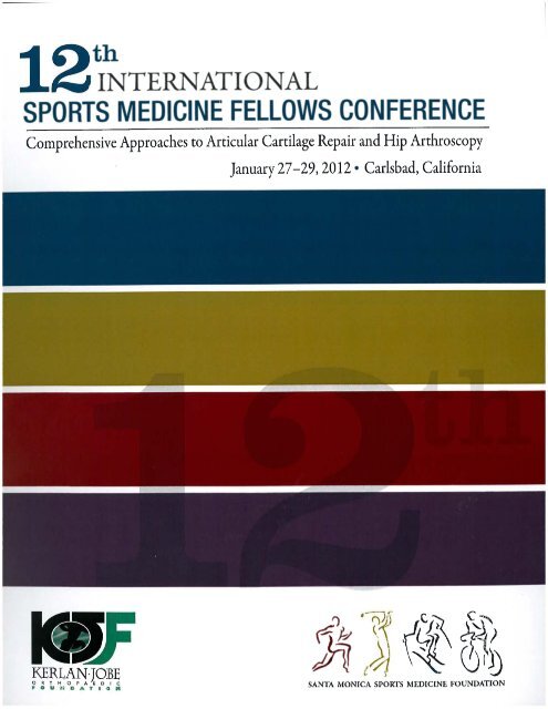
12 th International Sports Medicine Fellows Conference:<br />
Comprehensive Approaches to Articular Cartilage Repair and Hip Arthroscopy<br />
January 27-29, 2012 • Carlsbad, California<br />
The 12 th International Sports Medicine Fellows Conference gratefully<br />
acknowledges the generous support of:<br />
Platinum Sponsors:<br />
Gold Sponsors:<br />
Silver Sponsors:
12 th International Sports Medicine Fellows Conference:<br />
Comprehensive Approaches to Articular Cartilage Repair and Hip Arthroscopy<br />
January 27-29, 2012 • Carlsbad, California<br />
Wayne K. Augé II, MD<br />
Fall River, MA, USA<br />
Robert H. Brophy, MD<br />
Chesterfield, MO, USA<br />
William Bugbee, MD<br />
La Jolla, CA, USA<br />
J.W. Thomas Byrd, MD<br />
Nashville, TN, USA<br />
Susan Chubinskaya, PhD<br />
Chicago, IL, USA<br />
Brian J. Cole, MD<br />
Chicago, IL, USA<br />
Jack Farr, MD<br />
Greenwood, Indiana, USA<br />
Wayne K. Gersoff, MD<br />
Denver, CO, USA<br />
Alberto Gobbi, MD<br />
Milan, ITALY<br />
Course Chairs:<br />
Bert R. Mandelbaum, MD, Course Co-Chair<br />
Santa Monica, California, USA<br />
Ralph A. Gambardella, MD, Course Co-Chair<br />
Los Angeles, California, USA<br />
Michael B. Gerhardt, MD, Hip Course Chair<br />
Manhattan Beach, California, USA<br />
Course Faculty<br />
William Hutchinson, MD<br />
Santa Monica, CA, USA<br />
Warren Kramer, MD<br />
Newport Beach, CA, USA<br />
David R. McAllister, MD<br />
Los Angeles, CA, USA<br />
Kai Mithoefer<br />
Boston, MA, USA<br />
Marc Safran, MD<br />
Redwood City, CA, USA<br />
Robert L. Sah, MD, ScD<br />
La Jolla, CA, USA<br />
Jason M. Scopp, MD<br />
Salisbury, Maryland, USA<br />
Jason Snibbe, MD<br />
Beverley Hill, CA, USA<br />
Jason Theodosakis, MD<br />
Tucson, AZ, USA<br />
C. Thomas Vangsness, MD<br />
Los Angeles, CA, USA
Driving Directions from Hilton Garden Inn to DJO Inc.<br />
Start: Hilton Garden Inn Carlsbad Beach<br />
6450 Carlsbad Blvd<br />
Carlsbad, CA 92011<br />
(760) 476-0800<br />
End: DJO Incorporated<br />
1430 Decision Street<br />
Vista, CA 92081<br />
Directions Distance Time<br />
Start: Drive (West) toward Carlsbad Blvd. 0.2 0:01<br />
1: Turn RIGHT to merge onto Palomar Airport Rd [CR-S12] 6.3 0:07<br />
2: Turn LEFT (North) onto Business Park Dr. 0.6 0:01<br />
3: Take 2 nd RIGHT (North-East) onto Scott St 0.1 0:01<br />
4: Take 1 st RIGHT (South) onto Decision St < 0.1 0:01<br />
End: Arrive (DJO Inc) 1340 Decision Street is on the RIGHT < 0.1 < 1min<br />
Total Route (approximate-driving times vary) 7.1 mi 11 mins<br />
If you have problems getting into the lab call:<br />
Cheresse Edwards, CERF Coordinator<br />
760-597-3942 office<br />
760-535-4704 cell - Best number to reach her
12 th International Sports Medicine Fellows Conference<br />
January 27-29, 2012● Carlsbad, California<br />
Session I<br />
7:15 AM - 7:30 AM Basics of Articular Cartilage/Repair Robert Sah, MD, ScD USA<br />
7:30 AM - 7:45 AM Articular Cartilage Imaging: New Horizons Robert Brophy, MD USA<br />
7:45 AM - 8:00 AM Microfracture Kai Mithoefer, MD USA<br />
8:00 AM - 8:15 AM Medium and Small Defects OC AutoGrafts and New Scaffolds Ralph Gambardella, MD USA<br />
8:15 AM - 8:30 AM CAIS and Juvenile Allogenic Cells Clinical Trials Bert Mandelbaum, MD USA<br />
8:30 AM - 8:45 AM Osteochondral Allograft: Basic Science William Bugbee, MD USA<br />
8:45 AM - 9:00 AM Osteochondral Allograft: Clinical Experience David McAllister, MD USA<br />
9:00 AM - 9:15 AM Discussion
Basics of Articular Cartilage & Synovial Joints<br />
11 th Annual Articular Cartilage Repair Course<br />
for Sports Medicine Fellows<br />
January 15, 2011<br />
Robert L. Sah, MD, ScD<br />
Professor of Bioengineering &<br />
Adjunct Professor of Orthopaedic Surgery<br />
University of California, San Diego<br />
La Jolla, California 92093-0412<br />
Professor,<br />
Howard Hughes Medical Institute<br />
rsah@ucsd.edu
Articular Cartilage Repair Course for Sports Medicine Fellows Robert L. Sah, MD, ScD<br />
Basics of Articular Cartilage & Synovial Joints January 16, 2011<br />
OUTLINE<br />
1) Principles of load-bearing, low-friction, and wear-resistant biomechanical functions of<br />
articular cartilage in healthy synovial joints.<br />
� Articular cartilage bears load that is >NxBW, over contact areas of ~(cm) 2 , with<br />
corresponding stress levels of ~MPa.<br />
� Normally, articular cartilage (synovial joints) has an apparent friction coefficient, μ, of<br />
~0.001-0.05 that varies markedly with the method of measurement.<br />
� Normally, articular cartilage (synovial joints) has a wear coefficient, κ, of ~0.001 (μm) 2 /N.<br />
2) The basis of the biomechanical functions of cartilage and synovial joints.<br />
� The load-bearing properties of articular cartilage exhibit visco(poro)-elasticity.<br />
o Under compressive loading, normal cartilage becomes pressurized with little<br />
compressive strain, and then slowly depressurizes as fluid exudes out and<br />
compressive strain increases.<br />
o The content and organization of its matrix constituents, especially proteoglycan and<br />
collagens, contribute to high compressive modulus and low hydraulic permeability.<br />
o In osteoarthritis, the balance between proteoglycan swelling and collagen restraint is<br />
altered due to collagen network dysfunction, resulting in cartilage swelling.<br />
� The low-friction properties of articular cartilage are due to pressure-dependent and pressureindependent<br />
phenomena (intrinsic friction coefficient with normal synovial fluid is 0.025).<br />
o In the boundary mode of lubrication, the low-friction properties of cartilage are<br />
dependent on the lubricating effects of synovial fluid (Schmidt+, Osteoarthritis<br />
Cartilage, 2007) and lubricant molecules, proteoglycan-4 (aka lubricin, superficial<br />
zone protein, PRG4) and hyaluronan (Schmidt+, Arthritis Rheum, 2007).<br />
o The “intrinsic” (boundary-mode) steady-state friction coefficient of articular cartilage,<br />
lubricated with normal synovial fluid, is ~0.025 (Schmidt+, Osteoarthritis Cartilage,<br />
2007).<br />
o The boundary lubricants in synovial fluid normally serve to lower cartilage-cartilage<br />
friction, and, consequently, shear strain of cartilage during loading.<br />
o After injury, synovial fluid is deficient in lubricating properties (Elsaid+, Arthritis<br />
Rheum, 2005), in association with decreased hyaluronan (Ballard+, JBJS, 2012)<br />
and/or PRG4 (Jay+, Arthritis Rheum, 2007).<br />
� The wear-resistance of cartilage depends on synovial fluid components, probably also<br />
boundary lubricant molecules.<br />
p. 2
Articular Cartilage Repair Course for Sports Medicine Fellows Robert L. Sah, MD, ScD<br />
Basics of Articular Cartilage & Synovial Joints January 16, 2011<br />
3) The biological and biophysical basis of articular cartilage maintenance and repair.<br />
� Heterogeneity in the Cells and Matrix of Articular Cartilage and Synovial Tissues.<br />
o The chondrocytes within articular cartilage are heterogeneous.<br />
1. Cells in the superficial zones synthesize and secrete large amounts of<br />
proteoglycan-4 lubricant (Flannery CR+, BBRC, 1999), whereas cells in the<br />
middle and deep zones synthesize and secrete large amounts of aggrecan.<br />
Don’t let the articular cartilage surface dry out (killing the surface chondrocytes) during<br />
surgery.<br />
2. Progenitor cells within cartilage and surrounding tissues may be sources for<br />
repair.<br />
o The matrix of articular cartilage is heterogeneous and anisotropic.<br />
1. The tangentially-oriented collagen network of the superficial zone provides<br />
high tensile stiffness (Temple MM+, Osteoarthritis Cartilage, 2007) and<br />
resistance to wear.<br />
2. The aggrecan content of the middle and deep zones provides high<br />
compressive stiffness and low hydraulic permeability (Chen AC+, J<br />
Biomechanics, 2001).<br />
3. The depth-variation in aggrecan content, in association with collagen network<br />
content and function (Han EH+, Biophys J, 2011), underlies the depth-varying<br />
compressive modulus of articular cartilage (Schinagl RM+, J Orthop Res,<br />
1997)<br />
� Mechanobiological Homeostasis of Load-Bearing.<br />
o The dynamics of load-bearing proteoglycan and collagen molecules in cartilage<br />
extracellular matrix depends (Hascall and Sandy) on the balance between<br />
1. synthesis and secretion by chondrocytes<br />
2. degradation by enzymes and loss driven by fluid flow.<br />
o Articular cartilage responds to dynamic compression (Sah RL+, J Orthop Res, 1989)<br />
and shear (Jin M+, Arch Biochem Biophys, 2001) via chondrocyte altering synthesis<br />
of matrix constituents, important for load-bearing.<br />
o Excessive compression can induce lethal strains to the chondrocytes.<br />
Be gentle with articular cartilage. Do not poke it or whack it too hard.<br />
� Mechanobiological Homeostasis of Articulation.<br />
o The dynamics of lubricant molecules in synovial fluid depends (Blewis ME+., Eur<br />
Cell Mater, 2007) on the balance between<br />
1. the cells lining the synovial joint, the chondrocytes and synoviocytes,<br />
secreting proteoglycan-4 and hyaluronan lubricant into synovial fluid<br />
2. degradation/loss of lubricant molecules from synovial fluid.<br />
p. 3
Articular Cartilage Repair Course for Sports Medicine Fellows Robert L. Sah, MD, ScD<br />
Basics of Articular Cartilage & Synovial Joints January 16, 2011<br />
o Articular cartilage responds to dynamic shear via chondrocytes in superficial zone<br />
altering synthesis of PRG4 lubricant into the synovial fluid (Nugent GE+, Arthritis<br />
Rheum, 2006).<br />
o PRG4 lubricant molecules are absorbed to the articular surface through interactions<br />
allowing it to be depleted and then repleted (Nugent GE+, J Orthop Res, 2007).<br />
o Continuous passive motion (of knee joints ex vivo) enhances secretion of PRG4<br />
(Nugent-Derfus GE+, Osteoarthritis Cartilage, 2007).<br />
p. 4
ARTHRITIS & RHEUMATISM<br />
Vol. 58, No. 7, July 2008, pp 2065–2074<br />
DOI 10.1002/art.23548<br />
© 2008, American College of Rheumatology<br />
Biomechanics of Cartilage Articulation<br />
Effects of Lubrication and Degeneration on Shear Deformation<br />
Benjamin L. Wong, 1 Won C. Bae, 1 June Chun, 1 Kenneth R. Gratz, 1<br />
Martin Lotz, 2 and Robert L. Sah 1<br />
Objective. To characterize cartilage shear strain<br />
during articulation, and the effects of lubrication and<br />
degeneration.<br />
Methods. Human osteochondral cores from lateral<br />
femoral condyles, characterized as normal or<br />
mildly degenerated based on surface structure, were<br />
selected. Under video microscopy, pairs of osteochondral<br />
blocks from each core were apposed, compressed<br />
15%, and subjected to relative lateral motion with<br />
synovial fluid (SF) or phosphate buffered saline (PBS)<br />
as lubricant. When cartilage surfaces began to slide<br />
steadily, shear strain (E xz) and modulus (G) overall in<br />
the full tissue thickness and also as a function of depth<br />
from the surface were determined.<br />
Results. In normal tissue with SF as lubricant, E xz<br />
was highest (0.056) near the articular surface and<br />
diminished monotonically with depth, with an overall<br />
average E xz of 0.028. In degenerated cartilage with SF as<br />
lubricant, E xz near the surface (0.28) was 5-fold that of<br />
normal cartilage and localized there, with an overall E xz<br />
of 0.041. With PBS as lubricant, E xz values near the<br />
articular surface were �50% higher than those observed<br />
with SF, and overall E xz was 0.045 and 0.062 in normal<br />
and degenerated tissue, respectively. Near the articular<br />
Supported by the National Science Foundation and the NIH.<br />
Mr. Wong’s work was supported in part by the San Diego Fellowship.<br />
Dr. Sah’s work was supported by the Howard Hughes Medical Institute<br />
through the Professors <strong>Program</strong> Grant to the University of California,<br />
San Diego.<br />
1 Benjamin L. Wong, MS, Won C. Bae, PhD, June Chun, BS,<br />
Kenneth R. Gratz, MS, Robert L. Sah, MD, ScD: University of<br />
California, San Diego, La Jolla, California; 2 Martin Lotz, MD: Scripps<br />
Research Institute, La Jolla, California.<br />
Mr. Wong and Dr. Bae contributed equally to this work.<br />
Address correspondence and reprint requests to Robert L.<br />
Sah, MD, ScD, Department of Bioengineering, Mail Code 0412,<br />
University of California, San Diego, 9500 Gilman Drive, La Jolla, CA<br />
92093-0412. E-mail: rsah@ucsd.edu.<br />
Submitted for publication September 20, 2007; accepted in<br />
revised form March 18, 2008.<br />
2065<br />
surface, G was lower with degeneration (0.06 MPa,<br />
versus 0.18 MPa in normal cartilage). In both normal<br />
and degenerated cartilage, G increased with tissue<br />
depth to 3–4 MPa, with an overall G of 0.26–0.32 MPa.<br />
Conclusion. During articulation, peak cartilage<br />
shear is highest near the articular surface and decreases<br />
markedly with depth. With degeneration and diminished<br />
lubrication, the markedly increased cartilage<br />
shear near the articular surface may contribute to<br />
progressive cartilage deterioration and osteoarthritis.<br />
The tissue-scale mechanics of articular cartilage<br />
during the compressive and shear loading of joints with<br />
normal movement have not been fully characterized.<br />
After repeated knee bending (1) and running (2), articular<br />
cartilage is compressed by �5–20% of its overall<br />
thickness. Within the cartilage tissue of dynamically<br />
compressed osteochondral blocks (3), the magnitude of<br />
compressive strain is highest near the articular surface<br />
and minimal in deeper regions, a pattern consistent with<br />
the depth-varying compressive modulus of cartilage (4).<br />
Despite extensive knowledge of cartilage compressive<br />
behavior, the overall and depth-varying shear deformation<br />
of cartilage during joint movement remains to be<br />
elucidated. Theoretical models of articulating joints can<br />
be used to predict local cartilage deformation (5,6), but<br />
are highly dependent on assumptions about boundary<br />
conditions at articulating surfaces (e.g., frictionless or<br />
adherent) and depth-varying mechanical properties (5).<br />
Detailed experimental characterization of apposing cartilage<br />
samples sliding relative to one another would<br />
further the understanding of cartilage mechanics during<br />
joint movement by elucidating the actual boundary<br />
condition at the articulating cartilage surface as well as<br />
the deformation and properties of cartilage in shear.<br />
Lubrication of articulating cartilage by synovial<br />
fluid (SF) facilitates low friction in the boundary mode
2066 WONG ET AL<br />
and may therefore affect the shear deformation of<br />
cartilage. When articulating cartilage is tested in a<br />
configuration that reveals boundary lubrication effects,<br />
SF and lubricant molecules in SF are shown to reduce<br />
articular surface interaction, as indicated by decreased<br />
friction (7,8). The replacement of SF lubricant with<br />
phosphate buffered saline (PBS) results in an elevation<br />
of boundary-mode friction (7). The dependence of local<br />
and overall shear deformation on SF may be important<br />
for maintenance of cartilage health, since acute injury or<br />
inflammatory arthritis results in reduced lubricating<br />
function of SF, and this may be involved in postinjury<br />
cartilage degeneration (9).<br />
Cartilage degeneration may also affect the way in<br />
which the tissue deforms during joint movement. As<br />
cartilage undergoes degeneration, for example with aging<br />
(10), articular surfaces become fibrillated and roughened<br />
(11). Concomitantly, cartilage mechanical properties<br />
deteriorate under compressive, tensile, and shear<br />
loading (12), i.e., cartilage deformation and strain magnitude<br />
increase in response to a particular amplitude of<br />
applied load. Such mechanical deformation of cartilage<br />
affects chondrocyte metabolism, with moderate levels of<br />
dynamic compressive and shear strain stimulating matrix<br />
synthesis, but excessive levels inhibiting matrix synthesis<br />
(13). Thus, determination of the deformation and strain<br />
of articulating cartilage, with both normal and degenerated<br />
surfaces, would provide insight into the local mechanical<br />
cues that regulate chondrocyte behavior and<br />
the consequences of degenerative changes on such cues.<br />
One experimental approach used to elucidate the<br />
local strain deformation and strain of cartilage is to track<br />
displacement of fiducial markers using a video microscopy<br />
system. Previous studies have focused on cartilage<br />
deformation and strain when samples were subjected to<br />
compression in the absence of applied shear (4,14,15).<br />
The depth-dependent compressive behavior of cartilage<br />
found in these microscale studies was consistent with<br />
findings in macroscopic tissue explants investigated by<br />
magnetic resonance imaging (3), and provided a resolution<br />
sufficient to resolve small magnitudes of strain<br />
(�1%). Thus, microscale analysis may also provide<br />
insight into cartilage shear deformation, both locally and<br />
overall. For studying cartilage shear deformation during<br />
joint movement (Figure 1A), a pair of osteochondral<br />
blocks (Figure 1B) can be compressed in apposition<br />
(Figure 1C) and subjected to lateral shearing motion<br />
(Figure 1D). Such a configuration can be controlled to<br />
mimic the biomechanical behavior of articulating cartilage<br />
at one scale (full-thickness tissue) and allow elucidation<br />
of cartilage deformation at a finer scale (regions<br />
Figure 1. A and B, Schematic representation of knee joint movements<br />
at multiple scales (A), and deformation of cartilage under no load,<br />
compression (Comp.), and compression plus shear loading (B). C,<br />
Material points in compressed cartilage before and during shear<br />
loading used to determine u�(x,z) (depth-dependent displacement<br />
[Disp.] vectors), which were then used to assess intratissue deformation.<br />
D, Schematic representation of experimental setup and loading<br />
protocol for microscale shear testing.<br />
of tissue measurements). Combined measurements of<br />
strain and stress allow determination of local and overall<br />
biomechanical properties such as shear modulus.
LUBRICATION AND DEGENERATION EFFECTS ON CARTILAGE SHEAR DURING ARTICULATION 2067<br />
Table 1. Age of the donors, and thickness and Mankin-Shapiro<br />
scores of the cartilage samples*<br />
Normal<br />
(n � 3)<br />
The hypothesis of the present study was that<br />
during joint articulation, the shear deformation of cartilage<br />
is affected by both lubrication and degeneration.<br />
To test this, we implemented a cartilage-on-cartilage<br />
sliding test to microscopically observe and analyze cartilage<br />
deformation during compression and shear. With<br />
this system, we assessed the effects of lubrication (by SF<br />
versus PBS) and degeneration (normal versus mildly<br />
degenerated) on overall and depth-varying shear strain,<br />
and determined the overall and depth-varying shear<br />
modulus of normal and mildly degenerated cartilage.<br />
MATERIALS AND METHODS<br />
Mildly degenerated<br />
(n � 3)<br />
Age, years 48 � 1.7 78 � 9.2<br />
Thickness, mm 1.86 � 0.12 2.08 � 0.15<br />
Mankin-Shapiro score 1.89 � 0.29 9.11 � 1.24<br />
* Values are the mean � SEM.<br />
Sample isolation. Osteochondral cores (each with a<br />
diameter of 10 mm) were isolated from the anterior lateral<br />
femoral condyle of a single knee block from each of 6 fresh<br />
human cadaveric tissue donors (Table 1). Cores were chosen<br />
for this study based on the gross appearance of the articular<br />
surface, i.e., normal (n � 3) or mildly degenerated (n � 3). The<br />
surface was considered normal if the modified Outerbridge<br />
grade (16) was 1, and mildly degenerated if the grade was 3.<br />
Normal specimens were from donors ages 41–60 years at the<br />
time of death, and degenerated specimens were from donors<br />
age �60 years (Figures 2A and B). The cores were immersed<br />
in PBS containing proteinase inhibitors (PIs) and stored at<br />
�80°C until use.<br />
Experimental design. On the day of testing, each core<br />
was thawed and prepared. The core was scored vertically in the<br />
cartilage using a razor blade and cut through the bone using a<br />
low-speed saw with a 0.3-mm–thick diamond-edge blade<br />
(Isomet; Buehler, Lake Bluff, IL) to yield an osteochondral<br />
fragment for histopathologic analysis and 2 approximately<br />
rectangular blocks for biomechanical testing. Each of the 2<br />
blocks had a cartilage surface area of �3 � 8mm 2 and a total<br />
thickness of �7 mm.<br />
Histopathologic analysis. Histopathologic analysis was<br />
performed to assess and confirm the state of degeneration.<br />
Samples were fixed in 4% paraformaldehyde in PBS (pH 7.0)<br />
for 24 hours, decalcified with 18% disodium EDTA in PBS for<br />
7 days, and sectioned to 7 �m using a cryostat. Some sections<br />
were stained with Alcian blue to localize sulfated glycosaminoglycans<br />
(GAGs), as previously described (17), and other<br />
sections were stained with hematoxylin and eosin to highlight<br />
cellular detail (18). Staining of samples resulted in a gradation<br />
of intensity (Figures 2C and D), reflecting the variation in<br />
GAG concentration with depth; thus, the staining method was<br />
sufficient to delineate GAG variation, such as loss due to<br />
pathology, and allow qualitative scoring. Transmitted light<br />
micrographs of the stained sections were obtained, and histologic<br />
features (19) analyzed using the Mankin-Shapiro semiquantitative<br />
scale (20). Briefly, the histopathologic characteristics<br />
assessed included structural integrity (surface irregularity<br />
and vertical and horizontal clefts), cellularity (cloning and<br />
hypocellularity), and GAG loss. A relatively high score represented<br />
more degenerated cartilage. Grades from 3 independent<br />
observers were averaged for each sample. Interobserver<br />
errors (SD) for normal and degenerated samples were reasonably<br />
small (average 1.0 and 2.1, respectively).<br />
Biomechanical testing. Samples were first tested by<br />
microscale shear testing as described below, with PBS plus PI<br />
as the lubricant. Then, samples were allowed to reswell for �4<br />
hours at 4°C.<br />
Next, samples were tested again by microscale shear<br />
Figure 2. A and B, Photographs of cross-sections of samples of<br />
normal and degenerated cartilage. C and D, Representative micrographs<br />
of Alcian blue–stained sections, showing structure detail of<br />
normal (C) and degenerated (D) cartilage. Color figure can be viewed<br />
in the online issue, which is available at http://www.arthritisrheum.org.
2068 WONG ET AL<br />
testing, this time with SF (pooled from adult bovine knees and<br />
stored at �80°C) plus PI as the lubricant. The SF had been<br />
characterized previously for boundary lubrication properties<br />
(7) and for levels of lubricant molecules (�1 mg/ml of hyaluronan<br />
and 0.45 mg/ml of proteoglycan 4 [21]). The same<br />
regions of interest as used in studies with PBS were imaged.<br />
Then, samples were allowed to reswell.<br />
<strong>Final</strong>ly, samples were tested in a macroscale shear<br />
system to assess overall mechanical properties of cartilage in<br />
shear, as described below.<br />
Microscale shear testing. Each sample, consisting of<br />
paired osteochondral blocks, was bathed for �14–18 hours in<br />
test lubricant containing PI and propidium iodide (20 �g/ml) at<br />
4°C to fluorescently highlight cell nuclei prior to microscale<br />
shear testing. Each pair of osteochondral blocks (Figures 1B<br />
and D) was then placed with cartilage in apposition in a custom<br />
biaxial loading chamber mounted onto an epifluorescence<br />
microscope for digital video imaging (4). The chamber secured<br />
one block at the bone and allowed in-plane movement of the<br />
apposing mobile block along the x-directed 8-mm lengths of<br />
the samples (Figure 1D), with orthogonally positioned plungers<br />
interfaced with either a micrometer (for axial displacement)<br />
(model 262RL; Starrett, Athol, MA) or motioncontroller<br />
(for lateral displacement) (model MFN25PP;<br />
Newport, Irvine, CA). Fluorescence images (G-2A filter; Nikon,<br />
Melville, NY) with a field of view of �3 � 2mm 2 were<br />
obtained at 5 frames/second, showing a full-thickness region of<br />
cartilage in the secured block and a partial-thickness region of<br />
cartilage in the apposing block.<br />
Cartilage deformation in the secured block was assessed<br />
during axial and shear loading. First, the block was<br />
imaged in the reference state (uncompressed). Then, axial<br />
displacement (�40 �m/second) was applied by the micrometer<br />
to induce 15% compression (1 �� z, where � z is the stretch<br />
ratio [22]) of the cartilage tissue (Figure 1C), during which<br />
time sequential images (�1.5% compression increment/frame)<br />
were acquired. Samples were then allowed to stress-relax for 1<br />
hour, which was calculated to be sufficient to reach an approximate<br />
equilibrium stress based on consideration of the characteristic<br />
time constant. Experimentally, this duration of stressrelaxation<br />
was validated to be sufficient with load decreasing to<br />
50% of the peak by 130 seconds, and load at 1 hour being only<br />
3 � 1% (mean � SEM; n � 3) higher than the load at 16<br />
hours. Subsequently, lateral motion was applied to the mobile<br />
osteochondral block (Figure 1D). Two sets of lateral displacements<br />
(�x), each consisting of �1 mm and then �1 mm<br />
(returning to initial position), were applied at 100 �m/second<br />
to the bone portion of the mobile block. The first set, followed<br />
by a pause of 12 seconds, was for preconditioning (7), while the<br />
second set was recorded for analysis. The sliding velocity was<br />
chosen based on the range of velocities (0–0.1 m/second)<br />
occurring during the loading (stance) phase of gait (23,24).<br />
Before and during the application of lateral displacements<br />
(Figure 1D), sequential images, with �10 �m of lateral<br />
movement of the mobile block between frames, were obtained.<br />
Macroscale shear testing. To assess the overall compressive<br />
and shear biomechanical properties of the cartilage of the<br />
secured block, following microscale shear tests, each secured<br />
block was subjected to a macroscale test using a biaxial<br />
mechanical tester (Mach-1 V500CS; BioSyntech, Montreal,<br />
Quebec, Canada). Test conditions were chosen to match the<br />
mechanical deformation observed in the microscale shear test,<br />
but with instrumentation allowing measurement of axial and<br />
shear loads. Each sample block was affixed, compressed 15%<br />
at a rate of 0.03%/second using a rigid stainless steel platen<br />
(�2 �m pore size, providing a no-slip boundary condition),<br />
and allowed to stress-relax for 1 hour to reach equilibrium.<br />
With increasing lateral displacement, continuous and monotonically<br />
increasing shear load waveforms were observed,<br />
confirming no-slip conditions; a plateau in shear load, which<br />
would indicate slip, was not observed. Equilibrium force was<br />
recorded and normalized to the 3 � 8mm 2 area to yield an<br />
estimate of the equilibrium compressive stress. Next, 2 sets of<br />
lateral displacements of the amplitude occurring at the surface<br />
at the onset of sliding in microscale shear tests were applied at<br />
a rate of 100 �m/second, held for 10 seconds while shear load<br />
was being recorded, and released. A pause of �12 seconds<br />
followed the first set of lateral displacements, during which the<br />
cartilage sample was continuously hydrated in PBS plus PI. As<br />
in the microscale tests, the first set was for preconditioning,<br />
while the second set was used for analysis. The lateral displacements<br />
were then repeated in the reverse direction following a<br />
5-minute pause, and shear loads occurring in the last second of<br />
the 10-second load-capture period in both directions were<br />
averaged. This average shear load was normalized to the 3 �<br />
8mm 2 area to yield an estimate of the shear stress (at which<br />
sliding occurred in the microscale test).<br />
Data collection and calculations. Digital micrographs<br />
were analyzed to determine the depth-varying and overall<br />
strains in cartilage. Images were analyzed with MatLab 7.0<br />
(MathWorks, Natick, MA) using software routines similar to<br />
those described previously (14,25,26). Briefly, an evenly distributed<br />
set of cell nuclei (�250 cells/mm 2 ), which served as<br />
fiducial markers, was selected and tracked by maximizing<br />
cross-correlation of regions surrounding each marker with the<br />
preceding, and then initial, frames. Generally, chondrocyte<br />
nuclei in normal tissue were available, and were chosen in<br />
regions up to the true articular surface (defined by a discontinuity<br />
in displacement during sliding), while for degenerated<br />
tissue, nuclei were visible up to �50–70 �m (�3%) below the<br />
true surface, due to fibrillation. Local affine mappings of<br />
nuclei were used to calculate the displacement (u�) of uniformly<br />
spaced (10-pixel) mesh points in the region of interest (�1<br />
mm � full thickness) during deformation (Figure 1D). Displacement<br />
gradients (�u�/�z, �u�/�x) were then determined by<br />
finite difference approximations, and in turn, used to determine<br />
Lagrangian compressive strain (E zz) after axial compression<br />
and shear strain (E xz) after lateral shearing, with these<br />
Lagrangian strains being appropriate for finite strains as well<br />
as small strains (27).<br />
This method was validated, and interobserver variability<br />
was assessed. Using mathematically transformed images,<br />
the calculated strain values deviated by less than 0.005 � 0.005<br />
(mean � SEM) from the theoretical values, with the error<br />
being proportionately smaller in regions exhibiting lower amplitudes<br />
of strain. Interobserver variability, assessed as the<br />
average of the SD in calculated strain between 3 observers, was<br />
�0.01 for a subset of data from all sample types (n � 4). Here
LUBRICATION AND DEGENERATION EFFECTS ON CARTILAGE SHEAR DURING ARTICULATION 2069<br />
also, the variability was proportionately lower in regions of<br />
relatively small shear magnitudes.<br />
The calculated shear strain was interpolated, averaged<br />
depthwise, and plotted as a function of tissue depth, normalized<br />
to the cartilage thickness in the compressed state. Shear<br />
strain was interpolated linearly at every 5% of the normalized<br />
tissue thickness near the articular surface (i.e., 0–0.3) and<br />
every 10% for remaining regions of the tissue depth (i.e.,<br />
0.3–1) after applied compression and prior to lateral motion.<br />
To consolidate data, strain values were averaged at the same<br />
normalized depth (0 [surface] and 1 [tidemark]) to yield a<br />
depth profile. Shear strain results were determined when<br />
surfaces were sliding, at which time shear strain was at a peak<br />
and no further changes in deformation occurred with additional<br />
lateral displacement (i.e., sliding). The overall Lagrangian<br />
shear strain was defined as half the lateral surface displacement<br />
normalized to the compressed cartilage thickness. Since<br />
the shear strain peaked near the articular surface, shear strain<br />
occurring at the top 5% was also compared.<br />
Overall cartilage properties were determined from<br />
forces recorded during compression and shear. Under compression<br />
alone, the compressive modulus of cartilage was<br />
defined as the increment in stress divided by the increment in<br />
applied compressive strain. The overall shear modulus (G) for<br />
each sample was determined as the increment in shear stress<br />
(shear load divided by surface area) divided by the increment<br />
in shear strain.<br />
The depth-varying shear moduli were estimated from<br />
the overall shear modulus and local shear strain in each<br />
sample. Since shear modulus is inversely related to shear<br />
strain, and with shear stress assumed to be constant at all<br />
depths, the local shear modulus was estimated as the overall<br />
shear modulus multiplied by the overall shear strain and<br />
divided by the local shear strain. Because these estimates<br />
depend on shear strain in the denominator, shear modulus was<br />
calculated at every 10% of the tissue depth near the articular<br />
surface (up to 20% of the depth) and every 20% of the tissue<br />
depth in the subsequent regions, to reduce noise.<br />
Statistical analysis. Data are reported as the mean �<br />
SEM, unless noted otherwise. Repeated-measures analysis of<br />
variance was used to determine the effects of normalized tissue<br />
depth (0–1 [surface–bone]), lubricant (PBS or SF), and degeneration<br />
(normal or degenerated) on tissue shear strain, and to<br />
determine the effects of tissue depth and degeneration on<br />
shear modulus.<br />
RESULTS<br />
Histopathologic findings. The overall histopathology<br />
scores were consistent with the gross (Figures<br />
2A and B) and histologic (Figures 2C and D) appearance<br />
of normal and degenerated samples. Degenerated<br />
samples had significantly (P � 0.01) higher histopathology<br />
scores than normal samples (Table 1). In degenerated<br />
samples, structural (surface irregularity, vertical<br />
clefts to transitional zone, and transverse clefts) and<br />
cellular (cloning) features exhibited mild degeneration<br />
and were scored �1-point higher than the corresponding<br />
features in normal samples. In contrast, cellularity<br />
and GAG staining scores were similarly low (i.e., normal)<br />
in normal and degenerated samples. These results<br />
confirmed that gross visualization resulted in an appropriate<br />
selection of normal and mildly degenerated samples,<br />
for articulation testing.<br />
Compressive deformation and properties. The<br />
axial compression of cartilage in the secured block<br />
resulted in compressive strain that varied significantly<br />
with depth from the articular surface (P � 0.001) but not<br />
between normal and degenerated samples (P � 0.6). E zz<br />
was relatively high near the articular surface (mean �<br />
SEM 0.38 � 0.05) and relatively low in the deepest<br />
regions (0.06 � 0.01). Transverse-oriented (E xx) and<br />
shear (E xz) strain resulting from the applied compression<br />
was low, averaging �0.03 in all samples. The<br />
equilibrium compressive modulus (E) in the fullthickness<br />
cartilage was not significantly different between<br />
sample groups (P � 0.5) (mean � SEM 0.79 �<br />
0.13 MPa in normal cartilage and 0.61 � 0.23 MPa in<br />
degenerated cartilage).<br />
Shear deformation. Qualitatively, cartilage shear<br />
loading resulted in a sequence of 4 events: 1) At the<br />
onset of applied lateral displacement, cartilage surfaces<br />
initially adhered and began to move laterally in unison.<br />
2) With increasing lateral displacement, lateral deformation<br />
and hence E xz increased and occurred throughout<br />
the tissue depth. 3) Next, lateral deformation and E xz of<br />
cartilage peaked, just as the surfaces detached and slid<br />
relative to each other. 4) With additional lateral displacement,<br />
the cartilage deformation and E xz were<br />
maintained at a steady-state peak.<br />
At the steady state, tissue displacement and E xz<br />
(Figure 3) were nonuniform and depth-varying, with<br />
shear magnitudes that were high near the articular<br />
surface and low (almost indistinguishable) near the<br />
bone. The mean � SEM maximum lateral displacement<br />
of apposing surfaces before slipping when lubricated<br />
with SF was 115 � 14 �m and 82 � 16 �m in normal and<br />
degenerated tissue, respectively. When lubricated with<br />
PBS, maximum lateral displacement in normal and<br />
degenerated tissue was 169 � 12 �m and 125 � 18 �m,<br />
respectively, before slipping. Differences in shear deformation<br />
between normal and degenerated samples were<br />
also evident (Figure 3). Shear deformation in degenerated<br />
samples (Figures 3C and D) was concentrated near<br />
the surface, while in normal samples, deformation occurred<br />
further into the tissue depth (Figures 3A and B).<br />
Quantitatively, the E xz of articulating cartilage
2070 WONG ET AL<br />
Figure 3. Micrographs obtained during shear loading of apposing<br />
normal (A and B) and degenerated (C and D) cartilage samples<br />
lubricated with phosphate buffered saline (PBS) (A and C) or synovial<br />
fluid (SF) (B and D), after maximum shear strain was achieved. The<br />
cell nuclei tracking method was used to create maps of shear strain<br />
(magnified above each panel; boxes with dotted lines indicate magnified<br />
areas). Dashed lines surround the analyzed regions on the<br />
undeformed images. Schematic at the bottom depicts the sample and<br />
testing configuration, with the dotted-line box representing the area<br />
from which the images shown were obtained and analyzed. Bars � 100<br />
�m.<br />
varied with depth from the articular surface, lubricant,<br />
and surface degeneration. In the depth profile of E xz<br />
(Figures 4A and B), the highest values were obtained<br />
near the articular surface, and E xz decreased monotonically<br />
with depth (P � 0.001). The variation of E xz with<br />
depth depended on surface degeneration (P � 0.001 for<br />
the interaction), with E xz decreasing more with depth in<br />
the degenerated sample. A similar trend was noted with<br />
testing using PBS, where E xz decreased at greater rates<br />
with depth than when tested with SF (P � 0.001 for the<br />
interaction) in both normal and degenerated samples.<br />
The combined effect of depth and either degeneration<br />
or lubricant on shear strain was evidenced as varying E xz<br />
values near the surface, but similarly low values of E xz<br />
(�0.01) in the deep layer of cartilage.<br />
Peak E xz at the surface (z � 0) (Figure 5A) and<br />
overall E xz (Figure 5B) were each significantly affected<br />
by both the lubricant used and degeneration status. The<br />
peak surface E xz (Figure 5A) was �3–5 times higher in<br />
Figure 4. Biomechanical measures of adult human articular cartilage<br />
in shear. A and B, Local shear strain (E xz) versus normalized tissue<br />
depth in normal and degenerated cartilage, as determined by microscale<br />
shear testing. Samples were tested with phosphate buffered<br />
saline (PBS) (A) or synovial fluid (SF) (B) as lubricant. C, Local shear<br />
modulus (G) versus normalized tissue depth in normal and degenerated<br />
samples, as determined by macroscale testing. Values are the<br />
mean and SEM.
LUBRICATION AND DEGENERATION EFFECTS ON CARTILAGE SHEAR DURING ARTICULATION 2071<br />
Figure 5. Effect of sample group (normal or degenerated cartilage)<br />
and of lubricant (phosphate buffered saline [PBS] or synovial fluid<br />
[SF]) on A, peak surface shear strain and B, overall shear strain. Values<br />
are the mean and SEM.<br />
samples with surface degeneration than in normal tissue<br />
(P � 0.05) with both surface lubricants. With PBS, the<br />
mean of the peak surface (Figure 5A) E xz increased<br />
from 0.11 (normal samples) to 0.41 (degenerated samples).<br />
With SF, E xz increased from 0.056 (normal samples)<br />
to 0.28 (degenerated samples). In addition, peak<br />
surface E xz values obtained when tested with PBS were<br />
�1.5–2 times the values obtained when tested with SF<br />
(P � 0.05). Overall E xz (Figure 5B) was not detectably<br />
different between normal and degenerated samples (P �<br />
0.10) when tested using the same lubricant. However,<br />
overall E xz did vary with lubricant. The overall E xz of<br />
normal tissue was significantly lower with SF than with<br />
PBS (0.028 versus 0.045), and the overall E xz of degenerated<br />
tissue was 0.041 with SF versus 0.062 with PBS (P<br />
� 0.05 for both).<br />
Shear properties. To replicate sample shear deformation<br />
in microscale shear tests, lateral surface dis-<br />
placements in macroscale shear tests ranging from 70 to<br />
125 �m (measured from microscale shear tests) were<br />
applied to normal samples, and 120–160 �m was applied<br />
to degenerated samples. Overall G was not significantly<br />
different between normal and mildly degenerated tissue<br />
(mean � SEM 0.32 � 0.09 MPa in normal and 0.26 �<br />
0.07 MPa in degenerated cartilage; P � 0.7). G was<br />
lowest near the articular surface and increased significantly<br />
with tissue depth (P � 0.01) (0.2–-0.6 times and<br />
10–15 times the overall G near the surface and in the<br />
deepest region, respectively) (Figure 4C). At all depths<br />
below the surface, G was not significantly different<br />
between normal and degenerated samples (P � 0.5).<br />
Near the articular surface, there was a trend toward<br />
lower G values with mild surface degeneration (P �<br />
0.10) (surface G � 0.18 � 0.06 MPa and 0.06 � 0.02<br />
MPa in normal and degenerated samples, respectively).<br />
DISCUSSION<br />
This study elucidated the shear deformation and<br />
strain of cartilage during contact and sliding, as well as<br />
the effects of lubrication and mild degeneration, using a<br />
microscale cartilage-on-cartilage testing system. The<br />
present results indicate that cartilage–cartilage articulation<br />
results in 4 sequential events: adherence, adherence<br />
and deformation, detachment, and sliding. Peak E xz was<br />
highest near the surface and modulated by both the<br />
lubricant used and the condition of the sample itself<br />
(Figures 4A and B). Relative to the use of normal SF as<br />
lubricant, E xz increased with the use of PBS by �100%<br />
near the surface (Figure 5A) and �55% overall (Figure<br />
5B). In addition, when degenerated samples were tested,<br />
their E xz values near the surface were �3–5 times higher<br />
than those of normal samples (Figure 5A).<br />
Osteochondral samples used in this study were<br />
prepared from a site that is affected by age-associated<br />
degeneration and osteoarthritis. The samples were from<br />
the lateral femoral condyle, a load-bearing region where<br />
�22% of arthroscopically diagnosed chondral defects<br />
occur (28,29). The biomechanical factors associated with<br />
early degeneration in such load-bearing regions may<br />
contribute to the progressive degeneration of articular<br />
cartilage. Although the samples used had been previously<br />
frozen, a single freeze–thaw cycle does not result in<br />
marked changes in compressive properties of normal<br />
(30) or degenerated (31) articular cartilage or its structure<br />
(32), and is thus unlikely to have marked effects on<br />
shear properties. Although the distal femoral condyle<br />
normally apposes the tibial plateau, cartilage from the<br />
femoral condyle was apposed against itself to create a
2072 WONG ET AL<br />
simplified symmetric loading situation. Cartilage from<br />
other sites could be examined in future studies.<br />
The testing protocol used in this study provides a<br />
mechanical environment mimicking the compression<br />
and sliding of articular cartilage during normal joint<br />
loading. A knee undergoes a wide range of dynamic<br />
compression (up to 5–20%) (1,2) and sliding (up to �50<br />
mm) during normal activities (estimated from refs. 23<br />
and 24). The present analysis addresses certain parts of<br />
the gait cycle (e.g., contralateral toe-off and heel rise<br />
[23]) in which compressive loading is high and sliding<br />
velocity is low, during which time interaction of apposing<br />
tissue surfaces is likely to be initiated. Since samples<br />
were allowed to stress-relax for 1 hour, the pattern of<br />
cartilage deformation and strain is likely to be representative<br />
of that occurring after prolonged cyclic loading<br />
and sliding, rather than that which occurs at the onset of<br />
cyclic loading. Cartilage gradually depressurizes and<br />
reaches an averaged steady-state compression under<br />
prolonged cyclic loading. During this time, interstitial<br />
fluid pressure diminishes (33), and boundary lubrication<br />
becomes increasingly important (7). The ratedependence<br />
of shear deformation was not assessed;<br />
however, shear deformation may not change markedly<br />
with rate of lateral displacement since kinetic friction<br />
remains fairly constant with sliding velocity (7). Additional<br />
studies might elucidate the time course of cartilage<br />
shear after the onset of loading, as well as effects of<br />
sliding velocity on shear deformation.<br />
Although edge effects may occur in this shear<br />
testing protocol, such effects are likely minimal in the<br />
central area of tissue that was analyzed. Boundaries of<br />
the analyzed region were �2 mm away from both the<br />
leading and the trailing edges of the samples. Recent<br />
studies have shown that intratissue deformation (Gratz<br />
KR, et al: unpublished observations) and stiffness (34)<br />
are affected only �1–2 mm from vertical boundaries of<br />
cartilage. Thus, the results are likely to be representative<br />
of the major areas of cartilage contact.<br />
The reduction in E xz with SF as lubricant is<br />
consistent with predictions derived from a variety of past<br />
studies on cartilage friction. The friction coefficient<br />
decreases with the use of SF as lubricant compared with<br />
saline, both in cartilage-on-cartilage in the boundary<br />
mode (7,35) and in whole joint (36–38) friction tests.<br />
Assuming cartilage material properties (i.e., compressive<br />
and shear modulus) are maintained with the use of<br />
SF and PBS, the friction-reducing property of SF would<br />
be predicted to result in a reduction of tissue E xz.<br />
The overall E xz values obtained in this study are<br />
consistent with values estimated during physiologic joint<br />
articulation based on previously reported material properties<br />
of cartilage, as follows. To provide a first-order<br />
estimate of tissue properties, cartilage can be modeled<br />
as a linear, homogeneous, isotropic, and elastic tissue.<br />
With such assumptions and under small strain conditions,<br />
the overall stiffness of cartilage in shear (G) can be<br />
related to the shear stress (� xz) and the resulting overall<br />
E xz (27) as follows: G � � xz/2E xz (equation 1). In<br />
addition, � xz acting on cartilage can be related to the<br />
friction coefficient (�) and normal contact stress (� n)as<br />
follows: � xz � �� n (equation 2). Substituting equation 2<br />
into equation 1 and rearranging terms, cartilage E xz can<br />
be expressed as E xz � �� n/G (equation 3).<br />
Using reported values of � (�0.01) (39) and � n<br />
(�2 Mpa) (40) and the overall G values of �0.2–0.4<br />
MPa found in this study, the estimated overall E xz is<br />
0.02–0.05. The estimate of E xz is consistent with the<br />
range of overall E xz determined in this study (Figure<br />
5B). This suggests that an overall E xz ranging from 0.01<br />
to 0.1 may be suitable for biomimetic in vitro simulation<br />
of physiologic shear loading, for example during mechanical<br />
evaluation and mechanical stimulation for tissue<br />
engineering of cartilage.<br />
The increased E xz with degeneration, particularly<br />
near the surface with SF as lubricant (Figure 5A),<br />
appears to be due to both increased friction between<br />
sliding surfaces and a reduction in tissue shear modulus.<br />
Roughened surfaces have local asperities that can cause<br />
increased interaction and adherence between tissue surfaces,<br />
enhancing friction during sliding. Since boundary<br />
lubricants modulate friction, higher E xz in degenerated<br />
cartilage when tested in PBS (Figure 5A) indicates that<br />
friction between degenerated surfaces is higher than that<br />
between normal surfaces. Additionally, with degeneration,<br />
cartilage mechanical properties, such as G, become<br />
impaired (12). Diminished G would result in increased<br />
cartilage E xz at the same magnitude of � xz (equation 3),<br />
even if friction coefficient were similar. Since surface E xz<br />
increased with degeneration with the use of SF as<br />
lubricant, and G near the surface tended to diminish<br />
with degeneration, the elevated E xz at the surface in<br />
mildly degenerated tissue is likely a result of both<br />
increased � and reduced G (equation 3 and Figure 5A).<br />
Depth variations in E xz and G in the cartilage<br />
samples used in the present study provide additional<br />
information on the depth-varying biomechanical properties<br />
of articular cartilage. Compressive strain is depthvarying<br />
in both statically (4) and dynamically (3) compressed<br />
cartilage, decreasing monotonically with depth<br />
from the articular surface. E xz observed in this study was<br />
similarly depth-varying, being highest near the surface
LUBRICATION AND DEGENERATION EFFECTS ON CARTILAGE SHEAR DURING ARTICULATION 2073<br />
and lowest near the tidemark (Figures 4A and B).<br />
Compressive modulus, deduced from the overall compressive<br />
stress and local strain, was reflective of the<br />
depth-varying strain magnitudes, with cartilage increasing<br />
in stiffness with increasing depth (4). Similarly, G<br />
was lowest near the articular surface and increased<br />
monotonically to a maximum (20%) in the deepest<br />
region (Figure 4C). While G near the surface only<br />
tended to decrease with degeneration, the lack of statistical<br />
significance was likely due to the calculation from 2<br />
measurements, shear stress and strain (each having<br />
variability), as well as donor variability.<br />
The overall unconfined compressive modulus at<br />
equilibrium found in this study (E � 0.79 MPa) is<br />
consistent with previously reported values in human<br />
cartilage (0.58 MPa) (41). The overall shear properties<br />
of human cartilage have yet to be reported; however,<br />
overall G can be estimated from Poisson’s ratio (�) and<br />
the aggregate modulus (H A) demonstrated in indentation<br />
tests, using the relationship<br />
G � H A�1 � ���1 � 2��<br />
2�1 � ���1 � ��<br />
(27,42). The overall G found in this study (0.32 MPa) is<br />
consistent with that estimated from indentation tests<br />
(0.2–0.4 MPa) (43). Thus, for cartilage in compression,<br />
the deformation and mechanical properties we observed<br />
are consistent with findings of previous studies, and for<br />
cartilage in shear, the newly described depth-varying<br />
strain and shear properties are within values predicted<br />
for full-thickness human cartilage.<br />
With cartilage degeneration, tissue structure and<br />
low cellularity may affect the analysis and interpretation<br />
of deformation and strain. Degenerated cartilage contains<br />
clefts and areas of erosion (20), both of which<br />
result in material discontinuities within cartilage. Thus,<br />
continuum assumptions are not strictly valid. Also, compatibility<br />
conditions (27) are not maintained when fibrillated<br />
tissues overlap during deformation. Nevertheless,<br />
the present samples were only mildly degenerated and<br />
relatively intact except at the surface, and, whereas there<br />
were few visible cells at the surface of the degenerated<br />
samples, cells near the surface (3–5% depth) could be<br />
tracked. From a linear extrapolation, this may have<br />
resulted in a slight (�15%) underestimation of the<br />
absolute magnitude of superficial shear strain.<br />
The present results suggest that changes in E xz<br />
magnitudes and shear behavior may contribute to cartilage<br />
degeneration and pathogenesis. While samples<br />
were tested with a common overall compressive strain in<br />
this study, marked differences in E xz between groups<br />
occurred even with this conservative testing protocol.<br />
Under similar loads instead of overall strain, softer<br />
tissues (i.e., degenerated cartilage) would exhibit larger<br />
compressive and shear strain, excessive levels of which<br />
can contribute to cartilage degeneration. Excessive magnitudes<br />
of compressive strain result in mechanical injury<br />
to cells (44,45) and matrix (45,46), leading to reduced<br />
cartilage remodeling, maintenance, and repair. Likewise,<br />
high E xz that results from degenerated matrix,<br />
along with deficient lubrication, may lead to additional<br />
degeneration. Cell death and matrix damage may spread<br />
with continued exposure to high E xz, diminishing local<br />
mechanical properties of cartilage in both compression<br />
and shear. Cartilage E xz may then further increase and<br />
further the changes in tissue structure, resulting in a<br />
self-propagating cycle of degeneration. Such mechanical<br />
effects may also induce changes in surrounding joint<br />
tissues and the biochemical environment, which could<br />
supplement cartilage deterioration.<br />
ACKNOWLEDGMENTS<br />
We thank the many residents and staff at Dr. Lotz’s<br />
Laboratory at the Scripps Research Institute for harvesting<br />
and providing the human tissue used in this study, and Barbara<br />
Schumacher for guidance on histopathologic processing.<br />
AUTHOR CONTRIBUTIONS<br />
Dr. Sah had full access to all of the data in the study and takes<br />
responsibility for the integrity of the data and the accuracy of the data<br />
analysis.<br />
Study design. Wong, Bae, Sah.<br />
Acquisition of data. Wong, Chun, Lotz.<br />
Analysis and interpretation of data. Wong, Bae, Gratz, Sah.<br />
Manuscript preparation. Wong, Bae, Sah.<br />
Statistical analysis. Wong, Bae, Sah.<br />
REFERENCES<br />
1. Eckstein F, Lemberger B, Stammberger T, Englmeier KH, Reiser<br />
M. Patellar cartilage deformation in vivo after static versus dynamic<br />
loading. J Biomech 2000;33:819–25.<br />
2. Kersting UG, Stubendorff JJ, Schmidt MC, Bruggemann GP.<br />
Changes in knee cartilage volume and serum COMP concentration<br />
after running exercise. Osteoarthritis Cartilage 2005;13:<br />
925–34.<br />
3. Neu CP, Hull ML, Walton JH. Heterogeneous three-dimensional<br />
strain fields during unconfined cyclic compression in bovine<br />
articular cartilage explants. J Orthop Res 2005;23:1390–8.<br />
4. Schinagl RM, Gurskis D, Chen AC, Sah RL. Depth-dependent<br />
confined compression modulus of full-thickness bovine articular<br />
cartilage. J Orthop Res 1997;15:499–506.<br />
5. Ateshian GA, Lai WM, Zhu WB, Mow VC. An asymptotic<br />
solution for the contact of two biphasic cartilage layers. J Biomech<br />
1994;27:1347–60.<br />
6. Swanson SAV. Friction, wear, and lubrication. In: Freeman MAR,
2074 WONG ET AL<br />
editor. Adult articular cartilage. 2nd ed. Tunbridge Wells (UK):<br />
Pitman Medical; 1979. p. 415–60.<br />
7. Schmidt TA, Sah RL. Effect of synovial fluid on boundary<br />
lubrication of articular cartilage. Osteoarthritis Cartilage 2007;15:<br />
35–47.<br />
8. Schmidt TA, Gastelum NS, Nguyen QT, Schumacher BL, Sah RL.<br />
Boundary lubrication of articular cartilage: role of synovial fluid<br />
constituents. Arthritis Rheum 2007;56:882–91.<br />
9. Elsaid KA, Jay GD, Warman ML, Rhee DK, Chichester CO.<br />
Association of articular cartilage degradation and loss of boundary-lubricating<br />
ability of synovial fluid following injury and inflammatory<br />
arthritis. Arthritis Rheum 2005;52:1746–55.<br />
10. Clark JM, Simonian PT. Scanning electron microscopy of “fibrillated”<br />
and “malacic” human articular cartilage: technical considerations.<br />
Microsc Res Tech 1997;37:299–313.<br />
11. Meachim G, Emery IH. Quantitative aspects of patello-femoral<br />
cartilage fibrillation in Liverpool necropsies. Ann Rheum Dis<br />
1974;33:39–47.<br />
12. Setton LA, Elliott DM, Mow VC. Altered mechanics of cartilage<br />
with osteoarthritis: human osteoarthritis and an experimental<br />
model of joint degeneration. Osteoarthritis Cartilage 1999;7:2–14.<br />
13. Sah RL, Grodzinsky AJ, Plaas AHK, Sandy JD. Effects of static<br />
and dynamic compression on matrix metabolism in cartilage<br />
explants. In: Kuettner KE, Schleyerbach R, Peyron JG, Hascall<br />
VC, editors. Articular cartilage and osteoarthritis. New York:<br />
Raven Press; 1992. p. 373–92.<br />
14. Wang CC, Deng JM, Ateshian GA, Hung CT. An automated<br />
approach for direct measurement of two-dimensional strain distributions<br />
within articular cartilage under unconfined compression.<br />
J Biomech Eng 2002;124:557–67.<br />
15. Chahine NO, Wang CC, Hung CT, Ateshian GA. Anisotropic<br />
strain-dependent material properties of bovine articular cartilage<br />
in the transitional range from tension to compression. J Biomech<br />
2004;37:1251–61.<br />
16. Yamada K, Healey R, Amiel D, Lotz M, Coutts R. Subchondral<br />
bone of the human knee joint in aging and osteoarthritis. Osteoarthritis<br />
Cartilage 2002;10:360–9.<br />
17. Scott JE, Dorling J. Differential staining of acid glycosaminoglycans<br />
(mucopolysaccharides) by alcian blue in salt solutions. Histochemie<br />
1965;5:221–33.<br />
18. Ross MH, Kaye GI, Pawlina W. Histology: a text and atlas. 4th ed.<br />
Philadelphia: Lippincott Williams & Wilkins; 2003.<br />
19. Bae WC, Temple MM, Amiel D, Coutts RD, Niederauer GG, Sah<br />
RL. Indentation testing of human cartilage: sensitivity to articular<br />
surface degeneration. Arthritis Rheum 2003;48:3382-94.<br />
20. Shapiro F, Glimcher MJ. Induction of osteoarthrosis in the rabbit<br />
knee joint: histologic changes following menisectomy and meniscal<br />
lesions. Clin Orthop Rel Res 1980;147:287–95.<br />
21. Mazzucco D, Scott R, Spector M. Composition of joint fluid in<br />
patients undergoing total knee replacement and revision arthroplasty:<br />
correlation with flow properties. Biomaterials 2004;25:<br />
4433–45.<br />
22. Fung YC. Biomechanics: mechanical properties of living tissues.<br />
2nd ed. New York: Springer-Verlag; 1993.<br />
23. Whittle M. Gait analysis: an introduction. 3rd ed. Oxford (UK):<br />
Butterworth-Heinemann; 2002.<br />
24. Shelburne KB, Torry MR, Pandy MG. Muscle, ligament, and<br />
joint-contact forces at the knee during walking. Med Sci Sports<br />
Exerc 2005;37:1948–56.<br />
25. Bae WC, Lewis CW, Levenston ME, Sah RL. Indentation testing<br />
of human articular cartilage: effects of probe tip geometry and<br />
indentation depth on intra-tissue strain. J Biomech 2006;39:<br />
1039–47.<br />
26. Gratz KR, Sah RL. Experimental measurement and quantification<br />
of frictional contact between biological surfaces experiencing large<br />
deformation and slip. J Biomech 2008;41:1333–40.<br />
27. Fung YC. A first course in continuum mechanics. 2nd ed. Englewood<br />
Cliffs (NJ): Prentice-Hall; 1977.<br />
28. Hjelle K, Solheim E, Strand T, Muri R, Brittberg M. Articular<br />
cartilage defects in 1,000 knee arthroscopies. Arthroscopy 2002;<br />
18:730–4.<br />
29. Curl WW, Krome J, Gordon ES, Rushing J, Smith BP, Poehling<br />
GG. Cartilage injuries: a review of 31,516 knee arthroscopies.<br />
Arthroscopy 1997;13:456–60.<br />
30. Swann AC, Seedhom BB. The stiffness of normal articular cartilage<br />
and the predominant acting stress levels: implications for the<br />
aetiology of osteoarthrosis. Br J Rheumatol 1993;32:16–25.<br />
31. Kempson GE, Spivey CJ, Swanson SA, Freeman MA. Patterns of<br />
cartilage stiffness on normal and degenerate human femoral<br />
heads. J Biomech 1971;4:597–609.<br />
32. Kiefer GN, Sundby K, McAllister D, Shrive NG, Frank CB, Lam<br />
T, et al. The effect of cryopreservation on the biomechanical<br />
behavior of bovine articular cartilage. J Orthop Res 1989;7:<br />
494–501.<br />
33. Armstrong CG, Lai WM, Mow VC. An analysis of the unconfined<br />
compression of articular cartilage. J Biomech Eng 1984;106:<br />
165–73.<br />
34. Bae WC, Law AW, Amiel D, Sah RL. Sensitivity of indentation<br />
testing to step-off edges and interface integrity in cartilage repair.<br />
Ann Biomed Eng 2004;32:360–9.<br />
35. Forster H, Fisher J. The influence of loading time and lubricant on<br />
the friction of articular cartilage. Proc Inst Mech Eng [H] 1996;<br />
210:109–19.<br />
36. Wright V, Dowson D. Lubrication and cartilage. J Anat 1976;121:<br />
107–18.<br />
37. Linn FC. Lubrication of animal joints. II. The mechanism. J Biomech<br />
1968;1:193–205.<br />
38. Mabuchi K, Tsukamoto Y, Obara T, Yamaguchi T. The effect of<br />
additive hyaluronic acid on animal joints with experimentally<br />
reduced lubricating ability. J Biomed Mater Res 1994;28:865–70.<br />
39. Tanaka E, Kawai N, Tanaka M, Todoh M, van Eijden T, Hanaoka<br />
K, et al. The frictional coefficient of the temporomandibular joint<br />
and its dependency on the magnitude and duration of joint<br />
loading. J Dent Res 2004;83:404–7.<br />
40. Raimondi MT, Pietrabissa R. Contact pressures at grafted cartilage<br />
lesions in the knee. Knee Surg Sports Traumatol Arthrosc<br />
2005;13:444–50.<br />
41. Jurvelin JS, Buschmann MD, Hunziker EB. Mechanical anisotropy<br />
of the human knee articular cartilage in compression. Proc<br />
Inst Mech Eng [H] 2003;217:215–9.<br />
42. Mow VC, Hayes WC, editors. Basic orthopaedic biomechanics.<br />
2nd ed. New York: Raven Press; 1997.<br />
43. Athanasiou KA, Rosenwasser MP, Buckwlter JA, Malinin TI,<br />
Mow VC. Interspecies comparisons of in situ intrinsic mechanical<br />
properties of distal femoral cartilage. J Orthop Res 1991;9:330–40.<br />
44. Loening A, Levenston M, James I, Nuttal M, Hung H, Gowen M,<br />
et al. Injurious mechanical compression of bovine articular cartilage<br />
induces chondrocyte apoptosis. Arch Biochem Biophys 2000;<br />
381:205–12.<br />
45. Chen CT, Bhargava M, Lin PM, Torzilli PA. Time, stress, and<br />
location dependent chondrocyte death and collagen damage in<br />
cyclically loaded articular cartilage. J Orthop Res 2003;21:888–98.<br />
46. Thibault M, Poole AR, Buschmann MD. Cyclic compression of<br />
cartilage/bone explants in vitro leads to physical weakening, mechanical<br />
breakdown of collagen and release of matrix fragments.<br />
J Orthop Res 2002;20:1265–73.
Effect of a Focal Articular Defect on Cartilage Deformation during<br />
Patello-Femoral Articulation<br />
Benjamin L. Wong, Robert L. Sah<br />
Department of Bioengineering, University of California—San Diego, 9500 Gilman Drive, La Jolla, California<br />
Received 3 August 2009; accepted 22 April 2009<br />
Published online 2 July 2010 in Wiley Online Library (wileyonlinelibrary.com). DOI 10.1002/jor.21187<br />
ABSTRACT: The objective of this study was to determine cartilage strains near, and in apposition to, a focal defect during patello-femoral<br />
articulation. Bovine osteochondral blocks from the trochlea (TRO) and patella (PAT) were apposed, compressed 12%, and subjected to<br />
sliding under video microscopy. Samples, lubricated with synovial fluid, were tested intact and then with a full-thickness defect in PAT<br />
cartilage. Shear (Exz), axial (Ezz), and lateral (Exx) strains were determined locally for TRO and PAT cartilage. For articulation with a<br />
focal defect, the strain amplitudes of PAT cartilage near the surface were ∼2–8× lower in Exz and ∼1.4× higher in −Ezz than intact PAT<br />
cartilage. At 20% depth, Exz and Exx for PAT cartilage with a focal defect were ∼2× and ∼10–25× higher than intact PAT, respectively.<br />
For TRO articulating against a focal defect, Exz and −Ezz near the surface and at 20% depth were ∼2–4× lower than that for articulation<br />
against intact cartilage. The results elucidate dramatic region-specific changes in strain due to lateral motion. In these regions, such<br />
altered cartilage mechanics during knee movement may cause focal defects to extend by induction of damaging levels of strain to bordering<br />
regions of cartilage. © 2010 Orthopaedic Research Society. Published by Wiley Periodicals, Inc. J. Orthop. Res. 28: 1554–1561, 2010<br />
Keywords: focal defect; articulation; biomechanics; deformation; cartilage strains<br />
Focal articular defects are prevalent in symptomatic<br />
knees and are associated with progressive cartilage<br />
degeneration. In patients evaluated arthroscopically,<br />
focal defects were found in 20–60% of all symptomatic<br />
knees, occurring most frequently in the medial condyle<br />
and patella, both of which are load-bearing regions (Fig.<br />
1). 1–4 Such, focal defects typically ranged from 0.5 to<br />
4cm 2 in area with an average area of 2.1 cm 2 , and<br />
extended beyond half the cartilage thickness in depth<br />
in 50% of all visualized articular defects. 1 With continued<br />
joint loading and time, untreated defects enlarge, 5<br />
are associated with cartilage volume loss, 6 and exhibit<br />
histopathological signs of cartilage degeneration adjacent<br />
to the focal defect. 7,8 While such findings suggest<br />
that the presence of a focal defect predisposes joints<br />
to secondary osteoarthritis, the mechanism by which it<br />
causes cartilage to degenerate within the proximity of a<br />
focal defect remains to be elucidated.<br />
The effect of a focal defect on cartilage deformation<br />
has been examined during axially directed loading.<br />
Under axial compression alone, contact stress and<br />
stress gradients are elevated in areas of cartilage near<br />
the edges of a focal defect. 9,10 As a result, macroscopic<br />
tissue deformation 11 and local strains 12 are<br />
increased markedly in these regions. Elevated strains<br />
may reach levels that induce cell death 13 and matrix<br />
damage, 14 and thus, the elevated strains resulting from<br />
a focal defect may become injurious to cartilage and<br />
induce degeneration. Such tissue deformation may be<br />
altered by or accentuated by the addition of lateral<br />
motion superimposed on compression, as both occur<br />
Additional Supporting Information may be found in the online<br />
version of this article.<br />
Correspondence to: Robert L. Sah (T: 858-534-0821;<br />
F: 858-822-1614.; E-mail: rsah@ucsd.edu)<br />
© 2010 Orthopaedic Research Society. Published by Wiley Periodicals, Inc.<br />
1554 JOURNAL OF ORTHOPAEDIC RESEARCH DECEMBER 2010<br />
in load-bearing regions of cartilage during joint movement.<br />
During knee movement, articular surfaces contact,<br />
compress, and articulate against each other. Within the<br />
patellofemoral groove of the knee, patellar cartilage<br />
contacts and slides against trochlear cartilage during<br />
normal joint movement and loading (Fig. 2A). Under<br />
applied compressive loads of ∼1.5× the body weight,<br />
patellofemoral cartilage compresses ∼10% of its overall<br />
thickness in vitro following 14 min of static loading, 15<br />
and following knee bending, patellar cartilage alone<br />
compresses about ∼5–10% overall in vivo. 16 Collectively,<br />
such results provide physiologic mechanical parameters<br />
for the in vitro testing of patello-femoral articulation.<br />
Recently, local and overall deformation of cartilage<br />
during compression and cartilage articulation 17 were<br />
determined using video microscopy 18 and image correlation<br />
to track the displacement of fiducial markers. 19,20<br />
Such a test configuration can be applied to study contact<br />
of patellar and trochlear cartilage surfaces, 21,22 to<br />
examine the effects of a focal articular defect on the<br />
deformation of cartilage near, and in apposition to, the<br />
proximal defect edge that experiences oncoming forward<br />
motion of the apposing surface during compression and<br />
lateral displacement (Fig. 2). Effects of a focal defect on<br />
cartilage deformation may be distinctly and markedly<br />
different during compression than during shear.<br />
Therefore, the hypothesis of this study was that during<br />
patellofemoral cartilage articulation, the cartilage<br />
deformation of the patella and trochlea are markedly<br />
altered with the presence of a focal articular defect.<br />
The specific objective of this study was to determine the<br />
effects of a focal articular defect, created in the patellar<br />
tissue, on the local and overall strains of the patellar<br />
and trochlear cartilage during compression and sliding<br />
motion.
Figure 1. Anatomical distribution of focal articular defects averaged<br />
from four previous arthroscopy studies on the prevalence of<br />
cartilage defects in symptomatic knees. 1–4<br />
Figure 2. Schematic of (A) knee joint movements at multiple<br />
scales, (B) experimental setup and loading protocol for micro-shear<br />
testing, (C) and locations of sub-regions (EDGE, MID, FAR) used<br />
for statistical analysis of strains in patella and trochlear cartilage.<br />
EFFECT OF A FOCAL DEFECT ON CARTILAGE DEFORMATION 1555<br />
MATERIALS AND METHODS<br />
Sample Isolation and Preparation<br />
Osteochondral cores with macroscopically normal cartilage<br />
were harvested from the trochlea (TRO) and patella (PAT)<br />
of four adult bovine animals (1–2 years). Using a low speed<br />
drill press with custom stainless steel coring bits, a 10 mm<br />
diameter osteochondral core was isolated from both the TRO<br />
and PAT of each joint in a manner similar to that described<br />
previously. 23 The TRO and PAT cores were then trimmed to<br />
each yield one approximately rectangular block for biomechanical<br />
testing. 17 Each rectangular block had a cartilage surface<br />
area of ∼3 × 10 mm 2 and a total thickness of ∼1 cm (Fig. 2A).<br />
Each sample consisted of one TRO and one PAT block from the<br />
same knee, and was stored in phosphate buffered saline (PBS)<br />
containing proteinase inhibitors (PI) until testing.<br />
Prior to mechanical testing, samples were stained for<br />
∼2–4 h at 4 ◦ C in PBS + PI and propidium iodide (20 �g/mL) to<br />
fluorescently highlight cell nuclei. Blocks were then bathed in<br />
normal bovine synovial fluid (SF) containing PI and propidium<br />
iodide (20 �g/mL) at 4 ◦ C for 12–16 h to lubricate surfaces. The<br />
SF was pooled from adult bovine knees, previously characterized<br />
for boundary lubrication properties and lubricant levels<br />
and stored at −80 ◦ C, and characterized previously for boundary<br />
lubrication properties and lubricant molecules levels. 23<br />
Experimental Design<br />
To characterize the effect of a focal defect on cartilage deformation<br />
during articulation, samples were mechanically tested,<br />
first intact and then with a focal defect. In between tests, samples<br />
were rinsed, allowed to reswell, and incubated for ∼2–4 h<br />
in PBS + PI and then in SF + PI for an additional 12–16 h at<br />
4 ◦ C. Following the mechanical testing of intact samples and<br />
reincubation, a full thickness (∼2 mm in depth), 3 mm wide<br />
focal articular defect was created in the center of the PAT cartilage<br />
(Fig. 2A) as described previously. 12 The width of the focal<br />
defect was chosen so to be wide relative to the articulation distance<br />
(described below) in order to focus on the initial stages<br />
of articulation.<br />
Micro-Scale Shear Testing<br />
Samples were shear tested under video microscopy essentially<br />
as described previously. 17 Briefly, each TRO and PAT pair<br />
was secured in a custom bi-axial loading chamber mounted<br />
onto an epi-fluorescence microscope for digital video imaging<br />
(Fig. 2B). 18 The chamber secured the PAT block at the bone<br />
and allowed in-plane movement of the apposing mobile TRO<br />
block with orthogonally positioned plungers. 17 Subsequently,<br />
an axial displacement was applied (∼40 �m/s) to induce 12%<br />
compression (1 − �z, where �z is the stretch ratio 24 ) of the overall<br />
cartilage thickness (Fig. 2B). Samples were then allowed<br />
to stress relax for 1 h, determined to be sufficient to reach<br />
an approximate equilibrium stress for the current sample<br />
geometries. 17 Cartilage deformation was then captured during<br />
lateral motion separately in the TRO and PAT cartilage<br />
following axial compression. Three sets of applied lateral displacements<br />
(�x), each consisting of +1 mm and then −1mm<br />
(returning to initial position) were applied at 100 �m/s to the<br />
bone portion of the TRO block (Fig. 2B). The first set, was for<br />
preconditioning, 23 while the second and third set were recorded<br />
for the PAT and TRO blocks for analysis, respectively. Deformation<br />
during patello-femoral cartilage articulation was captured<br />
with sequential fluorescence images taken at ∼25 �m increments<br />
of lateral displacement.<br />
JOURNAL OF ORTHOPAEDIC RESEARCH DECEMBER 2010
1556 WONG AND SAH<br />
Data Collection and Calculations<br />
Acquired images were analyzed as described previously 17,20<br />
to determine the depth-varying and overall deformation<br />
and strain in cartilage. Evenly distributed cell nuclei<br />
(∼250 cells/mm 2 ) were tracked during lateral motion to determine<br />
the displacements of uniformly spaced (10 pixel) mesh<br />
points in local regions. Subsequently, Lagrangian shear (Exz),<br />
axial (Ezz), and lateral (Exx) strains were determined relative<br />
to the unloaded state, 24 when articular surfaces were sliding<br />
(�x = 0.8 mm) and strains had become steady. Local strains<br />
were determined by averaging both depth-wise and at various<br />
lateral distances from the defect edge. First, sample thickness<br />
was normalized and divided into 8 intervals, with 4 intervals<br />
being 0.083 times the normalized thickness near the articular<br />
surface (i.e., 0–0.333) and 0.167 times for the remaining tissue<br />
depth (i.e., 0.333–1). To reduce noise and consolidate data, PAT<br />
cartilage strains near the proximal defect edge, which experiences<br />
forward lateral motion, were averaged depth-wise for<br />
lateral regions (∼0.2 mm × full cartilage thickness) at (EDGE),<br />
∼0.4 mm (MID) and ∼0.8 mm (FAR) away from the defect edge<br />
to yield a depth-profile at varying lateral distances (Fig. 2C).<br />
For TRO cartilage in direct apposition to the focal defect prior<br />
to lateral motion, strains following lateral articulation were<br />
averaged depth-wise in lateral regions (∼0.15 mm × full cartilage<br />
thickness) at (EDGE), ∼0.3 mm (MID), and ∼0.5 mm<br />
(FAR) away from the defect edge. For intact tissue, strains<br />
in corresponding lateral regions were determined similarly.<br />
Overall strain values were determined as the mean of all local<br />
values.<br />
Statistical Analysis<br />
Data are reported as mean ± standard error of the mean<br />
(SEM). Repeated measures ANOVA was used to determine the<br />
effects of a focal defect (intact vs. defect), tissue depth, and lateral<br />
location from the defect edge (EDGE, MID, FAR) on local<br />
and overall strains. Differences between defect and intact samples<br />
at the various lateral locations were assessed by planned<br />
pair-wise comparisons.<br />
Figure 3. Representative micrographs of patellar cartilage as (A,D,G) intact or (B,E,H) with a focal defect with superimposed colormaps<br />
of (A,B) shear (Exz), (D,E) axial (−Ezz), and (G,H) lateral (Exx) strain maps when articulating against trochlear samples after articular<br />
surfaces have slid. Strain map boundaries encompass the corresponding deformed states. Local (C) shear (Exz), (F) axial (−Ezz), and (I)<br />
lateral (Exx) strain averaged depth-wise versus normalized tissue depth for patella cartilage as intact or with a focal defect. For samples<br />
with a focal defect, strains were also determined as a function of lateral distance from the defect edge (EDGE, MID, FAR).<br />
JOURNAL OF ORTHOPAEDIC RESEARCH DECEMBER 2010
RESULTS<br />
Qualitatively, the deformations of intact PAT and TRO<br />
cartilage were similar, being depth-varying during axial<br />
compression as well as during lateral motion. Cartilage<br />
thickness was similar between intact samples, being<br />
1.97 ± 0.13 mm and 1.93 ± 0.14 mm for PAT and TRO<br />
tissue, respectively. Under compression, axial deformation<br />
of intact PAT and TRO cartilage were similarly<br />
depth-varying, being highest near the surface and lowest<br />
near the tidemark. Also during compression, shear<br />
and lateral deformation were low (
1558 WONG AND SAH<br />
Figure 5. Representative micrographs of trochlear cartilage with superimposed colormaps of (A,B) shear (Exz), (D,E) axial (−Ezz), and<br />
(G,H) lateral (Exx) strain when in apposition with patellar cartilage as (A,D,G) intact or (B,E,H) with a focal defect during lateral motion.<br />
Dashed lines (– –) encompass the analyzed regions prior to lateral motion, while boundaries of strain maps encompass the corresponding<br />
deformed states. Local (C) shear (Exz), (F) axial (−Ezz), and (I) lateral (Exx) strain averaged depth-wise versus normalized tissue depth<br />
for trochlear cartilage, intact or with a focal defect. For samples apposing a focal defect, strains were also determined as a function of<br />
lateral distance from the defect edge (EDGE, MID, FAR).<br />
a defect decreased with increasing lateral distance from<br />
the defect edge (p < 0.01). Thus, when TRO cartilage filling<br />
the focal defect slides over the proximal defect edge,<br />
PAT cartilage near a focal defect compresses more than<br />
intact cartilage near the surface and overall.<br />
Lateral Strain (Exx)<br />
With articulation, the resultant Exx for intact cartilage<br />
did not vary significantly with tissue depth (p = 0.2) and<br />
lateral location (p = 1.0) (Fig. 3G), while for cartilage<br />
with a defect, Exx varied with tissue depth (p < 0.001)<br />
and lateral region (p < 0.01) (Fig. 3H). For all lateral<br />
regions, Exx remained negligible (≤0.01) for intact cartilage<br />
throughout tissue depth and peaked at ∼20% depth<br />
for defect samples (Fig. 3I). Near the articular surface,<br />
Exx for cartilage with a defect were statistically indifferent<br />
(p = 0.4) from that for intact cartilage and did not<br />
JOURNAL OF ORTHOPAEDIC RESEARCH DECEMBER 2010<br />
significantly (p = 0.6) vary with lateral distance (Fig.<br />
4G). At 20% tissue depth (Fig. 4H) and overall (Fig.<br />
4I), cartilage Exx peaked at a value of ∼0.08 and ∼0.03,<br />
respectively, in the MID region for cartilage with a defect<br />
(p < 0.01) and were significantly higher than Exx for<br />
intact cartilage (p < 0.01). Thus, PAT cartilage expands<br />
laterally at 20% tissue depth as it becomes further compressed<br />
when TRO cartilage filling the focal defect slides<br />
over the defect edge.<br />
Trochlear Cartilage Deformation<br />
During patello-femoral articulation, the depth-variation<br />
in strains of TRO cartilage in articulation with an intact<br />
and defect-containing PAT surface were similar to that<br />
of PAT cartilage (Fig. 5), and the effect of a focal defect<br />
on TRO cartilage strains were also marked (Fig. 6).<br />
For TRO cartilage in articulation with an intact PAT
Figure 6. Effect of a focal defect (intact (I) vs. defect (D)) on (A–C)<br />
shear, (D–F) axial, and (G–I) lateral strain (A,D,G) near the articular<br />
surface, (B,E,H) at 20% tissue depth, and (C,F,I) overall of<br />
trochlear cartilage during articulation.<br />
surface, Exz and −Ezz were highest near the surface and<br />
decreased with increasing depth (p < 0.001), while Exx<br />
remained negligible throughout tissue depth (p = 0.6).<br />
All strains did not vary with lateral region for TRO<br />
articulation with intact PAT cartilage (p = 0.15–0.6). For<br />
TRO cartilage in articulation with a focal defect, Exz<br />
and −Ezz were ∼2–3× (p < 0.05, Fig. 6A) and ∼2–4×<br />
(p < 0.01, Fig. 6D), respectively, less near the surface<br />
than when in articulation with an intact surface. Furthermore,<br />
TRO cartilage compressed laterally in the<br />
EDGE region near the surface, and Exx near the surface<br />
markedly increased with lateral distance (p < 0.05),<br />
transitioning into lateral tension in the MID and FAR<br />
regions (Fig. 6G). The effects of tissue depth, lateral<br />
region, and a focal defect on TRO strains during lateral<br />
articulation are described in further detail in the<br />
supplementary data.<br />
DISCUSSION<br />
This study elucidated the deformation of cartilage<br />
within the proximity of, and directly in apposition to,<br />
EFFECT OF A FOCAL DEFECT ON CARTILAGE DEFORMATION 1559<br />
a focal articular defect during patello-femoral cartilage<br />
articulation. As the TRO surface displaced laterally, the<br />
tissue partially filling the focal defect pushed and then<br />
plowed over the proximal defect edge. As a result, PAT<br />
cartilage proximal to the defect sheared markedly less<br />
(∼70–700%) near the surface and more (∼50–60%) at<br />
20% tissue depth than intact cartilage during lateral<br />
motion (Fig. 7). PAT cartilage near the focal defect also<br />
compressed more (30–40%) than intact cartilage, and<br />
expanded laterally ∼10–25× more at 20% tissue depth<br />
than intact cartilage as TRO cartilage translated laterally<br />
over the proximal defect edge. For regions directly in<br />
apposition to the focal defect, TRO cartilage sheared and<br />
compressed less (∼50–70%) than when it slid over intact<br />
cartilage. As the TRO surface plowed over the proximal<br />
defect edge, TRO cartilage near the surface also compressed<br />
laterally at the EDGE region, and expanded<br />
laterally at MID and FAR regions. Collectively, the current<br />
results indicate that with articulation, the tissue<br />
deformation of both the cartilage adjacent to, and in<br />
apposition to, a focal defect are altered drastically by<br />
the presence of a focal defect, extending the findings of<br />
strain analysis of cartilage when compressed. 12<br />
To replicate the physiologic articulation and lubrication<br />
of cartilage surfaces, osteochondral samples were<br />
isolated from the trochlear and patella and tested in<br />
apposition with normal bovine synovial fluid. While<br />
efforts were not taken to ensure trochlear and patellar<br />
samples were in exact apposition and registration<br />
in the native anatomical joint, patellar cartilage translates<br />
a distance of ∼2.7 cm over the trochlear surface<br />
(estimated from Refs. 25,26 ). Since samples were isolated<br />
from matching anatomical aspects (i.e., lateral or<br />
medial), samples likely apposed and slid against one<br />
another during anatomic articulation. Since the medial<br />
femoral condyle is the site of greatest incidence of focal<br />
lesions, 1–4 condylar cartilage are of interest. However, it<br />
has a greater curvature in the saggital plane and articulates<br />
against both tibial and meniscal cartilage during<br />
knee movement. Thus, effects of a focal defect on condylar<br />
cartilage are likely to be somewhat more complex and<br />
remain to be investigated. A relative sliding distance of<br />
1 mm was sufficient to result in deformations reaching<br />
a steady-state for both intact and defect samples (data<br />
not shown). Thus, deformations described in the present<br />
study would likely be representative of greater, more<br />
physiologic, sliding distances.<br />
The defect geometry for the present samples was<br />
a full thickness slot with sharp edges in a rectangular<br />
osteochondral block and is a simple model of focal<br />
articular defects. Such a configuration would be most<br />
representative of a partially apposed and loaded defect,<br />
since completely covered defects would have a significant<br />
hoop stress around the defect rim that would help<br />
resistance deformation during articulation. Additional<br />
analyses to address complex three-dimensional issues<br />
would be useful in future studies. Furthermore, defect<br />
edges that are rounded, smoothed, or blunted may also<br />
occur, which may minimize tissue strains near focal<br />
JOURNAL OF ORTHOPAEDIC RESEARCH DECEMBER 2010
1560 WONG AND SAH<br />
Figure 7. (A) Schematic depicting the effect of a focal defect on tissue deformation of trochlear and patellar cartilage when unloaded,<br />
compressed, and compressed and sheared in apposition. (B) Table of relative changes in shear (Exz), axial (−Ezz), and lateral (Exx)<br />
strains compared to normal intact cartilage (i.e., without a focal defect) for both trochlear and patellar cartilage when they are unloaded,<br />
compressed, and compressed and sheared in apposition.<br />
defects. While the present study provides analysis for<br />
a particular set of defect geometries, others remain to<br />
be investigated.<br />
While patellar cartilage was analyzed up to the articular<br />
surface, the region very close to the trochlear<br />
surface (∼10% of tissue depth), which partially filled the<br />
defect, was not analyzed because during lateral motion,<br />
tissue compression was so extensive in that region that<br />
cell nuclei coalesced and could not be discriminated and<br />
tracked. However, regions directly in apposition of the<br />
defect edge (EDGE) prior to lateral motion were tracked<br />
appropriately near the surface. Thus, strains in other<br />
lateral regions (MID, FAR) of the trochlear cartilage may<br />
be somewhat (∼10–15%) underestimated and may be<br />
more similar to the values of the EDGE lateral region.<br />
The dramatic elevation in cartilage strains near a<br />
focal defect during lateral articulation may be related<br />
and possibly contribute to, the progressive tissue degeneration<br />
associated with focal defects. With joint loading<br />
and time, increased fibrillation and decreased matrix<br />
staining were noted in regions adjacent to a focal defect<br />
in animal models. 7,8 Also, untreated defects in human<br />
knee joints have been shown to enlarge 5 and be associated<br />
with cartilage volume loss. 6 Markedly elevated<br />
strains likely make cartilage susceptible to damage in<br />
these regions, via cell death 13 and matrix damage, 14<br />
which can be induced by high magnitudes of compression.<br />
With repeated loading during joint movement, the<br />
increased Exz and Exx at 20% tissue depth, as well as<br />
the elevated −Ezz near the surface, may all contribute<br />
to inducing the indicators of tissue degeneration characterized<br />
previously near the focal defect edge. Thus, focal<br />
JOURNAL OF ORTHOPAEDIC RESEARCH DECEMBER 2010<br />
defects may enlarge due to articulation-induced strain,<br />
and damage the bordering regions of cartilage.<br />
For cartilage in direct apposition to focal defects, the<br />
reduction in localized strains during lateral articulation<br />
may be related to the low incidences of two focal defects<br />
being in direct apposition (i.e., “kissing lesions”). Focal<br />
defects found on both apposing cartilage surfaces were<br />
found in only ∼2% of the symptomatic knees arthroscopically<br />
diagnosed with focal defects. 3 The low frequency<br />
of kissing lesions may be attributed the reduction in<br />
Exz and −Ezz of cartilage when in articulation with<br />
a focal defect. Since strain magnitudes are lowered, a<br />
chondral defect may be unlikely to develop from a preexisting<br />
focal defect on the opposing surface. Instead,<br />
kissing lesions may form with the defects being initiated<br />
concurrently at the time of injury or with one lesion<br />
enlarging big enough to become full-thickness in depth<br />
and cause the apposing chondral surface to be in articulation<br />
against the subchondral bone, inducing abrasive<br />
wear.<br />
The present study suggests that focal articular<br />
defects drastically alter cartilage deformation of both<br />
apposing cartilage surfaces not only during axial loading,<br />
but also distinctly during lateral articulation. Focal<br />
defects, of which a majority (∼61%) has been associated<br />
with acute injury or trauma, 1 markedly alter the<br />
mechanical environment of cartilage, and the resulting<br />
abnormal strains may be injurious to cells and<br />
matrix. Mechanically induced cell death and tissue loss<br />
reduce cell population and likely compromise the overall<br />
biosynthetic response of the tissue. 27 The metabolic<br />
activities of the remaining cells that may continue
to experience injurious levels of strain, are markedly<br />
altered. 13,14 As a result, tissue repair and remodeling<br />
responses are likely compromised, facilitating changes<br />
in cartilage structure by degeneration and wear. Thus,<br />
rehabilitation protocols, such as a period of non-weight<br />
bearing following acute injury, may be beneficial for<br />
allowing repair as well as limiting damage to the<br />
remaining native cartilage following surgical procedures<br />
such as debridement. Thus with normal loading,<br />
the changes in cartilage deformation associated with<br />
focal effects during both axial loading and lateral articulation<br />
may contribute to the enlargement of focal defects<br />
and predispose joints to secondary osteoarthritis.<br />
ACKNOWLEDGMENTS<br />
This work was supported by NIH and the Howard Hughes<br />
Medical Institute through the Professors <strong>Program</strong> to UCSD (to<br />
R.L.S.). We thank Chris Kim and Alexander Cigan for assistance<br />
with the sample preparations.<br />
REFERENCES<br />
1. Hjelle K, Solheim E, Strand T, et al. 2002. Articular cartilage<br />
defects in 1,000 knee arthroscopies. Arthroscopy 18:730–734.<br />
2. Curl WW, Krome J, Gordon ES, et al. 1997. Cartilage<br />
injuries: A review of 31,516 knee arthroscopies. Arthroscopy<br />
13:456–460.<br />
3. Aroen A, Loken S, Heir S, et al. 2004. Articular cartilage<br />
lesions in 993 consecutive knee arthroscopies. Am J Sports<br />
Med 32:211–215.<br />
4. Widuchowski W, Widuchowski J, Trzaska T. 2007. Articular<br />
cartilage defects: Study of 25,124 knee arthroscopies. Knee<br />
14:177–182.<br />
5. Wang Y, Ding C, Wluka AE, et al. 2006. Factors affecting<br />
progression of knee cartilage defects in normal subjects over<br />
2 years. Rheumatology (Oxford) 45:79–84.<br />
6. Cicuttini F, Ding C, Wluka A, et al. 2005. Association of cartilage<br />
defects with loss of knee cartilage in healthy, middle-age<br />
adults: A prospective study. Arthritis Rheum 52:2033–2039.<br />
7. Lefkoe TP, Trafton PG, Ehrlich MG, et al. 1993. An<br />
experimental model of femoral condylar defect leading to<br />
osteoarthrosis. J Orthop Trauma 7:458–467.<br />
8. Jackson DW, Lalor PA, Aberman HM, et al. 2001. Spontaneous<br />
repair of full-thickness defects of articular cartilage<br />
in a goat model. A preliminary study. J Bone Joint Surg Am<br />
83-A:53–64.<br />
9. Brown TD, Pope DF, Hale JE, et al. 1991. Effects of osteochondral<br />
defect size on cartilage contact stress. J Orthop Res<br />
9:559–567.<br />
10. Guettler JH, Demetropoulos CK, Yang KH, et al. 2004. Osteochondral<br />
defects in the human knee: Influence of defect size<br />
on cartilage rim stress and load redistribution to surrounding<br />
cartilage. Am J Sports Med 32:1451–1458.<br />
EFFECT OF A FOCAL DEFECT ON CARTILAGE DEFORMATION 1561<br />
11. Braman JP, Bruckner JD, Clark JM, et al. 2005. Articular<br />
cartilage adjacent to experimental defects is subject to<br />
atypical strains. Clin Orthop Relat Res 430:202–207.<br />
12. Gratz KR, Wong BL, Bae WC, et al. 2009. The effects of focal<br />
articular defects on cartilage contact mechanics. J Orthop<br />
Res 27:584–592.<br />
13. Kurz B, Jin M, Patwari P, et al. 2001. Biosynthetic response<br />
and mechanical properties of articular cartilage after injurious<br />
compression. J Orthop Res 19:1140–1146.<br />
14. Quinn TM, Allen RG, Schalet BJ, et al. 2001. Matrix and cell<br />
injury due to sub-impact loading of adult bovine articular<br />
cartilage explants: Effects of strain rate and peak stress. J<br />
Orthop Res 19:242–249.<br />
15. Herberhold C, Faber S, Stammberger T, et al. 1999. In<br />
situ measurement of articular cartilage deformation in<br />
intact femoropatellar joints under static loading. J Biomech<br />
32:1287–1295.<br />
16. Eckstein F, Lemberger B, Stammberger T, et al. 2000. Patellar<br />
cartilage deformation in vivo after static versus dynamic<br />
loading. J Biomech 33:819–825.<br />
17. Wong BL, Bae WC, Chun J, et al. 2008. Biomechanics of cartilage<br />
articulation: Effects of lubrication and degeneration<br />
on shear deformation. Arthritis Rheum 58:2065–2074.<br />
18. Schinagl RM, Gurskis D, Chen AC, et al. 1997. Depthdependent<br />
confined compression modulus of full-thickness<br />
bovine articular cartilage. J Orthop Res 15:499–506.<br />
19. Wang CC, Deng JM, Ateshian GA, et al. 2002. An automated<br />
approach for direct measurement of two-dimensional<br />
strain distributions within articular cartilage under unconfined<br />
compression. J Biomech Eng 124:557–567.<br />
20. Gratz KR, Sah RL. 2008. Experimental measurement and<br />
quantification of frictional contact between biological surfaces<br />
experiencing large deformation and slip. J Biomech<br />
41:1333–1340.<br />
21. Erne OK, Reid JB, Ehmke LW, et al. 2005. Depth-dependent<br />
strain of patellofemoral articular cartilage in unconfined<br />
compression. J Biomech 38:667–672.<br />
22. Guterl CC, Gardner TR, Rajan V, et al. 2009. Twodimensional<br />
strain fields on the cross-section of the human<br />
patellofemoral joint under physiological loading. J Biomech<br />
42:1275–1281.<br />
23. Schmidt TA, Sah RL. 2007. Effect of synovial fluid on boundary<br />
lubrication of articular cartilage. Osteoarthr Cartilage<br />
15:35–47.<br />
24. Fung YC. 1977. A first course in continuum mechanics.<br />
Englewood Cliffs: Prentice-Hall.<br />
25. Whittle M. 2002. Gait analysis: An introduction. Oxford;<br />
Boston: Butterworth-Heinemann.<br />
26. Shelburne KB, Torry MR, Pandy MG. 2005. Muscle, ligament,<br />
and joint-contact forces at the knee during walking.<br />
Med Sci Sports Exerc 37:1948–1956.<br />
27. Hunziker EB, Quinn TM. 2003. Surgical removal of articular<br />
cartilage leads to loss of chondrocytes from cartilage<br />
bordering the wound edge. J Bone Joint Surg Am 85-A(Suppl<br />
2):85–92.<br />
JOURNAL OF ORTHOPAEDIC RESEARCH DECEMBER 2010
The Journal of Bone and Joint Surgery Page Proof 1<br />
Author(s): Paragraph text formatting will be adjusted prior to publication.<br />
NIH: Yes<br />
Article type: Scientific Articles<br />
Cover: Trauma, Basic Science<br />
Disclosure: One or more of the authors received payments<br />
or services, either directly or indirectly (i.e., via his or her<br />
institution), from a third party in support of an aspect of<br />
this work. In addition, one or more of the authors, or his or<br />
her institution, has had a financial relationship, in the<br />
thirty-six months prior to submission of this work, with an<br />
entity in the biomedical arena that could be perceived to<br />
influence or have the potential to influence what is written<br />
in this work. Also, one or more of the authors has had<br />
another relationship, or has engaged in another activity, that<br />
could be perceived to influence or have the potential to<br />
influence what is written in this work. The complete<br />
Disclosures of Potential Conflicts of Interest submitted<br />
by authors are always provided with the online version of<br />
the article.<br />
doi:10.2106/JBJS.K.00046<br />
Effect of Tibial Plateau Fracture on Lubrication<br />
Function and Composition of Synovial Fluid<br />
Brooke L. Ballard, MD*, Jennifer M. Antonacci, PhD*, Michele M. Temple-Wong, PhD, Alexander Y.<br />
Hui, BS, Barbara L. Schumacher, BS, William D. Bugbee, MD, Alexandra K. Schwartz, MD, Paul J.<br />
Girard, MD, and Robert L. Sah, MD, ScD<br />
Investigation performed at the Department of Orthopaedic Surgery, University of California, San Diego, San<br />
Diego, and Department of Bioengineering, University of California, San Diego, La Jolla, California
Effect of Tibial Plateau Fracture on Page 3 of 25<br />
Lubrication Function and Composition of Synovial Fluid 8/22/2011<br />
Effect of Tibial Plateau Fracture on Page 2 of 25<br />
Lubrication Function and Composition of Synovial Fluid 8/22/2011<br />
regression analysis indicated that kinetic friction coefficient increased as hyaluronan<br />
1<br />
Abstract<br />
1<br />
concentration decreased.<br />
2<br />
Background: Intra-articular fractures may hasten post-traumatic arthritis in patients who are<br />
2<br />
Conclusions: Joints afflicted with a tibial plateau fracture have synovial fluid with decreased<br />
3<br />
typically too active and young for joint replacement. Current orthopaedic treatment principles,<br />
3<br />
lubrication properties in association with a decreased concentration of hyaluronan.<br />
4<br />
including recreating anatomic alignment and establishing articular congruity, have not eliminated<br />
4<br />
Clinical Relevance: Tibial plateau fractures result in a post-traumatic deficiency in synovial fluid<br />
5<br />
post-traumatic arthritis. Additional biomechanical and biological factors may contribute to the<br />
5<br />
lubrication function.<br />
6<br />
development of arthritis. The objective of the present study was to evaluate human synovial fluid<br />
6<br />
7<br />
following tibial plateau fractures for friction-lowering function and the concentrations of putative<br />
7<br />
lubricant molecules.<br />
8<br />
Methods: Synovial fluid specimens were obtained from the knees of eight patients (25-57 years<br />
9<br />
old) with a tibial plateau fracture, with five specimens from the injured knee as injury<br />
10<br />
synovial fluid and six specimens from contralateral knees as control synovial fluid. Each<br />
11<br />
specimen was centrifuged to obtain a fluid sample, separated from a cell pellet, for further<br />
12<br />
analysis. For each fluid sample, the start-up (static) and steady-state (kinetic) friction coefficients<br />
13<br />
in the boundary mode of lubrication were determined from a cartilage-on-cartilage<br />
14<br />
biomechanical test of friction. Also, concentrations of the putative lubricants, hyaluronan and<br />
15<br />
proteoglycan-4, as well as total protein, were determined for fluid samples.<br />
16<br />
Results: The injury group of experimental samples was from patients 45±13 years old<br />
17<br />
(mean±SD), after a post-injury period of 11±9 days. Start-up and kinetic friction coefficient<br />
18<br />
demonstrated similar trends and dependencies. The kinetic friction coefficients for human<br />
19<br />
plateau fracture synovial fluid were ~100% higher than that of control human synovial fluid.<br />
20<br />
Hyaluronan concentrations were 9-fold lower for plateau fracture synovial fluid compared with<br />
21<br />
the control synovial fluid, whereas proteoglycan-4 concentrations were >2-fold higher in plateau<br />
22<br />
fracture synovial fluid compared with the control synovial fluid. Univariate and multivariate<br />
23
Effect of Tibial Plateau Fracture on Page 5 of 25<br />
Lubrication Function and Composition of Synovial Fluid 8/22/2011<br />
Effect of Tibial Plateau Fracture on Page 4 of 25<br />
Lubrication Function and Composition of Synovial Fluid 8/22/2011<br />
cellular components from damaged tissues and bone marrow infiltrate the joint space 13,19-22 .<br />
1<br />
Introduction<br />
1<br />
Such alteration of synovial fluid may disrupt its lubrication functions.<br />
2<br />
Traumatic intra-articular fractures, such as those of the tibial plateau, are at risk of joint<br />
2<br />
Alteration in the lubricating function and lubricant composition of synovial fluid appears to<br />
3<br />
degeneration and post-traumatic arthritis even when treated according to traditional orthopaedic<br />
3<br />
be involved in cartilage deterioration after anterior cruciate ligament (ACL) injury, as well as<br />
4<br />
principles to restore articular congruency and anatomic alignment 1-7 . The consequences of poor<br />
4<br />
other injuries of the knee joint. After ACL injury of human knees, the level of proteoglycan-4 in<br />
5<br />
results can be life-changing–painful weight-bearing, restricted activity and lost time in the work<br />
5<br />
synovial fluid was reduced, while levels of degradative enzymes, cartilage matrix degradation<br />
6<br />
force 1,8-10 . The costs of post-traumatic arthritis are a component of the estimated >$100 billion<br />
6<br />
products, and inflammatory markers were increased for up to 6-18 months after injury 15,23 .<br />
7<br />
annual burden of osteoarthritis on the U.S. economy, with 9.8% of knee osteoarthritis estimated<br />
7<br />
Deficient lubrication after an injury has similarly been detected in the synovial fluid from acutely<br />
8<br />
to be of post-trauma etiology; furthermore, patients with post-traumatic arthritis and its chronic<br />
8<br />
injured equine joints, in association with a decreased concentration of hyaluronan 24 . In guinea<br />
9<br />
consequences tend to be younger with an additional impact on employment 9 . Compared to<br />
9<br />
pig 25 and rat 26,27 models of knee joint injury, synovial fluid lubrication function and lubricant<br />
10<br />
patients without post-traumatic arthritis, patients with post-traumatic arthritis have worse clinical<br />
10<br />
levels are also diminished. These studies suggest that human knee trauma, and specifically intra-<br />
11<br />
outcomes after arthrodesis or arthroplasty 11,12 . Post-traumatic arthritis and its consequences have<br />
11<br />
articular tibial plateau fracture, may lead to a deficiency in synovial fluid lubrication.<br />
12<br />
not been eliminated by employing traditional orthopedic principles, suggesting a possible role for<br />
12<br />
The hypothesis tested in the present study was that tibial plateau fractures impair the friction-<br />
13<br />
additional biological and biomechanical factors.<br />
13<br />
lowering lubrication function of human synovial fluid in association with changes in the<br />
14<br />
The pathogenesis of post-traumatic arthritis is complex and multifactorial. Recent studies<br />
14<br />
concentrations of proteoglycan-4 and hyaluronan lubricant molecules. The objectives of this<br />
15<br />
have focused on articular chondrocyte death and cartilage damage due to direct trauma, enzyme-<br />
15<br />
study were to compare the trauma synovial fluid from joints afflicted with acute tibial plateau<br />
16<br />
mediated cartilage degradation, and the role of reactive oxygen species 13-16 . Deficient lubrication<br />
16<br />
fractures with normal synovial fluid in terms of 1) friction-lowering boundary lubrication<br />
17<br />
may also contribute to cartilage deterioration after trauma. Lubrication typically allows articular<br />
17<br />
function and 2) biochemical composition, including hyaluronan and proteoglycan-4<br />
18<br />
cartilage to bear load and slide with low friction and low wear 17 , and is mediated by high levels<br />
18<br />
concentrations. In addition, the possible biomechanical basis for impaired lubricant function was<br />
19<br />
of proteoglycan-4 and hyaluronan in synovial fluid 18 . Synovial fluid lubricant molecules are<br />
19<br />
assessed by correlating friction coefficient and lubricant concentration.<br />
20<br />
secreted by chondrocytes and synoviocytes lining the joint, and are concentrated through<br />
20<br />
21<br />
selective retention by synovium. However, after an intra-articular fracture, soft tissues that<br />
21<br />
Materials and Methods<br />
22<br />
normally produce and retain synovial fluid lubricants are damaged, and in addition, blood and<br />
22
Effect of Tibial Plateau Fracture on Page 7 of 25<br />
Lubrication Function and Composition of Synovial Fluid 8/22/2011<br />
Effect of Tibial Plateau Fracture on Page 6 of 25<br />
Lubrication Function and Composition of Synovial Fluid 8/22/2011<br />
but Ctrl-hSF were not of sufficient volume. Thus, three other Ctrl-hSF samples were used from<br />
1<br />
Materials. Materials for lubrication testing were obtained as previously described 28 . The<br />
1<br />
other patients. Radiographs of the contralateral knee were evaluated by radiologists and<br />
2<br />
antibody to proteoglycan-4 was that to Lubricin from AbCam (Cambridge, MA); Streptomyces<br />
2<br />
orthopaedists and confirmed to not have an acute injury. To obtain fluid, following sterilization<br />
3<br />
hyaluronidase was from Seikagaku (Tokyo, Japan); SeaKem gold agaraose (Lonza) was from<br />
3<br />
of skin overlying each joint, a standard 18 gauge hollow bore spinal needle from Becton-<br />
4<br />
Fisher Scientific (Pittsburg, PA) and SDS-horizontal agarose gel electrophoresis and Western<br />
4<br />
Dickinson attached to a 60cc syringe barrel from Becton-Dickinson (Laguna Hills, CA) was<br />
5<br />
blot materials were from Life Technologies (Carlsbad, CA). EDTA-treated adult human blood<br />
5<br />
introduced into the joint space and SF was aspirated. The site of needle introduction into the<br />
6<br />
was purchased from Golden West Biologicals (Temecula, CA). EDTA-treated bovine blood was<br />
6<br />
knee joint was at the level of the joint line, 1 cm medial or lateral to the patellar ligament on the<br />
7<br />
from Animal Technologies (Tyler, TX).<br />
7<br />
anterior aspect of the knee 30 .<br />
8<br />
Patient Samples and Fracture Classification. Samples of human synovial fluid (hSF) were<br />
8<br />
Experimental Design. Gross Analysis of hSF Samples. Samples were initially studied by<br />
9<br />
aspirated from consented patients at a level one trauma center under the auspices of an IRB-<br />
9<br />
gross examination for color and clarity. Samples were noted to be straw-colored or bloody.<br />
10<br />
approved research plan. Patients 21 years and older, scheduled for surgery after sustaining a<br />
10<br />
Clarity was described by the degree of opacity as clear or opaque based on the ability to read<br />
11<br />
closed tibial plateau fracture, were asked to be included in the research study prior to the<br />
11<br />
markings through a ~1cm distance through the tube. Samples were then centrifuged (3,000g for<br />
12<br />
procedure by an orthopaedic surgeon, and consented. A particular age range was not sought.<br />
12<br />
30 minutes at 4°C) to obtain discrete fractions. The relative volume of fractions was then<br />
13<br />
Exclusion criteria consisted of open fractures, and vulnerable groups including minors, known<br />
13<br />
estimated directly from the centrifuged tubes. Fluid samples were photographed before and<br />
14<br />
cognitively impaired or institutionalized individuals, patients with known HIV or HCV or HBV<br />
14<br />
after centrifugation. Following centrifugation, the supernatant and pellet were separated and<br />
15<br />
infections, and patients unable to provide consent.<br />
15<br />
stored in aliquots at -80°C. Light microscopy analysis of selected supernatants and pellets<br />
16<br />
Radiographs, and CT scans if available based on routine clinical care, were graded by an<br />
16<br />
indicated that the processing was sufficient to separate cells such that they were present in the<br />
17<br />
attending orthopaedic surgeon using the Schatzker classification system for tibial plateau<br />
17<br />
pellet and absent from the supernatant (data not shown). Samples with adequate volume (1 ml)<br />
18<br />
fractures 29 .<br />
18<br />
after processing were selected for use in the study.<br />
19<br />
A total of 11 synovial fluid (SF) samples collected from 8 patients were used in the present<br />
19<br />
Assay Validation for Biochemical Analysis of In Vitro Mixtures of SF and Blood. Due to the<br />
20<br />
study. Attempts were made in all consented patients for aspiration from both the injured joint and<br />
20<br />
blood present in the synovial fluid aspirated from patients with a tibial plateau fracture, it<br />
21<br />
contralateral knee. Synovial fluid samples of sufficient volume (see below) were successfully<br />
21<br />
was necessary to ensure that the presence of blood components in the synovial fluid did not<br />
22<br />
withdrawn from both the injured knee (Plat-hSF) and contralateral control knee (Ctrl-hSF) of<br />
22<br />
interfere with the accuracy of the biochemical assays used. Therefore, prior to biochemical<br />
23<br />
three donors with an acute tibial plateau fracture. For two other patients, Plat-hSF was obtained,<br />
23
Effect of Tibial Plateau Fracture on Page 9 of 25<br />
Lubrication Function and Composition of Synovial Fluid 8/22/2011<br />
Effect of Tibial Plateau Fracture on Page 8 of 25<br />
Lubrication Function and Composition of Synovial Fluid 8/22/2011<br />
subsequent test lubricant supplemented with PIs with the cartilage completely immersed for 16-<br />
1<br />
analysis of the clinical human synovial fluid samples, an in vitro model of mixtures of fresh<br />
1<br />
24 hours at 4°C prior to lubrication testing at room temperature. The lubricant sample and<br />
2<br />
EDTA-treated adult human blood (Golden West Biologicals) and human synovial fluid<br />
2<br />
cartilage were then tested at room temperature by preconditioning, compressing to 18% of the<br />
3<br />
collected during routine arthroscopic surgery was developed, in order to mimic the Plat-<br />
3<br />
total cartilage thickness, and allowing 30 minutes for stress relaxation and interstitial fluid<br />
4<br />
hSF, and used to validate the assays for protein, proteoglycan-4, and hyaluronan. First, the<br />
4<br />
depressurization. Then samples were rotated at an effective velocity of 0.3 mm/second with pre-<br />
5<br />
macroscopic appearance of in vitro mixtures of human synovial fluid and blood of varying<br />
5<br />
sliding durations (Tps; the duration the sample is stationary prior to rotation) of 120, 12, and 1.2<br />
6<br />
proportions were examined to identify variations after mixing and separation by centrifugation<br />
6<br />
seconds. Friction coefficients (μ) were calculated from the expression μ = ���Reff X�Neq), where<br />
7<br />
(3,000g for 30 minutes at 4°C). Portions of in vitro human mixtures prior to centrifugation<br />
7<br />
��is torque, Neq�is the equilibrium axial load after 30 minutes of stress relaxation, and Reff is the<br />
8<br />
(Mixture), and supernatant and pellet portions of in vitro mixtures after centrifugation were then<br />
8<br />
effective radius of the cartilage, as described previously 28,31 . Briefly, a static friction coefficient<br />
9<br />
assayed for the concentrations of total protein, hyaluronan and proteoglycan-4, relative to the<br />
9<br />
(μstatic) was calculated using the peak | τ |, measured immediately after (within 10° of) the start of<br />
10<br />
initial mixture volume. To demonstrate the visual appearance of in vitro mixtures, analogous<br />
10<br />
rotation, and Neq. A kinetic friction coefficient (μkinetic) was calculated using both the | τ |<br />
11<br />
but larger volumes (~5 ml) of bovine blood and bovine synovial fluid (Animal Technologies)<br />
11<br />
averaged during the second complete revolution of the test sample and also Neq.<br />
12<br />
were generated and photographed.<br />
12<br />
Consistent with previous results 31 comparing friction coefficients with increasing Tps, all test<br />
13<br />
Biomechanical and Biochemical Analyses of Ctrl- and Plat-hSF Samples. Portions of Ctrl-<br />
13<br />
lubricants demonstrated little variation in kinetic friction coefficients with values at Tps = 1.2<br />
14<br />
and Plat-hSF samples were analyzed by biomechanical lubrication tests for friction lowering<br />
14<br />
seconds remaining within 11% of values at Tps = 120 seconds; therefore, for brevity and clarity<br />
15<br />
properties, in addition to analysis by biochemical assays for the concentrations of total protein,<br />
15<br />
μkinetic data are presented as the average at all pre-sliding durations. As expected, the mean static<br />
16<br />
hyaluronan, and proteoglycan-4.<br />
16<br />
and kinetic friction coefficients measured for PBS were greater than 0.20 for all Tps, consistent<br />
17<br />
Analytical Methods. Lubrication Test. Portions of individual Ctrl- and Plat-hSF samples<br />
17<br />
with previous results 31 , so PBS data were not analyzed further.<br />
18<br />
were analyzed for static and kinetic friction coefficients in the boundary lubrication mode on<br />
18<br />
Biochemical Analyses. Portions of synovial fluid samples were analyzed biochemically for<br />
19<br />
articulating cartilage surfaces (one friction test per synovial fluid specimen) as described<br />
19<br />
absolute concentrations of lubricant molecules hyaluronan and proteoglycan-4, as well as for<br />
20<br />
previously 28,31 . Intact articular surfaces were in the form of osteochondral cores and annuli from<br />
20<br />
total protein. A minimum number of two replicate samples per specimen were used for<br />
21<br />
adult bovine knees, and stored in phosphate buffered saline (PBS) supplemented with protease<br />
21<br />
concentration measurements. Hyaluronan. The concentration of hyaluronan in SF samples was<br />
22<br />
inhibitors (PIs; 2 mM Na-EDTA, 1 mM PMSF, 5 mM Benz-HCL, and 10 mM NEM) at –80°C.<br />
22<br />
determined by an ELISA-like assay using hyaluronan binding protein (Corgenix). Proteoglycan-<br />
23<br />
For lubrication testing, cartilage samples were thawed at 4°C, and then bathed in ~0.5 ml of the<br />
23
Effect of Tibial Plateau Fracture on Page 11 of 25<br />
Lubrication Function and Composition of Synovial Fluid 8/22/2011<br />
Effect of Tibial Plateau Fracture on Page 10 of 25<br />
Lubrication Function and Composition of Synovial Fluid 8/22/2011<br />
RESULTS<br />
1<br />
4 (PRG4). The concentration of PRG4 in hSF samples was quantified by Western Blot using<br />
1<br />
Patient Demographics. A total of eleven synovial fluid samples from eight patients were<br />
2<br />
antibody to Lubricin after SDS-horizontal agarose gel electrophoresis on 2% 3mm thick agarose<br />
2<br />
used in the present study (Table I). For these patients, the tibial plateau fracture types ranged<br />
3<br />
gels and transfer to PVDF membrane (100mA, overnight). The immunoreactive proteins were<br />
3<br />
from Schatzker type II to type VI and from OTA class 41B2.2 to 41C3.3. The control group of<br />
4<br />
visualized by ECL-Plus detection and digital scanning with a STORM 840 Imaging System<br />
4<br />
normal samples (n=6) was from patients 42±16 years old (mean±SD), with a post-injury period<br />
5<br />
(Molecular Dynamics, Fairfield, CT). ImageQuant (Molecular Dynamics) was used to generate<br />
5<br />
before synovial fluid acquisition of 11±7 days. The injury group of experimental samples (n=5)<br />
6<br />
densitometric scans. The PRG4 in hSF was quantified using standards of PRG4 that were<br />
6<br />
was from patients 45±13 years old, with a post-injury period of 11±9 days. Of the eight patients,<br />
7<br />
purified 32 from conditioned medium of human cartilage explants and run on the same gels.<br />
7<br />
six were male, and two were female; both control and experimental synovial fluid samples were<br />
8<br />
Statistical Analysis. The data of continuous variables are presented as mean±SEM. The<br />
8<br />
successfully obtained from the two female patients.<br />
9<br />
effects of joint injury (trauma versus control) on SF properties (μkinetic and the concentration of<br />
9<br />
Gross Appearance of Control and Plateau Fracture Human Synovial Fluid Samples.<br />
10<br />
lubricants) were assessed by ANOVA. The effects of joint injury (trauma versus control, as a<br />
10<br />
The synovial fluid samples from the control and injured joints appeared markedly different.<br />
11<br />
fixed factor) and Tps (1.2, 12, and 120s, as a repeated factor) on synovial fluid static friction<br />
11<br />
Before centrifugation, the control synovial fluid showed only traces of blood (Fig. 1-A-i),<br />
12<br />
coefficient (μstatic) were assessed by two-level ANOVA. Planned comparisons for μstatic were<br />
12<br />
whereas the injury synovial fluid was grossly bloody (Fig. 1-A-ii). After centrifugation, the<br />
13<br />
performed between trauma and control groups at each Tps value. The dependencies of friction<br />
13<br />
supernatant of control synovial fluid samples was clarified and appeared clear or straw-colored<br />
14<br />
coefficients on the concentrations of putative lubricants (hyaluronan and proteoglycan-4) were<br />
14<br />
(Fig. 1-B-i). In contrast, the injury synovial fluid samples separated into a yellow supernatant<br />
15<br />
analyzed by univariate and also multivariate regression, with both the absolute and log10 value<br />
15<br />
and a dark red pellet of varying relative volumes (Fig. 1-B-ii).<br />
16<br />
of hyaluronan concentrations (since it varied by several orders of magnitude). Statistical analysis<br />
16<br />
Biochemical Assay Validation with in vitro Mixtures of SF and Blood. The in vitro<br />
17<br />
was performed using Systat 10.2 (Systat; Richmond, CA).<br />
17<br />
mixtures of bovine synovial fluid and bovine blood appeared similar to the range of control and<br />
18<br />
18<br />
injury human synovial fluid samples. Before centrifugation, mixtures with increasing proportion<br />
19<br />
Source of Funding<br />
19<br />
of blood were of an increasingly darker shade of red (Fig. 1-C). After centrifugation, samples<br />
20<br />
Supported by a grant from the Orthopaedic Trauma Association, NIH R01AR051565, an<br />
20<br />
separated into a supernatant that was increasingly yellow and a pellet that was increasing in size<br />
21<br />
NSF Graduate Research Fellowship, NIH 5T35HL007491, and an award under the Howard<br />
21<br />
as the proportion of blood increased (Fig. 1-D). The in vitro mixtures of human synovial fluid<br />
22<br />
Hughes Medical Institute Professors <strong>Program</strong>.<br />
22<br />
23
Effect of Tibial Plateau Fracture on Page 13 of 25<br />
Lubrication Function and Composition of Synovial Fluid 8/22/2011<br />
Effect of Tibial Plateau Fracture on Page 12 of 25<br />
Lubrication Function and Composition of Synovial Fluid 8/22/2011<br />
values were 49-120% higher for fracture synovial fluid than control synovial fluid, with the<br />
1<br />
and human blood appeared to separate similarly, although they were more difficult to visualize<br />
1<br />
percentage difference increasing as the pre-spin duration decreased from 120s to 1.2s. At the<br />
2<br />
due to the small volumes available.<br />
2<br />
short pre-spin duration of 1.2s, the μstatic values approached the μkinetic values (Fig. 3-A and B).<br />
3<br />
Biochemical analysis of the in vitro mixtures of human synovial fluid and blood, as well as<br />
3<br />
The average μkinetic value for fracture synovial fluid was double that of control synovial fluid<br />
4<br />
the supernatants and pellets, revealed that the hyaluronan and proteoglycan-4 constituents, when<br />
4<br />
(0.044 versus 0.022, p
Effect of Tibial Plateau Fracture on Page 15 of 25<br />
Lubrication Function and Composition of Synovial Fluid 8/22/2011<br />
Effect of Tibial Plateau Fracture on Page 14 of 25<br />
Lubrication Function and Composition of Synovial Fluid 8/22/2011<br />
fluid properties were not determined. Additionally, the number of samples was limited due to<br />
1<br />
friction coefficients (data not shown). Multivariate regression yielded similar results (elimination<br />
1<br />
the relatively small number of patients meeting the inclusion criteria and consenting to the<br />
2<br />
of variation due to proteoglycan-4, leaving only correlation with hyaluronan concentration).<br />
2<br />
study. Furthermore, the control samples were not only from contralateral knees due to the<br />
3<br />
3<br />
difficulty in obtaining such fluid.<br />
4<br />
Discussion<br />
4<br />
The present study expands upon previous research on the effects of human knee injuries on<br />
5<br />
The present study identifies marked alterations in the lubrication function and lubricant<br />
5<br />
synovial fluid lubricant properties by focusing on synovial fluid after tibial plateau fracture. In a<br />
6<br />
composition of SF from patients with intra-articular tibial plateau fractures in the initial stages of<br />
6<br />
previous study, synovial fluid aspirated from Emergency Department patients presenting with<br />
7<br />
treatment. Compared with control human synovial fluid, synovial fluid from knees with intra-<br />
7<br />
mono-articular knee effusions and no radiographic abnormalities (e.g., without intra-articular<br />
8<br />
articular fractures had markedly decreased lubrication ability (+100% increase in μkinetic, 0.022<br />
8<br />
fracture) also exhibited poor lubrication relative to saline (Δμ= –0.045) compared with human<br />
9<br />
versus 0.044) in the acute post-injury time period that was studied. Concomitantly, tibial plateau<br />
9<br />
control synovial fluid relative to saline (Δμ = –0.095) 33 . Though the lubrication test methods<br />
10<br />
fractures led to changes in the concentration of putative lubricant molecules, with a decrease in<br />
10<br />
were different, with the former study using a glass-on-latex arthrotripsometer versus articulating<br />
11<br />
hyaluronan (–87%, 2.53 mg/ml to 0.27 mg/ml) and an increase in proteoglycan-4 (+156%, 139<br />
11<br />
cartilage in the present study, the synovial fluid from intra-articular tibial plateau fractures also<br />
12<br />
μg/ml to 356 μg/ml). Regression analysis indicated that poorly lubricating synovial fluid was<br />
12<br />
resulted in a kinetic coefficient of friction approximately double that of the controls (Fig. 3).<br />
13<br />
associated with diminished hyaluronan concentration.<br />
13<br />
With attention to sample preparation and a previously validated boundary-mode friction<br />
14<br />
The experimental approach of the present study involved a number of considerations.<br />
14<br />
test protocol 28 , subsequent characterization of friction properties of synovial fluid at a<br />
15<br />
Lubrication testing of the human synovial fluid samples was performed with normal adult bovine<br />
15<br />
cartilage-cartilage interface can be obtained with low variability in measured friction<br />
16<br />
cartilage surfaces, minimizing effects of variation in cartilage surface properties. The test<br />
16<br />
coefficient with a coefficient of variation (CV) of 12 – 28%. However, the amount of<br />
17<br />
configuration facilitates boundary lubrication by apposed articular cartilage surfaces, maintaining<br />
17<br />
variability in the composition of the injury fluid may be due to a number of factors,<br />
18<br />
possible interactions between synovial fluid and the articular surfaces, and also minimizing<br />
18<br />
including donor age, severity of injury, days post injury that the sample was collected and<br />
19<br />
confounding factors such as ploughing 28 . Adult bovine cartilage appears to be lubricated<br />
19<br />
assay sensitivity. Thus, the lubrication properties of human knee synovial fluid may be disrupted<br />
20<br />
similarly by normal bovine synovial fluid (μkinetic=0.025) 31 and by control human synovial fluid<br />
20<br />
in a variety of scenarios of acute injury or inflammation.<br />
21<br />
(μkinetic=0.022, Fig. 3). Also, human synovial fluid was obtained only during surgical procedures<br />
21<br />
The present study is also consistent with and adds to information on the concentration of<br />
22<br />
under general anesthesia. These procedures occurred at various time points up to 30 days after<br />
22<br />
lubricant molecules in joint synovial fluid from humans and animals. The decreased<br />
23<br />
the injury, so the long-term and time-dependent effects of tibial plateau fracture on synovial<br />
23
Effect of Tibial Plateau Fracture on Page 17 of 25<br />
Lubrication Function and Composition of Synovial Fluid 8/22/2011<br />
Effect of Tibial Plateau Fracture on Page 16 of 25<br />
Lubrication Function and Composition of Synovial Fluid 8/22/2011<br />
and were present in the samples analyzed for both chemical constituents and lubrication<br />
1<br />
concentration of hyaluronan is consistent with effects of disease and injury of human knees 34,35 .<br />
1<br />
properties.<br />
2<br />
At the time of total knee arthroplasty, the hyaluronan concentration of synovial fluid from<br />
2<br />
Further studies on the components of the injury synovial fluid samples would be useful<br />
3<br />
osteoarthritic knees was 1.3 mg/ml 36 . Compared to these values and the control value of 2.53<br />
3<br />
to clarify the mechanism of altered lubrication. The structure of hyaluronan and<br />
4<br />
mg/ml in the present study, hyaluronan concentrations in synovial fluid from knees with tibial<br />
4<br />
proteoglycan-4 in intra-articular fracture conditions may affect their roles in the boundary<br />
5<br />
plateau fractures was markedly lower at 0.27 mg/ml. In horses, joint injury was also associated<br />
5<br />
lubrication of articular cartilage after joint injury. Although proteoglycan-4 levels were<br />
6<br />
with a decreased concentration of hyaluronan 24,37 . Studies of SF lubrication in experimental<br />
6<br />
higher in the plat-hSF, this elevated concentration may not have been sufficient to<br />
7<br />
animal injury models have focused on proteoglycan-4 25,27 but not hyaluronan. The correlation<br />
7<br />
compensate for the alterations in lubrication function after joint injury. Concurrently, the<br />
8<br />
between decreased lubrication properties and decreased HA concentration in the present study<br />
8<br />
structural quality of hyaluronan and proteoglycan-4 may have been compromised during<br />
9<br />
suggests the importance of diminished HA concentration in post-traumatic human synovial fluid.<br />
9<br />
injury, such as a reduction in the concentration of high molecular weight hyaluronan<br />
10<br />
The reported effects of injury and osteoarthritis on PRG4 concentration in humans and<br />
10<br />
present in the injury synovial fluid. Blood-derived components may also have contributed<br />
11<br />
animal models are quite varied, with some studies indicating an injury-associated decrease in<br />
11<br />
to deficient lubrication in plat-hSF.<br />
12<br />
concentration of PRG4 15,23,33 and others indicating an increase 24,38 . In osteoarthritic knees,<br />
12<br />
The consequences of increased friction coefficient, in the context of intra-articular<br />
13<br />
increasing PRG4 concentration correlated with worsening lubrication and OA severity 38 .<br />
13<br />
fracture and other joint injuries, remain to be fully elucidated. Increased friction and wear<br />
14<br />
Differences in the concentration of PRG4 in synovial fluid of human osteoarthritis (151 μg/ml)<br />
14<br />
appear to be related. It is possible that even a short period of deficient synovial fluid<br />
15<br />
and human trauma (356 μg/ml), may be due a difference in analysis methods including<br />
15<br />
lubrication, in the setting of soft-tissue or weight-bearing loads are sufficient to initiate<br />
16<br />
standards, or a difference in patient populations. Total protein levels were also markedly<br />
16<br />
damage that can have longer-term consequences. Studying early time points is the first step<br />
17<br />
different with 27 mg/ml for synovial fluid from end-stage osteoarthritic knees at the time of total<br />
17<br />
to studying and identifying a longer-term effect.<br />
18<br />
knee arthroplasty 36 , compared to 38 mg/ml in trauma knees and 22 mg/ml in control knees in the<br />
18<br />
The collective results here indicate that lubricant molecules in synovial fluid after a tibial<br />
19<br />
present study. This may be due to differences in the pathologic processes, since after trauma the<br />
19<br />
plateau fracture are acutely altered with diminished hyaluronan concentration, and elevated<br />
20<br />
joint space is compromised and SF may be diluted by extra-articular contents and infiltrated by<br />
20<br />
concentrations of proteoglycan-4 and total protein compared with controls. The correlation of<br />
21<br />
protein in blood. Although the specimens were processed to remove red blood cells, it is<br />
21<br />
impaired lubrication of trauma SF with diminished hyaluronan and elevated proteoglycan-4<br />
22<br />
likely that a considerable amount of blood components, such as serum proteins, remained<br />
22<br />
remains to be evaluated further. The proteoglycan-4 that is elevated may not be functional alone,<br />
23
Effect of Tibial Plateau Fracture on Page 19 of 25<br />
Lubrication Function and Composition of Synovial Fluid 8/22/2011<br />
Effect of Tibial Plateau Fracture on Page 18 of 25<br />
Lubrication Function and Composition of Synovial Fluid 8/22/2011<br />
References<br />
or functional in a manner dependent on high concentrations of hyaluronan. The presence of<br />
1<br />
1. Volpin G, Dowd GS, Stein H, Bentley G. Degenerative arthritis after intra-articular<br />
blood in the knee joint may lead to prolonged derangement of the articular cartilage<br />
2<br />
fractures of the knee. Long-term results. J Bone Joint Surg Br. 1990; 72:634-8.<br />
permanently 22,39,40 . The results and conclusions of the present study may also be pertinent to<br />
3<br />
2. Marsh JL, Buckwalter J, Gelberman R, Dirschl D, Olson S, Brown T, Llinias A. Articular<br />
other intra-articular fractures 20 , such as those of the distal radius, tibial plafond, calcaneus,<br />
4<br />
fractures: does an anatomic reduction really change the result? J Bone Joint Surg Am.<br />
and metacarpal. Post-traumatic changes in synovial fluid may lead to prolonged changes in<br />
5<br />
2002; 84:1259-71.<br />
synovial joint biomechanical function as well as biological function 15 . In view of these results,<br />
6<br />
3. Rasmussen PS. Tibial condylar fractures. Impairment of knee joint stability as an<br />
correction of lubrication and other pathologic changes may offer an opportunity to protect and<br />
7<br />
indication for surgical treatment. J Bone Joint Surg Am. 1973; 55:1331-50.<br />
preserve cartilage, facilitate lubrication for early range of motion activities, and ultimately<br />
8<br />
4. Jensen DB, Rude C, Duus B, Bjerg-Nielsen A. Tibial plateau fractures. A comparison of<br />
modulate the development of post-traumatic arthritis.<br />
9<br />
conservative and surgical treatment. J Bone Joint Surg Br. 1990; 72:49-52.<br />
5. Honkonen SE. Degenerative arthritis after tibial plateau fractures. J Orthop Trauma.<br />
1995; 9:273-7.<br />
6. Duwelius PJ, Connolly JF. Closed reduction of tibial plateau fractures. A comparison of<br />
functional and roentgenographic end results. Clin Orthop Relat Res. 1988; 230:116-26.<br />
7. Lucht U, Pilgaard S. Fractures of the tibial condyles. Acta Orthop Scand. 1971; 42:366-<br />
76.<br />
8. Stevens DG, Beharry R, McKee MD, Waddell JP, Schemitsch EH. The long-term<br />
functional outcome of operatively treated tibial plateau fractures. J Orthop Trauma. 2001;<br />
15:312-20.<br />
9. Buckwalter JA, Saltzman C, Brown T. The impact of osteoarthritis: implications for<br />
research. Clin Orthop Rel Res Relat Res. 2004; 427S:S6-15.<br />
10. Honkonen SE. Degenerative arthritis after tibial plateau fractures. J Orthop Trauma.<br />
1995; 9:273-7.
Effect of Tibial Plateau Fracture on Page 21 of 25<br />
Lubrication Function and Composition of Synovial Fluid 8/22/2011<br />
Effect of Tibial Plateau Fracture on Page 20 of 25<br />
Lubrication Function and Composition of Synovial Fluid 8/22/2011<br />
20. Dirschl DR, Marsh JL, Buckwalter JA, Gelberman R, Olson SA, Brown TD, Llinias A.<br />
11. Weiss NG, Parvizi J, Trousdale RT, Bryce RD, Lewallen DG. Total knee arthroplasty in<br />
Articular fractures. J Am Acad Orthop Surg. 2004; 12:416-23.<br />
patients with a prior fracture of the tibial plateau. J Bone Joint Surg Am. 2003; 85:218-<br />
21. Repo RU, Finlay JB. Survival of articular cartilage after controlled impact. J Bone Joint<br />
21.<br />
Surg Am. 1977; 59:1068-76.<br />
12. Civinini R, Carulli C, Matassi F, Villano M, Innocenti M. Total knee arthroplasty after<br />
22. Hooiveld M, Roosendaal G, Vianen M, van den Berg M, Bijlsma J, Lafeber F. Blood-<br />
complex tibial plateau fractures. Musculoskelet Surg. 2009; 93:143-7.<br />
induced joint damage: longterm effects in vitro and in vivo. J Rheumatol. 2003; 30:339-<br />
13. Furman BD, Olson SA, Guilak F. The development of posttraumatic arthritis after<br />
44.<br />
articular fracture. J Orthop Trauma. 2006; 20:719-25.<br />
23. Elsaid KA, Fleming BC, Oksendahl HL, Machan JT, Fadale PD, Hulstyn MJ, Shalvoy R,<br />
14. Ulrich-Vinther M, Maloney MD, Schwarz EM, Rosier R, O'Keefe RJ. Articular cartilage<br />
Jay GD. Decreased lubricin concentrations and markers of joint inflammation in the<br />
biology. J Am Acad Orthop Surg. 2003; 11:421-30.<br />
synovial fluid of patients with anterior cruciate ligament injury. Arthritis Rheum. 2008;<br />
15. Lohmander LS, Roos H, Dahlberg L, Hoerrner LA, Lark MW. Temporal patterns of<br />
58:1707-15.<br />
stromelysin-1, tissue inhibitor, and proteoglycan fragments in human knee joint fluid<br />
24. Antonacci JM, Schmidt TA, Serventi LA, Shu YL, Gastelum NS, Schumacher BL,<br />
after injury to the cruciate ligament or meniscus. J Orthop Res. 1994; 12:21-28.<br />
McIlwraith CW, Sah RL. Effects of joint injury on synovial fluid and boundary<br />
16. Martin JA, Buckwalter JA. Post-traumatic osteoarthritis: the role of stress induced<br />
lubrication of cartilage. Trans Orthop Res Soc. 2007; 32:156.<br />
chondrocyte damage. Biorheology. 2006; 43:517-21.<br />
25. Teeple E, Elsaid KA, Fleming BC, Jay GD, Aslani K, Crisco JJ, Mechrefe AP.<br />
17. Ateshian GA, Mow VC, Friction, lubrication, and wear of articular cartilage and<br />
Coefficients of friction, lubricin, and cartilage damage in the anterior cruciate ligament-<br />
diarthrodial joints, in Basic Orthopaedic Biomechanics and Mechano-Biology, Mow VC,<br />
deficient guinea pig knee. J Orthop Res. 2008; 26:231-7.<br />
Huiskes R, Editors. 2005, Lippincott Williams & Wilkins: Philadelphia. p. 447-494.<br />
26. Flannery CR, Zollner R, Corcoran C, Jones AR, Root A, Rivera-Bermudez MA, Blanchet<br />
18. Swanson SAV, Friction, wear, and lubrication, in Adult Articular Cartilage, Freeman<br />
T, Gleghorn JP, Bonassar LJ, Bendele AM, Morris EA, Glasson SS. Prevention of<br />
MAR, Editor. 1979, Pitman Medical: Tunbridge Wells, England. p. 415-460.<br />
cartilage degeneration in a rat model of osteoarthritis by intraarticular treatment with<br />
19. Guilak F, Fermor B, Keefe FJ, Kraus VB, Olson SA, Pisetsky DS, Setton LA, Weinberg<br />
recombinant lubricin. Arthritis Rheum. 2009; 60:840-7.<br />
JB. The role of biomechanics and inflammation in cartilage injury and repair. Clin<br />
27. Jay GD, Fleming BC, Watkins BA, McHugh KA, Anderson SC, Zhang LX, Teeple E,<br />
Orthop Relat Res. 2004; 423:17-26.<br />
Waller KA, Elsaid KA. Prevention of cartilage degeneration and restoration of
Effect of Tibial Plateau Fracture on Page 23 of 25<br />
Lubrication Function and Composition of Synovial Fluid 8/22/2011<br />
Effect of Tibial Plateau Fracture on Page 22 of 25<br />
Lubrication Function and Composition of Synovial Fluid 8/22/2011<br />
36. Mazzucco D, Scott R, Spector M. Composition of joint fluid in patients undergoing total<br />
chondroprotection by lubricin tribosupplementation in the rat following anterior cruciate<br />
knee replacement and revision arthroplasty: correlation with flow properties.<br />
ligament transection. Arthritis Rheum. 2010; 62:2382-91.<br />
Biomaterials. 2004; 25:4433-45.<br />
28. Schmidt TA, Sah RL. Effect of synovial fluid on boundary lubrication of articular<br />
37. Saari H, Konttinen YT, Tulamo RM, Antti-Poika I, Honkanen V. Concentration and<br />
cartilage. Osteoarthritis Cartilage. 2007; 15:35-47.<br />
degree of polymerization of hyaluronate in equine synovial fluid. Am J Vet Res. 1989;<br />
29. Schatzker J, McBroom R, Bruce D. The tibial plateau fracture: the Toronto experience<br />
50:2060-3.<br />
1968-1975. Clin Orthop Rel Res. 1979; 138:94-104.<br />
38. Neu CP, Reddi AH, Komvopoulos K, Schmid TM, DiCesare PE. Increased friction<br />
30. Pascual E, Doherty M. Aspiration of normal or asymptomatic pathological joints for<br />
coefficient and superficial zone protein expression in patients with advanced<br />
diagnosis and research: indications, technique and success rate. Ann Rheum Dis. 2009;<br />
osteoarthritis. Arthritis Rheum. 2010; 62:2680-7.<br />
68:3-7.<br />
39. Hooiveld M, Roosendaal G, Wenting M, van den Berg M, Bijlsma J, Lafeber F. Short-<br />
31. Schmidt TA, Gastelum NS, Nguyen QT, Schumacher BL, Sah RL. Boundary lubrication<br />
term exposure of cartilage to blood results in chondrocyte apoptosis. Am J Pathol. 2003;<br />
of articular cartilage: role of synovial fluid constituents. Arthritis Rheum. 2007; 56:882-<br />
162:943-51.<br />
91.<br />
40. Jansen NW, Roosendaal G, Wenting MJ, Bijlsma JW, Theobald M, Hazewinkel HA,<br />
32. Schumacher BL, Block JA, Schmid TM, Aydelotte MB, Kuettner KE. A novel<br />
Lafeber FP. Very rapid clearance after a joint bleed in the canine knee cannot prevent<br />
proteoglycan synthesized and secreted by chondrocytes of the superficial zone of<br />
adverse effects on cartilage and synovial tissue. Osteoarthritis Cartilage. 2009; 17:433-<br />
articular cartilage. Arch Biochem Biophys. 1994; 311:144-52.<br />
40.<br />
33. Jay GD, Elsaid KA, Zack J, Robinson K, Trespalacios F, Cha CJ, Chichester CO.<br />
Lubricating ability of aspirated synovial fluid from emergency department patients with<br />
knee joint synovitis. J Rheumatol. 2004; 31:557-564.<br />
34. Asari A, Miyauchi S, Sekiguchi T, Machida A, Kuriyama S, Miyazaki K, Namiki O.<br />
Hyaluronan, cartilage destruction and hydrarthrosis in traumatic arthritis. Osteoarthritis<br />
Cartilage. 1994; 2:79-89.<br />
35. Praest BM, Greiling H, Kock R. Assay of synovial fluid parameters: hyaluronan<br />
concentration as a potential marker for joint diseases. Clin Chim Acta. 1997; 266:117-28.
Effect of Tibial Plateau Fracture on Page 25 of 25<br />
Lubrication Function and Composition of Synovial Fluid 8/22/2011<br />
Effect of Tibial Plateau Fracture on Page 24 of 25<br />
Lubrication Function and Composition of Synovial Fluid 8/22/2011<br />
Figure 4. Effect of tibial plateau fracture on (A) hyaluronan, (B,C) proteoglycan-4, and (D)<br />
TABLE LEGENDS<br />
protein. (A,C,D) Concentrations were determined for synovial fluid from control (Ctrl-hSF, n=6)<br />
Table I. Summary of donor demographics. Tibial plateau fractures are categorized according<br />
or fractured (Plat-hSF, n=5) knees. Data are Mean±SEM. * indicates p
CME Question(s) Provisionally Accepted Manuscripts Only<br />
Acknowledgement<br />
CME Questions<br />
Acknowledgments<br />
1. Proposed mechanisms of post-traumatic arthritis do NOT include<br />
a. enzyme-mediated cartilage degradation<br />
b. articular chondrocyte death and cartilage damage due to direct trauma<br />
c. reactive oxygen species<br />
d. synovial fluid electrolyte abnormalities<br />
e. impaired lubrication<br />
Vedant Kulkarni, MD, Jonah Hulst, MD, Hugo Sanchez, MD, Suzanne Steinman, MD, UCSD<br />
orthopaedics residents, graciously contributed to clinical sample acquisition for this study. This<br />
work was supported by a grant from the Orthopaedic Trauma Association (BLB), NIH<br />
R01AR051565 (RLS), an NSF Graduate Research Fellowship (JMA), NIH 5T35HL007491 (for<br />
Page 4, lines 14-17: “Recent studies have focused on articular chondrocyte death and<br />
cartilage damage due to direct trauma, enzyme-mediated cartilage degradation, and the<br />
role of reactive oxygen species 13-16 . Deficient lubrication may also contribute to cartilage<br />
deterioration after trauma.”<br />
AYH), and an award to the University of California, San Diego under the Howard Hughes<br />
Medical Institute Professors <strong>Program</strong> (for RLS).<br />
2. In the early (day 1-30) period after tibial plateau fracture, synovial fluid exhibits a<br />
a. decrease in lubrication properties<br />
b. decrease in hyaluronan concentration<br />
c. decrease in PRG4 (lubricin) concentration<br />
d. decrease in total protein concentration<br />
e. decrease in RBC count<br />
Page 13, lines 2-9: “The concentration of hyaluronan was markedly lower in fracture<br />
synovial fluid than control synovial fluid (0.27 mg/ml versus 2.53 mg/ml, –87%,<br />
p460 kDa in synovial fluid samples (Fig. 4-B), the concentration<br />
of proteoglycan-4 was markedly higher in fracture synovial fluid than control synovial<br />
fluid (356 versus 139 μg/ml, +156%, p
Figure 1<br />
Click here to download high resolution image<br />
1 1 1 III 41B2.2 7 1 of 1 ORIF 25 F pedestrian v. auto<br />
2 2 2 V 41C3.3 8 1 of 2 ex fix 55 M fall from fence<br />
3 3 3 II 41B3.1 11 2 of 2 ORIF 57 F motor vehicle accident<br />
4 4 V 41C3.3 20 2 of 2 ORIF 57 M loading dock box crush<br />
5 5 VI 41C3.3 10 2 of 2 ORIF 25 M altercation<br />
6 6 II 41B3.3 8 1 of 1 ORIF 31 M fall from telephone pole<br />
7 4 V 41C3.3 27 1 of 1 ORIF 44 M soccer<br />
8 5 II 41B3.1 3 1 of 1 ORIF 42 M ATV acccident<br />
Gender Trauma Mechanism<br />
Age<br />
Procedure<br />
Type<br />
Procedures<br />
Procedure #<br />
of Total # of<br />
Day<br />
Postinjury<br />
Schatzker OTA<br />
Classification<br />
#<br />
Trauma<br />
Sample<br />
#<br />
Control<br />
Sample<br />
Donor<br />
#<br />
Table I
Figure 3<br />
Click here to download high resolution image<br />
Figure 2<br />
Click here to download high resolution image
Figure 5<br />
Click here to download high resolution image<br />
Figure 4<br />
Click here to download high resolution image
OSTEOCHONDRAL<br />
GRAFTING<br />
WHO,WHEN,WHERE,WHY,& HOW<br />
12/17/2010<br />
Ralph A. Gambardella, MD<br />
Kerlan-Jobe Orthopaedic Clinic<br />
Los Angeles, California<br />
Treatment Options<br />
Presentation Goals<br />
• Review of Clinical History<br />
• Review of Bioengineering History<br />
• Review of Surgical Technique<br />
Surgical Options<br />
• Lavage, debridement<br />
• Drilling, abrasion, microfracture<br />
• Osteochondral autograft g<br />
• Chondrocyte Implantation<br />
• Osteochondral allograft<br />
12/17/2010<br />
1
WHO ?<br />
• NO age limit<br />
• Generally under the age of 50 years<br />
• Focal Focal, full thickness articular cartilage<br />
defects<br />
WHEN ?<br />
• Defect size variable in the literature<br />
–One cm<br />
– Five cm<br />
• Literature supports best results in young<br />
• Femoral Condyle> Trochlea> Patella<br />
WHEN ?<br />
• Most often after failed debridement and or<br />
microfracture<br />
• Appropriate as the initial surgical treatment<br />
of focal full thickness lesions<br />
WHEN ?<br />
• Osteochondritis Dissecans<br />
• More Controversial<br />
– Osteonecrosis<br />
– Osteoarthritis<br />
2
WHERE ?<br />
ANYWHERE<br />
• Knee<br />
• Ankle<br />
• Shoulder<br />
•Hip<br />
WHERE ?<br />
WHY ? Osteochondral Autografts<br />
• Non-inflammatory healing response<br />
• Fill defects with osteochondral bone graft<br />
• Favorable results at 7-10 years<br />
• Immediate access to graft<br />
• Minimally-invasive procedure<br />
12/17/2010<br />
3
WHY ?<br />
CLINICAL STUDIES<br />
Bobic (1995)<br />
• Condyle lesion with ACL injury<br />
• Arthroscopic technique with ACL<br />
reconstruction<br />
• 29 patients with >1cm lesion<br />
• 19/22 excellent results @ 2-3 year followup<br />
• Hyaline cartilage biopsy specimens<br />
Autograft Development<br />
• Open mosaicplasty<br />
– Hangody<br />
• Open/arthroscopic OATS<br />
– Bobic<br />
• Open/arthroscopic COR<br />
– Barber-Chow<br />
Hangody (1997)<br />
• Preliminary report<br />
• 44 patients<br />
• Open technique with autograft<br />
• Open technique with autograft<br />
• HSS score<br />
–Pre: 62<br />
– Post: 94<br />
4
Hangody Results<br />
• Multicenter prospective study<br />
• 417 patients<br />
• 1992 to 1996<br />
• Arthroscopic technique<br />
• Femoral condyle lesions<br />
Hangody 2004 JBJS<br />
• 831 Patients<br />
• Up to 10 year follow-up<br />
• Good to excellent results:<br />
– 92% Femoral<br />
– 87% Tibial<br />
– 79% Trochlear and Patellar<br />
– 94% Talar Dome<br />
Post-op Improvement<br />
Modified Cincinnati Knee Score<br />
1 year 3 year 5 year<br />
Abrasion 58 % 28 % 0 %<br />
Microfracture 57 % 33 % 34 %<br />
Drilling 21 % 33 % 34 %<br />
Mosaicplasty 89 % 88 % 87 %<br />
Hangody 2004 JBJS<br />
• 3% morbidity<br />
• 69 of 83 second look<br />
arthroscopy with<br />
congruent surface and<br />
viable chondrocytes<br />
with histology<br />
5
Hangody 2004 JBJS<br />
• Histologic evidence of<br />
long term graft<br />
survival<br />
• Fibrocartilage filling<br />
of donor sites<br />
Marcacci 2005 Arthoscopy<br />
• 37 patients in a prospective study<br />
• 2 year f/u<br />
• 78 % good to excellent using ICRS Score<br />
• Young patients and LFC lesions did the best<br />
Hangody 2004 JBJS<br />
• Recommendations<br />
– Defects 1-4 square cms. in size<br />
– Attention to detail in technique<br />
– Upper limit age of 50 yrs<br />
WHY ?<br />
BASIC SCIENCE<br />
STUDIES<br />
6
Bioengineering Concerns<br />
• Proud Plug<br />
– Sees increased joint load<br />
– Progressive loss of surface<br />
– Damage to opposing surface<br />
• Recessed Plug<br />
– Sees decreased joint load<br />
– Integration of soft fibrous tissue<br />
– Decreased nutrition (fluid – bone)<br />
Topographic Considerations<br />
Osteochondral Grafting<br />
Ahmad et al AOSSM March 2001<br />
• Donor Sites<br />
– Medial trochlea, lateral trochlea, intercondylar notch<br />
• Small nonloading region<br />
• Si Similar il cartilage il thickness hi k (2.1mm (2 1 ave.) )<br />
• Recipient Sites<br />
– Lateral and medial trochlea curvature best match for<br />
femoral condyles<br />
– Intercondylar notch curvature best match for central<br />
trochlea<br />
– Cartilage thickness (2.5mm ave.)<br />
Topographic Matching for<br />
Osteochondral Grafting<br />
Bartz et al AOSSM March 2001<br />
• Loadbearing condylar recipient sites<br />
– MMost tmedial di l or lateral l t lpatellar t ll groove donors d are bbest t<br />
– Most inferior groove donor site provides best match<br />
• Intercondylar notch donor sites<br />
– Accurate surface restoration for 4-6mm defects<br />
– Inadequate surface restoration for >8mm defects<br />
Cole 2002 AOSSM<br />
• Contact pressure at donor site of<br />
patellofemoral joint<br />
• Pressure is not uniform<br />
Pressure is not uniform<br />
• Pressure is higher on the lateral condyle<br />
• Harvest grafts from medial condyle<br />
7
Koh 2002 AOSSM<br />
• Graft height mismatch<br />
• Small incongruities lead to significantly<br />
elevated contact pressures<br />
– 0.5mm proud worse than 0.5mm sunk<br />
Bioengineering Concerns<br />
Evans 2004 Arthroscopy<br />
• Manual versus Power Punch for Harvest<br />
• Chondrocyte viability better with the use of<br />
a manual punch<br />
Burks 2002 AOSSM<br />
• Pressure changes from defects in femoral<br />
condyle dl<br />
• 15mm diameter leads to 150% increase in<br />
pressure transference to normal cartilage<br />
Bioengineering Concerns<br />
Lane 2004 AJSM<br />
• Goat 6 Month Study<br />
• Cleft between host and transferred region<br />
remains<br />
8
Bioengineering Concerns<br />
Huntley 2005 JBJS<br />
• Chondrocyte Death From Graft Harvesting<br />
– Fresh human tissue<br />
– Confocal microscopy<br />
• Central 99 % viable<br />
• 382 micron margin of cell death<br />
• No change in 2 hours<br />
Autograft Systems<br />
•COR<br />
• OATS<br />
• Mosaicplasty<br />
• DBCS<br />
HOW ?<br />
Cartilage Repair System<br />
NEW GENERATION IN OSTEOCHONDRAL TRANSPLANTATION<br />
Improved Accuracy<br />
Reproducible and focused graft harvest and<br />
drilling with a first-of-kind perpendicularity device<br />
Protecting Chondrocyte Viability<br />
“No impact transfer” & “Low impact delivery”<br />
Ease of Use<br />
Intuitive handling and efficiency<br />
combined in a completely disposable system<br />
9
Improved Accuracy<br />
Harvester and Drill Guide w/ Perpendicularity<br />
Rod<br />
Cutting<br />
Tooth<br />
Underscores<br />
Graft<br />
Ensures<br />
complete<br />
uniform plug<br />
Ease of Use<br />
Improved Drill Bits 5mm-20mm<br />
depth<br />
Spade Cutting Tip<br />
• Single Use guarantees<br />
sharp tip<br />
• Minimizes tip wandering<br />
and cartilage damage<br />
Fluted Channels<br />
• Reduces drilling force<br />
by removing bone<br />
• Reduces friction and heat<br />
that may cause cell damage<br />
COR Precision Targeting… Easy to Use System<br />
INTUITIVE HANDLING – Easily identified components – labeled and color coded<br />
EFFICIENCY – Complete disposable system always available, no missing parts, no<br />
sterilization<br />
Protects Chondrocyte Viability<br />
Protecting Chondrocyte Viability with “No Impact<br />
Transfer”<br />
Harvester/Delivery Guide Cutter<br />
Interface<br />
• Preloaded System<br />
• Cutter protects and stores plug outside the<br />
guide tube until ready for transfer<br />
• No contact with cartilage surface at any time<br />
Technique Keys<br />
• Perpendicular graft insertion<br />
• Joint congruity restoration<br />
8<br />
5<br />
Graft Loader<br />
• No impact on cartilage surface<br />
• Single step<br />
• Loads plug with minimal<br />
force on cancellous bone<br />
10
Indications<br />
• Contained lesion<br />
• 1cm to 3cm defects<br />
• Normal mechanical alignment<br />
• No kissing lesions<br />
THE<br />
FUTURE<br />
Contraindications<br />
• Osteoarthritis<br />
• Instability<br />
• Patellar maltracking<br />
• Mechanical malalignment<br />
Ideal Cartilage Scaffold<br />
• Synthetic and biodegradable<br />
• Designed to match the physical and mechanical<br />
properties of the recipient tissue<br />
• Integrates to reproduce the native properties of<br />
articular cartilage<br />
– Biomechanical<br />
– Histological<br />
– Biochemical<br />
• Safe, effective and durable results<br />
– IKDC, Cincinnati, KOOS or other validated measures<br />
– MRI and/or similar quantitative assessments<br />
– Minimal adverse events, reoperations and failures<br />
11
Synthetic Osteochondral<br />
Grafting<br />
OBI<br />
SMITH &<br />
NEPHEW<br />
OBI Implant Design<br />
• OBI's multilayered product<br />
addresses the three critical<br />
portions of osteochondral<br />
healing:<br />
– articular cartilage<br />
– tidemark<br />
– subchondral bone<br />
• Each layer is designed to match<br />
the physical and mechanical<br />
properties of the adjacent tissue<br />
articular cartilage<br />
tidemark<br />
subchondral bone<br />
Patent 5,607,474 plus others issued and pending<br />
TruFit Surgical Technique<br />
12
Defect<br />
Preparation of Recipient Site<br />
BGS Plug - Case Study<br />
Plug Implantation<br />
9.0 mm<br />
�� ~ 88 % of treatment site had filled with cartilage<br />
�� Excellent integration with surrounding<br />
�� Normal thickness, homogeneous neocartilage<br />
2 nd Look - 8 months<br />
2.0 mm<br />
Presented by David Caborn, M.D.<br />
Original implant margin<br />
Original implant margin<br />
BGS Plug - Case Study<br />
2nd Look Arthroscopy at 8 months<br />
57-76<br />
51-80<br />
�� Indentation Stiffness measured<br />
with ACTAEON ACTAEON Probe<br />
�� Normal stiffness over 65 65-70% 70% of<br />
original defect area<br />
�� Stiffness above 55 shown to be hyaline (Bae Bae et al, Arth & Rheum 2003 2003)<br />
�� Directly comparable to animal study at same timeframe<br />
Presented by David Caborn, M.D.<br />
13
BGS Plug - Case Study<br />
3 rd Look - 21 months<br />
Original implant margin<br />
�� Entire site had filled with cartilage with<br />
smooth integration<br />
�� Cartilage stiffness of repair and<br />
surrounding tissue within normal range of<br />
hyaline cartilage<br />
�� Area of fibrillation caused by meniscal tear<br />
Presented by David Caborn, M.D.<br />
2007 ICRS WARSAW<br />
Cartilage Repair with TruFit CB<br />
Plug<br />
Spaulding, et.al.<br />
• 8 patients<br />
• Failed debridement or microfracture<br />
• IKDC from 44.6 to 79<br />
• 8 month f/u<br />
Biopsy:<br />
anterior area of<br />
treated site<br />
near center<br />
Saf O/Fast Green shows normal<br />
hyaline like cartilage and<br />
complete subchondral bone fill<br />
BGS Plug - Case Study<br />
3 rd Look - 21 months<br />
Original implant margin<br />
Presented by David Caborn, M.D.<br />
2007 ICRS WARSAW<br />
TruFit Early Results<br />
Sciarretta, et.al.<br />
• 15 patients<br />
15 patients<br />
• 11 mm plugs<br />
• Early improvemrnt with IKDC scoring<br />
14
Point-of-Care Cell Therapy:<br />
Autologous Tissue Repair and<br />
RRegeneration ti<br />
Regenerative Medicine at Johnson & Johnson<br />
CAIS Point-of-Care Cell Therapy<br />
1: BIOPSY<br />
Harvest/Mince Small Tissue Sample<br />
Tissue Loaded<br />
Scaffold<br />
Holding Ring<br />
2: AUTOGRAFT PREPARATION<br />
Fibrin Glue<br />
(Crosseal)<br />
3: SURGICAL IMPLANTATION<br />
Minced tissue<br />
loaded scaffold<br />
+<br />
Disposable Patient Kit:<br />
• Harvesting tool<br />
• Loading Device<br />
• Proprietary Scaffold<br />
• Fixation Staples<br />
Staple<br />
J&J’s Intraoperative, Point-of-Care,<br />
Cell Therapy Paradigm (CAIS)*<br />
Harvest<br />
Same day<br />
Implantation<br />
CAIS DEVICE KIT<br />
Minces tissue<br />
Prepares Implant<br />
*(Cartilage Autograft Implantation System)<br />
CAIS Preclinical Evidence<br />
(Goats and Horses)<br />
• Surgically created, clinically relevant cartilage defects<br />
– Goat: 7 mm diameter, one staple fixation<br />
– Horse: 15 mm diameter, three staple fixation<br />
Implant<br />
Goat<br />
Horse<br />
15
Controlled Rehabilitation in Horses:<br />
Progressive Treadmill Exercise<br />
30 mph Gallop at 6 Months<br />
Histological Analysis of Repair Tissue<br />
Goats (6-mos)<br />
Empty<br />
untreated<br />
Scaffold<br />
alone<br />
CAIS<br />
Horses (12-mos)<br />
Repair Site Appearance<br />
CAIS: one stage procedure<br />
(Horse) at 12 Months<br />
0 5 10 15 mm<br />
Two stage procedure Empty<br />
• CAIS provides excellent fill, uniformity and<br />
integration of repair tissue with the host<br />
CONCLUSIONS<br />
16
Indications<br />
• Contained lesion<br />
• 1cm to 3cm defects<br />
• Normal mechanical alignment<br />
• No kissing lesions<br />
FUTURE NEEDS<br />
• More clinical studies<br />
• Long term<br />
• MRI evaluations<br />
• Bone scan evaluations<br />
• Ultrasound evaluations<br />
• Computerized mapping techniques<br />
Contraindications<br />
• Osteoarthritis<br />
• Instability<br />
• Patellar maltracking<br />
• Mechanical malalignment<br />
FUTURE NEEDS<br />
• More basic science studies<br />
–Stiffness and biomechanical<br />
studies<br />
–Edge integration studies<br />
–Plug depth and Press-fit stability<br />
–Pulsed Electromagnetic field<br />
studies<br />
17
THANK YOU<br />
18
� Overview<br />
� Previous experience<br />
�� Rationale for CAIS<br />
� Animal study<br />
� Human Clinical Trials<br />
Safety<br />
� Human Clinical Trials<br />
Efficacy<br />
� Future<br />
CAIS and Allogeneic<br />
Transplantion: Clinical<br />
trials<br />
Bert R. Mandelbaum MD DHL<br />
(hon)<br />
FIFA Medical Committee<br />
F-MARC Member<br />
Team Physician US Soccer, LA Galaxy,<br />
Chivas USA, Pepperdine University<br />
CAIS<br />
Overview<br />
Historical Overview<br />
Evolving Concepts/Technology 2009<br />
Autologous Chondrocye Implantation<br />
� 1982-Grande, Peterson Rabbit<br />
study<br />
�� 1987 1987- Clinical Application in<br />
Gothenburg Sweden<br />
� 1996- Carticel Registry<br />
� 2000 -Multiple Labs, new scaffolds<br />
� Overview<br />
� Previous experience<br />
�� Rationale for CAIS<br />
� Animal study<br />
� Human Clinical Trials<br />
Safety and Efficacy<br />
� Rehabilitation and postoperative<br />
care<br />
� Future<br />
1st Surgery: Su ge y<br />
Harvest<br />
Day 1 Transport<br />
& Process Tissue<br />
CAIS<br />
Overview<br />
ACI Generation l Paradigm:<br />
Complex Two-Stage Procedure<br />
Day 28+:<br />
2 nd Surgery:<br />
Implantation<br />
� 2002-07<br />
Day 28<br />
Transport Day 24<br />
Cell Isolation Expansion<br />
Day 2<br />
Day 23<br />
Formulation<br />
Historical Overview<br />
Evolving Concepts/Technology 2012<br />
Autologous Chondrocye Implantation<br />
� RCT microfracture equivalence<br />
JBJS 2004 /2007<br />
Packaging<br />
ChondroCelect vs MFX<br />
� 2007-2011 Landmark StudiesKOOS<br />
Clinical benefit at 36m<br />
30<br />
� Trochlear, Patella, youth, athletes<br />
20<br />
� 10-20 year outcomes<br />
� RCT superiority over MFX<br />
10<br />
0<br />
BL 6M 12M 18M 24M 30M 36M<br />
1
Generation l<br />
Conclusions<br />
Favorable factors for ACI:<br />
1. Younger patient<br />
2. High pre-op. clinical score<br />
3. Duration
� Overview<br />
� Previous experience<br />
�� Rationale for CAIS<br />
� Animal study<br />
� Human Clinical Trials<br />
Safety and Efficacy<br />
� Rehabilitation and postoperative<br />
care<br />
� Future<br />
1: BIOPSY<br />
Harvest/Mince Small Tissue Sample<br />
Tissue Loaded<br />
Scaffold<br />
Holding Ring<br />
2: AUTOGRAFT PREPARATION<br />
Cartilage Alone<br />
CAIS<br />
Overview<br />
CAIS<br />
Technique<br />
Fibrin Glue<br />
(Crosseal)<br />
3: SURGICAL IMPLANTATION<br />
Minced tissue<br />
loaded scaffold Staple<br />
+<br />
Disposable Patient Kit:<br />
• Harvesting tool<br />
• Loading Device<br />
• Proprietary Scaffold<br />
• Fixation Staples<br />
Tissue Specific Response<br />
SCID Mouse<br />
Bone/cartilage Paste<br />
In Vitro<br />
H/E<br />
IIn Vi Vivo<br />
Saf-O<br />
In Vivo<br />
MMAB<br />
Cartilage Alone<br />
Bone/cartilage Paste<br />
Cartilage Autograft Implantation<br />
System (CAIS) DePuy Mitek J & J<br />
Harvest<br />
Same day<br />
Implantation<br />
CAIS DEVICE KIT<br />
� Overview<br />
� Previous experience<br />
�� Rationale for CAIS<br />
� Animal study<br />
� Human Clinical Trials<br />
Safety and Efficacy<br />
� Rehabilitation and postoperative<br />
care<br />
� Future<br />
CAIS<br />
Overview<br />
Minces tissue<br />
Prepares Implant<br />
Cole, Farr, Hosea, Richmond,<br />
Gambardella Mandelbaum<br />
Clinical Trial 2006-07<br />
30 pateints complete March<br />
31,2007<br />
CAIS Preclinical Evidence (Goats<br />
and Horses)<br />
�Surgically created, clinically relevant cartilage<br />
defects<br />
�Goat: 7 mm diameter, one staple fixation<br />
�Horse: 15 mm diameter, three staple fixation<br />
Goat<br />
Implant<br />
Horse<br />
3
Controlled Rehabilitation in<br />
Horses:<br />
Progressive Treadmill Exercise<br />
30 mph Gallop at 6 Months<br />
CAIS<br />
Histological Analysis of Repair Tissue<br />
Goats (6-mos)<br />
Horses (12-mos)<br />
Empty<br />
untreated<br />
Scaffold<br />
alone<br />
CAIS<br />
Summary of Goat and Horse<br />
Study<br />
Efficacy<br />
� Good integration with adjacent<br />
cartilage and bone<br />
� Cartilage loaded implant performed<br />
better than scaffold alone and empty<br />
ddefect f t<br />
� Long term under strenuous<br />
mechanical challenge in horses at<br />
12 months<br />
� More consistent fill<br />
� More uniform saffranin-O staining<br />
� More hyaline-like appearance<br />
� phenotypic expression of cartilage<br />
markers: Type II vs Type I collagen<br />
CAIS<br />
Morphologic Repair (Horse) 12m<br />
CAIS: one stage procedure<br />
0 5 10 15 mm<br />
Two stage procedure Empty<br />
�excellent fill, uniformity and lateral and basal<br />
integration of repair tissue<br />
CAIS<br />
Immunostaining (goats) 6 months<br />
Tissue<br />
+<br />
Scaffold<br />
Scaffold<br />
alone<br />
Summary of Goat and Horse<br />
Study<br />
Safety<br />
� No adverse events<br />
� graft preparation,<br />
� surgical implantation,<br />
� post-implantation rehab<br />
� clinical examinations<br />
� final necropsy analysis<br />
� Biocompatible<br />
Type I<br />
Type yp II<br />
Type I<br />
Type II<br />
4
� Overview<br />
� Previous experience<br />
�� Rationale for CAIS<br />
� Animal study<br />
� Human Clinical Trials<br />
Safety and Efficacy<br />
� Rehabilitation and postoperative<br />
care<br />
� Future<br />
CAIS<br />
Overview<br />
Articular Cartilage lesions (Grade lll-IV +OCD<br />
full thickness)<br />
1-8 cm2<br />
Repair Options<br />
� Cellular/Biologic<br />
Transplantation (ACI)<br />
� Periosteum<br />
� scaffolds (HA, collagen MACI,<br />
PLA/PGA fleece) with<br />
chondrocytes<br />
X-rays<br />
Key Milestone: Clinical Trial<br />
Initiation<br />
� Internal development program at<br />
DePuy Mitek and JNJ<br />
Regenerative Therapeutics<br />
� Ongoing g g IDE Clinical study y<br />
� Randomized Controlled Multi-center<br />
� 6 sites<br />
� Ongoing EU clinical study<br />
� Randomized Controlled Multi-center<br />
� 6 countries<br />
CAIS<br />
Case History<br />
Before After<br />
Large focal defect before and after<br />
implant placement using CAIS<br />
�44 y/o male<br />
�Twisted R knee getting out of bed 1-05. He<br />
developed R knee pain, locking and mechanical<br />
symptoms. t<br />
�MRI R knee revealed OCD lesion of MFC<br />
�6-05 underwent arthroscopic debridement and<br />
chondral biopsy followed by ACI 9-05.<br />
�R knee has done well<br />
MRI<br />
5
� Overview<br />
� Previous experience<br />
�� Rationale for CAIS<br />
� Animal study<br />
� Human Clinical Trials<br />
Safety and Efficacy<br />
� Rehabilitation and postoperative<br />
care<br />
� Future<br />
� Weeks 6-12<br />
CAIS<br />
Overview<br />
CAIS Post Operative and<br />
Rehabilitation <strong>Program</strong><br />
� Advance to FWB as tolerated<br />
�Discard crutch when pain free and gait pattern<br />
normalizes<br />
�� Discontinue brace when quad control achieved<br />
� Gain full A/PROM<br />
� Begin with open chain isometrics, progress to<br />
closed chain through pain free arc of motion;<br />
resistance less than patient’s body weight.<br />
Balance, proprioception, PRE hamstrings, calves,<br />
hips, upper quadrant.<br />
AJSM 2012<br />
Summary Cole, Farr, Mandelbaum of Goat et and al Horse<br />
Study<br />
Safety<br />
� No adverse events<br />
� graft preparation,<br />
� surgical implantation,<br />
� post-implantation rehab<br />
� clinical examinations<br />
� final necropsy analysis<br />
� Biocompatible<br />
Change from Baseline<br />
60<br />
40<br />
20<br />
0<br />
CAIS Post Operative and<br />
Rehabilitation <strong>Program</strong><br />
�Weeks 0-2<br />
�TDWB, brace locked in full extension<br />
�Begin CPM post-op<br />
�6-8 hours/day, 0-45� advance to 90� as tolerated<br />
�Weeks �Weeks 22-6 6<br />
�TDWB, brace unlocked<br />
�Continue CPM through week 3<br />
�Increase PROM as tolerated<br />
�Weeks 0-6<br />
�Isometric quad strengthening, SLR, Hip abd/add,<br />
hamstring isometrics, stationary bike for PROM<br />
CAIS Post Operative and Rehabilitation<br />
<strong>Program</strong><br />
� 12 weeks and beyond<br />
� FWB with normalized gait pattern<br />
� No brace<br />
� Full pain-free AROM<br />
� 3-6 months<br />
� Treadmill walking, g, bilateral closed chain exercises as tolerated, ,<br />
progression of stationary bike resistance, Stairmaster
� Overview<br />
� Previous experience<br />
�� Rationale for CAIS<br />
� Animal study<br />
� Human Clinical Trials<br />
Safety and Efficacy<br />
� Rehabilitation and postoperative<br />
care<br />
� Future<br />
CAIS<br />
Overview<br />
Autologous vs Allogeneic Cells<br />
• Autologous Cell-based Technologies<br />
- Advantages<br />
o No disease transmission concern<br />
o No immuno-rejection j concern<br />
o No donor tissue/cell shortage issue<br />
- Disadvantages<br />
o Repair efficacy with autologous tissue or cell grafts is<br />
effected by the age of the patient<br />
o Donor site morbidity concern<br />
o Limited availability of autologous donor tissue/cells<br />
o Two surgeries are required in the case of autologous<br />
cultured chondrocyte implantation<br />
Clinical Literature Related to<br />
Immunological Properties of Cartilage Allograft<br />
� Cartilage is immune privileged<br />
– Gibson et al. Brit J Plast Surg 1958; 11:177<br />
� Three decades of success with fresh osteochondral allograft<br />
transplantation – immune suppressive agents g not required<br />
– Gross (Clin Orthop Relat Res. 2005 435:79-87)<br />
– Bugbee (J Knee Surg. 2002 Summer;15(3):191-5)<br />
– Convery (Clin Orthop Relat Res. 1991:(273):139-45)<br />
� Osseous component occasionally could give rise to moderate<br />
immunologic reaction<br />
– Elves & Zervas Br J Exp Pathol 1974; 55:344-351<br />
� Pivotal study<br />
CAIS<br />
Future<br />
� Profiling and stratification of patients,<br />
timing and other cohort details<br />
� Arthroscopic techniques<br />
� Other technologies in conjunction<br />
…growth factors<br />
Autologous vs Allogeneic Cells<br />
• Allogeneic Cell-based Technologies<br />
- Advantages<br />
o No donor site morbidity issue<br />
o Si Single l surgery<br />
o Possible to obtain tissues/cells younger than patients’ own<br />
tissues/cells<br />
- Disadvantages<br />
o Disease transmission concern (actual risk is very low)<br />
o Immuno-rejection concern (actual risk is very low)<br />
o Donor joint supply limitation<br />
Immune-privilege of Juvenile Chondrocytes<br />
H. Nochi, P.R. Streeter, C. Milliman C, K.A. Hruska and H.D. Adkisson (2004),<br />
Transaction of the 50 th Annual Meeting of Orthopaedic Research Society<br />
San Francisco, CA. p.594<br />
Mixed lymphocyte reaction (MLR) assay was conducted to determine the proliferation of human<br />
peripheral blood lymphocytes when co-cultured with allogeneic human juvenile chondrocytes.<br />
Key y findings g from the study y are:<br />
• Juvenile chondrocytes do not elicit an allogeneic immune response.<br />
• Juvenile chondrocytes lack the expression of specific co-stimulatory molecules required for<br />
alloreactivity.<br />
• Juvenile chondrocytes express cell surface proteins that suppress lymphocytes proliferation.<br />
7
Adult vs Juvenile Cells<br />
Juvenile cartilage cell density ≈ 10 x Adult cartilage cell density<br />
Juvenile Cartilage Adult Cartilage<br />
Tissue Engineered Cartilage Grafts with Allogeneic<br />
Juvenile Chondrocytes<br />
(DeNovo ® ET)<br />
Developed by Zimmer, Inc. and ISTO Technologies, Inc.<br />
(under clinical investigation in the US; not approved for sale in US or European<br />
Union)<br />
Cell Morphology and collagen fibrillar structure<br />
Native Juvenile cartilage<br />
Juvenile Adult DeNovo Neocartilage ET<br />
Native Tissue Engineered Tissue DeNovo ET<br />
Adult vs Juvenile Cells<br />
Juvenile cells have better biosynthetic activities<br />
S-GAGG/DNA/Day<br />
3<br />
2<br />
1<br />
0<br />
0 10 20 30 40 50 60 70 80<br />
Age (years)<br />
J. Feder, H.D. Adkisson, N. Kizer, K.A. Hruska, R. Cheung, A. Grodzinsky, Y. Lu, J. Bogdanske and M. Markel (2004)<br />
In: Tissue Engineering in Musculoskeletal Clinical Practice (eds. L. Sandell & A. Grodzinsky),<br />
American Academy of Orthopaedic Surgeons, Rosemont, IL, pp.219-226<br />
DeNovo ET Implant: What is it?<br />
� Cartilage allograft grown with juvenile human chondrocytes<br />
(NO FETAL OR STILL-BORN INFANT TISSUES/CELLS ARE USED)<br />
� Secured Juvenile Donor into cartilage Minced defects with a fibrin adhesive<br />
Tissue<br />
Tissue<br />
Digestion<br />
Primary Cells<br />
Thaw and Seed<br />
Released Cells<br />
Expansion<br />
Culture<br />
Expanded Cells<br />
Implant<br />
Culture<br />
No scaffold<br />
Cryo-Preserve<br />
NC lot # 1<br />
NC lot # 2<br />
NC lot # 3<br />
NC lot # 4<br />
Donor “Cell Bank”<br />
DeNovo ET graft characteristics<br />
• intense staining for GAG indicating the abundance<br />
of proteoglycans in the extracellular matrix<br />
• Biochemical composition indicative of hyaline cartilage<br />
� Type II, VI, IX and XI collagens deposited in the ECM<br />
� No measurable Type I or X collagen in the ECM)<br />
DeNovo ET<br />
8
Allogeneic Particulated Cartilage Graft<br />
(DeNovo ® NT)<br />
Processed by ISTO Technologies, Inc.<br />
Marketed by Zimmer, Inc.<br />
(only available to surgeons in US currently in a limited release)<br />
(not approved for sale in the European Union)<br />
DeNovo NT Allograft Cartilage<br />
THANK YOU<br />
Bert R. Mandelbaum MD DHL<br />
What is it?<br />
� Cartilage tissue allograft composed of juvenile donor hyaline cartilage<br />
pieces<br />
� Mechanically minced with a high cell viability<br />
� Secured into cartilage defects with a fibrin adhesive<br />
Juvenile Donor Joint Minced<br />
DeNovo NT Implant<br />
Tissue<br />
Frisbie et. al. (2006)<br />
(a) DeNovo NT human cartilage graft<br />
(b) Implanted in chondral defects of the horse knee<br />
(c) Good qualities of both cartilage repair tissue and subchondral bone<br />
6-month horse study<br />
9
Osteochondral Allograft<br />
Transplantation:<br />
Basic Science<br />
William Bugbee, MD<br />
Att Attending di Ph Physician, i i Scripps S i Clinic Cli i<br />
Associate Professor<br />
Department of Orthopaedics<br />
University of California, San Diego<br />
Governing Principles for<br />
Osteochondral Allografting<br />
• Whole tissue or organ transplant<br />
• Viable chondrocytes<br />
• Mature hyaline cartilage matrix<br />
• Chondrocytes survive<br />
transplantation and maintain<br />
matrix<br />
• Cells within matrix are<br />
immunopriveleged<br />
How Do We Establish These<br />
“Governing Principles”?<br />
Basic Science Collaboration<br />
David Amiel, PhD<br />
Connective Tissue Biochemistry Lab<br />
Department of Orthopedics, UCSD<br />
Robert Sah, MD, ScD<br />
Cartilage Tissue Engineering Lab<br />
Department of Bioengineering, UCSD<br />
Basic Science<br />
• Historical Context<br />
• Surgical technique<br />
• Immunology<br />
• Disease transmission<br />
• Retrieval analysis<br />
• Allograft recovery and processing<br />
• Allograft storage<br />
• Fresh vs Frozen<br />
• Tissue engineering with allografts<br />
Governing Principles for<br />
Osteochondral Allografting<br />
• Osseous portion is a nonliving<br />
osteoconductive scaffold<br />
• Interface for attachment and<br />
integration<br />
• Transplant p minimal bone volume<br />
necessary for restoration or fixation<br />
• Graft incorporates by creeping<br />
substitution<br />
• Potential site for immunologic<br />
response<br />
• Behavior of osseous component is<br />
most important factor in clinical<br />
outcome<br />
Fresh Allograft<br />
Recovery and Processing<br />
• Before 2002<br />
– Institution associated tissue banks<br />
– “Exceptional release criteria”<br />
– IRB, independently developed recovery,<br />
processing and release protocols<br />
– Recovery y to implantation p less than 7 days y<br />
– Less than 100 per year<br />
• After 2002<br />
– Commercial distribution by national tissue<br />
banks<br />
– Standardized protocols, AATB and FDA<br />
oversight (CFR 1271)<br />
– Recovery to implantation 7-42 days<br />
– More than 2000 per year<br />
1/23/2012<br />
1
“Logistics” of Osteochondral<br />
Allograft Transplantation<br />
Donor<br />
Patient<br />
• Donor recovery<br />
• Evaluation and indication<br />
• Donor suitability<br />
• Consent and counseling<br />
evaluation<br />
• Communication with<br />
• Processing and testing tissue bank<br />
• Graft storage<br />
• Anatomic sizing<br />
• Donor and recipient • Insurance approval<br />
matching<br />
• Patient notification of<br />
• <strong>Final</strong> graft release<br />
potential donor<br />
• Shipping<br />
• Graft release<br />
• 28 day window to implant • Surgical scheduling<br />
Fresh Osteochondral Allografts are<br />
More Effective than Frozen<br />
Osteochondral Allografts in Repair<br />
of Cartilage Defects in the Goat<br />
*Andrea L Pallante, ‡Simon Görtz, +Won C Bae,<br />
*Albert C Chen, ‡Derek Chase, ‡Scott T Ball,<br />
+Christine B Chung, +Graeme M Bydder, ‡David Amiel, *Robert L Sah, ‡°William D<br />
Bugbee<br />
University of California-San Diego,<br />
Departments of *Bioengineering, +Radiology, ‡Orthopaedic Surgery, La Jolla, CA<br />
°Scripps Clinic, Division of Orthopaedic Surgery, La Jolla, CA<br />
Department of<br />
Bioengineering<br />
skeletally,<br />
mature goat, n = 8<br />
Non-OPERATED OPERATED<br />
O = 8 mm<br />
h = 5 mm<br />
Experimental Design<br />
MFC<br />
Treatment Storage<br />
Time [d]<br />
MFC<br />
n<br />
LT<br />
n<br />
Non-OP ── 8 8<br />
FROZEN ── 4 4<br />
FRESH 3 4 4<br />
LT<br />
harvest donor cores<br />
+<br />
mark k & ddrill ill recipient i i t sites it grafted ft d knee k<br />
LT<br />
1 cm<br />
MFC<br />
=<br />
Chondrocyte Viability<br />
• Living chondrocytes are the fundamental basis<br />
for fresh allografting<br />
• Maintenance of intact hyaline cartilage matrix<br />
(fresh vs. frozen)<br />
• Long term graft function<br />
Allograft Efficacy May Be Dependent on<br />
Viable Chondrocytes & Mechanical Stability<br />
• retrieved fresh grafts<br />
– patients required revision/TKA � bias<br />
– contain viable cells up to 29 yrs after implant 1<br />
– failures demonstrate: 2<br />
low # chondrocytes ↑ surface f roughness necrotic ti bbone<br />
articular surface<br />
articular surface<br />
cartilage<br />
tidemark<br />
• pannus-like covering, subchondral irregularities3 H & E<br />
Saf-O bone H & E<br />
1Jamali+ 2007. 2Gross 2008. 3Williams+ 2007.<br />
Goat Knee Analysis (6 months)<br />
STRUCTURE: MRI<br />
• en bloc knee samples<br />
• clinical sequences<br />
– T1w: T1-weighted w/o fat saturation<br />
– T2w FS: T2-weighted w/ fat saturation<br />
STRUCTURE: Gross Grade<br />
1 cm<br />
Non-OPERATED OPERATED<br />
LT<br />
Harvest Plugs LT<br />
MFC<br />
MFC<br />
HOST GRAFT HOST<br />
T1w T2w FS<br />
Loadd<br />
1 cm<br />
TISSUE MATRIX:<br />
Indentation Stiffness<br />
Time<br />
CHONDROCYTE<br />
VIABILITY &<br />
CELLULARITY<br />
• LIVE/DEAD: vertical & en face<br />
TISSUE MATRIX:<br />
mCT<br />
• w/ Hexabrix<br />
contrast<br />
Histology<br />
•H & E<br />
•Saf-O<br />
1/23/2012<br />
2
LIVE<br />
DEAD<br />
LIVE+DEAD<br />
Viability in Grafts @<br />
t=0 (implant)<br />
100 μm<br />
FROZEN<br />
FRESH<br />
LIVE<br />
DEAD<br />
LIVE+DEAD<br />
Reflects Cell Content @<br />
t = 6 months<br />
Non-OP<br />
250 μm<br />
FROZEN<br />
FRESH<br />
Media Optimization for the Storage<br />
of Fresh Osteochondral Allografts<br />
• Ongoing translational research project<br />
• Collaborative, interdisciplinary<br />
• NIH supported t d<br />
• Now part of standardized tissue bank recovery<br />
and processing protocols<br />
• Partially solved graft availability problem<br />
• Facilitated osteochondral transplantation surgery<br />
outside of specialized centers<br />
% Viability V<br />
100%<br />
90%<br />
80%<br />
70%<br />
60%<br />
50%<br />
40%<br />
30%<br />
20%<br />
10%<br />
0%<br />
FBS versus SFM: Viability by Layer<br />
superficial<br />
middle<br />
deep<br />
Day 1 Control Day 28 SFM Day 28 FBS<br />
MFC<br />
LT<br />
Frozen Grafts Exhibit ↓<br />
Matrix Content<br />
Non-OP FROZEN<br />
FRESH<br />
Allograft Storage Studies<br />
2002-2010<br />
• Williams SK, Amiel D, Ball ST, Allen RT, Wong VW, Chen AC, Sah RL, Bugbee WD: Prolonged<br />
Storage Effects on the Articular Cartilage of Fresh Human Osteochondral Allografts. J Bone Joint<br />
Surg Am 85-A(11):2111–20, November 2003.<br />
• Ball ST, Amiel D, Williams SK, Tontz W Jr., Chen A, Sah RL, Bugbee WD: The Effects of Storage<br />
on Fresh Human Osteochondral Allografts. Clin Orthop Relat Res (418): 246–56, January 2004.<br />
• Allen RT, Robertson CM, Pennock AT, Bugbee WD, Harwood FL, Wong VW, Chen AC, Sah RL,<br />
Amiel D: Analysis of Stored Osteochondral Allografts at the Time of Surgical Implantation. Am J<br />
Sports p Med 33(10):1479–94. ( ) October 2005.<br />
• Pennock AT, Robertson CM, Wagner F, Harwood FL, Bugbee WD, Amiel D: Does Subchondral<br />
Bone Affect the Fate of Osteochondral Allografts During Storage? Am J Sports Med 34(4):586–<br />
91, April 2006.<br />
• Robertson CM, Allen TR, Bugbee WD, Harwood FL, Healey RM, Amiel D: Upregulation of<br />
Apoptotic and Matrix-related Gene Expression During Fresh Osteochondral Allograft Storage.<br />
Clin Orthop Relat Res 442:260–66, January 2006.<br />
• Pennock AT, Wagner F, Harwood FL, Bugbee WD, Amiel D: Prolonged Storage of<br />
Osteochondral Allografts: Does the Addition of Fetal Bovine Serum Improve Chondrocyte<br />
Viability? J Knee Surg 19(4):265–72, October 2006.<br />
• Pallante AL, Bae WC, Chen AC, Görtz S, Bugbee WD, Sah RL: Chondrocyte Viability is Higher<br />
after Prolonged Storage at 37°C than at 4°C for Osteochondral Grafts. Am J Sports Med., October<br />
2009<br />
Superficial<br />
Middle<br />
Deep<br />
Day 1 Day 28 SFM Day 28 FBS<br />
1/23/2012<br />
3
CPM/mg Carrtilage<br />
80.0<br />
70.0<br />
60.0<br />
50.0<br />
40 40.00<br />
30.0<br />
20.0<br />
10.0<br />
0.0<br />
600<br />
500<br />
400<br />
300<br />
200<br />
100<br />
0<br />
Proteoglycan Synthesis<br />
35 SO4 Uptake<br />
Day 1 Day 7 Day 14 Day 28<br />
Williams et al., JBJS Am 2003<br />
Biomechanics Results:<br />
Indentation Stiffness [IRHI]<br />
57.2 ±<br />
10.9<br />
Not significant<br />
61.0 ±<br />
10.2<br />
7 Day 28 Day<br />
Gene Expression in<br />
Stored Allografts<br />
Day 1<br />
Day 21<br />
4.5%<br />
4.0%<br />
3.5%<br />
3.0%<br />
2.5%<br />
2.0%<br />
1.5%<br />
1.0%<br />
0.5%<br />
0.0%<br />
Results:<br />
GAG Content [% hexosamine/dry wt]<br />
Not significant<br />
Not significant<br />
Not significant<br />
4.2 ± 0.7 4.1± 1.3 4.0 ± 0.5 4.2 ± 0.5<br />
1 Day 7 Day 14 Day 28 day<br />
Gene Expression in<br />
Stored Allografts<br />
Microarray Technology Microarray Technology<br />
• RNA �DNA, labeled<br />
with fluorescent dyes, and<br />
then washed over a glass<br />
slide bearing a grid<br />
spotted with DNA<br />
sequences from known<br />
genes.<br />
140<br />
120<br />
100<br />
80<br />
60<br />
40<br />
20<br />
0<br />
Affymetrix<br />
• By analyzing the location<br />
and intensity of the<br />
fluorescent signals, you<br />
can determine the level of<br />
activity for each gene<br />
Affymetrix<br />
Gene Array Results<br />
Up Regulated Apoptosis Genes<br />
Baseline<br />
Day 21<br />
Day 35<br />
Casp6 Casp7 CD30 CD30L Fas FasL TNFa<br />
Robertson, et al. CORR 2006<br />
1/23/2012<br />
4
Mechanism of Cell Death<br />
Apoptosis Pathway<br />
Storage Studies:<br />
Conclusions<br />
• Storage does not affect cartilage matrix or<br />
biomechanical properties.<br />
• Cell viability, y, cell density y and cell metabolism<br />
decreased with increasing storage time.<br />
• Optimum storage conditions not yet defined<br />
– DMEM with FBS<br />
• Euthermic storage vs. refrigerated<br />
– Other potential anabolic/ anti-catabolic substances<br />
• Anti TNF agents (Etanercept)<br />
• In vivo studies are needed to define the<br />
parameters of acceptable graft tissue quality.<br />
Goat Allografts Stored in Media at 4°C<br />
Role of Etanercept<br />
Baseline With Etanercept Without Etanercept<br />
4 Weeks<br />
10%<br />
Superficial<br />
50% Middle<br />
40% Deep<br />
Future Allograft Innovations:<br />
The Next 100 Years<br />
• Changes in tissue banking paradigm<br />
– universal donation, testing/ processing<br />
• Storage or cryopreservation technology<br />
• Molecular modulation of graft<br />
– bone and cartilage growth factors<br />
– Immunoreactivity<br />
• Allografts as scaffolds for cell based technologies<br />
– mature matrix<br />
• Allograft cartilage as a cell source for tissue<br />
engineered repair<br />
• Joint fabrication<br />
1/23/2012<br />
5
Thank You<br />
Summary<br />
• Fresh grafts perform better than frozen<br />
grafts<br />
• Storage necessary for processing but<br />
decreases chondrocyte viability<br />
• If you use osteochondral allografts,<br />
understand what happens before the graft<br />
is delivered to your OR and after it is<br />
implanted.<br />
1/23/2012<br />
6
Osteochondral Allograft: Clinical Experience<br />
David R. McAllister, MD<br />
Professor<br />
Chief, Sports Medicine Service<br />
Department of Orthopaedic Surgery<br />
David Geffen School of Medicine at UCLA<br />
Indications<br />
• Focal articular cartilage lesions of the femoral condyles of the knee<br />
• Large with full-thickness articular cartilage loss (with or without subchondral bone<br />
loss-i.e. OCD)<br />
• Failed previous articular cartilage treatment<br />
Rationale<br />
• Replacing damaged articular cartilage with size-matched LIVE articular cartilage<br />
• Large single plug minimizes fibrocartilage formation (i.e. “grout”)<br />
• Allograft source alleviates donor site morbidity concerns seen with autogenous<br />
cartilage transplantation<br />
• Only articular cartilage procedure which results in hyaline cartilage<br />
Results<br />
• Improved with good fit, unipolar surface, and young graft donor<br />
• Survivorship:<br />
5 years-95%<br />
10 years-71%<br />
20 years-66%<br />
Shasha, et al: Cell and Tissue Banking, 2002<br />
Results<br />
• 73% G/E results after 10 years<br />
• 76 % G/E, overall<br />
• 84% G/E for unipolar lesions<br />
• 50% G/E for bipolar lesions<br />
Chu,et al, CORR, 1999<br />
Factors Affecting Success<br />
• Size<br />
• Fit<br />
• Match<br />
• Alignment<br />
• Fresh/Frozen
Drawbacks<br />
• Can be difficult to obtain fresh non-irradiated grafts<br />
• Risk of disease transmission and infection<br />
• Logistically challenging<br />
Osteochondral Allograft Transplantation<br />
• Allografts are most effective when transplanted fresh. When kept in tissue culture<br />
medium at 4°C, articular graft chondrocytes remain alive although viability decreases<br />
with time.<br />
• Avoid use of use fresh frozen grafts<br />
ACT Instruments Overview<br />
Lesion Gauge<br />
• Sized to Cover entire Lesion<br />
• Tripod Base<br />
– 12, 4, and 8 o’clock Positions<br />
– Best Stability on a Curved Surface<br />
• Cannulated to Accept Guide Pin<br />
• Color Coded Plastic<br />
Guide Pin<br />
• 2.4mm<br />
• Drill Tip<br />
• 30cm in Length<br />
• Calibrated from 0mm to 50mm<br />
– 1.0 cm Increments<br />
– Read from the Proximal End of Lesion Gauge<br />
Guide Pin<br />
Lesion Reamer<br />
• Dual-Cutting Surface<br />
– Outer Edge<br />
• Large Channels to Expel Debris<br />
• Calibrated in 5mm Increments<br />
• Cannulated<br />
• Color Code Banding<br />
Depth Gauge<br />
• Cylinder – One per Size<br />
• Calibrated Circumferentially in 1mm Increments<br />
– Numbers Appear at 12, 4, & 8 O’Clock<br />
• Use Skin Pen to Mark Three Spots<br />
• Transfer Readings to Graft<br />
• Stainless Steel<br />
• Color Code Banding
GraftStation <br />
• Holds Allograft for Graft Preparation<br />
• Guide Arm Adjusts<br />
• Insert Lesion Gauge<br />
• Lock Arm<br />
• Insert GraftMaker <br />
• Create Graft<br />
• Pads on Base Fine-Tune Depth and Chamfer Edges to Aid Insertion<br />
GraftMaker <br />
• Two-Piece Instrument<br />
– Cutting Section<br />
– Driving Section<br />
• Creates Cartilage Graft (core)<br />
• Windows in Cutting Section<br />
– 5mm Increments<br />
– Depth Reading<br />
GraftMaker <br />
• Cutting Section Detaches<br />
– Push Graft Out w/o Touching Cartilage<br />
• Cannulated<br />
• Color Code Banding<br />
Sizing Forceps<br />
• Capture Graft Circumferentially<br />
• Cartilage Not Touched<br />
• Act as a Cutting Guide<br />
– <strong>Final</strong> Depth Cut Made Here<br />
• Ratchet Handle Clamps and Holds Position<br />
Dilator<br />
• Use to Dilate Graft Site, If Needed<br />
• Optional Use to Push Graft Out of GraftMaker, If Using This Technique<br />
• Color Code Banding<br />
Tamp<br />
• Plastic, High-Concave Cut-out<br />
– Protects Cartilage<br />
– Impacts Only on Edge<br />
• Color-Coded Plastic<br />
Retrieval Pin<br />
• Threaded Tip<br />
• 30cm Length<br />
• Attach to Drill to Advance into Graft<br />
• Pull Graft Out of Misaligned Position<br />
• Reposition Graft
Mallet<br />
• Used for Tamping and Dilating<br />
• Slotted for Cannulated Option<br />
• Special Fitting Acts as Wrench<br />
• Used to Tighten GraftStation Clamp<br />
– Wrench Also Included<br />
Two Instrument Cases<br />
• Common Instruments<br />
– Guide Pins<br />
– Retrieval Pin<br />
– Hudson Adaptor<br />
– Pusher Rod<br />
– GraftStation <br />
– Mallet<br />
– Wrench<br />
• Size-Specific Instruments<br />
– Lesion Gauge<br />
– Lesion Reamer<br />
– GraftMaker <br />
– Depth Gauge<br />
– Sizing Forceps<br />
– Dilator<br />
– Tamp<br />
• 12 Steps to OC Tx Success!<br />
Step 1: Size Defect<br />
Step 2: Place Guidepin<br />
Step 3: Ream Lesion<br />
Step 4: Size Lesion and Measure Depth<br />
Step 5: Place donor in Workstation<br />
Step 6: Cut Graft<br />
Step 7: Make Crosscut<br />
Step 8: Remove Graft<br />
Step 9: Mark depth<br />
Step 10: Cut to proper depth<br />
Step 11: Place into defect<br />
Step 12: Tamp into place
12 th International Sports Medicine Fellows Conference<br />
January 27-29, 2012● Carlsbad, California<br />
Session II<br />
9:30 AM - 9:45 AM Autologous Chondrocyte Implantation from Bench to Present Jason Scopp, MD USA<br />
9:45 AM - 10:15 AM<br />
Complex Reconstruction and Osteotomy: Pre-Operative Planning<br />
and Technique (Presentation via Live Video Stream)<br />
Brian Cole, MD USA<br />
10:15 AM - 10:30 AM Patellofemoral Joint Algorithm and Osteotomy to Arthroplasty Jack Farr, MD USA<br />
10:30 AM - 10:45 AM Meniscus Allografts and Meniscal Implants Wayne Gersoff, MD USA<br />
10:45 AM - 11:00 AM<br />
Next Generation Experience BMAC Other Scaffolds: Europe and<br />
Beyond<br />
Alberto Gobbi, MD ITALY<br />
11:00 AM - 11:15 AM Stem Cells: Fact or Fiction and Opportunity C. Thomas Vangsness, MD USA<br />
11:15 AM - 11:30 AM Post Fellowship Award Presentation Christopher Kreulen, MD USA
Autologous Chondrocyte<br />
Implantation<br />
Jason M. Scopp, M.D.<br />
Director, Joint Preservation Center<br />
Peninsula Orthopaedic Associates, PA<br />
Salisbury, Maryland<br />
Functional Unit<br />
The Macroenvironment<br />
• Limb alignment<br />
• Meniscus status<br />
• Ligamentous status<br />
• Cartilage status<br />
• Any change in one factor can affect the<br />
others via Chondral Overload Syndrome<br />
Be a cartilage doctor<br />
An intact surface is an important<br />
thing<br />
Decision Making<br />
• Small lesions ≠ large lesions<br />
• Articular cartilage defect ≠ osteoarthritis<br />
2 Stage Technique<br />
1/4/12<br />
1
Indications<br />
• Symptomatic ICRS Grade III and IV<br />
lesions<br />
• High demand patients age 15-55 with<br />
motivation and potential for compliance<br />
and realistic expectations<br />
• Size >2cm² and < 12cm² (???)<br />
• Continued pain after primary treatment<br />
Arthroscopic Evaluation<br />
• Manage concomitant<br />
pathology<br />
• Size defect<br />
• Initiate primary<br />
treatment<br />
• Biopsy<br />
• Policy<br />
Insurance Policy<br />
Autologous chondrocyte implantation for the treatment of cartilage<br />
defects is considered medically necessary for patients with cartilage<br />
defects of the femoral condyle (medial, lateral or trochlear) when the<br />
case meets the criteria set forth in the Policy Guidelines.<br />
• Patients for treatment with autologous chondrocyte implantation will<br />
present with the following:<br />
– a cartilage defect within a range of 1.5 cm to 3.0 cm in largest<br />
diameter, and<br />
– for whom more conservative treatment has failed, or is likely to fail<br />
to relieve the symptoms.<br />
– The Medical Director may consider patients with variations in<br />
lesion size or location, or those patients with more than one lesion<br />
No mention of ACI as secondary treatment option for this size<br />
The Biopsy<br />
Implant Day The Tools<br />
• Superomedial or<br />
superolateral trochlea<br />
• Lateral intercondylar<br />
notch<br />
• 200-300 mg (2 tic<br />
tacs)<br />
• Cartilage and bone<br />
• 200Kcells à� 12<br />
million<br />
1/4/12<br />
2
Retractors Position<br />
Exposure<br />
• Consider concomitant procedures<br />
• Epi soaked pledgettes<br />
• Thrombin spray<br />
• Gel foam<br />
• Sponge<br />
• Fibrin glue<br />
• Needle-tip bovie<br />
• NO tourniquet<br />
Hemostasis<br />
Defect Preparation<br />
• Debride to normal<br />
cartilage margins<br />
• 15 blade and ring<br />
curette<br />
• Maintain calcified<br />
cartilage layer<br />
• Hemostasis<br />
• Make template for<br />
periosteum<br />
Periosteum Harvest<br />
• Oversize by 2 -3mm<br />
• 3-5 fb below pes<br />
– Thin to win<br />
• Remove fascia and<br />
fat with wet sponge or<br />
fine scissors<br />
– Be careful<br />
• Femoral metaphysis<br />
– Thicker--hypertrophy<br />
– Adhesions (Seprafilm)<br />
1/4/12<br />
3
Don’t forget to mark Graft fixation<br />
Tips for uncontained<br />
• Anchors<br />
– Mitek Microfix<br />
• Drill keith needles<br />
• OATS plug<br />
• 6-O vicryl DYED<br />
• 4 corners of compass<br />
– Trochlea and patella<br />
– Avoid manhole cover<br />
• Leave open region to<br />
implant<br />
• Seal with fibrin glue<br />
• Test for watertight<br />
– epi<br />
Sandwich Technique Lesions<br />
>8mm Deep<br />
Periosteal Replacement Post-op<br />
• Nerve blocks à� Outpatient<br />
• CPM 6-8 hours a day<br />
• PWB 6-8 weeks<br />
• Proliferation Phase: 0-6wà� Soft<br />
• Transition Phase: 7w-6mà� Putty<br />
• Remondeling Phase: 6m-36mà� Stiffens<br />
1/4/12<br />
4
Results<br />
• ACI can be used for defects
Profiling the Optimal Patient<br />
• Fewer surgeries
Brian J. Cole, MD, MBA Office: 312-243-4244 1<br />
Section Head, Rush Cartilage Restoration Center` Fax: 312-942-1517<br />
Section of Sports Medicine EM: bcole@rushortho.com<br />
Rush University Medical Center<br />
Chicago, IL<br />
Website: WWW.cartilagedoc.org<br />
Notes:<br />
Cartilage Repair: Complex Reconstruction with Pre-Operative<br />
Planning and Techniques<br />
1) Presentation<br />
a) Pain<br />
b) Instability<br />
c) Deformity<br />
d) Varus/Lateral Thrust<br />
e) Prior Meniscectomy<br />
2) Treatment Goals<br />
a) Reduced pain<br />
b) Protect cartilage repair<br />
c) Eliminate instability<br />
d) Improve function<br />
3) Cartilage Resurfacing<br />
a) Patient Evaluation<br />
i) Hx<br />
ii) PE<br />
iii) Ancillary Testing<br />
(1) Rosenberg<br />
(2) Long-leg alignment<br />
(3) MRI<br />
iv) Prior arthroscopic hx/records/photos<br />
b) Decision Variables<br />
i) Defect-Specific<br />
(1) Location<br />
(2) Size/Depth<br />
(3) Geometry<br />
(4) Containment<br />
ii) Patient-Specific<br />
(1) Age<br />
(2) Demand-Match<br />
(3) Response to prior tx<br />
(4) Comorbidities<br />
(a) OCD<br />
(b) Alignment<br />
(i) T-F<br />
(ii) P-F<br />
(c) Stability<br />
(d) Meniscal Deficiency<br />
c) General Rules
Brian J. Cole, MD, MBA Office: 312-243-4244 2<br />
Section Head, Rush Cartilage Restoration Center` Fax: 312-942-1517<br />
Section of Sports Medicine EM: bcole@rushortho.com<br />
Rush University Medical Center<br />
Chicago, IL<br />
Website: WWW.cartilagedoc.org<br />
Notes:<br />
i) Avoid thinking linearly<br />
ii) Repair when possible<br />
iii) Re-establish subchondral bone<br />
iv) Do not correct alignment with grafts<br />
v) Avoid burning bridges to future treatment options<br />
d) Options<br />
i) Palliative<br />
(1) Debridement/Lavage<br />
ii) Reparative<br />
(1) Microfracture<br />
(2) Abrasion<br />
(3) Drilling<br />
iii) Restorative<br />
(1) ACI<br />
(2) OC Grafting<br />
(a) Autografts<br />
(b) Allografts
Brian J. Cole, MD, MBA Office: 312-243-4244 3<br />
Section Head, Rush Cartilage Restoration Center` Fax: 312-942-1517<br />
Section of Sports Medicine EM: bcole@rushortho.com<br />
Rush University Medical Center<br />
Chicago, IL<br />
Website: WWW.cartilagedoc.org<br />
Notes:<br />
e) General Algorithm
Brian J. Cole, MD, MBA Office: 312-243-4244 4<br />
Section Head, Rush Cartilage Restoration Center` Fax: 312-942-1517<br />
Section of Sports Medicine EM: bcole@rushortho.com<br />
Rush University Medical Center<br />
Chicago, IL<br />
Website: WWW.cartilagedoc.org<br />
4) Osteotomy<br />
a) Osteotomy Goals (Mechanical Axis)<br />
i) Pain/Arthrosis � Overcorrect (62% across plateau)<br />
ii) Cartilage Restoration � Correct (50% across plateau minimum)<br />
iii) Instability � Sagittal Slope Correction<br />
Notes:<br />
b) Opening Wedge Advantages<br />
i) Avoid TF Joint/Peroneal Nerve<br />
ii) Maintains Proximal Tibial Shape<br />
iii) Avoids medial tibial displacement<br />
iv) Reduced Infera (less complicated arthroplasty)<br />
v) Intraoperative Correction<br />
vi) Biplane Osteotomy (Sagittal/Coronal)<br />
vii) Simplicity<br />
viii) Lower Morbidity<br />
(1) Single cut<br />
(2) No anterolateral dissection<br />
c) Disadvantages<br />
i) Bone Graft<br />
ii) Non-Union<br />
iii) Large Corrections Difficult<br />
d) Clinical Indications<br />
i) Malalignment + Arthrosis<br />
ii) Malalgnment + Instability<br />
iii) Malalignment + Arthrosis + Instability<br />
iv) Malalgnment + Cartilage Restoration (Meniscus/Articular)<br />
e) Contraindications<br />
i) Contralateral compartment arthrosis/prior lateral meniscectomy<br />
ii) Motion loss (< 0-75 o )<br />
iii) Inability to tolerate postoperative restrictions<br />
iv) Joint subluxation<br />
f) Patient Assessment<br />
i) History/PE<br />
ii) Operative Record Review<br />
iii) Instability Assessment<br />
iv) Short-Leg Cast/Brace Trial<br />
(1) Gait-Analysis<br />
g) Radiographic Assessment<br />
i) Bilateral Standing Hip to Ankle<br />
(1) Single-Leg Standing Hip to Ankle<br />
h) Alignment Assessment<br />
i) Tibiofemoral Angle (5-7 o Valgus): Single-Leg Standing Hip to Ankle
Brian J. Cole, MD, MBA Office: 312-243-4244 5<br />
Section Head, Rush Cartilage Restoration Center` Fax: 312-942-1517<br />
Section of Sports Medicine EM: bcole@rushortho.com<br />
Rush University Medical Center<br />
Chicago, IL<br />
Website: WWW.cartilagedoc.org<br />
Notes:<br />
ii) Mechanical Axis (Neutral): Deviation of weight-bearing line (preferred)<br />
iii) Sagittal Plane: Tibial Slope (1-10 o )<br />
(1) Increased slope = Increased Anterior Translation = Worsens ACL = Helps PCL<br />
(2) Decreased slope = Increases Posterior Translation = Helps ACL = Worsens ACL<br />
i) Surgical Technique<br />
i) Incision: Medial, 5 cm, start 1 cm below joint line extend to bottom of pes anserinus<br />
(similar to ACL incision for tibial tunnel centered b/w TT and posteromedial tibia)<br />
ii) Incise pes anserinus longitudinally and elevate and protect MCL<br />
iii) Elevate superficial MCL<br />
iv) Expose junction b/w fat pad and patellar tendon insertion<br />
v) Right angle under patellar tendon<br />
vi) Guide pin placement<br />
(1) Fluoroscopy<br />
(2) Perpendicular to tibia (coronal)<br />
(3) Start just below beginning of TT (4 cm below joint line)<br />
(4) Cross tibia obliquely at patellar tendon insertion<br />
(5) End at proximal fibula head (1 cm below joint line)<br />
(6) Place 2 nd pin more posterior/attach cutting guide<br />
(7) Cut below guide under fluoroscopic guidance leave lateral 1 cm<br />
(8) Finish with osteotomes<br />
(9) Gradual opening<br />
(10) Plate placement<br />
(a) 2-6.5 mm cancellous screws proximally<br />
(b) 2-4.5 mm cortical screws distally<br />
(11) Bone Graft<br />
(a) ICBG<br />
(b) Cortical ring allograft<br />
(c) DBM supplement<br />
j) Pearls and Pitfalls<br />
i) Expose patellar tendon insertion at tibial tubercle<br />
ii) Flouroscopic guidance<br />
iii) More horizontal cut for larger corrections to improve fixation stability<br />
iv) Osteotomy parallel to sagittal slope joint line<br />
v) Osteotomy below guide pin to avoid fx<br />
vi) Thin flexible osteotomes<br />
k) Rehabilitation<br />
i) Hinged knee brace<br />
ii) Immediate ROM<br />
iii) NWB/HTWB 4-6 Weeks<br />
iv) Gradual WB at 5-6 Weeks (Radiographic healing??)<br />
v) CPM with intraarticular work<br />
vi) 6-12 weeks: functional program strengthening<br />
vii) > 12 weeks: muscular endurance<br />
l) Complications<br />
i) Intraoperative<br />
(1) Fracture
Brian J. Cole, MD, MBA Office: 312-243-4244 6<br />
Section Head, Rush Cartilage Restoration Center` Fax: 312-942-1517<br />
Section of Sports Medicine EM: bcole@rushortho.com<br />
Rush University Medical Center<br />
Chicago, IL<br />
Website: WWW.cartilagedoc.org<br />
Notes:<br />
(2) Intraarticular screw<br />
(3) Medial cortex violation<br />
ii) Early Postoperative<br />
(1) Hematoma<br />
(2) Compartment syndrome<br />
iii) Late Postoperative<br />
(1) Delayed union<br />
(2) Non union<br />
(3) Hardware failure<br />
m) Special Situations<br />
i) ACL & Varus<br />
(a) ACL Alone (Shelbourne)<br />
(b) HTO Alone<br />
(i) Pain<br />
(ii) Open wedge posteriorly (decrease slope)<br />
(c) ACL & HTO<br />
(i) Pain & Instability<br />
(ii) Order<br />
1. Intra-articular preparation<br />
2. Shorter tibial tunnel<br />
3. Femoral tunnel<br />
4. Osteotomy (oblique and start low)<br />
5. Fix osteotomy<br />
6. Pass graft<br />
7. Recess femoral plug to handle graft mismatch<br />
8. Cut tibial plug after fixation<br />
(2) PCL & Varus<br />
(a) Opening wedge anterior (increased tibial slope)<br />
(3) Meniscus Transplant<br />
(a) Young: combine<br />
(b) Old: Osteotomy first, then transplant prn<br />
(4) Articular Cartilage<br />
(a) Young: combine<br />
(b) Old: Osteotomy first, then transplant prn<br />
5) Ligament Reconstruction<br />
a) Cartilage Resurfacing: ACL Always reconstruct<br />
i) Revision common<br />
ii) Avoid tunnel communication (meniscus transplantation)<br />
iii) Bone graft tunnel enlargement first and return<br />
iv) Timing<br />
(1) ACL & Meniscus transplant : Simultaneous<br />
(2) ACL & Malalignment & Cartilage<br />
(a) ACL first<br />
(b) Alighment and Cartilage Resurfacing<br />
(c) May try osteotomy first and then return prn<br />
(d) May try ligament first and then return prn
Brian J. Cole, MD, MBA Office: 312-243-4244 7<br />
Section Head, Rush Cartilage Restoration Center` Fax: 312-942-1517<br />
Section of Sports Medicine EM: bcole@rushortho.com<br />
Rush University Medical Center<br />
Chicago, IL<br />
Website: WWW.cartilagedoc.org<br />
Questions<br />
1. When performing marrow stimulation of the femoral condyle, it is important to:<br />
A. Create vertical walls at the transition zone<br />
B. Violate the calcified layer<br />
C. Initially protect the defect from weigthbearing<br />
D. Implement immediate post operative range of motion<br />
E. All of the above<br />
2. All of the following are true regarding osteochondral allograft transplantation except:<br />
A. The graft should be initially maintained at 37 o C for no more than 28 days<br />
B. A minimal amount of subchondral bone should be transplanted with the graft<br />
C. It is acceptable to utilize a contralateral femoral condyle for a donor graft<br />
D. The graft should be gently impacted<br />
E. Bipolar grafts are associated with a guarded prognosis<br />
3. Classic indications for autologous chondrocyte implantation assume all of the<br />
following except:<br />
A. The defect is associated with a minimal amount of bone loss<br />
B. The defect must be contained<br />
C. Sclerotic areas can be debrided even if bleeding occurs<br />
D. Patellar alignment can be corrected simultaneously or in a staged fashion when<br />
treating trochlear defects<br />
E. A meniscus deficient knee should undergo allograft meniscus transplantation at<br />
the same time the femoral condyle is treated.<br />
4. In a patient with ACL deficiency who is in varus, a high tibial osteotomy can help by:<br />
A. Increasing the posterior slope of the tibia in the sagital plane<br />
B. Decreasing the posterior slope of the tibia in the sagital plane<br />
C. Increasing contact pressures in the lateral compartment<br />
D. Protecting an ACL reconstruction by reducing stress on the ligament<br />
E. Both A and D<br />
Answers<br />
Question 1: E, All of the above<br />
Question 2: B, The graft should be initially maintained at 37 o C for no more than 28 days<br />
Question 3: B, The defect must be contained<br />
Question 4: E, Decreases the posterior slope of the tibia and protects the ACL reconstruction<br />
Notes:
Brian J. Cole, MD, MBA Office: 312-243-4244 8<br />
Section Head, Rush Cartilage Restoration Center` Fax: 312-942-1517<br />
Section of Sports Medicine EM: bcole@rushortho.com<br />
Rush University Medical Center<br />
Chicago, IL<br />
Website: WWW.cartilagedoc.org<br />
CPT Codes<br />
Meniscus Transplant 29868<br />
Microfracture 29879<br />
Osteochondral Allograft 27415<br />
Osteochondral Autograft 29866<br />
HTO 27457<br />
DFO 27450<br />
Notes:
Brian J. Cole, MD, MBA Office: 312-243-4244 9<br />
Section Head, Rush Cartilage Restoration Center` Fax: 312-942-1517<br />
Section of Sports Medicine EM: bcole@rushortho.com<br />
Rush University Medical Center<br />
Chicago, IL<br />
Website: WWW.cartilagedoc.org<br />
REFERENCES<br />
Cartilage Restoration<br />
Garretson RB, Katolik LI, Bach BR, Verma N, Cole BJ: Dynamic contact pressure in the<br />
patellofemoral joint. In Press: Am J Sports Med, 2004<br />
Cole BJ, Lee SJ: Complex knee reconstruction: Articular cartilage treatment options.<br />
Arthroscopy, (19) Supplement 1, 1-10, 2003.<br />
Freedman K, Nho S, Cole B: Marrow stimulating techniques to augment meniscus repair,<br />
Arthroscopy, 19(7):794-798, 2003.<br />
Fox JA, Kalsi RS, Cole BJ. Update on Articular Cartilage Restoration, Tech in Knee Surg<br />
2(1):2-17, 2003.<br />
Farr J, Meneghini RM, Cole BJ: Allograft interference screw fixation in meniscus transplantation.<br />
Arthroscopy, In press, March, 2004.<br />
Cole BJ, Rodeo S, Carter T: Allograft Meniscus Transplantation: Indications, Techniques,<br />
Results, J Bone Joint Surg, 84A:1236-1250, 2002.<br />
Fox J, Cole BJ. Management of Articular Cartilage Injuries. Orthopedic Knowledge Update in<br />
Sports Medicine, Volume #3, American Academy of Orthopedic Surgeons, 2003.<br />
Cole BJ, Fox JA, Lee SJ, Farr, J. Bone bridge in slot technique for meniscal transplantation. Op<br />
Tech Sports Med, 11:2, 144-155, April, 2003.<br />
Fox JA, Lee SJ, Cole BJ. Bone plug technique for meniscal transplantation. Op Tech Sports<br />
Med, 11:2, 161-169, April, 2003.<br />
Farr J, Cole BJ: Meniscus transplantation: Bone Bridge in slot technique. Op Tech Sports Med,<br />
10(3):150-156, 2002.<br />
Fox JA, Freedman KB, Lee SJ, Cole BJ. Fresh osteochondral allograft transplantation for<br />
articular cartilage defects. Op Tech Sports Med, 10(3): 168-173, 2002.<br />
Freedman JB, Coleman SH, Olenac C, Cole BJ: The biology of articular cartilage injury and the<br />
microfracture technique for the treatment of articular cartilage lesions. Seminar in Arthroplasty,<br />
13(3): 202-209, 2002.<br />
D’Amato M, Cole BJ: Autologous Chondrocyte Implantation, Orthopedic Techniques, 11:115-<br />
131, 2001.<br />
Cole BJ, Farr J: Putting it all together, Orthopedic Techniques, 11:151-154, 2001.<br />
Miller M, Cole BJ: Atlas on chondral injury, Orthopedic Techniques, 11:145-150:2001.<br />
Notes:
Brian J. Cole, MD, MBA Office: 312-243-4244 10<br />
Section Head, Rush Cartilage Restoration Center` Fax: 312-942-1517<br />
Section of Sports Medicine EM: bcole@rushortho.com<br />
Rush University Medical Center<br />
Chicago, IL<br />
Website: WWW.cartilagedoc.org<br />
Cole BJ, DiMasi M: Single-stage autologous chondrocyte implantation and lateral meniscus<br />
allograft reconstruction. Orthoped Tech Rev. 2:3, 44-59, 2000.<br />
Cole BJ (Section Ed.): Cartilage Restoration: Alternative techniques for the management of<br />
articular cartilage disease. In: The Arthritic Knee, Rosenberg, A.G., Mabrey, J.D., and Woolson,<br />
S.T. (Eds.). Rosemont, Illinois, American Academy of Orthopaedic Surgeons, 2000.<br />
Osteotomy<br />
Brown G, Amendola A: Radiographic evaluation and pre-operative planning for high tibial<br />
osteotomy. Op Tech Sports Med, 8:1, January 2000.<br />
Cole BJ, Freedman KB, Takasali S, Hingtgen B, DiMasi M, Bach Jr. BR, Hurwitz DE: Use of a<br />
lateral offset short-leg walking cast before high tibial osteotomy. Clin Ortho Rel Res, 408:209-<br />
217, 2003.<br />
Puddu G, Cipolla M, Franco V. A plate for open wedge tibial and femoral osteotomies.<br />
Presented at The Congress of the International Society of Arthroscopy, Knee Surgery and<br />
Orthopaedic Sports Medicine; 1999; Washington, DC.<br />
Hernigou P, Medevill D, Debeyre J, et al. Proximal tibial osteotomy with varus deformity: a ten- to<br />
thirteen-year follow-up study. J Bone Joint Surg Am, 69:332, 1987.<br />
Koshino T, Murase T, Saito T. Medial Opening-Wedge High Tibial Osteotomy with Use of Porous<br />
Hydroxyapatite to Treat Medial Compartment Osteoarthritis of the Knee. J Bone and Joint Surg<br />
Am, 85:78-85, January 2003.<br />
Koshino T, Morii T, Wada J, et al. High Tibial Osteotomy with Fixation by a Blade Plate for<br />
Medial Compartment Osteoarthritis of the Knee. Orthopedic Clinics of North America, 20:227-<br />
243, April 1989.<br />
Notes:
12 th Annual International Sports Medicine Fellows Conference<br />
Patellofemoral Joint Algorithm and Osteotomy to Arthroplasty<br />
January 28, 2012 10:15-10:30am<br />
Jack Farr, M.D.<br />
OrthoIndy Knee Care Institute<br />
1260 Innovation Parkway #100<br />
Greenwood, IN 46143<br />
Ph: 317-884-5200<br />
Fax: 317-884-5365<br />
E-mail: indyknee@hotmail.com<br />
Patellofemoral Compartment: AMZ and PFA<br />
Osteotomy Planning<br />
1. Step One: Define the Pathology<br />
a. Classifications to aid in Treatment<br />
i. Morphology<br />
ii. Traumatic vs. Atraumatic (Primary DJD)<br />
iii. Fulkerson Alignment Classification<br />
1. Fulkerson Alignment Classification: Alignment & TT-TG<br />
a. Correlate Patellar position with contribution of TT-TG<br />
b. Mean of Asymptomatic patients: 13 mm<br />
c. Outlier symptomatic group: >20 mm<br />
iv. Regional Chondrosis Mapping<br />
1. Normalization of PF stress without Cartilage Restoration: AMZ<br />
(Pidoriano and Fulkerson, 1997)<br />
a. Inferior Pole and Lateral Facet: 87% G/E<br />
b. Medial Facet: 55% G/E<br />
c. Proximal Pole and Diffuse: 20% GE<br />
d. Concomitant Central Trochlear Involvement: All Poor<br />
v. Soft Tissue Envelope<br />
1. Medial Patellofemoral Ligament Status<br />
2. Lateral Retinaculum Status<br />
2. Step Two: Identify Why There Is Pain<br />
a. Chondrosis ≠ pain<br />
Diagnosis of Exclusion<br />
i. Articular cartilage is aneural<br />
ii. Pain therefore originates from:<br />
1. Soft tissue (Synovium, Capsule, Tendons and Ligaments)<br />
2. Nerves (Local or Remote, e.g., saphenous, neuroma)<br />
3. Bone (Local or Remote, subchondral, referred hip)<br />
4. Need to identify those patients with pain on the basis of the<br />
mechanically identifiable factors and associated chondral defects;
exclude CRPS and dehabilitation (exceeding Dye Envelope of<br />
Function)<br />
3. Step Three: Right Surgery for the Right Patient - Patient Specific Demand Matching<br />
a. Treat both Mechanical and Chondral Pathologies<br />
4. Step Four: Applying the Specific Surgery<br />
a. Tibial Tuberosity Medialization<br />
i. TT-TG distance > 20mm<br />
ii. If suspected Femoral/Tibial rotational abnormalities (e.g., Increased<br />
femoral anteversion): CT assess hip/knee/ankle rotation (Teitge)<br />
b. Deciding on Tuberosity Transfer<br />
i. Degree of Anterization:<br />
1. Ferrandez 10 mm Clin Ortho 1989<br />
2. Ferguson 12.5 JBJS 1979<br />
3. Fulkerson 15 mm AJSM 1990<br />
ii. Medialization<br />
1. Do NOT over-medialize—Andrish 2005<br />
2. Goal to normalize in range of TT-TG 10-15 mm<br />
iii. FEA Patient-Specific Models for Tuberosity Transfer: AMZ<br />
20% mean decrease in stress<br />
c. Planning Tuberosity Transfer<br />
i. Preop Planning: Knee Specific<br />
ii. Steepest slope approx. 60 degrees<br />
iii. Applying Trigonometry to a constant elevation of 15mm:<br />
1. 60 degree slope & elevation 15 = 8.7 mm medialization<br />
2. 50 degree slope & elevation 15 = 12.5 mm medialization<br />
3. 45 degree slope & elevation 15 = 15 mm medialization<br />
4. Example of Typical Excessive TT-TG of 25 treated with<br />
a. 60 degree slope or 8.7 = 16.3 post op TT-TG<br />
b. 50 degree slope or 12.5 = 12.5 post op TT-TG<br />
d. AMZ-Indications<br />
i. TT-TG - abnormally elevated and distal lateral chondrosis of the patella<br />
without trochlear involvement<br />
ii. Very steep AMZ in severe lateral PF compartment overload (excessive<br />
lateral compression/ tilt)<br />
iii. Straight anterization when TT-TG is within normal limits and the goal is<br />
to decrease PF forces (Fulkerson osteotomy modified for straight<br />
anterization).<br />
e. AMZ-Contraindications<br />
i. Proximal pole, medial, panpatellar chondrosis or concomitant chondrosis<br />
of the trochlea<br />
ii. TT-TG within normal limits (Straight anterization is an option in this<br />
setting)<br />
iii. Pain is not directly related to a biomechanical abnormality that will be<br />
reversed by<br />
f. TTO-Indications<br />
i. Highly controversial
ii. Lyon France school of thought: Patellar alta and trochlear dysplasia are<br />
the main problems to be addressed for patellar instability<br />
1. Distalization of the tuberosity only<br />
iii. US surgeons are divided: patellar instability is treated with good reported<br />
results by:<br />
1. TTO alone (medialization +/- distalization)<br />
2. MPFL repair/ reconstruction alone<br />
3. MPFL surgery concomitant with TTO<br />
iv. TTO biomechanically indicated<br />
1. Normalization of the lateral resultant vector would appear to aid in<br />
the PF management when there is minimal chondrosis (grade 2 or<br />
AMZ<br />
Author Procedure Journal Subjects Follow up Results<br />
Fulkerson<br />
JP (11)<br />
Pidoriano<br />
AJ (5)<br />
Buuck D<br />
(12)<br />
Bellemans<br />
J (13)<br />
Naranja<br />
RJ (14)<br />
AMZ AJSM 1990 30 pts12<br />
pts<br />
AMZ AJSM<br />
1997<br />
AMZ Op Tech Spt<br />
Med<br />
> 2 y F/U>5 y<br />
F/U<br />
89-93% G/E<br />
Advance DJD<br />
subgroup: No E;<br />
75% G<br />
23 pts 10 F/U 87% G/E<br />
distal/lateral<br />
chondrosis<br />
55% G/E medial<br />
chondrosis<br />
poor results with<br />
trochlear<br />
42 knees<br />
in 36 pts<br />
AMZ AJSM 1997 29<br />
patients<br />
Elmslie-Trillat<br />
Maquet<br />
AJSM 1996 55 knees<br />
in 51<br />
patients<br />
chondrosis<br />
8.2 mean F/U 86% G/E<br />
32 mon mean<br />
(25- 44 mon)<br />
74.2 mon (13<br />
– 196 mon)<br />
28 successful<br />
73-84% G/E
PFA<br />
Authors Procedure Journal Subjects Follow up Results<br />
Sisto DJ,<br />
Sarin VK<br />
(15)<br />
Ackroyd<br />
CE, et.al.<br />
(16)<br />
Merchant,<br />
AC (17)<br />
Lonner,<br />
JH (18)<br />
Kooijman<br />
HJ, et.al.<br />
(19)<br />
Custom PFA<br />
(Kinamatch,<br />
Kinamed)<br />
Avon<br />
(Stryker)<br />
LCS based<br />
(DePuy)<br />
Lubinus<br />
(Waldemar<br />
Link) and<br />
Avon<br />
(Stryker)<br />
Richards<br />
(Smith<br />
Nephew)<br />
JBJS<br />
2007<br />
JBJS<br />
2007<br />
CORR<br />
2005<br />
CORR<br />
2004<br />
JBJS<br />
2003<br />
25 PFA<br />
in 22<br />
pts.<br />
109<br />
PFA in<br />
85 pts.<br />
16 PFA<br />
in 16<br />
pts<br />
30 PFA<br />
Lubinus 25<br />
PFA<br />
Avon<br />
56 PFA<br />
in 51<br />
pts.<br />
73m<br />
mean<br />
range; 32 to<br />
119 months<br />
Min 5<br />
yr F/U<br />
F/U<br />
2.75-<br />
6.25<br />
years<br />
4 yr<br />
mean<br />
(2-6) 6<br />
mon<br />
mean<br />
(1-12<br />
mon)<br />
17 y<br />
mean<br />
range<br />
15-21<br />
25/25 G/E<br />
80% success<br />
95.8%<br />
survival<br />
94% G/E<br />
84% G/E<br />
96% G/E<br />
86% G/E<br />
2% loosening
Meniscal Allograft Transplantation<br />
12 th Annual Articular Cartilage Course<br />
Wayne K. Gersoff, M.D.<br />
1. Goal of MAT is to preserve articular cartilage, inhibit<br />
degeneration, and prolong function.<br />
2. Historical Perspective- first MAT done in 1984, since<br />
then thousands have been performed.<br />
3. Basic Science Considerations<br />
Chondroprotective Potential<br />
Healing Potential<br />
Immunoligoc Factors<br />
Histological Analysis<br />
Preservation Techniques<br />
Biomechanical Considerations<br />
4.Tissue Preservation<br />
Fresh- logistically not practical<br />
Freeze Dried – cell death, structural damage<br />
Cryopreservation – cost, allow for slight increase in cell<br />
viability.<br />
Fresh Frozen – slight increase in cell death, minimal<br />
structural damage.<br />
Role of cell viability – limited, meniscus acts as a<br />
biological scaffold.
5. Tissue Preservation<br />
Gamma radiation negative effects- Naal, J. Biomed<br />
Mater. 2008.<br />
Sterilization doesn’t compromise tissue integrity –<br />
McNickle, COOR.2008.<br />
Significant deep freezing can alter meniscus collagen<br />
Gelber. Knee Surg Sports Trauma. 2008.<br />
Proprietary processing techniques<br />
6. Indications : subtotal or total menisectomy; pain secondary to<br />
meniscal deficiency; grade 3 or less chondromalacia; stable<br />
knee without malalignment.<br />
7. Contraindications : RA, Metabolic degenerative disease,<br />
obesity<br />
post-infectious disease, remodeling of femoral condyle.<br />
8. Techniques<br />
Double bone plug<br />
Bone bridge – slot and trough<br />
9. Double Bone Plug<br />
commonly used on medialside<br />
importance of anatomical placement of both horns<br />
maintain configuration<br />
avoids tibial spine debridement<br />
fragility of bone plugs and fixation<br />
10. Bone Bridge<br />
utilized medially or laterally<br />
allow for maintenance of anatomy and architecture
provides excellent bone fixation and healing<br />
Trough – press fit fixation<br />
Slot – interference screw fixation<br />
11. Sizing Considerations<br />
AP and lateral radiograph with radiologic marker – Pollard<br />
Width – tibial eminence to periphery of tibial compartment on<br />
AP radiograph<br />
Accuracy of predictability – McDermott, 2004<br />
MRI of contralateral knee – Prodromos. Arthroscopy. 2007<br />
12.Complications<br />
Arthrofibrosis<br />
Meniscal Detachment<br />
Meniscal Shrinkage<br />
Meniscal Failure<br />
Meniscal Extrusion<br />
13. Pitfalls and Pearls<br />
Appropriate surgical planning<br />
Don’t compromise exposure<br />
Adequate fixation techniques<br />
Presence of meniscal remnant<br />
Accurate positioning of bone<br />
Anterior horn fixation<br />
Placement of pull through suture<br />
14. Rehabilitation<br />
Early protected WB, Full WB at 4-6 weeks<br />
Limited early motion<br />
Similar to delayed meniscal repair
Use of Unloader brace<br />
Early treatment of concerns<br />
15.Concomitant Procedures<br />
Ligamentous Reconstruction<br />
Malalignment Correction<br />
Articular Cartilage Restoration<br />
16. Asymptomatic Post-Menisectomy Knee<br />
Education about signs and symptoms of knee degeneration<br />
45 degree PA radiographs<br />
? yearly bone scans<br />
Articular cartilage tends to rapidly break down, especially in<br />
lateral compartment.<br />
Younger patient- strongly consider early intervention<br />
17. Future Considerations<br />
Tissue Scaffolding<br />
Tissue Engineering<br />
Synthetic Replacements<br />
Allograft – immunologic and technical considerations<br />
Long Term Functional Results<br />
18. Treatment of Meniscal Deficiency and Chondral Defects<br />
19. Symbiotic Realtionship of Meniscus and Articular<br />
Cartilage<br />
Concept of Functional Unit
20.Common Factors in Treatment<br />
Knee Stability<br />
Knee Alignment<br />
Knee Environment<br />
21. MAT and ACI<br />
arthroscopic debridement<br />
arthrotomy and preparation of tibial bone anchor site<br />
meniscal allograft and chondral defect preparation<br />
placement and fixation of meniscal allograft<br />
completion of ACI procedure<br />
22. Applications for 2012<br />
Complex injuries to meniscus will continue and be problematic<br />
Articular cartilage injuries present challenge for athletic<br />
population especially competitive athletes.<br />
Primary goal is not return to competitive sports but restoration<br />
of functional knee.<br />
Challenging population is the adolescent and young adult.
BASIC SCIENCE<br />
NEXT GENERATION EXPERIENCE BMAC & OTHER SCAFFOLDS:<br />
EUROPE AND BEYOND.<br />
Alberto Gobbi,MD, Anup Kumar,MD, GeorgiosKarnatzikos, MD.<br />
Relevant Anatomy<br />
Articular cartilage is a thin layer of specialized connective tissue lining the articulations of<br />
diarthrodial joints. Properties, which are unique to this tissue, enable an almost frictionless joint movement<br />
and afford protection to the underlying bone from excessive load and trauma by dissipating the forces<br />
produced during movement.<br />
The structural organization of articular cartilage can be divided into four major zones: superficial,<br />
middle, deep and calcified [45, 69]. Each zone is distinctly structured with cells and extracellular matrix<br />
(ECM) organized in specific patterns. The cells, known as chondrocytes, make up 1-2% of the total weight<br />
of the articular cartilage. On the other hand, the ECM, which makes up the rest of the cartilage, is generally<br />
composed of type II collagen and proteoglycans.<br />
Biomechanics<br />
Within joints and in particular the knee, there are two types of cartilage: fibrocartilage and hyaline<br />
cartilage. Fibrocartilage is an elastic or “fibrous” cartilage of which the menisci are composed whereas<br />
hyaline cartilage is the tissue that covers the extremities of the bones that make up the joint. Hyaline cartilage<br />
has an extremely important biomechanical function as shock absorber as well as providing frictionless<br />
movement to the joint. Even though this layer of cartilage is only a few millimetres thick it has a significant<br />
capacity to absorb forces as well as distributing load to reduce stress on subchondral bone.<br />
Once this protective layer of articular cartilage is compromised, subsequent trauma and excessive<br />
loading can accelerate the progression of the “wear and tear”. Moreover, significant functional properties are<br />
lost leading to further pathological changes that can involve the surrounding cartilage and subchondral bone<br />
[66, 69].<br />
1
Biology<br />
As early as 1743, Hunter recognized that “articular cartilage lesions don’t heal”; the limited intrinsic<br />
healing potential of articular cartilage is attributed to the presence of few and specialized cells with a low<br />
mitotic activity [38]. As the human body matures, the cell density, which influences the amount of<br />
extracellular matrix produced, declines further, limiting its capacity to regenerate. The reparative capacity of<br />
cartilage is further reduced by the fact that the cartilage is avascular. The absence of a vascular network<br />
prevents access for mesenchymal stem cells and macrophages, which would normally help repairing tissues.<br />
Stem cells are responsible for the formation of new chondrocytes while macrophages remove debris<br />
associated with damaged cartilage. As a result, once injury occurs, surgical intervention may be necessary to<br />
achieve repair of the resulting focal chondral defects to obtain a good functional outcome.<br />
CLINICAL EVALUATION<br />
History<br />
Patients suffering from chondral injuries often find it difficult to recall a specific incident that<br />
triggered their symptoms. Swelling is usually present in the affected knee, which can sometimes be<br />
accompanied by mechanical symptoms like catching and clicking. A high index of suspicion for chondral<br />
injuries should be considered in patients who present with episodes of recurrent swelling in a knee that has<br />
effusion at the time of examination [52]. In addition, Mandelbaum emphasized that chondropenia (a process<br />
of loss of cartilage volume and elevation of contact pressures over time resulting in downward progression<br />
on the dose response curve and the eventual OA formation) as a possible causative factor, should not be<br />
overlooked when evaluating patients who present with these particular knee symptoms [46].<br />
Physical Findings<br />
Several authors have reported specific symptoms synonymous to chondral defects. Brittberg et al<br />
and Ochi et al [10, 11, 61] stressed that knee pain, symptoms of locking, retropatellar crepitus and swelling<br />
are among the prominent findings to look out for. Other authors including Hangody et al [34, 35] mentioned<br />
that instability could also be present. As signs and symptoms elicited during physical examination can mimic<br />
the presentation of other knee pathologies, authors agree that correlation with other diagnostic modalities<br />
should be made routinely to increase the accuracy of diagnosis.<br />
2
Imaging<br />
Among the diagnostic imaging modalities used, MRI has proven to be the most accurate having a<br />
sensitivity which is > 95% [14, 36, 37]. Aside from delineating the extent of the articular cartilage lesions,<br />
subchondral bone and associated ligament or meniscal injuries can also be assessed. The use of fast-spin-<br />
echo (with or without fat suppression) and/or fat-suppressed (or water-selective excitation) spoiled gradient-<br />
echo image for better resolution has been recommended. Signal properties of articular cartilage are<br />
dependent on: MR pulse sequence utilized, cellular composition of collagen, proteoglycans and water,<br />
orientation of collagen in different laminae of cartilage, and effective cartilage pulse sequencing (Potter et al)<br />
[70].<br />
Recently, surgeons used arthroscopy more extensively as a diagnostic tool: the benefit afforded by<br />
direct visualization of the extent of the defect, together with the information provided by MRI significantly<br />
enhances the surgeon’s capacity to precisely plan the appropriate treatment necessary to address the<br />
pathology.<br />
Decision Making, Algorithms and Classification<br />
The Outerbridge classification was the traditional system used but recently a more comprehensive<br />
system (ICRS Classification) has been adopted [10].<br />
Several options exist in the treatment of cartilage lesions; however, integrating all of these into a<br />
comprehensive algorithm has not been easy. Any form of planned treatment should be based on patient<br />
characteristics and expectations, clinical symptoms and type of lesions.<br />
One algorithm proposed by Cole and Miller [17, 51] presents an overview of a surgical-decision<br />
scheme where multiple options are presented for similar lesions without endorsing a specific treatment over<br />
the others.<br />
In general, surgeons agree that parameters such as lesion size, depth and associated issues,<br />
like alignment, ligament and meniscal integrity should be considered when planning treatment for<br />
chondral defects. Furthermore, other factors related to the patient (e.g. age, genetic predisposition,<br />
level of activity, associated pathologies and expectations) should not be overlooked.<br />
3
TREATMENT<br />
Operative<br />
Traditional palliative techniques or newer reparative treatment options have demonstrated variable<br />
results. Lavage and chondroplasty can provide symptomatic pain relief with no actual hyaline tissue<br />
formation. However, these techniques remove superficial cartilage layers, which include collagen fibers that<br />
are responsible for the tensile strength, creating a less functional cartilage tissue [49]. Bone marrow<br />
stimulation techniques, such as subchondral plate drilling or microfracture have been reported to stimulate<br />
production of hyaline-like tissue with variable properties and durability compared to normal cartilage, but it<br />
has been observed in many cases that these techniques tend to produce fibrocartilaginous tissue, which<br />
degenerates with time [29, 31, 53, 73]. Osteochondral autologous transplantation and mosaicplasty can<br />
restore normal cartilage tissue, but they can be applied only to small defects and there are some concerns<br />
regarding donor site morbidity [29, 34]. Autologous chondrocyte implantation, which was first introduced<br />
by Peterson, on the other hand, has been proven to be capable of restoring normal hyaline-like cartilage<br />
tissue, that is mechanically and functionally stable even in athletes at long-term follow up, but this method<br />
showed local morbidity for periosteal harvest and requires two surgical procedures [6, 36, 37, 54, 68].<br />
The apparent complexity of the Peterson periosteal technique and the possible complication of<br />
periosteal patch hypertrophy prompted surgeons to look for alternative techniques to enhance cell delivery<br />
and outcome. At present the most promising technique seems to be tissue engineering, where cells are<br />
combined with scaffolds to pre-form a given tissue. In general, the concept involves cultured autogenous or<br />
allogenous chondrocytes integrated in biodegradable and biocompatible scaffolds. Once cultivated on the<br />
scaffold, the chondrocytes must reacquire and maintain their chondrogenic phenotype in order to synthesize<br />
an extracellular matrix containing type II collagen, glycosaminoglycans and proteoglycans, all of which are<br />
necessary to produce hyaline cartilage.<br />
Scaffolds<br />
The use of a three-dimensional scaffold for autologous chondrocyte culture was developed with the<br />
aim to improve both the biological performance of chondrogenic autologous cells as well as renders the<br />
surgical technique easier, avoiding the use of the periosteal flap [30, 47, 48, 58]. A scaffold that is properly<br />
sized can be positioned directly into the articular defect under arthroscopic guidance; although, some<br />
4
technical limitations prevail, which include treating patellar lesions and posterior portions of femoral<br />
condyles or tibial plateau and could partly be resolved with the development of new arthroscopic tools.<br />
For some years now, different types of scaffolds with different matrices have been tested in animal<br />
models, and in some cases, human trials were carried out to determine the efficacy in facilitating and<br />
promoting cartilage repair. These scaffolds can be divided according to their chemical nature into protein<br />
based polymers (collagen, fibrin and gelatine), carbohydrate polymers (hyaluronan, agarose, polylactic acid,<br />
polyglycolic acid, chitosan and alginate) and other polymers (Teflon, Dacron, carbon fibers, polybutyric<br />
acid, hydroxyapatite). Combinations of these different polymers are also available.<br />
The ideal scaffold should be biocompatible (by not triggering any inflammatory response and not<br />
being cytotoxic), biodegradable by offering a temporary support to cells in order to promote replacement of a<br />
newly synthesized matrix and possibly induce proliferation of the transplanted cells. The matrix should also<br />
be permeable to nutrients and provide firm adhesion to the surrounding cartilage wound edges so, as to<br />
promote integration. Furthermore the scaffold must be reproducible and readily available and versatile for<br />
repair and resurfacing [39].<br />
Hyaluronan (hyaluronic acid, HA) is a naturally occurring and highly conserved<br />
glycosaminoglycans, which is widely distributed in the body. It has proven to be an ideal molecule for tissue<br />
engineering strategies in cartilage repair, given its impressive multi-functional activity through its structural<br />
and biological role [16].<br />
Through the chemical modification of HA, a scaffold can be obtained and may be processed into<br />
stable configurations to produce a variety of biodegradable structures with different physical forms and in-<br />
vivo residence times. Extensive biocompatibility studies have demonstrated the safety of these biomaterials<br />
and their ability to be resorbed in the absence of an inflammatory response [15].<br />
Three-dimensional non-woven scaffolds support the in vitro growth of highly viable chondrocytes<br />
and promote the expression of the original chondrogenic phenotype [12, 13].<br />
Chondrocytes, previously expanded on plastic and seeded into the scaffold produce a characteristic<br />
extracellular matrix rich in proteoglycans and express typical markers of hyaline cartilage, such as collagen<br />
II and aggrecan [1, 32, 55]. When implanted in full-thickness defects of the femoral condyle in rabbits,<br />
chondrocytes regenerated a cartilage-like tissue [32, 33, 75].<br />
5
An important issue to be addressed for the future perspective for cartilage repair is the quality of the<br />
bonding and the integration of the newly formed tissue to the native tissue. Different studies were completed<br />
and all seem to demonstrate the importance to the presence of both cells and the newly synthesized matrix<br />
for achieving a stable healing [65].<br />
Indications<br />
The main indications for cartilage transplantation are symptomatic focal, full thickness cartilage<br />
lesion (ICRS Grade III – IV) in the absence of significant arthritis in physiologically young patients (15 - 55<br />
years). Additional factors to consider include the patient’s motivation and willingness to comply with the<br />
post-implantation rehabilitation regimen. Defect size ranging from 2-12 cm 2 has been shown to be<br />
favourable to regeneration. Osteochondritis Dissecans is not a contraindication for cartilage transplantation<br />
as long as the bone loss does not exceed 8 mm [67].<br />
Second Generation Autologous Chondrocyte Implantation Technique<br />
Autologous Chondrocyte Implantation is carried out through the conventional arthrotomy approach;<br />
however, recent advances in scaffold technology have enabled surgeons to perform this technique in<br />
condylar lesions arthroscopically [2, 3, 26, 27, 42, 43, 48, 58, 74, 77].<br />
The chondrocytes obtained from the biopsy taken during initial arthroscopy are then expanded in<br />
vitro and finally seeded onto a 3-dimensional biomaterial where they will continue to proliferate and re-<br />
differentiate. After proliferation, chondrocytes organize three-dimensionally to stimulate the synthesis of<br />
extracellular matrix molecules and to prevent the loss of cell phenotype. The time frame observed for this<br />
cultivation with respect to ACI is three to four weeks. At the time of graft implantation, the lesion is prepared<br />
using a low-profile cannulated drill maintained in place by a Kirschner guide wire (0.9 mm diameter)<br />
anchored in the bone. A depth of 2 mm is observed while preparing the defect to avoid violating the<br />
subchondral bone plate. Once the defect is completely devoid of fibrotic tissues, the hyaluronic acid patch<br />
containing the cultured chondrocytes is obtained. At this point, the knee joint is then drained of fluid and the<br />
scaffold is loaded in the delivery system instrument using the cannula to position the patch into the defect.<br />
The intrinsic adhesive characteristics of the scaffold assure its stability once positioned in the defect and in<br />
the majority of cases there is no need for further fixation. Graft stability is then verified through a range of<br />
6
knee movement prior to closure of the portals. Otherwise, in large uncontained defects and in the<br />
patellofemoral compartment treated with open surgery, fibrin glue and/or other fixation systems maybe<br />
indicated to keep the graft in place [28].<br />
Several reports from controlled trials in patients operated with the use of these HA scaffolds have<br />
been presented [48, 58]. However, the largest collection of data (more than 4.000 cases in Europe) using the<br />
HA scaffold in clinical practice is represented by a multicenter observational study conducted in Italian<br />
Orthopaedic Centers since 2001 [48, 63, 64].<br />
EVALUATION OF OUTCOME<br />
Since 2001, we have participated in an ongoing observational multicenter investigation to evaluate<br />
the long- term clinical outcomes of the treatment with HA scaffold (HYAFF 11 ® Fidia Advanced<br />
Biopolymers, Abano Terme, Italy).<br />
141 patients with follow up assessments ranging from 2 to 5 years (average follow up time: 38<br />
months) were evaluated. At follow up 91.5% of patients improved according to the international knee<br />
documentation committee subjective evaluation; 76% and 88% of the patients had no pain and mobility<br />
problems respectively assessed by the Euro Qol-EQ5D measure. Furthermore, 95.7% of the patients had<br />
their treated knee normal or nearly normal as assessed by the surgeon; cartilage repair was graded<br />
arthroscopically as normal or nearly normal in 96.4% of the scored knees; the majority of the second – look<br />
biopsies of the grafted site, histologically, were assessed as hyaline-like. A very limited complication rate<br />
was recorded in this study [47].<br />
We also evaluated the patellofemoral full- thickness chondral defects treated with Hyalograft C.<br />
Thirty-two chondral lesions located in the patella and trochlea, were treated. The International Cartilage<br />
Repair Society-International Knee Documentation Committee and EuroQol EQ-5D scores demonstrated a<br />
statistically significant improvement (P < .0001). Objective preoperative data improved from 6/32 (18.8%)<br />
with International Knee Documentation Committee A or B to 29/32 (90.7%) at 24 months after<br />
transplantation. Mean subjective scores improved from 43.2 points preoperatively to 73.6 points 24 months<br />
after implantation. Magnetic resonance imaging studies at 24 months revealed 71% to have an almost normal<br />
cartilage with positive correlation with clinical outcomes. Second-look arthroscopies in 6 cases revealed the<br />
repaired surface to be nearly normal with biopsy samples characterized as hyaline-like in appearance [30].<br />
7
COMPLICATIONS AND SPECIAL CONSIDERATIONS<br />
The most commonly cited complications of first generation autologous chondrocyte implantation are<br />
graft hypertrophy and arthrofibrosis [8, 42, 46, 54, 66]. Graft hypertrophy has been noted to occur between 3<br />
to 9 months post-operatively and it is thought to result from abrasion of the periosteal patch, which<br />
overhangs from the margin of the defect.<br />
Adverse events of second generation ACI are apparently lower than first generation as reported by<br />
Mandelbaum et al [46] and today second generation ACI represents a modern and viable technique for<br />
cartilage full thickness chondral lesion repair but it is still not without problems: aside from the risk of<br />
harvest site morbidity and two surgical procedures, the total cost of the operation, scaffold and process of<br />
chondrocytes culture remain drawbacks of this procedures [6, 26].<br />
IMPROVEMENTS<br />
Very promising results have been shown with ACI to date, however, ACI is a technology that<br />
involves the implantation of an expanded chondrocyte population derived from a cartilage biopsy. These<br />
expansion results in the loss of its phenotypic traits also called “de-differentiation” [9, 44, 57, 79]. This<br />
produces chondrocytes with a decreased capacity to regenerate hyaline cartilage cells [73].<br />
Characterized Chondrocyte Implantation (CCI) is a second-generation ACI procedure that uses<br />
ChondroCelect. ChondroCelect (Ti-Genix NV, Haasrode, Belgium) was developed to limit this loss of<br />
phenotype and is an expanded population of chondrocyte that expresses a marker profile predictive of the<br />
capacity to form stable hyaline like cartilage in vivo in a consistent and reproducible manner.<br />
The chondrogenic character of the cells is confirmed with the ChondroCelect Score (CC Score)<br />
representing a potency assay done after every step of the process. In a mouse model, the CC-score and<br />
histological score showed a high degree of correlation. The CC-score is a range from -6 to +6 (4 positive<br />
and 2 negative genetic markers). Only chondrocytes with a CC-score of > 0 would be used ensuring a high<br />
cartilage-forming capacity.<br />
In a prospective, randomized controlled trial, that compared CCI versus Microfracture as treatment<br />
for single symptomatic cartilage defects of the femoral condyle, CCI produced a superior type of tissue<br />
regenerate. The primary aims of the trial were (a) to demonstrate superiority of CCI over microfracture in<br />
overall quality of structural regeneration of the articular tissue at 12 months post treatment using<br />
8
histomorphometry and Overall Histology Assessment Score; and (b) to demonstrate a clinical outcome as<br />
assessed by Overall Knee Injury and Osteoarthritis Outcome Score (KOOS) at 12-18 months post treatment<br />
that was at least comparable in both treatment groups. For the first time this trial proved that joint surface<br />
repair/regeneration using cell technology produced higher quality regenerate than intrinsic repair. It also<br />
showed clinical outcome similar in both treatments. These results suggest CCI may lead to an improved<br />
long-term clinical outcome [73].<br />
FUTURE DIRECTIONS<br />
In the future the use of autologous mesenchymal stem cells seeded in a temporary scaffold with<br />
growth factors could be an improvement of the currently available techniques.<br />
Mesenchymal Stem Cell<br />
ACI has the potential for repair of the damaged cartilage and, as of today, remains one of the more<br />
promising technologies of tissue engineering. Nevertheless, the chondrocyte being a quiescent cell, the<br />
expansion of the cell population to obtain a sufficient cell number for the surgical procedure appears critical<br />
and in this regard the approach has some limitations: (a) if the damaged surface is particularly wide, the<br />
availability of healthy cartilage to harvest appears limited; (b) in vitro expanded human articular<br />
chondrocytes reduce their chondrogenic potential after 5-6 cell doubling.<br />
Research is currently underway to develop alternatives to this protocol. Recent directions in cartilage<br />
repair are moving towards the possibility to perform one-step surgery; several groups are analysing the<br />
possibility to use mesenchymal stem cells (MSC) with chondrogenic potential and growth factors [19, 20,<br />
21, 22, 26, 44, 60].<br />
MSC have a self-renewal capacity and multi-lineage differentiation potential, which remain of great<br />
interest for scientists involved in cell therapy and tissue engineering.<br />
MSC can be characterized by their cultivation behaviour and their differentiation potential into<br />
adipogenic, osteogenic and chondrogenic cells, as well as form bone, cartilage and fat [32, 37, 40, 55, 79].<br />
Researches are currently exploring the possibility of manipulating stem cells in the laboratory to differentiate<br />
into chondrocytes and which can then be integrated into a synthetic scaffold for later implantation. If MSC<br />
are cultured in the appropriate microenvironment, they can differentiate to chondrocytes and form cartilage.<br />
9
Onset of chondrogenesis requires a chemically defined serum free medium supplemented with<br />
dexamethasone, ascorbic acid and growth factors such as TGF-B [72]. The micromass culture or pellet<br />
culture system is generally considered a good in vitro model of chondrogenesis [44]. Johnstone et al cultured<br />
MSC as pellets at the bottom of a tube for 2 weeks in a specific serum free cocktail medium. Under these<br />
conditions cells organize a cartilaginous matrix by secreting proteoglycans and type II collagen and cells<br />
appear as real chondrocytes embedded in their own matrix lacunae [40].<br />
Ochi et al found that injection of cultured MSCs combined with a bone marrow stimulation<br />
procedure in the chondral defect of rat model can accelerate the regeneration of articular cartilage. They<br />
believed that this cell therapy was a less invasive treatment for the patients with cartilage injury [62].<br />
Wakitani et al have used autologous culture expanded BMSC transplantation for repair of cartilage<br />
defects in osteoarthritic knees. They chose 24 knees of 24 patients with knee O.A. who underwent a high a<br />
high tibial osteotomy. Patients were divided into cell transplanted group and cell free group. After 16 months<br />
follow-up, they concluded that M.S.C. was capable of regenerating a repair tissue for large chondral defects<br />
[78]. Giannini et al [25] presented their one-step surgery procedure using MSC and scaffold.<br />
In order to find a more effective and simple technique for cartilage repair, we have attempted to use<br />
high concentration MSC combined with biologic scaffolds for osteochondral defect repair (Figures 7-06_1-<br />
6). For this operative procedure, we firstly prepared the osteochondral defect with debridement by<br />
arthroscopy or mini-open arthrotomy, and extracted bone marrow from iliac crest. High concentration MSC<br />
was obtained by centrifuge (four to six times the baseline value). Then, we activate the bone marrow<br />
concentrate and produce a sticky clot material. <strong>Final</strong>ly, we paste BMC into the osteo-chondral defect and<br />
cover it with a collagenic membrane. Due to the porous structure of the scaffold, we hypothesize it is easy<br />
for adhesion, proliferation and differentiation of MSC. Preliminary data from our institution and other Italian<br />
authors on MSC implantation with a one-step procedure seem to be promising, showing good clinical<br />
outcomes at early follow up [32].<br />
Plasma Rich in Growth Factors<br />
The plasma fraction just above the red and the white cells is called plasma rich in growth factors<br />
(PRGF). It is a plasma fraction with a high number of platelets; about three times the number of platelets in<br />
blood [71]. In these platelets there is a high density of alpha granules, which contain proteins. PRGF is an<br />
10
autologous preparation rich in growth factors, obtained from platelet-enriched plasma. Studies have been<br />
carried out using PRGF in articular cartilage lesion treatment, and presented the following positive results:<br />
PRGF can increase total G.A.G., collagen II synthesis and decrease degradation; induce chondrogenesis of<br />
MSCs and promote chondrocyte proliferation, differentiation and adhesion [18].<br />
PRGF contains different growth factors, such as PDGF, IGF-1, TGF-B, EGF, bFGF, VEGF, and<br />
others. They regulate key processes involved in tissue repair, including cell proliferation, chemotaxis,<br />
migration, cellular differentiation, and extracellular matrix synthesis [7]. Some growth factors in PRGF show<br />
good activity for chondrogenesis. Barry F et al demonstrated MSC cultured with TGF-β produced<br />
significantly more proteoglycans and collagen II [5]. Fukumoto T et al found that IGF-1 has a synergistic<br />
effect with TGF- β in promoting MSC chondrogenesis [23]. The studies by Stevens MM et al have shown<br />
that bFGF induce M.S.C. proliferation and chondrogenesis [76].<br />
Cugat et al have used PRGF treating chondral defects and have obtained good results. According to<br />
their experiences for other connective tissue repair, they think that PRGF in physiological concentrations is<br />
effective for the recovery of connective tissue. Local treatment is safe and does not alter the systemic<br />
concentrations of these proteins [18].<br />
Kon et al [41] have studied a group of 30 patients with symptomatic degenerative disease of the knee<br />
joints treated with three PRP intra-articular injections weekly; the follow up at 6 months showed positive<br />
effects on the function and symptoms.<br />
We have also attempted to use PRGF combined with a scaffold for osteochondral defects. We<br />
hypothesized that penetration of subchondral plate would allow the release of MSC, which provide the cell<br />
source for cartilage repair. 23 patients with a mean age of 44.3 years, among a group of 50 patients, were<br />
followed-up. We collected pain visual analogue scale (VAS) and KOOS score at pre-treatment, 3 and 6<br />
months post-treatment, and the preliminary results are encouraging. There was a trend towards improvement<br />
in both scores. PRGF can induce MSC chondrogenesis, promote cell proliferation and increase deposition of<br />
ECM.<br />
Recently authors have proven synergistic effects of PRP combined with MSC. Nishimoto et al [59]<br />
suggested that simultaneous concentration of PRP and bone marrow cells (BMC), could play important role<br />
in future regenerative medicine. Milano et al [50] in an in vivo study showed a more effective cartilage repair<br />
11
after microfracture associated to hydro gel scaffold with PRP. <strong>Final</strong>ly, several authors [1, 25, 26, 57, 61, 62,<br />
78, 79] have stated that growth factors could act like as a carrier to fix chondrocytes into cartilage defects<br />
and can be combined with mesenchymal stem cells. Preliminary data are encouraging but further studies on<br />
clinical efficacy will clarify if simultaneous use of PRP and MSC could represents a real solution for<br />
regenerative medicine in cartilage repair.<br />
However, application of growth factor proteins is hampered by their short pharmacological half-life.<br />
This has let to the application of gene transfer to achieve a localized delivery into the defect of therapeutic<br />
gene constructs. Among the most studied candidates, polypeptide growth factors have shown promise to<br />
enhance the structural quality of the repair tissue. These studies show less morbidities and complications<br />
inherent to cartilage surgical techniques by lessening surgical procedures translating to lower cost for the<br />
patient. However, medium-term prospective randomized studies are suggested to confirm these short-term<br />
results.<br />
CONCLUSIONS<br />
A number of viable options have been made available over the years to address problems concerning<br />
cartilage damage and each technique has its advantages and disadvantages. Numerous studies are currently<br />
under way to clarify some of the questions that still remain unanswered regarding the long-term durability of<br />
these procedures and the possible modifications that can still be made to achieve better results.<br />
From an initial arthrotomy approach, some of the techniques can now be entirely performed<br />
arthroscopically. This modification has enabled surgeons to avoid possible intra-operative problems and<br />
decreased operative time. By eliminating the need for an open procedure and harvesting of periosteal flap,<br />
joint trauma is significantly reduced. Complications, such as graft hypertrophy and ossification are also<br />
avoided with the use of this three-dimensional hyaluronic acid scaffold.<br />
At present, not all lesions can be addressed with a single technique. Retropatellar, tibial plateau and<br />
posterior condylar lesions remain a challenge. Development of better instruments and introduction of new<br />
techniques in the near future should be able to solve this dilemma.<br />
Characterized Chondrocyte Implantation is a second-generation ACI procedure and improves<br />
chondrocyte selection to form stable and consistent hyaline cartilage.<br />
12
Biotechnology is progressing at a rapid pace, allowing the introduction of numerous products for<br />
clinical application. The research on the identification or creation of the ideal material for engineering<br />
cartilage substitutes has been very prolific in the last decade. The use of polymers, both natural and<br />
synthetic, that undergo controllable bulk erosion or resorption have been largely investigated as they could<br />
represent a favorable solution for engineering cartilage tissues in vitro or in vivo.<br />
An important issue to be addressed for the future perspective for cartilage repair is the quality of the<br />
bonding and the integration of the newly formed tissue to the native tissue, and several studies seem to<br />
demonstrate the importance of both cells and the new matrix to achieve a stable healing. This possibly would<br />
lead to the generation of tissue-engineered strategies for the repair of complex lesions involving cartilage,<br />
together with the subchondral bone and other structures. Several scaffolds have been tested in animal models<br />
for regenerating cartilage tissue. However, carefully conducted randomised prospective studies for each of<br />
these innovations should be carried out to validate their efficacy for cartilage regeneration.<br />
REFERENCES<br />
1. Aigner J, Tegeler J, Hutzier P et al: Cartilage Tissue Engineering with Novel Non-Woven Structured<br />
Biomaterial Based on Hyaluronic Acid Benzyl Ester. J Biomed Mater Res. 1998; 42: 172-181.<br />
2. Ando W, Tateishi K, Nakamura N et al: Cartilage repair using an in vitro generated scaffold-free<br />
tissue-engineered construct derived from porcine synovial mesenchymal stem cells. Biomaterials.<br />
2007 Dec; 28(36): 5462-70. Epub 2007 Sep 14.<br />
3. Ando W, Tateishi K, Nakamura N et al: In vitro generation of a scaffold-free tissue-engineered<br />
construct (TEC) derived from human synovial mesenchymal stem cells: biological and mechanical<br />
properties and further chondrogenic potential. Tissue Eng Part A. 2008 Dec; 14(12): 2041-9.<br />
4. Anitua E, Sanchez M et al: The potential impact of the preparation rich in growth factors<br />
(PRGF) in different medical fields. Biomaterials 2007; 28:4551-4560.<br />
5. Barry F, Boynton RE, Liu B et al: Chondrogenic differentiation of mesenchymal stem cells from<br />
bone marrow: differentiation-dependent gene expression of matrix components. Exp Cell Res. 2001<br />
Aug 15;268(2):189-200.<br />
6. Behrens P, Bitter T, Kurz B, et al: Matrix-associated autologous chondrocyte<br />
transplantation/implantation (MATC/MACI)-5-year follow –up. The knee. 2006; 13:194-202.<br />
7. Bennett NT, Schultz GS: Growth factors and wound healing: biochemical properties of growth<br />
factors and their receptors. Am J Surg. 1993 Jun;165(6):728-37.<br />
13
8. Bentley G, Biant LC, Carrington RW et al: A Prospective, Randomised Comparison of Autologous<br />
Chondrocyte Implantation Versus Mosaicplasty for Osteochondral Defects in the Knee. J Bone Joint<br />
Surg Br Mar. 2003; 85(2):223-230.<br />
9. Benya, PD, Shaffer JD: Dedifferentiated chondrocytes reexpress the differentiated collagen<br />
phenotype when cultured in agarose gels. Cell. 1982 Aug; 30(1): 215-24.<br />
10. Brittberg M, Winalski C: Evaluation of Cartilage Injuries and Repair. J Bone Joint Surg Am. 2003;<br />
85-A Suppl 3:58-69.<br />
11. Brittberg M, Peterson L, Sjogren-Jansson E et al: Articular Cartilage Engineering with Autologous<br />
Chondrocyte Transplantation. A Review of Recent Developments. J Bone Joint Surg Am. 2003; 85-A<br />
Suppl 3:109-115.<br />
12. Brun P, Abatangelo G, Radice M et al: Chondrocyte Aggregation and Reorganization into Three-<br />
Dimensional Scaffolds. J. Biomed Mater Res. 1999; 46: 337-346.<br />
13. Brun P, Dickinson SC, Abatangelo G et al: Characteristics of repair tissue in second-look and third-<br />
look biopsies from patients treated with engineered cartilage: relationship to symptomatology and<br />
time after implantation. Arthritis Res Ther 2008; 10(6): R132. Epub 2008 Nov 11.<br />
14. Burstein D and Gray M: New MRI Techniques for Imaging Cartilage. J Bone Joint Surg Am. 2003;<br />
85-A Suppl: 70-77.<br />
15. Campoccia D, Doherty P, Radice M et al: Semisynthetic Resorbable Materials from Hyaluronan<br />
Esterification. Biomaterials. 1998; 19: 2101-2127.<br />
16. Chen WJ, Abatangelo G: Functions of Hyaluronan in Wound Repair. Wound Repair Regen. 1999; 7:<br />
79-89.<br />
17. Cole B and Farr J: Putting It All Together. Oper Tech Orthop. 2001; 11(2): 151-154.<br />
18. Cugat R, Carrillo JM, Serra I et al: Articular cartilage defects reconstruction by plasma rich growth<br />
factors. Basic science, clinical repair and reconstruction of articular cartilage defects: current status<br />
and prospects. TIMEO 801-807.<br />
19. Fortier LA, Balkman CE, Sandell LJ et al: Insulin-like growth factor-I gene expression patterns<br />
during spontaneous repair of acute articular cartilage injury. J Orthop Res. 2001 Jul; 19(4): 720-8.<br />
20. Fortier LA, Nixon AJ, Williams J et al: Isolation and chondrocytic differentiation of equine bone<br />
marrow-derived mesenchymal stem cells. Am J Vet Res 1998 Sep; 59(9): 1182-7.<br />
21. Fortier LA, Mohammed HO, Lust G et al: Insulin-like growth factor-I enhances cell-based repair of<br />
articular cartilage. J Bone Joint Surg Br. 2002 Mar; 84(2): 276-88.<br />
22. Fortier L, Potter H, Rickey E et al: Concentrated Bone Marrow Aspirate Improves Full-Thickness<br />
Cartilage Repair Combined to Microfracture in an Equine Model of Extensive Cartilage Loss. Journal<br />
of Bone and Joint Surgery: in Press.<br />
23. Fukumoto T, Sperling JW, Sanyal A et al: Combined effects of insulin-like growth factor-1 and<br />
transforming growth factor-beta1 on periosteal mesenchymal cells during chondrogenesis in vitro.<br />
Osteoarthritis Cartilage. 2003 Jan;11(1):55-64.<br />
14
24. Giannini S, Buda R, Vannini F et al: Arthroscopic autologous chondrocyte implantation in<br />
osteochondral lesions of the talus: surgical technique and results. Am J Sports Med. 2008 May; 36(5):<br />
873-80. Epub 2008 Jan 28.<br />
25. Giannini S, Buda R, Vannini F et al: One-step Bone Marrow-derived Cell Transplantation in Talar<br />
Osteochondral Lesions. Clin Orthop Relat Res. 2009 May;(467): 3307-3320.<br />
26. Gobbi A: Use of autologous MSC and PRP in the treatment of cartilage lesions. Ortopedia e<br />
Reumatologia vol.120; n. 3-4. Nov 2009.<br />
27. Gobbi A, Bathan L: Minimal Invasive Second-Generation Autologous Chondrocyte Implantation. In:<br />
Cole b, Gomoll A (2009) Biologic Joint Reconstruction. SLACK Incorporated, NJ 08086 USA.<br />
28. Gobbi A, Kon E, Berruto M et al: Patellofemoral Full-Thickness Chondral Defects Treated With<br />
Hyalograft- C: A Clinical, Arthroscopic, and Histologic Review Am. J. Sports Med., Nov 2006; 34:<br />
1763 – 1773.<br />
29. Gobbi A, Francisco R, Allegra F et al: Arthroscopic Treatment of Osteochondral Lesions of the<br />
Talus: A Prospective Study of Three Different Techniques. Arthroscopy. 2006 Oct; 22(10): 1085-92.<br />
30. Gobbi A, Kon E, Filardo G et al: Patellofemoral Full-Thickness Chondral Defects Treated with<br />
Second Generation ACI: A Clinical Review at 5 years Follow-up. Am J Sports Med. 2009 Jun; 37(6):<br />
1083-92.<br />
31. Gobbi A, Nunag P, Malinowski K: Treatment of Full Thickness Chondral Lesions of the Knee with<br />
Microfracture in a Group of Athletes. Knee Surg, Sports Traumatol, Arthro 2005 Apr; 13(3): 213-21.<br />
32. Grigolo B, Lisignoli G, Piacentini A et al: Evidence for Redifferentiation of Human Chondrocytes<br />
Grown on a Hyaluronan-based Biomaterial (HYAFF ® 11): Molecular, Immunohistochemical and<br />
Ultrastructural Analysis. Biomaterials. 2002; 23: 1187-1195.<br />
33. Grigolo B, Lisignoli G, Desando G et al: Osteoarthritis treated with M.S.C. on hyaluronan-based<br />
scaffold in rabbit. Tissue Eng Part C Methods. 2009 Dec; 15(4): 647-58.<br />
34. Hangody L, Fules P: Autologous Osteochondral Mosaicplasty for the Treatment of Full-Thickness<br />
Defects of Weight-bearing Joints. J Bone Joint Surg Am. 2003; 85-A Suppl 3:25-32.<br />
35. Hangody L, Kish G, Karpati Z et al: Mosaicplasty for the Treatment of Articular Cartilage Defects:<br />
Application in Clinical Practice. Orthopaedics. 1998; 21:751–756.<br />
36. Henderson I, Francisco R, Oakes B et al: Autologous Chondrocyte Implantation for Treatment of<br />
Focal Chondral Defects of the Knee: A Clinical, Arthroscopic, MRI and Histologic Evaluation at 2<br />
Years. 2005 Jun; 12(3): 209-16. Epub 2004 Nov 14.<br />
37. Henderson IJ, Tuy B, Oakes B et al: Prospective Clinical Study of Autologous Chondrocyte<br />
Implantation and Correlation with MRI at Three and 12 months. J Bone Joint Surg Br. 2003 ;<br />
85:1060-1066.<br />
38. Hunter W: On the Structure and Diseases of Articulating Cartilage. Philos Trans Rsoc Lond B Biol<br />
Sci; 1743; 9:277.<br />
15
39. Hunziker E: Articular Cartilage Repair: Basic Science and Clinical Progress. A Review of the Current<br />
Status and Prospects. Osteoarthritis Cart. 2002 433-463.<br />
40. Johnstone B, Hering TM, Caplan AI et al: In vitro chondrogenesis of bone marrow-derived<br />
mesenchymal progenitor cells. Exp Cell Res. 1998 Jan 10;238(1):265-72.<br />
41. Kon E, Buda R, Filardo G et al: Platelet-rich plasma: intra-articular knee injections produced<br />
favorable results on degenerative cartilage lesions. Knee Surg Sports Traumatol Arthrosc. 2010 Apr;<br />
18(4): 472-9. Epub 2009 Oct 17.<br />
42. Kon E, Gobbi A, Filardo G et al: Arthroscopic Second-Generation A.C.I. Compared with<br />
Microfractures for chondral lesions of the knee. AM J Sports Med 2009; 37:33.<br />
43. Kon E, Filardo G, Delcogliano M, et al: Arthroscopic second-generation autologous chondrocyte<br />
implantation at 48 months follow up. Osteoarthritis and Cartilage. 2007; Vol 15, Supplement B,<br />
22.1:44-45.<br />
44. Mackay AM, Beck SC, Murphy JM et al: Chondrogenic differentiation of cultured human<br />
mesenchymal stem cells from marrow. Tissue Eng. 1998 Winter;4(4):415-28.<br />
45. Mainil-Varlet P, Aigner T, Brittberg M, et al: Histological Assessment of Cartilage Repair. A Report<br />
by the Histology Endpoint Committee of the ICRS. J Bone Joint Surg Am. 2003; 85-A Suppl 2:45-57.<br />
46. Mandelbaum B, Browne JE, Fu F et al: Treatment outcomes of autologous chondrocyte implantation<br />
for full-thickness articular cartilage defects of the trochlea. Am J Sports Med. 2007; 35(6): 915-921.<br />
47. Marcacci M, Berruto M, Gobbi A, Kon E et al: Articular cartilage engineering with Hyalograft C: 3-<br />
year clinical results. Clin Orthop Relat Res. 2005 Jun;(435):96-105.<br />
48. Marcacci M, Zaffagnini S, Kon E et al: Arthroscopic Autologous Chondrocyte Transplantation:<br />
Technical Note. Knee Surg Sports Traumatol Arthrosc 2002; 10:154-159.<br />
49. McKinley BJ, Cushner FD, Scott WN: Debridement Arthroscopy. 10-year Follow-Up. Clin Orthop.<br />
1999; 367:190-194.<br />
50. Milano G, Zarelli D et al: Does Platelet Rich Plasma Injection enhance Cartilage Healing After<br />
Microfractures? An Animal Study. Poster no. 536. 54th Annual Meeting of the Orthopaedic Research<br />
Society.<br />
51. Miller M, Cole B: Atlas of Chondral Injury Treatment. Oper Tech Orthop. 2001; 11(2): 145-150.<br />
52. Miller M, Howard R, Plancher K: Treatment of Chondral Injuries and Defects. In: Surgical Atlas of<br />
Sports Medicine. Pennsylvania: Saunders, 2003: 110-115.<br />
53. Minas T, Gomoll AH, Rosenberger R et al: Increased failure rate of autologous chondrocyte<br />
implantation after previous treatment with marrow stimulation techniques. Am J Sports Med. 2009<br />
May; 37(5): 902-8. Epub 2009 Mar 4.<br />
54. Minas T, Peterson L: Advanced Techniques in Autologous Chondrocyte Transplantation. Clin Sports<br />
Med. 1999 Jan; 18(1): 13-44.<br />
55. Muraglia A, Cancedda R, Quarto R.: Clonal mesenchymal progenitors from human bone marrow<br />
differentiate in vitro according to a hierarchical model. J Cell Sci. 2000 Apr;113 ( Pt 7):1161-6.<br />
16
56. Nakamura N, Miyama T, Engebretsen L et al: Cell-based therapy in articular cartilage lesions of the<br />
knee. Arthroscopy 2009 May; 25(5): 531-52.<br />
57. Nakamura Y, Sudo K, Kanno M et al: Mesenchymal progenitors able to differentiate into osteogenic,<br />
chondrogenic, and/or adipogenic cells in vitro are present in most primary fibroplast like cell<br />
populations. Stem Cells. 2007 Jul; 25(7): 1610-7. (EPUB 2007).<br />
58. Nehrer S, Domayer S, Dorotka R et al: Three-year clinical outcome after chondrocyte transplantation<br />
using a hyaluronan matrix for cartilage repair. European Journal of Radiology. 2006; 57:3-8.<br />
59. Nishimoto S, Oyama T, Matsuda K: Simultaneous concentration of platelets and marrow cells: a<br />
simple and useful technique to obtain source cells and growth factors for regenerative medicine.<br />
Wound Repair Regen. 2007 Jan-Feb; 15(1): 156-62.<br />
60. Nixon AJ, Wilke MM, Nydam DV: Enhanced early chondrogenesis in articular defects following<br />
arthroscopic mesenchymal stem cell implantation in an equine model. J Orthop Res. 2007. Jul; 25(7):<br />
913-25.<br />
61. Ochi M, Adachi N, Nobuto H et al: Articular Cartilage Repair Using Tissue Engineering Technique:<br />
Novel Approach with Minimally Invasive Procedure. Artif Organs. 2004; 28(1): 28-32.<br />
62. Ochi M, Adachi N, Nobuto H et al: Effects of CD44 antibody-or RGDS peptide-immobilized<br />
magnetic beads on cell proliferation and chondrogenesis of mesenchymal stem cells. J Biomed Mater<br />
Res A. 2006 Jun 15; 77(4): 773-84.<br />
63. Pavesio A, Abatangelo G, Borrione A et al: Hyaluronan-based Scaffolds (Hyalograft ® C) in the<br />
Treatment of Knee Cartilage Defects: Preliminary Clinical Findings. Novartis Foundation<br />
Symposium, 2003; 249: 203-217.<br />
64. Pavesio Β, Abatangelo G, Borrione A et al: Hyaluronan-based Scaffolds (Hyalograft ® C) in the<br />
Treatment of Knee Cartilage Defects: Clinical Results. Tissue Engineering in Musculoskeletal<br />
Clinical Practice (AAOS), 2004; 8: 73-83.<br />
65. Peretti GM, Zaporojan V, Spangenberg KM et al: Cell-based bonding of articular cartilage: an<br />
extended study. J Biomed Mater Res, 1; 64A (3): 517-24, 2003.<br />
66. Peterson L, Brittberg M, Kiviranta I et al: Autologous Chondrocyte Transplantation. Biomechanics<br />
and Long-term Durability. Am J Sports Med 2002; 30: 2-12.<br />
67. Peterson L, Minas T, Brittberg M et al: Treatment of Osteochondritis Dissecans of the Knee with<br />
Autologous Chondrocyte Transplantation: Results at Two to Ten Years. J Bone Joint Surg Am. 2003;<br />
85-A Suppl 3:17-24.<br />
68. Peterson L, Vasiliadis HS, Brittberg M et al: Autologous Chondrocyte Implantation: A Long-term<br />
Follow-up. Am J Sports Med. 2010 Feb 24. [Epub/ahead of print].<br />
69. Poole R: What Type of cartilage Repair Are We Attempting to Attain? J Bone Joint Surg Am 2003;<br />
85-A Suppl 2:40-44.<br />
70. Potter H, Foo L et al: MRI and Articular Cartilage. Evaluating Lesions and Postrepair Tissue. In:<br />
Potter H, Foo L et al. (2007) Cartilage Repair Strategies. Humana Press (Springer) Berlin Heidelberg.<br />
17
71. Robert BD: The clinical and laboratory utility of platelet volume parameters. Aus J Med Sci. 1994;<br />
14: 625- 41.<br />
72. Robey PG, Bianco P: The use of adult stem cells in rebuilding the human face. J Am Dent Assoc.<br />
2006Jul; 137(7): 961-72. Review.<br />
73. Saris DBF, Vanlauwe J, Victor J et al: CCI Results in Better Structural Repair When Treating<br />
Symptomatic Cartilage Defects of the Knee in a Randomised Clinical Trial versus Microfracture. Am<br />
J Sports Med. 2008 Feb; 36(2): 235-46.<br />
74. Sgaglione NA, Miniaci A, Gillogly SD et al: Update on Advanced Surgical Techniques in the<br />
Treatment of Traumatic Focal Articular Cartilage Lesions in the Knee. Arthroscopy. 2002; 18 (Suppl<br />
1): 9-32.<br />
75. Solchaga LA, Yoo JU, Lundberg M et al: Hyaluronan-based Polymers in the Treatment of<br />
Osteochondral Defects. J Orthop Res. 2000; 18: 773-780.<br />
76. Stevens MM, Marini RP, Martin I et al: FGF-2 enhances TGF-beta1-induced periosteal<br />
chondrogenesis. J Orthop Res. 2004 Sep;22(5):1114-9.<br />
77. Tateishi K, Ando W, Nakamura N et al: Comparison of human serum with fetal bovine serum for<br />
expansion and differentiation of human synovial MSC: potential feasibility for clinical applications.<br />
Cell Transplant. 2008; 17(5): 549-57.<br />
78. Wakitani S, Imoto K, Yamamoto T et al: Human autologous culture expanded bone marrow<br />
mesenchymal cell transplantation for repair of cartilage defects in osteoarthritic knees. Osteoarthritis<br />
Cartilage. 2002 Mar;10(3):199-206.<br />
79. Wakitani S, Yokoyama M, Miwa H et al: Influence of fetal calf serum on differentiation of<br />
mesenchymal stem cells to chondrocytes during expansion. J Biosci Bioeng 2008 Jul; 106(1): 46-50.<br />
18
1<br />
STEM CELL BIOLOGY: FACT,<br />
FICTION AND OPPORTUNITY<br />
January 27-28, 2012<br />
C. Thomas Vangsness, JR., M.D.<br />
Professor of Orthopaedic Surgery<br />
Chief of Sports Medicine<br />
Keck School of Medicine<br />
University of Southern California<br />
Los Angeles, California<br />
vangsness@usc.edu<br />
1. What is a Stem Cell?<br />
a. A stem cell is a cell that has the ability to divide (self replicate) for<br />
indefinite periods – often throughout the life of the organism. Under the<br />
right conditions, or given the right signals, stem cells can give rise<br />
(differentiate) to the many different cell types that make up the organisms<br />
(plasticity).<br />
b. Naturally-occurring living cells that haven’t yet differentiated into the<br />
specialized cells.<br />
c. Part of the body’s repair system, and can divide to replenish other cells.<br />
d. Each new cell has the potential to become a cell with a more specialized<br />
function such as a red blood cell, bone marrow cell, muscle cell, nerve<br />
cell, etc.<br />
e. Stem cells have the ability to divide asymmetrically-one portion of the cell<br />
division becomes a differentiated cell while the other becomes another<br />
stem cell.<br />
f. Discovered at University of Wisconsin 1998-James Thomson<br />
2. Difference Between an Embryonic Stem Cell and Adult Stem Cell<br />
a. Embryonic stem cells are isolated from embryos only a few days old.<br />
i. Used to create stem cell “lines” – cultures that can be grown<br />
indefinitely in the laboratory and widely distributed to researchers.<br />
ii. Potential to develop into any type of cell in the body.<br />
b. Adult stem cells are found in many body parts such as cardiac tissue, liver<br />
and bone marrow<br />
c. Mesenchymal stem cells (MSC’s) are found in the bone marrow in<br />
varying quantities. Rare in numbers, approximately 1 in 100,000 cells<br />
(which will vary inversely with age).<br />
d. Scientists are only beginning to understand the mechanisms involved.<br />
e. Embryonic stem cells function to generate new organs and tissues.
2<br />
f. Adult stem cells function to replace cells during the natural course of cell<br />
turnover.<br />
g. Researchers continue to explore where stem cells might reside in the body<br />
and how many different organs in the body contain stem cells.<br />
3. Why are Doctors and Scientists So Excited About Them?<br />
a. Hold enormous promise for helping to understand and treat a wide variety<br />
of diseases and disorders.<br />
b. Research can provide unprecedented insight into cell interactions, cancer,<br />
birth defects and other disorders that develop during cell growth and<br />
transformation.<br />
c. Could become a renewable source of replacement cells and tissues that<br />
could be used to treat illnesses, conditions and disabilities.<br />
4. Are Stem Cells Being Used Successfully to Treat Any Human Disease?<br />
a. Blood-forming stem cells in bone marrow called homatopoietic stem cells<br />
(HSCs) are routinely being used to treat disease.<br />
b. Doctors are transferring these cells as bone marrow transplants for more<br />
than 40 years.<br />
c. Treat: leukemia, lymphoma and several inherited blood disorders.<br />
d. Only adult stem cells have been tested in humans.<br />
e. Tests have taken place in very limited clinical studies.<br />
5. The Controversy<br />
a. The mass of cells created during nuclear transfer is known as a blastocyst,<br />
or an early embryo, which theoretically could be implanted into a uterus to<br />
produce a baby.<br />
b. Groups that oppose abortion have objected to this research on the grounds<br />
that creating an embryo through nuclear transfer, only to destroy it to<br />
obtain the stem cells, is tantamount to destroying a human life. Newer<br />
technologies are moving past this.<br />
c. 1996 Law prohibits tax payer dollars in work that harms an embryo – law<br />
suits and appeals persist.<br />
d. In 2001, President Bush issued an executive order limiting federal funding<br />
of embryonic stem cell research to about 60 stem cells lines obtained from<br />
discarded embryos created in the in vitro process at fertility clinics. The<br />
order does not pertain to privately funded research.<br />
e. Scientists hope to find stem cells in the adult body that have a greater<br />
plasticity or potency – the ability to divide into multiple cell types.
3<br />
6. Media Influence<br />
a. Controversy and confusion commonly present in the media.<br />
b. Issues of Cloning<br />
c. Abortion Debate<br />
-This is rapidly becoming a moot point.<br />
d. False promises of treatments<br />
7. U.S. Technology Leader<br />
a. United States controls most of the world’s stem cell patents.<br />
b. June 2004, there were approximately 5,125 stem cell patents worldwide.<br />
c. 75% of these were held by U.S. companies and academic institutions.<br />
d. Top 30 stem cell corporations only hold close to 10% of all patents.<br />
e. The majority of stem cells patients are controlled by the government and<br />
academia.<br />
f. 5% of all patents are embryonic stem cell related.<br />
g. 2000 and 2004, the United States filed for patents four times as often as<br />
Japan. Australia and England combined.<br />
8. Government Funding<br />
a. 80% of NIH’s funding to 50,000 researches at 2.800 universities, medical<br />
schools and research institution<br />
b. 10% of the NIH’s budget goes to its own laboratories, most of which are<br />
in Bethesda, Maryland and 6,000 scientist.<br />
c. FY 2002-2010 the NIH funded approximately 2.85 billion for human stem<br />
cell research and 4.3 billion for non human stem cell research.<br />
d. September 2010: Obama admisistration expands the number of stem cells<br />
for government funding. Re-expanded to 142 hESC in December 2011.<br />
e. ClinicalTrials.gov<br />
i. Stem Cells -3861<br />
ii. Stem Cells – Orthopaedic Surgery -24<br />
9. State Funding<br />
a. 2004 Propsition 71 in California<br />
i. California Institute for Regenerative Medicine.<br />
ii. 3 Billion (300 Million/year for 10 Years).<br />
b. Several other states are getting involved with much smaller proposals.<br />
10. The Future<br />
a. Continued increase in funding<br />
b. More understainding of the biology of cell to cell interactions and<br />
commumication.<br />
c. Increase in clinical trials<br />
d. Minimal Evidence-Based Medicine to date.
4<br />
11. Most Advanced Stem Cell Companies
12 th International Sports Medicine Fellows Conference<br />
January 27-29, 2012● Carlsbad, California<br />
12:30 PM - 14:30 PM Session 3<br />
Session III<br />
12:30 PM - 13:00 PM Non-Operative Treatment - Viscosupplementation or PRP Alberto Gobbi, MD ITALY<br />
13:00 PM - 13:15 PM<br />
Nutrition and Cartilage Management; Glucosamine and<br />
What Else?<br />
Jason Theodosakis, MD USA<br />
13:15 PM - 13:30 PM Future Approaches to Cartilage: Growth Factors Susan Chubinskaya, PhD USA<br />
13:30 PM - 13:45 PM<br />
Overview - Algorithm for Cartilage Care: Where Do We<br />
Go From Here?<br />
Jason Scopp, MD USA<br />
13:45 PM - 14:00 PM The New Frontier of the Hip Jason Snibbe, MD USA<br />
14:00 PM - 14:15 PM<br />
Product Challenges for Partial Thickness Articular<br />
Cartilage Treatment<br />
Wayne Augé II, MD USA<br />
14:15 PM - 14:30 PM The Business of Cartilage Repair Ralph Gambardella, MD USA<br />
14:30 PM - 14:45 PM Discussion
Introduction<br />
“Non-Operative Treatment – Viscosupplementation or PRP”<br />
Alberto Gobbi, Kenneth Zaslav, Georgios Karnatzikos, Anup Kumar<br />
Articular cartilage damage of the knee is becoming increasingly identified as a source of<br />
functionally limiting injuries in athletes 1, 2 . Cartilage has long been recognized as having<br />
limited intrinsic healing potential 3 by the fact that it is avascular, has very few and<br />
specialized cells with a low mitotic activity and the lack of undifferentiated cells that can<br />
promote tissue repair. Once cartilage is injured it gradually degenerates, leading to<br />
Osteoarthritis (OA) 3, 4 . Many conservative treatment options such as Glucosamine,<br />
Chondroitin Sulfate and other dietary, diacerein and intra-articular and viscosupplementation<br />
have been utilized for the treatment of OA. Platelet-rich Plasma (PRP) intra-articular<br />
injections represent a therapeutic option with promising preliminary clinical results.<br />
Glucosamine, Chondroitin Sulfate and other dietary supplements<br />
Glucosamine (G) 1,500-2,000 mg/day and Chondroitin Sulfate (Cs) 800-1,200 mg/day and<br />
Avocado-Soy Unsaponifiables (ASU) 300-600 mg/day taken together or alone are useful as<br />
adjunct therapies in cartilage disorders 5 . Basic science studies indicate more anti-catabolic<br />
than anabolic effects, and all three probably act as signal modulators of inflammatory and<br />
degredative enzyme pathways. 6-8 Some animal evidence indicates pre-treatment with G, Cs,<br />
or ASU might delay the course of OA after traumatic injury, 9-11 but without human evidence,<br />
prophylactic use of these supplements cannot be recommended. G, Cs and ASU have<br />
NSAID-sparring effects. 12, 13 In acute pain, co-administration of fast acting agents such as<br />
acetaminophen or NSAIDs with G, Cs and/or ASU, followed by prompt removal of the<br />
former, may be advised. Most of the 50+ human clinical trials on G, Cs, or ASU, have<br />
shown positive results either for structure modifying effects or pain/function<br />
improvement. 14-19 20, 21<br />
Importantly, structural benefits were independent of symptom relief.
Viscosupplementation<br />
Viscosupplementation with intra-articular injection of exogenous Hyaluronic acid, has an<br />
integral role in the treatment algorithm of osteoarthritis and cartilage damage.<br />
The healthy human knee contains approximately 2cc of synovial fluid. In the<br />
osteoarthritic knee, the concentration of HA is reduced to one-half to one-third of the<br />
normal value. 22 Endogenous HA is produced by the type B synoviocytes and fibroblasts<br />
of the synovium and its role is multifactorial. 22 It provides joint lubrication and absorbs<br />
shock, while also promoting chondrocyte proliferation/ differentiation. 23-25 HA has also<br />
been shown to inhibit tissue nocioceptors and stimulate endogenous hyaluronan<br />
formation 26 Viscosupplements have both chondroprotective and anti-inflammatory<br />
effects. 27 Chondroprotection occurs through down regulation of the gene expression of<br />
osteoarthritis-associated cytokines and enzymes. 28 The anti-inflammatory effect occurs<br />
by down regulation of TNF-alpha, IL-8 and iNOS in synoviocytes. 27<br />
These chondroprotective effects have driven the use of viscosupplementation in the post-<br />
operative knee. Pain that persists after arthroscopy can be decreased with the use of<br />
Hyaluronic acid injection. It has been shown to result in decreased joint swelling and to be<br />
NSAID sparing. 29 Additionally, the disease modifying effects of HA have been shown to<br />
reduce cartilage degeneration and promote tissue repair after micro-fracture in an animal<br />
model. 30, 31 This occurs by inhibiting the production of nitric oxide and by stabilizing<br />
32, 33<br />
proteoglycan structure.<br />
Platelet-Rich Plasma (PRP)<br />
PRP can be defined as the volume of the plasma fraction from autologous blood with<br />
platelet concentration above baseline (200000 platelets/µl). 34 PRP contains different<br />
growth factors, which regulate key processes involved in tissue repair. 35, 36 The rationale<br />
for topical use of PRP is to stimulate the natural healing cascade and tissue regeneration<br />
by a “supra-physiological” release of platelet- derived factors directly at the site of
treatment. PRP has been successfully used in surgical and outpatient procedures in the<br />
treatment of several musculoskeletal problems. 37-39<br />
While recent published RCT’s using PRP in the Achilles and Rotator cuff tendons have<br />
shown little to no statistical improvement, various authors have used PRP to treat chondral<br />
defects in athletes and obtained good results. 40, 41 We prospectively followed up 50 patients<br />
active in sports with degenerative lesions of the knee 42 . All patients were treated with<br />
two intra-articular injections (one monthly) with autologous PRP (Regen® ACR-C,<br />
RegenLab, Switzerland) (Fig.1a-d). Study revealed that the use of PRP in patients with<br />
chronic degenerative disease of the knee could act to diminish pain and improve symptoms<br />
and quality of life. A prospective randomized study comparing PRP to HMW HA as well<br />
as LMW HA reported superior outcomes at 6 months with PRP injections. 43<br />
Figure 1. PRP preparation: a) blood aspiration b, c) centrifugation of the blood sample<br />
d) fraction of PRP after centrifugation (yellow upper part in tube)
Role of Pulsed Electromagnetic Fields (PEMFs)<br />
Pre-clinical studies have shown that pulsed electromagnetic fields (PEMFs), with specific<br />
physical signal parameters 44 , in vitro favour the proliferation of chondrocytes, stimulate<br />
proteoglycan synthesis and demonstrate an A2A adenosine receptor agonist activity 45-47 . In<br />
vivo, PEMFs prevent degeneration of articular cartilage and down-regulate the synthesis<br />
and release of pro-inflammatory cytokines in the synovial fluid 66-68 . We prospectively<br />
followed up 22 patients treated with PEMFs (I-ONE therapy, IGEA, Carpi, Italy) therapy 50<br />
for 1 year (fig. 2). Patients showed significant improvement in all scores at final follow up<br />
(p < .005).<br />
CONCLUSION<br />
Figure 2: I-ONE PEMFs generator<br />
A number of conservative treatment options have been utilized in order to prevent progression<br />
of OA. PRP represents a user friendly therapeutic application, which is well-tolerated and<br />
shows encouraging preliminary clinical results in active patients with knee OA.
REFERENCES<br />
1. Aroen A, Loken S, Heir S, Alvik E, Ekland A, Granlund OG, Engebretsen L.<br />
Articular cartilage lesions in 993 consecutive knee arthroscopies. Am J Sports Med.<br />
2004;32:211-5.<br />
2. Bartz RL, Laudicina L. Osteoarthritis after sports knee injuries. Clin Sports Med.<br />
2005;24:39-45.<br />
3. Hunter W. On the structure and diseases of articulating cartilage. Philos Trans Rsoc<br />
Lond B Biol Sci. 1743;9:277.<br />
4. Mankin HJ. The response of articular cartilage to mechanical injury. J Bone Joint<br />
Surg Am. 1982;64(3):460-6.<br />
5. Kenneth Zaslav K, McAdams T, Scopp J, Theosadakis J,Mahajan V, Gobbi A. New<br />
Frontiers for Cartilage Repair and Protection. Cartilage January 2012 3: 77S-86S.<br />
6. Iovu M, Dumais G, du Souich P. Anti-inflammatory activity of chondroitin sulfate.<br />
Osteoarthritis Cartilage. 2008;16 Suppl 3:S14-8.<br />
7. Campo GM, Avenoso A, Campo S, D'Ascola A, Traina P, Calatroni A. Chondroitin-<br />
4-sulphate inhibits NF-kB translocation and caspase activation in collagen-induced<br />
arthritis in mice. Osteoarthritis Cartilage. 2008;16(12):1474-83.<br />
8. Piepoli T, Zanelli T, Letari O, Persiani S, Rovati LC, Caselli G. Glucosamine sulfate<br />
inhibits IL-1-stimulated gene expression at concentrations found in humans after oral<br />
intake [abstract]. Arthritis Rheum 2005;52 Suppl 9:S502.<br />
9. Cake MA, Read RA, Guillou B, Ghosh P. Modification of articular cartilage and<br />
subchondral bone pathology in an ovine meniscectomy model of osteoarthritis by<br />
avocado and soya unsaponifiables (ASU). Osteoarthritis Cartilage. 2000;8(6):404-11.<br />
10. Boileau C, Martel-Pelletier J, Caron J, Msika P, Guillou GB, Baudouin C, et al.<br />
Protective effects of total fraction of avocado/soybean unsaponifiables on the structural<br />
changes in experimental dog osteoarthritis: inhibition of nitric oxide synthase and matrix<br />
metalloproteinase-13. Arthritis Res Ther. 2009;11(2):R41. Epub 2009 Mar 16.
11. Uebelhart D, Thonar EJ, Zhang J, Williams JM. Protective effect of exogenous<br />
chondroitin 4,6-sulfate in the acute degradation of articular cartilage in the rabbit.<br />
Osteoarthritis Cartilage. 1998;6 Suppl A:6-13.<br />
12. Conrozier T. [Chondroitin sulfates (CS 4&6): practical applications and economic<br />
impact]. Presse Med. 1998;27(36):1866-8. French.<br />
13. Appelboom T, Schuermans J, Verbruggen G, Henrotin Y et al. Symptoms modifying<br />
effect of avocado/soybean unsaponifiables (ASU) in knee osteoarthritis. A double blind,<br />
prospective, placebo-controlled study. Scand J Rheumatol. 2001;30(4):242-7.<br />
14. Ernst E. Avocado-soybean unsaponifiables (ASU) for osteoarthritis – a systematic<br />
review. Clin Rheumatol. 2003;22(4-5):285-8. Review.<br />
15. Bruyere O, Pavelka K, Rovati LC, Deroisy R, Olejarova M, Gatterova J, et al.<br />
Glucosamine sulfate reduces osteoarthritis progression in postmenopausal women with<br />
knee osteoarthritis: evidence from two 3-year studies. Menopause. 2004;11(2):138-43.<br />
16. Christensen R, Bartels EM, Astrup A, Bliddal H. Symptomatic efficacy of avocado-<br />
soybean unsaponifiables (ASU) in osteoarthritis (OA) patients: a meta-analysis of<br />
randomized controlled trials. Osteoarthritis Cartilage. 2008;16(4):399-408. Review.<br />
17. Towheed TE, Maxwell L, Anastassiades TP, Shea B, Houpt J, Robinson Vet al.<br />
Glucosamine therapy for treating osteoarthritis. Cochrane Database Syst Rev.<br />
2005;(2):CD002946. Review.<br />
18. Hochberg MC. Structure-modifying effects of chondroitin sulfate in knee<br />
osteoarthritis: an updated meta-analysis of randomized placebo-controlled trials of 2-year<br />
duration. Osteoarthritis Cartilage. 2010;18 Suppl 1:S28-31.<br />
19. Lee YH, Woo JH, Choi SJ, Ji JD, Song GG.Effect of glucosamine or chondroitin<br />
sulfate on the osteoarthritis progression: a meta-analysis. Rheumatol Int. 2010;30(3):357-<br />
63.<br />
20. Michel BA, Stucki G, Frey D, De Vathaire F, Vignon E, Bruehlmann P, et al.<br />
Chondroitins 4 and 6 sulfate in osteoarthritis of the knee: a randomized, controlled trial.<br />
Arthritis Rheum. 2005;52(3):779-86.
21. Kahan A, Uebelhart D, De Vathaire F, Delmas PD, Reginster JY. Long-term effects<br />
of chondroitins 4 and 6 sulfate on knee osteoarthritis: the study on osteoarthritis<br />
progression prevention, a two-year, randomized, double-blind, placebo-controlled trial.<br />
Arthritis Rheum. 2009;60(2):524-33.<br />
22. Watterson JR, Esdaile JM. Viscosupplementation: therapeutic mechanisms and<br />
clinical potential in osteoarthritis of the knee. J Am Acad Orthop Surg. 2000;8(5):277-<br />
84.Review.<br />
23. Kawasaki K, Ochi M, Uchio Y, Adachi N, Matsusaki M. Hyaluronic acid enhances<br />
proliferation and chondroitin sulfate synthesis in cultured chondrocytes embedded in<br />
collagen gels. J Cell Physiol. 1999;179(2):142-8.<br />
24. Kujawa MJ, Caplan AI. Hyaluronic acid bonded to cell-culture surfaces stimulates<br />
chondrogenesis in stage 24 limb mesenchyme cell cultures. Dev Biol. 1986;114(2):504-8.<br />
25. Yagishita K, Sekiya I, Sakaguchi Y, Shinomiya K, Muneta T. The effect of<br />
hyaluronan on tendon healing in rabbits. Arthroscopy. 2005;21(11):1330-6.<br />
26. Waddell DD, Bert JM. The use of hyaluronan after arthroscopic surgery of the knee.<br />
Arthroscopy. 2010;26(1):105-11. Review.<br />
27. Greenberg DD, Stoker A, Kane S, Cockrell M, Cook JL. Biochemical effects of two<br />
different hyaluronic acid products in a co-culture model of osteoarthritis. Osteoarthritis<br />
Cartilage. 2006;14(8):814-22.<br />
28. Wang CT, Lin YT, Chiang BL, Lin YH, Hou SM. High molecular weight hyaluronic<br />
acid down-regulates the gene expression of osteoarthritis-associated cytokines and<br />
enzymes in fibroblast-like synoviocytes from patients with early osteoarthritis.<br />
Osteoarthritis Cartilage. 2006;14(12):1237-47.<br />
29. Mathies B. Effects of Viscoseal, a synovial fluid substitute, on recovery after<br />
arthroscopic partial meniscectomy and joint lavage. Knee Surg Sports Traumatol<br />
Arthrosc. 2006; 14(1):32-9.
30. Goldberg VM, Buckwalter JA. Hyaluronans in the treatment of osteoarthritis of the<br />
knee: evidence for disease-modifying activity. Osteoarthritis Cartilage. 2005;13(3):216-<br />
24. Review.<br />
31. Strauss E, Schachter A, Frenkel S, Rosen J. The efficacy of intra-articular hyaluronan<br />
injection after the microfracture technique for the treatment of articular cartilage lesions.<br />
Am J Sports Med. 2009;37(4):720-6.<br />
32. Díaz-Gallego L, Prieto JG, Coronel P, Gamazo LE, Gimeno M, Alvarez AI.<br />
Apoptosis and nitric oxide in an experimental model of osteoarthritis in rabbit after<br />
hyaluronic acid treatment. J Orthop Res. 2005;23(6):1370-6.<br />
33. Hulmes DJ, Marsden ME, Strachan RK, Harvey RE, McInnes N, Gardner DL. Intra-<br />
articular hyaluronate in experimental rabbit osteoarthritis can prevent changes in cartilage<br />
proteoglycan content. Osteoarthritis Cartilage. 2004 ;12(3):232-8.<br />
34. Mazzucco L, Balbo V, Cattana E, Guaschino R, Borzini P. Not every PRP-gel is born<br />
equal. Evaluation of growth factor availability for tissues through four PRP-gel<br />
preparations: Fibrinet, RegenPRP-Kit, Plateltex and one manual procedure. Vox Sang.<br />
2009;97(2):110-8.<br />
35. Bennett NT, Schultz GS. Growth factors and wound healing: biochemical properties<br />
of growth factors and their receptors. Am J Surg. 1993 ;165(6):728-37. Review.<br />
36. Molloy T, Wang Y, Murrell G. The roles of growth factors in tendon and ligament<br />
healing. Sports Med. 2003;33(5):381-94. Review.<br />
37. Anitua E, Sánchez M, Orive G, Andía I. The potential impact of the preparation rich<br />
in growth factors (PRGF) in different medical fields. Biomaterials. 2007;28(31):4551-60.<br />
38. Mishra A, Pavelko T. Treatment of chronic elbow tendinosis with buffered platelet-<br />
rich plasma. Am J Sports Med. 2006;34(11):1774-8.<br />
39. Sampson S, Gerhardt M, Mandelbaum B. Platelet rich plasma injection grafts for<br />
musculoskeletal injuries: a review. Curr Rev Musculoskelet Med. 2008 ;1(3-4):165-74.<br />
40. Ares O, Cugat R. Biological Repair of Knee Injuries in Soccer Players. Techniques in<br />
Knee Surgery. 2010; 9(3): 132-138. doi: 10.1097/BTK.0b013e3181ef5012
41. Kon E, Filardo G, Di Martino A, Marcacci M. Platelet-rich plasma (PRP) to treat<br />
sports injuries: evidence to support its use. Knee Surg Sports Traumatol Arthrosc. 2011<br />
Apr;19(4):516-27. Epub 2010 Nov 17. Review.<br />
42. Gobbi A, Karnatzikos G, Mahajan V, Malchira S. Platelet-rich Plasma treatment in<br />
symptomatic patients with knee Osteoarthritis: Preliminary results in a group of active<br />
patients. Sports Health: A Multidisciplinary Approach, (In Press).<br />
43. Kon E, Mandelbaum B, Buda R, Filardo G, Delcogliano M, Timoncini A, Fornasari<br />
PM, Giannini S, Marcacci M. Platelet-rich plasma intra-articular injection versus<br />
hyaluronic acid viscosupplementation as treatments for cartilage pathology: from early<br />
degeneration to osteoarthritis. Arthroscopy. 2011 Nov;27(11):1490-501. Epub 2011 Aug<br />
10.<br />
44. De Mattei M, Fini M, Setti S, Ongaro A, Gemmati D, Stabellini G, et al.<br />
Proteoglycan synthesis in bovine articular cartilage explants exposed to different low-<br />
frequency low-energy pulsed electromagnetic fields. Osteoarthritis Cartilage.<br />
2007;15(2):163-8.<br />
45. Varani K, Gessi S, Merighi S, Iannotta V, Cattabriga E, Spisani S, et al. Effect of low<br />
frequency electromagnetic fields on A2A adenosine receptors in human neutrophils. Br J<br />
Pharmacol. 2002;136(1):57-66.<br />
46. Varani K, De Mattei M, Vincenzi F, Gessi S, Merighi S, Pellati A, et<br />
al. Characterization of adenosine receptors in bovine chondrocytes and fibroblast-like<br />
synoviocytes exposed to low frequency low energy pulsed electromagnetic fields.<br />
Osteoarthritis Cartilage. 2008;16(3):292-304.<br />
47. De Mattei M, Varani K, Masieri FF, Pellati A, Ongaro A, Fini M, et al. Adenosine<br />
analogs and electromagnetic fields inhibit prostaglandin E(2) release in bovine synovial<br />
fibroblasts. Osteoarthritis Cartilage. 2009 Feb;17(2):252-62.<br />
48. Fini M, Torricelli P, Giavaresi G, Aldini NN, Cavani F, Setti S, et al. Effect of pulsed<br />
electromagnetic field stimulation on knee cartilage, subchondral and epiphyseal
trabecular bone of aged Dunkin Hartley guinea pigs. Biomed Pharmacother. 2008<br />
Dec;62(10):709-15.<br />
49. Benazzo F, Cadossi M, Cavani F, Fini M, Giavaresi G, Setti S, et al. Cartilage repair<br />
with osteochondral autografts in sheep: effect of biophysical stimulation with pulsed<br />
electromagnetic fields. J Orthop Res. 2008 May;26(5):631-42.<br />
50. Gobbi A, Karnatzikos G, Malchira S, Maggi L, Ongaro A. The use of pulsed<br />
electromagnetic fields in symptomatic patients with degenerative cartilage lesions of the<br />
knee: a preliminary report. Journal of Sports and Traumatology (In Press).
Dr. Theo<br />
Nutrition and Cartilage Management:<br />
Glucosamine and What Else?<br />
Jason Theodosakis, MD, MS, MPH, FACPM<br />
“Dr. Dr. Theo” Theo<br />
University of Arizona College of Medicine<br />
Tucson, Arizona<br />
drtheo.com / drjtheo@gmail.com<br />
How Should You Use/Prescribe<br />
Supplement for Joint Health?<br />
• For symptom relief<br />
• To reduce need for acetaminophen/NSAIDs<br />
p<br />
• For structure modification/delay of OA<br />
progression<br />
• As an adjunct to cartilage repair surgeries?<br />
Prescription DMOADs do not exist<br />
for OA, and pharmacologic agents<br />
are known to have serious<br />
adverse events<br />
Some dietary supplements have<br />
DMOAD action, are safe and should<br />
be used as co-first line agents, but<br />
there is a lot of hype and concerns<br />
about product quality and efficacy<br />
1<br />
3<br />
Disclosures<br />
Stock Ownership, Past Speaker: Pfizer<br />
Consultant and/or Grants: Bioiberica,<br />
Ownership/Officer: Supplement Testing Institute -<br />
“Dr. Theo’s ® Dr. Theo s ” Joint health products manufacturer and<br />
seller, sell GS on website<br />
Book Author<br />
Future OA Therapy (Speculation) - Tailor made?<br />
OA Tyype<br />
Dr. Theo<br />
Ia<br />
Ib<br />
IIa<br />
IIb…<br />
Pharmacogenomic Profile<br />
A1 A2 B1 B2…<br />
Optimal Pharmacologic<br />
Therapy<br />
= COX/5-LOX Inh.<br />
= G/CS<br />
= Diacerrhein<br />
= IL-1 RA<br />
=ASU = ASU<br />
= HA injection<br />
= NO NSAID<br />
Other Factors: Age, OA grade, location of lesions, biomechanical factors, etc.<br />
Acetaminophen is not that safe<br />
or effective<br />
• No Significant DMOAD studies published through 12/11<br />
• Several pain/function studies are null, especially long-term 1-4<br />
• Safety is becoming more and more of an issue …<br />
– #1 cause of accidental and intentional acute liver failure in the US 5<br />
– Even short-term use (2 weeks) can elevate LFTs 6<br />
– Even low-dose (>500 mg/day) chronic use is associated with 2x HTN<br />
incidence in middle-aged women 7<br />
– Toxic/Lethal dose is only 2.5x7 /8x of max daily dose (4 gm/day)<br />
Dr. Theo<br />
1- Case JP. Arch Intern Med. 2003;163:169-178<br />
2- Miceli-Richard C. Ann. Rheum. Dis. 2004:923-30<br />
3- Pincus T. Ann Rheum Dis. 2004;63;931-939<br />
4- Herrero Herrero-Beaumont Beaumont G. Arth & Rheum 2007. 56;2<br />
pp 555 555-567. 567.<br />
•<br />
5- Larson AM. Hepatology 2005;42:1364-72<br />
6- Watkins P. JAMA 2006;296:87-93<br />
7- Forman JP. Hypertension. Vol. 46(3), 9/2005, pp 500-507.<br />
8- Hung OL. Emergency Medicine: Comprehensive Study Guide<br />
6 th Ed (2004) Section 14; 171.<br />
4<br />
6<br />
1
NSAIDs are not that safe and offer<br />
only short-term symptom relief<br />
• No Significant DMOAD studies published through 12/11<br />
• All long-term pain/function studies are null 1-4<br />
GI safety benefits of Celebrex ® • GI safety benefits of Celebrex are lost with the addition of<br />
® are lost with the addition of<br />
prophylactic aspirin<br />
• Chronic NSAID usage often requires co-administration of<br />
PPIs<br />
Dr. Theo<br />
1- Case JP. Arch Intern Med. 2003;163:169-178<br />
2- Miceli-Richard C. Ann. Rheum. Dis. 2004:923-30<br />
3- Pincus T. Ann Rheum Dis. 2004;63;931-939<br />
4- Herrero Herrero-Beaumont Beaumont G. Arth & Rheum 2007. 56;2<br />
pp 555-567. 555 567.<br />
Copyright ©2005 American Physiological Society<br />
5- Larson AM. Hepatology 2005;42:1364-72<br />
6- Watkins P. JAMA 2006;296:87-93<br />
7- Forman JP. Hypertension. Vol. 46(3), 9/2005, pp 500-507.<br />
8- Hung OL. Emergency Medicine: Comprehensive Study Guide<br />
6 th Ed (2004) Section 14; 171.<br />
Rationale for Weight Loss<br />
Intra-abdominal fat creates chronic low-grade systemic inflammation<br />
Petersen, A. M. W. et al. J Appl Physiol 98: 1154-1162 (2005)<br />
Mediterranean Diet<br />
Fat as an endocrine<br />
organ (especially<br />
omental fat)<br />
7<br />
9<br />
11<br />
What About Weight Loss?<br />
Effect of weight reduction in obese patients diagnosed<br />
with knee osteoarthritis: a systematic review and<br />
meta-analysis<br />
• 35 trials identified; 4 RCTs (5 comparisons) included (N=454)<br />
• Pooled ES for a weight reduction of ~6 kg:<br />
– pain 0.20 (0 - 0.4)<br />
– disability 0.23 (0.04 - 0.4)<br />
• Disability improved significantly when weight was reduced<br />
>5% or at a rate >0.24% per week<br />
• Clinical efficacy for pain was evident, but not predictable<br />
Christensen, et. al. Annals of the Rheumatic Diseases 2007;66:433-439<br />
About 90% of Arthritis Patients<br />
Use Dietary Supplements – Why?<br />
• “Conventional medicine is not optimally effective<br />
• Drugs frequently have adverse effects<br />
• Herbal medicines are heavily yp promoted, , for example, p , by ythe<br />
popular press, Internet sites, popular books, and celebrities<br />
• Herbal medicines are widely available, usually marketed as food<br />
supplements<br />
• Exaggerated claims are often made regarding their efficacy<br />
• The public tends to view herbs as natural and thus devoid of risks<br />
• Most consumers can afford the extra costs for herbal medicines”<br />
Dr. Theo<br />
Ernst E. Rheum Dis Clin N Am 37 (2011) 95–102<br />
8<br />
10<br />
12<br />
2
What Supplements May Help Gout?<br />
• Lifestyle modification and prescriptions are<br />
quite effective<br />
– Xanthine oxidase inhibitors, Uricosuric agents<br />
– New URAT 1 inhibitor, IV pegloticase<br />
• No major definitive supplement trials<br />
• Vitamin C has most evidence<br />
• Cherries and glucosamine may be of benefit<br />
Dr. Theo<br />
Osteoarthritis - Evolving Definition<br />
“Osteoarthritis is a disorder involving movable joints<br />
characterized by micro- and macro-injury–initiated cell<br />
and extracellular matrix degeneration that activates<br />
Dr. Theo<br />
maladaptive l d ti repair i responses, iincluding l di sterile t il<br />
autoinflammatory pathways of innate immunity, bone<br />
remodeling, and osteophyte formation.”<br />
Virginia Kraus MD, PhD 2011<br />
Nutraceutical Interventions for Cartilage/OA<br />
Dr. Theo<br />
• ASU (Avocado-Soybean Unsaponifiables)* 300-600 mg<br />
• Chondroitin* 800-1,600 mg (≥ USP Grade, single animal origin)<br />
• Glucosamine* 1,500-3,000 mg<br />
• SAM-e (s-adenosylmethionine)* 800-1,600 mg<br />
* Also sold as OTC or Prescription drugs in some countries<br />
13<br />
15<br />
17<br />
Dr. Theo<br />
Trauma<br />
Vitamin C and Serum Uric Acid review article<br />
NF-�B NF-�B<br />
I-�B I-�B<br />
Summary<br />
Median 500 mg C<br />
Reduction -0.35 mg/dl<br />
P = 0.032<br />
NF-kB activation pathway<br />
Grimm et al., J Inflamm 3: 1-5, 2006<br />
• 200-2,000 mg<br />
• Heterogeneous<br />
populations and<br />
research designs<br />
• Uric acid levelsnot<br />
gout attacks<br />
evaluated<br />
Juraschek SP. Arthritis care<br />
& research. 63(9):1295-306,<br />
2011 Sep<br />
Nutraceutical Interventions for Cartilage/OA<br />
Secondary agents<br />
Dr. Theo<br />
• MSM (methylsulfonylmethane) 1,500-6,000 mg<br />
• Anti-inflammatory enzymes*<br />
• Hyaluronic Acid, Oral 80 mg - 240 mg/day<br />
• Omega-3 Fish Oil 2 gm & up of Omega-3, especially EPA<br />
• French Maritime Pine Bark Extract 100 100-150 150 mg<br />
• Herbal derivatives<br />
– Devil’s Claw 1,800 to 3,600 mg<br />
– Boswellia 300 – 3,000 mg<br />
– Rose Hips 2,250 mg<br />
– Curcumin-phosphatidylcholine complex 1,000 mg<br />
• Egg Shell Membrane 500 mg<br />
* Also sold as OTC or Prescription drugs in some countries<br />
14<br />
18<br />
3
Effect Sizes for Pain Improvement in<br />
Major OA Treatments (Es pain, 95%Cl, Level of Evidence)<br />
Acupuncture 0.35 (0.15, 0.55), la<br />
Weight reduction 0.20 (0.06, 0.33), la<br />
Electrotherapy/EMG 0.15 (-0.09, 0.39), la<br />
Glucosamine sulphate 0.40 (0.15, 0.64), la<br />
Chondroitin sulphate p 0.68 (0.43, ( , 0.93), ), la<br />
ASU 0.38 (0.01, 0.76), la<br />
Acetaminophen (paracetamol) 0.15 (0.01, 0.76) la<br />
NSAIDs 0.29 (0.22, 0.35), la<br />
Topical NSAIDs 0.44 (0.27, 0.62), la<br />
Opioids 0.37 (0.23, 0.51), la<br />
IA corticosteroid 0.55 (0.34, 0.75), la<br />
Zhang W, Moskowitz R, Nuki G. OARSI Treatment Guideline Committee ACR 10/2008 Includes studies published up to 1/31/08<br />
ASU - Research Review<br />
• Five, randomized, double-blinded, placebo-controlled<br />
human clinical trials lasting three months to two years in<br />
duration 1-4<br />
• Three studies positive for pain and function<br />
improvement 1-3 and NSAID use reduction 3<br />
improvement 1 3 and NSAID use reduction 3<br />
• Two studies evaluated Joint Space Width (JSW) of hip<br />
on X-Ray 4-5<br />
• Meta-Analysis: “Give ASU a chance for e.g. 3 months;<br />
Knee results>hip” 6<br />
1- Blotman F. Rev Rheum 1997;64(12);825-834<br />
2- Maheu E. Arth & Rheum 1998;41(1):81-91<br />
3- Appelboom T. Scand J Rheum 2001;30:242-7<br />
4- Lequesne M. Arth & Rheum 47:50-58, 2002<br />
5- Maheu E. Ann Rheum Dis 2010;69(Suppl3):150<br />
6- Christensen R. OA&Cart 2008;6,399-408<br />
SAM-e (S-Adenosyl- L-methionine)<br />
• Naturally occurring methyl donor used as a drug in<br />
Europe for treatment of depression, OA and intrahepatic<br />
cholestasis<br />
• Dose used in OA studies = 800 - 1,600 mg<br />
• Symptom, but no disease-modifying data for OA<br />
• Onset of action 8-12 weeks<br />
Dr. Theo<br />
19<br />
21<br />
23<br />
All herbal medicines should be<br />
ceased 2 weeks before surgery<br />
The peri-operative implications of herbal medicines<br />
P. J. Hodges and P. C. A. Kam<br />
Anaesthesia. Volume 57; Page 889 - 9/2002<br />
SAM-e (S-Adenosyl- L-methionine)<br />
• Equivalent effect overall to low-dose NSAIDs or placebo for OA<br />
pain and anti-depressants for mild/moderate depression (but with<br />
less side effects than prescription anti-depressant drugs)*<br />
• Effect size of studies lower than that for glucosamine/chondroitin g<br />
or<br />
ASU<br />
• Synthetic version appears to be less active - common in retail<br />
brands in US<br />
• Too expensive for general use. Reserve for OA/depression combo<br />
Dr. Theo<br />
Dr. Theo<br />
* Agency for Healthcare Research and Quality (AHRQ)<br />
Publication No. 02-E033, August 2002. Agency for<br />
Healthcare Research and Quality, Rockville, MD.<br />
What’s New in<br />
Glucosamine/Chondroitin?<br />
• Once daily & 4-6 month trial for symptom relief<br />
• Symptom relief unrelated to structural effect<br />
• D Dose (1 (1,500 500 mg glucosamine) l i ) may bbe llow<br />
• Should use of 800 mg chondroitin now be<br />
mandatory?<br />
• Chondroitin evidence is very strong at 800 mg, ≥<br />
USP Grade, single animal sourced<br />
22<br />
24<br />
4
Example of what happens in a long-term OA Study<br />
Dr. Theo<br />
Dr. Theo<br />
Meta-Analysis of Chondroitin Sulfate for Structure<br />
Modification<br />
Positive effect size (0.26), highly statistically significant p< 0.0001<br />
Chondroitin can beneficially affect structure independent of symptom modification<br />
Even with the addition of GAIT part II, the effect size was 0.22<br />
and still significant (Hochberg, OARSI 2008)<br />
Hochberg M, et. al. Current Medical Research and Opinion Vol.24, No. 11, 3029-35 (2008) 27<br />
Chondroitin Biologic Activity Variation<br />
• Testing in vitro biologic activity of 10 chondroitin raw<br />
materials on bovine chondrocytes<br />
• About half had either reduced activity or activity not<br />
significantly different from negative control<br />
Int’l Cartilage Repair Society Meeting, San Diego 1/06<br />
25<br />
29<br />
TID Dosing May Lower Glucosamine’s Peak Plasma Concentration<br />
Dr. Theo<br />
[<br />
Peak Plasma<br />
Concentration<br />
C max (µM)<br />
GS 1.5 g/day over 3 days 1 8.9<br />
G•HCL 1.5 g One-Time Dose 2 3.0<br />
G•HCL 0.5 g TID Multiple Dose<br />
over 12 weeks 3<br />
1.2<br />
3,000 mg GS<br />
13.3 µM<br />
Glucosamine IC50 on IL-1b-stimulated gene expression 4<br />
Marker iNOS TNF-a COX-2 MMP-3 IL-1b IL-6 ADAMTS5 NF-kB Subunits<br />
P50 p52 p65 RelB<br />
IC 50 (µM) 13.8 12.8 11.2 10.2 6.2 4.4 2.8 0.4 / 0.6 / 1.0 / 0.3<br />
Dr. Theo<br />
Dr. Theo<br />
1- Persani S. Osteoarthritis and Cart. 13, 1041-1049 (2005)<br />
2- Jackson CG. ACR 2005 meeting Late Breaking Abstract L13<br />
3- Jackson CG. ACR 2006 meeting Poster 352<br />
4- Piepoli et al, Arthritis Rheum 2005; 9 Suppl: 1326<br />
• 69 patients with synovitis, 800 mg CS or Placebo, 6 mo then all<br />
given CS for 6 months more<br />
• Symptoms similar between groups but:<br />
– At 6 months, CS group had less cartilage volume loss in global knee, lateral<br />
compartment, and tibial plateaus (p = 0.030, 0.015 and 0.002)<br />
– At 12 months, CS group lower BML scores in Lat compartment and Lat<br />
femoral condyle (p = 0.035 and 0.044)<br />
Ann Rheum Dis 2011;70:982-89<br />
Glucosamine/Chondroitin Safety<br />
• “Anything that has an effect can have an adverse effect”<br />
Theodosakis 1995<br />
• Case Reports<br />
– INR values may be affected with lower quality chondroitin<br />
– Skin rashes have been reported for shellfish based glucosamine,<br />
especially in those with shellfish anaphylaxis history<br />
• No binding to plasma proteins, no use of Cytochrome<br />
enzymes<br />
• No statistically significant difference has been in any human<br />
controlled or observational trial to date, compared to<br />
placebo, for up to 8 years of assessment<br />
26<br />
28<br />
30<br />
5
What are the chances that<br />
Glucosamine and Chondroitin are<br />
Placebos?<br />
• Most studies show statistically significant effects on<br />
primary or secondary outcome measures such as<br />
pain, ffunction, quality of f life, f joint space narrowing,<br />
cost effectiveness<br />
• In no instance out of > 100 study arms has a<br />
placebo resulted in a statistically significant better<br />
primary or secondary outcome than G/CS<br />
• Odds of this due to chance are astronomical<br />
Dr. Theo<br />
Dr. Theo<br />
Dr. Theo<br />
Chondroitin Effect Size Review<br />
31<br />
Long-term structure<br />
studies that were<br />
positive for<br />
structure<br />
changes, but not for<br />
pain (since subjects<br />
had little pain to<br />
start)<br />
Reichenbach S. Ann<br />
Intern Med.<br />
2007;146:580-590.<br />
Was GAIT 1 Falsely Negative?<br />
• Before even evaluating the effect of the<br />
supplements, the effect of the active comparator<br />
drug (celecoxib) must be similar to all other<br />
celecoxib studies or the study may be void<br />
• 3 ways to determine the effect of the active<br />
comparator drug:<br />
– Study outcome measures X - 40/42 failed<br />
– Clinical relevance of treatment effect<br />
– Effect Sizes<br />
X – Not 15% > Placebo<br />
X X – Not Clinically Relevant or Statistically Significant<br />
33<br />
35<br />
Dr. Theo<br />
Dr. Theo<br />
Why Doesn’t Glucosamine and<br />
Chondroitin “Work” in Some<br />
Patients?<br />
• What does “Work” Mean?<br />
– Less pain? p Improved p activity y tolerance? Less<br />
Stiffness?<br />
– Decreased use of medications?<br />
– Less need for surgery or physical therapy?<br />
– Lower overall cost of care?<br />
Pharmacological response to Chondroitin as a function of<br />
baseline pain intensity and duration of treatment<br />
(data from Reichenbach et al. Ann Intern Med 2007;146:580-90)<br />
90<br />
80<br />
70<br />
60<br />
50<br />
40<br />
30<br />
Responders VAS/Lequesne<br />
Non responders VAS/Lequesne<br />
Non responders WOMAC<br />
20<br />
0 3 6 9 12 15 18 21 24<br />
Duration of treatment (months)<br />
War- Supplements vs Drugs<br />
• Manufacturers hype/skew data<br />
• Studies are designed or performed<br />
improperly to get intended result<br />
– Improper dose, form, or outcomes are used<br />
– Meta-analyses are altered to include<br />
inappropriate studies<br />
– Structure studies are used to negate positive<br />
pain data<br />
32<br />
36<br />
6
Dr. Theo<br />
Some of the Negative (Null) Studies on G/CS are so<br />
flawed that they should not even be included in the<br />
argument against G/CS effectiveness<br />
• Uncontrolled use of NSAIDs, opiates,<br />
acetaminophen<br />
• Insufficient dose of supplements<br />
• Insufficient duration of the study<br />
• Mixed conditions/joints being studied<br />
• Inappropriate outcome measures<br />
No Excuse for having low levels of<br />
Vitamin D<br />
• Work in sun w/o sunscreen and only partially<br />
clothed - problem: your dermatologist<br />
• Measure levels: optimal appears to be about 50<br />
ng/ml (conversion to nmol/L: multiply by 2.5)<br />
• Most people need 1,000-5,000 IU by<br />
supplements<br />
• With low levels, 1,000 IU raises serum by about<br />
10 ng/ml, but less as level rises<br />
Oral HA Products<br />
• 3 main origins of HA raw material<br />
– Low HA concentration (mainly collagens, e.g. BioCell ® )<br />
– High HA concentration extracted from Avian sources (e.g. Hyal-Joint ® )<br />
– Fermented (China, e.g. HyaMax®)<br />
• Evidence (Avian)<br />
– 1 pilot DBPC clinical positive for improved pain, ADLs and QOL<br />
– 2 positive human pain/synovitis studies: effect > acetaminophen<br />
– Equine oral absorption/effect study at human equivalent dose<br />
– Several positive basic science mechanism studies<br />
Dr. Theo<br />
37<br />
39<br />
41<br />
HSA - Health Savings Accounts<br />
FSA - Flexible Spending Accounts<br />
www.irs.gov/pub/irs-pdf/p502.pdf<br />
MSM (Methylsulfonylmethane)<br />
• Naturally occurring sulfur-containing supplement<br />
• Anti-oxidant and anti-inflammatory properties<br />
• In-vitro and animal study- anti-inflammatory @ 6g/d<br />
• 3 pilot studies on OA pain and function<br />
– 1,500 mg/day Indian study1 3 000 mg/day Israel study2 – 3,000 mg/day Israel study2 – 6,000 mg/day US study 3<br />
• Lower doses (e.g. 500 mg) may help offset sulfur<br />
losses in cartilage due to OA and medication use<br />
• Use distilled product to reduce the risk of DMSO or<br />
heavy-metal contaminants<br />
1- Usha, et. al. Clin. Drug Investigations. 24:6;pp.353-63<br />
2- Debi, et. al. OARSI 2007<br />
3- Kim, et. al. OA & Cartilage 14:(2006), 286-294<br />
Omega-3 considerations<br />
• Oxidized oil (rancid) is proinflammatory<br />
• People convert short chain to long chain oils<br />
variably (fish oil may be better than others)<br />
• Cod liver oil - too much Vit A?<br />
• TG vs. EE form<br />
• EPA/DHA ratios<br />
Supplements<br />
7
Study Number of<br />
patients<br />
Pain Joint stiffness Physical<br />
function<br />
Farid et al. 37 -43 % -35 % +52 %<br />
Cisar et al 100 -40 % -40 % *<br />
+22 % *<br />
Farid et al. 37 -43 % -35 % +52 %<br />
Cisar et al 100 -40 % -40 % *<br />
+22 % *<br />
Cisar et al. 100 -40 % -40 % +22 %<br />
Belcaro et al. 156 -55 % -53 % +56 %<br />
Farid et al., Nutr Res, 27:692-697, 2007<br />
Cisar et al. Phytother Res 22: 1087-1092, 2008 (* = 2 month results)<br />
Belcaro et al. Phytother Res, 22: 518-523, 2008<br />
Key Points<br />
• The evidence for most dietary supplements or nutraceuticals<br />
in OA is incomplete<br />
• The four that are OTC or prescription drugs throughout the<br />
world do have good evidence (ASU, glucosamine,<br />
chondroitin, , SAMe) )<br />
• The risk/benefit/cost ratio of the first three is better than that<br />
of other pharmacologic interventions for OA<br />
• Vitamin D levels should be optimized in all patients<br />
• Some herbals like curcumin, boswellia and oral, avianderived<br />
Hyaluronic acid appear to be promising<br />
Dr. Theo<br />
Book - Integrative OA Treatment <strong>Program</strong><br />
Purpose: How-To guide to get patients directly involved in care, integrate all areas of<br />
treatment and reduce the time needed to educate patients. 8th grade reading level.<br />
Nine Steps:<br />
1. Accurate diagnosis, modify<br />
reversible contributors to OA<br />
2. Use glucosamine and<br />
chondroitin (& ASU)<br />
3. Improve biomechanics<br />
4. Low-impact exercise<br />
Dr. Theo<br />
5. Mediterranean diet, lots of fruits &<br />
vegetables, anti-oxidants and<br />
Vitamin D<br />
66. Maintain ideal body weight, weight lose<br />
weight if overweight<br />
7. Treat associated depression<br />
8. Use conventional medicine as<br />
necessary<br />
9. Maintain positive attitude/social<br />
support<br />
45<br />
© Jason Theodosakis, M.D. 1996, 2004 47<br />
Curcumin<br />
• Component of Turmeric from root of Curcuma longa plant<br />
• Absorption in humans after oral consumption of unmodified<br />
curcumin extract nil - even at multi-gram doses1-2 • Good 90 day study on 50 subjects3 expanded to 8-month<br />
DBPC OA study with 100 subjects subjects. Used 11,000 000 mg of<br />
phosphatidylcholine bound curcumin4 • When combined with black pepper extract piperine,<br />
bioavailability increased 20-fold, but piperine is a CYP3A4<br />
cytochrome inhibitor – a concern for drug interactions<br />
Dr. Theo<br />
1- Anand p" et al. Mol. Pharmaceutics: 2007, 4(6), pp. 807-818.<br />
2- Marczylo T., et al. Chemother. Pharmacol. 2007, 60, 171·177.<br />
3- Belcaro G., et al. Panminerva Medica: 2010 Giugno; 52 (2 Suppll). 55·62<br />
4- Belcaro G., et al Altern Med Rev. 2010 Dec;15(4):337-44<br />
44<br />
1- Alt. Med Rev. 15:4, pp 337-344<br />
Key Points - Nutraceuticals<br />
• Regarding Glucosamine/Chondroitin… Most of the<br />
null/negative studies to date are so flawed that they<br />
should provide no evidence against efficacy, but are<br />
being used in such a manner anyway<br />
• ASU/Glucosamine/Chondroitin should be co-firstline<br />
pharmacologic therapy for OA<br />
• The quality, dose (potency), label claim accuracy,<br />
biologic activity, and wild claims of dietary<br />
supplement category for OA needs improvement<br />
Dr. Theo<br />
Appendices<br />
48<br />
46<br />
8
Dr. Theo<br />
1<br />
Reading a Label<br />
Ask:<br />
• Are the right ingredients included<br />
in the proper form and amount?<br />
• Is the the amount amount hidden hidden within within a<br />
“proprietary blend?”<br />
• Labeling law requires listing the<br />
ingredients from the highest to<br />
lowest quantity<br />
Does an Oral Cartilage Treatment Work?<br />
The Seven Stages of Evidence<br />
2<br />
3<br />
4<br />
5<br />
6<br />
7<br />
Is it absorbed?<br />
What’s the mechanism of action?<br />
Clinical/Animal symptom y p studies<br />
Clinical/Animal structure studies<br />
Comparison to known treatments<br />
Is it safe?<br />
Long-term outcome measures<br />
ASU - Research Review<br />
Anti-Catabolic, Anti-Inflammatory mechanisms<br />
• Suppresses TNF-a, IL-1b, COX-2, iNOS, PGE2 and NO production in LPS<br />
activated articular chondrocytes and TNF-a, IL-1b in monocyte/macrophages 1<br />
• Inhibits NFk-B, MAPK-14, c-fos and c-jun expression 1<br />
• Suppresses IL-1b stimulation of stromelysin, IL-6, IL-8 and PGE2 in human<br />
articular chondrocytes in culture 2<br />
• Using RA synoviocytes, ASU lowered levels of VGEF (vascular endothelial<br />
growth factor) while increasing levels of TIMP-1 (both p
45<br />
40<br />
35<br />
30<br />
25<br />
20<br />
15<br />
10<br />
5<br />
0<br />
Effect of ASU on NSAID use<br />
and Pain Reduction<br />
# days on NSAIDs VAS Pain<br />
= P < 0.01<br />
N = 260 subjects<br />
Placebo<br />
ASU 300 mg<br />
ASU 600 mg<br />
Appelboom T, Schuermans J, et. al. Symptoms modifying effect of<br />
ASU in knee osteoarthritis. Scan J. Rheumtol. 2001;30:242-7.<br />
55<br />
Where do you draw the line for<br />
acceptable evidence?<br />
Any kind of study of<br />
any quality with any<br />
dosage of any form<br />
of an ingredient, g ,<br />
performed only by<br />
those with direct<br />
personal gain from a<br />
positive outcome<br />
Dr. Theo<br />
Double-blinded,<br />
placebo-controlled<br />
studies, with animal<br />
and basic science<br />
support. Biologic<br />
plausibility and<br />
dosing<br />
Over-Hyped “Science”<br />
Dr. Theo<br />
Multiple, high quality,<br />
DBPC studies, active<br />
comparator trials,<br />
outcome studies and<br />
review articles by<br />
different, independent<br />
reviewers using the<br />
exact same product<br />
in the exact same<br />
population?<br />
57<br />
59<br />
Outcome Data on G/CS<br />
• CS has been shown to reduce NSAID usage by 63-85% in<br />
(11,000) OA patients. Several weeks of CS allowed for an<br />
elimination of about 50% of refill prescriptions for NSAIDs 1<br />
• GS also lowers cost of OA care 2 and decreased need for<br />
Total Knee Replacement 3<br />
• G/CS have shown beneficial f or promising effects ff in most of f<br />
the proposed biochemical targets/pathways in OA: IL-1b,<br />
TNF-a, NF-kB, MMPs, iNOS/NO, PG & HA synthesis,<br />
oxidation, apoptosis, others. 4,5,6<br />
1- Conrozier T. Presse Med 1998 Nov 21;27(36):1866-8 4- Herrero-Beaumont G. Future Rheumatology (2006):1(4);397-414<br />
2- Reginster JY. Arthritis Rheum 2003;48 (9 suppl):89 5- Bali J-P. Sem Arth Rheum. 2001:3(1);58-68 (review)<br />
3- Pavelka K et al, Arthritis Rheum 2004;50,(9 suppl): 251 6- Volpi N. Curr Drug Targets Immune Endocr Metabol Disord. 2004<br />
Jun;4(2):119-27 (review)<br />
Dr. Theo<br />
56<br />
Dr. Theo<br />
Dr. Theo<br />
Common Marketing Ploys<br />
“Fraud Dosing”<br />
• Fraud implies intentional deception for personal<br />
gain for self (or for harm to another)<br />
• Sophisticated, often Doctorate-level formulators<br />
from most large companies know the exact dose<br />
that corresponds with benefit in the research<br />
• Products with a fraction of the efficacious,<br />
researched dose commonly have, or imply claims<br />
and benefits that are not substantiated<br />
58<br />
60<br />
10
Dr. Theo<br />
Dr. Theo<br />
Dr. Theo<br />
“The Non-Inferiority Game”<br />
• Instead of doing multiple double-blinded, placebo<br />
controlled clinical trials…<br />
• Set-up a clinical study between a new agent and a<br />
known, effective agent<br />
• Manipulate the study to favor the new agent by:<br />
– Hand-picking the population to study<br />
– Fidgeting with the dose or form<br />
– Picking inappropriate time intervals<br />
– Being creative with the analysis and statistics used<br />
Deceptive Marketing?<br />
Over-Hyped “Science”<br />
61<br />
• Too short of a duration<br />
• Only 26 patients per<br />
group started<br />
• NNo Pl Placebo b group<br />
• C + Cs has never<br />
performed so poorly in<br />
any other study<br />
• Efficacy vs Effectiveness<br />
63<br />
“The Absorption Game”<br />
• Some marketers try to confuse the public into<br />
believing that absorption relates precisely to<br />
effect<br />
• Often, there’s a significant price premium for a<br />
small increase in absorption<br />
• Absorption helps determine dose, not effect<br />
Dr. Theo 62<br />
Over-Hyped “Science”<br />
Dr. Theo<br />
64<br />
11
Bone<br />
High<br />
Future Approaches to<br />
Cartilage: Growth Factors.<br />
Is that yes or no?<br />
Susan Chubinskaya, PhD<br />
The Ciba-Geigy Professor<br />
of Biochemistry<br />
Sport Fellows Course<br />
Carlsbad, 01-28-2012<br />
The Spectrum of Repair<br />
Potential of Musculoskeletal<br />
Tissues<br />
Muscle Tendon Ligament Meniscus<br />
Cartilage<br />
Sport Fellows Course<br />
Carlsbad, 01-28-2012<br />
Low<br />
•Current US Cartilage Repair<br />
Market value ~60M/1B<br />
•Focal Defects can eventually<br />
lead to OA and Total Knee<br />
Arthroplasty (TKA)<br />
•Cartilage C til regeneration ti<br />
represents an opportunity for<br />
early intervention and<br />
maintenance of an active<br />
lifestyle, delaying or avoiding<br />
TKA<br />
Science drives the ship<br />
Sport Fellows Course<br />
Carlsbad, 01-28-2012<br />
Treatment of Cartilage Injuries: A Medical Need!<br />
Osteochondral<br />
Graft<br />
8%<br />
MicroFracture<br />
20%<br />
Cartilage Repair: US<br />
Procedural Share<br />
ACI<br />
1%<br />
Allografts<br />
1%<br />
Debridment &<br />
Lavage<br />
70%<br />
Still we could not reproduce the native<br />
articular cartilage!<br />
Courtesy of Dr. Cole<br />
Natural History of Cartilage Injury & Repair<br />
Spontaneous Repair Integrative Repair Regeneration<br />
Fails to replicate<br />
normal<br />
structure,<br />
composition and<br />
function<br />
Sport Fellows Course<br />
Carlsbad, 01-28-2012<br />
Fails to repair<br />
interface<br />
between repair<br />
tissue &<br />
cartilage<br />
Does not form<br />
new tissue that<br />
duplicates<br />
structure,<br />
composition &<br />
function<br />
Courtesy of Dr. Hurtig<br />
1
Adult repair tissue tissue-- --Can Can it develop<br />
appropriate properties or…the challenge of<br />
an aging but active patient population!<br />
• But adult repair tissue will adapt slowly<br />
• Regional specialization will be limited<br />
• Need for specialization is greater because<br />
bi biomechanical h i l loads l d will ill be b higher hi h<br />
• Repair tissue will be susceptible to re-injury<br />
Sport Fellows Course<br />
Carlsbad, 01-16-2009<br />
IGFs<br />
Cell proliferation<br />
Chondrogenesis<br />
Images-Dr. A. Shimmin-Melbourne<br />
TGF TGF-β Osteogenesis<br />
(42members) ) Chondrogenesis<br />
BMPs Wound Healing<br />
GDFs/CDMPs BMP BMP-2 2 & 7<br />
EGF<br />
EGF<br />
GGrowth th Factors F t<br />
Proliferation of<br />
Mesenchymal, glial,<br />
and epithelial cells<br />
VEGF<br />
Role in tissue<br />
engineering,<br />
MSC proliferation,<br />
vascular system<br />
repair<br />
TGF TGF-α<br />
Potential role in<br />
normal wound<br />
healing<br />
Slide-Courtesy of Dr. Hurtig<br />
PDGF<br />
Proliferation of<br />
Connective tissue<br />
Glial & Smooth<br />
muscle l cells ll<br />
FGFs, FGF-18 FGF 18<br />
Cell proliferation<br />
Chondrogenesis<br />
Osteogenesis<br />
Sport Fellows Course<br />
Carlsbad, 01-28-2012<br />
Synovial environment<br />
The Diamond Model of<br />
Cartilage Repair<br />
Interactions<br />
Chondrogenic Cells Chondrogenic Scaffolds<br />
Host<br />
Mechanical Environment<br />
Sport Fellows Course<br />
Carlsbad, 01-28-2012<br />
Chondrogenic Cells<br />
Diamond Concept<br />
Host<br />
Bone<br />
Active Agents/Growth Factors<br />
Adapted from Giannoudis et al, 2008, Injury<br />
FGF-18 in cartilage repair/animal study<br />
Intra-articular injection j of FGF18 induced<br />
chondrogenesis and promoted repair of cartilage<br />
lesions in rats with meniscal tear-induced OA. Rats<br />
were subjected to meniscal tear and treatment with<br />
either vehicle alone (a–d) or vehicle containing 5.0 μg<br />
of FGF18 (e–h). (a–d) Black arrows indicate sites of<br />
cartilage damage; red arrows indicate chondrophytes;<br />
green arrow in (a) indicates collapse of the<br />
subchondral bone. (e–h) Black arrowheads indicate<br />
sites of chondrogenesis and repair of cartilage<br />
lesions; red arrows represent chondrophytes.<br />
Sport Fellows Course<br />
Carlsbad, 01-28-2012<br />
2
FGF-18 human trials<br />
Cell therapy with growth<br />
factors<br />
Sport Fellows Course<br />
Carlsbad, 01-28-2012<br />
OP-1/BMP-7 Repair Models<br />
Soft Tissue Models<br />
Ex Vivo Model<br />
•Cartilage Acute trauma/PTOA model<br />
• Joint Tissue Repair<br />
– Articular Cartilage<br />
In Vitro Studies<br />
• Osteochondral Defect<br />
Models<br />
• Chondral Defect Models<br />
• O Osteoarthritis t th iti M Models d l<br />
•Organ/explant cultures (fresh and<br />
allograft cartilage)<br />
•Isolated chondrocytes in<br />
monolayers (primary, ACI)<br />
•Isolated Chondrocytes in Alginate<br />
• Acute trauma/PTOA model<br />
Beads (primary, ACI)<br />
• Clinical data<br />
– Meniscal Cartilage<br />
Hard Tissue Models<br />
– Tendon/Ligament<br />
•Long Bone Defects<br />
• Intervertebral Disc Repair<br />
•Spinal Fusion<br />
•Cranialfacial Defects<br />
– Injurious compression model •Metal Implant Fixation<br />
Sport Fellows Course<br />
Carlsbad, 01-28-2012<br />
Loeser et al A&R<br />
2003<br />
Chubinskaya et al<br />
O&C 2007<br />
•Dentin Regeneration<br />
•Periodontal Defects<br />
Combined growth factor treatment: OP-1 & IGF-1<br />
Primary normal chondrocytes<br />
Alginate<br />
beads<br />
Safranin O<br />
Type II Col<br />
Sport Fellows Course<br />
Carlsbad, 01-28-2012<br />
3
ACI chondrocytes<br />
Chubinskaya et al, 2008 JKS<br />
Sport Fellows Course<br />
Carlsbad, 01-28-2012<br />
•Adult male goats<br />
•14 days follow-up<br />
•histological &<br />
immunohistochemical<br />
analysis<br />
•3.2 mm Ø x 2 mm (deep)<br />
OCD, medial femoral<br />
condyle & trochlea<br />
•Treatment Groups<br />
•100 mg collagen<br />
putty + 250 µg OP-1<br />
•100 mg collagen<br />
putty<br />
•Empty defect<br />
Sport Fellows Course<br />
Carlsbad, 01-28-2012<br />
Osteochondral Defect Model in Goats<br />
(Collaboration with Drs. Oakes and Hurtig)<br />
Day 0 Day 2<br />
Day 3 Day 5 Day 14<br />
Chubinskaya et al, 2004, ICRS<br />
• Adult male dogs<br />
• 6 and 12 weeks follow-up<br />
• Gross and histologic analysis<br />
• Bilateral, 5 mm (diameter) x<br />
5 mm (deep) osteochondral<br />
defects in the medial condyle<br />
• Treatment GGroups<br />
– 100 mg OP-1 collagen putty<br />
(250 µg OP-1)<br />
– 100 mg collagen putty<br />
Untreated<br />
(Collagen<br />
Putty)<br />
– Empty defect<br />
Sport Fellows Course<br />
Carlsbad, 01-28-2012<br />
Cook et al. JBJS 2003, 85-<br />
A(Suppl 3):116-123<br />
Osteochondral Defect Model<br />
OP-1<br />
6 Weeks 12 Weeks<br />
Summary<br />
•No ossification in<br />
any joint;<br />
•Significant<br />
enhancement of both<br />
cartilage and bone<br />
repair in the defects;<br />
•Less fibrocartilage<br />
and an increase in<br />
hyaline cartilage.<br />
Day 0<br />
Placebo Control<br />
Staining with pro-OP-1 antibody<br />
Day 14<br />
LMC LTP LMC LTP<br />
Sport Fellows Course<br />
Carlsbad, 01-15-2011<br />
4
Mature OP OP-1 1<br />
staining<br />
• rhOP-1 remains within collagen<br />
vehicle for two weeks;<br />
• healing and bridging of the defect is<br />
initiated by day 5;<br />
• activation of endogenous OP-1;<br />
14 days post op + OP-1<br />
Chubinskaya et al, 2004, ICRS<br />
Pro-OP-1 staining<br />
• Autocrine and rhOP-1 contribute to<br />
the repair process suggesting that<br />
both have to be considered for growth<br />
Sport factors Fellows therapy Course<br />
Carlsbad, 01-16-2009 RMC LTP RTP<br />
Mosaicplasty Augmentation<br />
Autograft Plug Control at 12 weeks<br />
Autograft Plug with OP-1 Putty at 12 weeks<br />
OP-1 augments both cartilage and bone integration in mosaicplasty<br />
Sport Fellows Course<br />
Carlsbad, 01-28-2012<br />
• 9 adult sheep<br />
• 3,6 and 12 weeks follow-up<br />
• Bilateral model, two osteochondral<br />
defects in each medial femoral<br />
condyle<br />
• Donor site (5.5 mm x 10 mm) and<br />
mosaicplasty plug recipient site (5.4<br />
mm x 10 mm) on each condyle<br />
• Treatment groups<br />
– Donor hole filled with 75 mg OP-1<br />
collagen putty (200 µg OP-1)<br />
– Donor hole left empty<br />
– Recipient hole with mosaicplasty<br />
plug augmented with 75 mg OP-1<br />
collagen putty (200 µg OP-1)<br />
– Recipient hole with mosaicplasty<br />
plug only<br />
Sport Fellows Course<br />
Carlsbad, 01-28-2012<br />
Shimmin et al. Trans ICRS 4 th<br />
Symposium, No. 16, 2002, Toronto<br />
• Experimental Procedure 24<br />
sheep (1year old, 60 kg)<br />
• Knee 10 mm chondral<br />
defects<br />
– Trochlea<br />
– Condyle<br />
• Continuous 2 week OP-1<br />
delivery via mini-osmotic<br />
mini osmotic<br />
pumps<br />
• Doses: 0, 55 μg and 170<br />
μg<br />
• Arthroscopic monitoring of<br />
repair<br />
• Sacrifice at 3 and 6 months<br />
Jelic et al (2001) Growth<br />
Factors 19:101-113<br />
Sport Fellows Course<br />
Carlsbad, 01-28-2012<br />
Sheep Mosaicplasty<br />
Augmentation Study<br />
Mosaicplasty Augmentation<br />
Donor Holes-Histology at 12 weeks<br />
Untreated<br />
OP-1 Treated<br />
Chondral Defect Repair: OP-1 Sheep Model<br />
Histology, 6 mo<br />
Buffer Treated<br />
OP-1 Treated Summary:<br />
•Significant g cartilage g repair p<br />
was observed with OP-1<br />
delivered into the joint fluid;<br />
•No cartilage repair was<br />
observed in control defects<br />
without OP-1;<br />
•New cartilage is filled with<br />
chondrocytes embedded into<br />
Type II collagen;<br />
•New cartilage was well<br />
bonded with the old<br />
cartilage.<br />
5
Rabbit ACLT model: Protective effect of OP-1<br />
on cartilage in the development of OA<br />
Badlani et al, 2008 OA&C<br />
India Ink<br />
Hayashi et al. Arth Res & Ther 2008<br />
Mini-osmotic pump delivery<br />
Injections of 5% lactose-buffered<br />
rhBMP-7 in PBS. Sport Fellows Course<br />
Carlsbad, 01-28-2012<br />
OP-1 PREVENTS DEGENERATIVE CHANGES IN A<br />
CONTUSSIVE IMPACT MODEL OF OA IN SHEEP<br />
Mark Hurtig, Susan Chubinskaya, Jim Dickey, David Rueger<br />
Is OP-1 as a Osteoarthritis-Modifying Peptide?<br />
Time=0 bilateral impact injuries (30 MPa, 6mm diameter x2 )<br />
Time=0 Vehicle control OP-1 Implant injection (340 µg) i.a. injection<br />
Time=1 week Vehicle control OP-1 Implant injection (340 µg) i.a injection<br />
Time=12 weeks Sacrifice<br />
Results: Results<br />
OP OP-1 suppressed leukocyte influx in acute phase<br />
OP OP-1 preserved sGAG content of cartilage<br />
OP OP-1 improved (P< (P
India ink stained area for the medial femoral condyle<br />
Group A Group B Group C<br />
OP-1 Vehicle OP-1 Vehicle OP-1 Vehicle<br />
Mean 11.1 44.8 15.7 30.0 30.0 28.0<br />
T-test p
μCT of impacted cartilage explants<br />
Histology Safranin O staining<br />
Disc degeneration model<br />
Untreated cntr Compressed disc ROP-1<br />
Sport Fellows Course<br />
Carlsbad, 01-28-2012<br />
Sham COP-1<br />
Chubinskaya et al, JOR 2007<br />
Kawakami et al, 2005; 2007<br />
Bajaj et al, ORS<br />
2011<br />
Attenuation levels<br />
at varying depths.<br />
• OP-1 injection induces:<br />
• anabolic<br />
– restoration of the<br />
ECM;<br />
– stimulation of OP-1<br />
synthesis;<br />
• anti-catabolic effects<br />
– downregulates:<br />
• Aggrecanase<br />
• Substance P<br />
• MMP-13<br />
• Interleukin-1β<br />
• TNF-α<br />
Degenerative disc with annular tear<br />
or Contained herniated disc<br />
Intervertebral Disc Degeneration Models<br />
The compressed NP<br />
OP OP-1 OP OP-1 1 injection injection Insertion of 0.8 mm K-wire<br />
Sport Fellows Course<br />
Carlsbad, 01-28-2012<br />
Spring<br />
HUMAN EXPERIENCE WITH INTRA-<br />
ARTICULAR OP-1<br />
more than 50 cases, up to 10 years follow up<br />
Example: 41 yo football player<br />
TIRM, Regensburg, November 2010<br />
Osteochondral fragment retrieved from the joint space<br />
Courtesy of Dr. Shimmin<br />
8
OP-1 OP 1 putty was placed beneath the defect<br />
12 MONTH ARTHROSCOPY<br />
•OP-1 has not done any harm;<br />
•Assisted fixation of the<br />
fragment;<br />
•Improved integration of the<br />
fragment to the surrounding<br />
normal cartilage<br />
Overall Conclusions<br />
• Application of a single growth factor or a combination<br />
of those is a promising approach in cartilage repair<br />
• OP-1 is definitely one of the best candidates so far<br />
for cartilage repair and showed similar efficacy in<br />
various in vivo and ex vivo models, both animal and<br />
human cartilage<br />
• Traditional approaches pp ( (allograft, g , autograft, g , ACI) )<br />
might be optimized by addition of growth factors<br />
• Combination of growth factors and other anticatabolic<br />
agents of various mode of action has a<br />
potential in cartilage repair<br />
• Activation of autocrine anabolic pathways has to be<br />
taken in consideration in designing growth factor<br />
therapy.<br />
Sport Fellows Course<br />
Carlsbad, 01-28-2012<br />
Therapeutic Application of OP-1<br />
Clinical Studies<br />
• Phase 1, double-blind, randomized, multicenter, placebo-controlled,<br />
single-dose escalation safety study in patients with knee OA<br />
OARSI responder criteria<br />
•All subjects completed the study<br />
•OP-1 groups had more responders<br />
with OARSI criteria than placebo<br />
Sport Fellows Course<br />
Carlsbad, 01-28-2012<br />
A trend toward greater<br />
symptomatic improvement<br />
than placebo<br />
A higher frequency of<br />
injection site pain<br />
Hunter et al, 2010<br />
Challenges of Growth Factors Therapy<br />
• Which one or which ones?<br />
• Alone or in combination with other agents of a<br />
different mode of action<br />
• Formulations<br />
• Scaffolds/Carriers<br />
• DDoses<br />
• Treatment regimes<br />
• Delivery methods: local vs systemic vs gene<br />
• Time of Intervention<br />
• Possible adverse effects<br />
Sport Fellows Course<br />
Carlsbad, 01-28-2012<br />
9
Charities<br />
giftofhope<br />
Organ&Tissue<br />
Donor Network<br />
TIRM, Regensburg, November Donor’s 2010Families<br />
Collaborators:<br />
Rueger D<br />
Hurtig M<br />
Kildey R<br />
Cole B<br />
Wimmer M<br />
Oegema T<br />
Oakes B<br />
Loeser R<br />
Chubinskaya’s<br />
Chubinskaya s<br />
lab:<br />
Lidder S<br />
Pascual Garrido C<br />
Hakimiyan A<br />
Rappoport L<br />
Yanke A<br />
Segalite D<br />
Stryker Biotech<br />
Bajaj S<br />
Kirk S<br />
Ruta D<br />
Ciba-Geigy Endowed Chair Meininger A<br />
Drs. Cole and Galante Research<br />
Funding<br />
10
Hip the New Frontier: Cartilage and Labral Considerations<br />
International Sports Medicine Fellowship Course<br />
Carlsbad, CA<br />
2012<br />
Michael Gerhardt, MD<br />
Santa Monica Orthopaedic and Sports Medicine Foundation<br />
2020 Santa Monica Blvd, 4 th Floor<br />
Santa Monica, CA 90404<br />
mgerhardt@smog-ortho.net<br />
I. Current Indications for Hip Arthroscopy (1)<br />
a. FAI – accounts for majority of arthroscopic procedures today<br />
i. Labral debridement/repair<br />
ii. Chondroplasty<br />
iii. Osteoplasty – femoroplasty and acetabuloplasty<br />
b. Ligamentum teres injuries (2)<br />
c. Snapping Hip (3-4)<br />
i. Internal<br />
1. Snapping Iliopsoas tendon<br />
ii. External<br />
1. Snapping iliotibial band<br />
2. Snapping glut max<br />
d. Loose Bodies<br />
e. Synovial disease<br />
f. Peritrochanteric disorders (5, 6)<br />
i. Trochanteric bursitis<br />
ii. Abductor injuries of the hip<br />
g. Instability (7)<br />
h. Joint Sepsis<br />
i. Early OA?<br />
j. List is expanding rapidly<br />
i. Subgluteal syndrome (8,9)<br />
ii. Proximal Hamstring tendon injuries<br />
iii. Osteitis pubis?<br />
II. Labral Injuries – Beyond FAI<br />
a. Function of the Labrum<br />
i. Maintains continual contact on femoral head<br />
ii. Augments stability of the joint (10,11)<br />
1. Provides suction seal of femoracetabular joint
2. Maintains negative intrarticular pressure – resists<br />
distractive forces<br />
iii. Disperses Contact stress during weight bearing<br />
iv. Synovial fluid distribution<br />
v. Proprioceptive function<br />
b. Management options for labral injuries (12)<br />
i. Debridement (13, 16)<br />
ii. Labral Repair (14)<br />
1. Primary repair of labral tears<br />
a. Only 4% labral tears amenable to primary repair<br />
(Gerhardt, 2010)<br />
b. Majority of “labral repair” is actually LABRAL<br />
REFIXATION<br />
iii. Labral Reconstruction (15-16)<br />
1. Theory<br />
a. Labrum plays critical role in normal mechanics<br />
of hip<br />
b. Labral deficiency leads to internal derangement<br />
i. Microinstability<br />
ii. Abnormal mechanics of femoroacetabular<br />
joint<br />
iii. Joint preservation depends on labral<br />
integrity<br />
2. Indications<br />
a. Segmental labral deficiency<br />
b. Minimal existing chondral damage<br />
c. Favorable biological and mechanical<br />
environment<br />
3. Clinical studies<br />
a. Literature review<br />
4. Options<br />
a. Autograft<br />
i. Iliotibial band<br />
ii. Gracilis (distal)<br />
iii. Ligamentum teres<br />
b. Allograft<br />
i. Soft tissue donor sites – Hamstring, post<br />
tib, etc<br />
c. Bioscaffolds?<br />
III. Chondral Injuries (16-19)<br />
a. Femoral Head Lesions<br />
i. Types of FH chondral injuries<br />
1. Primary chondral injury<br />
2. Osteochondritis Dessicans (OCD) - rare<br />
3. Lateral Impact Syndrome (20)<br />
4. AVN?
REFERENCES<br />
ii. Management of FH chondral injuries<br />
1. Microfracture<br />
2. Repair?<br />
3. Resurfacing<br />
a. Biologic options<br />
b. Arthroplasty options<br />
b. Acetabular Lesions<br />
i. Types of Acetabular chondral injuries<br />
1. Primary articular cartilage injury<br />
2. Osteochondritis Dessicans (OCD) – very rare!<br />
ii. Management options of acetabular chondral injuries (21-22)<br />
1. Microfracture<br />
2. Repair of “delamination” injuries<br />
a. Suture repair (23)<br />
b. Fibrin Glue augmentation (24)<br />
c. Resection and microfracture (25-26)<br />
d. Microfracture and repair of cartilage flap<br />
3. Resurfacing options (27)<br />
a. ACI<br />
b. Scaffolds<br />
c. OATS<br />
4. Arthroplasty<br />
a. Options<br />
i. Partial – Hemi caps<br />
ii. Hip Resurfacing<br />
iii. Total Hip Replacement<br />
1. Stevens MS, Legay DA, Glazebrook MA, Amirault D. The Evidence for hip<br />
arthroscopy: grading the current indications. Arthroscopy. 2010 Oct;26(10):1370-83<br />
2. Byrd JW, Jones, KS. Traumatic rupture of the ligamentum teres as a source of hip<br />
pain. Arthroscopy. 2004 Apr;20(4):385-91.<br />
3. Ilizaturri VM, Chaidez C, Villegas P, Briseno A, Camacho-Galindo J. Prospective<br />
randomized study of 2 different techniques for endoscopic iliopsoas tendon release in the<br />
treatment of internal snapping hip syndrome. Arthroscopy. 2009 Feb;25(2):159-63. Epub<br />
2008 Oct 10.
4. Voos JE, Rudzki JR, Shindle MK, Martin H, Kelly BT. Arthroscopic anatomy and<br />
surgical techniques for peritrochanteric space disorders in the hip. Arthroscopy. 2007<br />
Nov;23(11):1246.e1-5.<br />
5 Voos JE, Shindle MK, Pruett A, Asnis PD, Kelly BT, Endoscopic Repair of Gluteus<br />
Medius Tendon Tears of the Hip.Am J Sports Med. 2009 Feb 9.<br />
6 Robertson WJ, Gardner MJ, Barker JU, Boraiah S, Lorich DG, Kelly BT. Anatomy and<br />
dimensions of the gluteus medius tendon insertion. Arthroscopy. Feb;24(2):130-6, 2008.<br />
7. Phillippon MJ, Kuppersmith DA, Wolff AB, Briggs KK. Arthroscopic findings<br />
following traumatic hip dislocation in 14 professional athletes. Arthroscopy. 2009<br />
Feb;25(2):169-74.<br />
8. Martin H, Shears S, Palmer I. Evaluation of the Hip. Sports Medicine & Arthroscopy<br />
Review: June 2010;18(2):63-75<br />
9. Martin H. The Endoscopic treatment of sciatic nerve entrapment/deep gluteal<br />
syndrome. Presented at ISHA 2010 Oct 8.<br />
10. Ferguson SJ, Bryant JT, Ganz R, Ito K. The influence of the acetabular labrum on<br />
hip joint cartilage consolidation: a poroelastic finite element model. Journal of<br />
Biomechanics. 2000 Aug;33(8):953-60.<br />
11. Ferguson SJ, Bryant JT, Ganz R, Ito K. An invitro investigation of the acetabular<br />
labral seal in hip joint mechanics. Journal of Biomechanics. 2003;36:171-8.<br />
12. Kelly BT, Weiland DE, Schenker ML, Philippon MJ. Arthroscopic labral repair in the<br />
hip: surgical technique and review of the literature. Arthroscopy. 2006;21:1496-504.<br />
13. Santori N, Villar RN. Acetabular labral tears: result of arthroscopic partial<br />
limbectomy. Arthroscopy. 2000 Jan-Feb;16(1):11-15<br />
14. Larson CM, Giveans MR. Arthroscopic debridement versus refixation of the<br />
acetabular labrum associated with femoroacetabular impingement. Arthroscopy 2009<br />
Apr;25(4):369-76.<br />
15. Phillipon MJ, Briggs KK, Hay CJ, Kuppersmith DA, Dewing CB, Huang MJ.<br />
Arthroscopic labral reconstruction in the hip using iliotibial band autograft: technique and<br />
early outcomes. Arthroscopy 2010 Jun;26(6):750-6.<br />
16. Byrd JW, Jones KS. Hip arthroscopy for labral pathology: prospective analysis with<br />
10-year follow-up. Arthroscopy. 2009 Apr;25(4):365-8.<br />
17. Byrd JW, Jones KS. Prospective analysis of hip arthroscopy with 10-year followup.<br />
CORR. 2010 Mar;468(3):741-6.
18. Philippon MJ, Schenker M, Briggs K, Kuppersmith D. Femoroacetabular<br />
impingement in 45 professional athletes: associated pathologies and return to sport<br />
following arthroscopic decompression. Knee Surgery Sports Traumatology Arthroscopy.<br />
2007 Jul;15(7):908-14.<br />
19. Singh PJ, O’Donnell JM. The outcome of hip arthroscopy in Australian football<br />
league players: a review of 27 hips. Arthroscopy. 2010 Jun;26(6):743-9.<br />
20. Byrd JW. Lateral impact injury: A source of hip pathology. Clin Sports Med. 2001<br />
Oct;20(4):801-15.<br />
21. Beaule PE, O’Neill M, Rakhra. Acetabular labral tears. JBJS 2009 Mar 1;91(3):701-<br />
10.<br />
22. Gdalvitch M, Smith K, Tanzer M. Delamination cysts: a predictor of acetabular<br />
cartilage delamination in hips with a labral tear. CORR. 2009 Apr;467(4):985-91.<br />
23. Sekiya JK, Martin RL, Lesniak BP. Arthroscopic Repair of Delaminated Acetabular<br />
Articular Cartilage in Femoroacetabular Impingement. Orthopedics. September<br />
2009;32(9):692.<br />
24. Tzaveas AP, Villar RN. Arthroscopic repair of acetabular chondral delamination<br />
with fibrin adhesive. Hip International. 2010 Jan-Mar;20(1):115-9.<br />
25. Philippon MJ, Schenker ML, Briggs KK, Maxwell RB. Can microfracture produce<br />
repair tissue in acetabular chondral defects? Arthroscopy. 2008 Jan;24(1):46-50.<br />
26. Crawford K, Philippon MJ, Sekiya JK, Rodkey WG, Steadman JR. Microfracture of<br />
the hip in athletes. Clin Sports Med. 2006 Apr;25(2):327-35<br />
27. Ellander P, Minas T. Autologous Chondrocyte Implantation in the hip: Case report<br />
and technique. Operative Techniques in Sports Medicine. 2008 Oct;16(4)201-6
12th International Sports Medicine Fellows Conference<br />
Comprehensive approaches to articular cartilage repair and hip arthroscopy<br />
Carlsbad, CA January 27-29, 2012<br />
Product Challenges for Partial Thickness Articular Cartilage Treatment<br />
Wayne K. Augé II, MD<br />
Department of Research and Development<br />
NuOrtho Surgical, Inc. at the<br />
Advanced Technology & Manufacturing Center<br />
University of Massachusetts Dartmouth<br />
This presentation will focus on the treatment of partial thickness articular cartilage lesions with<br />
an emphasis on the challenges these lesions create for therapeutic product development.<br />
Traditionally, surgical intervention has been limited to palliative tissue removal because of<br />
volumetric and functional over-resection that enlarge lesion size and eliminate surrounding<br />
healing phenotypes. Viewing partial-thickness lesions as a wound healing problem has enabled<br />
new therapeutic approaches which have been designed to decrease the disease burden associated<br />
with these lesions.
Reimbursement and Coding<br />
Issues for Articular<br />
Cartilage Treatment<br />
Ralph A. Gambardella, M.D.<br />
Kerlan Kerlan-Jobe Jobe Ortho Clinic<br />
Carlsbad Cartilage Course 2011<br />
Linking Industry Practices<br />
and Technologies<br />
Experience gained<br />
from engagements<br />
with other<br />
Orthopaedic<br />
practices,<br />
Ambulatory Surgery<br />
Centers, and<br />
Hospitals<br />
How do we identify pilot opportunities?<br />
Potential<br />
Pilots for the<br />
COE<br />
Internal J&J Competencies<br />
COE Outputs<br />
•Strategic Partnerships with Technology Vendors<br />
•Research papers with repeatable findings for Alumni<br />
Ideas and<br />
concepts gained<br />
from knowledge<br />
partners<br />
(Gartner) and<br />
participation in<br />
industry<br />
technology<br />
summits<br />
Business Center of<br />
Excellence (COE) Concept<br />
The Business COE provides a<br />
mechanism for experimenting p gwith<br />
business concepts and technology to<br />
evaluate the potential for improving the<br />
performance of Orthopaedic practices<br />
SUGICAL<br />
REIMBURSEMENT<br />
� Accounts Receivable Duration<br />
– Driven by y ‘controllable’ activities<br />
� Creating appropriate coding supported by<br />
appropriate documentation<br />
� Time between surgery and submission of a ‘clean<br />
claim’<br />
– Driven by ‘uncontrollable’ activities<br />
� Time between submission of ‘clean claim’ and<br />
receipt of reimbursement<br />
1
TOTALCHART<br />
TECHNOLOGY<br />
A SURGEON’S SURGEON S PARTNER<br />
Enhance accuracy<br />
Enhance Reimbursement<br />
Pilot Timing and Metrics<br />
The cycle time data utilized in this presentation was<br />
captured for Dr. Gambardella during three distinct<br />
periods:<br />
Pilot Purpose<br />
The technology introduced in this study enables<br />
the surgeon to code and dictate via a handheld<br />
device and includes coding logic to ensure the<br />
appropriate use of codes and modifiers modifiers.<br />
This analysis focused on two questions:<br />
1. Will the introduction of the handheld device<br />
impact administrative cycle times?<br />
2. Will these cycle times change as a surgeon<br />
becomes more familiar with the technology?<br />
Summary of Findings – Faster<br />
Claim Submission<br />
Duration between surgery and claim submission<br />
� Manual: 9.9 Days<br />
� Technology enabled: 7.4 Days (a 25% reduction)<br />
Percent of claims submitted with 10 days<br />
� Manual: 72.0%<br />
� Technology enabled: 86.6% (a 20%<br />
improvement)<br />
Percent of claims submitted within one week<br />
� Manual: 49.5%<br />
� Technology enabled: 81.8% (a 65%<br />
improvement)<br />
2
Summary of Findings – Faster<br />
Claim Submission<br />
Duration between surgery and claim submission<br />
� Manual: 21.2 Days<br />
� Technology enabled: 14.0 Days (a 34%<br />
reduction)<br />
Percent of claims submitted with 10 days<br />
� Manual: 59.2%<br />
� Technology enabled: 70.8% (a 12%<br />
improvement)<br />
Percent of claims submitted within one week<br />
� Manual: 32.7%<br />
� Technology enabled: 50.8% (a 80%<br />
improvement)<br />
Summary of Findings – Faster<br />
Reimbursement<br />
Duration between claim submission and<br />
reimbursement<br />
� Manual: 51.2 Days<br />
� Technology enabled: 32.5 Days (a 36%<br />
reduction)<br />
Percent of reimbursements received within 40<br />
days<br />
� Manual: 40.8%<br />
� Technology enabled: 63.1% (a 36%<br />
improvement)<br />
Summary of Findings – Faster<br />
Reimbursement<br />
Duration between claim submission and<br />
reimbursement<br />
� Manual: 62.8 Days<br />
� Technology enabled: 43.7 Days (a 30%<br />
reduction)<br />
Percent of reimbursements received within 40<br />
days<br />
� Manual: 44.1%<br />
� Technology enabled: 49.2% (a 11%<br />
improvement)<br />
Summary of Findings – Fewer<br />
Past Due Accounts<br />
Percentage of claims with reimbursement<br />
cycle > 90 days<br />
�� Manual: 31.2% 31 2%<br />
� Technology enabled: 14.4% (a 54%<br />
reduction)<br />
3
Summary of Findings – Fewer<br />
Past Due Accounts<br />
Percentage of claims with reimbursement<br />
cycle > 90 days<br />
�� Manual: 30.6% 30 6%<br />
� Technology enabled: 18.5% (a 45%<br />
reduction)<br />
ARTICULAR CARTILAGE<br />
SURGICAL CODES<br />
� CPT 29886 OCD drill intact lesion<br />
� CPT 29887 OCD intact lesion with internal<br />
fixation<br />
� CPT 29885 OCD with bone graft with or<br />
without internal fixation<br />
ARTICULAR CARTILAGE<br />
SURGICAL CODES<br />
� CPT 29877 Chondroplasty/Debridement<br />
� CPT 29874 Removal of Loose Body<br />
� CPT 29879 Microfracture<br />
� CPT 29866 Arthroscopic Osteochondral Autograft<br />
� CPT 29867 Arthroscopic Osteochondral Allograft<br />
� CPT 27415 Open Osteochondral Allograft<br />
� CPT 27416 Open Osteochonral Autograft<br />
� CPT 29870 Genzyme Chondrocyte Biopsy<br />
ARTICULAR CARTILAGE<br />
SURGICAL CODES<br />
� CPT 27412 Autologous Chondrocyte<br />
Implantation<br />
�� CPT 29868 Meniscal Transplantation<br />
� CPT 27457 Osteotomy after epiphyseal<br />
closure<br />
� CPT 27418 Anterior Tibial Tubercleplasty<br />
� CPT 29870 Arthroscopy with biopsy<br />
4
ARTICULAR CARTILAGE<br />
SURGICAL CODES<br />
� CPT 27442 Arthrosurface Trochlea<br />
� CPT 27438 Arthrosurface Patella<br />
�� CPT 27446 Unicompartmental Knee<br />
ARTICULAR CARTILAGE<br />
CODES<br />
� Modifiers<br />
59 is used to identify a distinct procedural service<br />
Separate p incision or lesion<br />
Separate compartment<br />
Separate injury<br />
22 is for extraordinary situation ( ie,obesity)<br />
Must document with cover letter<br />
ARTICULAR CARTILAGE<br />
ICD ICD-9 9 CODES<br />
� 717.9 Unspecified internal derangement<br />
� 719.96 Unspecified disorder of joint<br />
�� 732 732.7 7 Osteochonditis Dissecans<br />
� 733.90 Disorder of bone and cartilage<br />
� 733.92 Chondromalacia<br />
� 836.0 Tear medial meniscus<br />
� 836.1 Tear lateral meniscus<br />
ARTICULAR CARTILAGE<br />
REIMBURSEMENT<br />
� Hospital or Surgicenter Problems<br />
– Gl Global b l CContract t t ffor services i<br />
�Differs for different payors<br />
�Differs for different geographic<br />
areas<br />
5
ARTICULAR CARTILAGE<br />
REIMBURSEMENT<br />
� Surgical Implant and Disposable Costs<br />
– Shaver ( $40-100 )<br />
– RF Device ( $112-185 )<br />
– Osteochondral autograft kit ( $380-618 )<br />
– Osteochondral OBI kit ($225 )<br />
– Osteochondral allograft kit (Rental)<br />
– Chondral pins/screws ( $695 )<br />
– Meniscal repair implants ( $95-220 )<br />
ARTICULAR CARTILAGE<br />
REIMBURSEMENT<br />
� DME’s<br />
– Braces<br />
– Cold Therapy<br />
– Pain pumps<br />
� Viscosupplementation<br />
� Medications<br />
� Physical Therapy<br />
ARTICULAR CARTILAGE<br />
REIMBURSEMENT<br />
� IMPLANT MATERIALS<br />
� Osteochondral allografts ($9,950)<br />
� OBI Plugs ($500)<br />
� Meniscal allografts ($4,750)<br />
� ACL allografts ($2,500)<br />
� Osteotomy implants ($795)<br />
� Cell implants<br />
– Carticel ($20,000)<br />
� OP-1<br />
CASE 1<br />
� Chondroplasty Patella $400<br />
� MFC defect (3.0cm) $1,500<br />
�� Meniscal Repair $440<br />
� ACL Allograft $2,500 & $500<br />
TOTAL $6,340<br />
6
CASE 2<br />
� Chondroplasty Patella $400<br />
� ACI MFC $20,000<br />
�� Meniscal Transplant $4,750 $4 750 & $1100<br />
� ACL Allograft $2500 & $500<br />
TOTAL $29,250<br />
CONCLUSIONS<br />
� Get involved early in your career with the<br />
business end of what you do<br />
� Be a leader and embrace industry as your<br />
� Be a leader and embrace industry as your<br />
partner to help improve your practice and<br />
improve your patients outcomes<br />
CONCLUSIONS<br />
Impact to Overall A/R Cycle<br />
�RAG : 30% improvement<br />
– 72.8 days Manual<br />
– 51 51.2 2 d days w/Tech /T h<br />
�JTD : 35.8% improvement<br />
– 72.5 days Manual<br />
– 46.5 days w/Tech<br />
7
General Criteria<br />
for<br />
Cartilage Repair<br />
� Age 15-50 years<br />
�� Di Disabling bli llocalized li d kknee pain i ffor six i months th<br />
which has failed conservative treatment<br />
� An intact meniscus is present<br />
� Lesion must be discrete, single and unipolar<br />
� Lesion is largely contained with near normal<br />
surrounding cartilage (Gr 0,1,2)<br />
General Criteria<br />
for<br />
Cartilage Repair<br />
�� No history of cancer in bones fat or<br />
muscle of affected limb<br />
� Body Mass Index (BMI) of < or = to<br />
30<br />
General Criteria<br />
for<br />
Cartilage Repair<br />
� A normal joint space is present<br />
� No active infection<br />
� No osteoarthritis or inflammation<br />
� Knee is stable with normal mechanical<br />
alignment<br />
� Patient can comply with post-operative<br />
restrictions for wt. bearing<br />
ACI Criteria<br />
� Inadequate response to prior surgery<br />
� Defect size 1.5-10cm sq<br />
�� Cartilage only defect<br />
� No allergy to gentamicin<br />
� Focal, full thickness isolated to<br />
MFC,LFC trochlea<br />
� Defect from acute or repetitive trauma<br />
� ALL General Criteria met<br />
8
Osteochondral<br />
Autograft Criteria<br />
� Arthroscopic examination<br />
� Defect size between 1.0 to 2.5cm sq<br />
� Focal, full thickness isolated to<br />
MFC,LFC or trochlea<br />
� Defect from acute or repetitive<br />
trauma<br />
� ALL General Criteria met<br />
Investigational<br />
Procedures<br />
� ALL procedures on joints other than<br />
the knee<br />
� ANY procedure d th that t ddoes not t meet t<br />
ALL of the criteria<br />
� ALL use of non-autologous synthetic<br />
bone filler materials<br />
� ALL use of minced cartilage<br />
Osteochondral<br />
Allograft Criteria<br />
� Arthroscopic examination<br />
�� Defect size = or > 22.0cm 0cm sq<br />
� Focal, full thickness isolated to<br />
MFC,LFC or trochlea<br />
� Defect from acute or repetitive<br />
trauma<br />
� ALL General Criteria met<br />
9
12 th International Sports Medicine Fellows Conference<br />
January 27-29, 2012● Carlsbad, California<br />
7:30 AM - 7:35 AM Introduction<br />
Session IV: Hip Arthroscopy<br />
7:35 AM - 7:45 AM The Basics - Indications Michael Gerhardt, MD USA<br />
7:45 AM - 8:00 AM<br />
8:00 AM - 8:15 AM<br />
8:15 AM - 8:30 AM<br />
How To Get Started - Patient Assessment, Treatment<br />
Algorithm<br />
ABC's in the OR - Patient Set-Up, Positioning, Central and<br />
Peripheral Compartment Access and Portal Placement<br />
FAI - Making the Diagnosis, Imaging, Management and<br />
Surgical Treatment<br />
Warren Kramer, MD USA<br />
J.W. Thomas Byrd, MD USA<br />
Mark Safran, MD USA<br />
8:30 AM - 8:40 AM Sports Hernia – Diagnosis, Algorithm and Surgical Treatment William Hutchinson, MD USA<br />
8:40 AM - 8:55 AM<br />
8:55 AM - 9:10 AM<br />
Case Presentation - Hip and Groin Injury in the Professional<br />
Athlete<br />
Hip Arthroscopy - Where Have We Been, Where Are We Now<br />
and Where Are We Going?<br />
Michael Gerhardt, MD USA,<br />
Mark Safran, MD USA,<br />
Warren Kramer, MD USA<br />
J.W. Thomas Byrd, MD USA
INTERNATIONAL SPORTS<br />
MEDICINE FELLOWSHIP<br />
SYMPOSIUM 2012<br />
Michael B. Gerhardt, MD<br />
Intrarticular Hip Disorders<br />
�� Commonly misdiagnosed<br />
�� Traditionally athletes told “live with it”<br />
�� Hip Arthroscopy now offers a<br />
minimally invasive treatment option !<br />
Assessment<br />
Assessment<br />
Is the problem truly<br />
the hip joint???<br />
Confounding disorders<br />
• Sports hernia/<br />
hernia/<br />
Pubalgia<br />
• Lumbar disease<br />
• Extrarticular<br />
disorders:<br />
bursitis, tendonitis,<br />
etc<br />
INDICATIONS<br />
Overview of Common<br />
Hip p Injuries j<br />
Michael B. Gerhardt, MD<br />
Team Physician, LA Galaxy and Chivas USA<br />
Team Physician, US Soccer<br />
Assessment<br />
Presentation<br />
�� History of trauma variable<br />
�� Mechanical symptoms common<br />
�� Exacerbating features<br />
1
Favorable Prognostic Indicators<br />
�� History of significant traumatic event<br />
�� Mechanical symptoms<br />
• Intermittent pain/catching<br />
• Sharp or stabbing pain<br />
Current Indications<br />
“The “The big big 3” 3”<br />
�� Labral Pathology<br />
�� Chondral Damage<br />
�� FAI<br />
Current Current Indications<br />
Indications<br />
�� Ligamentum teres rupture<br />
�� Loose Bodies<br />
�� Degenerative Arthritis?<br />
SALT Lesion<br />
Lesion<br />
(Superior Acetab Labral Tear)<br />
Common Lesions of the Hip<br />
Recently…<br />
indications have<br />
expanded…<br />
and the list is<br />
growing rapidly!<br />
Current Current Indications<br />
Indications<br />
�� Synovial disease / Rheumatologic<br />
�� Joint sepsis<br />
2
Current Current Indications<br />
Indications<br />
Capsular laxity / Instability<br />
-Microinstability<br />
Microinstability<br />
-Macroinstability<br />
Macroinstabilityy<br />
Current Current Indications<br />
Indications<br />
The Peritrochanteric Space<br />
�� Trochanteric bursitis<br />
�� Snapping IT band<br />
• The The External External Snapper Snapper<br />
�� Abductor tears – Glut Medius Tears<br />
“Rotator “Rotator Cuff Cuff Tears Tears of of the the Hip” Hip”<br />
Most Common Lesions of the Hip<br />
�� Labral Pathology<br />
�� Articular cartilage injury<br />
�� Femoroacetabular Impingement (FAI)<br />
�� Ligamentum teres rupture<br />
�� Degenerative disease<br />
Current Current Indications<br />
Indications<br />
�� Diagnostic – Unresolved hip pain<br />
�� �� AVN?<br />
AVN?<br />
�� Snapping hip<br />
• Internal snapper – ILIOPSOAS TENDON<br />
Relative Relative Contraindications Contraindications to to<br />
Hip Hip Arthroscopy<br />
Arthroscopy<br />
�� Severe degenerative arthritis<br />
�� Stress Riser – impending fracture<br />
�� Severe obesity<br />
Labral Labral pathology pathology<br />
Etiology<br />
• Most common injury in athletes<br />
• Twisting mechanism common<br />
�� Certain sports pre-dispose<br />
pre dispose<br />
• Macrotrauma ( (tackle tackle) ) vs. Microtrauma<br />
(FAI FAI)<br />
3
Labral Labral pathology pathology<br />
Natural History<br />
• Unknown<br />
• Do labral tears heal?<br />
Labral Labral pathology pathology<br />
�� �� Uncertain<br />
Uncertain<br />
�� Some patients improve with conservative<br />
treatment<br />
Arthroscopic Treatment<br />
• Labral debridement vs repair???<br />
�� �� Labral tissue contains important nociceptive<br />
and neural mechanoreceptors critical to joint<br />
�� Johnson Johnson & & Gerhardt, Gerhardt, 2010 2010<br />
Labral Repair Technique<br />
Hip Specific<br />
Implants,<br />
Instrumentation<br />
and Techniques<br />
rapidly improving!<br />
Cinch Stitch for PushLock use in the hip<br />
Labral Labral pathology pathology<br />
Arthroscopic Treatment<br />
• Goal of surgery<br />
Labral Labral pathology pathology<br />
�� Resect or repair damaged tissue<br />
�� Create stable transition zone at labral-<br />
cartilage junction<br />
�� Preserve as much normal tissue as possible<br />
Arthroscopic Treatment<br />
•Labral Labral debridement vs repair repair???<br />
???<br />
�� Repair is possible and preferable<br />
�� �� Most lesions not repairable<br />
�� Awaiting studies to assess success of<br />
repaired labrum<br />
�� Repair is critical if acetabular pincer lesion<br />
resected<br />
Will discuss controversies later in meeting!<br />
PEEK and Bio-Pushlock<br />
Bio Pushlock<br />
�� Hip specific push-<br />
locks<br />
(2.9 mm Bio,<br />
PEEK, , or<br />
Biocomposite)<br />
�� Pretension repair<br />
�� Knotless Option<br />
�� Good fixation!<br />
4
SutureTaks w/ FiberWire<br />
Bio, PEEK, or<br />
Biocomposite<br />
SutureTaks with<br />
FiberWire<br />
Cinch Stitch Repair<br />
Labral Base refixation Labral Base refixation<br />
Labral Base refixation<br />
Chondral Chondral Injuries Injuries<br />
�� Articular cartilage injuries can be<br />
devastating!<br />
�� Worst W WWorst t prognosis i<br />
�� MRI/MRA low sensitivity<br />
• Often Often not not diagnosed diagnosed until until arthroscopy<br />
arthroscopy<br />
5
Chondral Chondral Injuries Injuries<br />
Common locations<br />
• Femoral head – less common<br />
less common<br />
�� Lateral impact syndrome<br />
• Anterolateral acetabulum<br />
�� Common in impingement syndrome<br />
Chondral Chondral Injuries Injuries<br />
Lateral Impact Syndrome (Byrd, 2001)<br />
• Usually contact sports<br />
• Athlete lands directly on lateral hip<br />
�� �� Violent contact between femoral head and<br />
acetabulum<br />
�� Shear Shear force force creates creates medial medial femoral femoral head head<br />
articular articular cartilage cartilage injury! injury!<br />
Lateral Impact Syndrome Lateral Impact Syndrome<br />
Lateral Impact Syndrome<br />
Chondral Chondral Injuries Injuries<br />
Treatment<br />
• Debridement/chondroplasty<br />
• Microfracture<br />
�� Promising results acetabular defects (Byrd, 2006)<br />
• Resurfacing?<br />
R RResurfacing? f i ?<br />
�� Biologic – ACI?<br />
�� Non Non-biologic biologic<br />
• Arthrosurface?<br />
• Resurfacing Arthroplasties?<br />
• THR?<br />
6
Femoroacetabular Impingement<br />
(FAI)<br />
PINCER TYPE CAM TYPE<br />
Arthroscopic Rim<br />
Trim<br />
Ligamentum Ligamentum Teres Teres Injury Injury<br />
Ligamentum Ligamentum Teres Teres Injury Injury<br />
Acute tear<br />
Arthroscopic treatment<br />
• Debridement<br />
Femoroplasty<br />
�� Excellent results reported<br />
�� �� 96% improvement rate (Byrd 2004)<br />
• Repair – case report (Philippon, 2001)<br />
�� NOT recommended!<br />
Ligamentum Teres Injury<br />
�� Athletes can have acute tear<br />
�� Mechanism<br />
Mechanism<br />
• Twisting/rotating<br />
�� Symptoms<br />
• Vague, non specific groin pain<br />
•+/ +/- mechanical complaints<br />
�� Difficult diagnosis<br />
• Often not confirmed until arthroscopy<br />
Ligamentum Ligamentum Teres Teres Injury Injury<br />
Chronic tear<br />
Loose Bodies<br />
Clearest indication for hip arthroscopy<br />
• Important for future integrity of joint<br />
• Arthroscopic removal superior to open<br />
procedure<br />
7
Loose Loose Bodies Bodies<br />
�� Symptoms<br />
• Catching, locking, giving way<br />
�� Diagnosis<br />
• S Sometimes ti evident id t on plain l i xrays<br />
•MRI MRI<br />
• CT scan excellent for subtle<br />
osteochondral frags<br />
THANK YOU!<br />
Thank You!<br />
Loose Loose Bodies Bodies<br />
�� Always look in peripheral<br />
compartment!!<br />
�� Central compartment only allows<br />
ACCESS TO ~70% of the joint<br />
• Gerhardt, AAOS, 2006<br />
8
Case Report<br />
Warren G. Kramer III, MD<br />
� 23 year old female professional surfer with bilateral hip<br />
pain.<br />
� No specific injury but pain is made worse with surfing.<br />
patient complains of popping sensation in the groin.<br />
� Exam: +FAIR/FABER. +Pain with resisted straight leg raise<br />
(internal and external rotation).<br />
� Failed conservative care (PT, NSAIDs, Rest).<br />
� MRI and xrays were taken.<br />
� Labral tear<br />
Hip Injuries From Surfing<br />
� Pain secondary to FAI‐ CAM or PINCER Type<br />
� Snapping Psoas Tendon and/or Tendonitis<br />
� Proximal Hamstring Tear<br />
� Rectus Femoris Tear<br />
� Ligamentum Teres Tear<br />
� Avulsion of ASIS tensor Fascia Lata Sartorius<br />
� Sports Hernia<br />
� Trochanteric Bursitis<br />
� Osteitis Pubis<br />
� ericah.MOV<br />
Things to look out for…<br />
Clear Cell Chondrosarcoma<br />
Center Edge Angle 37 degrees (right hip)<br />
Center Edge Angle 40 degrees (left hip)<br />
1/23/2012<br />
1
Alpha Angle 95 degrees (right hip)<br />
Alpha Angle 95 degrees (left hip)<br />
(Subtle Anterior Prominence)<br />
Surgical photos of right hip<br />
with evidence of FAI and<br />
s/p osteoplasty<br />
Surgical photos of left hip<br />
with evidence of FAI and<br />
s/p osteoplasty<br />
MRI Left Hip<br />
(Labrum appears normal despite<br />
significant pain/limitation)<br />
MRI Left Hip<br />
(Labrum appears normal despite<br />
significant pain/limitation)<br />
Patient did well post op for 5 months but had<br />
recurrence of left hip groin pain…<br />
� NSAIDs were given, repeat MRI (possible labral re‐tear), U/S<br />
guided cortisone injection into the joint and psoas bursa<br />
� Patient reported some relief with intra‐articular joint/psoas<br />
bursa but continued to have pain that kept her from surfing.<br />
Surgical photos of right hip<br />
with labral tear and repair<br />
Surgical photos of left hip<br />
with labral tear and repair<br />
Surgical photos of left hip showing intact labral repair and<br />
thickened, yellow degeneration of psoas tendon<br />
1/23/2012<br />
2
Patient is 3 months post op and has<br />
returned to surfing without pain.<br />
� Pain after hip arthroscopy is not uncommon.<br />
� Is a psoas tendon release under or over utilized?<br />
� Does repairing the labrum or capsular release near the<br />
psoas cause adhesions around the psoas tendon?<br />
� Why would a 23 year old female have yellowish<br />
degeneration in her psoas tendon?<br />
Gracias!<br />
1/23/2012<br />
3
ABC's in the OR - Patient Set-Up, Positioning,<br />
Central and Peripheral Compartment Access and Portal Placement<br />
J. W. Thomas Byrd, M.D.<br />
info@nsmfoundation.org<br />
12th International Sports Medicine Fellows Conference<br />
Carlsbad, California<br />
January 29, 2012<br />
I. Equipment<br />
A. Fracture table<br />
- Tensiometer for monitoring traction forces intraoperatively is the most<br />
important modification<br />
B. C-arm image intensifier<br />
C. 70 and 30 video-articulated arthroscopes<br />
D. Fluid management system<br />
E. Specialized hip arthroscopy cannula system<br />
1. Extra length cannulas<br />
2. Shortened bridge accommodates extra length cannulas with standard<br />
arthroscope<br />
3. Cannulated obturators<br />
- Allows prepositioning with spinal needle. Cannula/obturator assembly<br />
can be passed over a guide wire initially placed through the needle<br />
F. Extra length slotted cannulas allow passage of curved shaver blades<br />
G. Extra length sturdy hand instruments<br />
- Avoid instruments designed for other endoscopic purposes that might be less<br />
sturdy and at greater risk of breakage<br />
H. Radiofrequency devices exhibit distinct advantages in the hip<br />
1. Maneuverability<br />
2. Ability to effectively ablate tissue despite limits of maneuverability<br />
II. Anesthesia<br />
- Typically performed under general anesthetic. Epidural anesthesia is an appropriate<br />
alternative but requires adequate block for assuring muscle relaxation.<br />
1
ABC’s in the OR<br />
J. W. Thomas Byrd, M.D.<br />
III. Patient Positioning<br />
A. Placed supine on the fracture table (Figure 1)<br />
B. Heavily padded perineal post, lateralized against the medial thigh of the operative leg<br />
(Figure 2)<br />
1. Lateralizing the post adds a slight transverse component to the traction vector<br />
(Figure 3)<br />
2. Also lessens the likelihood of compression and possible neuropraxia of the<br />
pudendal nerve<br />
C. Operative hip positioned in extension, approximately 25 of abduction, and neutral<br />
rotation<br />
1. Slight flexion might relax the capsule and facilitate distraction, but can place<br />
more traction on the sciatic nerve and draw it closer to the joint, making it<br />
more vulnerable to injury. Thus, significant flexion is avoided during<br />
arthroscopy.<br />
2. Neutral rotation is important during portal placement (Figure 4) although<br />
freedom of rotation during arthroscopy can facilitate visualization.<br />
D. The contralateral extremity is abducted as necessary to accommodate positioning of<br />
the image intensifier between the legs.<br />
- Prior to applying traction to the operative leg, counter force is created by<br />
placing the contralateral extremity under light traction. This stabilizes the<br />
pelvis so that it does not shift as traction is gradually applied to the operative<br />
extremity.<br />
E. Traction is applied to the operative extremity and distraction of the joint confirmed<br />
by fluoroscopy.<br />
1. Typically about 50 pounds is applied. More force may be necessary for a tight<br />
joint, but should be undertaken with caution.<br />
2. Initially, adequate distraction (8-10 mm) may not be readily achieved.<br />
a) Allowing a few minutes for the capsule to accommodate to the tensile<br />
forces often results in relaxation of the capsule (physiologic creep) and<br />
adequate distraction without excessive force.<br />
b) Also, a vacuum phenomenon created by the capsular seal will later be<br />
released when the spinal needle is introduced and the joint is distended<br />
with fluid, which may further facilitate distraction.<br />
F. After confirming the ability to distract the joint, the traction is released until ready to<br />
begin the surgical procedure.<br />
G. Peripheral compartment<br />
1. Inspected after arthroscopy of intraarticular compartment<br />
2. Traction released & hip flexed 45º<br />
- Relaxes capsule & allows access to peripheral compartment<br />
3. Especially useful for loose bodies, synovial disease, & thermal capsulorrhaphy
ABC’s in the OR<br />
J. W. Thomas Byrd, M.D.<br />
Figure1<br />
Figure 3<br />
Figure 4<br />
Figure 2
ABC’s in the OR<br />
J. W. Thomas Byrd, M.D.<br />
IV. Portals<br />
- The three standard portals are:<br />
Anterior, Anterolateral, and<br />
Posterolateral (Figures 5 & 6).<br />
Figure 5<br />
Table 1<br />
Distance from Portal to Anatomic Structures<br />
(Based on Anatomic Dissection of Portal Placements in Eight Fresh Cadaver Specimens)<br />
Portals Anatomic Structure Average Range<br />
Anterior Anterior Superior Iliac Spine<br />
a Lateral Femoral Cutaneous Nerve<br />
b Femoral Nerve (level of Sartorius)<br />
(level of Rectus Femoris)<br />
(level of Capsule)<br />
Ascending branch of Lateral Circumflex<br />
Femoral Artery<br />
c Terminal Branch<br />
(cm)<br />
6.3<br />
0.3<br />
4.3<br />
3.8<br />
3.7<br />
3.7<br />
0.3<br />
(cm)<br />
6.0-7.0<br />
0.2-1.0<br />
3.8-5.0<br />
2.7-5.0<br />
2.9-5.0<br />
1.0-6.0<br />
0.2-0.4<br />
Anterolateral Superior Gluteal Nerve 4.4 3.2-5.5<br />
Posterolateral Sciatic Nerve 2.9 2.0-4.3<br />
a<br />
Nerve had divided into three or more branches and measurement was made to the closest<br />
branch.<br />
b<br />
Measurement made at superficial surface of sartorius, rectus femoris, and capsule.<br />
c<br />
Small terminal branch of ascending branch of lateral circumflex femoral artery identified in<br />
three specimens.<br />
Figure 6
ABC’s in the OR<br />
J. W. Thomas Byrd, M.D.<br />
A. Anterior portal (Figure 7)<br />
1. Positioning<br />
a) Entry site is at the<br />
intersection of a sagittal<br />
line drawn distally<br />
from the ASIS and a<br />
transverse line across<br />
the superior margin of<br />
the greater trochanter<br />
b) Directed approximately<br />
45 cephalad and 30<br />
towards the midline<br />
c) Enters the joint under<br />
the anterior margin of Figure 7<br />
the acetabular labrum<br />
(Position is facilitated by direct arthroscopic visualization)<br />
2. Relationship to extraarticular anatomic structures<br />
a) Penetrates the sartorius and rectus femoris before entering the anterior<br />
capsule<br />
b) At the level of this portal, the lateral femoral cutaneous nerve has<br />
trifurcated<br />
(1) One of these branches will usually lie close to the portal<br />
(2) Most branches lie lateral to the portal<br />
- Moving portal more laterally does not reliably avoid these<br />
branches<br />
- Moving portal medially is ill-advised because of closer<br />
proximity to femoral nerve<br />
(3) Laceration of the LFCN is avoided by utilizing careful technique,<br />
not incising too deeply with the skin incision<br />
(4) Although laceration can be avoided, neuropraxia of one of these<br />
branches may occur due to manipulation of the cannula and<br />
instrumentation from the anterior position (
ABC’s in the OR<br />
J. W. Thomas Byrd, M.D.<br />
B. Anterolateral (Anterior Paratrochanteric) Portal (Figure 8)<br />
1. Positioning<br />
a) Entry site is over the superior<br />
margin of the greater trochanter at<br />
its anterior border<br />
b) Direction<br />
(1) In the AP fluoroscopic view,<br />
the portal passes immediately<br />
above the greater trochanter<br />
and then close to the superior<br />
surface of the femoral head<br />
to stay underneath the lateral<br />
acetabular labrum.<br />
(2) Accounting for normal<br />
femoral neck anteversion,<br />
with the hip in neutral rotation,<br />
Figure 8<br />
the portal courses parallel to the floor thus entering the hip joint<br />
just anterior to its mid-coronal plane.<br />
2. Relationship to the extraarticular structures<br />
a) The anterolateral portal lies most centrally in the "Safe Zone" for<br />
arthroscopy (Consequently it is the first portal established)<br />
b) The portal penetrates the gluteus medius before entering the lateral<br />
capsule<br />
c) The superior gluteal nerve runs transversely an average of 4.4 cm cephalad to<br />
the portal<br />
C. Posterolateral (Posterior Paratrochanteric) Portal (Figure 9)<br />
1. Positioning<br />
a) Entry site is over the superior<br />
margin of the greater trochanter<br />
at its posterior border<br />
b) Directed slightly cephalad and<br />
anterior (converges toward<br />
anterolateral portal)<br />
c) Enters the joint underneath the<br />
posterolateral margin of the labrum<br />
(Entry location is performed under<br />
direct arthroscopic visualization)<br />
Figure 9
ABC’s in the OR<br />
J. W. Thomas Byrd, M.D.<br />
2. Relationship to the extraarticular structures<br />
a) The portal pierces the gluteus medius and minimus before entering the<br />
lateral aspect of the capsule posteriorly.<br />
b) Like the anterolateral portal, the superior gluteal nerve averages a<br />
distance of 4.4cm.<br />
c) It enters the capsule superior and anterior to the piriformis tendon.<br />
d) It lies closest to the sciatic nerve at the level of the capsule with an<br />
average distance of 2.9 cm.<br />
(1) Inadvertent external rotation of the hip during portal placement<br />
will move the greater trochanter more posterior relative to the hip<br />
joint. This will unnecessarily cause the posterolateral portal to<br />
pass closer to the sciatic nerve. Consequently, external rotation is<br />
avoided during initial portal placement.<br />
(2) Hip flexion might partially relax the capsule and improve<br />
distraction. However this will place more traction on the sciatic<br />
nerve and may draw it closer to the joint, again placing it at more<br />
risk for inadvertent damage. Thus, inordinate hip flexion during<br />
hip arthroscopy should be avoided.<br />
V. Portal Placement and Normal Arthroscopic Exam<br />
A. Traction is applied to the hip<br />
B. Anterolateral Portal<br />
1. Placed first because it lies most centrally in the “safe zone” for arthroscopy<br />
2. Prepositioning performed with a custom spinal needle (6", 17 gauge)<br />
under fluoroscopic guidance<br />
3. Joint is then distended with saline<br />
4. Important to avoid penetrating the lateral labrum with the needle<br />
a) The needle meets greater resistance penetrating the labrum and can be<br />
felt during placement.<br />
b) After distending the joint, if necessary, the needle can be repositioned<br />
closer to the femoral head, further lessening the likelihood of piercing<br />
the labrum.<br />
5. A guide wire is passed through the needle and the<br />
needle is withdrawn.<br />
6. The cannula/obturator assembly is then passed over the<br />
guide wire into the joint. (Figures10 & 11)<br />
- As the assembly pierces the capsule, it is lifted up<br />
to avoid grazing the articular surface of the<br />
femoral head.<br />
7. The 70 arthroscope is then introduced in the<br />
anterolateral cannula.<br />
Figure 10<br />
a) Use of the 70 scope allows a direct view of where<br />
the anterior and posterolateral portals enter the<br />
joint simply by rotating the lens anteriorly and<br />
posteriorly.<br />
b) If there is a chance that the cannula may still have<br />
pierced the labrum, at this point, excessive<br />
maneuvering of the cannula should be minimized.<br />
Figure 11
ABC’s in the OR<br />
J. W. Thomas Byrd, M.D.<br />
C. Anterior Portal<br />
1. Prepositioned with spinal needle<br />
a) Facilitated by fluoroscopy<br />
b) Precise intracapsular positioning confirmed by direct arthroscopic view<br />
2. As the cannula/obturator assembly enters the joint, again by utilizing<br />
arthroscopic visualization, the labrum is avoided and the assembly is lifted off<br />
the articular surface of the femoral head.<br />
D. Posterolateral Portal<br />
1. Rotating the arthroscope posteriorly in the anterolateral portal allows viewing<br />
of the entry site for the posterolateral position.<br />
2. Needle placement and introduction of the cannula are then carried out in the<br />
standard fashion.<br />
E. With all three portals established, the inflow is switched to the posterolateral portal.<br />
1. A separate inflow optimizes flow dynamics.<br />
2. With the inflow in the posterolateral cannula, bubbles tend to be flushed out<br />
anteriorly, keeping them from obscuring the view.<br />
F. The arthroscope is then switched to the anterior portal to critique positioning of the<br />
anterolateral cannula.<br />
- Once it is assured that the cannula is free of the labrum, it is then safer to<br />
freely maneuver the instrumentation.<br />
G. Systematic examination of the joint is then carried out from the three interchangeable<br />
cannulas, using both the 70 and 30 arthroscopes.<br />
1. The 70 scope is better for viewing the labrum, periphery of the acetabulum<br />
and femoral head, and the deep recesses of the acetabular fossa and<br />
ligamentum teres.<br />
2. The 30 scope is better for viewing the central portions of the acetabulum and<br />
femoral head and the superior portion of the acetabular fossa.<br />
H. Instrumentation<br />
1. Transverse capsular incisions improve maneuverability of the instruments<br />
within the joint.<br />
- These are created by passing an arthroscopic knife through the cannula<br />
to incise the surrounding capsule.<br />
2. Interchanging flexible cannulas allow passage of curved shaver blades for<br />
maneuvering around the convex surface of the femoral head.<br />
3. Extra length hand instruments can be passed through the cannulas.<br />
4. With careful attention to the orientation, once the portal tracts have been<br />
established, larger hand instruments can be maneuvered free-hand through<br />
these tracts into the joint, such as for retrieving large loose bodies.
ABC’s in the OR<br />
J. W. Thomas Byrd, M.D.<br />
I. Peripheral compartment<br />
1. Instruments removed, traction released, hip flexed 45º (Figure 12)<br />
2. Arthroscope repositioned from anterolateral portal onto anterior neck of femur<br />
(Figure 13)<br />
- Prepositioned under fluoroscopy with spinal needle, guide wire &<br />
cannulated obturator<br />
3. Ancillary portal established 4-5 cm distally (Figure 14)<br />
- Placed under arthroscopic visualization; prepositioning with needle<br />
4. Peripheral compartment best inspected by:<br />
a) Combination of 30 & 70º scopes<br />
b) Switching between two peripheral portals<br />
c) Rotation of hip<br />
5. Often useful adjunct to arthroscopy of intraarticular compartment<br />
a) Common area for loose bodies to reside, escaping detection when<br />
viewing the intraarticular compartment<br />
b) Allows access to synovium for generous synovectomy<br />
c) Allows access to capsule for effective thermal capsulorrhaphy in cases of<br />
instability due to incompetent capsule<br />
Figure 12 Figure 13<br />
Figure 14
ABC’s in the OR<br />
J. W. Thomas Byrd, M.D.<br />
VI. Arthroscopic Anatomy<br />
A. Intraarticular compartment (Figure 15)<br />
1. The following structures are reliably viewed:<br />
a) Entire labrum<br />
b) Entire lunate articular<br />
surface of the acetabulum<br />
c) The acetabular fossa<br />
(including ligamentum<br />
teres and pulvinar)<br />
d) Most of the articular<br />
surface of the femoral<br />
head<br />
e) Portions of the transverse<br />
acetabular ligament<br />
f) Capsule and capsular<br />
reflection from the<br />
labrum<br />
2. Structures best viewed from the<br />
anterolateral portal include:<br />
a) Anterior acetabular<br />
labrum<br />
b) Anterior acetabular wall<br />
c) Anterior surface of the<br />
femoral head<br />
Figure 15<br />
3. Structures best visualized from<br />
the posterolateral portal include:<br />
a) Posterior acetabular labrum<br />
b) Posterior acetabular wall<br />
c) Posterior portion of the femoral head<br />
4. Structures best visualized from the anterior portal include:<br />
a) Lateral acetabular labrum and the relation of the lateral two portals<br />
b) Anterior aspect of the transverse acetabular ligament<br />
5. The acetabular fossa and its contents can be viewed with a different<br />
perspective from each portal.<br />
6. Approximately 75% of the weight bearing articular surface of the femoral head<br />
can be viewed, facilitated by intra-operative rotation of the hip.
ABC’s in the OR<br />
J. W. Thomas Byrd, M.D.<br />
B. Peripheral compartment (Figures 16 & 17)<br />
1. Visualization most reliably achieved for medial, anterior, and lateral portions<br />
a) Peripheral labrum & capsular reflection<br />
b) Peripheral aspect of femoral head<br />
- Facilitated by ROM<br />
c) Femoral neck<br />
d) Medial synovial fold<br />
- Consistent anatomic landmark<br />
e) Zona orbicularis<br />
- Circumferentially oriented coalescence of fibers creating capsular<br />
collar around femoral neck<br />
2. Posterior compartment least well visualized<br />
a) Capsule tighter<br />
b) Femoral capsular attachment more proximal<br />
c) Closer proximity of sciatic nerve<br />
Figure 16<br />
REFERENCES<br />
Byrd JWT: The Supine Position. In Byrd JWT (ed)<br />
Operative Hip Arthroscopy Second Edition, New York,<br />
Springer, 2005, 145-169.<br />
Byrd JWT: Hip Arthroscopy, principles and application:<br />
Smith & Nephew Endoscopy, 160 Dascomb Road, Andover,<br />
MA 01810, 1996.<br />
Byrd JWT: Operative Hip Arthroscopy (video). New<br />
York, Thieme, 1998.<br />
Byrd JWT, Pappas JN, Pedley MJ: Hip Arthroscopy: an<br />
anatomic study of portal placement and relationship to the<br />
extra-articular structures, Arthroscopy, 1995;11(4):418-423.<br />
Byrd, JWT, Chern KY: Traction vs. distension for<br />
distraction of the hip joint during arthroscopy, Arthroscopy,<br />
1997;13(3):346-349.<br />
Figure 17<br />
Byrd JWT: Portal Anatomy. In Byrd JWT (ed) Operative<br />
Hip Arthroscopy Second Edition, New York, Springer,<br />
2005, 110-116.<br />
Byrd JWT: Avoiding the labrum in hip arthroscopy,<br />
Arthroscopy, 2000;16(7):770-773.<br />
Dienst M, Gödde S, Seil R, Hammer D, Kohn D: Hip<br />
arthroscopy without traction: in vivo anatomy of the<br />
peripheral hip joint cavity. Arthroscopy 2001;17(9):924-<br />
931.<br />
Dienst M: Hip arthroscopy without traction. In Byrd JWT<br />
(ed) Operative Hip Arthroscopy Second Edition, New York,<br />
Springer, 2005, 170-188.
ABC’s in the OR<br />
J. W. Thomas Byrd, M.D.<br />
NOTES:
Safran<br />
FAI – Dx & Rx<br />
Page 1<br />
Femoroacetabular Impingement (FAI):<br />
What Is It, Making The Diagnosis, Imaging, Management and<br />
Surgical Treatment<br />
For 12 th Annual International Sports Medicine Fellows Conference<br />
Carlsbad, California<br />
January 29, 2012<br />
MARC R. SAFRAN, MD<br />
Professor, Orthopaedic Surgery<br />
Associate Director, Sports Medicine<br />
Stanford University<br />
1) INTRODUCTION<br />
a. Ganz and Associates Described FAI<br />
i. First described in the Swiss Literature 1995<br />
ii. First described in English Literature 1999<br />
b. Conflict / abutment of the femoral head-neck region against the acetabulum<br />
i. Generally antero-superior acetabulum<br />
ii. Occurs with internal rotation<br />
1. Less internal rotation needed to impinge with<br />
a. Increasing flexion<br />
b. Increasing adduction<br />
2. May impinge in abduction<br />
3. May impinge in flexion<br />
4. Dependent on location of bony dysmorphology<br />
c. Types<br />
i. Cam<br />
1. Loss of Offset at the femoral head neck junction<br />
ii. Pincer<br />
1. Overcoverage of the Femoral Head by the Acetabulum<br />
iii. Combined<br />
1. Most common that there are components of both<br />
NOTES:
Safran<br />
FAI – Dx & Rx<br />
Page 2<br />
2) CAM IMPINGEMENT<br />
a. Loss of Femoral Head-neck offset<br />
b. Result of aspherical head-neck region<br />
i. Femoral head epiphysis protrudes laterally out of a circle outlining femoral head<br />
ii. Pistol Grip deformity<br />
iii. Lateral contour of the femoral head extends in a convex shape to the base of the<br />
femoral neck<br />
iv. Pathophysiology<br />
1. Cam lesion goes under the labrum resulting initially in a labral-chondral<br />
separation.<br />
a. Later intra-substance labral tearing<br />
2. Articular cartilage delamination of acetabulum<br />
3. Predominantly anterosuperior damage<br />
a. Deep and more focal<br />
3) PINCER<br />
a. Relative overcoverage of the femoral head by the acetabulum<br />
b. Several types / causes<br />
i. Coxa Profunda (deep hip)<br />
1. Floor of the Acetabular fossa touches the ilioischial line and center edge<br />
angle >35 degrees<br />
ii. Protrusio (otto pelvis, coxa protrusio)<br />
1. Femoral head crosses the ilioischial line<br />
iii. Retroversion<br />
1. Posterior wall of acetabulum is medial to center of the femoral head<br />
iv. Cranial Retroversion<br />
1. Upper portion of the anterior acetabulum crosses the posterior border<br />
medial to the lateral edge of the acetabulum<br />
2. Figure of 8 or crossing sign.<br />
v. Ossified Labrum<br />
1. Deepens acetabulum functionally<br />
c. Pathophysiology<br />
i. Labrum crushed between the Acetabular rim and femoral head-neck region<br />
1. Crush labrum<br />
2. notching femoral head – neck region<br />
3. Chondral damage shallow but more global<br />
a. Not just antero-superior<br />
4. ContreCoup injury<br />
a. Femoral head-neck levers on Acetabular rim<br />
b. Posterior femoral head Chondral damage<br />
c. Posterior labral and acetabular injury<br />
NOTES:
4) COMPENSATORY ISSUES<br />
a. Concept – some evidence suggesting (Birmingham, et al)<br />
b. Hip with limited range of motion at femoroacetabular joint<br />
c. As try to maintain stride length or hip motion with athletic activities, compensate for<br />
limited femoroacetabular motion<br />
i. Increase stress on other local joints<br />
1. Pubic symphysis<br />
a. Result in osteitis pubis<br />
b. Sports Hernia as try to stabilize pubic symphysis<br />
2. Sacro-Iliac Joint<br />
a. SI Joint Dysfunction<br />
3. Lumbar Spine<br />
a. Facet joint inflammation<br />
b. Disc degeneration / herniation<br />
5) PRESENTATION<br />
a. History<br />
i. Uncommon to have a history of trauma<br />
1. Usually insidious onset<br />
2. Traumatic injury may be the straw that broke the camel’s back<br />
ii. Groin Pain<br />
iii. Aching<br />
iv. Difficulty putting on socks and/or shoes<br />
v. Worse with activity<br />
vi. Worse with prolonged sitting<br />
vii. Pain with<br />
1. Squatting<br />
2. Cutting / Pivoting<br />
3. Sudden Stop / Starts<br />
4. Going up stairs<br />
viii. May report limited range of motion<br />
1. Flexion<br />
2. Internal rotation<br />
3. Adduction<br />
ix. Often reported as a recalcitrant hip flexor strain<br />
x. May have mechanical symptoms<br />
1. Locking<br />
2. Catching<br />
3. Sharp pain<br />
xi. Not uncommon to have had other previous surgeries without success<br />
NOTES:<br />
Safran<br />
FAI – Dx & Rx<br />
Page 3
NOTES:<br />
b. Physical Examination<br />
i. See Hal Martin’s Work for complete Evaluation<br />
ii. My exam<br />
1. Observe Sitting<br />
2. Trendelenberg<br />
3. Gait<br />
4. Hip Flexion<br />
5. Hip Rotation (in 90 degrees flexion)<br />
a. Internal<br />
b. External<br />
6. Impingement Test<br />
a. Internal rotation of hip that is flexed to 90 degrees and adducted<br />
7. Labral Stress (Scour) Test<br />
a. Start in Flexion, Abduction and external rotation<br />
i. Move into adduction, extension and internal rotation<br />
b. Then Start in Flexion, Adduction and internal rotation<br />
i. Move into abduction, extension and external rotation<br />
8. FABER for motion<br />
9. Other Commonly Performed Tests<br />
a. Supine<br />
i. Hesselbach’s Test<br />
ii. Resisted Sit Up<br />
iii. Resisted Straight Leg Raise<br />
iv. Patrick’s Test<br />
v. Log Roll<br />
1. Pain<br />
2. Amount of Rotation<br />
vi. Thomas Test<br />
vii. Hyperextension – External Rotation<br />
1. Posterior Impingement<br />
2. Anterior Instability<br />
viii. McCarthy’s Test<br />
ix. Byrd’s Test for Snapping Hip<br />
1. Variations<br />
x. Posterior Apprehension<br />
xi. Lasegue’s<br />
xii. SI Joint Stress with Leg over table<br />
b. Lateral<br />
i. Tenderness of the Trochanter<br />
ii. Tenderness of the Gluteus Medius<br />
iii. Tenderness of the Piriformis<br />
iv. Ober Test<br />
v. Bicycle Test for Snapping<br />
vi. Pelvic compression<br />
Safran<br />
FAI – Dx & Rx<br />
Page 4
6) IMAGING<br />
a. Plain XR<br />
i. AP Pelvis<br />
1. Coccyx Centered 1-3 cm above Pubic Symphysis<br />
2. Symmetric Obturator Fossae<br />
ii. Lateral of the hip<br />
1. Cross Table<br />
2. Dunn View or Modifications<br />
iii. Pitfalls<br />
1. Frog Lateral good lateral for proximal femur but not acetabulum<br />
2. AP of the Hip is not the Same as an AP of the Pelvis<br />
iv. Additional views<br />
1. AP of the Hip<br />
2. False Profile View<br />
v. Proximal Femur<br />
1. Sphericity<br />
2. Symmetry<br />
3. Version<br />
4. Neck Shaft Angle<br />
5. Notching<br />
vi. Look at Acetabulum<br />
1. Depth<br />
a. Relative to Ilioischial Line<br />
i. Floor of Cotyloid Fossa (profunda)<br />
ii. Femoral head (protrusio)<br />
b. Center-edge angle<br />
2. Acetabular Index<br />
3. Crossing Sign<br />
4. Peri-articular Ossifications<br />
a. Labral Ossification<br />
b. Os Acetabuli<br />
c. Rim Stress Fracture<br />
b. MRI<br />
i. Small field of view ((FOV) MRI better<br />
1. MRI of the hip better than MRI of the Pelvis<br />
ii. Arthrography more sensitive<br />
iii. 3T better than 1.5 T<br />
iv. I prefer adding Anesthetic (Ropivicaine) to see if pain reduced<br />
1. Assure pain coming from inside the joint<br />
c. 3D CT Scan<br />
i. Better assessment of bony morphology<br />
ii. Some get all the time<br />
iii. Risk of fair amount of radiation<br />
NOTES:<br />
Safran<br />
FAI – Dx & Rx<br />
Page 5
d. Alpha Angle<br />
i. Defined on Radial Cut MRI Imaging<br />
1. People use with XR, CT, and non Radial MRI<br />
ii. Angle Made by drawing a line from the Center of the femoral head<br />
1. To the center of a line at the narrowest part of the femoral neck<br />
2. To the point where the femoral head exceeds the a sphere set to the<br />
circumference of the femoral head<br />
iii. Normal less than 50 – 55 degrees<br />
iv. Insurance companies want to see this number<br />
v. Poor inter and intra-observer reliability<br />
vi. Does not correlate with clinical success of the surgery<br />
7) TREATMENT<br />
a. Non operative<br />
i. No proven benefit<br />
ii. Treat compensatory changes / loss of strength<br />
1. Core strengthening<br />
2. Often hip abductor weakness<br />
iii. Do not force ROM<br />
1. Increases impingement<br />
iv. Avoid Deep Squats<br />
v. Recreational athletes – shorten stride length with running / jogging<br />
b. Operative<br />
i. ROM without abutment<br />
1. Eliminate impingement<br />
2. Restore Femoral Head-Neck offset<br />
a. Chielectomy / Femoral Osteoplasty<br />
3. Address Acetabular overcoverage<br />
a. Acetabuloplasty / Acetabular osteoplasty<br />
ii. Address associated pathology<br />
1. Labral<br />
a. Repair<br />
b. Resect<br />
c. Reconstruct ?<br />
2. Chondral<br />
a. Debride<br />
b. Microfracture<br />
c. Repair ?<br />
NOTES:<br />
Safran<br />
FAI – Dx & Rx<br />
Page 6
Safran<br />
FAI – Dx & Rx<br />
Page 7<br />
8) RESULTS<br />
a. Problems with results<br />
i. Most are short term<br />
ii. Published studies to date lack good quality of life score for non-arthritic hips in<br />
younger active patients<br />
1. IHOT<br />
b. Comparative Systematic reviews<br />
i. Matsuda, et al, Arthroscopy, 2011<br />
ii. Botser, et al, Arthroscopy, 2011<br />
c. Systematic Reviews<br />
i. Ng, et al, Am J Sports Med 2010<br />
ii. Clohisy, et al, Clin Orthop, 2010<br />
iii. Bedi, et al, Arthroscopy 2008<br />
d. Results similar<br />
e. Morbidity and Complications less with Arthroscopy<br />
9) CONCLUSION<br />
a. FAI is an important cause of hip pain<br />
i. Associated with Labral Damage<br />
ii. Associated with Chondral Damage<br />
iii. Associated with Early Arthritis<br />
b. 2 Main Types<br />
i. Cam<br />
ii. Pincer<br />
iii. Most people have combination of both to varying degrees<br />
c. Treatment is Usually Surgical<br />
d. Open, Arthroscopic and Combined techniques are safe and effective<br />
i. Long Term Follow Needed<br />
ii. Follow Up using validated outcomes measures for active patients without arthritis<br />
critical<br />
e. Must treat impingement as well as associated labral and Chondral pathology
Definition:<br />
Sports Hernia – Diagnosis, Algorithm and Surgical Treatment<br />
William Hutchinson, MD USA<br />
Sports hernias include basically the area of the umbilicus to the mid thigh and<br />
are without a bulge in the inguinal area. There are many definitions and<br />
names for these so-called sports hernias and none seems to incorporate the<br />
exact problem. These hernias include skin, fat, fascia, muscle, vessels,<br />
tendons, cartilage, bone, and nerves. The nerves seem to be the most<br />
important as they transmit the pain or discomfort from the site of pathology.<br />
Occasionally they are the only thing involved in the so-called sports hernia.<br />
Diagnosis:<br />
There are a myriad of presentations with the various injury involved and to try<br />
to use a simple set of questions is almost impossible with these hernias. An<br />
attempt will be made to outline a set of questions that will help separate the<br />
various types of sports hernias.<br />
Sports Hernias Examples:<br />
Pictures and cases reports will be included to show of the more common<br />
presentations.<br />
Treatment:<br />
An attempt will be made to try to outline some of the various treatments for the<br />
more common sports hernias. These will all be related to those treated by the<br />
general surgeon as opposed to the orthopedic injuries which will be covered<br />
by other presentors.
Michael B. Gerhardt, MD<br />
Player details<br />
� 28 year old male professional soccer<br />
player<br />
� Midfielder for Major League Soccer<br />
franchise in the United States<br />
� Drafted in 2002 after four year college<br />
career in the United States<br />
History of present complaint<br />
� Right hip pain developed in opening<br />
months of MLS season 2009<br />
� Able to complete competition with<br />
conservative measures<br />
� NSAIDs<br />
� Stretching<br />
� Ice<br />
� Modified practice schedule<br />
� Progressed from soccer related pain to<br />
daily pain in most activities<br />
Chief Complaint<br />
Michael B. Gerhardt, MD<br />
Team Physician<br />
LA Galaxy, CD Chivas USA<br />
US Soccer<br />
� Right hip and groin pain<br />
Past surgical history<br />
� Right sports hernia repair 1 year prior*<br />
*2008 Muschaweck repair, Munich,<br />
Germany – successful return to play<br />
4 weeks after surgery<br />
1/23/2012<br />
1
Presenting physical examination<br />
� Pain with impingement maneuver of<br />
right hip<br />
� Flexion, internal rotation<br />
�� Ten degrees internal rotation right side<br />
vs twenty degrees on the left<br />
Ext Flex IR ER Abd<br />
Right -10 10 125 10 60 65<br />
Left -10 10 125 20 60 65<br />
Physical examination<br />
� Positive sports hernia exam<br />
○ Rocker’s test<br />
○ Resisted hip adduction<br />
○ Hesselbach’s test<br />
AP Pelvis Lateral Right Hip<br />
CAM Lesion<br />
1/23/2012<br />
2
CAM Lesion Labral Tear<br />
Summary<br />
� 28 year old male professional soccer<br />
with new onset HIP PAIN and<br />
RECURRENT GROIN PAIN<br />
� Hip pain secondary to CAM impingement<br />
and labral tear<br />
� Groin pain secondary to recurrent sports<br />
hernia<br />
� Failed prolonged conservative treatment<br />
� Desires return to professional soccer<br />
Labral tear Synovitis<br />
Management – Phase I<br />
� Right hip arthroscopy (November 2009)<br />
� Findings<br />
� Primarily CAM impingement<br />
�� Anterolateral labral fraying<br />
� Grade II articular cartilage defect at<br />
anterolateral acetabulum<br />
� Partial tear of ligamentum teres<br />
� Synovitis<br />
1/23/2012<br />
3
Ligamentum teres partial tear CAM Lesion<br />
What was done<br />
� Labral debridement<br />
� Femoroplasty<br />
� Chondroplasty<br />
� SSynovectomy t<br />
� Ligamentum teres debridement<br />
Cam lesion – requiring<br />
femoroplasty<br />
Labral tear and chondral<br />
delamination – consistent<br />
with cam FAI<br />
Postoperative AP Pelvis<br />
1/23/2012<br />
4
Postoperative Lateral<br />
Management – Phase II<br />
� Right groin sports hernia repair<br />
(December 2009 - 3 weeks following<br />
hip arthroscopy)<br />
�� Findings<br />
� Posterior inguinal deficiency (transversalis<br />
fascia)<br />
� Ilioinguinal nerve entrapment<br />
Rehabilitation<br />
� No crutches – weight bearing as<br />
tolerated<br />
� Gentle progressive stretching program<br />
�� Stationary bike starting POD #7<br />
� Hip arthroscopy protocol continues<br />
Rehabilitation<br />
� Partial weight bearing 14 days on<br />
crutches<br />
� Continuous passive motion for two<br />
weeks<br />
� Rehabilitation inititated at 14 days<br />
What was done<br />
� Sports hernia repair<br />
� Posterior ing wall imbrication (no mesh)<br />
� Ilioinguinal neurolysis<br />
Current Status<br />
� Rehabilitated with MLS team staff<br />
� Trainer<br />
� Strength and Conditioning coach<br />
�� Returned to full competition March 2010<br />
MLS season opener (5 months)<br />
1/23/2012<br />
5
Relationship of Hip Injuries and<br />
Sports Hernia<br />
� Dual diagnosis<br />
� Likely relationship between hip<br />
morphology and development of hip and<br />
groin injuries<br />
� VERY COMMON PROBLEM IN<br />
SOCCER PLAYERS THROUGHOUT<br />
THE WORLD!!!<br />
� Area of ongoing research – FIFA funded<br />
Sports Hernia<br />
Current research<br />
-Professional soccer<br />
players<br />
-Incidence of xray<br />
abnormalities – FAI<br />
-Correlation with<br />
previous sports hernia<br />
**Prospective look at development<br />
of FAI symptoms and/or sports<br />
hernia<br />
Scope of the Problem<br />
� Harris, Dietz, Gerhardt, and<br />
Mandelbaum<br />
� Identifies relationship between<br />
RADIOGRAPHIC abnormalities of the hip<br />
and prevalence of HIP AND GROIN<br />
INJURIES in the professional soccer athlete<br />
Sports Hernia<br />
Common duality<br />
Sports Hernia +<br />
Hip Disorder<br />
� Theory<br />
1. Pre-existing FAI<br />
2. Limited Hip ROM<br />
3. Compensatory stress groin/pelvis<br />
4. Development of Sports Hernia<br />
Scope of the Problem<br />
� Harris, Dietz, Gerhardt, and<br />
Mandelbaum<br />
� “The Prevalence of Radiographic<br />
Abnormalities in Elite Soccer Players” y<br />
� Radiographs of 95 elite level soccer players<br />
� Abnormal radiographs<br />
○ 72% men<br />
○ 50% female<br />
Labral Tears in young population<br />
� Ganz et al. CORR 2003<br />
� Femoroacetabular impingement as the primary<br />
cause of labral tears in the nondysplastic hip<br />
� CAM effect<br />
○ Anterosuperior acetabular cartilage and labral shearing<br />
○ Anterosuperior acetabular cartilage and labral shearing<br />
� Pincer effect<br />
○ Labrum caught between acetabulum and femoral neck<br />
○ Peripheral and circumferential labral degeneration<br />
1/23/2012<br />
6
Bedi et al. The Management of Labral Tears and FAI of the hip in the<br />
Young, Active Patient. Arthroscopy 2008 Return to play after hip<br />
arthroscopy<br />
� Phillipon et al AJSM 2009<br />
� 28 professional hockey players<br />
� Acetabular rim trimming, femoral neck<br />
osteoplasty, p y and labral repair p<br />
� Average age 27 years old<br />
� Return to skating/drills at 3.8 months<br />
� Average of 94 games played at 2 years<br />
postop<br />
Is it the labrum or FAI?<br />
� Philippon et al. AJSM 2007<br />
� 37 patients for revision hip arthroscopy<br />
� Persistent pain 21 mos after index<br />
procedure p<br />
� 36/37 had evidence of FAI on xray that was<br />
not/inadequately addressed<br />
� Underlying FAI pathology treatment is critical<br />
in treating labral pathology<br />
Sports Hernia<br />
History<br />
Current research<br />
US Men’s National<br />
Soccer Team<br />
-37.5% sports<br />
hernia surgery<br />
-12.5% bilateral<br />
-66% dual diagnosis<br />
>>sports hernia + FAI morphology<br />
Return to play after hip<br />
arthroscopy<br />
� Bharam Clin Sports Med 2006<br />
� Average return to play for labral surgery<br />
� No impingement surgery performed<br />
� Golf: 6 weeks<br />
� Hockey: 10 weeks<br />
� Baseball and soccer: 12 weeks<br />
Sports Hernia + FAI<br />
Conclusion<br />
*FAI and Sports<br />
Hernia common<br />
in in elite elite athletes athletes<br />
*Addressing BOTH<br />
diagnoses may be<br />
indicated<br />
*May result in lower<br />
recurrence rates<br />
1/23/2012<br />
7
Thank You<br />
1/23/2012<br />
8