The Having a Baby in Queensland Book Dec 2010 v1.0
The Having a Baby in Queensland Book Dec 2010 v1.0
The Having a Baby in Queensland Book Dec 2010 v1.0

You also want an ePaper? Increase the reach of your titles
YUMPU automatically turns print PDFs into web optimized ePapers that Google loves.
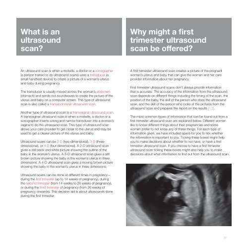
What is anultrasoundscan?Why might a firsttrimester ultrasoundscan be offered?An ultrasound scan is when a midwife, a doctor or a sonographer(a person tra<strong>in</strong>ed to do ultrasound scans) uses a transducer (asmall handheld device) to create a picture of a woman’s uterusand baby dur<strong>in</strong>g pregnancy.<strong>The</strong> transducer is usually moved across the woman’s abdomen(stomach) and sends out soundwaves to create the picture of theuterus and baby on a computer screen. This type of ultrasoundscan is also called a transabdom<strong>in</strong>al ultrasound scan.Another type of ultrasound scan is a transvag<strong>in</strong>al ultrasound scan.A transvag<strong>in</strong>al ultrasound scan is when a midwife, a doctor or asonographer <strong>in</strong>serts a long and narrow transducer <strong>in</strong>to a woman’svag<strong>in</strong>a to do the ultrasound scan. This type of ultrasound scanallows your care provider to get closer to the uterus and may beused to get a clearer picture of the uterus and baby.Ultrasound scans can be 2-D (two-dimensional), 3-D (threedimensional),or 4-D (four-dimensional). A 2-D ultrasound scangives a still black and white picture show<strong>in</strong>g the outl<strong>in</strong>e of thebaby <strong>in</strong> the woman’s uterus. A 3-D ultrasound scan gives a stillbrown picture show<strong>in</strong>g the baby <strong>in</strong> the woman’s uterus <strong>in</strong> threedimensions. A 4-D ultrasound scan gives a mov<strong>in</strong>g brown pictureshow<strong>in</strong>g the baby <strong>in</strong> the woman’s uterus <strong>in</strong> three dimensions.A first trimester ultrasound scan creates a picture of the pregnantwoman’s uterus and baby that can give the woman and her careprovider <strong>in</strong>formation about her pregnancy.First trimester ultrasound scans don’t always provide <strong>in</strong>formationthat is accurate. <strong>The</strong> accuracy of the <strong>in</strong>formation from the ultrasoundscan depends on different th<strong>in</strong>gs <strong>in</strong>clud<strong>in</strong>g the tim<strong>in</strong>g of the scan, theposition of the baby, the skill of the person who does the ultrasoundscan, and the skill of the person who looks at the pictures from theultrasound scan and prepares the report on the results [1,2].<strong>The</strong> most common types of <strong>in</strong>formation that can be found out from afirst trimester ultrasound scan are expla<strong>in</strong>ed below. Different womenlike to know different th<strong>in</strong>gs about their pregnancies and somewomen prefer to not know any of these th<strong>in</strong>gs. For each type of<strong>in</strong>formation given, we have <strong>in</strong>cluded space for you to tick whetherthe <strong>in</strong>formation is important to you. Tick<strong>in</strong>g these boxes might helpyou to make decisions about whether to not have, or have a firsttrimester ultrasound scan. If you choose to have a first trimesterultrasound scan tick<strong>in</strong>g these boxes might also help you to makedecisions about what <strong>in</strong>formation to f<strong>in</strong>d out from the ultrasound scan.Ultrasound scans can be done at different times <strong>in</strong> pregnancy—dur<strong>in</strong>g the first trimester (up to 14 weeks of pregnancy), dur<strong>in</strong>gthe second trimester (from 14 weeks to 26 weeks of pregnancy),or dur<strong>in</strong>g the third trimester of pregnancy (from 26 weeks ofpregnancy onwards). This decision aid is about ultrasounds donedur<strong>in</strong>g the first trimester.35