Autogenous tooth transplantation: Evaluation of pulp tissue ... - RIHUC
Autogenous tooth transplantation: Evaluation of pulp tissue ... - RIHUC
Autogenous tooth transplantation: Evaluation of pulp tissue ... - RIHUC

You also want an ePaper? Increase the reach of your titles
YUMPU automatically turns print PDFs into web optimized ePapers that Google loves.
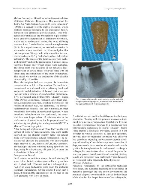
Med Oral Patol Oral Cir Bucal. 2011 Nov 1;16 (7):e984-9.<strong>Autogenous</strong> <strong>tooth</strong> <strong>transplantation</strong>Malmo, Sweden) on 14 teeth, or saline (isotonic solution<strong>of</strong> Sodium Chloride - Paracelsus - Pharmaceutical Industry,SA Porto-Portugal) also on 14 teeth. Emdogain ®(EMD) is a derivative <strong>of</strong> the matrix <strong>of</strong> enamel, whichcontains proteins belonging to the amelogenin family,extracted from embryonic porcine enamel. This productnot only stimulates the proliferation <strong>of</strong> pre-odontoblastsand the differentiation <strong>of</strong> immature osteoblasts,it also has an antibacterial action, due to its pH beingbetween 4 and 5 and effective in <strong>tooth</strong> <strong>transplantation</strong>(8-13). As a negative control, we used saline solution. Itwas used as a local anesthetic, the lidocaine hydrochlorideanhydrous, 20 mg / mL with adrenaline tartrate,corresponding to 12.5 g / ml <strong>of</strong> adrenaline, Adrenalinexylocaine ® . The space <strong>of</strong> the local receptor was evaluatedclinically and on the radiographs. The mesi-distal,vestibular-lingual and vertical spaces were evaluated.The donor <strong>tooth</strong> was measured in the periapical radiographyand an acrylic model <strong>tooth</strong> was made with thesame shape and dimensions <strong>of</strong> the <strong>tooth</strong> to transplant.This model was used in the preparation <strong>of</strong> the alveolarreceptor (Fig. 1A and B).Thus, the recipient bed was prepared for immediate<strong>transplantation</strong> or deferred by ten days. The teeth to betransplanted were cleaned with a polishing brush and<strong>tooth</strong>paste, and disinfection <strong>of</strong> the oral cavity was carriedout with a solution <strong>of</strong> chlorhexidine digluconate,0.1%; clorbutanol hemi-hydrate 0.1% (Eludril ® - PierreFabre Dermo-Cosmétique, Portugal). After local anesthetic,atraumatic extraction, avoiding disruption <strong>of</strong> theroot sheath and root buds, was performed. The extra alveolartime was minimal (less than 5 minutes), in orderto maintain the vitality <strong>of</strong> periodontal ligament. Whereintentional reimplantation was performed, the extraoraltime was longer (about 15 minutes), due to theperformance <strong>of</strong> apicoectomy, for the preparation <strong>of</strong> theapical cavity and placing the sealing material (MTA ® -Mineral Trioxide Agregate).After the topical application <strong>of</strong> SS or EMD on the rootsurface <strong>of</strong> teeth for <strong>transplantation</strong>, they were gentlyinserted into the alveolar, slightly below the oclusalplane to avoid premature oclusal contacts (11). The occlusionwas checked and controlled with interarticularpaper blue/red 60 μm, Bausch KG ® , (Köln, Germany).The setting <strong>of</strong> the teeth was done during a period <strong>of</strong> tendays, using for this purpose, silk yarn 3/0, to cross theocclusal face <strong>of</strong> the <strong>tooth</strong> (14).Postoperative PeriodIn all patients an antibiotic was performed, starting 24hours before the intervention (amoxicillin - 1 gram tablets:1 tablet each 12 hours), and for a subsequent period<strong>of</strong> six days. Indications were given for analgesictreatment (paracetamol, 500 mg tablets: 1 tablet each 6hours, if pain) and the application <strong>of</strong> an ice-pack on theface, protected with fabric or paper.ABFig. 1. Image <strong>of</strong> the model <strong>tooth</strong> made in acrylic resin (A),and periapical radiograph (B), after the socket was made, inthe region <strong>of</strong> the <strong>tooth</strong> 46 (clinical case 3).A s<strong>of</strong>t diet was advised for the 48 hours after the <strong>transplantation</strong>.Chewing with the quadrant was contra-indicatedfor a period <strong>of</strong> seven days. Careful oral hygienewas also recommended, followed by a mouthwash withchlorhexidine digluconate to 1mg/ml (Eludril ® - PierreFabre Dermo-Cosmétique, Portugal), diluted in 5 ml<strong>of</strong> water, to remove the suture, 10 days post-operation.The day after the treatment the patient was observedand questioned regarding the occurrence <strong>of</strong> pain, swellingand bleeding. Control check-ups were done after 10days, one month, three months, six months and annuallyafter the <strong>transplantation</strong>. In each control check-up, aradiographic examination, observation <strong>of</strong> the gums, themarginal groove, dental mobility and sensitivity testingto cold and percussion were performed. These data wereall referenced in the previously defined protocol.Methods <strong>of</strong> AnalysisPeriapical radiographs by the orthogonal techniquewere taken, and evaluated according to the existence <strong>of</strong>periapical pathology, the state <strong>of</strong> root development, thepresence <strong>of</strong> apical closure and the state <strong>of</strong> the hard layer<strong>of</strong> the alveolar. For the performance <strong>of</strong> the radiographs,e986