Autogenous tooth transplantation: Evaluation of pulp tissue ... - RIHUC
Autogenous tooth transplantation: Evaluation of pulp tissue ... - RIHUC
Autogenous tooth transplantation: Evaluation of pulp tissue ... - RIHUC

You also want an ePaper? Increase the reach of your titles
YUMPU automatically turns print PDFs into web optimized ePapers that Google loves.
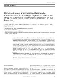
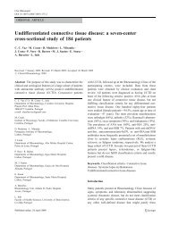
Med Oral Patol Oral Cir Bucal. 2011 Nov 1;16 (7):e984-9.<strong>Autogenous</strong> <strong>tooth</strong> <strong>transplantation</strong>Malmo, Sweden) on 14 teeth, or saline (isotonic solution<strong>of</strong> Sodium Chloride - Paracelsus - Pharmaceutical Industry,SA Porto-Portugal) also on 14 teeth. Emdogain ®(EMD) is a derivative <strong>of</strong> the matrix <strong>of</strong> enamel, whichcontains proteins belonging to the amelogenin family,extracted from embryonic porcine enamel. This productnot only stimulates the proliferation <strong>of</strong> pre-odontoblastsand the differentiation <strong>of</strong> immature osteoblasts,it also has an antibacterial action, due to its pH beingbetween 4 and 5 and effective in <strong>tooth</strong> <strong>transplantation</strong>(8-13). As a negative control, we used saline solution. Itwas used as a local anesthetic, the lidocaine hydrochlorideanhydrous, 20 mg / mL with adrenaline tartrate,corresponding to 12.5 g / ml <strong>of</strong> adrenaline, Adrenalinexylocaine ® . The space <strong>of</strong> the local receptor was evaluatedclinically and on the radiographs. The mesi-distal,vestibular-lingual and vertical spaces were evaluated.The donor <strong>tooth</strong> was measured in the periapical radiographyand an acrylic model <strong>tooth</strong> was made with thesame shape and dimensions <strong>of</strong> the <strong>tooth</strong> to transplant.This model was used in the preparation <strong>of</strong> the alveolarreceptor (Fig. 1A and B).Thus, the recipient bed was prepared for immediate<strong>transplantation</strong> or deferred by ten days. The teeth to betransplanted were cleaned with a polishing brush and<strong>tooth</strong>paste, and disinfection <strong>of</strong> the oral cavity was carriedout with a solution <strong>of</strong> chlorhexidine digluconate,0.1%; clorbutanol hemi-hydrate 0.1% (Eludril ® - PierreFabre Dermo-Cosmétique, Portugal). After local anesthetic,atraumatic extraction, avoiding disruption <strong>of</strong> theroot sheath and root buds, was performed. The extra alveolartime was minimal (less than 5 minutes), in orderto maintain the vitality <strong>of</strong> periodontal ligament. Whereintentional reimplantation was performed, the extraoraltime was longer (about 15 minutes), due to theperformance <strong>of</strong> apicoectomy, for the preparation <strong>of</strong> theapical cavity and placing the sealing material (MTA ® -Mineral Trioxide Agregate).After the topical application <strong>of</strong> SS or EMD on the rootsurface <strong>of</strong> teeth for <strong>transplantation</strong>, they were gentlyinserted into the alveolar, slightly below the oclusalplane to avoid premature oclusal contacts (11). The occlusionwas checked and controlled with interarticularpaper blue/red 60 μm, Bausch KG ® , (Köln, Germany).The setting <strong>of</strong> the teeth was done during a period <strong>of</strong> tendays, using for this purpose, silk yarn 3/0, to cross theocclusal face <strong>of</strong> the <strong>tooth</strong> (14).Postoperative PeriodIn all patients an antibiotic was performed, starting 24hours before the intervention (amoxicillin - 1 gram tablets:1 tablet each 12 hours), and for a subsequent period<strong>of</strong> six days. Indications were given for analgesictreatment (paracetamol, 500 mg tablets: 1 tablet each 6hours, if pain) and the application <strong>of</strong> an ice-pack on theface, protected with fabric or paper.ABFig. 1. Image <strong>of</strong> the model <strong>tooth</strong> made in acrylic resin (A),and periapical radiograph (B), after the socket was made, inthe region <strong>of</strong> the <strong>tooth</strong> 46 (clinical case 3).A s<strong>of</strong>t diet was advised for the 48 hours after the <strong>transplantation</strong>.Chewing with the quadrant was contra-indicatedfor a period <strong>of</strong> seven days. Careful oral hygienewas also recommended, followed by a mouthwash withchlorhexidine digluconate to 1mg/ml (Eludril ® - PierreFabre Dermo-Cosmétique, Portugal), diluted in 5 ml<strong>of</strong> water, to remove the suture, 10 days post-operation.The day after the treatment the patient was observedand questioned regarding the occurrence <strong>of</strong> pain, swellingand bleeding. Control check-ups were done after 10days, one month, three months, six months and annuallyafter the <strong>transplantation</strong>. In each control check-up, aradiographic examination, observation <strong>of</strong> the gums, themarginal groove, dental mobility and sensitivity testingto cold and percussion were performed. These data wereall referenced in the previously defined protocol.Methods <strong>of</strong> AnalysisPeriapical radiographs by the orthogonal techniquewere taken, and evaluated according to the existence <strong>of</strong>periapical pathology, the state <strong>of</strong> root development, thepresence <strong>of</strong> apical closure and the state <strong>of</strong> the hard layer<strong>of</strong> the alveolar. For the performance <strong>of</strong> the radiographs,e986
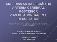
Med Oral Patol Oral Cir Bucal. 2011 Nov 1;16 (7):e984-9.<strong>Autogenous</strong> <strong>tooth</strong> <strong>transplantation</strong>a generated X-ray source was used, a portable devicePort-X II (Genoray Co. Ltd.) producing up to 60 kVp,for a current intensity <strong>of</strong> 2mA. The timer was set to theminimum value <strong>of</strong> 0.03 s. These images were viewed ona computer monitor, using for this purpose, the VixWinPro S<strong>of</strong>tware, version 1.5 (KaVo Dental Gendex DentalSystems, Germany) and a qualitative assessment wasdone. The maintenance <strong>of</strong> the <strong>pulp</strong> vitality and the rootdevelopment occurs when the donor <strong>tooth</strong> joints the openapex at the time <strong>of</strong> being transplanted (14). One <strong>of</strong> thesigns to assess the <strong>pulp</strong> vitality is the consequent partialor total obliteration <strong>of</strong> the canal and the root development(14). For the evaluation <strong>of</strong> root growth, we developeda protocol, as described below, which was adoptedin the controls <strong>of</strong> the teeth with open apex. The periapicalradiographs were evaluated on a computer programNemocef 5.0 ® (Nemotec Dental Systems, Spain). Thisprogram allowed measurements with approximationto a hundredth <strong>of</strong> a millimeter to be made. This waywe obtained values for the length <strong>of</strong> the root (from theamelo-cemental junction to the most apical point <strong>of</strong> theroot). The root growth was calculated by subtracting theinitial length to the final length <strong>of</strong> the root. A correctionfactor was introduced (f), to correct any differencebetween the initial and final radiographs:f = C1: C2, where C1 corresponds to the height <strong>of</strong> thecrown in the initial radiograph and C2 to the crown inthe final radiograph. The height <strong>of</strong> the crown was definedas the distance between the midpoint <strong>of</strong> the line <strong>of</strong>union <strong>of</strong> the amelo-cemental junction and the midpoint<strong>of</strong> the line that joins the tips <strong>of</strong> the cusps (Fig. 2).Then the increment <strong>of</strong> root growth was calculated usingthe following formula:FLR = FMR x fRI = CFR-ILRWhere FLR = final length <strong>of</strong> the root; FMR = final measurement<strong>of</strong> the root; ILR = initial length <strong>of</strong> the rootand RI=root increase (15). It was considered that rootgrowth occurred when there was an increase <strong>of</strong> at least0.5 mm. The cold test was done with a small ball <strong>of</strong>cotton soaked in ethyl chloride, placed on the cervicalthird <strong>of</strong> the dry <strong>tooth</strong>, which was isolated with cottonrolls. Whenever a reference <strong>of</strong> pain existed, its durationwas measured in seconds. For the cold test four gradationswere considered: T0 for the absence <strong>of</strong> response tothe test, T1 for a positive response, which ceases whenthe stimulation ends, T2 for the duration <strong>of</strong> a responsebetween 6 to 9 seconds and T3 for the duration <strong>of</strong> sensitivitysuperior or equal to 10 seconds. The percussiontest was performed with the lead <strong>of</strong> the clinical mirror,exerting a gentle swat on the occlusal surface <strong>of</strong> the <strong>tooth</strong>in evaluation. To test the impact felt P0, for no pain,P1 for the presence <strong>of</strong> pain and P2 for when the percussionsound was metallic.Statistical analysis <strong>of</strong> dataIn this clinical study, we used the Chi-square, as proportionswere compared. The tests were evaluated at asignificance level <strong>of</strong> 0.05.ResultsOf the 28 teeth transplanted, monitored for 24 to 65months (48 ± 12.96; MED ± SD), one <strong>of</strong> these, whichhad the open apex and appeared only for the first controlconsultation, was excluded and 4 were intentional reimplantations.For the remaining 23 an endodontic therapywas performed on 9 teeth (39%), 1 with an open apexand 8 with a closed apex. The remaining 14 teeth werenot subjected to endodontic therapy (Fig. 3).From the results <strong>of</strong> this clinical study, an analytical evaluationwas done, where we found that there were significantdifferences between teeth with an open apex, incomparison to those which had a closed apex. In these,<strong>of</strong> the 11 transplants performed it was necessary to performan endodontic therapy in 8 teeth, corresponding to73% <strong>of</strong> the cases. From the statistical analysis, there isa tendency that the need for endodontic therapy occurssignificantly more <strong>of</strong>ten (p=0.083), although this significanceis not confirmed to the level <strong>of</strong> 5%.On the 12 teeth with an open apex, it was only necessaryto perform an endodontic therapy in one case, correspondingto a value <strong>of</strong> 8% and a statistically significantdifference (p=0.0002).Fig. 2. Method used to analyse root development.C= crown height; CR= length <strong>of</strong> theroot.61%n=23ETHVital39%Fig. 3. Endodontic therapy (ETH), performed on the transplanted.e987
Med Oral Patol Oral Cir Bucal. 2011 Nov 1;16 (7):e984-9.<strong>Autogenous</strong> <strong>tooth</strong> <strong>transplantation</strong>Table 2. Comparison between the length <strong>of</strong> the root and revascularization.Open apex (N=12)Closed apex (N=11)Length <strong>of</strong> the root (mm) 8.07±2.88 10.82±1.89Revascularization n=11 n=3The relationship between the root length and the revascularization,although this occurred more frequently inteeth with short roots, there no statistically significantdifference (p=0.149) (Table 2). The relationship betweenthe two surgical techniques used and the Emdogain ® orsaline solution, there´s no difference.DiscussionStudies on the complications <strong>of</strong> root resorption after<strong>tooth</strong> <strong>transplantation</strong> have been based on models inmonkeys, rats and dogs (4,10). Transplantation to a recipientbed in which the <strong>tissue</strong> was under regenerationdescribed by Nethander et al. (4) in 2003, implies thatthe recipient bed is prepared surgically prior to <strong>transplantation</strong>and allowed to heal for 5 days. In this study,we used transplant teeth after 10 days <strong>of</strong> regeneration.The periodontal ligament has demonstrated a remarkablecapacity for repair and regeneration.Katayama et al. (5) demonstrated in dogs that transplantedproliferating <strong>tissue</strong>s promoted reconstruction<strong>of</strong> a normal periodontium in injured teeth. The effectis probably due to the expression <strong>of</strong> fibroblast growthfactor (FGF) that is a potent mitogen for mesenchymalcells, including bone and periodontal ligament cells(PDL), and also stimulates osteogenic expression <strong>of</strong>stromal bone marrow cells. They demonstrated thatthe mRNA expression <strong>of</strong> FGF and alkaline phosfatase(ALP) increased in proliferating <strong>tissue</strong>. They concludedthat this <strong>tissue</strong> may promote the regeneration <strong>of</strong> PDL<strong>tissue</strong> and prevent ankilosis and root resorption followingthe <strong>transplantation</strong> <strong>of</strong> teeth.This experimental study demonstrated that there wereno differences in the wound healing process betweenone-stage and two-stage techniques in transplantedteeth. From the results obtained we considered the existence<strong>of</strong> <strong>pulp</strong>al necrosis, when the sensitivity tests werenegative, when there was pain on the vertical percussionand when radiographically there were signs <strong>of</strong> inflammatoryreabsorption with the presence <strong>of</strong> a periapicalradio transparent area (15,16).These signals allow us todraw conclusions on performing the revascularizationor not <strong>of</strong> the transplanted <strong>tooth</strong>. On the transplantedteeth that presented an open apex, revascularizationoccurred in 92% <strong>of</strong> cases, which is in agreement withthe literature data (3, 16). It should also be taken intoaccount that the revascularization and <strong>pulp</strong>ar healingare dependent on the diameter <strong>of</strong> the foramen and thelength <strong>of</strong> the root (17).Our results indicate that the shorter the root is(L1mm), the greater the possibility <strong>of</strong> <strong>pulp</strong> revascularization.When during our study clinical signs andradiographic indicators <strong>of</strong> <strong>pulp</strong> or periapical pathologyappeared, we performed endodontic therapy, which occurredin 39% <strong>of</strong> transplants performed. On the otherhand, on the teeth with an open apex it was only necessaryto perform endodontic therapy on 8% <strong>of</strong> them. Althoughour results are in agreement with the literaturean issue that remains controversial is knowing the righttime for the performance <strong>of</strong> endodontic therapy post<strong>transplantation</strong>. Our option was to do it when there wereclinical and radiographic signs compatible with <strong>pulp</strong>necrosis, and not during a constant time interval rightafter the transplant, as suggested by other authors (18).In our favor are also results from animal experiments,which refer that there are no differences in performingthe endodontic treatment on the 15th or 40th days afterthe <strong>transplantation</strong>, since the inflammatory reabsorptionwas not significantly different (19). According toour results and supported by this experimental study,we can say that the performance <strong>of</strong> endodontic treatmentshould be done after the complete healing <strong>of</strong> theperiodontal ligament. This has a great significance andclinical relevance, as a very early endodontic treatment,may find the <strong>tooth</strong> with some mobility, which is uncomfortablefor the patient and for the dentist.Almost all teeth with no root canal filling showed <strong>pulp</strong>obliteration. This can be explained by the fact that <strong>pulp</strong>obliteration increases with time.These results have clinical implications and suggest thatif nonfunctional teeth such as third molars are available,auto<strong>transplantation</strong> can be considered the treatment <strong>of</strong>choice. Auto<strong>transplantation</strong> can be an alternative to dentalimplant in some patients in whom dental implantsbecome impossible due to inadequate bone support andpatients in growing stages.References with links to Crossref - DOIReferences1. Clokie CM, Yau DM, Chano L. <strong>Autogenous</strong> <strong>tooth</strong> <strong>transplantation</strong>:an alternativeto dental implant placement? J Can Dent Assoc. 2001;67:92-6.2. Northway WM, Konigsberg S. Autogenic <strong>tooth</strong> <strong>transplantation</strong>.The “state <strong>of</strong> the art”. Am J Orthod. 1980;77:146-62.3. Andreasen JO, Paulsen HU, Yu Z, Schwartz O. A long-term study<strong>of</strong> 370 autotransplanted premolars. Part III. Periodontal healing subsequentto <strong>transplantation</strong>. Eur J Orthod. 1990;12:25-37.4. Nethander G, Skoglund A, Kahnberg KE. Experimental autoge-e988
Med Oral Patol Oral Cir Bucal. 2011 Nov 1;16 (7):e984-9.<strong>Autogenous</strong> <strong>tooth</strong> <strong>transplantation</strong>nous <strong>tooth</strong> <strong>transplantation</strong> in the dog: a comparison between one- andtwo-stage surgical techniques. Acta Odontol Scand. 2003;61:223-9.5. Katayama A, Ota M, Sugito H, Shibukawa Y, Yamada S. Effect<strong>of</strong> proliferating <strong>tissue</strong> on transplanted teeth in dogs. Oral Surg OralMed Oral Pathol Oral Radiol Endod. 2006;101:e110-8.6. Lindskog S, Pierce AM, Bloml<strong>of</strong> L, Hammarstrom L. The role<strong>of</strong> the necrotic periodontal membrane in cementum resorption andankylosis. Endod Dent Traumatol. 1985;1:96-101.7. Kim E, Jung JY, Cha IH, Kum KY, Lee SJ. <strong>Evaluation</strong> <strong>of</strong> the prognosisand causes <strong>of</strong> failure in 182 cases <strong>of</strong> autogenous <strong>tooth</strong> <strong>transplantation</strong>.Oral Surg Oral Med Oral Pathol Oral Radiol Endod.2005;100:112-9.8. Schwartz Z, Carnes DL Jr, Pulliam R, Lohmann CH, Sylvia VL,Liu Y, et al. Porcine fetal enamel matrix derivative stimulates proliferationbut not differentiation <strong>of</strong> pre-osteoblastic 2T9 cells, inhibitsproliferation and stimulates differentiation <strong>of</strong> osteoblast-like MG63cells, and increases proliferation and differentiation <strong>of</strong> normal humanosteoblast NHOst cells. J Periodontol. 2000;71:1287-96.9. Ninomiya M, Kamata N, Fujimoto R, Ishimoto T, Suryono, KidoJ, et al. Application <strong>of</strong> enamel matrix derivative in auto<strong>transplantation</strong><strong>of</strong> an impacted maxillary premolar: a case report. J Periodontol.2002;73:346-51.10. Spahr A, Lyngstadaas SP, Boeckh C, Andersson C, PodbielskiA, Haller B. Effect <strong>of</strong> the enamel matrix derivative Emdogain onthe growth <strong>of</strong> periodontal pathogens in vitro. J Clin Periodontol.2002;29:62-72.11. Reich PP. <strong>Autogenous</strong> <strong>transplantation</strong> <strong>of</strong> maxillary and mandibularmolars. J Oral Maxill<strong>of</strong>ac Surg. 2008;66:2314-7.12. Jiang J, Goodarzi G, He J, Li H, Safavi KE, Spångberg LS, etal. Emdogain-gel stimulates proliferation <strong>of</strong> odontoblasts and osteoblasts.Oral Surg Oral Med Oral Pathol Oral Radiol Endod.2006;102:698-702.13. Hamamoto Y, Takahashi K, Sakurai H, Akiba K, Izumi N, KanohH, et al. The use <strong>of</strong> enamel matrix derivative (Emdogain) for improvement<strong>of</strong> probing attachment level <strong>of</strong> the autotransplanted teeth.Dent Traumatol. 2005;21:336-40.14. Nasjleti CE, Castelli WA, Caffesse RG. The effects <strong>of</strong> differentsplinting times on replantation <strong>of</strong> teeth in monkeys. Oral Surg OralMed Oral Pathol. 1982;53:557-66.15. Bauss O, Schilke R, Fenske C, Engelke W, Kiliaridis S. Auto<strong>transplantation</strong><strong>of</strong> immature third molars: influence <strong>of</strong> different splintingmethods and fixation periods. Dent Traumatol. 2002;18:322-8.16. Andreasen JO, Borum MK, Jacobsen HL, Andreasen FM. Replantation<strong>of</strong> 400 avulsed permanent incisors. 2. Factors related to<strong>pulp</strong>al healing. Endod Dent Traumatol.1995;11:59-68.17. Gault PC, Warocquier-Clerout R. Tooth auto-<strong>transplantation</strong> withdouble periodontal ligament stimulation to replace periodontallycompromised teeth. J Periodontol. 2002;73:575-83.18. Skoglund A, Tronstad L, Wallenius K. A microangiographicstudy <strong>of</strong> vascular changes in replanted and autotransplanted teeth <strong>of</strong>young dogs. Oral Surg Oral Med Oral Pathol. 1978;45:17-28.19. Azevedo PC, Moura CC, Zanetta-Barbosa D, Bernadineli N.Time <strong>of</strong> endodontic treatment in autogenic transplants <strong>of</strong> matureteeth: histological study in dogs. Oral Surg Oral Med Oral PatholOral Radiol Endod. 2007;104:287-93.e989