Permanent Mycoplasma Removal from Tissue Culture Cells: A ...
Permanent Mycoplasma Removal from Tissue Culture Cells: A ...
Permanent Mycoplasma Removal from Tissue Culture Cells: A ...

Create successful ePaper yourself
Turn your PDF publications into a flip-book with our unique Google optimized e-Paper software.
Biotechnol. Bioprocess Eng. 2000, Vol. 5, No. 2 85and thaw a sterile back-up stock vial. In instances,however, where no stock is available and mycoplasmafree cells can not be replaced by other sources, attemptsneed to be made to rid the cells of the mycoplasma.Different methods have been employed in order toremove mycoplasma <strong>from</strong> tissue culture cells [15]. Theseinclude hypotonic treatment, exposure to elevated heat,the use of mycoplasma-specific anti-serum [16], and,most commonly, the application of antibiotics [6].A wide variety of antibiotics have been demonstratedto be lethal, even at very low concentrations, to individualmycoplasma grown in broth media [17]. Whenassociated with tissue culture cells, however, mycoplasmaoften are protected [26], requiring much higherantibiotic concentrations for successful elimination. Inthis instance, at the concentration necessary to eliminatemycoplasma, the antibiotic often becomes toxic tothe host cells also [15,16,18]. Moderate antibiotic concentrationstolerable to the host cell often suppress mycoplasmagrowth temporarily but do not lead to successfulpermanent elimination [18,19,26]. Upon removalof the antibiotic, mycoplasma become detectable again.In addition, moderate antibiotic concentrations werereported to lead to the selection and outgrowth of mycoplasmasubstrains highly resistant to the antibioticapplied [15,20,21].A variety of toxic agents specifically designed for mycoplasmaremoval are commercially available [3], includingthe <strong>Mycoplasma</strong> <strong>Removal</strong> Agent (ICN/FlowLaboratories), BM Cyclin (Boehringer Mannheim), andAnti-PPLO Agent (Gibco). These cures meet with onlylimited success [3].Here, a genetic approach for the elimination of mycoplasma<strong>from</strong> tissue culture cells is described. Ninetyfivepercent of mycoplasma contaminants in tissue culturereportedly are caused by only four mycoplasmaspecies, one of which is Acholeplasma laidlawii [2]. Acholeplasmacommonly are introduced into tissue culturecells by contaminated bovine serum [1]. In this study,baby hamster kidney BHK-21 cells infected by Acholeplasmalaidlawii were thus chosen as the model system.The infected cells were genetically manipulated so as totemporarily tolerate antibiotic concentrations highenough to be lethal to associated mycoplasma whileleaving the host cells unharmed. Withdrawal of theantimicrobial agent and subcultivation of the curedhost cells led to the identification and subsequent isolationof cell clones that had lost the foreign DNA previouslyconferring increased antibiotic tolerance to thecell. Thus, the cells were cured yet remained biochemicallyunaltered.MATERIALS AND METHODS<strong>Mycoplasma</strong> DetectionRoutinely, three different tests were employed.DNA detectable in the cytoplasm of cells is a strongindication of mycoplasma contamination and was assayedemploying a DNA fluorochrome staining kit (Seromed/Biochrom,Germany, cat. no. D 7001). The tissueculture cells to be tested were grown to 30% confluencyon slideflasks (Nunc, Denmark, cat. no. 170920). Subsequently,the cells were treated according to the manufacturer'sinstructions.For the Hybricomb test (Biological Industries, Israel;cat. no. MTK-12), the cells to be tested were seeded ona tissue culture petri dish at low density so as to reach30% confluency within four days of incubation withoutrequiring any further medium change. Thereafter, analiquot of the supernatant was processed according tothe manufacturer’s description. The Hybricomb test isbased on specific mycoplasma DNA hybridization.Isolation of mycoplasmas <strong>from</strong> infected tissue culturecells and subsequent in vitro cultivation was performedas follows:10 mL of 96 h conditioned medium <strong>from</strong> confluentcells were inoculated into 100 mL of <strong>Mycoplasma</strong> LiquidMedia BT1/1 and BT2/1 (Amimed/Biotrade, Austria),respectively. Subsequently, the samples were incubatedfor 14 days at 37 o C, 5% CO 2in a regular cell cultureincubator, in order to allow for growth/division ofmycoplasma potentially present. After 3, 7, and 14 daysof incubation, 800 µL were taken <strong>from</strong> the incubationreactions and spread onto two BT3 and two BT4 <strong>Mycoplasma</strong>agar plates. These media were prepared byautoclaving 35 g of PPLO agar (Difco, USA; cat. no.B412) in 1,000 mL H 2O, and, thereafter, adding 400 mLof either <strong>Mycoplasma</strong> Nutrient Component BT3 or BT4(Amimed/Biotrade, Austria) prior to solidification.One plate was incubated aerobically and the other anaerobicallyfor three weeks. They were regularly inspectedmicroscopically for growth and the presence of mycoplasma,which exhibit a typical ‘fried egg’ appearance.<strong>Tissue</strong> <strong>Culture</strong><strong>Mycoplasma</strong> negative BHK-21 cells (ATCC CCL 10;BHK/Myco−cells) were routinely grown in DMEM:Ham's F12 medium (BioWhittaker, Belgium; cat. no. 12-719F) supplemented with 10% fetal calf serum, 2 mMglutamine, and 0.075% bicarbonate. Acholeplasma laidlawii[21] infected BHK-21 cells (BHK/Myco+) weregrown under identical conditions.Plasmids, Transfection and Selection1 µg pUCSV-Neo [22], in combination with 4 µgpUCSV-dhfr where applicable, was introduced into 30%confluent cells on a 5 cm tissue culture petri dish by theCaPO 4coprecipitation technique essentially as described[23]. pUCSV-dhfr was constructed by cloning theKpnI/SalI fragment <strong>from</strong> pNUT [24] into analogouslydigested pUC19. The volume of the DNA containingsolution was adjusted to 250 µL with 1 mM Tris/Cl pH8.0, 0.1 mM EDTA, and 25 µL 2.5 mM CaCl 2wereadded. This solution was added to 250 µL 2xHBS (280mM NaCl, 2.5 mM Na 2HPO 4, 50 mM Hepes/NaOH pH7.12), mixed, and spread onto the cells. After three hours,the medium was removed, and 1 mL of PBS containing15% glycerol added. After one minute, the PBS/ glycerolwas removed by aspiration, the cells were thoroughlyrinsed twice with PBS, and fresh medium was added.Two days after transfection, the cells were split intolarge tissue culture petri dishes at different densities,
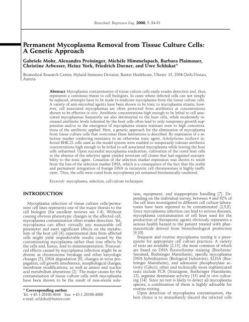
86 Biotechnol. Bioprocess Eng. 2000, Vol. 5, No. 2and either 1 mg G418 (Gibco, USA; cat. no. 11811-049;10 mg G418/mL PBS sterile filtered stock solution) or,in the case of double selection, 0.5 mg G418/mL mediumand 400 nM methotrexate (Sigma, cat. no. M 8407)were added. Approximately two weeks post transfection,cell clones became clearly visible whereas unmanipulatedcells did not survive under these conditions.The cell clones were trypsinized, and seeded ontonew petri dishes. For continued passage, these cellswere grown either in the presence or absence of G418.For isolation of individual cell clones, cells were splitinto tissue culture dishes at very low densities. Individualcolonies that arose were transferred, by means oftrypsin/EDTA saturated cotton swabs, to 24 well dishes,grown to confluency, and split equally into 6 welldishes in the absence and presence of G418 as describedin the text.PCR Testing for Foreign DNAIn order to test for the presence of the selectionmarker plasmid in the cells by PCR, cellular DNA wasprepared essentially as described [25]: 5 × 10 5 cells werepelleted, 50 µL of 2× lysis buffer (with Proteinase K)added and incubated for 4 h at 56 o C, yielding 100 µLlysate. Thereafter, Proteinase K was inactivated by incubatingthe sample for 10 min at 95 o C, followed byvigorous shaking of the sample for 5 mins at 95 o C. Sampleswere stored at -20 o C. PCR was performed with 1µL lysate in a 100 µL reaction consisting of 1 × PCRbuffer (Boehringer Mannheim), 200 µM of dATP, dGTP,dTTP, and dCTP each, 1 µM of each primer, and in thepresence of 1 µL Taq polymerase (Boehringer Mannheim).The PCR primers were 5′-GACGTTGTCACTG-AAGCGGGAA-3′ and 5′-TCACGGGTAGCCAACGCTA-TGTC-3′, and the PCR cycle consisted of 1 min at 55 o Cfor annealing, 1 min at 72 o C for extension, and 1 min at95 o C for denaturation. Thirty cycles were run.RESULTSThe goal of this study was to investigate whethersimple genetic manipulation of the infected cell couldconfer sufficient antibiotic resistance to the host cell,such that these cells would tolerate high concentrationsof the antibiotic without suffering any adverse effects,while simultaneously ensuring the permanent eliminationof mycoplasma. Since, by conventional antibioticsusceptibility determination of mycoplasma in vitro, thedeoxystreptamine group of antibiotics (e.g. hygromycinB, kanamycin, and neomycin) had previously beenshown to be lethal to a wide variety of mycoplasmastrains [6,17,21,27], neomycin phoshotransferase waschosen as a model selection marker. Neomycin phosphotransferasemediates resistance to neomycin andkanamycin in bacteria as well as to the neomycin analogG418/geneticin in tissue culture cells. The latterselection system is popularly used in many laboratoriesfor the establishment of stably transfected cell lines,hence widely available, and many experimentors arefamiliar with its use. BHK/Myco+ cells, harboring Acholeplasma laidlawii,were shown to test positive for mycoplasma by DNAstaining, hybridization, and in vitro culturing. <strong>Mycoplasma</strong>free BHK (BHK/Myco−) cells tested negative inall three tests. These cells were passaged and served asthe positive and negative controls for subsequent tests.Plasmid pUCSV-Neo, mediating expression of thebacterial neomycin phosphotransferase, was introducedinto BHK/Myco+ cells (Fig. 1(A)). Stably transfectedcolonies resistant to 1 mg G418/mL were selected; cellsthat had not taken up the plasmid died. Approximately200 colonies were pooled and grown to confluency. After18 cell divisions post transfection under permanentexposure to G418 selection, the cells were subjected tomycoplasma detection. In the three tests employed, thecells were found to be negative (Fig. 1(G)). The generationtime of untransfected cells (in the absence of G418)was found to be equal to that exhibited by the transfectedcells resistant to G418 when cultivated in thepresence of G418. This finding shows that the transfectedcells grew well in the presence of the high G418concentration.In order to determine whether the inability to detectmycoplasma was only due to the suppression of mycoplasmagrowth below the limits of detection or, rather,to a permanent eradication, the cells were subsequentlycultivated for 19 cell divisions in the absence of selectionpressure. Thereafter, the cells still tested negativefor mycoplasma (Fig. 1(I)). However, after passaging thecells for an additional 11 cell divisions without G418,they were found to be mycoplasma positive again (Fig.1(B)). Thus, mycoplasma recurrence had occurred 30cell divisions after omission of the selective agent G418.This finding indicated that exposure to G418 for 18generations was not sufficient to permanently removemycoplasma. Rather, this temporary exposure of mycoplasmato G418 had only resulted in growth suppressionto the extent that allowed them to evade detection.Historically, it has been a matter of dispute when acell was considered cured. Our observation of mycoplasmarecurrence after 30 cell divisions in the absenceof the antimicrobial agent confirms the necessity for a30 cell division post-treatment follow-up testing, as hasbeen suggested by some reports [2,15].Transfected cells permanently cultivated in selectivemedium for 110 cell divisions after transfection remainedmycoplasma negative (Fig. 1(F)). <strong>Cells</strong> exposedto G418 for a prolonged period of time after transfection,i.e. 53 generations (Fig. 1(H)), tested mycoplasmanegative when cultivated for additional 64 cell divisionswithout G418 (Fig. 1(C)). Since growth-suppressed mycoplasmain tissue culture had been shown to regaindetectability at approximately 30 cell divisions afterwithdrawal of the selective agent (see above), this findingsuggests that the mycoplasma were, in fact, permanentlyeliminated <strong>from</strong> the transfected infected cellswhen exposed to the toxic agent for 53 cell divisionsprior to its withdrawal.Thus, expression of an antibiotic resistance marker ininfected cells allows exposure to antibiotic concentra-
Biotechnol. Bioprocess Eng. 2000, Vol. 5, No. 2 87tions high enough to lead to the permanent eradicationof mycoplasma while ensuring host cell survival.Genetic Instability of the Transfected ResistanceMarker GeneExposure to the toxic agent selects for those cells retainingthe resistance marker expression independent ofwhether the corresponding genetic information is stablyor instably maintained in the cell population. Uponremoval of selection pressure, expression of the foreignprotein often ceases quickly unless the heterologousgenetic information has integrated stably into a hostcell chromosome. Foreign genetic information isthought to be genetically labile when present episomallyin the nucleus, or when integrated within a structurallyinstable chromosomal locus [28- 30].When equal numbers of cells, having undergone 18cell divisions in the presence and 30 cell divisions in theabsence of the toxic agent (Fig. 1(B)), were cultivatedfor an additional 30 cell divisions either in the presenceor absence of G418 (Fig. 1(E) and (D)), twice as manycell clones were present in the medium lacking G418.This finding suggested potential genetic instability ofthe selection marker in the transfected BHK/Myco+cells: when grown in the presence of the toxic agent, allcells having lost the vector harboring the selectionmarker gene died, resulting in a reduced number ofclones (Fig. 1(E)). In contrast, in the absence of thetoxic agent, cells survive even when they have lost theselection marker plasmid.Rescue of Cured Albeit Antibiotic SusceptibleRevertant <strong>Cells</strong>Fig. 1. Eradication of mycoplasma <strong>from</strong> infected cells by useof a selection marker plasmid and subsequent selection. Acholeplasmalaidlawii contaminated BHK-21 cells were transfectedwith the plasmid pUCSV-Neo, which mediates resistance tothe selective agent G418 (A). Stably transfected cells wereselected (G) and subsequently cultivated either in the presence(H) or absence (I) of G418. Although mycoplasma couldnot be detected after the host cells had been exposed to G418for 18 cell divisions (G), the mycoplasma had not been eliminated,but could be readily detected after an additional 30host cell divisions in the absence of G418 (B). <strong>Cells</strong> seeded,after initial selection and subsequent omission of G418 (I),into medium lacking the selective agent yielded twice asmany clones (D) compared to the number of clones detectedin medium containing G418 (E). Similarly to cells cultivatedfor 110 divisions in medium harboring G418 (F), cells initiallyexposed to G418 for 53 generations post transfection (H) andpassaged for 64 cell divisions in the absence of the toxic agentthereafter remained mycoplasma negative (C). Large circlesrepresent cells at individual time points during anti-mycoplasmatreatment. The distance between the cells is proportionalto the number of cell divisions. ‘+’ and ‘−’ in squaresrepresent positive and negative Acholeplasma laidlawii (A.l.)/mycoplasma test results, and cultivation of cells in the presenceand absence of G418 when in small circles.The permanent expression and presence of the selectionmarker protein in the cured cells may influenceintracellular processes directly or indirectly and, thus,might be undesirable in certain instances. If, after successfulmycoplasma eradication, the absence of the selectionmarker protein is required, the fact that stableintegration of foreign DNA into the host cell chromosomesoccurs very inefficiently can be exploited. Giventhe potential genetic instability of the resistance markergene, as described above, it should be possible to identifyindividual cell clones having lost neomycin phosphotransferaseexpression after successful anti-mycoplasmatreatment.BHK/Myco+ cells cotransfected with vectors pUCSV-Neo and pUCSV-dhfr, mediating resistance towardsG418 and methotrexate (MTX), were used in order toinvestigate the postulated loss of the selection markerexpression vector (Fig. 2(A)). Based on the previous experiment,exposure of the transfected cells to the toxicagent for 53 cell divisions is considered sufficient forcomplete mycoplasma eradication (Fig. 1(C)).Stable anchorage of foreign DNA in the host cell isgenerally known to increase with extended cultivationin the presence of the selective agent. In order to investigatewhether, even under stringent conditions (i.e.even upon cultivation in the presence of G418 in significantexcess to 53 generations) cell clones having lostthe resistance marker DNA upon permanent myco-
88 Biotechnol. Bioprocess Eng. 2000, Vol. 5, No. 2 !"#$%Fig. 2. Isolation of cell clones susceptible to the selectiveagent upon withdrawal of the latter after anti-mycoplasmatreatment. Acholeplasma laidlawii infected BHK-21 cells werecotransfected with plasmids pUCSV-Neo and pUCSV-dhfr,me-diating resistance against G418 and increased concentrationsof methotrexate (MTX), respectively. Transfected cellswere selected (F) and subsequently cultivated in the absenceof the selective agents (B). Individual clones were isolated (C)and tested for their ability to grow either in the presence (E)and absence (D) of the toxic agents. Large circles representcells at individual time points during anti-mycoplasma treatment.The distance between the cells is proportional to thenumber of cell divisions. ‘+’ and ‘−’, in the squares representpositive and negative Acholeplasma laidlawii(A.l.)/mycoplasma test results, and cultivation of cells in thepresence and absence of G418 when in small circles.plasma eradication could be identified, the transfectedBHK/Myco+ cells were exposed to G418/MTX for 80cell divisions. As expected, mycoplasma could not bedetected in the pool of resistant cells (Fig. 2(F)). Thereafter,the selective agents were omitted. After additional50 cell divisions, the cells were split in order to allow forclone isolation (Fig. 2(B)). Individual subclones wereisolated (Fig. 2(C)) and cultivated in medium eitherlacking any toxic agents (Fig. 2(D)) or containing G418(Fig. 2(E)). Several clones were shown to be able togrow only in the absence of G418 (Fig. 2(D)). Whenassayed for the presence of mycoplasma, these clonestested negative, even after cultivation for 73 cell divisionsin the absence of selective agents, and thus weredemonstrated to be cured <strong>from</strong> mycoplasma. Employingthe DNA-staining detection test, a representativeclone, cured <strong>from</strong> mycoplasma and incapable of growingin the presence of G418, is shown in Fig. 3(D). TheFig. 3. <strong>Mycoplasma</strong> detection by DNA staining: mycoplasmafree, contaminated, and permanently cured BHK cells. (A)Acholeplasma laidlawii infected BHK-21 cells; (B) transfectedcells constantly cultivated in the presence of the selectiveagents; (C) sterile BHK cells; (D) a cured BHK cell clone, aftertreatment for permanent mycoplasma eradication and subsequentprolonged cultivation in the absence of the selectiveagent, resulting in loss of resistance (corresponding to theclones in Fig. 2(D)). BHK/Myco +, −, mycoplasma infected andsterile BHK-21 cells; pUCSV-Neo/dhfr +, transfected withselection marker plasmids pUCSV-Neo and pUCSV-dhfr andselected thereafter; G418/MTX, cultivated either in the permanentpresence (+) or, after initial presence, upon omissionof G418/methotrexate. The large arrow depicts a nucleus; thesmall arrows point at intracellular fluorescence typical to mycoplasmalocated in the cytoplasm.control cells used in this assay were mycoplasma infected(Fig. 3(A)) and mycoplasma free (Fig. 3(C)) cells,as well as treated cells permanently exposed to the selectiveagents (Fig. 3(B)).This finding demonstrated that, upon successful mycoplasmaeradication, cell clones could be identifiedwhose selection marker expression had ceased.Antibiotic Sensitivity Revertance as the Result ofLoss of the Antibiotic Resistance MediatingExpression VectorIn order to test whether clones exhibiting G418 sensitivityafter mycoplasma eradication had regained sensitivityto G418 as a consequence of the loss of the resistancemarker gene, rather than due to simple cessationof expression by other cellular mechanisms (e.g. transcriptionalsilencing subsequent to methylation), polymerasechain reaction (PCR) analysis was performedwith four clones. Total cell lysates were prepared, andprimers specific for the neomycin phosphotransferase
Biotechnol. Bioprocess Eng. 2000, Vol. 5, No. 2 89gene in pUCSV-Neo were used for amplification. Thesize of the fragment to be amplified was expected to be437 base pairs.PCR of DNA derived <strong>from</strong> transfected BHK cells,permanently exposed to G418/MTX for 153 generationsand used as a control (Fig. 3(B)), exhibited the expectedPCR product (Fig. 4, lane 6 ‘Positive Control II’).Similarly, this fragment could be detected by PCR reactionsemploying either the neomycin resistance mediatingplasmid alone (Fig. 4, lane 2 ‘Plasmid Positive ControlI’) or a mycoplasma negative BHK lysate to whichthe plasmid DNA was added (Fig. 4, lane 7 ‘PositiveControl III’). In contrast, a PCR fragment was not obtained<strong>from</strong> mycoplasma negative (Fig. 4, lane 5 ‘NegativeControl II’; Fig. 3(C)) or <strong>from</strong> mycoplasma positivebut untreated BHK cells (Fig. 4, lane 4 ‘Negative ControlIII’; Fig. 3(A)). Likewise, no fragment was detectedby PCR in the four cell clones that originally had beenmycoplasma positive but were considered cured upontreatment and shown to have regained G418 susceptibilityupon prolonged omission of the selective agents(Fig. 4, lanes 8-11). These findings strongly suggest thatloss of G418 resistance resulted <strong>from</strong> the loss of the expressionmarker DNA previously mediating the resistanceto G418.Assuming the presence of one neomycin phosphotransferaseplasmid per cured cell, cultivated in thepermanent presence of the selective agents, the resultingPCR band was expected to exhibit an intensity correspondingto that shown in the PCR reaction employing20 fg pUCSV-Neo plasmid (Fig. 4, lane 2). The PCRreaction in these cells, however, revealed quantities ofthe PCR fragment (Fig. 4, lane 6) slightly less than thatshown by 100 fg plasmid (Fig. 4, lane 7). 100 fg plasmidcorresponds to 2.5 × 10 4 copies of the neomycin phosphotransferasegene, whereas 1 µL of lysate is derived<strong>from</strong> <strong>from</strong> 5 × 10 3 cells. Thus, the experimental datasuggest that each G418 resistant cell contains approximatelyfour (i.e. slightly less than five) copies of theresistance marker plasmid.DISCUSSIONFig. 4. Detection of selection marker vector DNA in mycoplasmacontaminated, free, and treated BHK-21 cells. PCRreactions for the amplification of a DNA fragment, 437bp inlength and specific for the expression vector conferring G418resistance to the cells, were performed in the lysates of mycoplasmafree, mycoplasma contaminated, and ‘cured’ cells aswell as in appropriate control reactions (lanes 2-11). 1, DNAmolecular weight marker; 2, 20 fg selection marker vectorDNA (pUCSV-Neo); 3, No DNA added; 4, mycoplasma infectedBHK cell lysate; 5, sterile BHK cell lysate; 6, lysate <strong>from</strong>mycoplasma positive cells after treatment and grown in thepresence of the selective agent; 7, mycoplasma free cell lysatesupplemented with 100 fg selection marker plasmid DNA; 8 -11, lysates <strong>from</strong> clones of infected cells, exhibiting regainedselective agent susceptibility upon successful treatment andsubsequent cultivation in the absence of the selective agent.When cultured in broth media, mycoplasma are verysensitive to a wide range of antimicrobial agents. Uponinfection of tissue culture cells, however, cell associatedmycoplasma often are protected <strong>from</strong> these agents. Theconcentration of drugs, e.g. kanamycin and neomycin,necessary to permanently eliminate mycoplasma <strong>from</strong>tissue culture cells frequently is toxic for the cells [31].Antibiotic concentrations low enough to be toleratedby the cell often are reported to suppress mycoplasmagrowth only temporarily (rather than causing their eradication);in many cases, e.g. when Acholeplasma laidlawiiis exposed to neomycin, mutant strains occurwhich have developed resistance to high concentrationsof the antibiotic in use [21].In this report, a genetic approach of mycoplasma eradication<strong>from</strong> tissue culture cells is described. Transfectionof the neomycin phosphotransferase gene confersresistance against high concentrations of the antimicrobialagent G418, a neomycin analog, to the host cells.Thus, the cells can be exposed to antibiotic concentrationshigh enough to permanently eliminate mycoplasmawhile simultaneously tolerating high concentrationsof the antibiotic chosen without suffering <strong>from</strong>any adverse effects.Exposure of mycoplasma infected baby hamster kidneycell line BHK-21, chosen as the model system cells,to G418 for 18 cell divisions after transfection suppressedgrowth of cell associated mycoplasma belowthe limit of detection. However, this period of time wasnot sufficient for permanent mycoplasma removal, sincemycoplasma reccurrence was detected after growth ofthe cells in the absence of G418 for an additional 30 cell