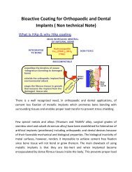
View details... - IFGL Bio Ceramics Limited
View details... - IFGL Bio Ceramics Limited
View details... - IFGL Bio Ceramics Limited

Create successful ePaper yourself
Turn your PDF publications into a flip-book with our unique Google optimized e-Paper software.
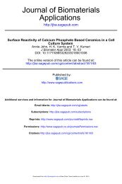
journalJ. Am. Ceram. Soc., 86 [2] 357–59 (2003)Synthesis of Nanosized and Microporous Precipitated Hydroxyapatite inSynthetic Polymers and <strong>Bio</strong>polymersArvind Sinha, Suprabha Nayar, and Archana AgrawalNational Metallurgical Laboratory, Jamshedpur 831007, IndiaDebasish BhattacharyyaIndian Institute of Chemical <strong>Bio</strong>logy, Kolkata 700 032, IndiaPatcha RamachandraraoBanaras Hindu University, Varanasi 221005, IndiaIn situ precipitation of microporous and nanosized hydroxyapatiteparticles (5–40 nm) has been conducted in poly(vinylalcohol) and bovine serum albumin gels. The process, which issimilar to biomineralization, is highly controlled with respectto microstructural features, such as size and shape, and toprecipitation of hydroxyapatite phase having a calcium:phosphorusstoichiometric ratio of 1.67. Nanosized precipitatedhydroxyapatite particles show remarkable thermal stabilityand do not decompose to other calcium phosphate phases, evenat higher temperatures.I. IntroductionOUT of the six principal calcium salts of orthophosphoric acid,precipitated hydroxyapatite (PHA) is most similar to theapatite present in bone, and it is known for its excellent biocompatibilityand nontoxicity. 1 PHA is the as-precipitated phase ofhydroxyapatite (HA) in aqueous solution, and it normally exists ina poorly crystalline state. 2 Several techniques based on wet, dry,hydrothermal, and alkoxide methods have been developed for thesynthesis of PHA and HA. 3–6 However, the chemistry of PHA isvery complex, and, when attempts have been made to precipitate itin solution, uncontrolled agglomerates of apatite products thathave a calcium:phosphorus ratio ranging from 1.5 to 1.66 havebeen produced. 7 Apatite structures are normally characterized bythe presence of channels along their hexagonal axes, and ions inthese channels are less rigorously bound in comparison with thosein the bulk. 8 This results in many specific structural, thermal, andchemical properties associated with PHA that makes the materialsuitable for bone implantation and for chromatographic separationas catalysts, ion exchangers, and drug-delivery systems. 9Nature produces a variety of minerals in nano and bulk sizeswith an optimum degree of crystallinity and porosity through theprocess of biomineralization. Recent experiments have revealedbiomimetics (polymer matrix mediated in situ synthesis of inorganicmaterials) as a powerful tool for the synthesis of monodispersednanoparticles and hierarchical structures produced by theself-assembly, self-construction, and self-organization of basicbuilding components. 10,11 In the present manuscript, we discuss aR. Condrate—contributing editorManuscript No. 186907. Received June 17, 2002; approved September 4, 2002.biomimetic approach for the synthesis of monodispersed nanosizedPHA particles and microporous polymer–HA compositeswith a calcium:phosphorus molar ratio of 1.67. The supramolecularmatrices used are bovine serum albumin (BSA) and poly(vinylalcohol) (PVA). This simple process provides nanosized andporous PHA–bio/synthetic polymer composites that can be directlyused for various orthopedic applications.II.Experimental ProcedureIn situ synthesis of nanosized and microporous HA particleswas conducted using gels of PVA and BSA, respectively. Two 54mL sets of freshly prepared 0.4 M calcium nitrate tetrahydratesolution were made alkaline using 11 mL of 1:2 ammonia:watersolution. A 70 mL aqueous solution of 0.1g of BSA (i.e., 0.1%Fraction V powder, SRL, India) in 100 mL of double-distilledwater was added to one of the above sets, and 70 mL aqueoussolution of 4% PVA was added to the other set. The calciumnitrate–BSA system was gently stirred to avoid denaturation of theprotein, but the calcium nitrate–PVA system was vigorouslystirred for 10 min, and both solutions were poured into Petri dishesand allowed to form transparent gels by incubating at 75°C in ahot-air oven for 24 h. A 77 mL amount of 0.156 M diammoniumhydrogen phosphate was made alkaline using 7 mL of 1:1ammonia:water, and this was added to the above gels. Milky whitecoloration was observed almost instantaneously, and the gels wereallowed to stand for 48 h, after which they were decanted, and themineralized gels washed thoroughly with deionized water. The HAformed was structurally characterized using scanning electronmicroscopy (SEM; Model JSM-840 A, JEOL, Tokyo, Japan),transmission electron microscopy (TEM; Model CM 200, Philips,Eindhoven, The Netherlands), X-ray diffractometry (XRD; ModelPTS 3003, Seifert), Fourier-transform infrared spectroscopy(FTIR), and differential thermal analysis (DTA, Model A-320,Seiko) techniques.III.Results and DiscussionThe applications of synthetic polymers and biopolymers aspreorganized matrices for controlled nucleation and growth ofminerals is of paramount importance in biomimetic studies. 12Gelation of PVA solution under optimum conditions leads to theformation of a noncovalent spatial network. The major types ofintermolecular links in the junction knot are hydrogen bondingbetween –OH groups of neighboring polymer chains. Syndiotacticsites in the chains are responsible for the formation of hydrogen357
358 Communications of the American Ceramic Society Vol. 86, No. 2Fig. 1.XRD pattern of nanosized HA particles in (a) PVA gel and (b) BSA gel.bonds, while the isotactic sites participate in intermolecular interactionsto sterically entrap cations. 13 Similarly, BSA is a globularprotein that has nanosized cavities in its native folded form. Thespecific spatial chemistry of nanosized cavities present in thequaternary structure of BSA attracts cations through interfacialmolecular recognition. 14 Based on their conformation, the aboveorganic systems dictate size, morphology, and orientation ofinorganic particles and kinetically control the nucleation andgrowth process.The formation of PHA phases in PVA and BSA has beenconfirmed using XRD. Characteristic (002), (112), (211), and(212) reflections in the diffraction pattern of the calcium phosphatein situ synthesized in PVA confirms the formation of the HA phase(Fig. 1(a)). An underlying broadening of peaks reflects thenanometer range of the particle size. Peak analysis of the (211)reflection (scanned at 0.5° min 1 ) using the Scherer formula (L 0.9/B cos , where is the wavelength of the X-ray, B the fullwidth at half maximum, and the Bragg angle) predicts an averageparticle size of 5 nm embedded in the PVA matrix. The analysis,however, does not take into account the instrumental and straininducedcomponents of the peak broadening. Similarly, the XRDpattern of PHA precipitated in BSA exhibits characteristic reflectionsfrom (002), (102), (211), (300), (202), and (301) planes of thepure HA phase (Fig. 1(b)). The Bragg peaks obtained in thissample are relatively sharper with respect to the particles producedin the PVA matrix. Peak width analysis of a slow-scanned (211)reflection of the diffraction pattern indicates an average size of 25nm, as compared with 35 nm obtained by plotting the sizedistribution curve based on TEM micrographs. No decrease in thepeak broadening can be observed, even after storing the sample for30 d. This indicates the absence of aggregation-based growth ofnanosized particles in the organic matrix.SEM studies of PHA–PVA composite film manifest the presenceof regularly arranged and interconnected uniform-sized poresof the order of 1–2 m (Fig. 2(a)), a requirement of the osteoblastprocess attributed to the preorganized porous PVA matrix. Thelimited solubility of PVA in water makes it possible to form amicellelike structure that leads to the huddling of hydrophilicgroups to form tubular conformations that minimize the surfaceenergy. As a result of solvent loss, the tubules assemble themselvesinto a long-range-ordered structure under mild conditions ofgelation. 15,16 During gelation, the present calcium ions stericallyentrap on the diffused surface of the PVA, transform the texture ofthe gel from regularly arranged tubular array to a fibrillikeinterconnected porous structure. These cations then react withphosphate ions at alkaline pH to precipitate the apatite phase,which keeps the porous network intact. A high nucleation rate ofapatite particles followed by a very slow growth rate in highlyviscous PVA matrix leads to the formation of poorly crystallinePHA particles in the nanosized range. Stability of 4–5 nmPHAparticles can be explained as surface-energy minimization as aresult of interaction of supercritical nuclei with PVA. In theabsence of the matching of interatomic distances in PVA and HA,this process resembles the biologically induced precipitation ofFig. 2. (a) SEM micrograph of in situ-synthesized microporous PVA–PHA composite. (b) TEM micrograph exhibiting monodispersed and nanosized HAparticles (30–40 nm) in a BSA matrix. (c) SAD pattern from PHA particles in BSA gel, revealing a polycrystalline ring from (100) reflection of HA.