
Create successful ePaper yourself
Turn your PDF publications into a flip-book with our unique Google optimized e-Paper software.
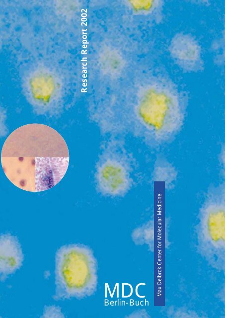
<strong>Research</strong> <strong>Report</strong> <strong>2002</strong>(covers the period 2000-2001)Max Delbrück Center forMolecular Medicine (<strong>MDC</strong>)Berlin-BuchRobert-Rössle-Str. 10D-13125 BerlinTel.: +49-30-9406-0Fax: + 49-30-949-4161e-mail: mdc@mdc-berlin.deThis <strong>Research</strong> <strong>Report</strong> is also availableon the World Wide Webhttp://www.mdc-berlin.deThe <strong>MDC</strong> is a member of theHGF (Helmholtz Associationof National <strong>Research</strong> Centers)The following <strong>Research</strong> <strong>Report</strong>shave been published previously:<strong>Research</strong> <strong>Report</strong> 1996(covers the period 1992-1995)<strong>Research</strong> <strong>Report</strong> 1996/97(covers the period 1996-1997)<strong>Research</strong> <strong>Report</strong> 2000(covers the period 1998-1999)Editorial board:Walter BirchmeierCarmen BirchmeierThomas BlankensteinUdo HeinemannHelmut KettenmannFriedrich C. LuftPeter M. SchlagCoordination:Barbara BachtlerHans-Josef LinkensVolker WunderlichBook design:Hoch Drei GmbH, 10963 BerlinPrinting:Druckhaus Berlin-Mitte GmbHPrinted in Germany <strong>2002</strong>Legend to Cover Figures:In situ hybridization with the patched-1 cDNAreveals a block in embryonic hair placodeformation in the absence of -catenin/wntsignaling. In addition, tissue-specific ablation ofthe -catenin gene in the skin of mice demonstratesthe essential role of -catenin signalingin the fate of skin stem cells in the adult: in theabsence of -catenin skin stem cells fail todifferentiate into follicular keratinocytes, takingan epidermal route instead (from Huelsken etal., Cell (2001) 105, 533-545; this report p. 97)
<strong>Research</strong> <strong>Report</strong><strong>2002</strong>Covers the period 2000/2001
2ContentInhaltIntroductionEinführung .................................................................................................................................................................... 8OverviewÜberblick .................................................................................................................................................................... 21Genetics, Bioinformatics and Structural BiologyGenetik, Bioinformatik und Strukturbiologie ........................................................................................................ 40Molecular Biology and Genetics of Cardiovascular DiseasesMolekularbiologie und Genetik kardiovaskulärer ErkrankungenDetlev Ganten, Norbert Hübner ................................................................................ 43Molecular Biology of Peptide HormonesMolekularbiologie von Peptid-HormonenMichael Bader .......................................................................................................... 45Genetics, Etiology, and Pathogenesis of Hypertension,Vascular Injury, and Renal DiseasesGenetik, Ätiologie und Pathogenese von Bluthochdruck,Gefäßschädigung und NierenerkrankungenFriedrich C. Luft ....................................................................................................... 47Gene Mapping and Identification in Monogenic and Complex DiseasesGenkartierung und Identifizierung bei monogenen und komplexen ErkrankungenPeter Nürnberg ........................................................................................................ 49Cardiovascular Molecular GeneticsKardiovaskuläre MolekulargenetikLudwig Thierfelder .................................................................................................... 52Obesity and HypertensionFettleibigkeit und BluthochdruckArya M. Sharma ....................................................................................................... 54Disorders of the Autonomic Nervous SystemErkrankungen des autonomen NervensystemsJens Jordan (Helmholtz Fellow) ................................................................................ 56Characterization of newly identified human importin proteinsCharakterisierung von neuen humanen Importin-alpha ProteinenMatthias Köhler (Helmholtz Fellow) ........................................................................... 57Tumor GeneticsTumorgenetikSiegfried Scherneck ................................................................................................. 58Mouse GeneticsMausgenetikCarmen Birchmeier .................................................................................................. 61Developmental GeneticsEntwicklungsgenetikAndreas Schedl ....................................................................................................... 64
3Lipids and Experimental Gene TherapyLipide und experimentelle GentherapieThomas Willnow ....................................................................................................... 66BioinformaticsBioinformatikJens Reich, Peer Bork .............................................................................................. 68Protein Misfolding: From Basic Biopolymer Physicsto Conformational DiseasesProtein-Fehlfaltung: von der Biopolymerphysik zu Konformations-ErkrankungenGregor Damaschun .................................................................................................. 70Structural Studies of Proteins and Nucleic Acidsby X-ray CrystallographyStrukturstudien an Proteinen und Nukleinsäuren mit Röntgenstrahl-KristallographieUdo Heinemann ....................................................................................................... 72Role of Protein Dynamics in Enzyme FunctionRolle der Proteindynamik bei der EnzymfunktionChristiane Jung ........................................................................................................ 75Computer Simulation of Nucleic Acid Structure and InteractionsComputersimulation von Nukleinsäure-Strukturen und WechselwirkungenHeinz Sklenar ........................................................................................................... 77Conformation, Stability and Interaction of Biological MacromoleculesKonformation, Stabilität und Wechselwirkung von biologischen MakromolekülenHeinz Welfle ............................................................................................................. 79Bioethics and Science CommunicationBioethik und WissenschaftskommunikationChristof Tannert ....................................................................................................... 81Cell Growth and DifferentiationZellwachstum und Differenzierung ................................................................................................................. 84Growth Control and Gene Regulation in the Hematopoietic SystemWachstumskontrolle und Genregulation im hämatopoetischen SystemAchim Leutz ............................................................................................................. 88Signal Transduction in Tumor CellsSignaltransduktion in TumorzellenClaus Scheidereit ..................................................................................................... 90Differentiation and Growth Control in Lymphocyte Developmentand ImmunopathogenesisDifferenzierung und Wachstumskontrollebei der Lymphozyten-Entwicklung und ImmunpathogeneseMartin Lipp .............................................................................................................. 92Initiation of DNA ReplicationInitiierung der DNA-ReplikationManfred Gossen ...................................................................................................... 95Epithelial Differentiation, Invasion and MetastasisEpitheliale Differenzierung, Invasion und MetastasierungWalter Birchmeier ..................................................................................................... 97
4Surgical OncologyChirurgische OnkologiePeter M. Schlag ..................................................................................................... 100Molecular Muscle PhysiologyMolekulare MuskelphysiologieIngo L. Morano ...................................................................................................... 103Cell Biology of Cardiovascular DiseasesZellbiologie kardiovaskulärer ErkrankungenHeinrich Leonhardt, M. Cristina Cardoso ................................................................ 105Intracellular ProteolysisIntrazelluläre ProteolyseThomas Sommer ................................................................................................... 107Nucleocytoplasmic Transport in the Yeast Saccharomyces cerevisiaeNukleo-zytoplasmatischer Transport bei der BäckerhefeKatrin Stade (Helmholtz Fellow) .............................................................................. 109Cytochrome P450 and Endoplasmic ReticulumCytochrom P450 und endoplasmatisches RetikulumWolf-Hagen Schunck ............................................................................................. 110Cell Polarity and Epithelial Formation in Development and DiseaseZellpolarität und Epithelbildung in Entwicklung und KrankheitSalim Abdelilah-Seyfried ......................................................................................... 112Electron MicroscopyElektronenmikroskopieGerd Kempermann, Bettina Erdmann ..................................................................... 114Molecular TherapyMolekulare Therapie ....................................................................................................................................... 118Myocardial RegenerationRegeneration des MyokardsRainer Dietz ............................................................................................................ 119Control of Smooth Muscle Cell FunctionKontrolle der Funktion der glatten MuskelzelleMaik Gollasch (Helmholtz Fellow) ............................................................................ 121Immunology of Cardiovascular DiseasesImmunologie kardiovaskulärer ErkrankungenGerd Wallukat ........................................................................................................ 123Hematology, Oncology and Tumor ImmunologyHämatologie, Onkologie und TumorimmunologieBernd Dörken ......................................................................................................... 125Molecular ImmunotherapyMolekulare ImmunotherapieAntonio Pezzutto .................................................................................................... 128Molecular Immunology and Gene TherapyMolekulare Immunologie und GentherapieThomas Blankenstein ............................................................................................. 130
5Cellular Immunology of Autoimmune ReactionsZelluläre Immunologie von AutoimmunreaktionenKirsten Falk, Olaf Rötzschke ................................................................................... 132Molecular and Cell Biology of Hematopoietic CellsMolekular-und Zellbiologie hämatopoietischer ZellenMartin Zenke .......................................................................................................... 134Cell Cycle Regulation and Gene TherapyZellzyklus-Regulation und GentherapieForschungsgruppe der Humboldt-Universität zu Berlin am <strong>MDC</strong> ............................. 136Evolution, Regulation and Genetic Applications of TransposableElements in VertebratesEvolution, Regulation und genetische Anwendungen von transposablenElementen bei VertebratenZoltán Ivics ............................................................................................................. 138Experimental PharmacologyExperimentelle PharmakologieIduna Fichtner ........................................................................................................ 139Drug Targeting„Drug Targeting“ (Zielgenaue Medikamentenentwicklung)Regina Reszka ....................................................................................................... 141RNA ChemistryRNA ChemieEckart Matthes ....................................................................................................... 143Molecular and Developmental NeurosciencesMolekulare Neurowissenschaft und Entwicklungsneurobiologie ............................................................... 146NeurodegenerationNeurodegenerationChristiane Alexander .............................................................................................. 148Neuronal Stem CellsNeuronale StammzellenGerd Kempermann ................................................................................................ 150Cellular NeurosciencesZelluläre NeurowissenschaftenHelmut Kettenmann ............................................................................................... 152Growth Factor and Regeneration GroupWachstumsfaktoren und RegenerationGary R. Lewin ........................................................................................................ 154Synapse Formation and FunctionBildung und Funktion von SynapsenFrank W. Pfrieger .................................................................................................... 156Developmental NeurobiologyEntwicklungsneurobiologieFritz G. Rathjen ...................................................................................................... 157Proteomics and Molecular Mechanisms of Neurodegenerative DisordersProteomik und molekulare Mechanismen neurodegenerativer ErkrankungenErich Wanker .......................................................................................................... 160
6Structure and OrganizationStruktur und OrganisationOrganizational StructureStiftungsorgane ................................................................................................... 164The Board of Trustees ............................................................................................ 164KuratoriumMembers of the Board of Trustees ............................................................... 164Mitglieder des KuratoriumsMembers of the Scientific Committee .......................................................... 165Mitglieder des Wissenschaftlichen AusschussesThe Management Board ......................................................................................... 166StiftungsvorstandScientific Council .................................................................................................... 166Wissenschaftlicher RatStaff Council .......................................................................................................... 167PersonalratSupporting Divisions ........................................................................................... 168StabsstellenSafety ......................................................................................................... 168SicherheitBuilding Coordination, Engineering and Reconstruction ............................... 168BauangelegenheitenAuditing and Legal Affairs ............................................................................ 168Innenrevision und RechtPatents/Licences ........................................................................................ 169Patente und LizenzenTechnology Transfer .................................................................................... 169TechnologietransferPress and Public Relations ................................................................................ 170Presse- und ÖffentlichkeitsarbeitAdministrationVerwaltung ........................................................................................................... 172Personnel ................................................................................................... 172PersonalFinances .................................................................................................... 172FinanzenPurchasing and Materials Management ....................................................... 173Einkauf und Materialwirtschaft
7Central FacilitiesZentrale Dienste .................................................................................................. 174Library ........................................................................................................ 174BibliothekAnimal Facilities ........................................................................................... 174TierhaltungCampus Net Management ........................................................................... 174Campusnetz-ManagementData and Image Processing ......................................................................... 175Daten- und BildverarbeitungTechnical Affairs .......................................................................................... 175TechnikMeetings, Workshops and Symposia ................................................................ 176Wissenschaftliche VeranstaltungenAwards ................................................................................................................. 178AuszeichnungenAddresses of Scientific Journals at the Berlin-Buch Campus ......................... 179Adressen wissenschaftlicher Zeitschriften auf demCampus Berlin-BuchIndex .................................................................................................................... 180IndexOrganigram .......................................................................................................... 188OrganigrammCampus Map ................................................................................. Inside Back CoverCampusplan ............................................................................. Innenumschlag hintenHow to find your way to the <strong>MDC</strong> ................................................ Inside Back CoverWie gelangen Sie zum <strong>MDC</strong> .................................................... Innenumschlag hinten
8IntroductionEinführungMolecular Medicine –A Concept and its Application atthe <strong>MDC</strong> in Berlin-BuchMolekulare Medizin –Ein Konzept und seine Umsetzungam <strong>MDC</strong> in Berlin-BuchHistory and FoundationOn January 1, <strong>2002</strong>, the Max Delbrück Center for MolecularMedicine (<strong>MDC</strong>) Berlin-Buch celebrated its 10th birthday. Itwas founded on January 1, 1992, from the Institutes of theEast German Academy of Sciences, the Central Institute forCancer <strong>Research</strong>, the Central Institute for Cardiovascular<strong>Research</strong> and the Central Institute for Molecular Biology. Thecenter is named after Max Delbrück, a native of Berlin, andone of the founding fathers of molecular biology who won theNobel Prize in 1969. Delbrück collaborated at the Kaiser WilhelmInstitute for Brain <strong>Research</strong> in Berlin-Buch with theRussian geneticist, N. W. Timoféev-Ressovsky and the physicist,K.G. Zimmer. In 1935, their interdisciplinary researchled to the publication of the epoch-making paper “The Natureof Gene Structure and Gene Mutation”.Anyone who experienced the reunification of Germanybetween November 9, 1989, and January 1, 1992 – involvingthe break up of the GDR, the unification of the two Germanies,with the evaluation of the entire scientific system of theGDR, the many plans for the restructuring of the scientificestablishments and institutional and personal uncertainties ofevery kind – can imagine the problems people experienced,as well as the hope and expectations during this two-year period.In 1991, in Berlin-Buch, there were over 1,000 staff,physicians, researchers, and technicians working in the institutesof the Academy and the clinics. At its foundation, the<strong>MDC</strong> had 350 budgeted posts. In such circumstances it wasno simple matter to set up a new organization to match thestandard of the former institutes of the Academy and guaranteeall qualified staff a future post. The details of this criticaland exciting period of the restructuring of science on theBuch Campus has been comprehensively described else-Geschichte und GründungAm 01. Januar <strong>2002</strong> beging das Max-Delbrück-Centrum fürMolekulare Medizin (<strong>MDC</strong>) Berlin-Buch sein 10-jährigesBestehen. Es ist am 01. Januar 1992 aus den Instituten derAkademie der Wissenschaften der DDR, dem Zentralinstitutfür Krebsforschung, dem Zentralinstitut für Herz-Kreislaufforschungund dem Zentralinstitut für Molekularbiologie hervorgegangen.Die Benennung des Institutes erfolgte nachMax Delbrück, dem aus Berlin stammenden Wegbereiter derMolekularbiologie, der 1969 mit dem Nobelpreis ausgezeichnetworden war. Delbrück hatte am Kaiser-Wilhelm-Institutfür Hirnforschung in Berlin-Buch zusammen mit dem russischenGenetiker N. W. Timoféeff-Ressovsky und dem PhysikerK.G. Zimmer gearbeitet, und bei dieser interdisziplinärenForschung war 1935 die epochemachende Arbeit über die„Natur der Genstruktur und der Genmutation“ entstanden.Wer die Zeit der deutschen Wiedervereinigung zwischen dem09. November 1989 und dem 1. Januar 1992 persönlich miterlebthat – mit dem Zusammenbruch der DDR, mit dem Prozessder Vereinigung der beiden deutschen Staaten, mit derEvaluation des gesamten Wissenschaftssystems der DDR, mitden vielen Plänen für die Neustrukturierung der wissenschaftlichenEinrichtungen und mit den Unsicherheiten in institutionellerund persönlicher Art –, kann sich vorstellen, welche Belastungen,aber auch welche Hoffnungen und Erwartungen indiesen zwei Jahren des Prozesses der deutschen Wiedervereinigungdurchlebt wurden. In Berlin-Buch waren 1991 weitüber 1.000 Mitarbeiter, Ärzte, Wissenschaftler, TechnischeAngestellte in den Akademieinstituten und Kliniken beschäftigt.Das <strong>MDC</strong> hatte bei der Gründung 350 budgetierte Stellen.Es war in dieser Situation nicht einfach, eine dem Leistungsniveauder Akademieinstitute entsprechende neue
9where. It was only possible because everyone was sympatheticto the final aim and worked together to ensure that itwas achieved.The significant and unavoidable problems at personal and institutionallevels were made easier by the fact that everyonewas aware of the great tradition of the Buch Institutes whichhad supported outstanding researchers like Oskar and CécileVogt, Walter Friedrich, Karl Lohmann, Arnold Graffi, ErwinNegelein, Albert Wollenberger and Hans Gummel. The realand positive role played by tradition in building new structuresat a time of great change can be particularly well observedand experienced in Berlin. Attention to tradition wasand still is important as far as setting up the <strong>MDC</strong> and theBerlin-Buch Campus are concerned.The clinics of the Academy of Sciences were incorporatedinto the university system – thanks to the energetic efforts ofthe then Senator for Science, Prof. Manfred Erhardt – as theRobert Rössle Cancer Clinic and the Franz Volhard CardiovascularClinic of the Charité, Berlin-Buch Campus. Thanksto particularly well defined cooperation contracts, they werelinked to the <strong>MDC</strong> in both personnel and institutional terms.BBB Biotechnologie Berlin-Buch Management GmbH set upa biotechnology park. This has allowed the basic researchactivities at the <strong>MDC</strong>, and clinical research at the Charitéclinics and the Buch Clinic as well as the Technology Park establishedby BBB Management GmbH to develop a synergywith respect to all research efforts that has subsequently beenshown to be successful based on any number of criteria. Thepure research carried out on the Berlin-Buch Campus wasboosted in1999 by the arrival of the Forschungsinstitut fürMolekulare Pharmakologie (FMP; <strong>Research</strong> Institute forMolecular Pharmacology), which is part of the Wilhelm GottfriedLeibniz Association. The institute collaborates closelywith the <strong>MDC</strong> on structural research into biologically activemolecules. The privatization of the clinics under the auspicesof Helios Kliniken GmbH in summer 2001 has created evenmore opportunities for cooperation.Bundespräsident Johannes Rau (Mitte) im Gespräch mit Prof. Detlev Ganten (<strong>MDC</strong>-Stiftungsvorstand)und dem damals designierten und jetzigen Administrativen Vorstand des<strong>MDC</strong>, Dr. Waltraud Kreutz-Gers, anläßlich der Verleihung des Deutschen Zukunftspreisesdes Bundespräsidenten am 29. November 2001 im <strong>MDC</strong> Kommunikationszentrum.The President of the Federal Republic of Germany, Johannes Rau (in the middle), chatswith the Scientific Director of the <strong>MDC</strong>, Detlev Ganten (MD. Ph.D), and the thenAdministrative Director designate, Dr. Waltraud Kreutz-Gers, after the awarding of thePresident's “Prize for Technology and Innovation” which took place at the <strong>MDC</strong>`s newMax Delbrück Communications Center (<strong>MDC</strong>.C) on November 29, 2001.Copyright: <strong>MDC</strong>; Annett KrauseOrganisationsform zu finden und allen qualifizierten Mitarbeiterndie ihnen zukommende Position zuzusichern. ÜberEinzelheiten dieser entscheidenden und aufregenden Monateund Jahre der Neuorientierung der Wissenschaft auf demCampus Buch ist an anderer Stelle ausführlich berichtet undgeschrieben worden. Sie konnten nur bewältigt werden, weiles von allen Seiten viel Verständnis und Unterstützung gab.Die großen und wohl unvermeidbaren Brüche in institutionellerund personeller Hinsicht wurden unter anderem dadurcherleichtert, dass ganz bewusst die große Tradition derBucher Institute mit so herausragenden Wissenschaftlern wieOskar und Cécile Vogt, Walter Friedrich, Karl Lohmann, ArnoldGraffi, Erwin Negelein, Albert Wollenberger und HansGummel fortgeführt wurde. Die reale und konkrete positiveBedeutung der Tradition für die Zukunftsgestaltung in einerZeit großer Umbrüche, kann in Berlin besonders gut beobachtetund erlebt werden. Traditionspflege wurde und wirdbewußt für den Aufbau des <strong>MDC</strong> und des Campus Berlin-Buch eingesetzt.Die Kliniken der Akademie der Wissenschaften wurden mittatkräftiger Unterstützung des damaligen Senators für Wissenschaft,Prof. Manfred Erhardt, in das universitäre Systemeingegliedert als Robert-Rössle-Krebsklinik und Franz-Volhard-Herz-Kreislaufklinikder Charité, Campus Berlin-Buch.Durch einen besonders engen Kooperationsvertrag wurdensie mit dem <strong>MDC</strong> personell und institutionell verbunden. DerAufbau eines Biotechnologieparks wurde durch die BBBBiotechnologie Berlin-Buch Management GmbH begründet.Auf diese Weise wurde mit der Grundlagenforschung am<strong>MDC</strong>, der klinischen Forschung an den Charité-Kliniken unddem Klinikum Buch sowie mit dem von der BBB ManagementGmbH eingerichteten Technologiepark ein Synergienschaffendes komplettes Forschungssystem eingerichtet, dassich in den folgenden Jahren in jeder Hinsicht als erfolgreicherwiesen hat. 1999 wurde die Grundlagenforschung auf demCampus Berlin-Buch durch den Zuzug des Forschungsinstitutsfür Molekulare Pharmakologie (FMP), das zur Wilhelm-Gottfried-Leibniz-Gemeinschaft gehört, verstärkt. DiesesInstitut arbeitet mit dem <strong>MDC</strong> insbesondere auf dem Gebietder Strukturforschung für biologisch aktive Moleküle intensivzusammen. Die Privatisierung der Kliniken durch dieHelios Kliniken GmbH im Sommer 2001 schafft erweiterteMöglichkeiten der Kooperation.Aufbruchstimmung in Berlin-BuchAuf dem Campus Berlin-Buch herrscht Aufbruchstimmung:Das Max Delbrück Communications Center (<strong>MDC</strong>.C) mitHörsälen, Seminar- und Laborräumen für Kongresse, Fortbildungund Kursen ist im Jahre 2001 fertig gestellt und in Betriebgenommen worden. Der erste Bauabschnitt des HelmholtzHauses für Tierlaboratorien und Büroräume wird noch imJahre <strong>2002</strong> bezogen. Ein neues Zentrum für Medizinische Genomforschungdes <strong>MDC</strong> ist im Entstehen. Vier neue Laborgebäudefür Existenzgründer und Biotechnologiefirmen sindfertig gestellt bzw. befinden sich zur Zeit im Bau. Das neue„Helios Klinikum Berlin“ wird auch die universitären Klinikender Charité in einem Neubau für 400 Millionen Mark mit aufnehmen,so dass eine der modernsten und größten KlinikenBerlins in unmittelbarer Nachbarschaft des Forschungscampus
10Developments in Berlin-BuchSpirits on the Berlin-Buch Campus are high as the site develops:the Max Delbrück Communications Center (<strong>MDC</strong>.C),with its lecture theaters, seminar rooms and laboratory spacefor congresses, further education and training courses, wasopened in 2001. The first section of the “Helmholtz Haus” foranimal laboratory facilities and offices will be ready in <strong>2002</strong>.A new <strong>MDC</strong> Center for Medical Genome <strong>Research</strong> is beingcreated. Four new laboratory buildings for start-up companiesand biotechnology companies have been completed orare under construction. The new “Helios Klinikum Berlin”will also incorporate the university clinics of the Charité in anew setting costing approximately 200 million Euro so thatone of the most modern and largest clinics in Berlin will belocated right next to the research campus. The space occupiedby the clinics previously represents an area of over 100 haand this will allow the Technology Park to expand. The clinicbuildings designated as being of historic interest will be usedas a “Cité Universitaire”, and will be adopted by the EuropeanCollege of Liberal Art (ECLA). A new shopping centerwill be built right next to the S-Bahn station. It is also plannedto build a hotel for visitors and congress delegates as well asrelatives of the patients being treated in the clinics.The health theme, which has made Berlin-Buch, the “HealthRegion” is becoming more and more important as a part ofthe modern knowledge-based society. In the future, Berlin-Buch will be a center for molecular medicine where the latestadvances in genome research and gene technology will beapplied to the clinical setting and in the field of business,allowing biotech companies and service firms to develop andcreate new jobs.entstehen wird. Die freiwerdenden Klinikgebäude in einemAreal von über 100 Hektar werden für den expandierendenTechnologiepark zur Verfügung stehen. DenkmalgeschützteKlinikgebäude sollen für eine „Cité Universitaire“, die auchdas European College of Liberal Art (ECLA) aufnehmen wird,genutzt werden. In unmittelbarer Nähe des S-Bahnhofes wirdein neues Einkaufszentrum gebaut. Dazu ist geplant, ein Hotelzu errichten für Besucher und Kongressteilnehmer sowie fürAngehörige von Patienten der Kliniken.Das Thema Gesundheit, dem sich Berlin-Buch, die „Gesundheitsregion“,verschrieben hat, wird als Teil der modernenWissensgesellschaft immer weiter an Bedeutung gewinnen.Berlin-Buch soll in Zukunft ein Zentrum für die MolekulareMedizin sein, in dem die Fortschritte der Genomforschungund Gentechnik verantwortungsvoll in Klinik und Wirtschaftangewendet werden und um das herum sich Biotechnologiefirmenund Serviceeinrichtungen entwickeln und neue Arbeitsplätzeentstehen lassen. Der Skulpturenpark auf demCampus Berlin-Buch mit Werken unter anderem von Ipousteguy,Szymanski und Kriester ist Ausdruck der engen Ver-Dr. Erwin Jost, Administrativer Vorstand des <strong>MDC</strong>, Dr. Arend Oetker (Vorsitzender desKuratoriums Deutscher Zukunftspreis und Präsident des Stifterverbandes) und Prof.Detlev Ganten (<strong>MDC</strong>-Stiftungsvorstand) beim Empfang nach der Vergabe des Zukunftspreisesdes Bundespräsidenten am 29. November 2001 im neuen Max Delbrück CommunicationsCenter (<strong>MDC</strong>.C)Reception after the awarding of the “Federal President's Prize for Technology and Innovation”in the new Max Delbrück Communications Center (<strong>MDC</strong>.C) on November 29,2001 (from left): Dr. Erwin Jost (Administrative Director of the <strong>MDC</strong>), Dr. Arend Oetker(Chairman of the Board of Trustees of the President's “Prize for Technology and Innovation“and President of the Stifterverband für die Deutsche Wissenschaft – Donor´s Associationfor the Promotion of the Sciences and Humanities) and Detlev Ganten (MD.,Ph.D, <strong>MDC</strong>`s Scientific Director).Copyright: <strong>MDC</strong>; Siegfried RöpkeThe Sculpture Park on the Berlin-Buch Campus with works by,among others, Ipousteguy, Szymanski and Kriester, reflects theclose relationship between the Arts and Sciences, which is particularlyencouraged in Berlin-Buch. In this way, Buch will beseen as an intellectual focal point where the development ofmolecular medicine and gene technology is being advancedalongside a continuing dialog with people from many differentbackgrounds. In this way Berlin-Buch will become a model ofa future human knowledge-based society. On the <strong>MDC</strong> Campusover 2000 staff are currently working in the research institutes,clinics and biotech companies, many more than beforereunification in 1989. During the last 10 years many new jobshave been created in this science-based environment.The rapid connections linking Berlin-Buch to the culturalcenter of central Berlin and the proximity of the Barnim NatureConservation Park have made Berlin-Buch one of themost attractive and dynamic suburbs of the new Berlin.Many of the staff who work in the research institutes and thebiotech companies live only 15 minutes away in PrenzlauerBerg, one of the favorite parts of Berlin with many theaters,bars and cinemas. Prenzlauer Berg is close to the cultural andspiritual heart of the city with the Humboldt University, theMuseum Island, the main theaters, the Berlin Philharmonicand the Kulturforum.
11Buch is a place of synergies. As a link between town andcountry, between pure research and the clinics as well asscientific applications of this research, living, working andrelaxing in Berlin-Buch are all equally attractive.The concept of molecular medicineAs far as the success of any scientific institution is concerned,the key factors are the original idea upon which it is based,the efforts of its scientists and other key staff, their commitment,the quality of the research activities, projects and itspublication record.bindung von Kunst und Wissenschaft, die in Berlin-Buch besondersgepflegt wird. Buch soll sich auf diese Weise als intellektuellerKristallisationspunkt profilieren, in dem die Entwicklungder Molekularen Medizin und Gentechnologievorangebracht und durch den Dialog mit breiten Kreisen derGesellschaft kritisch begleitet wird. Auf diese Weise wird Berlin-Buchzu einem Modell für die zukünftige humane Wissensgesellschaft.Auf dem Campus Berlin-Buch arbeiten zurZeit in den Forschungseinrichtungen, Kliniken und Biotechnologiefirmenüber 2.000 Mitarbeiter, weit mehr als vor derWiedervereinigung 1989. Im Umfeld der Wissenschaft sinddamit in den vergangenen 10 Jahren viele neue anspruchsvolleund zukunftsfähige Arbeitsplätze geschaffen worden.Die schnelle Anbindung von Berlin-Buch an das kulturelleZentrum in Berlin-Mitte und die direkte Nachbarschaft zumNaturschutzpark Barnim machen Berlin-Buch zu einem derattraktivsten und dynamischsten Vororte des neuen Berlin.Viele Mitarbeiter der Forschungseinrichtungen und Biotechnologiefirmenwohnen in dem 15 Minuten entfernten PrenzlauerBerg, einem der beliebtesten Stadtteile Berlins mit vielenTheatern, Kneipen und Kinos. Dem Prenzlauer Berg schließtsich direkt das kulturelle und geistige Zentrum der Stadt an mitder Humboldt Universität, der Museumsinsel, den großenTheatern, der Philharmonie und dem Kulturforum.Buch ist ein Ort der Synergien. Als Nahtstelle zwischen Stadtund Land, zwischen Forschung und klinischer sowie wirtschaftlicherAnwendung ist das Leben, das Arbeiten und dasErholen in Berlin-Buch gleichsam attraktiv.Abschied nach acht Jahren <strong>MDC</strong>: Prof. Jutta Schnitzer, wissenschaftliche Referentindes <strong>MDC</strong> und jetzige Generalsekretärin der Leopoldina, Halle. <strong>MDC</strong>-Vorstand Prof.Detlev Ganten beim Überreichen des Blumenstrausses.Farewell after eight years at the <strong>MDC</strong>: Prof. Jutta Schnitzer, <strong>MDC</strong>`s former ScientificCoordinator and now Secretary General of the “Leopoldina” (Halle) receives a bouquetof flowers from Detlev Ganten (MD., PhD), <strong>MDC</strong>`s Scientific DirectorCopyright: <strong>MDC</strong>; Siegfried EndruweitWhen the <strong>MDC</strong> was founded, we took advice based on therecommendations of the Scientific Council and the FoundersCommittee regarding ideas about the long-term developmentsin medicine and the latest results of molecular geneticsand genome research.There are two reasons for the extraordinary successful andsustained development of the modern life sciences. On theone hand, there were the scientists themselves, who highlightedthe new, often physico-chemical, methods whichcould identify the macromolecules which had key biologicalfunctions in biology and medicine, thereby allowing a betterunderstanding of the interplay between the form and functionof cells. One the other hand, institutions like the RockefellerFoundation play an important role. For example, in 1938, itsupported a research program for the advancement of geneticsgiving it the name “molecular biology”. One aim of thisproject was to raise the level of biosciences at that time andturn it into a quantitative “Science of Man”, which went beyondsimply describing events that happened in Nature. Overthe period 1937–39, the person who gave his name to the<strong>MDC</strong>, Max Delbrück, held a fellowship from the RockefellerFoundation and their support helped him become one of theDas Konzept der Molekularen MedizinEntscheidend für den Erfolg einer wissenschaftlichen Institutionist das inhaltliche Konzept, die wissenschaftliche Leistung,die tragenden Personen, das Engagement der Wissenschaftlerinnen,Wissenschaftler und Mitarbeiter, die Qualitätder Forschungstätigkeit, Projekte und Publikationen.Zur Zeit der Gründung des <strong>MDC</strong> haben wir uns auf der Basisder Empfehlungen des Wissenschaftsrates und des Gründungskomiteesbei den Konzepten von langfristigen Entwicklungender Medizin und neuesten Ergebnisse der molekularenGenetik und Genomforschung leiten lassen.Es gibt zwei Quellen für das außerordentlich erfolgreiche undnachhaltige Konzept der modernen Lebenswissenschaft. Zumeinen waren dies die Wissenschaftler selbst, die mit neuen,über die Biologie und Medizin hinausreichenden, häufig physikalisch-chemischenMethoden die Strukturen von Makromolekülenmit wichtigen biologischen Funktionen erkundenkonnten und sich anschließend daran machten, das Wechselspielvon Form und Funktion in der Zelle zu verstehen. Zumzweiten spielten Institutionen wie etwa die Rockefeller Stiftungeine wichtige Rolle, die im Jahre 1938 einem Forschungsprogrammzur Förderung der Genetik den Namen„Molekularbiologie“ gab. Ein Ziel dieser Initiative lag darin,das Niveau der damaligen Biowissenschaften anzuheben undzu einer quantitativ operierenden „Science of Man“ auszuweiten,die über die reine Beschreibung der Natur hinausging.Der Namenspatron des <strong>MDC</strong>, Max Delbrück, gehörte in
12founding fathers of molecular biology; there is much that wecan still learn from him today.When the <strong>MDC</strong> was established ten years ago in Berlin-Buchand raised a flag as far as the development of molecular medicinewas concerned, there were only a few institutes aroundthe world engaged in applying the latest results of pure researchto medical research in a closely-knit institutional setting.At that time, no one could have known the kind ofprogress that would take place in genome analysis in the nextten years. The mission of the <strong>MDC</strong> is to combine modernmedical and clinical research with the methods of molecularbiology and cell biology as well as the expansion in genetechnology, which systematically concentrates on the genomeand the latest results arising from research into it. As far aspure research is concerned, key areas should be those that areof vital importance for the analysis of disease phenomena andwhich lead to new opportunities firmly based on the naturalsciences for diagnosis, treatment and prevention.den Jahren 1937–39 zu den Stipendiaten der Rockefeller Stiftungund ist mit ihrer Hilfe zu dem Wegbereiter der Molekularbiologiegeworden, von dem wir heute noch lernen können.Als das <strong>MDC</strong> vor zehn Jahren in Berlin-Buch gegründetwurde und sich die Entwicklung einer Molekularen Medizinauf die Fahnen schrieb, gab es weltweit nur wenige Institute,die so konsequent die Anwendung neuester Methoden in derGrundlagenforschung für medizinische Forschung in engerinstitutioneller Verbindung verfolgten. Niemand konnte damalswissen, welche Fortschritte die Genomanalyse in diesemZeitraum machen würde. Die Aufgabe des <strong>MDC</strong> bestanddarin, moderne medizinische und klinische Forschung imVerband mit molekularbiologischen, zellbiologischen sowiezunehmend gentechnischen Methoden zu betreiben, die sichsystematisch auf das Genom konzentrierten und von ihm ausgingen.Wegweisend für die Grundlagenforschung solltenThemen sein, die von prinzipieller Bedeutung für die Analysevon Krankheitsphänomenen sind und aus denen sich naturwissenschaftlichbegründet neue Möglichkeiten für Diagnostik,Therapie und Prävention ableiten lassen.The multitalented molecules of molecular medicineTouch and Pain: two senses, one ion channel named DRASICOur senses of touch and pain, opposites one might think, have somethingin common, an ion channel called DRASIC. DRASIC, whichstands for dorsal root ganglion acid sensitive channel, has beenshown to be critical for both these senses. The DRASIC protein formsa channel in the membrane that when opened conducts sodium ionswhich then leads to the electrical excitation of neurons. A first clueabout the possible function of the DRASIC protein came from a geneticscreen for mutations in the nematode worm C.elegans whichshowed that similar proteins in the worm are necessary for the wormto sense touch. These proteins including DRASIC are all members ofthe Deg/Enac channel superfamily. Thus arose the idea that mechanicalforces on the membrane could open the channel: this would makeit a mechanical sensor. Thus the same channel obviously helps sensoryneuron to detect innocuous and painful stimuli in a completely differentway. It is known that DRASIC can potentially form heteromericion channel complexes with other members of the Deg/Enac family ofproteins.Another aspect of DRASIC channel function is the fact that acidificationie. protons are a very effective stimulus to open the channel. Acidificationis a universal consequence of injury or inflammation of peripheraltissues. Mice lacking DRASIC were much less affected by muscleacidification, in other words the lack of the channel protein had an analgesiceffect. The diversity of function for the DRASIC channel reportedhere for the first time shows directly that the channel does notwork alone but in concert with other as yet unknown proteins. It is preciselythis kind of in vivo analysis of protein function that highlights theprinciple that the function of a single protein can vary enormously dependingon the cellular context. Thus products of single genes are notsimple entities that work alone to accomplish a well-defined functionbut rather execute their function in a multitude of ways depending theother proteins that they can interact with. For further details see GaryLewin´s research group.Die vielseitigen Moleküle der Molekularen MedizinBerührung und Schmerz: Zwei Sinne, ein Ionenkanal namensDRASICDer Tastsinn und der Schmerz erscheinen zwar als gegenteilige Sinne,sie haben aber etwas gemeinsam, einen Ionenkanal mit Namen DRA-SIC. DRASIC kürzt den englischen Ausdruck „dorsal root ganglionacid sensitive channel“ ab, womit ein säureempfindlicher Ionenkanalim dorsalen Wurzelganglion gemeint ist. Es konnte gezeigt werden,dass DRASIC für die beiden genannten Sinne wesentlich ist. DasDRASIC Protein formt einen Kanal durch die Membran, durch den imgeöffneten Zustand Natriumionen strömen, die dann zur elektrischenErregung von Neuronen führen. Einen ersten Hinweis auf die möglicheFunktion des DRASIC Proteins ergab eine genetische Reihenuntersuchungvon Mutationen in dem Fadenwurm C. elegans. Sie zeigte,dass ähnliche Proteine nötig sind, damit der Wurm berührungsempfindlichwird. Diese Proteine – einschließlich DRASIC – sind alle Mitgliederder Deg/Enac Kanal Großfamilie. Aus dieser Kenntnis leitetsich die Idee ab, dass mechanische Kräfte, die auf die Membran wirken,zur Öffnung des Kanals führen, der auf diese Weise ein mechanischerSensor würde. Derselbe Kanal hilft offenbar sensorischen Neuronen,harmlose und schmerzhafte Reize auf vollständig verschiedeneWeise zu registrieren. Es ist bekannt, dass DRASIC in der Lage ist,heteromere Kanalkomplexe mit anderen Mitgliedern der Deg/EnacFamilie zu bilden.Ein weiterer Aspekt der Funktion des DRASIC Kanals steckt in derTatsache, dass eine Zunahme des Säuregrads – also das Auftretenvon Protonen – als effektiver Reiz zur Kanalöffnung funktioniert. DieZunahme des Säuregrads ist die universelle Konsequenz einer Verletzungoder Entzündung des peripheren Gewebes. Mäuse, die nichtüber DRASIC verfügten, waren jedoch viel weniger durch die Muskelsäuerungbetroffen. Mit anderen Worten, die Abwesenheit des Kanalproteinshatte einen analgetischen Effekt. Die Verschiedenheit derFunktionen für den DRASIC Kanal, die hier zum ersten Mal vorgestelltwird, zeigt direkt, dass der Kanal nicht allein, sondern im Verbund mitanderen Proteinen funktioniert, die bislang unbekannt sind. Es ist nungenau diese Art der in vivo Analyse einer Proteinfunktion, die das Prinzipdeutlich macht, demzufolge die Funktion eines einzelnen Proteinsje nach dem zellulären Kontext enorm variieren kann. Produkte einzelnerGene sind also nicht bloß einfache Einheiten, die alleine operieren,um wohldefinierte Funktionen zu erfüllen. Sie üben ihre Aufgaben vielmehrin einer Vielzahl von Wegen aus, die von anderen Proteinen abhängen,mit denen sie in Wechselwirkung treten können. WeitereDetails siehe Arbeitsgruppe von Gary Lewin.
13Die Interdisziplinarität in der Grundlagenforschung spiegeltesich wider in der Tatsache, dass die Ergebnisse Bedeutunghaben und Anwendung finden in fast allen klassischen klinischenFächern. Die gleichen Moleküle der Zelldifferenzierung,Wachstumsfaktoren, Ionenkanäle in den Membranensind für Krebs, Herzkreislauf- und Nervenerkrankungen ingleicher Weise wichtig. Die Molekulare Medizin überwindetdamit die traditionellen Grenzen der Disziplinen und schafftdie Voraussetzung für wahrhaft inhaltliche und strukturelleInterdisziplinarität.<strong>MDC</strong>-Neujahrsveranstaltung am 28. Januar 2000: Prof. Fritz Melchers (Mitte), derfrühere Vorsitzende des Wissenschaftlichen Ausschusses des <strong>MDC</strong> (Basel, Schweiz,Mitte) erhält für seine Verdienste um das <strong>MDC</strong> von Prof. Detlev Ganten (<strong>MDC</strong>-Stiftungsvorstand)(re.) die Max-Delbrück-Medaille. Interessierter Zuschauer: Wolf-Michael Catenhusen(Kuratoriumsvorsitzender des <strong>MDC</strong> und Parlamentarischer Staatssekretär imBundesforschungsministerium).At the <strong>MDC</strong>`s New Year Reception on January 28, 2000: Prof Fritz Melchers (in themiddle), former chairman of the Scientific Committee of the <strong>MDC</strong> from Basle (Switzerland)receives the Max Delbrück Medal for his contributions to the development of the<strong>MDC</strong> from <strong>MDC</strong>`s Scientific Director, Detlev Ganten (MD., Ph.D; right) as Wolf-MichaelCatenhusen, chairman of the <strong>MDC</strong>`s Board of Trustees and Parliamentary State Secretaryfrom the Federal Ministry for Education and <strong>Research</strong> looks on.Copyright: <strong>MDC</strong>; Siegfried EndruweitThe interdisciplinary nature of pure research is reflected inthe fact that its results are of importance and have applicationsin almost all classic clinical disciplines. The same moleculesinvolved in cell differentiation, growth factors, and ionchannels in membranes also play important roles in cancer,cardiovascular and nervous system diseases. Molecular medicineovercomes the traditional boundaries of these disciplinesand provides conditions for true content-based andstructural interdisciplinary activities.At the start of the 1980s, people began to understand for thefirst time how the tools of gene technology could be used tomap the human genes, but for a long time many scientistswere unable to make any sense of the immense efforts associatedwith the identification of the human gene sequence andthe three billion or so building stones involved. The hope of agenetic analysis of diseases was based more on optimismthan any empirical evidence. Only when clinical observationsin combination with molecular biology research clearlyshowed that cancer, for example, is a genetic disease and theformation of tumors is affected by DNA variants, did the projectreally get underway, leading to the sequencing of the humangenome and other model genomes. Due to these genomeprojects the ideas of molecular medicine became accepted bythe general public with their promise of sweeping perspectivesfor the future.The 1990s saw the start of a new form of collaboration inmedicine whereby scientists involved in molecular geneticsand genome research, using genetic engineering, tried to answerclinically relevant questions and develop new productsfor the healthcare market. Today, it is perhaps possible tothink of the past decade as the first period of biomedicine,during which the scope of gene diagnosis was enormouslyextended as well as the identification of many new targets asstarting points for the development of drugs of the future.These developments are not only characterized by immenseZu Beginn der achtziger Jahre war zum ersten Mal verstandenworden, wie die Werkzeuge der Gentechnik es erlaubten, Genkartendes Menschen anzufertigen, aber lange Zeit hindurchkonnten viele Wissenschaftler keinen Sinn in dem ungeheurenAufwand erblicken, der mit der Erstellung der menschlichenGensequenz und ihren rund drei Milliarden Bausteinen verbundensein würde. Die Hoffnung auf genetische Analysenvon Krankheiten beruhte mehr auf Optimismus als auf empirischerEvidenz. Erst als klinische Beobachtungen in Verbindungmit molekularbiologischen Forschungen deutlich vorAugen führten, dass zum Beispiel Krebs eine genetischeKrankheit ist und die Bildung von Tumoren von DNA Variantenbeeinflusst wird, kam das Projekt in Schwung, in dessenVerlauf das humane Genom und andere Modellgenome sequenziertwurden. Mit diesen Genomprojekten konnte sich dieIdee der Molekularen Medizin öffentliche Anerkennung verschaffenund umfassende Perspektiven für die Zukunft bieten.In den neunziger Jahren hat dann zunehmend eine neue Formder Zusammenarbeit auf dem Gebiet der Medizin begonnen.Dabei versuchen molekulargenetisch und genomisch orientierteWissenschaftler mit gentechnischen Methoden klinischrelevante Fragestellungen zu beantworten und ihre Lösungenin neue Produkte für den Gesundheitsmarkt umzusetzen.Es ist heute vielleicht möglich, vom vergangenen Jahrzehntals einem ersten Zeitalter der Biomedizin zu sprechen, in demnicht nur das Spektrum der Gendiagnostik enorm erweitertwurde, sondern in dem auch viele neue Zielstrukturen (Targets)erkannt wurden, die als Ausgangspunkt für die Entwicklungkünftiger Medikamente dienen. Diese Entwicklungist nicht nur durch ihre ungeheure Dynamik und das konsequentinterdisziplinäre Vorgehen der Wissenschaft charakterisiert,sondern auch durch die nahtlose Zusammenarbeit vonGrundlagenforschung, Klinik und Industrie, die – so lässtsich vorhersagen – in Zukunft noch intensiviert wird. DerCampus in Berlin-Buch hat von Anfang an diese Entwicklungwahrgenommen, mitgeprägt, zu ihr beigetragen und sein Vorgehenan diesem wissenschaftlichen, gesellschaftlichen undökonomischen Verbund orientiert.Das <strong>MDC</strong> mit seinen mehr als 700 Mitarbeiterinnen und Mitarbeitern,von denen 300 wissenschaftlich tätig sind, konzentriertseine Forschungen auf insgesamt sechs Felder:• Herz-Kreislaufforschung,• Krebsforschung,• Neurowissenschaften,• Genetik und Genomforschung, Strukturbiologie, Bioinformatik,• Zellwachstum und Zelldifferenzierung• Molekulare Therapie.
14The multitalented molecules of molecular medicineC/EBPs flip a switch in chromatin remodelling and gene activationThe human genome contains over 30,000 different genes. Expressionof various combinations of these genes gives rise to specific cell types,such as muscle, skin, blood or nerves, and also determines whethercells remain in a resting state, grow and proliferate or die and self-destruct.Inappropriate gene regulation causes autoimmune diseases, avariety of other genetic diseases and cancer. <strong>Research</strong>ers in the <strong>MDC</strong>laboratory directed by Achim Leutz are interested in how cells makedecisions to differentiate and how these processes are linked to thedevelopment of disease, especially leukemia. By focusing on a familyof transcription factors called C/EBP proteins, which were originallyidentified as regulators of liver-specific genes but which are nowknown to be important regulators for many cell types, the <strong>MDC</strong> scientistshave found that the ability of C/EBP proteins to influence variousprograms of differentiation depends on their ability to producechanges in chromatin structure. Chromatin is the higher order structureof chromosomes in a cell that helps keep some genes inaccessibleand inactive, while others are exposed and activated. The C/EBPproteins work in concert with other transcription factors to change thestructure of chromatin, in essence allowing some genes to switch froman inactive state to an activated one. The <strong>MDC</strong> research group hasshown that abrogation of the interaction between C/EBP and collaboratingtranscription factors, such as c-Myb, is an important event duringleukemogenesis. Meanwhile, workers in other laboratories haveidentified frequent mutations in C/EBP genes in patients with severalhematological disorders. <strong>Research</strong>ers at the <strong>MDC</strong> believe that C/EBPproteins can integrate signals elicited by nutrients or growth factors,translating them into changes in chromatin structure, gene regulationand cell fate. As a key regulator of cellular differentiation, and an importantcontributor to oncogenesis, C/EBP proteins are ideal candidatesfor the development of novel therapeutic agents.Die vielseitigen Moleküle der Molekularen MedizinC/EBP Proteine: Schalter der Chromatinumordnung undGenaktivierungDas menschliche Genom enthält mehr als 30.000 verschiedene Gene.Die Expression von verschiedenen Kombinationen dieser Gene führtnicht nur zu spezifischen Zelltypen wie etwa Muskel-, Haut-, Blut- undNervenzellen, sondern bestimmt auch, ob Zellen in einem Ruhezustandbleiben oder ob sie wachsen, sich teilen, sterben und selbst zerstören.Die ungeeignete Regulation von Genen kann zu Autoimmunkrankheitenund einer Reihe von anderen Genkrankheiten bis hin zu Krebsführen. Das von Achim Leutz geleitete <strong>MDC</strong>-Laboratorium ist an derFrage interessiert, wie Zellen Entscheidungen bei der Differenzierungtreffen und wie diese Vorgänge mit dem Entstehen von Krankheitenverbunden sind, wobei das besondere Augenmerk der Leukämie gilt.Indem sie sich auf eine Familie von Transkriptionsfaktoren mit NamenC/EBP Proteinen konzentriert haben, die ursprünglich als Regulatorenvon leberspezifischen Genen identifiziert worden waren und von denenman inzwischen weiß, dass sie wichtige Regulatoren für viele Zelltypensind, haben die Wissenschaftler am <strong>MDC</strong> herausfinden können, dassdie Fähigkeit der C/EBP Proteine, verschiedene Differenzierungsprogrammezu beeinflussen, auf ihrem Vermögen beruht, Änderungen derChromatinstruktur zu bewirken. Chromatin heißt die höhere Ordnungsstrukturder Chromosomen in einer Zelle, mit deren Hilfe einige Geneunzugänglich und inaktiv gehalten werden, während andere exponiertund aktiviert werden. Die C/EBP Proteine arbeiten mit anderen Transkriptionsfaktorenzusammen, um die Struktur des Chromatins zuändern, wobei vor allem einige Gene ihren inaktiven Zustand verlassenund aktiv werden. Die <strong>MDC</strong> Forschungsgruppe hat gezeigt, daß dieAufhebung der Wechselwirkung zwischen den C/EBP Proteinen undanderen Transkriptionsfaktoren wie etwa c-Myb ein wichtiger Schrittwährend der Entstehung der Leukämie ist. In der Zwischenzeit habenandere Laboratorien häufige Mutationen in C/EBP Genen in Patientenmit mehreren hämatologischen Störungen identifiziert. Forscher am<strong>MDC</strong> glauben, daß C/EBP Proteine Signale integrieren könnten, dievon Nährstoffen oder Wachstumsfaktoren stammen, um sie in Änderungender Chromatinstruktur, der Genregulation und des Schicksalseiner Zelle umzusetzen. Als Schlüsselmoleküle der Zelldifferenzierungund als wichtige Faktoren der Onkogenese sind die C/EBP Proteineideale Kandidaten für die Entwicklung neuartiger therapeutischerSubstanzen.dynamic and subsequent interdisciplinary scientific activitiesbut also by the ready collaboration between pure research, theclinics and industry, which – as previously said – will be evenmore intense in the future. Right from the start, the Berlin-Buch Campus has recognized the importance of this and beenan enthusiastic participant by adopting a perspective that combinesscientific, social and economic attitudes.The <strong>MDC</strong>, with over 700 staff, 300 of whom are engaged inscientific work, is concentrating its research efforts in sixareas:• Cardiovascular research,• Cancer research,• Neurosciences,• Genetics and Genome <strong>Research</strong>, Structural-Biology, Bioinformatics,• Cell Growth and Cell Differentiation• Molecular Therapy.These are described in detail in this <strong>Research</strong> <strong>Report</strong>. TheForschungsinstitut für Molekulare Pharmakologie with almost200 staff is collaborating closely with the <strong>MDC</strong> in itssearch for targets for new drugs. They are investigating thethree-dimensional structure of biological macromoleculesand using this information to characterize their pharmacolog-Die Schwerpunkte werden in diesem Forschungsbericht imDetail vorgestellt.Das Forschungsinstitut für Molekulare Pharmakologie (FMP)mit seinen knapp 200 Mitarbeiterinnen und Mitarbeitern, mitdem das <strong>MDC</strong> eng zusammenarbeitet, fahndet nach Zielpunktenfür neue Medikamente. Man versucht, biologischeMakromoleküle unter dieser Vorgabe in ihrer Raumstrukturaufzuklären und auf pharmakologische Wirkungen hin zucharakterisieren, ohne dabei die komplexe Biologie einerZelle aus den Augen zu verlieren.Die Besonderheit des <strong>MDC</strong> besteht in der Orientierung seinerForschungen an den klinischen Fragestellungen, und die beidenKliniken, mit denen die Wissenschaftler kooperieren,sind die Robert-Rössle-Krebs-Klinik und die Franz-Volhard-Herz-Kreislauf-Klinik.Oben wurde der Hinweis gegeben, dass das Humane Genomprojektunter anderem deshalb in die Wege geleitet wurde,weil erkannt worden war, dass zahlreiche genetische Komponentenzur Entwicklung von Krebs beitragen können. Inzwischensetzt sich die wissenschaftlich begründete Ansichtdurch, dass es für eine Behandlung besser ist, einen Tumor(Krebsgeschwulst) nicht mehr nur durch das Organ zu
15ical effects, without losing sight of the complex biologicalprocesses that take place within cells.The uniqueness of the <strong>MDC</strong> lies in the application of its researchefforts to answer clinical questions, and the two clinicswith which the researchers collaborate are the RobertRössle Cancer Center and the Franz Volhard Clinic for CardiovascularDiseases.It was mentioned earlier that the Human Genome Project, dueto the way it was conducted, showed that many genetic componentscould contribute to the development of cancer. Sincethen, researchers have found that it is better for treatment if atumor is no longer just characterized by the organ in which itoccurs. It is much more desirable to carry out a precise analysisof as many genes as possible in the cancerous transformedcells and associated tissues, as is possible today withgene chips. Now scientists talk about the genetic profile of atumor and they are able to use this to develop more specifictreatments which are based on particular situations.The application of molecular medicine has a number of advantages,such as a better choice of available drugs, since agenetic analysis can show which drugs have too narrow aspectrum of activity or highlight cases where undesirable sideeffects are likely to occur. Similar developments are expectedin the treatment of cardiovascular diseases, if researchersmanage to obtain more detailed information on the geneticcauses of cardiomyopathies or learn more about the molecularmechanisms which lead to high blood pressure or heartfailure.These problems are the subject of intensive study at the <strong>MDC</strong>where pure scientists have to rely on simplified systems,models and animal experiments because of the complexity ofthe problems. In our laboratories genes have been identifiedwhich in altered (mutated) form can trigger changes in heartfunction, blood pressure regulation or the nervous systemwhich clinicians see everyday in their patients.charakterisieren, in dem er entstanden ist. Vielmehr ist es geboten,in den krebsartig transformierten Zellen und dem erkranktenGewebe eine genaue Analyse von möglichst vielenGenen durchzuführen, wie sie heute mit sogenannten Genchipsgelingt. Man spricht dabei von dem genetischen Profileines Tumors und kann mit seiner Hilfe das Ziel einer Therapiein Angriff nehmen, die der individuellen Situation angemessenund insofern massgeschneidert ist.Der Ansatz der Molekularen Medizin erlaubt unter anderemeine bessere Auswahl unter den verfügbaren Medikamenten,da eine Genanalyse Hinweise gibt, welche Arzneistoffe übereine zu geringe Wirksamkeit verfügen bzw. in welchen Fällenunzumutbare Nebenwirkungen auftreten können. ÄhnlicheEntwicklungen sind für die Behandlung von Herz-Kreislauferkrankungenzu erwarten, wenn es gelingt, die genetischenUrsachen etwa von Kardiomyopathien genauer zu verstehenoder mehr von den molekularen Mechanismen zu erfahren, dieBluthochdruck- oder eine Herzinsuffizienz zur Folge haben.Am <strong>MDC</strong> wird intensiv an diesen Fragen gearbeitet, wobeidie Grundlagenforschung wegen der Komplexität der Fragestellungenauf reduktionistische Systeme, Modelle undTierversuche angewiesen bleibt. Dabei konnten in unserenLaboratorien Gene identifiziert werden, die in veränderter(mutierter) Form Störungen bei der Herzfunktion, Blutdruckregulationoder im Nervensystem auslösen, wie sie den Klinikernauch bei erkrankten Menschen bekannt sind.Wenn die Forscher des <strong>MDC</strong> in Zusammenarbeit mit den Klinikenerforschen, wie es möglich wird (und hoffentlich zuverhindern ist), dass aus einer Genvariante eine krankhafteStörung – Krebswachstum oder Herzkreislaufkrankheit –wird, dann legen sie ihren Überlegungen ein Konzept zugrunde,das sich zum ersten Mal bei Max Delbrück findet. Es gehtum die Idee der Signalumwandlung bzw. der Signalkette undA simple blood test may increase patient safety before genetherapy with adenovirusesAdenoviruses are the most widely used vectors in gene therapy studiesand their role is to transport the therapeutic genes to their site ofaction. Viruses are particularly versatile and effective gene transporters.There are great expectations that they will useful in the treatrmentof cancer. However, two years ago, the 18-year-old Jesse Gelsingerdied suddenly in the USA from organ failure after he had received aninjection of a genetically modified adenovirus, used as a gene taxi, directlyinto his bloodstream. The exact molecular biological cause of hisdeath remains unknown. However, new information about a previouslyunknown immune reaction to adenoviruses, or as “Science” commentedon January 25, <strong>2002</strong>, “a new piece in the puzzle”, has now comefrom a research team led by Günter Cichon from the Humboldt University,Berlin, at the Max Delbrück Center for Molecular Medicine(<strong>MDC</strong>) and Reinhard Burger from the Robert Koch Institute, Berlin,and published in the journal “Gene Therapy” (Vol. 8 (2001), pp. 1794-1800). Under laboratory conditions, high concentrations of the adenovirusesused in gene therapy produce an unexpected intense activationof the so-called complement system. The complement systemconsists of a group of proteins which circulate in the blood and act asan initial defense mechanism against an assault by infectious pathogens.In order to increase patient safety, the authors suggest carryingout a simple blood test to measure the degree of the complementreaction before gene therapy.More detailed information can be obtained in the report of the HumboldtUniversity research group at the <strong>MDC</strong>.Ein einfacher Bluttest könnte die Sicherheit der Patienten voreiner Gentherapie mit Adenoviren erhöhenAdenoviren sind die am meisten benutzten Vektoren bei Gentherapie-Studien. Ihre Aufgabe ist es, therapeutische Gene an ihren Wirkort zutransportieren. Viren sind besonders vielseitige und wirksame Gentaxis.Große Hoffnungen, z. B. bei der Behandlung von Krebs, gründensich auf deren Einsatz. Vor zwei Jahren starb jedoch plötzlich der18-jährige Jesse Gelsinger in den USA an Organversagen, nachdem ereine Injektion eines genetisch veränderten, als Gentaxi benutztenAdenovirus direkt in den Blutstrom erhalten hatte. Die genaue molekularbiologischeUrsache seines Todes blieb bisher rätselhaft. Neue Informationenüber eine zuvor unbekannte Immunreaktion gegenüberAdenoviren, oder wie die „Science“ am 25. Januar <strong>2002</strong> kommentierte„ein neues Stück des Puzzles“ haben nun ein Team um Günter Cichonvon der Humboldt-Universität zu Berlin am Max-Delbrück-Centrum fürMolekulare Medizin (<strong>MDC</strong>) und Reinhard Burger vom Robert-Koch-Institut Berlin vorgelegt und in der Zeitschrift „Gene Therapy“ (Band 8(2001), pp. 1794–1800) publiziert. Unter Laborbedingungen könnendie bei der Gentherapie verwendeten hohen Konzentrationen anAdenoviren eine unerwartet heftige Aktivierung des so genanntenKomplementsystems hervorrufen. Dieses System besteht aus einerGruppe von Proteinen, die im Blut zirkulieren und gemeinsam infektiösePathogene angreifen. Um die Sicherheit von Patienten zu erhöhen,schlagen die Autoren vor, den Grad der Komplementreaktionkünftig vor einer Gentherapie mit einem einfachen Bluttest zu messen.Weitere Einzelheiten im Bericht der Forschungsgruppe der HumboldtUniversität am <strong>MDC</strong>.
16If <strong>MDC</strong> researchers work in close collaboration with the clinicsto discover how it is possible (and hopefully how it can beprevented) that a gene variant can cause a serious change –the growth of a tumor or cardiovascular disease – then theirconjectures will form the basis of a concept that was first proposedby Max Delbrück. This involves the idea of signaltransformation or the signal chain and signal transduction. Inthis context, Delbrück meant the molecular route that ledfrom an altered gene sequence via the changed gene product(protein), then via another intermediate information carrier tothe final result which manifested itself as a disease. As far asDelbrück was concerned, a mechanism could only be understoodif the route of signal transfer was completely known.This has often led to a number of major surprises, such as justhow many different effects a signal molecule can exhibit.One example of this is “-catenin”, which appears to have akey function in the life of cells.Signaltransduktion. Delbrück meinte damit den molekularenWeg, der von einer veränderten Gensequenz über das dannveränderte Genprodukt (Protein) und weiter über andere Zwischenträgerder Information bis zum Endergebnis führt, dassich als Krankheit zeigt. Für Delbrück war ein Mechanismuserst dann verstanden, wenn sich der Signalübertragungsweglückenlos nachvollziehen ließ. Dabei treten oft große Überraschungenzutage, nämlich dann, wenn sich herausstellt, wievielfältig einige Signalmoleküle sein können. Ein Beispielhierfür ist das „Beta-Catenin“, das im Leben von Zellen eineSchlüsselfunktion zu haben scheint.Unter dem oben genannten Gesichtspunkt der Signalumwandlunglassen sich auch neurobiologische Arbeiten ausdem <strong>MDC</strong> anführen, in denen es nicht nur wie früher schonbei den Arbeiten von Timoféeff-Ressovsky um die Neurogenese– die Entwicklung des Nervensystems – geht, sondernmit denen die Mechanismen erkundet werden, die dem Ab-The multitalented molecules of molecular medicineOne single signal molecule controls the programs responsiblefor the development of both hair and skinAdult skin stem cells are able to produce two different types of cells:they can develop into skin cells of the outer layer of the skin (epidermalcells) as well as hair follicles with the horn cells of the hair. <strong>MDC</strong> researchersWalter Birchmeier and Jörg Hülsken have now discoveredthe signal that drives the development potential of skin stem cells inthe direction of the skin as well as the signal that drives them in thedirection of hair. The answer is a surprising one in that the choice ofdirection involves a single signal molecule, -catenin. This key moleculeis well known to Walter Birchmeier´s group since it plays a centralrole in an important cell communication system, the Wnt/-cateninsignal pathway. For years the group has been studying this signalpathway which controls the transfer of signals from the cell surface toits nucleus. For some time researchers have believed that -cateninmust be involved in the formation of skin and hair. -Catenin plays arole in a type of tissue related to hair, the feathers of birds. In addition,according to Birchmeier, mutations in other components of the Wntpathwayin the mouse have provided “certain information” about defectsin teeth, mammary glands and hair. However, absolutely nothingwas known about which cells were involved and how -cateninworked. It was mice, which had been modified by Jörg Hülsken sothat only the -catenin gene in their skin had been switched off (socalledconditional knock-out), that provided the explanation: thesemice lost their hair 20 to 30 days after birth. They then remainednaked, because they were unable to produce new hair follicles whichwould have led to the growth of new hair. In the adult, healthy animalthe hair follicles undergo a continuous process of self-renewal, whichis triggered by stem cells producing hair follicles. Only then can thehair continue to grow. In addition, it was found that knock-out micewere not just bald: the skin of these animals contained unusual cystscoated with stem cells. These cysts were produced by a defect instem cell regulation, which resulted in stem cells losing their ability toform hair follicle cells. Therefore, the results obtained by the <strong>MDC</strong> researchersshowed two things: 1. Skin stem cells require -catenin, inorder to produce hair follicles. 2. If there is no –catenin available, thenthe stem cells cannot produce hair cells, only skin cells. The fact that-catenin controls the development program for stem cells is a newdiscovery. Investigations by other groups have shown that the Wnt/cateninsignal pathway also plays an important role in the control ofother stem cells (e.g. the Lieberkühn crypts in the small intestine).For further details see research group of Walter BirchmeierDie vielseitigen Moleküle der Molekularen MedizinEin einziges Signalmolekül steuert die Entwicklungsprogrammefür Haut und HaareAdulte Stammzellen der Haut haben die Fähigkeit, zwei verschiedeneZelltypen auszubilden: Aus ihnen können sowohl Hautzellen der Oberhaut(epidermale Zellen) als auch Haarfollikel mit den Hornzellen derHaare hervorgehen. Welche Signale dieses Entwicklungspotenzialder Hautstammzellen einmal in Richtung Haut und einmal in RichtungHaar regulieren, haben Wissenschaftler des <strong>MDC</strong> um WalterBirchmeier und Jörg Hülsken herausgefunden. Dabei stellte sichüberraschenderweise heraus, dass für die Wahl der Richtung nur eineinziges Signalmolekül, das -Catenin, ausschlaggebend ist. DiesesSchlüsselmolekül ist ein alter Bekannter von Walter Birchmeier´sGruppe, da es eine zentrale Rolle in einem wichtigen Kommunikationssystemder Zelle, dem Wnt/-Catenin-Signalweg einnimmt. ZurKenntnis dieses Signalweges, der dafür sorgt, dass Signale von derOberfläche der Zelle in den Zellkern gelangen, hat die Gruppe seitJahren wesentlich beigetragen. Schon seit einiger Zeit hatte man dahervermutet, dass -Catenin in die Haut- und Haarbildung eingebundensein muss. So spielt -Catenin bei einem dem Haar verwandtenOrgan, den Federn von Vögeln, eine Rolle. Auch Mutationen in anderenKomponenten des Wnt-Weges in der Maus haben, so Birchmeier,„gewisse Hinweise” auf Defekte in Zähnen, den Brustdrüsen undeben in den Haaren ergeben. Völlig unbekannt war jedoch, in welchenZellen und auf welche Weise -Catenin wirkt. Erst Mäuse, bei denendurch Jörg Hülsken das -Catenin-Gen nur in der Haut ausgeschaltetwurde (sogenannter konditionaler knock-out), lieferten eine Erklärung:solche Mäuse verloren 20 bis 30 Tage nach der Geburt ihr Haarkleid.Danach blieben sie nackt, weil sich keine neuen Haarfollikel mehr bildenund damit auch kein Haar mehr nachwachsen konnte. Im erwachsenen,gesunden Organismus sind die Haarfollikel einem ständigenSelbsterneuerungsprozess unterworfen, der durch Stammzellenan den Seiten der Haarfollikel ausgelöst wird. Nur dann kann das Haarständig nachwachsen. Weiterhin wurde festgestellt, dass Knock-out-Mäuse nicht bloß kahl waren: in der Haut der Tiere fanden sich ungewöhnliche,mit Hautzellen ausgekleidete Zysten. Diese Zystenentstehen aus fehlgeleiteten Stammzellen, die ihre Fähigkeit verlorenhaben, Haarfollikelzellen zu bilden. Mit ihren Forschungen konnten die<strong>MDC</strong>-Forscher demnach zweierlei zeigen: 1. Stammzellen der Hautbenötigen -Catenin, um Haarfollikel zu bilden. 2. Fehlt -Catenin, sobilden sich aus den Stammzellen keine Haare sondern lediglich Hautzellen.Die Tatsache, dass -Catenin die Entwicklungsprogramme vonStammzellen steuert, ist eine neue Erkenntnis. Untersuchungen andererGruppen haben ergeben, dass der Wnt/-Catenin-Signalwegauch bei der Kontrolle anderer Stammzellen (z. B. der Lieberkühn-Krypten im Dünndarm) eine wichtige Rolle spielt.Weitere Details siehe Arbeitsgruppe Walter Birchmeier
17From the above viewpoint of signal transfer, one can also citeneurobiological studies from the <strong>MDC</strong>, not only the earlywork involving the research of Timoféev-Ressovsky on neurogenesis– the development of the nervous system – but alsoon how those mechanisms are signaled, which are fundamentalfor the death of brain tissue. For example, at the <strong>MDC</strong>, researchersare investigating the signals that are responsible forthe stability of the connections (synapses) between neuronsor initiate their destruction. Scientists are also carrying outresearch on glial cells and neurons and the sites where thesignals manifest their effects (receptors), as well as the genesthat are involved in the production of the relevant proteins.The entrepreneurial sideAlongside these research facilities and clinics, over forty biotech-orientedcompanies have set themselves up in Berlin-Buch using the Campus as a biotechnology park and, togetherwith the associated service companies and supply firms, theyemploy over 550 staff. These entrepreneurial activities arecoordinated by “BBB Management GmbH Campus BerlinBuch” which was set up in 1995. Since 1998, it has operatedan Innovation and Start-up Center (German abbrev. IGZ), thesecond stage of which was completed in Spring 2001. Withits help, it should be easier for scientists to transfer the resultsof their basic research into useful applications and assist inthe commercial exploitation of their business ideas. The biotechcompanies, which are located on the Buch Campus offera variety of new techniques, for example, to improve the diagnosisof myocardial infarction; they are also trying to linkthe vast synthetic ability of micro-organisms with modernsterben von Hirngewebe zugrunde liegen. Gefragt wird am<strong>MDC</strong> konkret zum Beispiel nach den Signalen, die für dieStabilität der Verbindungen (Synapsen) zwischen Neuronensorgen oder ihren Abbau in die Wege leiten und wie sichSynapsen bei der Entwicklung des Gehirns entfalten. Erforschtwird auch die Funktion von Gliazellen und Neuronenund ihren Rezeptoren. Weiter wird erforscht, welche Gene ander Herstellung der entsprechenden Proteine beteiligt sind.Die unternehmerische SeiteNeben den genannten Forschungseinrichtungen und den Klinikenhaben sich in Berlin-Buch mehr als vierzig biotechnischausgerichtete Firmen eingerichtet, die den Campus alsBiotechnologiepark nutzen und zusammen mit dazugehörigenDienstleistungsunternehmen und Zulieferfirmen mehr als550 Angestellte haben. Koordiniert werden die unternehmerischenAktivitäten durch die BBB Management GmbH, die1995 gegründet worden ist. Sie betreibt seit 1998 ein Innovations-und Gründerzentrum (IGZ), dessen zweite Baustufe imFrühjahr 2001 fertig geworden ist. Mit seiner Hilfe soll es fürWissenschaftler leichter werden, Ergebnisse aus der Grundlagenforschungin verwertbare Anwendungen zu überführenund ihrer Geschäftsidee den Weg auf den Markt zu ermöglichen(Existenzgründungen). Die Biotechfirmen, die auf demCampus in Buch vertreten sind, bieten unter anderem neueVerfahren an, um Herzinfarkte besser diagnostizieren zu können;sie versuchen, die vielfältigen Syntheseleistungen vonMikroorganismen mit den modernen Methoden der Genomanalysezu verbinden, um rascher Zugang zu bioaktivenSubstanzen und damit zu neuen Medikamenten zu bekom-The multitalented molecules of molecular medicineCholesterol in the brain: Making or breaking synapses“The smooth operation of the nervous system depends on rapid communicationbetween nerve cells at meeting areas called synapses. Althoughsynapses were first identified 100 years ago, their formation, aprocess called synaptogenesis, has remained something of a mystery.For example, it is still not clear how many synapses a neuron canmake with other nerve cells. Is the number of synapses rigidly preprogrammedor is it fluid, governed by interactions with neighboring cells?Thanks to a simple model system in which a defined type of neuronfrom the central nervous system is purified and cultured, two intriguingbut unanticipated conclusions about synaptogenesis have beenreached. The first is that neurons by themselves form few synapsesunless they have help from other nerve cells called glial cells, the secondis that the synapse-promoting signal released by glial cells is cholesterol.”Cholesterol, hitherto known as an essential neutral lipid constituent,makes its “nervous debut”.“These studies suggest that glial cells, long thought to be passive bystandersin the formation and operation of our neural circuitry, activelyparticipate in the making and breaking of synapses.”Excerpted from a comment of B.A. Barres and S.J. Smith (StanfordUniversity) to papers from Pfrieger´s group at the <strong>MDC</strong> - SCIENCE 294(2001), 1296-97So, cholesterol is not only of interest to cardiovascular researchers becauseof the way it coats arteries, it is also being studied by neurologists.Further details of this are given in the report of the work of FrankPfrieger and his group.Die vielseitigen Moleküle der Molekularen MedizinDas Cholesterol im Gehirn: Aufbau oder Abbau der Nervenübertragung„Das reibungslose Operieren des Nervensystems hängt von der raschenKommunikation zwischen Nervenzellen ab, die sich an Ortenbegegnen, die Synapsen heißen. Obwohl Synapsen zum ersten Malvor 100 Jahren identifiziert wurden, stellen ihre Formation und der dazugehörigeProzeß der Synaptogenese nach wie vor ein Geheimnisdar. So ist zum Beispiel nicht klar, wie viele Synapsen ein Neuron mitanderen Nervenzellen ausbilden kann. Ist die Zahl der Synapsen starrprogrammiert, oder ist sie flexibel in Abhängigkeit von der Wechselwirkungmit benachbarten Zellen? Dank eines einfachen Modellsystems,in dem eine bestimmte Art von Neuron aus dem zentralen Nervensystemgereinigt und kultiviert wird, konnten gleich zwei verblüffendeund unerwartete Schlüsse über die Synaptogenese gezogenwerden. Der erste besteht darin, dass Neuronen selbst nur wenigeneue Synapsen ausbilden, solange sie keine Hilfe von anderen Nervenzellennamens Gliazellen bekommen, und der zweite bestehtdarin, dass das Signal, das Gliazellen freisetzen, um die Bildung vonSynapsen anzuregen, das Cholesterin ist, das auf diese Weise seinDebüt im Nervensystem macht.„Diese Experimente legen den Gedanken nah, daß die Gliazellen, dieman lange als passive Zuschauer sowohl bei der Bildung von Synapsenals auch bei der Operation des Nervensystems angesehen hat,sich vielmehr aktiv am Auf- und Abbau der Synapsen beteiligen.“Auszüge aus einem Kommentar von B. A. Barres und S. J. Smith(Stanford Universität) zu Arbeiten der Pfrieger Gruppe vom <strong>MDC</strong>:SCIENCE 294 (2001), 1296–97Cholesterin interessiert also nicht nur die Herz-Kreislaufforscher bezüglichder Entstehung von Arterienverkalkung, sondern auch die Hirnforscher.Näheres im Bericht der Arbeitsgruppe von Frank Pfrieger.
18methods of genome analysis in order to obtain more rapid accessto bioactive agents, leading eventually to new drugs;they are also involved in the development of new types ofagents which can be used in cancer therapy, and drugs whichcan modulate the immune system so that the body’s defensivereaction after organ transplantation is reduced to a minimum;in addition, they offer tools for the reliable sequencing ofDNA-fragments on a large scale, in order to produce biomedicallyrelevant genes and establish the associated risk profileor to predict both the beneficial and side effects of drugs(Pharmacogenetics).men; sie bemühen sich um die Entwicklung von neuartigenSubstanzen, die zur Krebstherapie eingesetzt werden können,und von Arzneien, die das Immunsystem so modulieren, dassdie Abwehrreaktion nach Organtransplantationen minimalwird; sie bieten die verlässliche Sequenzierung von DNA-Abschnitten in großem Maßstab an, um biomedizinisch relevanteGene und dazugehörige Risikoprofile zu erstellen oderum die Wirkungen und Nebenwirkungen von Medikamentenvorherzusagen (Pharmakogenetik).Many of the firms have been able to obtain venture capital inrecent years in order to enter into an expansive phase of development.The resulting demand for laboratory and officespace cannot be met in the long-term by the Campus itself.Dealing with requests from people from other regions athome and abroad to come to Berlin-Buch, is the responsibilityof BBB Management GmbH, which is currently closelyinvolved in expanding the Biotechnology Park beyond theCampus itself.When scientists become involved in medical problems, a discoverycan be made in the laboratory at any time which canhave long-term commercial implications. Scientific institutionsmust, therefore, think about exploiting their researchfindings (technology transfer). In collaboration with the otherHelmholtz <strong>Research</strong> Centers, the GBF (Braunschweig), theGSF (Munich) and the DKFZ (Heidelberg), the <strong>MDC</strong> has setup a company called Ascenion, whose task it is to undertakethe centralized marketing of the results of the biomedicallyoriented research institutions that belong to the Helmholtz-Association of German <strong>Research</strong> Centers. Any income fromthese activities will be ploughed back into the participatingresearch institutes and research groups or to those responsiblefor the discoveries.The background to this coordinated activity is the increasingsensitivity of scientists regarding the potential economicapplications of their research results and this is reflected inthe increasing number of patent applications and start-upcompanies. This also includes the <strong>MDC</strong>.The public understanding of scienceThe future of molecular medicine does not just depend on theresults of research and their associated application in the clinicsand in general practice. It is also determined by the responseto the questions: “How much do the general publicunderstand about modern biomedicine and genetics?” and“How well informed do individual citizens feel?” Patient educationwill be an important “drug” in the future.As far as these questions are concerned, the Campus is tryingto help in two ways. On the one hand, a Life Science LearningLaboratory has been built where, on a daily basis, school children,their teachers and other groups are taught about the elementarymethods which are needed to understand genetics,gene technology and genome research. The Life ScienceLearning Laboratory is currently being extended with financialsupport from the European Regional Development Fund (Germanabbrev. EFRE). On the other hand, the <strong>MDC</strong> – also withinDie zwischen 1998 und 2001 errichteten neuen Laborgebäude des Biotechnologieparks.The new laboratory buildings in the Biotechnology Park erected between 1998 and2001.Copyright: BBB GmbH/Dr. Ulrich SchellerMehrere der Unternehmen konnten in den vergangenen Jahrenerfolgreich Risikokapital („venture capital“) einwerbenund in eine expansive Phase der Entwicklung eintreten. Diedaraus resultierende Nachfrage nach Labor- und Büroflächekann langfristig auf dem Campusareal selbst nicht mehrgelöst werden. Die Nachfrage nach Ansiedlungsmöglichkeiten,die aus anderen Bundesländern und dem Ausland in Berlin-Bucheintreffen, veranlasst die BBB Management GmbH,sich daher zur Zeit intensiv um eine Erweiterung des Biotechnologieparksüber den Campus hinaus zu bemühen.Wenn sich Grundlagenforscher medizinischen Fragestellungenzuwenden, kann jederzeit im Laboratorium eine Entdeckunggemacht werden, die sich langfristig vermarktenlässt. Wissenschaftliche Institutionen müssen sich daherrechtzeitig Gedanken um die Verwertung von Forschungsergebnissenmachen (Technologietransfer). Das <strong>MDC</strong> hat zusammenmit anderen, ebenfalls der Helmholtz-Gemeinschaftgehörenden Zentren, der GBF (Braunschweig), der GSF(München) und dem DKFZ (Heidelberg), die Firma Asceniongegründet, deren Aufgabe die zentrale Vermarktung vonArbeitsresultaten aus biomedizinisch orientierten Forschungsinstitutionenist, die zur Helmholtz-Gemeinschaft DeutscherForschungszentren e. V. gehören. Eventuell erwirtschafteteErträge werden dabei an die beteiligten Forschungseinrichtungenund Arbeitsgruppen bzw. die Erfinder zurückfließen.Hintergrund dieser koordinierten Aktivität ist die zunehmendeSensitivität von Wissenschaftlern für eine mögliche ökonomischeUmsetzung ihrer Forschungsergebnisse, die sichunter anderem an der zunehmenden Zahl von Patentanmeldungenund Existenzgründungen auch am <strong>MDC</strong> zeigt.
19the context of the infrastructure of EFRE support – has obtainedfunding for the construction of a communications centerfor science (<strong>MDC</strong>.C), which was opened in Fall 2001 by theFederal President Johannes Rau on the occasion of the awardingof the President’s “Prize for Technology and Innovation”.Starting in <strong>2002</strong>, the Communications Center in associationwith the Berlin company, K.I.T. GmbH, holds scientific conferencesand other cultural activities which are compatiblewith the proclaimed aims of the <strong>MDC</strong> and try to give a newdimension to the “public understanding of science”. The<strong>MDC</strong>.C promotes communication both at a scientific level –by means of specialist conferences – as well as at a publiclevel between interested groups and individual members ofthe public to demonstrate the role the scientific process playsin our culture and the close links between the sciences, thearts and culture. Ethical questions are also discussed. TheGerman <strong>Research</strong> Organizations have designated 2001 as the“Year of the Life Sciences” and the <strong>MDC</strong> has been closelyinvolved in many of the activities.The future of molecular medicineWhen, at the start of 2001, the first reliable total sequences ofthe human genome were presented, some scientists dared topredict that the future would be shaped by this new informationin the context of a new molecular form of medicine. Asfar as the predictions for 2010 are concerned, these includethe availability of genetic tests which can be used for the(“preventive”) prediction of about a dozen diseases. This willallow prevention of diseases on an individual basis. Thiswould also be accompanied by effective legislation whichwould prevent any form of genetic discrimination. The year2020 will see the announcement of methods for the treatmentof cancer which will be tailor-made to suit the genetic profileof a particular tumor and, in addition, scientists expect drugsto be tailor-made to suit individual patients, with their developmentand selection being based on genetic testing. Even 10years ago, anyone who had dared to make these predictionswould not have been believed. Now, they are made by successfulscientists and accepted as sensible. One can understandthis and, thanks to molecular medicine, they can beDas neue Kommunikationszentrum des <strong>MDC</strong>, das Max Delbrück CommunicationsCenter (<strong>MDC</strong>.C)<strong>MDC</strong>`s new Max Delbrück Communications Center (<strong>MDC</strong>.C)Copyright: <strong>MDC</strong>/Uwe EisingDas öffentliche Verständnis von WissenschaftDie Zukunft der Molekularen Medizin wird nicht nur vonForschungsergebnissen und der dazugehörenden Umsetzungin die klinische oder allgemeine Praxis abhängen. Sie wirdauch durch die Antwort auf die Frage bestimmt werden, wiegroß das Verständnis in der Öffentlichkeit für die moderneBiomedizin und Genetik sein wird und wie gut informiertsich der einzelne Bürger fühlt. Die Bildung der Patientenwird ein wichtiges „Arzneimittel“ der Zukunft werden.Der Campus versucht, bei diesen Fragen auf zwei Weisen zuhelfen. Auf der einen Seite wurde ein Gläsernes Labor eingerichtet,in dem täglich Schüler mit ihren Lehrern und andereGruppen in die elementaren Methoden eingeführt werden, diezum Verständnis der Genetik, Gentechnologie und Genomforschunggehören. Das Gläserne Labor wird zur Zeit mit Unterstützungdes Europäischen Fonds zur regionalen Entwicklung(EFRE) weiter ausgebaut. Auf der anderen Seite konntedas <strong>MDC</strong> – ebenfalls im Rahmen der EFRE-Infrastrukturförderung– Mittel zum Bau eines Kommunikationszentrumsfür die Wissenschaft einwerben (<strong>MDC</strong>.C), das im Herbst2001 von Bundespräsident Johannes Rau im Rahmen der Verleihungdes „Deutschen Zukunftspreises“ eröffnet wurde.Das Kommunikationszentrum führt in Verbindung mit der inBerlin ansässigen K.I.T. GmbH wissenschaftliche Konferenzenund andere kulturelle Veranstaltungen durch, die mit dengeschilderten Zielen des <strong>MDC</strong> vereinbar sind und denBemühungen um ein „Public Understanding of Science“ eineneue Dimension geben. Das <strong>MDC</strong>.C fördert die Kommunikationsowohl auf der wissenschaftlichen Ebene – durchFachkonferenzen – als auch im gesellschaftlichen Bereichzwischen interessierten Gruppen und einzelnen Menschen.Ethikdiskurse wird es ebenso geben wie Veranstaltungen, diezeigen, welchen Anteil das wissenschaftliche Vorgehen anunserer Kultur hat und wie eng Wissenschaft, Kunst und Kulturverbunden sind. Die Deutschen Forschungsorganisationenhatten 2001 zum „Jahr der Lebenswissenschaften“ ausgerufenund das <strong>MDC</strong> hatte sich intensiv daran beteiligt.Die Zukunft der Molekularen MedizinAls zu Beginn des Jahres 2001 die ersten zuverlässigen Gesamtsequenzendes menschlichen Genoms vorgelegt wurden,wagten einige Wissenschaftler vorherzusagen, welche Möglichkeitensich mit den neu gewonnenen Informationen fürdie Molekulare Medizin eröffnen könnten. Zu den Vorhersagenfür das Jahr 2010 gehört die Verfügbarkeit von genetischenTestverfahren, die zur („präventiven“) Vorhersage fürrund ein Dutzend Krankheiten eingesetzt werden können.Dieses ermöglicht eine individuelle Vorbeugung (Prävention)von Krankheiten. Dazu wird begleitend zugleich eine wirksameGesetzgebung kommen, die jedwede Form von genetischerDiskriminierung verhindert. Für das Jahr 2020 werdenMethoden der Krebsbehandlung angekündigt, die an dem genetischenProfil des Tumorgewebes ausgerichtet sind, undaußerdem erwarten die Wissenschaftler individualisierte – füreinzelne Patienten massgeschneiderte – Medikamente, dienach genetischen Tests entwickelt und ausgewählt werden.Vor 10 Jahren wäre jeder ungläubig angesehen worden, der es
20riskiert hätte, solche Prognosen abzugeben. Jetzt kommen sievon erfolgreichen Wissenschaftlern und machen Sinn. Mankann sie nachvollziehen und dank der Molekularen Medizinfür realistisch halten. Auf dem Campus in Berlin-Buch arbeitenwir mit daran, sie in die Tat umzusetzen. Die nächstenzehn Jahre werden zeigen, wie weit wir dabei kommen. Sicherist, dass die ärztliche Fürsorge und menschliche Zuwendungin Zukunft eher noch wichtiger wird.Prof. Eric S. Lander vom Whitehead Institute des Massachusetts Institute of Technology(MIT), Cambridge, USA bei seiner „Berlin Lecture“ über „The Human Genome andBeyond“ am 5. Dezember 2001 im neuen Kommunikationszentrum des <strong>MDC</strong>.Professor Eric S. Lander (Whitehead Institute, Massachusetts Institute of TechnologyMIT, Cambridge, USA) delivering his “Berlin Lecture” on “The Human Genome andBeyond” on December 5, 2001 in <strong>MDC</strong>`s new Max Delbrück Communications Center(<strong>MDC</strong>.C)Copyright: <strong>MDC</strong>; Dr. Ulrich Schellerregarded as realistic. On the Berlin-Buch Campus we areworking to make these predictions come true. The next tenyears will show just how successful we will be. One thing iscertain, that the medical care and human investment in the futurewill be more important than ever.As far as we in Berlin-Buch are concerned, the term “molecularmedicine” is more than just the application of the methodsof molecular biology, gene technology and genome researchto the understanding of the molecular biologicalmechanisms governing health and disease. We regard molecularmedicine as a comprehensive interdisciplinary concept,which combines all the new methodological opportunities ofgenome research with its use in experimental research in thelaboratory, the transfer of these results to the clinics andeveryday general practice, addressing new questions in bioethicsas well as the practical and commercial use of the resultsfor the diagnosis, prevention and treatment of diseasesaffecting the cardiovascular and nervous systems as well ascancer.Over the last ten years we have established a research systemto achieve exactly this on the Berlin-Buch Campus. In additionto the Max Delbrück Center for Molecular Medicine (<strong>MDC</strong>)Berlin-Buch and the basic research of the Forschungsinstitutfür Molekulare Pharmakologie, there are clinical applicationsat the Robert Rössle Cancer Center (German abbrev. RRK)and the Franz Volhard Clinic for Cardiovascular Diseases(German abbrev. FVK) and the Buch Clinic as well as thecommercial applications of our research results in the BiotechnologyPark with its 40 companies. This system also includesuniversity teaching. The Life Science Learning Laboratoryand the <strong>MDC</strong>.C, the clinics and the <strong>MDC</strong> take activelypart in the debate about the future of medicine and the role ofbioethics. In this context, Berlin-Buch can be seen as a suitablemodel for a future knowledge-based world. Science willshape our future.Mit dem Begriff „Molekulare Medizin“ verbinden wir in Berlin-Buchmehr als den Einsatz von Methoden der Molekularbiologie,Gentechnologie und Genomforschung für das Verständnisder molekularen biologischen Mechanismen beiGesundheit und Krankheit. Wir verstehen die MolekulareMedizin als ein umfassendes interdisziplinäres Konzept, dasdie neuen methodischen Möglichkeiten der Genomforschungin ihrer ganzen Breite mit der Anwendung in der experimentellenForschung im Labor und der Übertragung der Ergebnissein die Klinik und in die ärztliche Praxis verbindet. Dazugehört weiter die praktische und wirtschaftliche Nutzung derErgebnisse für Diagnose, Vorbeugung und Therapie vonKrankheiten im Bereich Herz-Kreislauf, Krebs und des Nervensystemssowie die Auseinandersetzung mit neuen Fragender Bioethik.Wir haben auf dem Campus in Berlin-Buch dazu in den vergangenen10 Jahren ein Forschungssystem aufgebaut. Diesesumfasst neben dem Max-Delbrück-Centrum für MolekulareMedizin (<strong>MDC</strong>) Berlin-Buch und der Grundlagenforschungdes Forschungsinstitutes für Molekulare Pharmakologie(FMP), die klinische Anwendung in der Robert-Rössle-Krebsklinik (RRK) und Franz-Volhard-Herz-Kreislauf-Klinik(FVK) und dem Klinikum Buch und die wirtschaftlicheNutzung der Ergebnisse im Biotechnologiepark mit seinen 40Firmen aber auch die Lehre an den Universitäten. Mit demGläsernen Labor und dem <strong>MDC</strong>.C sowie in verschiedenennationalen und internationalen Gremien beteiligen wir unsaktiv an der Debatte über die Fragen zur Zukunft der Medizinund zur Rolle der Bioethik. Berlin-Buch kann auf diese Weisezu einer erlebbaren Modellregion einer zukünftigen humanenWissensgesellschaft werden. Die Wissenschaft wird unsereZukunft prägen.Detlev GantenDetlev Ganten
21OverviewÜberblickClinical <strong>Research</strong>The collaboration between the <strong>MDC</strong> and the two universityclinics, the Franz Volhard Clinic for Cardiovascular Diseases(FVK) and Robert Rössle Cancer Center (RRK), have developedin an exceedingly satisfactory manner in recent years.Annually, the <strong>MDC</strong> makes available about 7 million Eurofrom its budget for joint research projects. The following procedureshave been set up to allocate <strong>MDC</strong> research funds toclinics:The four heads of departments of the FVK and RRK alsohead research groups at the <strong>MDC</strong>. This means that the cliniciansare part of the <strong>MDC</strong> research activities and memberswith equal rights on all committees of the <strong>MDC</strong>, includingthe management working party. This ensures that there is anunhindered institutional link between clinical and basic research.The same refers to teaching activities at the universities.Clinical <strong>Research</strong> Units (CRUs) form the basis of collaborativeprojects. These CRUs are not involved in routine patientcare but are exclusively intended for diagnostic proceduresand the scientific investigation of patients required as part ofthe collaborative projects. In this respect, they represent anunusual feature of the research structure of a university clinic.Examples of <strong>MDC</strong>-funded measures include the equipping ofa center for the identification of genetic diseases and for establishingexperimental protocols in the field of circulatorydiseases at the FVK as well as setting up a tumor bank at theRRK. This is available to all researchers for their researchprojects and provides a useful service involving future highthroughputprocedures based on DNA chip technology tohelp identify altered disease genes.All the research funding which the <strong>MDC</strong> provides for CollaborativeProjects (“Twinning Grants”) between clinicians andbasic research groups is monitored by internal and externalexperts.Klinische ForschungDie Zusammenarbeit zwischen dem <strong>MDC</strong> und den beidenUniversitätskliniken – der Franz-Volhard-Klinik für Herz-Kreislauferkrankungen (FVK) und der Robert-Rössle-Krebsklinik(RRK) hat sich in den vergangenen Jahren sehr gut entwickelt.Das <strong>MDC</strong> stellt jährlich etwa sieben Millionen Euroaus seinem Budget für gemeinsame Forschungsvorhaben zurVerfügung. Für die Vergabe von <strong>MDC</strong>-Forschungsgeldern indie Kliniken wurden folgende Verfahren eingerichtet:Die vier leitenden Ärzte der FVK und der RRK leiten gleichzeitigeine eigene Forschungsgruppe am <strong>MDC</strong>. Als <strong>MDC</strong>-Forschungsgruppenleiter sind sie zugleich Mitglieder in allen<strong>MDC</strong>-Gremien einschließlich des Leitungskollegiums. Damitwird eine institutionelle Verbindung zwischen Klinik undGrundlagenforschung sichergestellt.Klinische Forschungsgruppen bilden die Basis der gemeinsamenProjekte. Sie beteiligen sich nicht an der Routineversorgungder Patienten, sondern ausschließlich an diagnostischenVerfahren und wissenschaftlichen Untersuchungen von Patientenim Rahmen von gemeinsamen Forschungsprojekten.Sie stellen in dieser Hinsicht eine Besonderheit in der Forschungsstruktureiner Universitätsklinik dar.Zu den Beispielen der vom <strong>MDC</strong> geförderten Maßnahmengehören die Ausstattung eines Zentrums für die Identifizierunggenetisch bedingter Krankheiten sowie die Etablierungexperimenteller Protokolle auf dem Gebiet der Herz-Kreislauferkrankungenan der FVK oder einer Tumorbank an derRRK. Diese Einrichtungen stehen allen Forschern für ihreArbeiten zur Verfügung. Sie sind ein nützliches Instrumentauch für die künftig verfügbaren Verfahren mit hohen Durchlaufkapazitäten,die auf der Chip-Technologie basieren unddabei helfen, veränderte Gene, die eine Bedeutung für die Entstehungvon Krankheiten haben könnten, zu identifizieren.Die gesamte Forschungsförderung, die das <strong>MDC</strong> für die gemeinsamenProjekte („Twinning Grants“) von Klinikern und
22The multitalented molecules of molecular medicineNF-B: a protein that switches on genesNF-B is the name for a small group of related proteins that are to befound in all cells in the body. They normally exist in their inactive formin the cytosol but, after the external effect of a variety of signalingagents, like cytokines, growth factors or bacterial pathogens, theNF-Bs are transported from the cytosol into the cell nucleus wherethey bind to the genomic DNA. This triggers the copying of a group ofgenes which results in a subsequent increase in the proteins coded bythese genes. These proteins, which are under the control of NF-B, includea number of important switching molecules, some of which controlcellular growth properties and programmed cell death, while otherscan trigger signal-regulated gene activation in other cells. A numberof studies have shown that NF-B- regulated signal-dependentgene regulation is a pre-requisite for the correct functioning of the cellsof the immune system. No humoral or cellular immune reactions cantake place without NF-B. Since NF-B activates the expression ofcertain cytokines, such as tumor necrosis factor and a number ofinterleukins, it also plays a key role in chronic inflammation. NF-B-associatedconditions include a wide spectrum of autoimmune and inflammatoryconditions, such as rheumatoid arthritis, psoriasis andother skin conditions, asthma, Crohn’s disease and atherosclerosis.Due to its anti-apoptotic and cell-division-activating effects, NF-B isof general importance as a potential therapeutic target as far as the resistanceof tumors to chemo- or radiotherapy is concerned. Defectivecellular control of NF-B is considered to be a critical step in the pathogenesisof certain lymphomas (e.g. Hodgkin’s disease) and leukemia.Biochemical and biological investigations of NF-B have provided fundamentalinformation which is making an increasing impact on clinicalresearch. Due to its widespread clinical significance, <strong>MDC</strong> researchershave been studying NF-B for a long time at both a basic level and incollaboration with its two clinics.For further details see the report of Claus Scheidereit´s research groupDie vielseitigen Moleküle der Molekularen MedizinNF-B: ein Eiweiß, das Gene anschaltetNF-B bezeichnet eine kleine Gruppe verwandter Proteine, die in allenZelltypen des Körpers vorkommen. Dort befinden sie sich in inaktiverForm im Zytosol. Nach äußerer Einwirkung verschiedener als Signalewirkender Substanzen, wie Zytokine, Wachstumsfaktoren oder bakteriellerPathogene, werden die NF-Bs aus dem Zytosol in den Zellkerntransportiert und binden dort an die genomische DNA. Hierdurch wirddie Abschrift einer Gruppe von Genen aktiviert, was zu einer nachfolgendenZunahme der von diesen Genen kodierten Proteine führt.Diese durch NF-B kontrollierten Proteine umfassen eine Anzahl vonwichtigen Schaltermolekülen, von denen einige das zelluläre Wachstumsverhaltenund den programmierten Zelltod steuern, andere wiederumeine Signal-vermittelte Genaktivierung in anderen Zellen auslösenkönnen. Eine Anzahl von Untersuchungen haben gezeigt, daßNF-B vermittelte Signal-abhängige Genregulation eine Voraussetzungfür die Funktionsweise von Zellen des Immunsystems ist. Wederhumorale noch zelluläre Immunreaktionen können ohne NF-B zustandekommen. Da NF-B die Expression von bestimmten Zytokinen,wie Tumornekrosisfaktor und eine Reihe von Interleukinen aktiviert,spielt es auch eine Schlüsselrolle bei chronischen Entzündungen.NF-B-assoziierte Erkrankungen schließen ein weites Spektrumvon Autoimmun- und Entzündungserkrankungen ein, wie rheumatoideArthritis, Psoriasis und andere Dermatosen, Asthma, MorbusCrohn und Artherosclerose. Wegen seiner anti-apoptotischen sowiezellteilungs-aktivierenden Wirkungen erlangt NF-B bei der Resistenzvon Tumoren gegen Chemo- oder Radiotherapie als mögliches therapeutischesTarget eine generelle Bedeutung. Eine defekte zelluläreKontrolle von NF-B wird zudem bei bestimmten Lymphomen (z. B.Morbus Hodgkin) und Leukämien als einer der entscheidendenSchritte der Krankheitsentstehung angenommen.Biochemische und biologische Untersuchungen von NF-B haben einumfassendes Grundlagenwissen geschaffen, welches in zunehmendemMaße auf die klinische Forschung Einfluß nimmt. Wegen der breitenklinischen Signifikanz werden am <strong>MDC</strong> seit längerem NF-B Forschungsarbeitennicht nur im Grundlagenlabor, sondern auch in Kooperationmit den beiden Kliniken durchgeführt.Weitere Einzelheiten im Bericht der Arbeitsgruppe von Claus ScheidereitThe clinical research supported by <strong>MDC</strong> funds is based exclusivelyon research projects. These projects obtain financialsupport for research-associated additional expenditure providedthat it involves collaborative projects between the<strong>MDC</strong> and the clinics. These projects are proposed followingclose agreement with the coordination sectors of the <strong>MDC</strong>and, as far as the topics are concerned, they are classifiedunder the research specialities of molecular therapy, genetics,cell growth and differentiation as well as neurosciences. Ingeneral, such projects run for 2–3 years. The maximum periodof funding is 5 years.This allows the application of research methods under quality-controlledconditions and the support of collaborationbetween basic research and the clinics. In addition, the twoclinics have the opportunity to gain access to third-partyfunding, independently of the <strong>MDC</strong>, to help support theirclinical research.The fundamental idea behind a special clinical training programfor young doctors is to use targeted trainee sponsorshipof doctors who have completed their clinical training to facilitatethe switch to basic research at the <strong>MDC</strong> and to lay thefoundation for their further scientific development. At present16 young physicians are taking part in this training program.These four <strong>MDC</strong>-sponsored programs for clinical researchGruppen der Grundlagenforschung aufbietet, wird von internenund externen Experten überwacht.Die Finanzierung der klinischen Forschung aus <strong>MDC</strong>-Mittelnerfolgt ausschließlich für gemeinsame Forschungsprojektemit dem <strong>MDC</strong>. Diese Projekte werden in enger Übereinstimmungmit dem <strong>MDC</strong> vorgeschlagen und thematischden Forschungsschwerpunkten des <strong>MDC</strong> zugeordnet, alsoder Molekularen Therapie, Genetik, Zellwachstum und Zelldifferenzierungund den Neurowissenschaften. Im allgemeinenhaben diese Projekte eine Laufzeit von zwei bis drei Jahren.Die maximale Förderdauer liegt bei fünf Jahren.Damit wird die Unterstützung der Zusammenarbeit zwischenGrundlagenforschung und klinischer Forschung sowie derEinsatz von Forschungsmethoden und ihrer Qualitätskontrollesichergestellt. Daneben haben die beiden Kliniken dieMöglichkeit, sich Zugang zur Drittmittelförderung – unabhängigvom <strong>MDC</strong> – zu verschaffen.Grundidee eines speziellen Ausbildungsprogramms für jungeÄrzte ist es, Medizinern mit abgeschlossener klinischer Ausbildunggezielt den Übergang in die Grundlagenforschungam <strong>MDC</strong> zu erleichtern und die Basis für ihre weitere wissenschaftlicheEntwicklung zu legen. Zur Zeit nehmen 16junge Ärzte an diesem Ausbildungsprogramm teil.
23form a unit and have been set up by agreed procedures toguarantee that outstanding clinical research projects can besupported for specific periods from <strong>MDC</strong> resources in theRRK and FVK.To bring together the two different worlds of basic and clinicalresearch was quite unique in Germany in 1992. However,this model of close collaboration between these two worldshas turned out to be very promising.One example of this is the area of molecular therapy. The<strong>MDC</strong> and the clinics have developed various strategies forgene therapeutic approaches. The <strong>MDC</strong> and the clinics havecome to an agreement with other working groups and institutionsin conjunction with the Union of Clinical PharmacologyBerlin/Brandenburg and obtained substantial funding fromthe BMBF for a period of 8 years.Moreover, laboratories meeting the good manufacturingpractice standards (GMP laboratories) have been set up in the<strong>MDC</strong> in conjunction with one partner of the pharmaceuticalindustry, Schering AG (Berlin). This GMP facility involvesfour laboratories for vector production and two for analysisand cell culture. It is also available for use in collaborativeprojects with other partners e.g. biotechnology companieslocated on the Campus.Diese vier Programme für die klinische Forschung bildeneine Einheit. Sie sind nach gemeinsam aufgestellten Kriterieneingerichtet und begutachtet worden. Damit wird sichergestellt,dass hervorragende klinische Forschungsprojekte inder RRK und FVK zeitlich befristet mit <strong>MDC</strong>-Mitteln gefördertwerden können.Es war im Deutschland des Jahres 1992 einzigartig, die beidenso verschiedenen Welten von Grundlagenforschung undklinischer Forschung zusammenzubringen. Das Modell derengen Zusammenarbeit zwischen diesen beiden Bereichenhat sich jedoch viel versprechend entwickelt.Ein Beispiel betrifft dabei das Gebiet der Molekularen Therapie.Das <strong>MDC</strong> und die Kliniken haben verschiedene gentherapeutischeStrategien entwickelt. Das <strong>MDC</strong> und die Klinikenkonnten sich zu diesen Arbeiten mit anderen Gruppenund Institutionen abstimmen und zusammen mit dem Verbundfür Klinische Pharmakologie Berlin/Brandenburg erheblicheFördermittel des Bundes (BMBF) für den Zeitraumvon acht Jahren einwerben.Darüber hinaus konnte das <strong>MDC</strong> mit einem Partner aus derpharmazeutischen Industrie – der Schering AG –, GMP-Laboratorien(GMP – Good Manufacturing Practice) einrichten.Diese GMP Einrichtung verfügt über vier Labore zur Vektorproduktionund zwei für Analyse und Zellkultur. Die Laboratorienstehen für Projekte mit anderen Partnern – wie etwaBiotechnologiefirmen auf dem Campus – zur Verfügung.GTB GenTherapeutika Berlin-Buch GmbHGTB GenTherapeutika Berlin-Buch is a company specializing in themanufacture of clinical trial samples for clinical studies in phases I, IIand III. The trial samples are viral and non-viral vectors as well as liposomalcarrier systems for gene therapy, cellular products and oncolyticviruses. It was founded in August 2000 and is a cooperation betweenthe pharmaceutical company Schering AG Berlin and the MaxDelbrück Center for Molecular Medicine (<strong>MDC</strong>) in Berlin-Buch.GTB GenTherapeutika is a contract development and manufacturingorganization. Besides manufacturing clinical trial samples, the companyalso offers manufacture of non-GMP material for pre-clinicalstudies and development of lab scale processes for GMP-compatibleprocesses. As manufacturer of biopharmaceutical products, GTBGenTherapeutika has established a quality management and a qualityassurance system according to German laws and regulations governingthe manufacture of drugs.The manufacture facility has been established as a cleanroom facility.The facility consists of several cleanrooms which represent the cleanroomclassification A, B and D. Cleanroom areas of categories A andB are equipped and devised for aseptic works, the areas of category Dare for analytics (molecular-biological and cell-based analytics) and forproduct storage (cryo cell storage). The entire facility as well as the singleprocessing steps are being monitored by a complex supervisionand documentation system. The cleanroom facility and the operationalequipment of the clean room areas are qualified, validated where necessaryand in compliance with the current GMP guidelines of the EUand the FDA (USA). In addition, the facility is suitable for work with geneticallymodified organisms of Risk Group 2 (GenTG).GTB GenTherapeutika Berlin-Buch GmbHDie GTB GenTherapeutika Berlin-Buch GmbH ist ein Unternehmen,das sich auf die Herstellung von Proben für klinische Versuche spezialisierthat, die für Arzneimittelstudien in den klinischen Phasen I, II undIII eingesetzt werden. Die Versuchsproben enthalten neben viralenund nicht-viralen Vektoren auch liposomale Transportsysteme für dieGentherapie und darüber hinaus zelluläre Produkte und onkolytischeViren. Das Unternehmen ist im August 2000 gegründet worden, undzwar als Kooperation zwischen der Schering AG und dem <strong>MDC</strong>.Die GTB GenTherapeutika Berlin-Buch GmbH agiert als Organisationim Bereich der Kontraktentwicklung und der Herstellung. Neben derAnfertigung von klinischen Versuchsproben bietet das Unternehmendie Herstellung von Materialien für präklinische Studien an, die nichtden GMP Richtlinien unterliegen, und es entwickelt Abläufe im Labormaßstabfür solche Verfahren, für die der GMP Standard verlangtwird. Als Hersteller von biopharmazeutischen Produkten hat die GTBGenTherapeutika Berlin-Buch GmbH ein Qualitätsmanagement undein Qualitätssicherungssystem entwickelt, das den deutschen ArzneimittelgesetzenRechnung trägt.Die Produktionsanlagen sind als „cleanroom facilities“ (Reinräume) angelegt.Sie bestehen aus mehreren Reinräumen entsprechend derReinraum-Klassifikation A, B und D. Zonen der Kategorie A und Bsind ausgerüstet für aseptische Arbeiten, während die Kategorie D füranalytische Arbeiten (molekularbiologischer und zellanalytischer Art)und Produktlagerung (eingefrorene Zellen) vorgesehen ist. Sowohl diegesamte Anlage als auch die einzelnen Verarbeitungsschritte unterliegeneinem komplexen Überwachungs- und Dokumentationssystem.Die Reinraum-Einrichtungen und die operationelle Ausrüstung sindqualifiziert, wenn nötig validiert und in Übereinstimmung mit den derzeitigenGMP Richtlinien der EU und der amerikanischen FDA. Danebenist die Anlage auch für genetisch modifizierte Organismen derRisikogruppe 2 (GenTG) geeignet.
24A new partner: the Helios-Kliniken GmbHSince June 2001, the Franz Volhard Clinic for CardiovascularDiseases and Robert Rössle Cancer Clinic of the “Charité”Medical School of the Humboldt University of Berlin, haveprivatized their health care facilities which are now managedby Helios-Kliniken GmbH. This will ensure that, in the future,healthcare and research will be provided by means ofclose cooperation between the Humboldt University and theprivate sector. Helios-Kliniken GmbH will erect a new cliniccosting approximately 200 million Euro in Berlin-Buch andincorporate the Berlin-Buch university clinics of the Charité,FVK and RRK, in this new building together with the former1000 bed Community Hospital. Thus, one of the most modernand largest clinics of Berlin will be located in the vicinity ofthe Berlin Buch campus.Ein neuer Partner – die Helios-Kliniken GmbHPlans for a new Medical Genomics Building atthe <strong>MDC</strong>The Max Delbrück Center is planning to set up a new researchfacility on the Berlin-Buch campus, a laboratory building forMedical Genomics. It will be devoted to the application ofmodern genomics for the analysis of gene functions in healthand complex genetic diseases. The <strong>MDC</strong> staff has developeda concept which has been refined in expert meetings withscientists from various European countries and the USA. Theconcept has also been presented to the Federal Ministry ofEducation and <strong>Research</strong> and the Senate of Berlin. The ScientificAdvisory Board of the <strong>MDC</strong> approved the concept inJuly 2000.The following research technologies were identified as of primaryimportance for future research in the center for medicalgenomics: (i) bioinformatics, (ii) structural biology, (iii) humangenetics and epidemiology, (iv) genetics of animal modelsand (v) the genome-wide analysis of the expression of geneticinformation. These technologies are to be aimed at theanalysis of normal physiological processes and complex dis-Bundesgesundheitsministerin Ulla Schmidt (Mitte) mit <strong>MDC</strong>-Stiftungsvorstand Prof.Detlev Ganten (2.v.re) sowie Klinikern und Wissenschaftlern bei ihrem Besuch desCampus Berlin-Buch am 8. Mai 2001.Federal Secretary of Health, Ulla Schmidt (in the middle), listening to <strong>MDC</strong>`s ScientificDirector Detlev Ganten (2nd from right) during her visit to the Berlin-Buch Campus onMay 8, 2001, as clinicians and researchers from Buch look on.Copyright: <strong>MDC</strong>/Uwe EisingDer frühere Wissenschaftssenator Dr. Christoph Stölzl (2. v. l.) besuchte am 3. Juli 2000das <strong>MDC</strong>. Mit dabei (v.l): Prof. Bernd Dörken (damaliger Ärztlicher Leiter der Robert-Rössle-Klinik der Charité), Dr. Gudrun Erzgräber (Geschäftsführerin der BBB ManagementGmbH Campus Berlin-Buch), Prof. Rainer Dietz (Ärztlicher Leiter der Franz-Volhard-Herz-Kreislauf-Klinik) und Prof. Detlev Ganten (<strong>MDC</strong>-Stiftungsvorstand).Former Berlin Senator for Science, Dr. Christoph Stölzl (2nd from left) visited the <strong>MDC</strong>on July 3rd, 2000. With him (from left) are: Prof. Bernd Dörken (former Medical Directorof the Robert Rössle Cancer Center of the Charité), Dr. Gudrun Erzgräber (ManagingDirector of the BBB Management GmbH Campus Berlin-Buch), Prof. Rainer Dietz(Medical Director of the Franz Volhard Clinic for Cardiovascular Diseases of the Charité)and Detlev Ganten (MD, Ph.D, <strong>MDC</strong>`s Scientific Director).Copyright: <strong>MDC</strong>/Uwe EisingSeit Juni 2001 ist die Patientenversorgung der FVK und derRKK (beide gehören zur Charité, der Medizinischen Fakultätder Berliner Humboldt-Universität ) privatisiert und wird vonder Helios-Kliniken GmbH gemanagt. Zukünftig ist damitsicher gestellt, dass Krankenversorgung und Forschung inenger Kooperation von Humboldt-Universität und dem privatenTräger erfolgen können. Helios-Kliniken GmbH wird inBerlin-Buch eine neue Klinik für rund 200 Millionen Euroerrichten und die Bucher Universitätskliniken der Charité,der FVK und des RRK, zusammen mit dem Bucher Klinikummit ehemals 1.000 Betten, in das neue Gebäude einbeziehen.Damit wird sich eine der modernsten und größten Klinikenvon Berlin in der Nähe des Buches Campus befinden.Pläne für ein neues Gebäude für MedizinischeGenomforschung am <strong>MDC</strong>Das <strong>MDC</strong> plant, eine neue Forschungseinrichtung auf demCampus Berlin-Buch zu bauen, und zwar ein Laborgebäudefür Medizinische Genomforschung. Es wird sich der Anwendungder modernen Genomforschung durch die Analyse vonGenfunktionen für die Gesundheit und für komplexe genetischeKrankheiten widmen. Am <strong>MDC</strong> ist dafür ein Konzeptentwickelt worden, das in Fachtagungen mit Wissenschaftlernaus verschiedenen europäischen Ländern und den USAweiter ausgearbeitet worden ist. Das Konzept ist ebenfallsdem Bundesministerium für Bildung und Forschung und dem
25eases (neurological diseases, cancer, cardiovascular or metabolicdiseases). A major financial contribution is to be providedby EFRE (European Fund for Regional Development).The architects have been chosen, and the laboratory buildingis planned to be ready for use in September 2004.Biotechnology Park with its Innovation andStart-up CenterSince the beginning of the 1990s, right next to the basic researchand clinical activities, there has been a technologypark on the Berlin-Buch Campus with an Innovation andStart-up Center (German abbrev IGZ) where small and medium-sizedcompanies can carry out research and productionactivities. At present, 39 companies are located on the campus.Of these, 31 are biotechnology companies and the remainingeight are involved in providing biotech support andservices. The companies employ about 550 staff in all.Young and start-up companies are currently located in fivemodern buildings on the IGZ site covering an area of about14,100 square meters of attractive specially adapted space. In2001, BBB Management GmbH Campus Berlin-Buch, thecompany responsible for the management and developmentof the campus, took over the running of another two buildings,the Otto Warburg House (House 80) and the Karl LohmannHouse (House 82). With their laboratory and officespace, these buildings are able to meet the needs of a series ofcompanies including three newcomers (Combinature BiopharmAG, Custos Biotech AG and Semaia GmbH).Senat von Berlin vorgestellt worden. Der WissenschaftlicheBeirat des <strong>MDC</strong> hat dem Konzept im Juli 2000 zugestimmt.Die folgenden Forschungstechniken wurden als äußerstwichtig für künftige Forschungen im Zentrum für MedizinischeGenomforschung angesehen. (1) Bioinformatik und (2)Strukturbiologie, (3) Humangenetik und Epidemiologie, (4)Genetik von Tiermodellen und (5) Genomweite Analyse derGenexpression. Diese Technologien sollen zur Analyse gesunderphysiologischer Prozesse und von komplexen Krankheiten(neurologischen Erkrankungen, Krebs, Herz-KreislaufoderStoffwechselstörungen) genutzt werden. Ein maßgeblicherfinanzieller Beitrag wird vom Europäischen Fonds fürdie Regionalförderung (EFRE) erwartet. Die Architekten sindausgewählt, und das Laborgebäude könnte im September2004 bezugsfähig sein.Biotechnologiepark mit Innovations- undGründerzentrumIm direkten Umfeld von Grundlagenforschung und klinischerPraxis ist auf dem Campus Berlin-Buch seit Beginn der1990er Jahre ein Technologiepark mit Innovations- undGründerzentrum (IGZ) entstanden, in dem kleine und mittelständigeUnternehmen forschen und produzieren. Gegenwärtigsind auf dem Campus 39 Unternehmen angesiedelt. Davonsind 31 Biotechnologieunternehmen, die verbleibendenacht Unternehmen sind in den Bereichen Support und Servicesfür Biotechs tätig. Die Firmen beschäftigen insgesamtcirca 550 Mitarbeiter.Junge Unternehmen und Existenzgründer finden im IGZ derzeitin fünf modernen Gebäuden auf 14.100 Quadratmeternbranchenspezifische, attraktive Rahmenbedingungen. 2001nahm die BBB Management GmbH Campus Berlin-Buch,die Betreiber- und Entwicklungsgesellschaft des Campus, mitdem Otto-Warburg-Haus (Haus 80) und dem Karl-Lohmann-Haus (Haus 82) zwei weitere Gebäude in Betrieb. Mit diesenbeiden Labor- und Bürogebäuden konnte zunächst der Bedarfeiner Reihe von Unternehmen nach Erweiterung sowie vondrei Neugründungen (Combinature Biopharm AG, CustosBiotech AG und Semaia GmbH) nach Ansiedlung auf demCampus befriedigt werden.Die früheren Berliner Senatoren Wolfgang Branoner (re.) und Dr. Christoph Stölzl am13. Juli 2000 bei der Grundsteinlegung für das <strong>MDC</strong>-Kommunikationszentrums.Two former Berlin Senators, Wolfgang Branoner (right) and Dr. Christoph Stölzl layingthe foundation stone of the Max Delbrück Communications Center (<strong>MDC</strong>.C).Copyright: BBB GmbHIn September 2001, building was started on the third stage ofthe IGZ. BBB Management GmbH is providing support inthe region of 21 million Euro. The extension with its 8,500 m 2working area will be available to future users by the firstquarter of 2003. The building is designed so that future users,especially those engaged in bioinformatics, will have a suitableplace to carry out their activities on the campus. Completionof this third and final stage of its building program meansIm September 2001 wurde mit der Errichtung der 3. Ausbaustufedes IGZ begonnen. Der BBB Management GmbH stehendafür Fördermittel in Höhe von insgesamt etwa 21 MillionenEuro zur Verfügung. Der Erweiterungsbau mit 8.500m 2 Nutzfläche wird bis zum I. Quartal 2003 den künftigenNutzern zur Verfügung stehen. Das Gebäude ist so ausgelegt,dass künftig insbesondere auch Bioinformatik-Unternehmenauf dem Campus angesiedelt werden können. Mit dieser drittenBaustufe wird das IGZ mit insgesamt ca. 22.500 m 2 seineEndausbaustufe erreichen. Damit verfügt der Campus übereines der größten branchenspezifischen Gründerzentren inDeutschland.Die BBB Management GmbH verfolgt zielstrebig Pläne zurErweiterung des Biotechnologieparks über den jetzigen Campushinaus. Im Mittelpunkt stehen Firmenentwicklungenaußerhalb des IGZ-Bereichs mit Flächenbedarf um oder über
262.000 m 2 . Die BBB Management GmbH arbeitet intensiv ander Nachnutzung des ehemaligen örtlichen Bereiches V desKlinikums (Campus Hobrechtsfelder Chaussee). Das Nachnutzungskonzeptder BBB sieht für das 15 ha große Areal vor,in Bestandsgebäuden durch Sanierung und Modernisierungca. 14.000 m 2 Büro- und Laborfläche und sukzessive weitereNeubauten mit nochmals insgesamt 14.000 m 2 Nutzfläche zuschaffen.Dr. Gudrun Erzgräber (l.) von der BBB Management GmbH Campus Berlin-Buch unddie Berliner Bildhauerin Sabina Grzimek enthüllen die Büste von Otto Warburg bei derEröffnung des Otto-Warburg-Hauses am 12. Oktober 2001.Dr. Gudrun Erzgräber (left), BBB Management GmbH Campus Berlin-Buch, and theBerlin sculptress, Sabina Grzimek, unveil the bust of Otto Warburg at the opening of theOtto Warburg House on October 12, 2001.Copyright: BBB GmbH; Stefan Kühl, Helios Klinikum BerlinDurch die Tätigkeit der BBB GmbH hat der Campus denCharakter eines biomedizinischen Wissenschafts- und Wirtschaftsparkserhalten. Das enge Zusammenwirken von Forschung,Kliniken und Unternehmen prägt das Campusleben.Die BBB GmbH betreibt das moderne Breitbandkommunikationssystemdes Campus, ist aktiver Partner in Berliner,nationalen und internationalen Netzwerken und führt dasStandortmarketing für den Campus durch. Seit ihrer Gründung1995 hat die BBB GmbH rund 65,3 Millionen Euro inden Ausbau und die Modernisierung des Campus sowie seinenBiotechnologiepark investiert. Darin sind Mittel derGemeinschaftsaufgabe zur Verbesserung der regionalen Wirtschaftsstruktur(GA) und des Europäischen Fonds für RegionaleEntwicklung enthalten.Die BBB GmbH wurde durch das Max-Delbrück-Centrumfür Molekulare Medizin (<strong>MDC</strong>) Berlin-Buch gegründet. Mitgesellschaftersind das Forschungsinstitut für MolekularePharmakologie (20 %) sowie die Schering AG (20 %).that the IGZ will cover an area of ca. 22,500 m 2 . This willprovide the campus with one of the largest specialized startupcenters in Germany.BBB Management GmbH is determined to pursue its plansfor the expansion of the Biotechnology Park beyond the limitsof the present campus. At the heart of this are business developmentsover and above that of the IGZ requiring 2,000 m 2or more of space. BBB Management GmbH is working hardto convert the site occupied by the former Section V of theClinic (Hobrechtsfelder Chaussee Campus). According to therenovation plans of the BBB, after renovation and modernization,there will be a 15 hectare facility which will provideca. 14,000 m 2 office and laboratory space and this will be followedby new building work to provide a further 14,000 m 2 ofworking space.Das ehemalige Dr. Heim-Krankenhaus in Berlin-Buch – Standort für die Erweiterung desBiotechnologieparksThe former Dr. Heim Hospital in Berlin-Buch – site for expansion of the BiotechnologyParkCopyright: BBB GmbH/Annett KrauseDue to the activities of BBB GmbH, the campus has taken onthe character of a biomedical research and business park. Theclose collaboration between research, the clinics and thecompanies makes this a very lively campus.BBB GmbH manages the campus’s modern broad-band communicationssystem, and is an active partner in Berlin, nationaland international networks and is responsible for targetedmarketing for the Campus.Since it was set up in 1995, BBB GmbH has invested about65.3 million Euro in the expansion and modernization of thecampus and its Biotechnology Park. This involves fundingfrom the joint task force for the improvement of regional economicstructures (German abbrev GA) and the EuropeanRegional Development Fund.
27BBB GmbH was set up by the Max Delbrück Center for MolecularMedicine (<strong>MDC</strong>) Berlin-Buch. Its other partners arethe <strong>Research</strong> Institute for Molecular Pharmacology (20%)and Schering AG (20%).Life Science Learning Laboratory and activitiesinvolving the general publicThe activities involving the general public organized by theCampus Users Committee has been able to handle the growinginterest in the Campus by politicians, management, professionalbodies from business and industry as well as thepublic in the from of visits by delegations, on-site meetings,campus and company visits, inquiries and events. The LifeBesucher experimentieren im Gläsernen Labor bei der „Langen Nacht der Wissenschaften“am 15. September 2001Visitors carry out experiments in the Life Science Learning Laboratory during the „LongNight of the Sciences“ on September 15, September 2001Copyright: BBB GmbH; Thomas Oberländer/Helios-KlinikenGläsernes Labor und ÖffentlichkeitsarbeitDie von der Nutzergemeinschaft des Campus gemeinsam getrageneÖffentlichkeitsarbeit war in der Lage, das wachsendeInteresse am Campus durch Politik, Verwaltung, Fachkreiseaus Wirtschaft und Unternehmertum sowie Öffentlichkeit inForm von Delegationsbesuchen, Vorortterminen, CampusundFirmenbesichtigungen, Anfragen und Veranstaltungen zubewältigen. Das Gläserne Labor (Life Science Learning Lab)unter Leitung von Dr. Ulrich Scheller erfüllt zunehmend Aufgabeneines zentralen Info-Center für den Campus. Es bietetseit 1999 praktischen Biologieuntericht für Schüler der gymnasialenOberstufe an. Pro Jahr besuchen rund 3.500 Schülervorwiegend aus Berlin und Brandenburg die Kurse im GläsernenLabor. Über die Schülerkurse hinaus liegt ein Schwerpunktder Arbeit des Gläsernen Labors in der Weiterbildungvon Lehrern, der Entwicklung von neuen Schülerexperimenten– die idealer Weise auch in jeder Schule realisierbar seinsollten. Für die Förderung des naturwissenschaftlichen Nachwuchswird ein 5-tägiger Schülerintensivkursen in den Ferienangeboten.Seit September 2001 ist das Gläserne Labor erfolgreich in dasBerliner Modellprojekt „Life Science in die Schule“ eingebunden.Ziel dieses vom Landesschulamt, BIOTOP undder TSB geförderten Projektes ist es, über 5.000 Schülern proJahr das praktische Erlebnis eines Labortags zu ermöglichen.Unter Federführung des Gläsernen Labors werden ähnlicheKurse seit Herbst 2001 auch am BerufbildungszentrumChemie in Adlershof sowie an den Oberstufenzentren „Lise-Meitner“ und „Emil-Fischer“ angeboten.Besucher in der Ausstellung des Gläsernen Labor während der „Langen Nacht der Wissenschaften“am 15. September 2001Visitors to the Life Science Learning Laboratory`s exhibition during the „Long Night ofthe Sciences“ on September 15, 2001Copyright: BBB GmbH; Thomas Oberländer/Helios-KlinikenScience Learning Lab, headed by Dr. Ulrich Scheller, has fulfilledthe increasing need for a centrally located Info-Centerfor the Campus. Since 1999, the Life Science Learning Labhas offered practical instruction in biology for children fromhighschools. Annually, about 3,500 pupils mainly from Berlinand Brandenburg visit the Life Science Learning Lab. Inaddition to school courses, the Life Science Learning Labalso plays an important role in teacher training and the developmentof new pupil experiments – which ideally should beable to be carried out in every school. A 5-day intensivecourse for pupils is offered during the vacation period to encouragepupils interested in the natural sciences.Since September 2001, the Life Science Learning Lab hasbeen successfully associated with the Berlin model project“Life Science in Schools”. The aim of this project supportedby the regional schools office, BIOTOP and the TSB is to allowover 5000 pupils a year to gain practical experience ofworking in a laboratory. Under the overall control of the LifeScience Learning Lab, similar courses have also been heldsince Autumn 2001 at the Professional Education ChemistryCenter in Adlershof as well as the “Lise Meitner” and “EmilFischer” upper-school centers.
28The Helmholtz AssociationThe <strong>MDC</strong> belongs to the Helmholtz Association of German <strong>Research</strong>Centres (Helmholtz-Gemeinschaft Deutscher Forschungszentren,HGF). The Helmholtz Association is Germany´s largest governmentfundedresearch organization consisting of 15 national research centers.A total of 10 Helmholtz Centers contribute to research in thehealth field. The major four Helmholtz Centers in health research areGSF-Forschungszentrum für Umwelt und Gesundheit GmbH, DKFZ-Deutsches Krebsforschungszentrum Heidelberg, GBF-Gesellschaftfür Biotechnologische Forschung mbH and the Max Delbrück Centerfor Molecular Medicine (<strong>MDC</strong>) Berlin-Buch.The members of the Helmholtz Association have a high research profilein various fields of medical research, particularly in the areas of basic,clinical and public health research and they have key skills in researchfields with complex, interdisciplinary and long-term scientific programs.Recently, the Helmholtz Association has been reorganized. Under thereforms, most of the Helmholtz Association´s annual budget will beearmarked for six research fields, among them health research. Theseactivities and capacities are structured in programs which are of along-term, but not permanent, nature. In future, the funding of theHelmholtz Centers will be program-oriented. This means that fundingwill take place through the provision of resources for programs aimedat long-term research objectives carried out on the basis of cooperationand competition with external evaluation. Program-oriented fundingis intended to raise the collaboration between the legally autonomousHelmholtz Centers to an even higher standard.The <strong>MDC</strong> takes part in three out of seven programs in the researchfield, Health. These are the programs Cancer <strong>Research</strong>, Cardiovascularand Metabolic Disease <strong>Research</strong> and the program Function andDysfunction of the Nervous System. The strategic evaluation of healthresearch of the Helmholtz Centers is scheduled for summer <strong>2002</strong>.During the conduct of these programs, the four centres devoting morethan 60 percent of their total budget to the research field, Health(DKFZ, <strong>MDC</strong>, GBF, GSF), will strive to harmonize their managerialstructures. For example, by setting up a steering committee consistingof the managerial boards of the four centres in order to achieve veryclose coordination. Furthermore, it is being considered to establishone common scientific advisory board for future program evaluations.Die Helmholtz GemeinschaftDas <strong>MDC</strong> gehört zur Helmholtz Gemeinschaft Deutscher Forschungszentren(HGF). Die HGF ist Deutschlands größte staatlich finanzierteForschungsorganisation, die aus 15 nationalen Zentren besteht.Insgesamt zehn Helmholtz Zentren arbeiten auf den Gebiet derGesundheit. Die vier größten Helmholtz Zentren sind das GSF-Forschungszentrumfür Umwelt und Gesundheit GmbH, das DKFZKrebsforschungszentrum Heidelberg, die GBF-Gesellschaft für BiotechnologischeForschung mbH und das <strong>MDC</strong>. Die Mitglieder derHGF weisen hohe Forschungsprofile in verschiedenen Bereichen dermedizinischen Wissenschaft auf, insbesondere bei der Erforschungder biologischen Grundlagen, der klinischen Anwendung und der allgemeinen,die Gesundheit fördernden Maßnahmen („public healthresearch“). Sie verfügen über Schlüsselkompetenzen in Forschungsgebietenmit komplexen, interdisziplinären und langfristigen wissenschaftlichenProgrammen.Vor kurzem ist die HGF restrukturiert worden. Als Konsequenz derReform wird der größte Teil des jährlichen Budgets auf sechs Forschungsfelderverteilt werden, von denen eines die Gesundheit ist.Innerhalb dieser Forschungsfelder werden die Aktivitäten und Kapazitätenin Programmen strukturiert, die zwar langfristig, aber nicht aufDauer angelegt sind. In Zukunft wird die Mittelvergabe bei der HGFprogrammorientiert sein. Dies bedeutet, daß die Förderung durch dieVergabe von Mitteln für Programme vor sich geht, die langfristige Forschungszielehaben, die auf der Basis von Kooperation und Kompetitionmit externer Evaluierung durchgeführt werden.Das <strong>MDC</strong> nimmt an drei von sieben Programmen auf dem Gebiet derGesundheit teil. Dies sind die Programme Krebsforschung, Erforschungvon Herz-Kreislauferkrankungen und Stoffwechselkrankheitensowie das Programm Funktion und Fehlfunktion des Nervensystems.Die strategische Bewertung der Gesundheitsforschung ist für denSommer <strong>2002</strong> vorgesehen. Im Verlauf der Fortschritte in diesen Programmenwerden die vier Zentren (DKFZ, <strong>MDC</strong>, GBF, GSF), die mehrals 60% ihres Gesamtbudgets dem Thema Gesundheit widmen, sichbemühen, ihre Managementstrukturen zu harmonisieren. So lassensich zum Beispiel Lenkungsausschüsse einrichten, zu denen dasManagement der vier Zentren gehört, um eine möglichst enge Kooperationzu erreichen. Darüber hinaus ist vorgesehen, einen gemeinsamenWissenschaftsausschuss für die kommenden wissenschaftlichenEvaluierungen einzurichten.External EvaluationDuring the period November 1996 to April 1998, three externalevaluations of its entire research activities were carriedout at the <strong>MDC</strong> and, in early <strong>2002</strong>, there was an evaluation ofthe clinical research programs in which basic <strong>MDC</strong> researchersclosely collaborated with the clinicians at the RobertRössle and Franz Volhard Clinics. A comprehensive strategicevaluation as part of the HGF Health program took place insummer <strong>2002</strong>.The evaluation were conducted in accordance with the samebasic principle as that employed by special research areas ofthe German <strong>Research</strong> Society (DFG). The evaluation committeespent two days on-site to allow them the opportunity toget to know the researchers who gave a number of presentations.Following this site visit, the evaluation committee prepareda report which included a number of recommendations.Following these recommendations, the <strong>MDC</strong> has undertakena series of financial and structural measures following detaileddiscussions by the Scientific Management Board. Theresults of these measures are regularly monitored using acheck-list.Externe EvaluierungIn der Zeit von November 1996 bis April 1998 ist am <strong>MDC</strong> indrei externen Begutachtungen die gesamte Forschung evaluiertworden. Im Frühjahr <strong>2002</strong> sind die klinischen Forschungsprogrammebegutachtet worden, in denen Grundlagenwissenschaftlerdes <strong>MDC</strong> mit den Klinikern derRobert-Rössle-Klinik und der Franz-Volhard-Klinik eng zusammenarbeiten,Eine umfassende strategische Evaluationim Rahmen des Gesundheitsprogramms der HGF erfolgt imSommer <strong>2002</strong>.Die bisherigen Begutachtungen verliefen nach dem gleichenGrundprinzip wie bei den Sonderforschungsbereichen derDeutschen Forschungsgemeinschaft (DFG). Die Gutachterkommissionenhatten auf der Grundlage ausführlicher Unterlagenvor Ort zwei Tage lang Gelegenheit, die Wissenschaftlerinnenund Wissenschaftler bei Vorträgen und Laborbesuchenkennenzulernen. Im Anschluss an die „site visit“ erstellten dieGutachterkommissionen einen Bericht, in dem Empfehlungenabgegeben wurden.
29In addition, the <strong>MDC</strong> has carried out an evaluation of itsmanagement procedures. This involved all aspects of thestructure, organization, quality and financial efficiency of theinfrastructure and management of the <strong>MDC</strong>. In 1998, 1999and 2000 there were also three rounds of evaluations involvingStaff and Social Affairs, Purchasing and Materials as wellas Finance and Accounting Procedures.The Helmholtz Society centers were also evaluated by theScientific Advisory Board as part of a system evaluation in2000. On March 16 and 17, 2000, the <strong>MDC</strong> had a visit fromthe Scientific Advisory Board. In January 2001 the results ofthe evaluation by the Scientific Advisory Board were published.Based on this expert report, a fundamental reorganizationof the entire Helmholtz Association has taken place.Graduate student education – dean of graduatestudentsThe support and structure of graduate student education at the<strong>MDC</strong> is given extreme importance. We have established agraduate program to accommodate highly qualified candidatesand prepare them for careers in research science. The programprovides training and research opportunities at the highestlevel within existing resources of the <strong>MDC</strong>. Ph.D./M.D.students participate in lectures and seminars held at the <strong>MDC</strong>and gain broad knowledge in biomedical sciences. A strongstudent-advisor relationship is essential for superior academicperformance and is the basis for development of studentsinto independent and creative researchers.The approximately 120 <strong>MDC</strong> graduate students elect representativeswho negotiate graduate student affairs with the instituteand interact closely with the elected dean of graduatestudents. In addition, the student representatives organizescientific and social meetings for their fellow graduate studentssuch as the traditional annual Student Symposium.Helmholtz fellowsHelmholtz Fellowships are intended to allow promisingyoung scientists an early opportunity to carry out their ownindependent research. The Helmholtz Fellowships have beenestablished for young scientists who have already demonstratedthat they are capable of carrying out high quality researchon their own but who do not yet qualify for C3-gradeposts or equivalent positions with their own facilities and personalfreedom. The fellowships support the positions for upto a maximum of five years. The fellows are given their ownequipment and the cost of this is borne by the research group.Generally, working space and personal equipment is suppliedby the research group of the institution they join. It is expectedthat support is provided by the research group to allow theresearch fellow full independence. The aim is to allow theHelmholtz Fellows during the period of their award to setthemselves up as independent researchers and compete on anequal footing. They are also expected to apply for third-partyfunding in their own right. Fellows are chosen following apositive assessment on the basis of suggestions from the researchgroups involved.Das <strong>MDC</strong> hat aufgrund der Empfehlungen der Gutachter finanzielleund strukturelle Maßnahmen ergriffen, die im WissenschaftlichenAusschuss des Kuratoriums detailliert diskutiertworden sind. Die Ergebnisse der Maßnahmen werdenanhand einer Checkliste regelmäßig überprüft.Darüber hinaus hat das <strong>MDC</strong> eine Begutachtung der Verwaltungdurchgeführt. Diese bezog sich auf alle Fragen der Gliederung,Organisation, Qualität und Wirtschaftlichkeit von Infrastrukturund Verwaltung des <strong>MDC</strong>. Sie fand 1998, 1999und 2000 in drei Runden, jeweils für die Abteilungen Personal-und Sozialwesen, Einkauf und Materialwirtschaft sowieFinanz- und Rechnungswesen statt.Alle Zentren der Helmholtz Gemeinschaft wurden im Rahmeneiner Systemevaluation im Jahre 2000 durch den Wissenschaftsratevaluiert. Am 16. und 17. März 2000 wurde das<strong>MDC</strong> durch den Wissenschaftsrat besucht. Im Januar 2001wurden die Ergebnisse der Begutachtung durch den Wissenschaftsratveröffentlicht. Auf der Basis dieser Begutachtungerfolgte die grundlegende Reorganisation der gesamtenHelmholtz Gemeinschaft.Ausbildung von Doktoranden –Der VertrauensdozentDer Förderung und Struktur der Ausbildung von Doktorandenwird im <strong>MDC</strong> besondere Aufmerksamkeit geschenkt. Eswurde ein Graduiertenprogramm eingerichtet, um hochqualifizierteKandidaten auf ihre Karriere in der Forschung vorzubereiten.Das Programm sieht Trainings- und Forschungsmöglichkeitenauf höchstem Niveau im Rahmen der vorhandenen<strong>MDC</strong> Ressourcen vor. Doktoranden nehmen an Vorlesungenund Seminaren des <strong>MDC</strong> teil und erlangen dabei ein breitesWissen auf dem Feld der biomedizinischen Wissenschaften.Eine enge Verbindung zwischen Student und Lehrer ist wesentlichfür eine herausragende akademische Qualifikation,und sie stellt die Grundlage dafür dar, dass aus den Doktorandenunabhängige und kreative Forscher werden.Es gibt rund 120 Doktoranden am <strong>MDC</strong>. Sie wählen ihre Vertreter,welche die Belange der Doktoranden mit dem Institutverhandeln und die engen Kontakt mit dem gewählten Vertrauensdozenthaben. Darüber hinaus organisieren die Studentenvertreterwissenschaftliche und soziale Zusammenkünftefür ihre Kommilitonen wie zum Beispiel das traditionelle jährlicheDoktoranden-Symposium.Helmholtz-StipendiatenHelmholtz-Stipendien sind vorgesehen, um die frühe Unabhängigkeitjunger, erfolgversprechender Wissenschaftler zuermöglichen. Die Helmholtz-Stipendien werden für jungeWissenschaftler eingerichtet, die bereits nachgewiesen haben,dass sie hervorragende eigenständige wissenschaftliche Arbeitleisten, die aber sich noch nicht für C3-Stellen oder andereäquivalente Positionen mit eigener Ausstattung und persönlicherUnabhängigkeit qualifizieren. Die Stipendien finanzierendie Stelle des Stipendiaten für einen Zeitraum bis zu maximalfünf Jahren. Sie werden mit eigenen Sachmitteln ausgestattet,
30Scientific JournalsThe <strong>MDC</strong> is the site of several editorial offices.Journal of Molecular Medicine (JMM)The Journal of Molecular Medicine (JMM) is publishedmonthly by Springer Verlag Heidelberg since 1995. The focusis in molecular medicine, a field which applies the methodsand knowledge of molecular biology and gene technologyto medical research, therapy, and disease prevention.JMM’s goal is to bring together basic science and clinical medicinein the field of molecular and gene technology research,which has become particularly important for the progress ofmedicine in all aspects. The journal’s editor-in-chief is DetlevGanten, Scientific Director of the <strong>MDC</strong>.NeuroforumNeuroforum is the Newsletter of the German NeuroscienceSociety (Neurowissenschaftliche Gesellschaft). The journalwas founded in 1994, Neuroforum gives an overview of theactivities in the field of neuroscience research in Germany.The journal publishes review articles covering all aspects ofneuroscience research. Besides that, Neuroforum publishesarticles on key persons in the history of the neurosciences,meeting reports, methodological aspects, book reviews, opinions,portraits of industrial research institutions, informationon educational and research programs, and news from theGerman Neuroscience Society. The journal is published bySpektrum Akademischer Verlag, Heidelberg, Germany. Theeditor-in-chief is Helmut Kettenmann.GliaGlia, founded in 1988, provides a dedicated forum for a broadrange of experimental topics in the field of glial research andis an indispensible medium for scientific exchanges amongresearchers interested in neuroglial research. Original articles,short communications, review articles and Special Issueson the physiology, anatomy, pharmacology, chemistry, andpathology of glia are published. The publisher is WILEY-LISS,New York, USA, editors-in-chief are Bruce Ransom andHelmut Kettenmann.die sich an den Kosten für die Arbeitsrichtung orientieren. Dieräumliche und personelle Ausstattung soll im Allgemeinendurch die aufnehmende Arbeitsgruppe der Institution gewährleistetwerden. Es wird erwartet, dass von dieser ArbeitsgruppeUnterstützung geleistet wird und die Unabhängigkeitdes Wissenschaftlers gefördert wird. Ziel ist es, dass dieHelmholtz-Stipendiaten während der Zeit ihres Stipendiumsin unabhängige Positionen hineinwachsen und sich darum bewerben.Für selbst eingeworbene Drittmittel wird eine selbstständigeVerantwortlichkeit vorgesehen. Das Auswahlverfahrenerfolgt über geeignete Begutachtung auf Vorschlag deraufnehmenden Arbeitsgruppe.Wissenschaftliche JournaleDas <strong>MDC</strong> ist Sitz mehrerer wissenschaftlicher Redaktionen.Journal of Molecular Medicine (JMM)Das JMM wird seit 1995 monatlich vom Springer Verlag publiziert.Die Zeitschrift konzentriert sich auf das Gebiet dermolekularen Medizin, in dem die Methoden und die Ergebnisseder Molekularbiologie und Gentechnologie für die medizinischeForschung, Therapie und Prävention genutzt werden.Das Ziel des JMM besteht darin, Grundlagenforschungund klinische Medizin zusammenzubringen, weil das Zusammengehenvon Molekularbiologie und Gentechnologie einebesondere Bedeutung für alle Aspekte des Fortschritt in derMedizin hat. Der verantwortliche Herausgeber der Zeitschriftist Detlev Ganten, der wissenschaftliche Vorstand des <strong>MDC</strong>.NeuroforumNeuroforum ist das Mitteilungsblatt der deutschen NeurowissenschaftlichenGesellschaft. Die Zeitschrift ist 1994 gegründetworden. Neuroforum bietet einen Überblick über dieAktivitäten auf dem Gebiet der Neurowissenschaften inDeutschland. Die Zeitschrift publiziert Übersichtsartikel zuallen Aspekten der neurowissenschaftlichen Forschung. Darüberhinaus publiziert Neuroforum Aufsätze über Schlüsselpersonenin der Geschichte der Neurobiologie, bringt Berichteüber Konferenzen, stellt methodologische Aspekte vor, bringtBuchrezensionen, Porträts von Industrieforschungsinstitute,informiert über Ausbildungs- und Forschungsprogramme undmeldet die Neuigkeiten der deutschen NeurowissenschaftlichenGesellschaft. Die Zeitschrift erscheint im SpektrumAkademischer Verlag, Heidelberg. Der Herausgeber ist HelmutKettenmann.GliaGlia wurde 1988 gegründet und stellt ein engagiertes Forumfür einen weiten Bereich an experimentellen Themen aus derGliazellenforschung dar. Es ist ein maßgebliches Medium fürden wissenschaftlichen Austausch unter Forschern in diesemFeld. Glia publiziert Originalbeiträge, kurze Mitteilungen,Übersichtsartikel und Sonderhefte über die Physiologie, Anatomie,Chemie, Pharmakologie und Pathologie der Gliazellen.Die Zeitschrift erscheint im Verlag Wiley-Liss, New York(USA), die Herausgeber sind Bruce Ransom und HelmutKettenmann.
31Chinese-German Laboratory for MolecularMedicine in BeijingThe identification of genes which are involved in causing cardiovascularand associated diseases is the aim of the Chinese-German Laboratory for Molecular Medicine which has recentlybeen set up in Beijing by the Fu Wai Hospital of theChinese Academy of Medical Sciences (CAMS) and the<strong>MDC</strong>. This laboratory includes a “Genome Screening Center”which was set up in 1999 with funds from the GermanFederal <strong>Research</strong> Ministry (German abbrev. BMBF). There,scientists are trying to identify the genes that are responsiblefor these diseases. This has led to more intensive collaborationbetween researchers at the CAMS and the <strong>MDC</strong>. FromNovember 2000, the Beijing Laboratory for Molecular Medicine(headed by Hui Rutai and Liu Lisheng) has been assistedby the activities of two “Young Investigator PartnershipGroups”. The laboratory in the Fu Wai Hospital is supportedby the CAMS, the Chinese Ministry of <strong>Research</strong>, the <strong>MDC</strong>and the Swiss pharmaceutical company, Hoffmann-La Roche,in Basle. A number of other groups are also taking part inthis research including the Chinese Human Genome Projectand the Franz Volhard Clinic for Cardiovascular Diseases(Charité University Clinic on the Berlin-Buch Campus). Fellowshipsto support guest scientists in Beijing and Berlin areprovided by the Society of Friends and Sponsors of the <strong>MDC</strong>.The Fu Wai Hospital, a specialist facility for cardiovasculardiseases on western lines, also acts as a reference center forthese diseases in China. Since 1995, the <strong>MDC</strong> has provided acenter for gene mapping. This specialist facility is headed byNorbert Hübner and Peter Nürnberg and is funded by theBMBF under its German Human Genome Project.Chinesisch-deutsches Labor für MolekulareMedizin in PekingDie Identifizierung von Genen, die an der Entstehung vonHerz-Kreislauferkrankungen und ihren Folgekrankheiten beteiligtsind, ist das Ziel des chinesisch-deutschen Labors fürMolekulare Medizin, welches das Fu Wai Krankenhaus derChinesischen Akademie der Medizinischen Wissenschaften(CAMS) und das <strong>MDC</strong> in Peking in den vergangenen Jahrenaufgebaut haben. Zu diesem Pekinger Labor gehört ein 1999mit Mitteln des Bundesforschungsministeriums (BMBF) eingerichtetes„Genom-Screening-Zentrum“, in dem die Forschersystematisch nach Genen fahnden, die Krankheiten verursachen.Damit konnte die Zusammenarbeit zwischen Wissenschaftlernder CAMS und dem <strong>MDC</strong> intensiviert werden. SeitNovember 2000 ist das Pekinger Labor für Molekulare Medizin(Leitung Hui Rutai und Liu Lisheng) am Fu Wai Hospitaldurch zwei junge Forschergruppen („Young InvestigatorPartnership Groups“) verstärkt worden. Das Labor im Fu WaiHospital wird gefördert von der CAMS, dem chinesischenForschungsministerium, dem <strong>MDC</strong> sowie dem SchweizerPharmakonzern Hoffmann-La Roche in Basel. An den Forschungsprojektenbeteiligt sind auch das Chinesische Humangenomprojektund die Franz-Volhard-Klinik für Herz-Kreislauferkrankungen(Universitätsklinikum Charité, CampusBerlin-Buch). Mit Stipendien für Forschungsaufenthalte vonGastwissenschaftlern in Peking und Berlin unterstützt derVerein der Freunde und Förderer des <strong>MDC</strong> diese Initiative.Das Fu Wai Krankenhaus, eine nach westlicher Medizin ausgerichteteSpezialklinik für Herz-Kreislauf-Erkrankungen, istzugleich ein Referenzzentrum für diese Erkrankungen inChina. Das <strong>MDC</strong> verfügt seit 1995 über ein Zentrum für Genkartierung.Dieses von Norbert Hübner und Peter Nürnberggeleitete Speziallabor wird im Rahmen des DeutschenHumangenomprojekts vom BMBF gefördert.CongressesIn the years reported, two major conferences took place inBerlin which were organized by scientists from the <strong>MDC</strong>.International Conference on “Cell Migration in Developmentand Disease”Cell migration is an important process in development. Frequentlyprecursor cells are generated at one site in the embryoand then migrate over long distances to targets where theydifferentiate. Migration processes in the embryo are well controlled,and migrating cells are generated during distinct stagesand use characteristic and even invariant paths to reachtheir targets. Ordered migration of cells can also be observedin the adult, e.g. in the immune system. Such well controlledprocesses are in contrast to the aberrant migration of cells inpathological processes such as cancer.Important recent insight into these processes was provided atan international conference on “Cell Migration in Developmentand Disease” (November 29 to December 1, 2001),which was held by the <strong>MDC</strong> in Berlin. The conference wasfunded by the Deutsche Forschungsgemeinschaft (DFG; German<strong>Research</strong> Fondation) and the <strong>MDC</strong>. More than 200scientists from the USA, Australia, Israel, Japan, and manyKongresseIn den vergangenenen zwei Jahren fanden zwei wichtigeKonferenzen in Berlin statt, die von Wissenschaftlern des<strong>MDC</strong> organisiert wurden.Internationaler Kongress zu „Cell Migration in Developmentand Disease“Die Fähigkeit von Zellen, während der Entwicklung einesOrganismus zu wandern, ist ein lebenswichtiger Prozess.Sehr häufig entstehen in einem Embryo so genannte Vorläuferzellen,die dann zu weit entfernten Zielorganen wandernmüssen, wo sie differenzieren, das heißt, zu ganz spezifischenKörperzellen werden. Diese Wanderungsprozesse von Zellenim Embryo sind sehr genau gesteuert. Die Fähigkeit von Zellen,ihren Entstehungsort zu verlassen, wird in bestimmtenEntwicklungsphasen des Organismus benötigt. Sie nutzenganz charakteristische, vorgebene und sich niemals änderndePfade, um ihre Zielpunkte zu erreichen. Eine geordnete Zellwanderungwird auch im erwachsenen Organismus beobachtet,zum Beispiel im Abwehrsystem des Körpers, dem Immunsystem.Gerät die Zellwanderung aus dem Takt, dann könnenKrankheiten, wie zum Beispiel Krebs, entstehen.
32European countries attended. The organisers were WalterBirchmeier (<strong>MDC</strong>), Doris D. Wedlich (Technical Universityof Karlsruhe, Germany), Carmen Birchmeier and MartinLipp (both <strong>MDC</strong>).2nd International Symposium on Obesity and HypertensionOverweight and obesity currently affect approximately 100million adults in the countries of the European Union and inmost European countries more than 20 per cent of the populationare obese. In Germany, more than 16 million people areobese while more than 40 million, i.e. half the population, isoverweight. Obesity is now recognized as an important riskfactor for a number of medical problems including cardiovasculardiseases, such as hypertension, and diabetes. Healthcarerelated costs are around 200 times higher for obese individuals,says Arya Sharma, physician and nephrologist at theFranz Volhard Clinic (FVK) for Cardiovascular Diseases ofthe Charité, Medical School of the Humboldt University ofBerlin. In recent years, there have been remarkable advancesin our understanding of the biology of fat (adipose) tissue. Fatcells (adipocytes) are now seen as an important source of avariety of molecules that can affect blood pressure regulationand kidney function. Scientists and physicians hope that thesefindings will eventually lead to new preventive strategies andtherapies for managing obesity and related cardiovasculardiseases. These diseases were the key topics of the 2nd InternationalSymposium on Obesity and Hypertension held inBerlin from October 25 to October 27, 2001 at the <strong>MDC</strong>. 200scientists and physicians from the USA, Canada, Japan, Australia,Israel and Europe attended this symposium. The organiserswere Arya Sharma, Friedrich Luft (FVK and <strong>MDC</strong>) andAchim Leutz (<strong>MDC</strong>). This symposium was officially endorsedby the European Society of Hypertension, the EuropeanAssociation for the Study of Obesity and the InternationalObesity Task Force. It was supported by the Deutsche Forschungsgemeinschaft,and by the “High-Level ScientificMeeting Programme” of the European Union.Academic AppointmentsSix new group leaders have been appointed during the lasttwo years (2000 and 2001).The biologist Christiane Alexander was appointed head ofthe research group “Neurodegeneration” in the Neurosciencessection of the <strong>MDC</strong> for a period of five years in July2001. She received her degree from the University of Düsseldorfin 1994 and her doctorate four years later. Her postdoctoralresearch involved molecular genetics, first one year atTübingen University Clinic, followed by two years at UniversityCollege, London, supported by a DFG-Postdoctoralscholarship.Gerd Kempermann was appointed head of the “NeuronalStem Cells” research group at the <strong>MDC</strong> in September 2000.He completed his medical studies in Cologne and Freiburg imBreisgau with a practical year in New York in 1993. In thesame year, he obtained his doctorate in the Department ofNeuropathology, Pathology Institute, University of Freiburg.He spent a period of two years practical training as a physician.He then obtained a DFG scholarship for three years atMit diesen Prozessen befasste sich ein Internationaler Kongress„Cell Migration in Development and Disease“ (Zellwanderungin der Entwicklung von Organismen und bei derEntstehung von Krankheiten), den das <strong>MDC</strong> vom 29. Novemberbis 1. Dezember 2001 in Berlin veranstaltet hatte. DerKongress wurde unterstützt von der Deutschen Forschungsgemeinschaft(DFG) und dem <strong>MDC</strong>. Über 200 Wissenschaftleraus den USA, Australien, Israel, Japan und mehrereneuropäischen Ländern nahmen an dem Kongress teil. Organisatorenwaren Walter Birchmeier (<strong>MDC</strong>), Doris D. Wedlich(Technische Universität Karlsruhe), Carmen Birchmeier undMartin Lipp (beide <strong>MDC</strong>).2. Internationales Symposium zu Fettsucht undBluthochdruckEtwa 100 Millionen Erwachsene in den Ländern der EuropäischenUnion sind übergewichtig oder zu dick. Allein in derBundesrepublik bringen 40 Millionen Menschen, und damitnahezu die Hälfte der Bevölkerung, nach Ansicht von Medizinernzuviel Gewicht auf die Waage. 20 Prozent, rund 16Millionen Menschen, gelten als adipös. Menschen mit Übergewichtund Fettsucht sind häufig auch chronisch krank. Siesind anfällig für Herz-Kreislauf-Erkrankungen, zum BeispielBluthochdruck (Hypertonie), sowie für Diabetes. Sie verursachennach Aussage von Arya Sharma (Franz-Volhard-Klinik(FVK) der Charité, Humboldt-Universität zu Berlin, CampusBerlin-Buch) darüber hinaus 200 mal höhere Gesundheitskostenals schlanke Menschen. In den vergangenen Jahren habenGrundlagenforscher sehr viele neue Erkenntnisse überdie Biologie des Fettgewebes gewonnen. So wissen sie inzwischen,dass eine Vielzahl von Molekülen, die von Fettzellenproduziert werden, direkt das Herz-Kreislaufsystem unddie Nieren schädigen können. Fettsucht und Bluthochdruckstanden im Mittelpunkt des 2. internationalen Symposiumsfür Grundlagenforscher und Kliniker, das Arya Sharma gemeinsammit Friedrich Luft (FVK und <strong>MDC</strong>) und AchimLeutz (<strong>MDC</strong>) in Berlin-Buch organisiert hatte. An dem Symposiumvom 25. Oktober bis 27. Oktober 2001 hatten rund200 Wissenschaftler aus den USA, Kanada, Japan, Australien,Israel und Europa teilgenommen. Es wurde unterstütztvon der Europäischen Gesellschaft für Bluthochdruck, derEuropäischen Vereinigung für die Erforschung der Adipositas,der Internationalen Adipositas Task Force sowie derDeutschen Forschungsgemeinschaft (DFG) und dem „High-Level Scientific Conference Programme“ der EuropäischenUnion.Akademische BerufungenSechs neue Gruppenleiter sind in den beiden vergangenenJahren (2000 und 2001) berufen worden :Die Biologin Christiane Alexander leitet seit Juli 2001 fürfünf Jahre die Forschungsgruppe „Neurodegeneration“ imBereich Neurowissenschaften am <strong>MDC</strong>. 1994 legte sie an derUniversität Düsseldorf die Diplomprüfung ab und promoviertedort vier Jahre später. In ihrer Postdoc-Zeit beschäftigtesie sich mit molekularer Genetik, zuerst für ein Jahr amUniversitätsklinikum Tübingen, anschließend zwei Jahre amUniversity-College London/Großbritannien, finanziert miteinem DFG-Postdoc-Stipendium.
33the Genetics Laboratory of the Salk Institute, La Jolla, California.In October 1998, he was appointed a scientific assistantat the Regensburg Neurological University Clinic beforecoming to the <strong>MDC</strong>.Peter Nürnberg was appointed head of the research group“Molecular Genetics and Center for Gene Mapping” at the<strong>MDC</strong> for a period of five years in February 2001. He studiedbiology until 1981 at the Humboldt University of Berlin. Aftercarrying out research at the Institute of Biochemistry,Charité, and the Central Institute of Molecular Biology inBerlin-Buch, he obtained his doctorate in 1984. He has alsoworked at the Institute of Medical Genetics, Charité, wherehe was appointed head of the Molecular Genetics Departmentin 1989, and the Max Planck Institute of Psychiatry, Martinsried.In 1997, he qualified as a university lecturer in humangenetics and was appointed senior assistant in September1998.Erich Wanker was appointed head of the “Proteome <strong>Research</strong>and Molecular Mechanisms of Neurodegenerative Diseases”research group following a C4-grade appointment in “MolecularMedicine” at the Humboldt University of Berlin and the<strong>MDC</strong>, in September 2001. He is also responsible for the corefacility “Proteomics” at the <strong>MDC</strong>. The Austrian-born scientiststudied biochemistry at the Technical University in Graz,obtaining his doctorate in 1992. After a three-year post-doctoralfellowship at the University of California in Los Angeles,he led the “Huntington’s Disease Project” at the MaxPlanck Institute of Molecular Genetics in Berlin-Dahlem. In1999, he was appointed to a C3-grade post there before comingto the <strong>MDC</strong>.Salim Abdelilah-Seyfried has been head of the research group“Cell Polarity and Epithelium Formation” since September2001. After obtaining his high school leaving certificate inDüsseldorf and studying biochemistry, molecular biology andzoology in Manchester, Tübingen and Harvard MedicalSchool, Boston/USA, the Algerian-born scientist graduatedin biology at the University of Tübingen. He was interested inthe Zebra fish as a model for genetic research and obtainedhis doctorate in 1996 from the Faculty of Biology, Universityof Tübingen. After a short period at the Max Planck Instituteof Biochemistry in Martinsried, he undertook postdoctoral researchat the University of California in San Francisco, beforemoving to the <strong>MDC</strong> for a period of 5 years.Christof Tannert is principal investigator in a project “Discourseto the Ethical Questions of Biomedicine”, funded bythe Federal Ministry for Education and <strong>Research</strong>, and hasheaded a group “Bioethics and Scientific Communication” atthe <strong>MDC</strong> since January <strong>2002</strong>. He studied biology at the Universityof Leipzig. He then carried out research at an AcademyInstitute in Leipzig, became an assistant at the Charité,Humboldt University, Berlin, studying the destruction of mitochondriaand lipid peroxidation as well as the biochemistryof erythrocyte aging, and obtained his doctorate in biology in1979. From 1980 to 1984, he studied theology by enrolling ina distance-learning course in the diocese of Saxony and thenworked freelance on questions involving scientific ethics. In1987, he started the Independent Institute for EnvironmentalProblems, Berlin/Halle, which he headed from 1989 to 1993.Gerd Kempermann ist im September 2000 für fünf Jahre alsLeiter der Forschungsgruppe „Neuronale Stammzellen“ andas <strong>MDC</strong> berufen worden. Er studierte Medizin in Köln undFreiburg i. Br. und schloß es mit einem praktischen Jahr inNew York 1993 ab. Im gleichen Jahr verteidigte er seine Promotionin der Abteilung Neuropathologie des PathologischenInstitutes der Universität Freiburg. Dort arbeitete er auchzwei Jahre als Arzt im Praktikum. Anschließend ging er miteinem Ausbildungsstipendium der DFG für drei Jahre an dasGenetische Labor des Salk Instituts, La Jolla, Kalifornien/USA. Im Oktober 1998 hatte er die Stelle eines wissenschaftlichenAssistenten an der Neurologischen UniversitätsklinikRegensburg erhalten, bevor er ans <strong>MDC</strong> kam.Peter Nürnberg leitet seit Februar 2001 für den Zeitraum vonfünf Jahren die Forschungsgruppe „Molekulare Genetik undZentrum für Genkartierung“. Er studierte bis 1981 an derHumboldt-Universität zu Berlin Biologie. Nach Forschungsarbeitenam Institut für Biochemie der Charité und am Zentralinstitutfür Molekularbiologie in Berlin-Buch promovierteer 1984. Stationen seiner weiteren wissenschaftlichen Laufbahnwaren neben Berlin-Buch, das Institut für MedizinischeGenetik der Charité der Humboldt-Universität zu Berlin, andem er ab 1989 die Abteilung „Molekulargenetik“ leitete, unddas Max-Planck-Institut für Psychiatrie, Martinsried. 1997hatte er sich für das Fach Humangenetik habilitiert.Erich Wanker hat im September 2001 von der Humboldt-Universitätzu Berlin und dem <strong>MDC</strong> einen Ruf als C4-Professorauf den Lehrstuhl „Molekulare Medizin“ sowie als Forschungsgruppenleiter„Proteomforschung und molekulareMechanismen der neurodegenerativen Erkrankungen“ erhalten.Am <strong>MDC</strong> leitet er zudem die Core-facility „Proteomics“.Der in Österreich geborene Wissenschaftler studierte Biochemiean der Technischen Universität in Graz, wo er 1992 auchpromovierte. Nach einer dreijährigen Postdoc-Zeit an derUniversität von Kalifornien in Los Angeles leitete er amMax-Planck-Institut für Molekulare Genetik in Berlin-Dahlemdas „Huntington’s Disease Project“. 1999 wurde er dortin eine C3-Position berufen.Salim Abdelilah-Seyfried leitet seit September 2001 die<strong>MDC</strong>-Forschungsgruppe „Zellpolarität und Epithelbildung“.Nach Abitur in Düsseldorf und Studien der Biochemie, MolekularenBiologie und Zoologie in Manchester/Großbritannien,Tübingen und an der Harvard Medical School in Boston/USA, legte der in Algerien geborene Wissenschaftler 1993seine Diplomprüfung in Biologie an der Universität Tübingenab. Er beschäftigte sich mit dem Zebrafisch als Modell genetischerUntersuchungen und promovierte 1996 an der Fakultätfür Biologie der Universität Tübingen. Als Postdoc ging ernach kurzem Aufenthalt am Max-Planck-Institut für Biochemiein Martinsried, an die Universität von Kalifornien/USAin San Francisco. Von dort wurde er für 5 Jahre an das <strong>MDC</strong>berufen.Christof Tannert verantwortet ein vom BMBF gefördertesProjekt „Diskurs zu den ethischen Fragen der Biomedizin“und leitet seit Januar <strong>2002</strong> am <strong>MDC</strong> die Arbeitsgruppe„Bioethik und Wissenschaftskommunikation“. Er studiertean der Universität Leipzig Biologie und arbeitete danach ineinem Leipziger Akademie-Institut und an der Charité der
34From 1990 to 1994, he was a member of the Berlin State Parliamentand, from 1994 to 1999, a member of the EuropeanParliament in Strasbourg/France.Further appointments at the <strong>MDC</strong>Thomas Willnow received a tenured position as researchgroup leader at the <strong>MDC</strong> in February 2001. A joint C4-gradeappointment with the Free University of Berlin in MolecularCardiovascular <strong>Research</strong> was made in September of thatsame year. He studied biology in Munich and obtained hisdoctorate in 1991. He then worked at the Southwestern MedicalCenter in Dallas/USA. He received a Heisenberg-Stipendand has been research group leader at the <strong>MDC</strong> since 1996.He succeeded Ernst-Georg Krause, who had headed the researchgroup “Intracellular Signal Transduction in the CardiovascularSystem” until his retirement in December 2000.Wolfgang Uckert has been offered a joint C3-grade professorshipin “Molecular Cell Biology and Gene Therapy” at the<strong>MDC</strong> and the Humboldt University, Berlin.Appointments of <strong>MDC</strong> scientists to other scientificInstitutionsYoung-Ae Lee from the <strong>MDC</strong> Molecular Genetics and GeneMapping Center has been appointed to a junior professorshipin pediatrics for an initial period of 3 years at the CharitéMedical School of the Humboldt-University of Berlin. Shereceived her doctorate in 1999. Her research focusses on theanalysis of complex genetic diseases.André Wiesmann Da Silva Reis, research group leader of<strong>MDC</strong>’s Molecular Genetics and Gene Mapping Center, wasappointed to the Chair of Human Genetics at the FriedrichAlexander University Erlangen-Nürnberg in June 2000.Jürgen Behrens from the research group “Epithelial Differentiation,Invasion and Metastasis” accepted a C4-grade professorshipin Experimental Medicine (Molecular Tumor<strong>Research</strong>) in September 2000 at the Friedrich Alexander UniversityErlangen-Nürnberg.Reinhold Förster from the research group “Molecular TumorGenetics” was appointed to a grade C3 chair in ExperimentalSurgery at the University Erlangen-Nürnberg in January 2001.Frank W. Pfrieger, research group leader “Formation andFunction of Synapses”, was appointed to a 5-year post tohead a junior group at the Institute of Genetics and Molecularand Cellular Biology in Strasbourg/France. It is jointly fundedby the French CNRS and the the Max Planck Societystarting in January 2001.Andreas Schedl, research group leader “Developmental Geneticsin the Cardiovascular System”, was appointed to a professorshipin murine genetics at the Institute of Human Geneticsat the International Centre for Life in Newcastle uponTyne/United Kingdom in March 2001.Hans-Dieter Royer, research group leader “Cell Cycle Regulation”,was appointed group leader and chief physician at theInstitute of Transplantation Diagnosis and Cell Therapeuticsat the University of Düsseldorf in December 2000.Humboldt-Universität zu Berlin. 1979 promovierte er imFach Biologie. Von 1980 bis 1984 studierte er in einem Fernkursder Kirchenprovinz Sachsen Theologie und arbeitete danachfreiberuflich über Fragen der Wissenschaftsethik. 1987initiierte er das Unabhängige Institut für Umweltfragen Berlin/Halle,das er von 1989 bis 1993 leitete. Von 1990 bis 1994war er Mitglied des Berliner Abgeordnetenhauses sowie von1994 bis 1999 Mitglied des Europäischen Parlaments inStraßburg/Frankreich.Weitere Berufungen im <strong>MDC</strong>Thomas Willnow ist im Februar 2001 als unbefristeter Forschungsgruppenleiteran das <strong>MDC</strong> berufen worden. Die gemeinsameBerufung mit der Freien Universität Berlin auf dieC4-Position für das Fachgebiet Molekulare Herz-Kreislauf-Forschung erfolgte im September des gleichen Jahres. Er tratdamit die Nachfolge von Ernst-Georg Krause an. ThomasWillnow studierte Biologie in München. Nach seiner Promotion1991 folgte ein mehrjähriger Forschungsaufenthalt amSouthwestern Medical Center in Dallas/USA. Der HeisenbergStipendiat hatte 1996 eine Forschungsgruppe am <strong>MDC</strong>erhalten.Wolfgang Uckert von der <strong>MDC</strong>-Forschungsgruppe „MolekulareImmunologie und Gentherapie“ hat im Frühjahr <strong>2002</strong>einen Ruf auf eine gemeinsame C 3-Professur „MolekulareZellbiologie und Gentherapie“ am <strong>MDC</strong> und der Humboldt-Universität zu Berlin erhalten.Berufungen von <strong>MDC</strong>-Wissenschaftlern an andere wissenschaftlicheEinrichtungenYoung-Ae Lee vom Zentrum für Genkartierung des <strong>MDC</strong> undder Kinderklinik der Charité der Humboldt-Universität zuBerlin hat den Ruf auf eine vorerst auf 3 Jahre befristeteStelle der Juniorprofessur des Faches Kinderheilkunde an derCharite. Sie hatte 1999 an der Freien Universität (FU) Berlinpromoviert. Ihr Forschungsschwerpunkt ist die genetischeAnalyse multifaktorieller Erkrankungen.André Wiesmann Da Silva Reis, Forschungsgruppenleiter desZentrum für Genkartierung des <strong>MDC</strong> hat den Ruf auf denLehrstuhl für Humangenetik der Friedrich-Alexander-UniversitätErlangen-Nürnberg im Juni 2000 angenommen.Jürgen Behrens, Forschungsgruppe „Epitheliale Differenzierung,Invasivität und Metastasierung“, hat im September2000 den Ruf an die Friedrich-Alexander-Universität Erlangen-Nürnbergals C 4-Professor für Experimentelle Medizin(Molekulare Tumorforschung) angenommen.Reinhold Förster, Forschungsgruppe „Molekulare Tumorgenetikund Immungenetik“, hat im Januar 2001 den Ruf alsC3-Professor auf den Lehrstuhl für Experimentelle Chirurgie,an die Universität Erlangen-Nürnberg angenommen .Frank W. Pfrieger, Forschungsgruppenleiter „Bildung undFunktion von Synapsen“, hat ab Januar 2001 eine auf 5 Jahrebefristete Anstellung bei der Max-Planck-Gesellschaft erhalten,um im Rahmen eines Austauschprogramms eine Nachwuchsgruppedes CNRS am Institut de Genetique et de BiologieMoleculaire et Cellulaire in Strasbourg/Frankreich zuleiten.
35Heinrich Leonhardt research group leader “Cell Biology ofCardiovascular Diseases” was appointed to a grade C3 universityprofessorship in Molecular Human Genetics at theLudwig Maximilian University, Munich, starting April 1,<strong>2002</strong>.RetirementsBrigitte Wittmann-Liebold, research group leader “ProteinChemistry and Proteomic Analysis” since 1992 remainedactive although reaching retirement age until her group wastaken over by Erich Wanker at the end of 2001. Her groupcollaborated closely with and received financial support fromthe Max Planck Society.Dietrich Arndt, research group leader “Phospholipids”, retiredfrom his scientific activities at the <strong>MDC</strong> on reachingretirement age in December 2001. He had a PhD in chemistryand had worked at the Buch research institutes since 1972.Uwe Karsten, head of the research group “Tumor-associatedGlycoconjugates” will have complete his scientific work inBerlin-Buch at the end of <strong>2002</strong>. He has a PhD in biology andworked in a number of the Academy’s institutes on the campussince 1967 .Ernst-Georg Krause retired in December 2000 as head of the<strong>MDC</strong> research group “Intracellular Signal Transduction inthe Cardiovascular System”. His successor is Thomas Willnow.After obtaining his doctorate and qualifying as a universitylecturer, he was appointed a professor in 1979 at theHumboldt University of Berlin. He had worked on the BerlinCampus since 1958 and headed the research group “IntracellularSignal Transduction in the Cardiovascular System” untilhis retirement. He is a member of the renowned DeutscheAkademie der Naturforscher Leopoldina in Halle (GermanAcademy of Natural Scientists Leopoldina).AwardsA number of prestigious prizes have been awarded to scientistsof the <strong>MDC</strong> and clinicians of the collaborating universityaffiliated Robert Rössle Cancer Clinic and Franz VolhardClinic for Cardiovascular Diseases in 2000 and 2001.Leibniz Prize awarded to Carmen BirchmeierDevelopmental biologist and gene researcher Carmen Birchmeier,research group leader at the <strong>MDC</strong>, has won the mostvaluable award of the German <strong>Research</strong> Society (DeutscheForschungsgemeinschaft, DFG), the Gottfried Wilhelm LeibnizPrize. The prize is awarded for a period of five years. andis worth 1.55 million Euro (3 million Deutsch marks). CarmenBirchmeier’s main research interests center on molecularbiology and mammalian embryo and organ development.She is particularly interested in signal transmission which isof critical importance for communication between cells duringdevelopment. The ability of cells to maintain a continuousdialogue with each other and coordinate their functions isalso fundamentally important for sustaining all the key functionsthat are essential to human and animal life. A breakdownin the communication pathways can lead to severe developmentalproblems in the embryo and result in seriousAndreas Schedl, Forschungsgruppenleiter „Entwicklungsgenetikdes Herz-Kreislauf-Systems“, hat im März 2001 einenRuf auf die Professur für Mausgenetik am Institute of HumanGenetics des International Centre for Life in Newcastle uponTyne/Grossbritannien angenommen.Hans-Dieter Royer, Forschungsgruppenleiter „Regulationdes Zellzyklus“, hat im Dezember 2000 die Stelle einesArbeitsgruppenleiters und leitenden Oberarztes am Institutfür Transplantationsdiagnostik und Zelltherapeutika der UniversitätDüsseldorf angetreten.Heinrich Leonhardt, Forschungsgruppe „Zellbiologie kardiovaskulärerErkrankungen“, hat im Juni 2001 einen Ruf alsC3-Universitätsprofessor für Molekulare Humangenetik andie Ludwig-Maximilians-Universität München erhalten undihn zum 1. April <strong>2002</strong> angenommen.EmeritierungenBrigitte Wittmann-Liebold leitete seit 1992 die Arbeitsgruppe„Proteinchemie und Proteomanalyse“, die in enger Kooperationund unter Förderung der Max-Planck-Gesellschaft tätigwurde. Auch nach ihrer Emeritierung war sie am <strong>MDC</strong> biszur Übernahme der Arbeitsgruppe durch Erich Wanker, Endedes Jahres 2001, wissenschaftlich aktiv.Dietrich Arndt, Leiter der <strong>MDC</strong>-Arbeitsgruppe „Phospholipide“,hat nach Erreichen des Rentenalters seine wissenschaftlicheTätigkeit am <strong>MDC</strong> im Jahre <strong>2002</strong> beendet. Derpromovierte Chemiker hatte seit 1972 in Bucher Forschungsinstitutengearbeitet.Uwe Karsten hat mit Ablauf des Jahres <strong>2002</strong> seine wissenschaftlicheTätigkeit in Berlin-Buch beendet. Er leitete dieArbeitsgruppe „Tumorassoziierte Glykokonjugate“. Als promovierterBiologe arbeitete er seit 1967 auf dem Wissenschaftscampusin verschiedenen Akademieinstituten undschließlich am <strong>MDC</strong> in der Krebsforschung.Ernst-Georg Krause, Leiter der Forschungsgruppe „IntrazelluläreSignalumsetzung im Herz-Kreislauf-System“, wurdeam 31. Dezember 2000 emeriert. Er war seit 1958 auf demBucher Campus wissenschaftlich tätig. Nach Promotion undHabilitation an der Humboldt-Universität zu Berlin, wurde er1979 zum Professor berufen. Ernst-Georg Krause ist Mitgliedder Deutschen Akademie der Naturforscher Leopoldina inHalle. Sein Nachfolger ist Thomas Willnow.PreiseEine Reihe angesehener Preise wurde in den Jahren 2000 und2001 Wissenschaftlern des <strong>MDC</strong> und der kooperierendenuniversitären Kliniken zuerkannt.Leibniz-Preis für Carmen BirchmeierMit dem höchstdotierten Preis der deutschen Wissenschaft,dem Gottfried Wilhelm Leibniz-Preis der Deutschen Forschungsgemeinschaft(DFG), ist für das Jahr <strong>2002</strong> die Entwicklungsbiologinund Genforscherin Carmen Birchmeiervom <strong>MDC</strong> ausgezeichnet worden. Sie erhielt für die Dauervon fünf Jahren 1,55 Millionen Euro (drei Millionen Mark).
36conditions in the fully developed animal. Carmen Birchmeieris engaged in the study of the function of a range of growthfactors. The internationally renowned researcher was able toidentify some of these factors as key role players in the developmentof the nervous system and heart.Berlin-Buch and Munich scientists win the Erwin SchrödingerPrize worth 100,000 Deutsch marksMartin Lipp (<strong>MDC</strong>), Reinhold Förster (formerly <strong>MDC</strong>, currentlyprofessor at the Medical University, Hannover), ElisabethKremmer (GSF-<strong>Research</strong> Center for Health and theEnvironment, Neuherberg) and Eckhard Wolf (Gene Center,Ludwig Maximilian University, Munich) have been awardedthe Erwin Schrödinger Prize 2000 for their discovery of thefundamental mechanism that allows immune cells to trackdown and destroy pathogens and which may also play a rolein autoimmune diseases and the rejection of transplanted organs.The prize was established by the Donors’ Associationfor German Science (Stifterverband für die Deutsche Wissenschaft)in 1999 and is awarded by the Helmholtz Associationfor interdisciplinary research.Friedrich Luft wins the Lingen PrizeFriedrich Luft was awarded the Prize from the Helmut andRuth-Lingen Foundation (Cologne), worth 100,000 Deutschmarks on November 30, 2001, for his “outstanding efforts inthe pathophysiology, clinical aspects and treatment of renaland vascular disease”. Luft is an nephrologist at the FranzVolhard Clinic for Cardiovascular Diseases at the Charité,Humboldt University Berlin, and also leads a research groupat the Max-Delbrück Center for Molecular Medicine (<strong>MDC</strong>)Berlin-Buch. He intends to use his prize money to supportyoung researchers.Domagk Prize for Jürgen BehrensJürgen Behrens (formerly of the <strong>MDC</strong>, currently professor atthe University of Erlangen) has been awarded the GerhardDomagk Prize for his “ground-breaking research” on the etiologyof cancer. He received the prize worth 20,000 Deutschmarks on June 5, 2000, at the University of Münster. Since1963, the prize has been awarded biannually. Behrens and hisgroup have discovered a number of control molecules which,when they undergo alteration, play an important role in theetiology of intestinal cancer because cellular signal cascadesare not under proper control.Young-Ae Lee receives the SmithKline Beecham PrizeThe prize for clinical research, worth 25,000 Deutsch marks,donated by the SmithKline Beecham Foundation wasawarded in 2001 to Young-Ae Lee who works at the <strong>MDC</strong>and the Charité Children’s Clinic of the Humboldt UniversityBerlin. The pediatrician and researcher received the prize onApril 24, 2001, in Wiesbaden for her research into neurodermitis,one of the most common chronic diseases of childhood.In a study involving countries across Europe she collaboratedwith researchers from Germany, Italy, Sweden and the Netherlandsand identified for the first time a region on chromosome3 which contains the neurodermitis gene. A significantsystem involved in a predisposition to allergies has beentraced to this same site on chromosome 3.Carmen Birchmeier befasst sich vor allem mit molekularbiologischenFragen der Embryonal- und Organentwicklung derSäuger. Dabei erforscht sie insbesondere die Signalübertragung,die für ein geordnetes Zusammenspiel von Zellenwährend dieser Entwicklungsperioden eine wichtige Rollespielen. Die Fähigkeit von Zellen, in ständigem Dialog miteinanderzu stehen und ihre Funktionen aufeinander abzustimmen,ist auch für die Aufrechterhaltung der Lebensfunktionenvon Mensch und Tier von grundlegender Bedeutung.Sind die fein aufeinander abgestimmten Signalwege gestört,kann es im Embryo zu Entwicklungsstörungen und im erwachsenenOrganismus zur Entstehung schwerer Krankheitenkommen.Carmen Birchmeier und ihrer Forschungsgruppe ist es gelungen,die Rolle einer Reihe von Wachstumsfaktoren bei derEntwicklung des Organismus aufzuklären. Einige von ihnensind entscheidend für die Entstehung von Brustkrebs oder lösenFehlfunktionen des Nervensystems und des Herzens aus.Berlin-Bucher und Münchner Wissenschaftler erhalten denmit 100.000 Mark dotierten Erwin Schrödinger-PreisFür die Aufklärung eines grundlegenden Mechanismus,durch den Immunzellen ihre Fähigkeit erhalten, Krankheitserregeraufzuspüren und zu vernichten und der auch bei Autoimmunerkrankungenund der Abstoßung von Organtransplantateneine Rolle spielen könnte, haben Martin Lipp(<strong>MDC</strong>), Reinhold Förster (früher <strong>MDC</strong>, jetzt Professor an derMedizinischen Hochschule Hannover), Elisabeth Kremmer(GSF-Forschungszentrum für Umwelt und Gesundheit, Neuherberg)und Eckhard Wolf (Gen-Zentrum der Ludwig-Maximilians-UniversitätMünchen) den Erwin-Schrödinger-Preis2000 erhalten. Der vom Stifterverband für die Deutsche Wissenschaftinitiierte Preis wird seit 1999 von der Helmholtz-Gemeinschaft für interdisziplinäre Forschung vergeben.Friedrich Luft erhält Lingen-PreisFür seine „herausragenden Leistungen auf dem Gebiet derPathophysiologie, Klinik und Therapie der Nieren- undGefäßerkrankungen“ hat Friedrich Luft am 30. November2001 den mit 100.000 Mark dotierten Preis der Helmut undRuth-Lingen Stiftung (Köln) erhalten. Luft ist Nephrologe ander Franz-Volhard-Klinik für Herz-Kreislauf-Erkrankungender Charité der Humboldt-Universität zu Berlin und leitetdarüber hinaus eine Forschungsgruppe am Max-Delbrück-Centrum für Molekulare Medizin (<strong>MDC</strong>) Berlin-Buch. Mitdem Preisgeld will er junge Wissenschaftler fördern.Domagk-Preis an Jürgen BehrensFür seine „bahnbrechenden Forschungen“ über die Entstehungvon Krebs ist Jürgen Behrens (damals <strong>MDC</strong>, jetztProfessor an der Universität Erlangen) mit dem Gerhard-Domagk-Preis ausgezeichnet worden. Er erhielt den mit20.000 Mark dotierten Preis am 5. Juni 2000 in der UniversitätMünster überreicht. Der Preis wird seit 1963 alle zweiJahre verliehen. Behrens und Mitarbeiter entdeckten verschiedeneKontrollmoleküle, deren Veränderung bei der Entstehungvon Darmkrebs eine wichtige Rolle spielt, weil zelluläreSignalkaskaden fehlreguliert werden.
37SmithKline Beecham Prize for Thomas WillnowThomas Willnow, a research group leader at the <strong>MDC</strong>, haswon the Basic Medicine <strong>Research</strong> Prize 2000 awarded by theSmithKline Beecham Foundation for his research into themetabolism of Vitamin D. He has shown how Vitamin D, asteroid hormone which is essential for healthy bone growth,is taken up from the bloodstream and stored in the kidneyswhere it can be activated. The results of his research suggesthow other steroid hormones in the body reach their targetsites, including those that promote the growth of certainbreast and prostate tumors. The prize is worth 25,000Deutsch marks and was presented to him on May 3, 2000, inWiesbaden.Sofja Kovalevskaja Prize for Michael GotthardtPhysician Michael Gotthardt (32) is one of 29 trainee researcherswho received the new and valuable Sofja KovalevskajaPrize of the Alexander von Humboldt Foundation, to allowthem to set up their own research groups in Germany.Several years ago he worked at the <strong>MDC</strong> in the group led byProf. Michael Strauss, before going to the USA. There heworked at the Universities of Dallas (Texas) and Pullman(Washington). With the help of the prize fellowship worth 1.2million Euro, he returned to the <strong>MDC</strong> in <strong>2002</strong>. The programprovides support for a period of three years.<strong>MDC</strong> and the Karlsruhe <strong>Research</strong> Center win the first technologytransfer prize of the Federal <strong>Research</strong> MinistryThe 400,000 Deutsch mark “Prize of the Federal Ministry forformation and research for start-up initiatives, awarded by theKarl Heinz Beckurts Foundation” is divided equally betweenthe <strong>MDC</strong> and the Karlsruhe <strong>Research</strong> Center. This is the firsttime the prize has been awarded and the Federal <strong>Research</strong>Ministry (Bundesministerium für Bildung und Forschung,BMBF) wishes to support initiatives by extra-university researchinstitutions to develop new commercial ideas and encouragethe setting up of technology-based companies. Theprize was awarded to the two members of the HelmholtzAssociation of Helmholtz Association of National <strong>Research</strong>Laboratories (Helmholtz-Gemeinschaft Deutscher Forschungszentren,HGF) on December 8, 2000, during a ceremony heldby the Beckurts Foundation in Munich by the ParliamentarySecretary of State to the Federal <strong>Research</strong> Ministry, Wolf-Michael Catenhusen. The <strong>MDC</strong> received the prize for “Settingup a network of biomedical companies and researchfacilities for venture capital companies leading to a largenumber of companies establishing themselves on the campus”.Young-Ae Lee erhielt SmithKline Beecham PreisDer mit 25 000 Mark dotierte Preis für klinische Forschungder SmithKline Beecham Stiftung ist im Jahr 2001 an Young-Ae Lee vom <strong>MDC</strong> und der Charité-Kinderklinik der Humboldt-Universitätzu Berlin gegangen. Die Kinderärztin undWissenschaftlerin erhielt den Preis am 24. April 2001 inWiesbaden für ihre Forschungen über die Neurodermitis, eineder häufigsten chronischen Krankheiten im Kindesalter. In einereuropaweiten Studie hatte sie gemeinsam mit Wissenschaftlernaus der Bundesrepublik, Italien, Schweden undden Niederlanden erstmals eine Genregion auf Chromosom 3identifiziert, die ein Krankheitsgen der Neurodermitis enthält.Eine erbliche Anlage zur Allergieneigung konnte andemselben Genort auf Chromosom 3 nachgewiesen werden.SmithKline Beecham Preis für Thomas WillnowFür die Erforschung des Vitamin D-Stoffwechsels ist ThomasWillnow, Forschungsgruppenleiter im <strong>MDC</strong>, mit dem Preisfür medizinische Grundlagenforschung 2000 der SmithKlineBeecham Stiftung ausgezeichnet worden. Er hatte nachgewiesen,auf welche Weise Vitamin D, ein Steroidhormon, dasfür gesundes Knochenwachstum unerlässlich ist, aus demBlutkreislauf in die Niere geschleust wird, um dort aktiviertzu werden. Die Forschungsergebnisse geben möglicherweiseauch einen Hinweis darauf, wie andere Steroidhormone imKörper an ihren Zielort gelangen, darunter auch solche, diedas Wachstum bestimmter Brust- und Prostatatumoren fördern.Die mit 25.000 Mark dotierte Auszeichnung wurde ihmam 3. Mai 2000 in Wiesbaden überreicht.Sofja Kovalevskaja-Preis für Michael GotthardtDer Mediziner Michael Gotthardt (32) ist einer von 29 jungenForschern, die den neuen Sofja-Kovalevskaja-Preis der Alexandervon Humboldt-Stiftung erhalten haben, um eine eigeneForschungsgruppe in Deutschland aufzubauen. Einige Jahrezuvor hatte er in der Gruppe von Prof. Michael Strauss am<strong>MDC</strong> gearbeitet, bevor er in die USA ging. Dort war er anden Universitäten in Dallas (Texas) und Pullman (Washington)tätig. Mit Hilfe des mit 1,2 Millionen Euro dotierten Stipendiumskehrte er <strong>2002</strong> für eine Förderperiode von 3 Jahrenan das <strong>MDC</strong> zurück.<strong>MDC</strong> und Forschungszentrum Karlsruhe erhalten erstenTechnologietransferpreis des BundesforschungsministeriumsDer mit insgesamt 400.000 Mark dotierte „Preis des Bundesministeriumsfür Bildung und Forschung für Gründungsinitiativen,vergeben durch die Karl Heinz Beckurts-Stiftung“ist zu gleichen Teilen an das <strong>MDC</strong> und das ForschungszentrumKarlsruhe gegangen. Mit dem zum ersten Mal verliehenenPreis will das Bundesforschungsministerium (BMBF)Initiativen außeruniversitärer Forschungseinrichtungen beider Entwicklung von Geschäftsideen und bei der Gründungtechnologieorientierter Unternehmen unterstützen. Der Preiswurde den beiden Mitgliedern der Helmholtz-GemeinschaftDeutscher Forschungszentren (HGF) am 8. Dezember 2000im Rahmen einer Festveranstaltung der Beckurts-Stiftung inMünchen vom Parlamentarischen Staatssekretär im Bundesforschungsministerium,Wolf-Michael Catenhusen, überreicht.Das <strong>MDC</strong> erhält den Preis für den „Aufbau eines Netzwerkesvon biomedizinischen Firmen und Forschungseinrichtungenbis hin zu Venture-Capital-Gesellschaften, aus dem einegroße Zahl von Firmenansiedlungen resultiert“.
38InnoRegio project launched in Berlin-BuchIn the InnoRegio competition announced by the Federal Ministryof Education and <strong>Research</strong> (Bundesministerium fürBildung und Forschung, BMBF) for regional development inthe new German states, Berlin-Buch, represented by the Gesundheitsregion(Health Region) Berlin-Buch e.V., was oneof the few winners of the second round. Almost 5 millionEuro was granted by the BMBF to establish a regional networkof competence in order to encourage the developmentof novel therapeutic approaches, biomedical technologiesand clinical applications that will have a decisive impact onregional development. As a first step in achieving this aim,the network management was set up at the beginning of <strong>2002</strong>on the biomedical campus Berlin-Buch. The main task of themanagement team, consisting of a project manager and a projectassistant, is to encourage and coordinate joint projectsbetween science, biotechnology and clinical research. Thekey goal of InnoRegio and its management is to initiate a networkof experts and technological platforms for the developmentand testing of new therapeutic agents, beginning withearly studies of Human Genome Epidemiology, functionalgenomics, screening molecular targets for drugs, drug design,GMP-production of samples for clinical trials and finallytesting new agents in Phase I–III clinical trials. All theseactivities involving local partners from diverse backgroundswill be supported by a company, which will be founded in thenear future to provide site management of the activities of ourpartners in clinically oriented research and development. Projectsconcerned with education of study personnel and theimprovement of the public understanding of genetics andclinical trials will complement the portfolio of InnoRegio.The first projects initiated in spring <strong>2002</strong> are focusing on thedevelopment of novel procedures for the screening of moleculardrug targets and subsequent drug design as well as onthe establishment of a new technological platform for geneassays to allow drug monitoring at a molecular level duringclinical trials.Ein InnoRegio Projekt für Berlin-BuchIm InnoRegio Wettbewerb, den das BMBF zur Förderung derregionalen Entwicklung in den neuen Bundesländern ausgerufenhatte, gehörte Berlin-Buch zu den wenigen Gewinnern derzweiten Runde. Buch wurde repräsentiert durch die GesundheitsregionBerlin-Buch e.V. Das BMBF stellte einen Betragvon nahezu 5 Millionen Euro zur Verfügung, um ein regionalesKompetenz-Netzwerk zu etablieren, mit dessen Hilfe dieEntwicklung neuartiger therapeutischer Ansätze, biomedizinischerTechnologien und klinischer Anwendungen erleichtertwerden soll, die einen entscheidenden Einfluss auf die regionaleEntwicklung ausüben können. Als ersten Schritt zur Realisierungdes Programms wurde zu Beginn des Jahres <strong>2002</strong> aufdem biomedizinischen Campus Berlin-Buch das NetzwerkManagement etabliert. Die Hauptaufgabe dieses Management,zu dem ein Projektmanager und eine Projektassistentingehören, liegt in der Stimulierung und Koordinierung gemeinschaftlicherProjekte zwischen der Wissenschaft, der Biotechnologieund der klinischen Forschung. Das oberste Ziel derInnoRegio und des dazugehörigen Managements bestehtdarin, ein Netzwerk von Experten und technologischen Plattformenzu knüpfen, das der Entwicklung und Prüfung vonneuen therapeutischen Agenzien dient, beginnend mit frühenStudien der Humangenom-Epidemiologie, der funktionellenGenomik, der Reihenuntersuchung von molekularen Angriffspunktenfür Medikamente, dem Design von Medikamenten,der Produktion von Proben für klinische Prüfungen nach denGMP-Richtlinien bis hin zu der Prüfung neuer Agenzien inder Klinik von Phase I bis Phase III. Alle diese Aktivitäten vonverschiedenen Partnern mit jeweils verschiedenem Hintergrundwerden von einer Firma unterstützt, die in nächster Zukunftgegründet wird, um die verstreuten Aktivitäten der Partnerin der klinisch orientierten Forschung und Entwicklungvon einer Stelle aus zu managen. Projekte, die der Ausbildungdes Prüfungspersonals und dem verbesserten „public understanding“von Genetik und klinischen Prüfungen dienen, werdendas Portfolio von InnoRegio ergänzen.Die ersten Projekte, die im Frühjahr <strong>2002</strong> in die Wege geleitetwurden, konzentrieren sich sowohl auf die Entwicklungneuartiger Verfahren für die Reihenuntersuchung von molekularenAngriffspunkten für Medikamente mit anschließendem„drug design“ als auch auf die Errichtung einer neuentechnologischen Basis für Gentests, die auch eine Ermittlungder Medikamente auf der molekularen Ebene während klinischerPrüfungen erlauben.
Genetics,Bioinformatics andStructural Biology
40Genetics, Bioinformatics andStructural BiologyGenetik, Bioinformatik undStrukturbiologieThis <strong>Research</strong> Program combines groups that use approachesbased on genetics, functional genomics, bioinformatics andstructural biology to study the genetic and molecular basis ofdisease. Problems addressed in this context range from thegenetic epidemiology of cardiovascular disease and cancer todevelopmental biology, signal transduction and macromolecularstructure, function, and stability. Animal models are ofparticular importance for elucidating gene function duringembryonic development and in the adult organism. Close collaborationwith the Franz Volhard Clinic promotes researchinto the molecular basis of cardiovascular disease. Otherresearch, carried out within the Genetics, Bioinformatics andStructural Biology Program, is directed at breast cancer. Indepthstudies of the function of single genes and gene productsin cells and tissues are complemented by systematic linkageanalyses of genetic markers as carried out in the <strong>MDC</strong>Gene Mapping Center. Along with various functional genomicsapproaches, gene mapping and genetic analyses will beimportant in the planned <strong>MDC</strong> Center of Medical Genomicsthat is a logical consequence of the work currently carried outin this <strong>Research</strong> Program. Another research discipline ofgreat importance to the new Center is bioinformatics, whichalready supports and connects the genetic, genomic andstructure-oriented research in the department.Carmen Birchmeier and colleagues are studying signal transductionprocesses that regulate cell migration and cell lineagedetermination during development. Using recently establishedmouse models with ubiquitous or conditional genedefects in the tyrosine kinase receptor ErbB2, its ligand neuregulin-1,and the transcription factor Sox10, they have identifiedsignaling pathways controlling the migration of neuralcrest cells and their differentiation into the Schwann cell lineage.Furthermore, they identified crucial roles for the scatterfactor/hepatocyte growth factor, its receptor c-Met, and thehomeobox gene Lbx1 in the migration of myogenic precursorsto the limb buds and in skeletal muscle formation.The group of Thomas Willnow also employs mouse modelsand conditional mutagenesis to elucidate the role of novelgene products. The focus of these studies is the functionalDie in diesem Experimentellen Forschungsprogramm vereintenGruppen untersuchen genetische und molekulareGrundlagen von Erkrankungen mit Ansätzen, die auf Genetik,funktioneller Genomforschung, Bioinformatik und Strukturbiologieberuhen. Die bearbeiteten Problemfelder reichen vonder genetischen Epidemiologie kardiovaskulärer Erkrankungenund von Krebs zu Aspekten der Entwicklungsbiologie, zellulärenSignalübertragung und makromolekularen Struktur,Funktion und Stabilität. Von besonderer Bedeutung für dieAufklärung genetischer Faktoren bei Embryonalentwicklungund im adulten Organismus sind Tiermodelle. In enger Kooperationmit der Franz-Volhard-Klinik der Charité werden molekulareGrundlagen kardiovaskulärer Erkrankungen erforscht.Daneben wird im Rahmen des Forschungsprogramms auch dieGenetik von Brustkrebs untersucht. Vertiefte Untersuchungender Funktion einzelner Gene oder Genprodukte in Zellen undGeweben werden im Genkartierungszentrum des <strong>MDC</strong>s durchsystematische Analyse der Kopplung zwischen genetischenMarkern ergänzt. So wie diverse Ansätze der funktionellen Genomforschungwerden auch Genkartierungen und genetischeAnalysen für das geplante Zentrum für Medizinische Genomforschungdes <strong>MDC</strong>s wichtig sein. Dieses Zentrum darf alslogische Fortentwicklung der jetzt im ForschungsprogrammGenetik, Bioinformatik und Strukturbiologie durchgeführtenArbeiten angesehen werden. Die Forschungsdisziplin Bioinformatikwird für das neue Zentrum von ähnlich großer Bedeutungsein wie für das bestehende Forschungsprogramm, in demsie genetische, genomische und strukturbiologische Forschungschon jetzt unterstützt und verbindet.Carmen Birchmeier und ihre Kollegen studieren Signalübertragungsvorgänge,welche die Zellmigration und Zelltyp-Spezifizierungwährend der Embryonalentwicklung regulieren.Ausgehend von unlängst etablierten Maus-Modellen mit ubiquitärenoder konditionalen Defekten in den Genen des Tyrosinkinase-RezeptorsErbB2, seines Liganden Neuregulin-1 unddes Transkriptionsfaktors Sox10 konnten Signalübertragungswegeaufgezeigt werden, die die Migration von Neuralleistenzellenund ihre Differenzierung in Schwann-Zellen steuern.Darüber hinaus konnte die entscheidende Bedeutung des Pro-
41characterization of the LDL receptor gene family, a class ofmultifunctional endocytic receptors. Recent work has uncoveredan important role of megalin, a member of this genefamily, in the cellular uptake and metabolism of the steroidhormone vitamin D. Megalin defects have been identified asa possible cause of vitamin D deficiency in humans.Cardiovascular research depends heavily on model systemsto elucidate genetic and non-genetic factors. Mouse, rat, andhuman disease models form the basis of a broad researchspectrum targeted at unravelling critical pathways in cardiovascularpathology. Key molecules of the renin-angiotensinsystem and its counterplayer, the kallikrein-kinin system,have been modified in transgenic rodent models by MichaelBader’s group to define organ-specific effects. In addition toendocrine systems, unrecognized genetic factors also appearto play a major role in systemic hypertension. Detlev Gantenand his team are addressing the issue of genetic factors bymapping quantitative trait loci in congenic rat strains. Toolssuch as transgenic technologies, linkage maps, genomic libraries,and expression chips, will provide the necessary resourcesfor rat genomics, and not only for hypertension research.Genetic mapping information is often limited in thecase of human diseases. However, with the identification oflarge families, twins, and other disease populations, severalgroups (Friedrich Luft, Arya Sharma, Jens Jordan, Maik Gollasch,Matthias Köhler, and Ludwig Thierfelder) have successfullyused these resources to map and identify geneticfactors relevant to human hypertension, obesity, metabolicand vascular diseases, as well as heart failure.Genetic defects that predispose to the onset and progressionof breast cancer in humans are the topics being addressed bySiegfried Scherneck and his coworkers. New studies includethe detailed characterization of germ-line mutations in thebreast cancer susceptibility genes BRCA1 and 2, and the finemapping of novel tumor suppressor genes on chromosomes 6,8 and 17.The Gene Mapping Center of the <strong>MDC</strong> (Peter Nürnberg) providestechniques that permit the positional cloning of genesunderlying genetic diseases. This facility provides highthroughputgenetic linkage analysis with polymorphic microsatellitemarkers to identify the chromosomal location of candidategenes. Recently, gene mapping has been extendedfrom the localization of genes underlying monogenic traits toidentifying genetic factors involved in complex disease. Thebioinformatics group of Jens Reich and Peer Bork is analyzingindividual variations in the human genome and their rolein lipid metabolism. By evaluating the importance of singlenucleotide polymorphisms and splicing variants in the humangenome, they are attempting to identify lipid traits as risk factorsfor arteriosclerosis.Within the structural biology program of the <strong>MDC</strong>, three-dimensionalstructure analysis of proteins and nucleic acids byX-ray diffraction is a central theme of Udo Heinemann’sresearch. His group has recently addressed problems of structure-and sequence-specific protein-nucleic-acid recognition,electron transfer in cytochrome P450 systems, the conformationalstability of the ubiquitous cold shock domain, the functionof LG domains in the sex-hormone binding globulin, andteins Scatter Factor/Hepatozyten-Wachstumsfaktor, seines Rezeptorsc-Met und des Homeobox-Gens Lbx1 für die Migrationmyogener Vorläufermoleküle zu den Extremitäten-Knospenund bei der Skelettmuskelbildung nachgewiesen werden.Die Gruppe um Thomas Willnow setzt ebenfalls Maus-Modelleund konditionelle Mutagenese ein, um die Funktion neuerGenprodukte zu untersuchen. Der Schwerpunkt ihrer Untersuchungenliegt in der funktionellen Beschreibung der LDL-Rezeptor-Genfamilie, einer Klasse multifunktioneller Transportrezeptoren.Unlängst konnte eine wichtige Rolle desMegalins, eines Mitglieds dieser Genfamilie, in der zellulärenAufnahme und dem Metabolismus des Steroidhormons VitaminD beschrieben werden. Megalindefekte wurden als möglicheUrsache des Vitamin-D-Mangels beim Menschen entdeckt.Kardiovaskuläre Forschung bedarf zum Studium genetischerund nicht-genetischer Faktoren geeigneter Modellsysteme.Maus, Ratte und menschliche Erkrankungsmodelle bilden dieGrundlage eines auf die Aufklärung kritischer Mechanismenkardiovaskulärer Pathologie abzielenden Forschungsspektrums.Schlüsselmoleküle des Renin-Angiotensin-Systems undseines Gegenparts, des Kallikrein-Kinin-Systems, wurden vonMichael Baders Gruppe zur Beschreibung organspezifischerEffekte modifiziert. Zusätzlich zu endokrinen Systemen scheinenbei der Ausbildung des systemischen Bluthochdrucks nochunbekannte genetische Faktoren eine Rolle zu spielen. DetlevGanten und seine Gruppe untersuchen genetische Faktorenkardiovaskulärer Erkrankungen durch Kartierung quantitativerLoci, die einen Phänotyp in congenen Rattenstämmen bestimmen.Analytische Werkzeuge wie transgene Techniken, genetischeKopplungs-analysen, genomische Bibliotheken und Expressions-Chipsversprechen die bei der Genomanalyse derRatte benötigten Ressourcen, nicht nur für die Erforschung desBluthochdrucks, bereit zu stellen. Für Erkrankungen des Menschenliegen oft nur begrenzte Genkartierungsdaten vor. Mitder Identifizierung großer Familien, von Zwillingen und anderenkrankheitsrelevanten Populationen haben einige Gruppen(Friedrich Luft, Arya Sharma, Jens Jordan, Maik Gollasch,Matthias Köhler und Ludwig Thierfelder) diese Ressourcenjedoch erfolgreich genutzt, um genetische Faktoren mit Bezugzu Bluthochdruck, Übergewicht, metabolischen und Gefäßerkrankungen,sowie Herzversagen beim Menschen zu identifizieren.Genetische Anlagen für das Auftreten und Fortschreiten vonBrustkrebs in der Bevölkerung werden von Siegfried Scherneckund seinen Mitarbeitern untersucht. Neuere Studienbeschäftigten sich mit eingehenden Analysen von Keimbahnmutationenin den Brustkrebs-Genen BRCA1 und 2, sowie derFeinkartierung neuer Tumorsuppressorgene auf den Chromosomen6, 8 und 17.Das Genkartierungszentrum des <strong>MDC</strong>s (Peter Nürnberg) stelltTechniken zur Verfügung, die letztendlich die Positions-Klonierung krankheitsauslösender Gene erlaubt. Diese Einrichtungist für hohen Durchsatz bei genetischen Kopplungsanalysenmit polymorphen Mikrosatellitenmarkern zurIdentifizierung der chromosomalen Lage eines Kandidatengensausgelegt. Die Anwendbarkeit der Genkartierung wurdein jüngster Zeit von der Genanalyse bei monogenen Defektenauf die Identifizierung genetischer Faktoren, die an komplexen
42the recognition of cell surface receptors, such as tissue factor,by monoclonal antibodies. The group also plays a leadingrole in the Protein Structure Factory, a Berlin-based structuralgenomics project devoted to establishing a technical basis forhigh-throughput protein structure analysis.Computer algorithms for the systematic conformational descriptionof nucleic-acid helices and the treatment of electrostaticsin simulations of nucleic-acid structure and ligandbinding are being developed in the group led by HeinzSklenar. These tools are being used to predict conformationalproperties of gene regulatory DNA sequences, non-canonicalstructural motifs in RNA, and singlet-oxygen generating dyesbound to DNA.Protein misfolding and non-native protein conformations relatedto amyloidoses, diseases characterized by deposits of-stranded protein aggregates in tissue, are in the focus ofGregor Damaschun’s research. By combining several experimentaltechniques, his group is studying the folding pathwaysand kinetics of a number of polypeptides, including the AlzheimerA peptides, to help understand the mechanisms ofpathological protein misfolding.The group of Christiane Jung is interested in the dynamic behaviorof the thiolate heme proteins, cytochrome P450 andNO synthase, during redox reactions. Using time-resolvedFourier-transform infrared spectroscopy discrete reactionintermediates associated with electron transfer have beenidentified for cytochrome P450, and structural features of theactive site of NO synthase have been characterized. Antibody-peptideinteractions and initiator-tRNA binding by thebacterial initiation factor IF2 have been investigated in thelaboratory of Heinz Welfle. These studies used spectroscopic(circular dichroism, fluorescence, infrared, and Raman), aswell as calorimetric methods.Carmen Birchmeier-Kohler, Udo Heinemann, Friedrich C.Luft, Jens G. Reich, Ludwig Thierfelder, Thomas WillnowErkrankungen beteiligt sind, erweitert. Die Bioinformatik-Gruppe von Jens Reich und Peer Bork beschäftigt sich mit derAnalyse individueller genetischer Variationen beim Menschenund ihrer Rolle im Fettstoffwechsel. Durch Untersuchung derBedeutung von Einzel-Nukleotid-Polymorphismen und Spleißvariantenim Humangenom wird versucht, Fettstoffwechsel-Anlagen als Risikofaktoren für Arteriosklerose zu identifizieren.Innerhalb der Strukturbiologie am <strong>MDC</strong> beschäftigt sich UdoHeinemanns Gruppe mit der Analyse dreidimensionalerStrukturen von Proteinen und Nukleinsäuren durch Röntgenbeugungsmethoden.In jüngerer Zeit hat sie Aspekte der struktur-und sequenzspezifischen Protein-Nukleinsäure-Erkennung,des Elektronentransfers im Cytochrom P450-System,der konformationellen Stabilität der ubiquitären Kälteschockdomäne,der Funktion von LG-Domänen, wie sie im SexualhormonbindendenGlobulin vorkommen, sowie der Erkennungvon Zelloberflächen-Rezeptoren vom Typ des Gewebsfaktorsdurch monoklonale Antikörper untersucht. Darüber hinausspielt die Gruppe eine führende Rolle in der „Proteinstrukturfabrik“,einem Berliner Gemeinschaftsprojekt der strukturellenGenomforschung mit dem Ziel der Etablierung einer technischenInfrastruktur für die Proteinstrukturanalyse bei hohemDurchsatz.Computeralgorithmen für die systematische Konformationsanalysevon Nukleinsäurehelices und die Behandlung derElektrostatik in Simulationen der Nukleinsäurestruktur und derLigandenbindung werden in Heinz Sklenars Gruppe entwickelt.Diese Werkzeuge dienen der Vorhersage von Konformationseigenschaftengenregulatorischer DNA-Sequenzen,nicht-kanonischer Strukturmotive in RNA und der Bindungvon Singulett-Sauerstoff freisetzenden Farbstoffen an DNA.Die Fehlfaltung von Proteinen und nicht-native Proteinkonformationenmit Bezug zu Amyloiderkrankungen, die durch dieAblagerung -strukturierter Proteinaggregate in Gewebe charakterisiertsind, stehen im Mittelpunkt der Arbeit bei GregorDamaschun. Durch Kombination verschiedener experimentellerTechniken untersucht seine Gruppe Faltungswege und-kinetik einer Reihe von Polypeptiden, darunter AlzheimerA-Peptide, um Mechanismen pathologischer Proteinfehlfaltungenverstehen zu helfen.Christiane Jungs Gruppe interessiert sich für das dynamischeVerhalten der Thiolat-Haem-Proteine Cytochrom P450 undNO-Synthase während Redoxreaktionen. Unter Anwendungzeitaufgelöster Fourier-Transform-Infrarotspektroskopie wurdendiskrete Reaktionsintermediate beim Elektronentransferbei Cytochrom P450, sowie strukturelle Eigenschaften des aktivenZentrums der NO-Synthase beschrieben. Wechselwirkungenzwischen Antikörpern und Peptiden, sowie zwischenInitiator-tRNA und dem bakteriellen Initiationsfaktor IF2 wurdenim Labor von Heinz Welfle untersucht. Dabei kamen spektroskopische(Circulardichroismus, Fluoreszenz, Infrarot undRaman) und kalorimetrische Methoden zum Einsatz.Carmen Birchmeier-Kohler, Udo Heinemann, Friedrich C.Luft, Jens G. Reich, Ludwig Thierfelder, Thomas Willnow
43Molecular Biology and Genetics ofCardiovascular DiseaseDetlev GantenNorbert HübnerAnalysis of complex cardiovascular diseasesin the ratThe rat is one of the most important model systems for complex,polygenic diseases. Since all epidemiologically importanthuman diseases belong to this category, the potential formajor advances through systematic genetic investigation ofthe rat is substantial.Over the past years we have demonstrated that multiple chromosomalloci in rat models contribute to blood pressure regulationand hypertension. Independent of elevated blood pressure,additional genetic factors contribute to end-organdamage and stroke in these animals.Ongoing research in our laboratory is directed towards theidentification of the underlying predisposing genes and thesubsequent identification of their molecular variants whichare causative for different cardiovascular disease phenotypes.To localize the disease genes within chromosomal regionslinked to quantitative traits (e.g. blood pressure), we are establishingmultiple congenic rat strains. These congenic strainsare being developed by introgressing disease alleles encompassingthe quantitative trait locus (QTL) into non-affectedreference strains by successive backcrossing and molecularanalysis. This strategy allows the observation of the effect andthe genetic analysis of a single QTL. We are currently applyingthis strategy to a number of QTLs for blood pressure regulation,stroke, and kidney disease in the stroke-prone spontaneouslyhypertensive rat. A similar strategy is currentlybeing adopted in collaboration with our Israeli partners to elucidatethe genetic basis of salt-sensitive hypertension in theSabra rat model.Production and high-throughput characterizationof genomic resources for the rat genomeThe ultimate identification of disease-relevant genes withinQTLs by positional cloning requires the availability of a varietyof genomic tools, such as large-insert genomic libraryclones, cDNA libraries and mapping resources. As a partnerof national and international rat genome research efforts ourgroup has produced various tools for the rat genome, amongthem the first rat YAC library, a high-resolution mappingcross, and a hybridization based Interspersed RepetitiveSequence (IRS-)PCR marker system. A set of about 800 IRSmarkers has been assigned to the rat genetic and radiationhybrid (RH) maps. A preliminary physical framework maphas been produced based on hybridization data from this setof markers against high density gridded filters representingabout 90,000 YAC clones (corresponding to 20-fold coverage)of the rat genome.The mapping efforts of complex cardiovascular traits by congenicexperimentation and positional cloning will be used inongoing projects jointly with the establishment of gene expressionsignatures in target organs of congenic animals andtheir parental progenitors. High density arrays of cDNA clonesor gene-specific oligonucleotides are used for this approach.A combinatorial approach of positional cloning andexpression profiling will provide a powerful tool to identifypositional candidate genes within chromosomal regions forgenetically determined cardiovascular diseases (see Figure).Transgenic rat technologyIn order to study the functional relevance of genes linked tohypertension and stroke, transgenic rats have been producedwith alterations in the expression of these genes. The powerof this technology has been demonstrated in several transgenicrat models with modifications in the renin-angiotensinsystem. Rats expressing the mouse renin-2 gene have helpedto reveal the physiological functions of local renin-angiotensinsystems in tissues. Furthermore, transgenic rats carryingthe human renin and angiotensinogen genes are excellent modelsto study pregnancy-induced hypertension and hypertension-inducedend-organ damage, particularly in the kidney. Inaddition, numerous other transgenic rat models for the studyof cardiovascular physiology have been generated and analyzedin collaboration with other groups. Furthermore, transgenictechnology in the rat is being developed further by thegeneration of transgenic animals with large genomic constructsand the establishment of knockout technology for thisspecies. Major efforts are directed towards the establishmentof ES cells in rats.The combination of congenic experimentation with the developmentof subcongenic animals, with only a fraction of theinitial congenic segment will allow the successive fine mappingwithin a QTL.
44critical regionSchematic representation of a congenic rat strain and a gene chip analysis. The transcriptional level of several thousand genes can be investigated simultaneously. Genes presentingevidence for allele specific transcriptional regulation are mapped to the rat genome. If they map to the identified critical region they represent a positional candidate gene.Selected PublicationsBohlender, J., Ganten, D., and Luft, F.C. (2000) Rats transgenicfor human renin and human angiotensinogen as a modelfor gestational hypertension. J. Am. Soc. Nephrol. 11, 2056-2061.Fändrich F, Lin X, Chai GX, Schulze M, Ganten D, Bader M,Holle J, Huang DS, Parwaresch R, Zavazava N, Binas B.(<strong>2002</strong>). Preimplantation-stage stem cells induce long-term allogeneicgraft acceptance without supplementary host conditioning.Nat. Med. 8, 171-178Gosele, C., Hong, L., Kreitler, T., Rossmann, M., Hieke, B.,Gross, U., Kramer, M., Himmelbauer, H., Bihoreau, M.T.,Kwitek-Black, A.E., Twigger, S., Tonellato, P.J., Jacob, H.J.,Schalkwyk, L.C., Lindpaintner, K., Ganten, D., Lehrach, H.,and Knoblauch, M. (2000). High-throughput scanning of therat genome using interspersed repetitive sequence-PCR markers.Genomics 69, 287-294.Huang, B.S., Ganten, D., and Leenen, F.H. (2001) Responsesto central Na(+) and ouabain are attenuated in transgenic ratsdeficient in brain angiotensinogen. Hypertension 37, 683-686.Monti, J., Gross, V., Luft, F.C., Franca Milia, A., Schulz, H.,Dietz, R., Sharma, A.M., and Hübner, N. (2001). Expressionanalysis using oligonucleotide microarrays in mice lackingbradykinin type 2 receptors. Hypertension. 38, E1-E3.Nadeau, J.H., Balling, R., Barsh, G., Beier, D., Brown, S.D.,Bucan, M., Camper, S., Carlson, G., Copeland, N., Eppig, J.,Fletcher, C., Frankel, W.N., Ganten, D., Goldowitz, D., Goodnow,C., Guenet, J.L., Hicks, G., Hrabé de Angelis, M.,Jackson, I., Jacob, H.J., Jenkins, N., Johnson, D., Justice, M.,Kay, S., Kingsley, D., Lehrach, H., Magnuson, T., Meisler,M., Poustka, A., Rinchik, E.M., Rossant, J., Russell, L.B.,Schimenti, J., Shiroishi, T., Skarnes, W.C., Soriano, P., Stanford,W., Takahashi, J.S., Wurst, W., and Zimmer, A. (2001).Sequence interpretation. Functional annotation of mouse genomesequences. Science 291, 1251-1255.Structure of the GroupGroup leaderProf. Dr. Detlev GantenDr. Norbert HübnerScientistsDr. Margit Knoblauch*Dr. Kathrin Meissner*Dr. Jan MontiDr. Herbert Schulz*Dr. Yaxin Xu*Graduate and undergraduate studentsClaudia GöseleMaolian GongMathias Rickert*Yinyan SunHeike ZimdahlTechnical assistantsSusanne BlachutCorinna BräunerAnja FeldnerHeide KistelAnita MüllerSabine Schmidt* part of the time reported
45Molecular Biology of PeptideHormonesMichael BaderKallikrein-kinin systemThe kallikrein-kinin system (KKS) is an important hormonesystem for cardiovascular regulation mostly counteracting theeffects of the RAS. As a model to study the functions of theKKS in an intact animal, transgenic rats were developed expressingthe human tissue kallikrein gene under the control ofthe heavy-metal responsive metallothionein promoter. Theanimals express the transgene in all organs tested and excretehuman tissue kallikrein in the urine. In these rats, blood pressureand its diurnal rhythm, as measured by telemetry, is significantlyreduced compared with control rats. The hearts ofthe animals are protected against ischemic and hypertrophicinjury.The functions of the kinin B1 receptor are an enigma. To investigatethem, we developed mice lacking this subtype. Theresulting animals exhibited analgesia and altered inflammatoryreactions demonstrating the important role of the B1 receptorin pain transmission and inflammation.Our group is interested in the molecular biology and functionof hormone systems involved in cardiovascular regulation.Besides the cloning and characterization of genes for theircomponents, the physiological functions of the systems areanalyzed by the production and analysis of transgenic andgene-targeted animal models.Renin-angiotensin systemThe renin-angiotensin system (RAS) is central to blood pressureregulation and, therefore, has been studied in detail bytransgenic techniques.A major focus of our research is the tissue RAS in the brain.Transgenic rats expressing an antisense RNA against angiotensinogenexclusively in brain astrocytes have been developedand show a decreased local concentration of this proteinand lowered blood pressure and plasma vasopressin levels.These animals are suitable models for the study of the functionof local angiotensin production in the brain. Using theserats, we showed that central angiotensin modulates circadianrhythms and the baroreceptor reflex. Furthermore, it is involvedin the hypertensive and hypertrophic effects of circulatingangiotensin.The role of the RAS in hypertension-induced end-organ damageis of major clinical importance. In a novel transgenicmouse model, we studied the function of locally produced angiotensinin the development of cardiac hypertrophy andnephrosclerosis. These mice carried a targeted disruption ofthe angiotensinogen gene which was compensated by a rattransgene exclusively expressed in liver and brain, but not inkidney and heart, where the angiotensinogen gene is expressedin normal mice. Because of elevated plasma angiotensinogenlevels, the animals are hypertensive but they suffer less damageto the target organs as a result of the lack of local angiotensinsynthesis.Embryonic stem cell technologyUsing embryonic stem cell technology, the gene for thesmooth muscle myosin heavy chain has been deleted by homologousrecombination. Lack of this gene still allowedsmooth muscle contraction indicating redundancy of the contractileproteins. Furthermore, mice lacking tryptophan hydroxylasewere developed and the genotype of these serotonin-deficientanimals is under investigation.In order to also allow gene-targeting experiments in the rat,which is more suitable for research on cardiovascular diseasesthan the mouse, we are establishing embryonic stemcells and cloning technology for this species.Selected PublicationsBaltatu, O., Janssen, B.J., Bricca, G., Plehm, R., Monti, J.,Ganten, D., and Bader, M. (2001). Alterations in blood pressureand heart rate variability in transgenic rats with low brainangiotensinogen. Hypertension 37, 408-413.Fändrich F, Lin X, Chai GX, Schulze M, Ganten D, Bader M,Holle J, Huang DS, Parwaresch R, Zavazava N, Binas B.(<strong>2002</strong>). Preimplantation-stage stem cells induce long-termallogeneic graft acceptance without supplementary host conditioning.Nat. Med. 8, 171-178Krivokharchenko, A., Galat, V., Ganten, D., and Bader, M.(2001). In vitro formation of tetraploid rat blastocysts afterfusion of two-cell embryos. Mol. Reprod. Dev., 61, 460-465.Morano, I.L., Chai, G.X., Baltas, L.G., Lamounier-Zepter, V.,Lutsch, G., Kott, M., Haase, H., and Bader, M. (2000).Smooth muscle contraction without smooth muscle myosin.Nature Cell Biol. 2, 371-375.
46Pesquero, J.B., Araujo, R.C., Heppenstall, P.A., Stucky, C.L.,Silva, J.-A.Jr., Walther, T., Oliveira S.M., Pesquero, J.L.,Paiva, A.C., Calixto, J.B., Lewin, G.R., and Bader, M. (2000).Hypoalgesia and altered inflammatory responses in micelacking kinin B1 receptors. Proc. Natl. Acad. Sci. USA 97,8140-8145.Silva, J.-A.Jr., Araujo, R.C., Baltatu, O., Oliveira, S.M.,Tschöpe, C., Fink, E., Hoffmann, S., Plehm, R., Chai, K.X.,Chao, L., Chao, J., Ganten, D., Pesquero, J.B., and Bader, M.(2000). Reduced cardiac hypertrophy and altered blood pressurecontrol in transgenic rats with the human tissue kallikreingene. FASEB J. 14, 1858-1860.Structure of the GroupGroup leaderDr. Michael BaderScientistsDr. Ovidiu BaltatuDr. Cécile CaylaDr. Alexander KrivokharchenkoDr. Thomas LangenickelDr. Elena PopowaDr. Mihail TodirasDr. Diego WaltherGraduate and undergraduate studentsNatalia AleninaHeloisa Baptista Allegro*Saleh BashamekhLuciana Aparecida Campos*Cibele Campos CardosoRadu IliescuJens-Uwe PeterClaudia WilhelmIoulia ZaitsevaTechnical assistantsAdelheid BöttgerMonika NitzLiselotte WinklerSecretariatDana Lafuente* part of the period reported
47Genetics, Etiology, andPathogenesis of Hypertension,Vascular Injury, and Renal DiseasesFriedrich C. LuftSummaryThis group is interested in the genetics and pathogenesis ofhypertension, vascular, and renal diseases. Sylvia Bähringleads a team concentrating on the molecular genetics of bloodpressure regulation and lipid metabolism. Here, the aim is tofind important regulatory genes by means of linkage, association,and positional cloning. Volkmar Gross focuses on sophisticatedcardiovascular physiology in gene-targeted mice.He combines this technology with gene expression arrays.Dominik N. Müller concentrates his attention on the vascularwall. His team uses unique, transgenic rodent models to elucidatemechanisms responsible for hypertension-induced endorgandamage. Ralf Dechend is pursuing preeclampsia researchin patients and in animal models. Ralf Kettritz isstudying human vasculitis while Marek Drab has succeededin disrupting the gene for caveolin-1.Molecular geneticsSylvia Bähring has been pursuing the gene responsible for autosomal-hypertensionand brachydactyly. She, Hakan Toka,and Friedrich C. Luft have studied families from Turkey,France, Canada, and the United States with this problem. Affectedpersons have severe hypertension and die of stroke beforethe age of 50. The gene is mapped on chromosome 12pand attempts are being made to find the gene by positionalcloning. Hussam Al-Kateb is working on autosomal-recessivehypercholesterolemia. His family has a mutation in the genecoding for an LDL receptor adapter protein on chromosome 1.Interestingly, the family also shows linkage to a site on chromosome13q that the team has previously identified as the siteof a putative “lipid-lowering” gene. Andreas Busjahn has focusedon monozygotic and dizygotic twins. He and his associatesrecently identified the PPAR gene locus as the site ofa gene involved in the process of dizygotic twinning.Vascular researchVolkmar Gross has succeeded in establishing blood pressuretelemetry, cardiac catheterization, and sophisticated methodsof renal function monitoring in gene disrupted mice. He hascharacterized mice lacking the angiotensin AT2 receptor andother mice lacking the bradykinin BK2 receptor. Other novelmodels are currently being studied. Coupling detailed physiologywith microarray analyses promises to identify novelgenes involved in cardiovascular disease. Dominik N. Müllerhas focused on angiotensin II-induced vascular damage. Reactiveoxygen species generation, and activation of associatedtranscription factors, such as NF-B and AP-1, are pivotal tothis process. In collaboration with Claus Scheidereit, Müllerhas shown that aspirin treatment and other novel anti-inflammatoryapproaches can reduce vascular damage. A uniquerole for angiotensin II-induced adaptive immunity has beenrecently identified by the group.Ralf Dechend, a young cardiologist from the department ofRainer Dietz, has become interested in preeclampsia. Theteam had previously shown that women with preeclampsiadevelop autoantibodies capable of stimulating the angiotensinII AT1 receptor. Dechend has pursued this issue further andhas shown that NADPH oxidase is activated by these antibodiesin human placental tissue. Ralph Kettritz is an authority onvasculitis, particularly on Wegener’s granulomatosis and herecently observed that anticytoplasmic antibodies signal neutrophilsvia the MAP kinase pathway. The process has an importantbearing on neutrophil apoptosis.Finally, Marek Drab spent four years in the laboratory of TeymurasKurzchalia at the <strong>MDC</strong> where they succeeded in disruptingthe caveolin-1 gene. The absence of caveolae in themice impaired nitric oxide and calcium signaling in the cardiovascularsystem causing aberrations in endothelium-dependentrelaxation, contractility, and maintenance of myogenictone. In addition, the lungs of knockout animals displayedthickening of the alvaeolar septa caused by uncontrolled endothelialcell proliferation and fibrosis, resulting in severephysical limitations in caveolin-1-disrupted mice. The miceshould prove invaluable for clarifying the importance of caveolae.MilestonesHermann Haller left the Nephrology Hypertension Departmentat the Franz Volhard Clinic to assume chairmanship ofthe Nephrology Department at the University of HannoverMedical School in Hannover, Germany. His position has beenfilled by Arya M. Sharma, whose research report is in thisvolume. Herbert Schuster founded the biotech company, IN-FOGEN, which specializes in genetic evaluations and risk assessment.He has been joined by Andreas Busjahn and HansKnoblauch and close collaborations with the <strong>MDC</strong> have beenmaintained by these investigators. Ralf Dechend was awardedthe Jan Brod prize (2000) for his preeclampsia work and FriedrichC. Luft was awarded the Arthur Corcoran Award of theAmerican Heart Association (2000) and the Lingen Prize(2001) of the Lingen Foundation.
48dTGR dTGR+DEXA SDHydroxy-ethidium method for determining reactive oxygen free radical production within the vascular wall (upper) and glomeruli (lower). Untreated double transgenic rats (dTGR)produce high amounts via local Ang II. Following dexamethasone treatment, this production is reduced to Sprague-Dawley (SD) levels.Selected publicationsBusjahn, A., Knoblauch, H., Faulhaber, H.D., Aydin, A., Uhlmann,R., Tuomilehto, J., Kaprio, J., Jedrusik, P., Januszewicz,A., Strelau, J., Schuster, H., Luft, F.C., Muller-Myhsok,B. (2000) A region on chromosome 3 is linked to dizygotictwinning. Nat Genet. 26, 398-9.Milia, A.F., Gross, V., Plehm, R., De Silva, J.A. Jr., Bader, M.,Luft, F.C. (2001) Normal blood pressure and renal function inmice lacking the bradykinin B(2) receptor. Hypertension. 37,1473-9.Muller, D.N., Heissmeyer, V., Dechend, R., Hampich, F.,Park, J.K., Fiebeler, A., Shagdarsuren, E., Theuer, J., Elger,M., Pilz, B., Breu, V., Schroer, K., Ganten, D., Dietz, R.,Haller, H., Scheidereit, C., Luft, F.C. (2001) Aspirin inhibitsNF-kappaB and protects from angiotensin II-induced organdamage. FASEB J. 15, 1822-4Dechend, R., Homuth, V., Wallukat, G., Kreuzer, J., Park,J.K., Theuer, J., Juepner, A., Gulba, D.C., Mackman, N.,Haller, H., Luft, F.C. (2000) AT(1) receptor agonistic antibodiesfrom preeclamptic patients cause vascular cells to expresstissue factor. Circulation. 101, 2382-7.Kettritz, R., Schreiber, A., Luft, F.C., Haller, H. (2001) Roleof mitogen-activated protein kinases in activation of humanneutrophils by antineutrophil cytoplasmic antibodies. J AmSoc Nephrol. 12, 37-46.Drab, M., Verkade, P., Elger, M., Kasper, M., Lohn, M., Lauterbach,B., Menne, J., Lindschau, C., Mende, F., Luft, F.C.,Schedl, A., Haller, H., Kurzchalia, T.V. (2001) Loss of Caveolae,Vascular Dysfunction, and Pulmonary Defects in Caveolin-1Gene-Disrupted Mice. Science. 293, 2449-52Structure of the groupGroup leaderProf. Dr. Friedrich C. LuftScientistsAtakan AydinDr. Sylvia BähringDr. Marek DrabDr. Volkmar GrossDr. Volker HomuthDr. Ralph KettritzDr. Dominik MüllerTechniciansSabine GrügerChristine JunghansEireen KleinAstrid MühlYvette NeuhausRegina UhlmannDoctoral studentsHussam Al-KatebMarkus BieringerFranziska HampigErdenechimeg Shagdarsuren
49Gene Mapping and Identification inMonogenic and Complex DiseasesPeter NürnbergPositional cloning is now widely used for the identification ofgene defects that are the basis of inherited diseases. A necessaryfirst step for positional cloning is the mapping of thegene locus that co-segregates within families with a particulardisease or trait, which allow allocation of a specific chromosomalposition to the responsible gene. Although mappingwas initially developed for monogenic traits, it has now becomethe most widely used strategy to locate genetic factorsinvolved in the etiology of complex diseases. The most powerfultechnique currently available is linkage analysis withhighly polymorphic microsatellite markers, which involvesan examination of the entire genome with a set of evenlyspaced markers. This type of study is usually referred to as awhole genome scan.The Gene Mapping Center is a specialized laboratory for suchhigh-throughput genotyping for gene mapping in monogenicas well as complex diseases. We have developed various setsof well established markers from the Généthon and the CHLCgenetic maps with different marker densities to accommodatethe requirements of special study designs. The laboratory ismainly funded by grants from the German Federal Ministry ofEducation and <strong>Research</strong> (BMBF). Since January 1997, wehave participated in the German Human Genome Project and,since 2001, in the National Genome <strong>Research</strong> Network as oneof the major core facilities. The laboratory also undertakesmapping projects for researchers from Germany and abroad.Mapping of complex diseasesThe main focus of the Gene Mapping Center is mapping ofgenetic factors in multifactorial diseases. This type of study involvesthe analysis of large numbers of phenotypically wellcharacterized families. Hundreds of markers are used for genotypingand sophisticated biostatistical analyses are subsequentlyrequired to identify genetic loci that contribute to acomplex disease. For this purpose we have established thenecessary techniques and, particularly, automation of the experimentalprocedures. Our annual capacity is about 2,000,000high-quality genotypes and we are working towards doublingthis capacity by the end of the year. Currently, mapping isbased upon highly informative microsatellite markers but, inthe future, analysis will shift more towards single-nucleotidepolymorphisms (SNPs). Five scientists are involved in projectmanagement, genotyping and technology development whiletwo scientists concentrate on laboratory information management(LIM) which involves the integration of genotype andphenotype data and the processing of these data for biostatisticalanalyses. This is done in close collaboration with the bioinformaticsgroup (Dr. K. Rohde) and the University of Bonn(Prof. T. Wienker) who are also involved in data analysis.A total of seven genome scans for complex diseases havebeen completed and two further studies are underway. In aEuropean collaborative study of the genetics of atopic dermatitis,199 families with two or more affected siblings each,totalling 839 probands, have been investigated and a majorsusceptibility locus identified on chromosome 3q21. In a secondEuropean collaborative study, 130 families comprising617 probands have been investigated to identify genetic factorsinvolved in susceptibility to common idiopathic generalizedepilepsies (IGE). The linkage results provided significantevidence for a novel IGE susceptibility locus on chromosome3q26 and suggestive evidence for two IGE loci on chromosomes14q23 and 2q36.1. The susceptibility loci of both diseasesare currently being pursued further with refined mappingand testing of positional candidate genes. We expect togain important insights into the etiology of both diseasegroups.Ongoing studies include an affected sib-pair study of type 2diabetes in about 400 affected sib pairs from Saxony (PI Dr.Tom Lindner, University Hospital Würzburg) and genotypingof a study of the genetic factors in hypertension in collaborationwith the Franz-Volhard Clinic on the <strong>MDC</strong> campus (Prof.Friedrich Luft). The design of the latter study is based on isolatedpopulations and takes advantage of the restricted geneticheterogeneity in these populations.Mapping of monogenic diseasesIn contrast to complex diseases, mapping of monogenic traitsrequires less genotyping. Usually, it is sufficient to analyze 30probands or less. The statistical evaluation is different andoften requires skilled interpretation, e.g. haplotyping. In thesix years the laboratory has been in existence, about 50 monogenictraits have been mapped in humans. The underlyinggene defect has been identified for several of these, completingthe process of positional cloning. For instance, an autosomaldominant bone dysplasia, craniometaphyseal dysplasia(CMD), was found to be caused by heterozygous mutations inANKH, the human ortholog of the mouse progressive ankylosisgene. The gene product is a multipass transmembrane proteinand seems to form a channel for inorganic pyrophosphate(PPi). These data are important since they link the PPi pathwaywith bone formation and remodelling. Further analysis ofthe molecular pathology of CMD and related conditions is inprogress. Understanding this group of monogenic diseaseswill provide us with novel insights into more general processescontrolling bone density and may offer new approachesto the therapy of osteoporosis.
50Cosegregation of ANKH mutations with the CMD phenotype. Eight families with mutations in ANKH are shown. In family 6,no DNA samples from the unaffected parents were available for analysis. Presence or absence of ANKH mutations previouslyidentified by genomic sequencing is demonstrated by restriction analysis (families 1, 2, 6-8) or fragment size determination ofPCR products (families 3-5). Filled square, affected male; filled circle, affected female; open square, normal male; open circle,normal female; /, deceased; M, size marker.Another focus of the group is the molecular characterizationof hereditary skin diseases. Congenital ichthyosis is a severegenodermatosis characterized by scaling of the skin over theentire body. Autosomal recessive congenital ichthyosis isboth clinically and genetically heterogeneous. A new locusfor this disorder has been identified on chromosome 17p bycombination of linkage analysis and homozygosity mappingin families from Germany and Turkey.The etiology of chronic pancreatitis, a continuing or relapsinginflammatory disease of the pancreas, is still unclear in manycases. Mutations in the cationic trypsinogen gene were onlyfound in 11 of 96 unrelated cases analyzed. However, we haveidentified a strong association between chronic pancreatitisand mutations in the pancreatic secretory trypsin inhibitorgene (SPINK1) in this group.In addition, we have also initiated mapping of monogenictraits in animal models, mainly the mouse. Several spontaneousand ENU-induced mutants have been mapped and, in sixcases, the underlying mutations have been identified. The majorityof the projects originated in external laboratories andclinics in Germany as well as England, France, The Netherlands,Canada, the Republic of South Africa, the United ArabEmirates, Australia, and other countries.
51Selected PublicationsKrebsová, A., Küster, W., Lestringant, G.G., Schulze, B.,Hinz, B., Frossard, P.M., Reis, A., and Hennies, H.C. (2001).Identification, by homozygosity mapping, of a novel locus forautosomal recessive congenital ichthyosis on chromosome17p, and evidence for further genetic heterogeneity. Am. J.Hum. Genet. 69, 216-222.Lee, Y.A., Wahn, U., Kehrt, R., Tarani, L., Businco, L.,Gustafsson, D., Andersson, F., Oranje, A.P., Wolkertstorfer,A., v. Berg, A., Hoffmann, U., Küster, W., Wienker, T.,Rüschendorf, F., and Reis, A. (2000). A major susceptibilitylocus for atopic dermatitis maps to chromosome 3q21. NatureGenet. 26, 470-473.Nürnberg, P., Thiele, H., Chandler, D., Hohne, W., Cunningham,M.L., Ritter, H., Leschik, G., Uhlmann, K., Mischung,C., Harrop, K., Goldblatt, J., Borochowitz, Z.U., Kotzot, D.,Westermann, F., Mundlos, S., Braun, H.S., Laing, N., andTinschert, S. (2001). Heterozygous mutations in ANKH, thehuman ortholog of the mouse progressive ankylosis gene, resultin craniometaphyseal dysplasia. Nature Genet. 28, 37-41.Sander, T., Schulz, H., Saar, K., Gennaro, E., Riggio, M.C.,Bianchi, A., Zara, F., Luna, D., Bulteau, C., Kaminska, A.,Ville, D., Cieuta, C., Picard, F., Prud’homme, J.F., Bate, L.,Sundquist, A., Gardiner, R.M., Janssen, G.A.M.A.J., de Haan,G.J., Kasteleijn-Nolst-Trenité, D.G.A., Bader, A., Lindhout,D., Riess, O., Wienker, T.F., Janz, D., and Reis, A. (2000). Genomesearch for susceptibility loci of common idiopathic generalisedepilepsies. Hum. Mol. Genet. 9, 1465-1472.Witt, H., Luck, W., Hennies, H.C., Classen, M., Kage, A., Lass,U., Landt, O. and Becker, M. (2000). Mutations in the gene encodingthe serine protease inhibitor, Kazal type 1 are associatedwith chronic pancreatitis. Nature Genet. 25, 213-216.Graduate and undergraduate studentsSilke Appel*Anja Brinckmann*Katja-Martina Eckl*Stefan Ey*Lisa Haucke*Julia KrauseInés Mácha*Dietlind PachaleKaren Uhlmann*Andreas Wollstein*Technical assistantsFrancoise AndréChristian Becker*Ingelore Bäßmann*Elisabeth Kirst*Barbara Lucke*Susanne Lützkendorf*Gudrun NürnbergRegina Pospiech*Madeleine SkornaInka Szangolies*Guest assistantsHeike Fischer*Jenny Pech*Monika SchwarzSecretariatKornelia Dokup* part of the period reportedStructure of the GroupGroup leaderDr. Peter NürnbergScientistsArif Bülent Ekici*Dr. Hans Christian HenniesDr. Birgit Meyer*Dr. Holger Thiele*Mohammad Reza Toliat*Dr. Kathrin Saar*Guest ScientistsDr. Katrin HoffmannDr. Young-Ae LeeDr. Franz Rüschendorf
52Cardiovascular Molecular GeneticsLudwig Thierfeldertwo unrelated families. Interestingly, although both mutationsare expressed in cardiac and skeletal muscle, only the heart isclinically affected.Molecular genetics of arrhythmogenic rightventricular cardiomyopathyArrhythmogenic right ventricular cardiomyopathy (ARVC) iscardiac condition and is associated with sudden death andheart failure. We have identified an autosomal dominantfounder mutation on chromosome 3p25 in a large Canadiancardiomyopathy population of ~400 individuals at 50% riskof inheriting the condition. ARVC in these individuals is associatedwith a distinct electrocardiographic pattern and a lifeexpectancy of
53Modular structure of the titin filament (adapted from Gregorio et al., Curr. Opin. Cell Biol.11, 18, 1999). Arrowheads indicate the position of two titin mutations identified in largeDCM pedigrees.Witt, C.C., Gerull, B., Davies, M.J., Centner, T., Linke, W.A.,and Thierfelder, L. (2001). Hypercontractile properties of cardiacmuscle fibers in a knock-in mouse model of cardiac myosin-bindingprotein-C. J. Biol. Chem. 16, 5353-5359.Cai L, Struk B, Adams MD, Ji W, Haaf T, Kang HL, Dho SH,Xu X, Ringpfeil F, Nancarrow J, Zach S, Schaen L, StummM, Niu T, Chung J, Lunze K, Verrecchia B, Goldsmith LA,Viljoen D, Figuera LE, Fuchs W, Lebwohl M, Uitto J, RichardsR, Hohl D, Ramesar R. (2000) A 500-kb region on chromosome16p13.1 contains the pseudoxanthoma elasticum locus:high-resolution mapping and genomic structure. J MolMed. 78:36-46.Struk B, Cai L, Zach S, Ji W, Chung J, Lumsden A, Stumm M,Huber M, Schaen L, Kim CA, Goldsmith LA, Viljoen D, FigueraLE, Fuchs W, Munier F, Ramesar R, Hohl D, Richards R,Neldner KH, Lindpaintner K. (2000) Mutations of the geneencoding the transmembrane transporter protein ABC-C6cause pseudoxanthoma elasticum. J Mol Med. 78:282-6.Gerull B, Gramlich M, Atherton J, McNabb M, Trombitas K,Sasse-Klaassen S, Seidman JG, Seidman C, Granzier H, LabeitS, Frenneaux M, Thierfelder L. (<strong>2002</strong>) Mutations of TTN,encoding the giant muscle filament titin, cause familial dilatedcardiomyopathy. Nat. Genet. 30:201-4.Structure of the GroupGroup leaderProf. Dr. Ludwig ThierfelderScientistsDr. Brenda GerullDr. Christine KupprionDr. Sabine Sasse-KlaassenDr. Bertold StrukDr. Dirk Wernicke*Graduate studentsAnke GeupelMichael Gramlich*Technical assistantsRenate KühnSigrid MilanIlona Trippmacher* part of the period reported
54Obesity and HypertensionArya M. SharmaRole of the renin-angiotensin system in adipocytegrowth and differentiationWe have recently reported that all essential components of therenin-angiotensin system are widely expressed in human adiposetissue (Figure). We have now shown that this system isintimately involved in the adipogenic differentiation of humanpreadipocytes and also that the activity of this system ismarkedly elevated in patients with obesity, hypertension andinsulin resistance. We hypothesize that increased activity ofthis system accounts for failure of adipocyte differentiationand may, therefore, contribute to the development of type 2diabetes and/or hypertension in obese individuals.Recent years have seen a world-wide increase in the prevalenceof overweight and obesity, affecting more than 30-40%of the populations in many countries. Obesity has long beenrecognized as one of the main risk factors for hypertension,but the molecular mechanisms underlying this relationshiphave yet to be understood. Recent studies have shown that adiposetissue is an important source of a host of factors that candirectly contribute to cardiovascular function. Furthermore,recent developments have led to the identification of a varietyof pharmacological targets for obesity therapy. The main researchfocus of this group is a study of the importance of adiposetissue for cardiovascular regulation and to address thecardiovascular effects and efficacy of antiobesity medications.Expression of “cardiovascular” genes in humanadipose tissueAdipose tissue is now recognized to express a variety ofgenes with well-known cardiovascular function. These includeangiotensinogen, endothelin, nitric oxide, and leptin.The function of these genes in adipose tissue is not clear. Inorder to explore the expression pattern of cardiovascular andother genes in human adiposes tissue and their relationship toobesity, hypertension and the metabolic syndrome, we areperforming adipose-tissue biopsies in a large number of clinicallywell-characterized obese and non-obese patients withand without hypertension. Gene-expression profiles are beingexamined using a glass – slide-based microarray technique.These studies are funded by the Deutsche Human-Genomprojektand should provide important insights into the molecularmechanisms related to obesity and hypertension.Expression of AT1 receptors on a human fat cell(Confocal microscopy: Gorzelniak/Quass)Role of perivascular adipose tissue in vascularregulationMost blood vessels are intimately surrounded by substantialamounts of adipose tissue. As adipose tissue is now recognizedas a source of a variety of vasoactive substances, we examinedthe potential role of perivascular adipose tissue in theregulation of arterial function. We found that perivascular adiposetissue markedly attenuated the contractile response tovasoactive substances (Ang II, serotonin, phenylephrine), aneffect that appears to be mediated by one or more factors thatinhibit the ATP-dependent K-channel in vascular smoothmuscle cells and are not dependent on NO production. Basedon these findings, we propose that adventitial adipose tissue isan important regulator of vascular tone and may serve as animportant interface between tissue demand and blood flow.Following two decades of intensive research on endothelialdysfunction, we believe that it is now time to consider therole of “adventitial dysfunction” in vascular disease.Genetics of obesity and hypertensionBoth obesity and hypertension are complex disorders influencedboth by environmental as well as genetic factors. Severalgenes have recently been identified in rare monogenic
55forms of obesity and hypertension, but the importance ofthese genes for the development of non monogenic forms ofhypertension remains to be explored. We are currently conductingseveral studies using both a population-based crosssectionalas well as a family-based approach in an attempt toidentify genetic variants that can account for the developmentof these disorders.Pharmacological management of obesityhypertensionAlthough obesity accounts for a considerable proportion ofcardiovascular morbidity and mortality, the efficacy and outcomeof cardiovascular therapies have not been extensivelystudied in obese patients. Thus, whereas for non-obese patientsthe utility and efficacy of pharmacological interventionhas been well established, these principles need to be reassessedregarding their utility and efficacy in obese patients.This is important, since obesity can affect both the pharmacokineticsand pharmacodynamics of various cardiovasculardrug treatments. Furthermore, recent antiobesity drugs havebeen found to have a significant impact on cardiovascularregulation, but their role in the management of obesity hypertensionremains to be defined. We are currently conductingseveral studies addressing these issuesSelected PublicationsBoschmann, M., Ringel, J., Klaus, S., and Sharma, A.M.(2001). Metabolic and hemodynamic response of adipose tissueto angiotensin II. Obes. Res. 9, 486-491.Sharma, A.M., Pischon, T., Engeli, S., and Scholze, J. (2001).Choice of drug treatment for obesity-related hypertension:where is the evidence? J. Hypertens. 19, 123-134.Structure of the GroupGroup leaderProf. Dr. Arya M. SharmaScientistsStefan EngeliDr. Jürgen JankeDr. Margaret WellnerKerstin GorzelniakDr. Heidrun MehlingDr. Michael StoeterGalina DubrovskaDr. Carmen KresseGraduate and undergraduate studentsMareike FeldpauschClaudia GösselFrauke HartwigDieticianJana BöhnkeStudy NurseIris GottschalkGrit StoffelsAnke StraußTechniciansBärbel GirreschHenning DammBettina JunkerUte GerhardtMathilde SchmidtSecretariatSigrid RehmSharma, A.M., Pischon, T., Hardt, S., Kunz, I., and Luft, F.C.(2001). Beta-adrenergic receptor blockers and weight gain: Asystematic analysis. Hypertension. 37, 250-254.Kunz, I., Klaus, S., Kallies, B., Schorr, U., and Sharma, A.M.(2000). Kinetic analysis of the thermic effect of food and itsrelationship to body composition in humans. Metabolism 49,1340-1345.Engeli, S., and Sharma, A.M. (2000). Role of adipose tissuefor cardiovascular-renal regulation in health and disease.Horm. Metab. Res. 32, 485-499.Brand, E., Schorr, U., Kunz, I., Kertmen, E., Ringel, J., Distler,A., and Sharma, A.M. (2001). Tumor necrosis factoralpha-308G/A polymorphism in obese Caucasians.Int. J. Obes. Relat. Metab. Disord. 25, 581-585.
56Disorders of the AutonomicNervous SystemJens Jordan (Helmholtz fellow)Selected PublicationsShannon, J. R., Flattern, N., Jordan, J., Jacob, G., Black, B.K., Biaggioni, I., Blakely, R. D., and Robertson, D. (2000).Orthostatic Intolerance intolerance and tachycardia associatedwith norepinephrine transporter deficiency. N. Engl. J.Med. 342, 541-548.Jordan, J., Shannon, J. R., Black, B. K., Ali, Y., Farley, M.,Costa, F., Diedrich, A., Robertson R. M., Biaggioni, I., andRobertson, D. (2000). The pressor response to water drinkingin humans: a sympathetic reflex? Circulation. 101, 504-509.Jordan, J., Toka, H., Heusser, K., Toka, O., Shannon, J. R.,Tank, J., Diedrich, A., Stabroth, C., Stoffels, M., Oelkers, W.,Schuster, H., Schobel, H. P., Haller, H., and Luft, F. C. (2000).Severely impaired baroreflex-buffering in patients withmonogenic hypertension and neurovascular contact. Circulation.102, 2611-2618.The main interest of the group is basic (mechanism-oriented)patient-oriented research in the field of clinical autonomicdisorders, arterial hypertension, obesity, and genetics. One intentionof our group is to combine patient-oriented researchwith basic science and genetics in the field of cardiovasculardiseases. The purpose of our research is to develop new treatmentstrategies for patients with orthostatic hypotension, orthostaticintolerance, multiple system atrophy, pure autonomicfailure, neurally-mediated syncope, and neurogenic hypertensionbased on a better understanding of the pathophysiologyof these clinical syndromes. To elucidate the potential influenceof candidate genes we participate in twin studies.Genetic influences on cardiovascular regulation inhealth and diseaseIn a patient with orthostatic intolerance and her identical twin,we conducted extensive tests of autonomic nervous systemregulation. We found evidence for impaired norepinephrineuptake mechanisms. Genetic testing revealed a functional mutationof the norepinephrine transporter gene causing the syndrome.We investigated blood pressure regulation in patientswith monogenic hypertension, brachydactyly, and neurovascularbrainstem contact. We found no difference in sympatheticnerve traffic or activation. However, blood pressureincreases to phenylephrine were markedly augmented, comparedwith controls. Complete ganglionic blockade largely attenuatedthis difference, suggesting marked impairment ofbaroreflex buffering in these patients. In healthy dizygotic andmonozygotic twins, we tested the hypothesis that baroreflexfunction, which has a central role in blood pressure regulation,is influenced by genetic factors. A large part of the variabilityin baroreflex function could be explained by genetic influences.In a subsequent association study, we found that part ofthe genetic influence on baroreflex function may be explainedby a gene encoding a calcium-activated potassium channel.Jordan, J., Tank, J., Stoffels, M., Franke, G., Luft, F. C., andBoschmann, M. (2001). Interaction of beta-adrenoreceptorstimulation and nitric oxide release on tissue perfusion andmetabolism. J. Clin. Endocrinol. Metab. 86, 2803-2810.Tank, J., Jordan, J., Diedrich, A., Stoffels, M., Franke, G.,Faulhaber, H. D., Luft, F. C., and Busjahn, A. (2001). Geneticinfluences on baroreflex function in normal twins. Hypertension.37, 907-910.Structure of the GroupGroup leaderDr. Jens JordanScientistsFrauke AdamsMarina BrinsukDr. Christoph SchröderDr. Jens TankGraduate and undergraduate studentsGötz KruppAndreas BirkenfeldStudy NursesMandy StoffelsNadine KrügerTechniciansGabriele Franke
57Characterization of newly identifiedhuman importin proteinsMatthias Köhler (Helmholtz fellow)non-ribosome-associated yeast NAC subunits can indeedtranslocate into the nucleus in vivo. Using in vitro assays andin vivo experiments, she has demonstrated that this transportis specific and can be mediated by several of the known importfactors. Jacqueline Franke is now trying to find newimport substrates via GST-pulldown assays using immobilizedimportin proteins. Christina Quensel recently startedto work as a postdoc in this group and she is trying to establisha model for inhibition of distinct importins in culturedcells. The aim of this project is to test the hypothesis that theimportin proteins play a functional role during cellulardifferentiation and proliferation. Since the importins aredifferentially expressed in various tissues, the group is alsoinvestigating if they are differentially regulated in chronicdiseases. Together with Hermann Haller’s group in Hannoverand Eero Mervaala in Helsinki, Matthias Köhler and his teamrecently found that the newly identified importin 7 isstrongly upregulated in two different rat models of diabeticnephropathy. This finding supports a role for importin proteinsduring the development of this common renal disease.Matthias Köhler and his group are working on nuclear proteinimport. In collaboration with Enno Hartmann and DirkGörlich, he has identified four novel human importin isoformsand demonstrated that they function as nuclear importfactors. Using an in vitro import assay, he has shown that the importins differ in their substrate specific import activity.He and his colleagues are trying to further characterize thefunction of the six known importins in vitro and in vivo. Togetherwith Jacqueline Franke, who is a doctoral fellow in hislaboratory, he has recently demonstrated that nuclear importof an adenoviral E1A-NLS-BSA fusion protein is preferablymediated via importin 3 in vitro. Jacqueline Franke has beenfocusing on the analysis of the mechanisms mediating the nuclearimport of nascent polypetide-associated complex (NAC).In collaboration with Brigitte Wiedmann, she has shown thatSelected Publications:Köhler, M., Görlich, D., Hartmann, E., and Franke, J. (2001).Adenoviral E1A protein nuclear import is preferentially mediatedby importin 3 in vitro. Virology 289, 186-191Köhler, M., Buchwalow, I.B., Alexander, G., Christiansen,M., Shagdarsuren, E., Samoilova, V., Hartmann, E., Meervala,E.M.A., and Haller, H. (2001). Increased Importin Proteins Expression in Diabetic Nephropathy. Kidney Int. 60,2263-2273Franke, J., Reimann, B., Hartmann, E., Köhler, M., and Wiedmann,B. (2001). Evidence for a nuclear passage of nascentpolypeptide-associated complex subunits in yeast. J. Cell Sci.114, 2641-2648.Nuclear import of adenoviral E1A-NLS-BSA fusion proteins is mediated via importin 3in vitro.Structure of the groupGroup leaderDr. Matthias KöhlerScientistsDr. Christina QuenselGraduate studentsJacqueline FrankeTechnical assistantsBrigitte Nentwig
58Tumor GeneticsSiegfried ScherneckThe research program of this group is aimed at obtaining abetter understanding of the genetic basis of cancers, particularlyhuman breast cancers (BC). BC, one of the most commoncancers affecting women, has been demonstrated to arisethrough a multi-step process in which a number of oncogenesand tumor suppressor genes (TSG) contribute to the cancerwhen their function is altered. Although about 90% of BC aresporadic, the remaining cases are heritable and caused by mutationsof at least two TSG, BRCA1 and BRCA2. It is reasonablylikely that at least one more highly penetrant susceptibilitygene will emerge. However, attention is now shiftingtoward more common low-penetrance mutations and theirpossible contribution to BC. Therefore, multiple approaches,such as linkage in high-risk families and association studiesin large BC case-control studies, are being used to identify additionalhigh- and low penetrance genes.Furthermore, identification and characterization of a numberof genes that are somatically altered in sporadic BC as well assubsequent analysis of respective alterations in the germlineof individuals of BC families (predisposing genes) will leadto a better understanding of the development and progressionof BC. Advances in these areas may provide informationabout the clinical uses of these genes for predisposition testing,early detection, prognostication, and therapy selection.Comprehensive analysis of German breast/ovariancancer families for BRCA1/BRCA2 germlinemutations: mutation profiles and frequencies inthe German populationW. Hofmann, E. Claßen, D. Horn, B. Jandrig, S. Seitz, A.Nothnagel, I. Sümnich, H. Zeidler, K. Krause, the “Berlin Centerfor Hereditary Breast and Ovarian Cancer” in cooperationwith the “German Consortium for Hereditary Breast andOvarian Cancer (GCHBOC)” and M. Schwab (DKFZ, Heidelberg)Germline mutations in the BC susceptibility genes, BRCA1/BRCA2, jointly explain the most significant part of the familialbreast/ovarian cancer syndrome. Within the GCHBOC,which was initiated and supported by the “Deutsche Krebshilfe”,we have participated in a comprehensive study to analyzethe entire coding sequence of the BRCA1 and BRCA2genes in about 1000 patients from German breast/ovariancancer families. A total of about 80 BRCA1 and about 70BRCA2 distinct deleterious mutants in about 300 patientshave been identified by the GCHBOC. More than one third ofthese mutations are novel and might be specific for the Germanpopulation. The mutation study has defined groups ofhigh-risk families. Mutation rates of only 50% and lower inthe higher-risk groups provide evidence for further predisposinggenes. At present, identified mutations and unclassifiedvariants in the BRCA genes, as well as specific rearrangementsin mutation carriers have been characterized for clarificationof genotype-phenotype correlations by extended controlstudies and investigation of the corresponding tumors.The identification of BRCA1 and BRCA2 genes has led tochanges in the management and treatment of high-riskwomen.Systematic search for genes contributing to thegenesis and progression of sporadic and hereditarybreast cancerB. Jandrig, S. Seitz, I. Lapidous, M. Plaumann, A. Schwartz,K. Wenzel, C. Zeller, R. Frege, K. Poppe, S. Werner, in cooperationwith A. Meindl (Munich), N. Arnold (Kiel), D. Niederacher(Düsseldorf), K. Schmutzler (Bonn), B.M. Jockusch(Braunschweig), I. Petersen (Berlin), B. Hinzmann, A. Rosenthal(Berlin)Over the last few years, the increasing resolution of geneticand physical maps has allowed the identification of recurrentgenetic alterations in BC development. We have focused ourresearch on genes whose function is impaired or lost duringBC development, with particular emphasis on chromosomeregions 6q23-q25, 8p12-p21 and 17p13.3. Chromosometransfer studies using cell lines as target cells derived frombreast carcinoma suggest the existence of TSG within theseregions. Deletion fine mapping in combination with sequencing,sequence database searches and microarray technologyhave uncovered sequences of several candidate genes on 6q(i.e. <strong>MDC</strong> 11 and 12 genes), 8p (i.e. TRAIL receptor genes,EXTL3, LOXL2, BZF) and 17p13 (i.e. profilin 1) which arecurrently being tested. To validate breast cancer associatedgenes, several positional and functional approaches are beingused in combination: identification of differentially expressedESTs by electronic- and real Northern blotting and RT-PCR;
59fine mapping of LOH hotspots; microarray-technology; mutationanalysis. In addition, transfection assays and functionalcomplementation tests have been applied.Somatic genetic alterations in breast cancer:correlation of genetic data with clinicopathologicalparametersS. Seitz, A. Nothnagel, K. Poppe, S. Werner in cooperationwith P.M. Schlag (RRK, Berlin), J. Fischer (RRK, Berlin)Specific genetic alterations could become new diagnosticmarkers for BC prognosis, providing defined individualizedinformation about clinical outcome and response to therapy.In particular, LOH has been shown to occur commonlyamong various alleles at specific loci. A significant correlationwas found between loss of 17p13 and parameters associatedwith more aggressive tumor behaviour, including largetumor size, grade, proliferative activity and estrogen receptorstatus. At present we are examining the contribution andprognostic relevance of genetic alterations on chromosomeregions 8p, 8q, 6q and 17p at the genomic and transcriptionallevel.Molecular pathology of solid tumorsK. Kölble, B. Barthel, H. Fischer, M. SimonWe have investigated the patterns of chromosomal and microsatelliteinstability in human tumors of the breast, gastrointestinaland urogenital tracts and have correlated these with theprevalence of germline and somatic mutations in a range ofgenes implicated in tumorigenesis (APC, CTNNB1, AXIN2).The development of a novel instrument and advanced methodologiesfor laser-assisted microdissection of human tissuesand their integration with microscale real-time PCR and immunohistologicalexpression analyses allows to detect quantitativeexpression patterns in morphologically selected smallnumbers of normal and neoplastic cells. Such multi-dimensionalassays reflecting the various physiological and pathologicaldifferentiation states of tissues may help to refine theconcepts and practice of molecular diagnostics.Antibody engineeringB. Micheel, J. Schenk, G. ScharteAntibody technology has been used to produce and modifyantibodies against different antigens, including tumor-associatedgene products. Several monoclonal antibodies havebeen produced against epitopes of the BRCA1 gene product.The antibodies have been tested by immunohistochemicaltechniques for their value as diagnostic reagents. Metaboliclabelling experiments have been performed to screen forthose antibodies reacting with the intracellular BRCA1 geneproduct that is biologically active. Binding partners of theBRCA1 gene product will be identified with the help of theseantibodies. Monoclonal antibodies have also been producedagainst the products of newly identified tumor associatedgenes and these will be used to study the biological functionof the gene products.Selected PublicationsJanke, J., Schlüter, K., Jandrig, B., Theile, M., Kölble, K., Arnold,W., Grinstein, E., Schwartz, A., Estevéz-Schwarz, L.,Schlag, P.M., Jockusch, B., and Scherneck, S. (2000). Suppressionof tumorigenicity in breast cancer cells by microfilamentprotein profilin 1.J. Exp. Med. 191, 1675-1685.Seitz, S., Werner, S., Fischer, J., Nothnagel, A., Schlag, P.M.,and Scherneck, S. (2000). Refined deletion mapping in sporadicbreast cancer at chromosomal region 8p12-p21 and associationwith clinicopathological parameters. Eur. J. Cancer36, 1507-1513.Zhang, Y., Siebert, R., Matthiesen P., Harder, S., Theile, M.,Scherneck, S., and Schlegelberger, B. (2000). Feasibility ofsimultaneous fluorescence immunophenotyping and fluorescencein situ hybridization study for the detection of estrogenreceptor expression and deletions of the estrogen receptorgene in breast carcinoma cell lines. Virchows Arch 436, 271-275.Seitz, S., Poppe, K., Fischer, J., Nothnagel, A., Estevéz-Schwarz, L., Haensch, W., Schlag, P.M., and Scherneck, S.(2001). Detailed deletion mapping in sporadic breast cancerat chromosomal region 17p13 distal to the TP53 gene: associationwith clinicopathological parameters. J. Pathol. 194,318-326.Salvelyeva, L., Claas, A., Matzner, I., Schlag, P.M., Hofmann,W., Scherneck, S., Weber, B., and Schwab, M. (2001). Constitutionalgenomic instability with inversions, duplications, andamplifications in 9p23-24 in BRCA2 mutation carriers. CancerRes. 61, 5179-5185.Structure of the GroupGroup leaderProf. Dr. Siegfried ScherneckScientistsDr. Burkhard JandrigDr. Helmuth Prokoph*Dr. Arnfried SchwartzDr. Susanne SeitzDr. Elke Burghardt-Göttges*Dr. Denise HornDr. Irina LapidousDr. Marlies Plaumann*Dr. Katrin Wenzel*Elvira ClaßenGraduate and undergraduate studentsUnn-Hilde Grasmo-Wendler*Wera HofmannConstanze Zeller*Peter Waßmuth*Friedrich Wiedemann*
60Technical assistantsRenate FregeKonstanze PoppeKarin RückerIngeborg SümnichSabine WernerHelga ZeidlerKerstin Krause*Guest scientistsDr. Dr. Konrad KölbleDr. Beatrix Barthel*Heike FischerDr. Lope Estevez-Schwarz*SecretariatRenate GalleAssociated <strong>Research</strong> GroupGroup leaderProf. Dr. Burkhard MicheelGraduate studentJörg SchenkTechnical assistantGudrun Scharte* part of the time reported
61Mouse GeneticsCarmen BirchmeierWe are using mice as a model organism for the functionalanalysis of genes important for development and disease.Tools for the molecular genetics are well established in mice,and homologous recombination and embryonal stem celltechnology make it possible to introduce targeted deletions orinsertions into the mouse genome. A further development ofthe technique, the Cre-LoxP technology, now allows us to introducesubtle alterations like point mutations, or conditionalmutations that are restricted to a particular cell lineage.The role of Sox10 and the Neuregulin signalingsystem in development of neural crest cells and theperipheral nervous systemS. Britsch, M. Woldeyesus, A. Garratt, L. Li, M. SieberNeuregulin-1 is an EGF-like growth and differentiation factorthat signals via tyrosine kinase receptors of the ErbB family.Two receptors, ErbB3 and ErbB4, bind Neuregulin-1 withhigh affinity, and are expressed in distinct patterns during development:ErbB3 is expressed in neural crest and glial cells,whereas ErbB4 is expressed in the heart. The third receptor,ErbB2, is present ubiquitously and acts as an essential co-receptor.The functional receptor complexes in vivo are thus eitherErbB2/3 or ErbB2/4 heteromers, depending on the organor cell type. We have introduced null-mutations into mouseNeuregulin-1, ErbB2 and ErbB3 genes. All these mutationscause embryonic or postnatal lethality. We, therefore, also introducedmore subtle mutations, for instance point mutationsthat eliminate either the alpha or beta isoform of Neuregulin,or conditional mutations in the ErbB2 receptor. Together, theanalysis of these mutants revealed the important role of thissignaling system in the development of neural crest cells andof the peripheral glial lineage.Animals with mutations in the Neuregulin/ErbB signalingsystem lack Schwann cells in the late developmental stages.The first changes observed in the development of the peripheralglial lineage appear very early, i.e. before differentiationof neural crest cells into early glial precursors. Since the sympathogenicneural crest sub-populations require Neuregulin/ErbB2 for migration, we have concluded that a deficit in migrationof neural crest cells along the developing axons is thecause of this phenotype. Interestingly, work on the conditionalmutants demonstrated that Neuregulin/ErbB2 has additionalroles during subsequent stages of development of peripheralglial cells and that a single signaling system takesover multiple roles during the development of the lineage.Firstly, the early function in migration of neural crest cells,the precursors of the glial lineage; secondly, an essential rolefor growth and survival of early glial precursors, that allowsan adjustment of cell numbers in the precursor pool. Upon terminaldifferentiation of the lineage, the glia becomes independentof Neuregulin/ErbB2 signals in terms of growth andsurvival. However, this is when the third role in myelinationof Schwann cells becomes apparent.Sox10 and ErbB3 have very similar expression patterns inneural crest cells and their derivatives. This prompted us toinvestigate a genetic interaction between the two genes. Followingthe analysis of Sox10 mutant mice, we found that appropriateErbB3 expression in neural crest cells requiresSox10. Accordingly, Sox10 and ErbB3 mutant mice sharephenotypes, for instance reduced numbers of neural crestcells that migrate along the outgrowing peripheral axons.However, analysis of the Sox10 mutants demonstrated thatSox10 has additional roles in development of the glial lineage,that are not mediated by ErbB3. Neuronal cells form indorsal root ganglia in mice that carry a spontaneous or a targetedmutation of Sox10 but early Schwann cells or satellitecells are not generated. Sox10 is thus a key regulator of thedifferentiation of peripheral glial cells.The role of the Neuregulin signaling system in heartdevelopment and functionC. Özcelik and A. GarrattMice with null-mutations in Neuregulin/ErbB2 display deficitsin heart development, that cause embryonic lethalityat mid-gestation. This genetic analysis revealed for the firsttime a role of Neuregulin/ErbB2 in cardiomyocytes, whichwas also of interest for subsequent observations of side-effectsof ErbB2 antibody therapy used now in patients for tumortreatment: a proportion of such patients develop cardiomyopathies.To analyze whether ErbB2 has an essential role inadult heart function, we used the Cre-loxP technology to mutateErbB2 specifically in cardiomyocytes. Such conditionalmutant mice develop a severe dilated cardiomyopathy, withsigns of cardiac dysfunction appearing by the second postnatalmonth. We infer that signaling from the ErbB2 receptor,which is enriched in T-tubules in cardiomyocytes, is crucialnot only during heart development, but also for the correctfunctioning of the adult heart. As in the development of theperipheral glial lineage, the signaling system is thus essentialat distinct stages in the life of a cardiomyocyte. ConditionalErbB2 mutants provide a novel animal model of dilated cardiomyopathy,and will allow a rigorous assessment of the adverseeffects of anti-ErbB2 antibodies on cardiac function.
62Genes that control migration of muscle precursorcellsH. Brohmann, M. Strehle, E. VassioutinaSkeletal muscles in vertebrates, despite their functional andbiochemical similarities, are generated by diverse developmentalmechanisms. A major subclass of hypaxial musclegroups are derived from long-range migrating progenitorcells that delaminate from the dermomyotome. Our previousexperiments have demonstrated that SF/HGF, its receptorc-Met and the adaptor, Gab1, are all essential for delaminationof migrating myogenic precursors. A hypomorphic c-Met mutationand the mutation in Gab1 (the Gab1 gene encodes anadaptor important in c-Met signaling) reduce, but do not completelyeliminate, c-Met signaling. Compound mutants inc-Met and Gab1 are able to “titrate” the signaling efficiency ofthe receptor further, without eliminating the signal completely.In such mutants, delamination of myogenic precursor cells occursand, on occipital levels, precursors begin to migrate in theventral direction. Instead of turning at a particular point alongtheir route in an anterior direction, they stop migrating at thisturning point, and appear to aggregate. Similarly, we have observedthat myogenic precursor cells delaminate from somitesat the forelimb level but they do not move distally and, instead,appear to aggregate in the proximal limb bud. We concludefrom this that c-Met is not only essential for delamination, butalso for migration of muscle precursor cells.The homeobox gene Lbx1 has been identified as a mouse homologueof the Drosophila ladybird gene. During muscle development,expression of the Lbx1 gene is restricted to themigratory lineage. To analyze Lbx1 function during development,we have developed mice with a targeted mutation in thegene. In such Lbx1 mutants, migratory precursors form anddelaminate from the dermomyotome, but migrate in an abnormalmanner. Most strongly affected are those cells destined tomove to the extremities, which fail to move laterally towardsthe limb buds and migrate ventrally instead. These misroutedcells accumulate in the mesoderm of the ventral body wall,and their migratory path is similar to that taken by cells thatmove towards the septum transversum, the analog of the diaphragm.Selected PublicationsSox10 is essential for the differentiation of peripheral glial cells. Shown are crosssections through the spinal cord, dorsal root ganglia and spinal nerves of wild-type(upper image) and homozygous Sox10 mutant mice (lower image). Peripheral glial cellsand neuronal cells are visualized with antibodies directed against B-FABP (red) andTuJ-1 (green), respectively. On the left side of each image only the red signal is shown;an overlay of the red and green signals is shown on the mirrored right side. Note that inhomozygous Sox10 mutant animals, neuronal cells within the dorsal root ganglia form,but glial cells in the dorsal root ganglia and along the spinal nerves are not generated.Britsch, S., Goerich, D., Riethmacher, D., Peirano, R., Rossner,M., Nave, K. A., Birchmeier, C., and Wegner, M. (2001).The transcription factor Sox10 is a key regulator of peripheralglial development. Genes & Dev. 15, 66-78.Garratt, A. N., Voiculescu, O., Topilko, P., Charnay, P., andBirchmeier, C. (2000). A dual role of erbB2 in myelinationand in expansion of the Schwann cell precursor pool. J. CellBiol. 148, 1035-1046.Woldeyesus, M. T., Britsch, S., Riethmacher, D. Xu, L., Sonnenberg-Riethmacher,E., Harvey, R., Caroni, P., and Birchmeier,C. (1999). Genetic rescue of cardiac morphogenesis inerbB2 mutant mice reveals functions of the ErbB2 receptor indevelopment of the peripheral nervous system. Genes & Dev.19, 2538-2548.
63Birchmeier, C., and Brohmann, H. (2000). Genes that controldevelopment of migrating myogenic precursors in the embryo.Curr. Opin. Cell Biol. 12, 725-731.Brohmann, H., Jagla, K., and Birchmeier, C. (2000). TheLbx1 gene is essential for migration of muscle precursor cellsinto the limb buds. Development 127, 437-445.Structure of the GroupGroup leaderDr. Carmen BirchmeierScientistsDr. Stefan BritschDr. Ursula Gaio*Dr. Alistair GarrattDr. Thomas MüllerDr. Cemil Özcelik*Dr. Masresha Woldeyesus*Graduate and undergraduate studentsMalgorzata Borowiak*Henning BrohmannLi LiMartin SieberMichael StrehleElena Vassioutina*Hendrik Wildner *Technical AssistantsSven BuchertKarin GottschlingAndrea LeschkeCathrin RudolphVerena SasseTanja Schalow*SecretariatBrigitta Wedekind* part of the period reported
64Developmental GeneticsAndreas SchedlStudying the molecular mechanisms underlying organ developmentis one of the key approaches to understanding thepathways leading to disease. We are particularly interested intwo genes, the Wilms’ Tumor suppressor WT1 and the HMGbox-containing gene SOX9, both of which are involved in anumber of genetic disorders in humans. Using transgenic andknock-out strategies in mice, we are analysing the molecularfunction of these transcription factors, developing modelsystems for human diseases and trying to understand the cellularand pathophysiological processes leading to the diseasephenotype in mouse and man.WT1 function in development and diseaseWT1 is required for development of the adrenal gland andnephrogenesis.Wilms’ tumour is a paediatric kidney tumour occurring in 1 inevery 10000 children. A proportion of patients show mutationsin the Wilms’ tumour suppressor WT1, which encodes azinc finger protein with the ability to bind to both DNA andRNA. WT1 has also been implicated in embryonic development.Mice carrying homozygous mutations for WT1 fail todevelop kidneys and gonads and die at day 13.5 in utero. Weperformed complementation experiments involving themouse mutation by introducing human WT1 YAC constructsof 280kb or 470kb into the mouse genome. Both constructsrescued the knock-out mutation to different extents, which allowedus to look at WT1 function during later stages of development.Mice complemented with a 280kb construct surviveduntil birth, but died within 48 hours due to defects in kidneydevelopment. Initial steps of nephrogenesis were completed,but mature epithelium never formed indicating a second rolefor WT1 during later stages of kidney development. Surprisingly,adrenal glands were also affected demonstrating the requirementof WT1 for this organ.A crucial role for WT1 in the kidney podocyte: reduced expressionlevels cause glomerular diseaseAs described above, complementation experiments with thehuman 280kb construct were only partially successful, possiblybecause of the lack of a regulatory region on the constructused for our rescue experiments. Indeed, when usingthe 470kb human transgene, all developmental defects werecomplemented. Interestingly, rescued mice developed progressiveglomerulosclerosis and died of renal failure a fewmonths after birth. Using quantitative analysis we demonstratedthat the observed phenotype is caused by too low levelsof WT1 expression within the podocytes of the kidney.Moreover, we showed that nephrin and podocalyxin, two majorcomponents of the slit diaphragm of the podocyte, are dramaticallydownregulated in rescued animals, offering an attractivehypothesis for the molecular mechanism leading torenal failure. Our data have demonstrated for the first time anessential role for WT1 in the maintenance of podocyte functionin adult life (Guo et al., in press).Two splice variants of WT1 gene have distinct functions duringsex determination and nephron formationAlternative splicing of WT1 results in the production of variousisoforms. Of particular interest is an alternative splice donorsite at the end of exon 9, which introduces the three aminoacidsKTS between zinc fingers 3 and 4. In vitro analysesindicated that + and -KTS isoforms fulfil distinct functions.Whereas –KTS isoforms are likely to act as transcriptionalregulators, proteins harbouring the +KTS sequence have beenproposed to act at the posttranscriptional level. To investigatethese functions in vivo we have introduced subtle mutationsinto the endogenous Wt1 locus using ES cell technology. Thepoint mutations were designed to interfere with splice donorsite selection and, hence, result in the production of only the+KTS or –KTS allele from the targeted allele. Heterozygousmice carrying +KTS ablated ES-cells developed kidney abnormalitiesreminiscent of the Frasier/Denys-Drash syndrome(progressive mesangial sclerosis). When crossed to homozygosity+KTS mice survived embryonic development, but diedperinatally possibly due to kidney failure. Podocytes completelyfailed to develop footprocesses demonstrating the importanceof +KTS variants for the terminal differentiation ofthis cell type. Excitingly, these mice show male-to- femalesex reversal and completely lack Sox9 expression in the developinggonad. In contrast, mice lacking –KTS isoforms(production of +KTS only) show streak gonads with a limitedamount of Sox9. These data demonstrate a different role forthe + and –KTS isoforms in vivo and place +KTS forms beforeSRY in the sex determination cascade.SOX9 - a sex-determining geneMutations in SOX9 have been found in patients sufferingfrom the semidominant campomelic dysplasia syndromewhich is characterised by bowing of the long bones, XY sexreversal and perinatal death. The observed sex reversal suggeststhat SOX9 is required for male sex determination and,indeed, it is the first gene with a male-specific expression patternafter the expression of the sex-determining gene Sry. Toaddress whether Sox9 is not only required, but also sufficient,to induce the sex determination pathway, we performed ectopicexpression studies in which the mouse Sox9 locus wasbrought under control of Wt1 regulatory regions. Transgenic
65mice generated with this construct expressed Sox9 in bothmale and female gonads. Interestingly, XX transgenic micedeveloped as phenotypical males and expressed male-specificmarkers such as MIS. Our results demonstrate that Sox9 issufficient to induce the male sex determination pathway andsuggests that Sry acts only as a molecular switch (Vidal et al.,2001).Selected PublicationsHammes, A., Guo, J. K., Lutsch, G., Leheste, J. R., Landrock,D., Ziegler, U., Gubler, M. C., and Schedl, A. (2001). Twosplice variants of the Wilms' tumor 1 gene have distinct functionsduring sex determination and nephron formation. Cell106, 319-329.Vidal, V. P., Chaboissier, M. C., de Rooij, D. G., and Schedl,A. (2001). Sox9 induces testis development in XX transgenicmice. Nat. Genet. 28, 216-217.Drab, M., Verkade, P., Elger, M., Kasper, M., Lohn, M., Lauterbach,B., Menne, J., Lindschau, C., Mende, F., Luft, F. C.,Schedl, A., Haller, H., and Kurzchalia, T. V. (2001). Loss ofcaveolae, vascular dysfunction, and pulmonary defects in caveolin-1gene-disrupted mice. Science 293, 2449-2452.Structure of the GroupGroup leaderDr. Andreas SchedlScientistsDr. Marie-Christine ChaboissierDr. Annette HammesDr. Christian MrowkaDr. Valerie VidalGraduate and undergraduate studentsJiankan GuoYiming XingFrances WongTechnical assistantsDanilo LandrockSabine SchmidtUlrike ZieglerPresent addressProf. Dr. Andreas Schedl, Institute of Human Genetics, InternationalCentre for Life Sciences, Newcastle upon Tyne, NE13BZ, Central Parkway, e-mail Andreas.Schedl@ncl.ac.ukKleinjan, D. A., Seawright, A., Schedl, A., Quinlan, R. A.,Danes, S., and van, H., V (2001). Aniridia-associated translocations,DNase hypersensitivity, sequence comparison andtransgenic analysis redefine the functional domain of PAX6.Hum. Mol. Genet. 10, 2049-2059.Schedl, A. and Hastie, N.D. (2000). Cross-talk in kidney development.Curr. Opin. Genet. Dev. 10, 543-549.Moore, A.W., McInnes, L., Kreidberg, J., Hastie, N.D., andSchedl, A. (1999). YAC complementation shows a requirementfor Wt1 in the development of epicardium, adrenalgland and throughout nephrogenesis. Development 126,1845-1857.
66Lipids and Experimental GeneTherapyThomas E. Willnowby glomerular filtration. Furthermore, it delivers 25-(OH)vitamin D 3 to tubular epithelial cells for conversion into 1,25-(OH) 2 vitamin D 3 , the active form of the vitamin and a potentregulator of the systemic calcium and bone metabolism.Urinary excretion of 25-(OH) vitamin D 3 in megalin -/- miceresults in vitamin D deficiency and impaired bone calcification.Thus, megalin acts as an endocytic receptor for uptake oflipophilic vitamins and regulates a crucial step in the transportand renal conversion of vitamin D 3 metabolites. In parallelstudies, we were able to identify patients that exhibitpathological symptoms highly reminiscent of murine megalindeficiency including urinary excretion of DBP, plasma vitaminD 3 deficiency and impaired calcification of bones. Thisfinding suggests that megalin dysfunction may be the underlyingcause of a human disease.Ongoing research in our laboratory is directed towards thegeneration of new mouse models with obligate and conditionalgene defects of LDL receptor-related receptors and theelucidation of their roles in the (patho)physiology of the cellularand systemic lipid metabolism.IntroductionThe low-density lipoprotein (LDL) receptor is a 150-kDa endocyticreceptor that mediates the cellular uptake of lipoproteinparticles and plays a central role in the removal of lipidsfrom the systemic circulation. In patients with a genetic defectof the LDL receptor (Familial Hypercholesterolemia,FH), massive increase in the concentration of plasma lipoproteinsresults in hyperlipidemia and, as a consequence, in atherosclerosisand coronary artery disease. In recent years, anumber of novel receptors have been identified that are structurallyrelated to the LDL receptor and are designated membersof the LDL receptor gene family (figure 1). Given thecentral role of the LDL receptor in the cardiovascular system,equally important roles for other receptors in this gene familyare anticipated. Focus of our studies is the elucidation of thevarious functions that receptors of the LDL receptor genefamily play in the (patho)physiology of the lipid metabolism.Towards this goal, we are using gene targeting approaches togenerate mouse models with deficiencies in LDL receptor-relatedreceptors and study the consequences of the receptorgene defects in vivo.Analysis of LDL receptor-related receptorsIn recent studies, we have focused on the functional characterizationof megalin, a member of the LDL receptor genefamily predominantly expressed in the proximal tubules ofthe kidney. Experimental evidence suggested that this receptoris involved in the clearance of macromolecules from theglomerular filtrate. To test this hypothesis and to identify itsendogenous ligands, we generated mice genetically deficientfor the receptor and analyzed their tubular resorptive function.These studies identified megalin as a receptor for vitaminD binding protein (DBP), the plasma carrier for the steroid25-(OH) vitamin D 3 , and demonstrated that the receptormediates the tubular retrieval of vitamin D 3 -DBP-complexesfiltered through the glomerulus. This receptor-mediated uptakeis required to prevent the loss of vitamin D 3 metabolitesIdentification of endocytic receptors for lipophilicvitamins and steroid hormonesMechanistically, the cellular uptake of 25-OH vitamin D 3 -DBP-complexes by megalin resembles the endocytosis oflipoproteins by the LDL receptor and other lipoprotein receptors(figure 2). This observation suggests a common evolutionaryorigin of both lipid uptake pathways. In both cases,target cells express endocytic receptors that recognize a proteinmoiety, either a carrier protein, such as DBP, or an apoproteinembedded in a larger lipoprotein particle. After internalizationof these complexes, the proteins are degraded inlysosomes while the lipid/vitamin moieties are transportedinto the cytosol where they are further metabolized. The identificationof an endocytic pathway (megalin) for the steroid25-OH vitamin D 3 may have important implications for ourFigure 1 Structural organization of some members of the LDL receptor gene familyand related receptors.Structural elements found in the various receptor species are depicted. These elementsinclude ligand binding type (filled dots) and epidermal growth factor (EGF) precursortype repeats (open dots), transmembrane domain (filled square), fibronectin typeIII repeat (open square) and Vps10p homology domain (filled oval).LDL ReceptorGene FamilyLDLReceptorNH 2Ligand binding type repeatEGF precursor type repeatMembrane anchorFibronectin type III domainVps 10p homology domainVLDLReceptorApoEReceptorNH 2 NH 2LRP-6LRPNH 2MegalinNH 2Related Vps 10pReceptorsSorLA-1NH 2NH 2NH 2NH 2SortilinSorCSCOOH COOH COOH COOH COOH COOH COOH COOH COOH
67understanding of the cellular metabolism of other steroid hormones.Because all steroid hormones are transported by specificplasma carrier proteins, similar endocytic uptake pathwaysmay exist whereby bound steroids such as androgensand estrogens are specifically delivered to target cells. In particular,tissues that require large amounts of steroids, such asreproductive organs or steroid-responsive tumors, may useendocytosis to fulfill their needs for these essential regulators.In recent studies, we were able to identify additional endocyticreceptors for DBP and for uteroglobin (Clara cell secretoryprotein), a proposed carrier for progesterone in the uterusand the lung, supporting a role of endocytic receptors in cellularsteroid hormone uptake. In collaboration with industrialpartners we are currently investigating the existence of additionalpathways for the endocytic uptake of other steroid carrierproteins, and the role of the LDL receptor gene family inthis process.Functional characterization of cellular sortingreceptorsPreviously identified members of the LDL receptor gene familysolely exhibit structural motifs found in the LDL receptor(figure 1). This observation suggests a role of these receptorsin endocytosis of extracellular ligands, a hypothesis supportedby our findings in receptor-deficient mouse models.Recently, a novel receptor sorLA-1 was uncovered that combinesmotifs of the LDL receptor gene family with structuralelements found in the yeast vacuolar sorting receptor Vps10pand in the mannose 6-phosphate receptor (figure 1). SorLA-1in turn is highly homologous to a novel class of mammalianVps10p-related receptors designated sortilins or sorCS (figure1). The physiological role of these sorting receptors andtheir relevance for the activities of the LDL receptor genefamily is unclear at present. However, expression of thereceptors in kidney, adipose tissue and heart suggests importantfunctions in the cardiovascular systems. We have generatedknockout mouse models lacking functional expression ofthe various members of this gene family and are currentlyworking on the phenotypic analysis of these animal models.Selected PublicationsNykjaer, A., Dragun, D., Walther, D., Vorum, H., Jacobsen,C., Herz, J., Melsen, F., Christensen, E.I. and Willnow, T.E.(1999). An endocytic pathway essential for renal uptake andactivation of the steroid 25-(OH) vitamin D3. Cell 96, 507-515.Willnow, T. E., Nykjaer, A. and J. Herz. (1999). Lipoproteinreceptors: new roles for ancient proteins. Nature Cell Biol. 1,E157-E162.Herz, J., Gotthard, M. and T. E. Willnow. 2000. Cellular signalingby lipoprotein receptors. Curr. Opin. Lipid. 11: 161-166.Schmitz, C., Gotthardt, M., Hinderlich, S., Leheste, R.-J.,Gross, V., Vorum, H., Christensen, E.I., Luft, F.C., Takahashi,S. and T. E. Willnow. 2000. Normal blood pressure andFigure 2 Endocytosis of lipoproteins and lipid-carrier-complexes by the LDL receptorgene family.(A) Lipoproteins are taken up by the LDL receptor and other lipoprotein receptors viabinding of the proteinaceous components of the lipoprotein particle, the apoproteins.The apoproteins are degraded in lysosomes, while cholesterol enters the cellular membranepool or is stored as cholesterol esters. (B) Lipid-carrier-complexes are internalizedvia megalin through recognition of the carrier protein DBP. The carrier is degradedwhile the bound steroid 25-OH vitamin D 3 is converted into 1,25-(OH) 2 vitamin D 3 andre-secreted.plasma renin activity in mice lacking the renin-binding protein,a cellular renin inhibitor. J. Biol. Chem. 275: 15357-15362.Burmeister, R., Boe, I.-M., Nykjaer, A., Jacobsen, C., Moestrup,S. K., Verroust, P., Christensen, E.I., Lund, J. and T.E.Willnow. 2001. A two receptor pathway for catabolism ofclara cell secretory protein in the kidney. J. Biol. Chem. 276:13295-13301.Structure of the GroupGroup leaderProf. Dr. Thomas E. WillnowScientistsDr. Christian Bönsch*Dr. Jan HilpertDr. Jörg-Robert LehesteDr. Helle Heibroch Petersen*Dr. Christian Schmitz*Graduate and undergraduate studentsRegina BurmeisterRobert Spoelgen*Technical AssistantsMarc Eigen*Charlotte RäderHannelore SchulzDonate Vetter*SecretariatVerona Kuhle** part of the period reported
68BioinformaticsJens ReichPeer Bork(P. B. is jointly appointed as guest researcher at EMBL)Individual variation of the human genome and itsrole in lipid metabolism: genetic-epidemiologicalstudy of risk factors for arteriosclerosisTwo subgroups of our team (genome analysis, P.B., metabolicpathway analysis, S. Schuster, and lipid network, J.R.) continuedin a joint endeavour to evaluate the importance of SNPsand splicing variants in the human genome. This was combinedwith an application study on 1250 normal probands (recruitedby INFOGEN-VALIGEN Medical Genetics GmbH[H. Schuster], and Franz-Volhard-Clinic, FVK [F. Luft]), inwhich 30 SNP variants at 6 relevant gene loci (for names andabbreviations see figure legend) were measured together with5 clinically important indicators of human lipoprotein metabolism.This is the first time that the individual genotype ofcommon SNPs (i.e. > 3% population frequency in a Germanpopulation sample) relevant for a metabolic pathway werecorrelated with the physiological level of the resultant phenotype.Lipid traits are the major risk factor for arteriosclerosisand its severe complications (myocardial infarction, stroke,a.o.). Physiologically valid phenotypic values (plasma levelsof total cholesterol, TC; triglyceride, TG; low density lipoprotein,LDL, high density lipoprotein, HDL; and the importantclinical risk factor LDL/HDL) were measured at FVK understandardized conditions and SNP genotyping was done byVALIGEN´s genomic center in Paris. Earlier comparativestudies on homozygotic and dizygotic twins (Busjahn & Luft)had established that a global heritability component of between30 and 40% contributes to the individual lipid level inhumans. As SNPs are the most common genetic variants wewanted to learn which of them change the phenotype levels toa statistically significant extent. This genotype-phenotypecorrelation was to be integrated into a pathway model that wehave developed on the basis of theoretical studies of metabolicmodels (S. Schuster et al. 2000, H. Knoblauch et al.,2000). The data obtained were subjected to an exhaustivemathematical-statistical analysis using advanced computertechniques.The main result is that 25 of the studied polymorphismsshowed common variation in the sample. The allelic associationof these SNPs (expressed as score of linkage disequilibrium,LD) varied with their distance on the genome. The fullgenomic structure of all loci could be established for all geneloci by reference to the NCBI data base of the human genome(incidentally: Peer Bork and two further members of ourgroup are the only German theorists co-authoring the milestoneNATURE paper of Lander et al. that published the fullreference sequence of the human genome on February 15 th ,see publication list). About half of the SNPs were in the codingregion (non-synonymous and synonymous) the others inpromoter and intron segments.We found that within 1 kbp distance (= 1000 nucleotide letters)all SNP pairs showed complete LD, since only 2 or 3 ofthe 4 possible 2-SNP haplotypes were found, pointing to theabsence of recombination effects in the population at suchsmall inter-SNP distance. At larger distances (our gene locispan between 30 and 140 kbp on the genome) the LD levelsoff and reaches full equilibrium due to recombination at about50 kbp.These data are in agreement with population-genetic modelsfor a fully outbred panmictic population in Germany, and alsowith published data on other genome sections. Such a situationallows the mapping of functional gene alleles only atsmall distances from the marker SNP. However, we could establishthe SNP-haplotype structure of all loci by way of analysisof the family pedigrees of the 1250 subjects, since theywere recruited as nuclear families in a systematic field workingproject of INFOGEN. Haplotyping was achieved with anewly developed computer program (Rohde & Fürst, 2001)that permits the establishment, with high confidence, of thechromosomal phase of SNP positions when diploid genotypedata from nuclear families are available, as was our case. Itwas found that in 4 of the studied gene loci only a few (4-5)haplotypes accounted for the genotype of about 80-90% ofthe population sample (see fig.). This means that chromosomalhaplotypes of common SNPs are very “old”genomicstructures (i.e. many tens of thousands generations old, notdissolved in the whole population by extensive recombination)and may therefore be reliable markers of functional allelesthat explain the variation and risk status of the phenotype.This warrants a genotype-phenotype association study ifcontrolled for stratification into subpopulations.The association was evaluated in combination with linkagetests according to modern models of biometric genetics, whichpartition the total variance into additive genetic, polygenicand environmental components. Comparison of genotypephenotypecorrelation between families and within familiespermit to control for stratification (sample inhomogeneity) effects.We found that at 5 of the loci there are chromosomal haplotypesin the population which change the mean lipid levels oftheir carriers in a significant way. Most impressive is the variationeffect of apolipoprotein E (ApoE), which confirms resultspublished for Sweden, America and other countries.ApoE, whose variants play a major role as risk or protectionfactors in lipid disorders as well as in the causation of Alzhei-
69mer disease, explains about 5-10% of the global variance ofthe lipid traits in normal people. The contribution of SNPs atthe other loci, notably lipoprotein lipase, hepatic lipase, cholesterylester transfer protein and the famous LDL receptor,was demonstrated for the first time. Their effect is less pronounced,but still significant. The SNP haplotype variation ofall 5 variant loci studied by us explains between 10 and 20%of the whole variance and amounts therefore to one third toone half of the genetic inheritance that was established in thetwin studies.The results (to be submitted) are a “proof of principle” of the“common-variant-explains-common-trait”-hypothesis (Collins& Chakravarti) for complex pathways. This is of high relevancefor the prediction of the genetic contribution to the riskstatus of individuals. Before publication we will have to enlargethe sample and include some relevant loci (apoproteinA,apoproteinB) for which hitherto no SNP variants were obtainedsimultaneously with lipid values.Genetic diversity of lipid genes in the German populationSNP-haplotype frequencies from 616 chromosomes of unrelated persons, coded 1(= more frequent SNP-variant) and 2 (= less frequent SNP-variant) from 5’ to 3’ on thegenome: a) apolipoprotein E (ApoE), b) low density lipoprotein cholesterol receptor(LDLR), c) cholesteryl ester transfer protein (CETP), d) lipoprotein lipase (LPL), e) hepaticlipase (HL).Selected PublicationsKnoblauch, H., Schuster, H., Luft, J. C., and Reich, J. (2000).A pathway model of lipid metabolism to predict the effect ofgenetic variability on lipid levels. J.Mol.Med. 78,507-515.Schultz, J., Doerks, T., Ponting, C.P., Copley, R., and Bork, P.(2000). More than 1,000 putative new human signalling proteinsrevealed by EST data mining. Nature Genet. 25, 201-204.Schuster, S., Fell, D., and Dandekar, T. (2000). A general definitionof metabolic pathways useful for systematic organizationand analysis of complex metabolic networks. Nature Biotechnol.18, 326-332.Lander, E. S., et al. (International human genome sequencingconsortium, 246 authors, including Bork, P., Doerks, T.,Schultz, J.) (2001). Initial sequencing and analysis of the humangenome. Nature 409, 860-921,see also Bork, P. and Copley, R. (2001). Nature 409 (sameissue), 818-820.Rohde, K., and Fuerst, R. (2001). Haplotyping and estimationof haplotype frequencies for closely linked biallelic multilocusgenetic phenotypes including nuclear family information.Human Mutation 17, 289-295.Structure of the GroupGroup leaderProf. Dr. Jens G. ReichScientistsDr. Miguel Andrade*Dr. Peer Bork*Dr. David Brett*Dr. Thomas DandekarTobias Doerks*Eva Eicher*Dr. Wilfried GuniaAlexander Herrmann*Dr. Martijn HuijnenDr. Ina Koch*Jan Korbel*Dr. Warren Lathe*Gerrit Lehmann*Dr. Ferdinand MoldenhauerHarald PankowDr. Carolina Perez-Iracheta*Dr. Heike Pospisil*Dr. Klaus RohdeRaun Rune Linding*Dr. Mauro Santibanez-Koref*Steffen SchmidtDr. Willy Schmidt*Daniel Schober*Dr. Stefan SchusterBernd Simon*Dr. Chamil Siouniaev*Dr. Imre Törö*Alexej TschapekGraduate and undergraduatestudentsBirgit EisenhaberJoerg Schultz*Inga ZastrowTechnical assistantsAnja Bauerfeind*Anita NothnagelGudrun NürnbergBrunhilde PoppeVerena ThieleEdelgard Wolf* part of the period reported
70Protein Misfolding: From BasicBiopolymer Physicsto Conformational DiseasesGregor DamaschunA-Amyloids(in cooperation with E. Krause, FMP)A critical event in Alzheimer’s disease is the transition of Apeptides from their soluble forms into disease-associated-sheet-rich conformers. Structural analysis of a completeD-amino acid replacement set of A(1-42) has enabled us tolocalize in the full-length 42-mer peptide the region responsiblefor the conformational switch into a -sheet structure.Although NMR spectroscopy of trifluoroethanol-stabilizedmonomeric A(1-42) revealed two separated helical domains,only the destabilization of helix I, comprising residues 11-24,caused a transition to a -sheet structure. This conformational-to- switch was directly accompanied by an aggregationprocess leading to the formation of amyloid fibrils.Kinetics of amyloid formation(in cooperation with H. Fabian, <strong>MDC</strong>)Amyloid fibers are the characteristic common to a number ofso-called protein conformational disorders. Systemic or organspecificamyloidoses, including Alzheimer’s disease, and thetransmissible spongiform encephalopathies belong to thisclass of disorders. More than twenty of these human and animaldiseases are known where a specific protein is misfoldedto form amyloid fibers in each case. In spite of the very differentproteins involved, the amyloid fibers have very similarconformations. Slow kinetics of progression is typical of allamyloidoses and the process of fiber formation is only poorlyunderstood up to now.Protein conformational disorders can be promoted by pointmutations but appear also spontaneously or are caused by infectiousprotein conformations (prions).In recent years, it has been observed that about thirty proteinsunrelated to so far known diseases can form amyloid fibers.This observation has led to the suggestion that the ability toform amyloid fibers is a common phenomenon and a genericproperty of polypeptide chains. The ability to form amyloidfibrils from a wide range of proteins allows a large number ofmodel systems to be used to study the process of fibril formationin more detail.We have recognized that the adage “one sequence, one conformation”is not strictly true. Depending on solvent conditionsat folding, probably any protein chain can adopt a varietyof conformations in which there is a degree of periodicorder, i.e., extensive-regions of secondary structure. A recentsystematic study of the conformations adopted by the glycolyticenzyme, phosphoglycerate kinase, shows that in differentmedia the chain can adopt five distinct states in vitro.However, such alternative states do not have the precisely andtightly packed side-chains, which are the hallmark of the nativestate of orthodox globular proteins.One of these alternatives is the amyloid state, the result ofprotein conformational disorders in vivo.We have studied the transformation of different model proteinsinto the amyloid conformation by time-resolved simultaneousmeasurements by means of dynamic and static lightscattering, X-ray scattering, circular dichroism and Fouriertransforminfrared spectroscopy. This combination of methodsallows us to determine simultaneously changes in particledimensions and mass as well as changes in the secondarystructure.The main results are the following. Monomers having the typicalamyloid-like cross- structure do not exist as was postulatedby Prusiner for the prion. The conformational changefrom -helices into -sheets is triggered by the onset ofmonomer aggregation. The amount of -structure grows untilthe formation of an ensemble of critical oligomers is finished.These critical oligomers assemble into wormlike amyloid fibersin a second step. The fractal dimensions of the twogrowth mechanisms are different.Protein misfolding and structure transformationProtein molecules adopt their native conformation in solutionand within the cell only under specific environmental conditions.Alcohols are suitable for modulating the interactionsbetween the polypeptide chain and the solvent as well as theinteraction between different segments of the polypeptidechain. Recent investigations have dealt with the influence ofcosolvents on the folding kinetics of proteins, the tuning ofsolvent conditions for studies of the amyloid formation ofpeptides and proteins, the dissection and reassembly of amyloidfibrils, and the --transition of proteins, which plays anessential role in various conformational diseases.The conformational transitions of bovine -lactoglobulin Aand phosphoglycerate kinase from yeast induced by hexafluoroisopropanol(HFIP) and trifluoroethanol (TFE) have beenstudied by dynamic light scattering and circular dichroismspectroscopy in order to elucidate the potential of fluoroalcoholsto bring about structural changes in proteins. Moreover,pure fluoroalcohol-water mixed solvents were investigated toprove the relationship between hydrophobic cluster formation
71Critical Oligomer(s)Amyloid FibrilsCoagulation model for the formation of amyloid fibers from misfolded protein molecules. The blue bars and circular areas represent the increasing amount of newly formed cross- structure of the polypeptide chains. Cross- structure is typical of amyloid fibers. The conformational transformation is caused by aggregation.and the effects on proteins. The results demonstrate that clusterformation is mostly an accompanying phenomenon becauseimportant structural changes in the proteins occur wellbelow the critical concentration of fluoroalcohol at which theformation of clusters occurs. According to our light scatteringexperiments, the remarkable potential of HFIP is a consequenceof extensive preferential binding. Surprisingly, preferentialbinding seems to play a vanishing minor role in thecase of TFE. However, the comparable Stokes radii of bothproteins in the highly helical state induced by either HFIP orTFE point to a similar degree of solvation in both mixed solvents.This shows that both direct binding and an indirectmechanism must be taken into consideration to explain theeffects of alcohols on proteins.Selected PublicationsDamaschun, G., Damaschun, H., Gast, K., and Zirwer, D.(1999). Proteins can adopt totally different folded conformations.J. Mol. Biol. 291, 715-725.Damaschun, G., Damaschun, H., Fabian, H., Gast, K.,Kröber, R., Wieske, M., and Zirwer, D. (2000). Conversion ofyeast phosphoglycerate kinase into amyloid-like structure.Proteins Struct. Funct. Genet. 39, 204-211.Gast, K., Zirwer, D., and Damaschun, G. (2000). Time-resolveddynamic light scattering as a method to monitor compactionduring protein folding. Macromol. Symp. 162, 205-220.Gast, K., Siemer, A., Zirwer, D., and Damaschun, G. (2001).Fluoralcohol-induced structural changes of proteins: some aspectsof cosolvent-protein interactions. Eur. Biophys. J. 30,273-283.Janek, K., Rothemund, S., Gast, K., Beyermann, M., Zipper,J., Fabian, H., Bienert, M., and Krause, E. (2001). Study ofthe conformational transition of A beta (1-42) using D-aminoacid replacement analogues. Biochemistry 40, 5457-5463Structure of the GroupGroup leaderProf. Dr. Gregor DamaschunScientistsHilde DamaschunDr. Klaus GastDr. Dietrich ZirwerGraduate and undergraduate studentsAndreas Modler*Ansgar Siemer*Technical AssistantReinhard Kröber* part of the period reported
72Structural Studies of Proteinsand Nucleic Acids by X-rayCrystallographyUdo Heinemannshaped sugar-phosphate backbone. The single base contactmade is with the C8 atom of a guanine which is exposed forintermolecular contact only in the Z-form. The crystal structureof the Z DLM -Z-DNA complex thus proves the existenceof a family of Z-DNA-binding proteins which includes furthermolecules such as the vaccinia virus protein E3L. Inwhich way these proteins use the B↔Z transition of DNA tomodulate biological processes remains to be established.Electron transport in cytochrome P450 systemsJ. J. MüllerMacromolecular crystallography is a uniquely powerful toolto study the three-dimensional structures of proteins, nucleicacids and their complexes. It permits to determine the precisearrangement of all atoms and the shape and property of allsurfaces in small or medium-sized molecules as well as hugemolecular complexes. This knowledge can be used to explainbiochemical observations, to predict biological functions andto design ligands specific to a given protein molecule. Wecombine X-ray diffraction studies with biochemical and biophysicalexperiments of proteins involved in a variety of biologicalprocesses. Crucial to this work is the ability to preparecrystallizable samples of nucleic acids, proteins and proteindomains by chemical, biochemical and gene-technologicalmeans. The scientific infra-structure at our disposal is currentlybeing extended with the completion of a state-of-theartprotein crystallography station at the electron storage ringBESSY II in Berlin-Adlershof.Below, selected examples from the different areas of our researchare presented. They include work by Yves Muller, whohas recently accepted an offer to join the faculty of the Universityof Sussex, UK, and his colleagues.Nucleic acid-interacting proteinsR. Bienert, H. Delbrück, D. Khare, Y. Roske, T. Schwartz,E. WernerRecent work in the group of A. Rich (MIT, Cambridge, USA)had shown that the unusual left-handed Z-form of DNA canbe induced and stabilized by binding of the Z domain of theRNA-editing enzyme ADAR1. To examine whether ADAR1is part of a larger family of Z-DNA-binding proteins, theweakly sequence-related N-terminal domain of the tumor-associatedprotein DLM-1 from mouse was bound to a selfcomplementarysix-base-pair oligonucleotide and crystallized.The crystal structure of this Z DLM -DNA complexclearly showed the DNA to adopt the Z-conformation. Z DLM ,as well as Z ADAR bind the DNA conformation- rather thansequence-specifically by contacting mainly the uniquelyTo continue our studies of electron transport to cytochromesP450, carried out in collaboration with K. Ruckpaul (<strong>MDC</strong>)and R. Bernhardt (Universität des Saarlandes), a complex ofthe [2Fe-2S] ferredoxin adrenodoxin cross-linked to its cognateadrenodoxin reductase was crystallized. The structureanalysis of this complex was a challenge, since the crystalsdisplayed an unusually long primitive axis of >600 Å. Thestructure (see figure) confirmed biochemical data regardingresidues of adrenodoxin involved in reductase binding andidentified two additional interaction regions of the ferredoxin.A probable route for the electron transfer from the FAD ofadrenodoxin reductase to the iron cluster of adrenodoxin couldbe delineated based on the crystal structure.Structural basis of protein stabilityH. Delbrück, U. MuellerThe small, bacterial cold shock proteins (Csp's) are extremelywell suited for studies of the structural basis of protein stability,since they fold in a reversible two-state process and theiranalysis is not complicated by disulfide bonds, bound cofactorsor multimerization. In collaboration with F.X. Schmid(Universität Bayreuth) we have studied two Csp's from differentBacillus strains that differ in only a small number ofamino-acid side chains but show a large difference in thermalstability. We could show that the backbone structure of theseproteins is virtually unchanged and the stability difference ismainly caused by electro-static interactions between sidechains. Studying a set of Csp mutants it could be shown by biophysicalexperiments that only two surface residues are responsiblefor the entire stability difference and by crystalstructure analysis that these side chains do not engage in definedpairwise salt bridges but rather help establish the globalsurace charge balance of the proteins. This work permits theimportant conclusion that proteins may be easier stabilized bythe removal of electrostatic repulsion between side chainsthan the engineering of salt bridges.
73Crystal structure of the electron-transfer complex formed between adrenodoxin (Adx) and adrenodoxin reductase (AR). The redox-active groups of the two proteins and the aminoacidside chains involved in complex formation are shown in a schematic drawing of the molecules. From Müller et al. (2001).LG domains and their functionsI. Grishkovskaya, G. Sklenar, Y. A. MullerLaminin G-like domains (LG domains), also called “LNS”domains, are commonly found building blocks in extracellularproteins, where they bind a diverse range of ligands andare involved in a host of biological functions. The comparisonof the crystal structure of sex hormone-binding globulin(SHBG) solved at <strong>MDC</strong> with the LG/LNS domains from neurexinand laminin 2 chain revealed a close structural relationshipbetween LG domains and legume lectins. Of theseproteins, SHBG appears to be a rather complex molecule. Thecrystal structures of various steroid complexes reveal that androgensand estrogens are bound in different orientations inthe steroid-binding pocket. Steroid binding is modulated byzinc. The crystal structures of SHBG in presence and absenceof zinc show how the binding of zinc to a site near the steroidsite induces conformational disorder in a loop segment thatcovers the steroid.The cell surface receptors tissue factor and skyK. Faelber, C. Heiring, Y. A. MullerThe outstanding importance of the antigen-antibody recognitionprocess for the survival and defence strategy of higher organismsis in sharp contrast to the limited number of high-resolutionstructures available on antibody-antigen pairs withantigenic proteins. We solved the crystal structure of the complexbetween tissue factor (TF) and the humanized Fab fragmentD3h44 at high resolution as well as the structure of uncomplexedD3h44. This gave us a unique opportunity to studythe recognition process in detail. We observed that upon complexformation conformational changes are very small and almostexclusively limited to the reorientation of the sidechains. Presently our focus has shifted to structural studies onthe sky receptor. The sky receptor belongs to the Tyro-3 receptortyrosine kinase family and is activated by the ligandGas6.Structural genomicsT. NiedenzuA recently established international initiative to determine thethree-dimensional structures of a set of protein molecules representingall known sequence families has been given thename structural genomics. Our group has assumed a leadingrole in the Berlin-based structural genomics project, the ProteinStructure Factory, which is at the forefront of research inthis area. The Factory is setting up an infrastructure for highthroughputprotein structure analysis by X-ray crystallographyand NMR spectroscopy. Important parts of the Factoryare centralized expression cloning and protein purification facilities,a large robotic station for protein crystallization andprotein crystallography beamlines at the electron storage ringBESSY II in Berlin-Adlershof.
74Selected PublicationsAvvakumov, G.V., Grishkovskaya, I., Muller, Y.A., and Hammond,G.L. (2001). Resolution of the human sex hormonebindingglobulin dimer interface and evidence for two steroid-bindingsites per homodimer J. Biol. Chem. 276,34453-34457.Faelber, K., Kirchhofer, D., Presta, L., Kelley, R., and Muller,Y.A. (2001). The 1.85 Å resolution crystal structures of tissuefactor in complex with humanized Fab D3h44 and of free humanizedFab D3h44; revisiting the solvation of antigen combiningsites. J. Mol. Biol. 313, 83-97.Heinemann, U., Frevert, J., Hofmann, K.-P., Illing, G.,Maurer, C., Oschkinat, H., and Saenger, W. (2001). An integratedapproach to structural genomics. Prog. Biophys. Mol.Biol. 73, 347-362.Müller, J.J., Lapko, A., Bourenkov, G., Ruckpaul, K., andHeinemann, U. (2001). Adrenodoxin reductase-adrenodoxincomplex structure suggests electron transfer path in steroidbiosynthesis. J. Biol. Chem. 276, 2786-2789.Perl, D., Mueller, U., Heinemann, U., and Schmid, F.X.(2001). Two exposed amino acid residues confer thermostabilityon a cold shock protein. Nature Struct. Biol. 7, 380-383.Schwartz, T., Behlke, J., Lowenhaupt, K., Heinemann, U.,and Rich, A. (2001). Structure of the DLM-1–Z-DNA complexreveals a conserved family of Z-DNA-binding proteins.Nature Struct. Biol. 8, 761-765.Structure of the GroupGroup leaderProf. Dr. Udo HeinemannScientistsDr. Michael Kolbe*Dr. Jürgen J. MüllerDr. Yves A. Muller*Timo Niedenzu*Dr. Thomas Schwartz*Graduate and undergraduate studentsRalf BienertHeinrich DelbrückKatja FaelberOlaf GaiserIrina GrishkovskayaDheeraj KhareUwe Mueller*Yvette RoskeGisela TünnemannErik WernerTechnical AssistantsAnette FeskeAndreas KnespelGisela SklenarSecretariatBirgit Cloos
75Role of Protein Dynamics inEnzyme FunctionChristiane JungThere are two important steps in the reaction cycle which areinvolved in the reduction process - these are redox-partnercomplex formation and electron transfer to the heme iron.Using Fourier-transform infrared spectroscopy (FTIR) wehave resolved changes in the protein structure occuring inboth steps. In the first step, the iron-sulfur protein putidaredoxin(Pdx) is the natural redox partner for cytochrome P450-cam. If Pdx is titrated by P450cam and analyzed by FTIR andprincipal-component analysis, an infrared difference spectrumis obtained which clearly indicates structural changes inthe protein and amino-acid side-chains. We found that therewas a salt-link formation between an arginine and an aspartate.In addition, there was deprotonation of a glutamate andchanges in turn-regions of the secondary structure. This exampleshows that we have successfully established a new approachto studying protein-protein interactions by FTIR. Forthe second step, we previously showed that photoreduction isan appropriate tool to uncover structural differences betweenthe oxidized and reduced form of cytochrome P450. Using rutheniumcomplexes covalently bound to amino acid sidechainson the protein surface, a long-range intramolecularelectron transfer over 27 Å to the heme iron was detected byFTIR spectroscopy. The structural changes connected withthis process are similar to those observed for photoreductionwith unbound ruthenium complexes.Reaction intermediates of cytochrome P450The dynamic behavior of protein structures and its relationshipto protein folding and function are the main focus of ourresearch group. The thiolate heme proteins, cytochrome P450and NO synthase, are the main subjects studied. While P450sare involved in several metabolic processes in animal and humanorgans, e.g. the biotransformation of drugs and the biosynthesisof steroid hormones, the NO synthases play a criticalrole in the production of the important signal moleculeNO. Both enzymes have a very similar heme-iron coordinationsphere and analogous intermediate steps in the reactioncycle, although their function, protein structure and interactionwith redox partners are very different. Uncovering thefundamental structural requirements for this different behaviorwill contribute to a better understanding of the reactionmechanisms and to the development of new strategies for thedesign of enzyme inhibitors of great medical significance.The reaction cycle of thiolate heme proteins procedes via anunstable intermediate, called compound I or [Fe-O] species.For many years it was assumed that this intermediate wassimilar to the corresponding intermediates of peroxidases. Incollaboration with the group of A.X. Trautwein, MedicalUniversity of Lübeck, we have shown for the first time thatchloroperoxidase and cytochrome P450cam have a differentintermediate. This intermediate has been produced and stabilizedby freeze-quench experiments and characterized byMössbauer and EPR spectroscopy. It turns out that the hemeiron is in the Fe(IV) state. In addition, a tyrosine radical hasbeen found for P450cam in contrast to the porphyrin -cationradical in chloroperoxidase. These results have made an importantimpact on our understanding of the complex reactionmechanism for thiolate heme proteins.In the last two years we have continued our studies of thestructural analysis of cytochrome P450s from various sourcesand of inducible NO synthase using various techniques.Structural changes implicated in the reductionof Cytochrome P450Active-site structure of iNO synthase indicating the hydrogen bond between theguanidinium nitrogen atom of the arginine substrate and the heme iron CO ligand (PDBentry code 1nod, the CO ligand is sketched).Active-site structure of NO synthaseIn cooperation with D.K. Ghosh, Duke University MedicalCenter, Durham, USA, and G. Guillemette, Dept. of Chemistry,University of Waterloo, Canada, we have analyzed the effectof arginine and tetrahydrobiopterin on the active-sitestructure of inducible NO synthase (oxygenase domain) frommouse and human full-length iNOS using FTIR. The stretchmode of the heme iron CO ligand was used as a spectroscopic
76probe. We found that arginine forms a hydrogen bond with theCO ligand (Figure). High-pressure FTIR using a membranedrivensapphire-anvil cell allowed us to study the pressure-inducedshift of the CO stretch mode and the compressibility ofthe active site was determined. It turns out that the active-sitestructure in arginine-bound iNOSox is less compressible thanin camphor-bound P450cam.Selected PublicationsJung, C. (2000). Insight into protein structure and proteinligandrecognition by Fourier transform infrared spectroscopy.J. Mol. Recogn. 13, 325-351.Schünemann, V., Jung, C., Trautwein, A.X., Mandon, D., andWeiss, R. (2000). Intermediates in the reaction of substratefreecytochrome P450cam with peroxyacetic acid. FEBS Letters479, 149-154.Jung, C., Stuehr, D., and Ghosh, D.K. (2000). FT-infraredspectroscopic studies of the iron ligand CO stretch mode ofiNOS oxygenase domain: Effect of arginine and tetrahydrobiopterin.Biochemistry 39, 10163-10171.Lei, C., Wollenberger, U., Jung, C., and Scheller, F.W. (2000).Clay-bridged electron transfer between cytochrome P450camand electrode. Biochem. Biophys. Res. Commun. 268, 740-744.Simgen, B., Contzen, J., Schwarzer, R., Bernhardt, R., andJung, C. (2000). Substrate binding to 15-hydroxylase(CYP106A2) probed by FT infrared spectroscopic studies ofthe iron ligand CO stretch vibration. Biochem. Biophys. Res.Commun. 269, 737-742.Structure of the GroupGroup leaderDr. Christiane JungGraduate and undergraduate studentsJörg ContzenAndrei KariakineRolf Schwarzer*Technical AssistantUrsula Klamm* part of the time reported
77Computer Simulation of NucleicAcid Structure and InteractionsHeinz SklenarFunctional annotation of regulatory regions ingenomic sequencesSubtle sequence effects on the helical geometry of DNA havebeen found to be critically important for selective recognitionof specific base sequences by regulatory proteins. Structurallibraries, derived from the analysis of experimentally solvedstructures and modeling results, allow for a structural descriptionof binding sites for specific transcription factors and helpin the search for sites with characteristic and common featuresin long sequences with unknown function. We are participatingin an international initiative started during the expertmeeting in Ascona 2001, with the goal to improve theunderlying data by using large-scale Molecular Dynamicssimulations on the current state-of-the-art level.Dynamics of DNA and RNA structuresUnderstanding nucleic acid structures is important due to thevariety of biological functions fulfilled by DNA and differentclasses of RNA molecules. To contribute to the functional annotationof regulatory regions in genomic sequences we havefocussed on the development of both structural analysis toolsand new simulation techniques. Computer simulations arebased on physical models that describe the driving forces forthe formation of molecular structures. The results lead to abetter understanding of biomolecular structures in terms oftheir physical properties, help to predict what structures areformed and how these structures interact in living systems.The computational approach complements high-resolutionstructure determination using X-ray crystallography andNMR spectroscopy, with the advantage that the full sequencespace can be readily explored.Structural analysis toolsThe accumulation of structural data on nucleic acid fragmentsand nucleic acid complexes with proteins and other moleculeshas emphasized the need for clear and standardized analysistools. In the case of helical structures, helical parametersmake important geometrical features easily readable, providedthey faithfully quantify sequence-dependent helicaldeformations which are generally present. The CURVES approachsatisfies this goal by providing a full set of helical parametersand by defining an unique, generally curvilinear,helical axis.The revised version (CURVES 2000) takes into account thedecisions from the 1999 Tsukuba meeting on DNA structuralanalysis and the new IUPAC-IUBMB approved base coordinatesystem (Olson et al. (2001) J. Mol. Biol. 313, 229-237).The new program is both simpler from a mathematical pointof view and easier to use, and should avoid confusionbetween local and global helical parameters. It has also beenextended to include a detailed analysis of groove geometry,accompanied by new graphical representations.Besides the detailed atomic resolution structure an understandingof the mobility and conformational deformability ofDNA and RNA molecules is important for interpreting theirfunction. Currently, only some aspects of the dynamic behaviorof nucleic acids can be measured experimentally. In a detailedstudy on the conformational deformability of Watson-Crick paired, mismatch and bulge containing RNA fragments,the harmonic-mode analysis method has been used. To overcomethe limitations of this method we have now focussed onthe development of a practicable simulation technique usingthe Monte Carlo Metropolis algorithm. In contrast to the morerigorous Molecular Dynamics approach, the new technique iscomputationally very fast. First encouraging results showexperimentally known conformational conversions of thesugar-phosphate backbone as frequently observed events, andB-A-B transitions of DNA can easily be induced by changingsolvent conditions. Thus, even folding simulations of smallRNA fragments could become feasible by means of thismethod.DNA-ligand interactionsMethylene blue, an efficient singlet oxygen generating dye,binds to DNA and allows photosensitized reactions to be usedfor sequence-specific cleavage of the DNA backbone. Intercalationand groove binding are possible binding modes of thedye, depending on base sequences and environmental conditions.The relative stability of six alternative structures, derivedfrom our former modeling study of methylene bluebinding to a DNA decamer with alternating GC base sequence,has been analyzed as a function of salt concentration.The results of finite difference numerical solutions of the nonlinearPoisson-Boltzmann equation show that the stabilizingeffect of salt is larger for free DNA than for the modeled complexes.Accordingly, the estimated binding energies decreasewith increasing ionic strength. A slightly higher stabilizationof the groove binding complexes results in comparable bindingenergies for symmetric intercalation and minor groovebinding at high salt concentration. Both results are in agreementwith experimental data.
78Conservation and diversification of C2H2 zincfinger proteins in eukaryotic genomesC2H2 zinc fingers, short repetitive sequence modules in zincfinger proteins, are the most frequent nucleic acid bindingmotifs in eukaryotic genomes. Their high sequence variability,combined with different arrangements of a variable numberof fingers, results in rather diverse functions of the zincfinger proteins, ranging from sequence-specific binding toDNA or DNA/RNA hybrids, binding to RNA or non B-formDNA, to their involvement in protein-protein interactions.Using a substantially improved Pfam search pattern, developedin joint work with the Sanger Centre (UK), the completesets of zinc finger proteins have been identified in the genomesof Arabidopsis, yeast, C. elegans, Drosophila andman. Comparative sequence analysis of these five sets hasrevealed a new level of zinc finger protein complexity due totheir diversification in evolution. 90% of the zinc finger proteinsencoded by the Arabidopsis and yeast genomes are speciesspecific and not found in the three other genomes. 75% ofC. elegans and Drosophila zinc finger proteins are speciesspecific as well and not conserved in man. This diversificationis mainly caused by independent expansion of the zincfinger proteins in each genome of the major eukaryotic lineages.Most of the zinc finger proteins conserved betweeninvertebrates and man belong to subfamilies of DNA bindingtranscription factors. However, several of these are also expandedindependently in the different species, with anincreasing number of family members in the order C. elegans< Drosophila < man.Selected PublicationsZacharias, M., and Sklenar, H. (1999). Conformational analysisof single base bulges in A-DNA and RNA using a hierarchicalapproach with a continuum solvent model. J. Mol.Biol. 289, 261-275.Zacharias, M., and Sklenar, H. (2000). Conformational deformabilityof RNA: A harmonic mode analysis. Biophys. J.78, 2528-2542.Rohs, R., Sklenar, H., Lavery, R., and Röder, B. (2000).Methylene Blue binding to DNA with alternating GC base sequence:A modeling study. J. Am. Chem. Soc. 122, 2860-2866.Graphical representation of the CURVES analysis for a DNA fragment bound to theTATA box binding protein, showing the global curvilinear helical axis (green) and thecalculated major (red) and minor (blue) groove geometry.Structure of the GroupGroup leaderDr. Heinz SklenarScientistsDr. Siegfried BoehmDr. Wolfgang Schulz*Dr. Martin Zacharias*Graduate StudentsAlexandra Maier*Annekathrin Müller*Remo RohsTechnical assistentWerner Leistner* part of the period reportedRohs, R., and Sklenar, H. (2001). Methylene Blue binding toDNA with alternating GC Base sequence: Continuum treatmentof salt effects. Ind. J. Biochem. Biophys. 38, 1-6.Olson, W. K., Bansal, M., Burley, S. K., Dickerson, R. E.,Gerstein, M., Harvey, S. C., Heinemann, U., Lu, X. J., Neidle,S., Shakked, Z., Sklenar, H., Suzuki, M., Tung, C. S., Westhof,E., Wolberger, C., Berman, H. M. (2001). A standard referenceframe for the description of nucleic acid base-pairgeometry. J. Mol. Biol. 313, 229-237.
79Conformation, Stabilityand Interaction of BiologicalMacromoleculesentropy contributions to the free energy differ significantly,but in each system the complex formation is enthalpicallydriven.Heinz WelfleThe main areas of our research involve studies of the physicochemicalproperties of nucleic acid-binding proteins andtheir complexes with RNA or DNA, and of antibodies andantibody-peptide complexes. The main tools used are circulardichroism, fluorescence, infrared and Raman spectroscopy,and calorimetric methods.Antibody-peptide interactionsEssentially the same features determine the recognition ofproteins and peptides by antibodies. However, the usually unfoldedand flexible structure of peptide ligands in solutioncomplicates the first steps of recognition; large changes in theconformational entropy contribute to the energetics of thebinding reaction. Isothermal titration calorimetry provides anexperimental tool for the investigation of the free energy ofbinding, G°, separating it into enthalpic and entropic components.In collaboration with Wolfgang Höhne and JensSchneider-Mergener, Humboldt-Universität Berlin, we havestudied in detail the binding of peptides by high affinity antibodiesCB/RS/1 (anti-interleukin 10 monoclonal antibody),CB4-1 (anti-p24 (HIV-1) monoclonal antibody), and tAB2(anti-TGF monoclonal antibody). Although monospecificinteraction of an endogeneous epitope is expected for thephysiological function of antibodies, antibodies can bind tohomologous and non-homologous peptides; high affinitybinders were identified by screening of large combinatorialpeptide libraries. For example, the murine anti-p24 (HIV-1)antibody CB4-1, binds to a linear peptide epitope of the capsidprotein and also to several unrelated peptides. X-ray crystalstructure analyses show that these peptides assume differentbackbone conformations and utilize different interactionsin CB4-1 Fab fragment complexes with epitope-homologousand non-homologous peptides (T. Keitel, A. Kramer, H.Wessner, C. Scholz, J. Schneider-Mergener, Cell 91, 1997,811-820). Binding constants, binding stoichiometry and bindingenthalpies were obtained experimentally to allow the calculationof binding free energies and binding entropies. Forthe antibody-peptide interactions studied so far, enthalpy andTranslational initiation factor IF2 from BacillusstearothermophilusInitiation factor IF2 is involved in the initiation step of eubacterialtranslation, and its main known function is the correctpositioning of initiator fMet-tRNA fMet in the ribosomal P site.In collaboration with C.O. Gualerzi, University of Camerino,Italy, and U. Heinemann, <strong>MDC</strong>, we have provided clear evidencefor the localization of the entire fMet-tRNA fMet bindingsite in a small C-terminal subdomain, IF2 C-2, from the Bacillusstearothermophilus factor. Our earlier protein unfoldingstudies had suggested that IF2 C, the 24.5-kDa fMet-tRNAbinding domain of IF2, may consist of two subdomains. Replacementof the four Phe residues of IF2 C yielded four variantproteins having intrinsic fluorescence markers in differentpositions of the molecule. Comparison of the circulardichroism and Trp fluorescence changes induced by increasingconcentrations of guanidine hydrochloride demonstratedthat IF2 C indeed consists of two subdomains: the more stableN-terminal (IF2 C-1) subdomain and the less stable C-terminal(IF2 C-2) subdomain. Isolated subdomain C-2, whichconsists of just 110 amino acids, was found to bind fMettRNAfMet with the same specificity and affinity as native IF2 orIF2 C-domain. Characterization of IF2 C-2 by circular dichroism,by urea-, guanidine hydrochloride-, and temperatureinducedunfolding, and by differential scanning calorimetryindicates the properties of a globular molecule containingpredominantly structures (33%) and turns (19%). Complexformation between fMet-tRNA fMet and IF2 C or IF2 C-2 is accompaniedby barely detectable spectral changes as demonstratedby a comparison of the Raman spectra of the complexeswith the calculated sum of the spectra of the individualcomponents. These results and the temperature-dependenceof the Kd of the protein-RNA complexes, determined by analyticalultracentrifugation studies, indicate that complex formationis not accompanied by obvious conformationalchanges in the components and, possibly, depends on a rathersmall binding site comprising only a few interacting residuesof the two components.Plasmid pSM19035-encoded proteinsBecause of the central role of the pSM19035-encoded -proteinin regulating the maintenance of low-copy-number plasmids,we initiated a detailed analysis of this protein. Thelongterm goal of this project is to elucidate, in collaborationwith W. Saenger, FU Berlin, and J. Alonso, C.S.I.C. Madrid,the cellular systems that ensure stable inheritance of lowcopy-numberplasmids within bacterial cells by an accuratecontrol of DNA replication and an ordered partition at cell division.pSM19035-encoded products that avoid the appearanceof plasmid-free cells, map within the SegA and SegB regions.The SegB region, that makes plasmids ~1000 timesmore stable than expected for random segregation, includesthe , , and genes. At the transcriptional level, protein,the product of the gene, coordinates the expression of pro-
80Krafft, C., Diehl, A., Laettig, S., Behlke, J., Heinemann, U.,Pon, C., Gualerzi, C.O., and Welfle, H. (2000). Interaction offMet-tRNA fMet with the C-terminal domain of translational initiationfactor IF2 from Bacillus stearothermophilus. FEBSLetters 471, 128-132.Hahn, M., Winkler, D., Welfle, K., Misselwitz, R., Welfle, H.,Wessner, H., Zahn, G., Seiffert, M., Schneider-Mergener, J.,and Höhne, W. (2001). Crossreactive binding of a cyclic peptideto an anti-TGF antibody Fab fragment. J. Mol. Biol.,314, 293-309Misselwitz, R., de la Hoz, A.B., Ayora, S., Welfle, K., Murayama,K., Saenger, W., Alonso, J.C., and Welfle, H. (2001).Stability and DNA binding properties of the regulator proteinfrom the broad-host range Streptococcus pyogenes plasmidpSM19035. FEBS Letters, 505, 436-440.Welfle, K., Misselwitz, R., Sabat, R., Volk, H.-D., Schneider-Mergener, J., Reineke, U., and Welfle, H. (2001). Interactionof a designed interleukin-10 epitope mimic with an antibodystudied by isothermal titration microcalorimetry. J. Mol. Recogn.14, 89-98.Structure of the GroupGroup leaderProf. Dr. Heinz WelfleTitration of monoclonal anti-p24 (HIV-1) antibody CB4-1 with peptide ATPQDLNTn-LeuL. Titration was performed at 25°C in 50 mM sodium phosphate, pH 7.2, injecting100 µl of 0.53 mM peptide solution in 5 µl steps into 0.016 mM antibody solution. Thebinding isotherm is calculated from the experimental data and provides the number ofbinding sites n, the binding constant K a , the free energy G°, the entropy TS° and theenthalpy H° of binding.teins required to control the copy number and maintainance ofplasmids. Using circular dichroism, fluorescence spectroscopy,analytical ultracentrifugation and an electrophoreticmobility shift assay, the wild type protein and a variant witha C-terminal hexa-histidine tag (-H6) were characterized.The -protein is mainly -helical (42%), occurs as a dimer insolution, unfolds thermally with half transition temperatures,Tm, between ~43 and ~78°C depending on the ionic strengthof the buffer, and binds PcopS DNA with high affinity. Furtherstudies will focus on DNA binding properties of the protein.ScientistsDr. Karin WelfleDr. Rolf MisselwitzGraduate studentsStefan LättigLubomir DostalTechnical assistantBrunhilde KannenSelected PublicationsSpurio, R., Brandi, L., Caserta, E., Pon, C.L., Gualerzi, C.O.,Misselwitz, R., Krafft, C., Welfle, K., and Welfle, H. (2000).The C-terminal sub-domain (IF2 C-2) contains the entirefMet-tRNA binding site of initiation factor IF2. J. Biol.Chem. 275, 2447-2454.
81Bioethics and ScienceCommunicationChristof TannertAt the beginning of <strong>2002</strong> a new research unit was establishedat <strong>MDC</strong> to collaborate closely with a sociological researchgroup from the <strong>Research</strong> Center Juelich (ForschungszentrumJülich, FZJ) on the project “Discourse on the Ethical Questionsof Biomedicine”. The project is funded by the FederalMinistry for Education and <strong>Research</strong>.Brief description of the project• A public initiative designed to serve as a model of a biomedicalresearch centre in close collaboration with a sociologicalunit of the FZJ on matters involving public information,dialog and consensus processes concerningethically relevant biomedical questions of today• Testing and exploitation of a variety of structured discourseoptions to assist in the formation of public opinion/consensusbuilding over a wide range of issues and atvarious levels• The testing of new communication media for discussions(Internet discussion)• A one-year scientific and interdisciplinary discussion processaccompanied and moderated by experts and “advisers”from specific social groups on the subject of “Ethicalquestions regarding research on human stem cells andtheir medical exploitation”• Implementation of a conference of laymen on the samesubject• Scientific evaluation and further development of the appliedprocedures to help resolve value conflicts in societyTarget discussion groups• The general public throughout the Federal Republic• Decision-makers from politics and business• Scientists and other expertsPrinciplesThe guiding principle of the project is a permanent, informeddiscourse between researchers and the public which is structuredtowards creating consensus processes. In this connection,the group aims to develop an interface for German biomedicinethat will serve as an example in the future. Thiscomplex and long-term project will take place in close associationwith the <strong>MDC</strong> and its staff and take direct advantageof their expertise. However, it will not be thematically limitedto research matters of interest to the <strong>MDC</strong>, but will extend beyondthis and deal with the issues in a complex and interlinkedmanner and in close collaboration with the social sciences.It is of fundamental importance for achieving a consensusopinion of experts and the general public that the discourse isnot designed to be simply an instrument for gaining acceptance,but to be more a form for identifying common socialconcerns. Declarations of ethical dissent based on technicallyargumentative discussion must, therefore, be considered asbeing completely valid.Finally, the essential objectives of the project areas follows:• gathering experience by means of an interdisciplinary discoursein a controlled environment, which will be recordedin a report and scientifically evaluated• introducing scientific and methodological evaluation andfurther development of interactive media forms for thepublic discussion of ethical matters (Internet discussion)• evaluating and further developing all the discussion optionsused with regard to their suitability for resolvingvalue conflicts in societyStructure of the Interdisciplinary GroupGroup leaderDr. Christof Tannert (Biologist)Sub-group leader FZJDr. Peter Wiedemann (Psychologist)*ScientistsDr. Susanne Reif (Biologist)Dr. Silke Schicktanz (Philosopher)*Graduate studentSilke Domasch (Linguist)*Management assistantAli ben Salem* Guest Scientists (FZJ)
Cell Growth andDifferentation83
84Cell Growth and DifferentiationZellwachstum und DifferenzierungApproximately 35,000 genes make up the human genome.Although we know the DNA sequence of all these genes, wehave only a basic idea of how they are regulated and the functionsthey exhibit, in particular those that can induce disease.Genes encode structural and regulatory proteins that balancecell growth, differentiation and homeostasis through the controlof cell-cell interactions, signal transduction, cell cycleprogression, and gene regulation. In many diseases, criticalgenes are mutated or misexpressed so that cell growth anddifferentiation are distorted. Thus, it is a formidable task toidentify the critical disease genes and to find out how theycommunicate with each other and with the rest of the genome.This is essential for developing future rational therapiesfor almost all diseases, including cancer and cardiovasculardisease, Alzheimer disease, cystic fibrosis, or diseasesaffecting bone and muscle development.In the Cell Growth and Differentiation Programme of the<strong>MDC</strong>, we study how genes and their products interact to controlnormal tissue development and function, and how disruptionof their interactions leads to disease. A major focus of theProgramme is to unravel all the features of cancer and cardiovasculardisease at the molecular level using methods ofmolecular genetics, biochemistry and cell biology.A reductionistic approach is often required to track the functionof a gene. Accordingly, we use model systems, such asDrosophila or baker’s yeast, to analyze basic gene functions.However, highly sophisticated tissue functions developedlater during evolution and, so, we also need to study higherorganisms that are amenable to genetic analysis such as thezebrafish or the laboratory mouse, to understand complex geneticfunctions. Such a combination of approaches has helpedus to uncover several important mechanisms of disease developmentand major advances have been made in our Programmeduring the last two years.For example, an important role is played by chemokines andtheir receptors in the process of dissemination and metastasisof solid and of haematopoietic tumors. It was found that theDas menschliche Genom umfaßt rund 35.000 Gene. Obwohluns die DNA Sequenz aller Gene bekannt ist, wissen wir nurwenig über ihre Funktion und Regulation, insbesondere auchihre Rolle bei der Entstehung von Krankheiten. Gene kodierenfür Strukturproteine und Regulatorproteine, die Zellwachstum,Differenzierung und Homöostase durch Kontrollevon Zell-Zell-Interaktionen, Signaltransduktionen und Zellteilungin eine Balance bringen. In vielen Krankheiten sindkritische Gene mutiert oder fehlerhaft exprimiert, so dassZellwachstum und Differenzierung gestört ablaufen. UnsereAufgabe besteht somit darin, kritische Gene mit Krankheitswertzu ermitteln und herauszufinden, wie sie untereinanderund mit dem Rest des Genoms kommunizieren. Dies ist dieprimäre Aufgabe, die es zu lösen gilt, um in Zukunft rationaleTherapien für alle Krankheiten zu entwickeln, einschließlichKrebs, kardiovaskulären und neurodegenerativen Krankheiten,für die Zystische Fibrose oder für Krankheiten, die dieEntwicklung der Knochen und Muskeln betreffen.Im <strong>MDC</strong>-Programm für Zellwachstum und Differenzierunguntersuchen wir, wie Gene und ihre Produkte miteinander interagieren,um die Entwicklung und die Funktion von normalemGewebe zu kontrollieren, und wie Veränderungen dieserWechselwirkung zu Krankheiten führen. Ein wesentlicherProgrammpunkt besteht darin, die molekulare Basis vonKrebs und kardiovaskulären Krankheiten mit den Methodender Molekulargenetik, der Biochemie und der Zellbiologieaufzudecken.Um der Funktion eines Gens auf die Spur zu kommen, mußman sich oft reduktionistischer Methoden bedienen. Demgemäßsetzen wir einfache Modellsysteme wie z. B. dieFruchtfliege oder die Bäckerhefe ein, um grundlegende Genfunktionenzu analysieren. Jedoch haben sich komplexereGewebefunktionen erst sehr spät im Verlauf der Evolutiongebildet. Wir müssen deshalb auch höhere Organismen wieden Zebrafisch oder die Labormaus genetisch analysieren,wenn wir komplexe genetische Funktionen verstehen wollen.Durch die Kombination dieser Ansätze ist es in unserem Programmin den letzten zwei Jahren gelungen, mehrere wich-
85chemokine receptors CCR7 and CXCR4 are expressed onReed-Sternberg cells of classical Hodgkin’s disease. Upregulationof CCR7 correlates with the dissemination of neoplasticcells to the interfollicular T cell zone of lymph nodeswhere chemokine ligands are expressed. In Hodgkin-derivedcell lines, CCR7 upregulation depends on constitutive NFBactivity (Höpken et al., Blood 99, 1109-1116, <strong>2002</strong>).The cell adhesion and signalling protein, -catenin, is importantin embryonic development and in the formation of carcinomas.Using Cre/loxP technology, a conditional deletion ofthe -catenin gene was introduced in the epidermis and hairfollicles. Hair is completely lost in mutants after the first haircycle, and regressing hair follicles are transformed into epidermalcysts. It has been demonstrated that -catenin is essentialfor the ultimate fate of stem cells in the skin: in the absenceof -catenin, stem cells do not differentiate intofollicular keratinocytes, but instead have an epidermal fate(Huelsken et al., Cell 105, 533-545, 2001).Using a gene targeting approach in mice, NF-B was shownto be required for the early morphogenesis of hair follicles,exocrine glands and teeth. The phenotype of NF-B inhibitionclosely resembles hypohydrotic ectodermal dysplasia(HED) in humans and is identical to that in mice deficient insignalling molecules, called EDA or EDAR. Our data showthat defective signalling through the EDA/NF-B pathwaycauses HED (Schmidt-Ullrich et al., Development 128, 3843-3853, 2001).Novel genes that are associated with metastatic tumor progressionhave been identified. The expression of sialyl transferaseST6Gal-II is related to poor survival of patients withcolorectal carcinomas. ST6Gal-I also plays a crucial role inthe adhesion and metastasis of carcinoma cells. Breast carcinomacells that express sialyltransferase display reduced cellcelladhesion and an enhanced invasion capacity, whereascell clones that express an antisense-construct exhibit stronghomotypic cell-cell adhesion (Schneider et al., Cancer Res.61, 4605-4611, 2001).C/EBP transcription factors are involved in many developmentalprocesses that regulate proliferation and terminaldifferentiation in a wide variety of cell types. C/EBP andC/EBP proteins occur in different isoforms that display entirelydifferent biological activities. It has been found thatregulation of translational initiation is responsible for thegeneration of C/EBP isoforms. Dysregulation of translationinitiation pathways or misexpression of C/EBP isoforms disruptingthe coupling between proliferation and differentiationprograms has direct implications for cancer research, haematopoiesis,adipogenesis, lung function and female reproductivebiology (Calkhoven et al., Genes & Development 14,1920-1932, 2000).Ubiquitin-conjugation during endoplasmic reticulum-associateddegradation (ERAD) is essential for the elimination ofmisfolded proteins from the secretory pathway. It has beenshown that the basic ERAD activity is sufficient to eliminateunfolded proteins under normal growth conditions. However,under stress, ERAD activity is increased by the UPR (UnfoldedProtein Response) through transcriptional induction oftige Mechanismen der Krankheitsentstehung aufzuklären unddabei die folgenden, spürbaren Fortschritte zu erzielen.Eine wichtige Rolle bei der Verbreitung und Metastasierungvon soliden und hämatopoietischen Tumoren spielen die Chemokineund ihre Rezeptoren. Es konnte gezeigt werden, dassdie Chemokinrezeptoren CCR7 und CXCR4 von Reed-Sternberg-Zellender klassischen Hodgkin-Krankheit exprimiertwerden. Die Erhöhung von CCR7 korreliert mit der Verbreitungneoplastischer Zellen in der interfollikulären T-Zell-Zone der Lymphknoten, in denen Chemokin-Liganden exprimiertwerden. In Zelllinien aus Hodgkin-Tumoren wiederumhängt die CCR7-Hochregulierung an der konstitutiven Aktivitätvon NF-B (Höpken et al., Blood 99, 1109-1116, <strong>2002</strong>).Das Zelladhäsions- und Signalprotein -Catenin spielt einewichtige Rolle in der Embryonalentwicklung und bei der Bildungvon Karzinomen. Mit Hilfe der Cre/lox-Technologiekonnte eine konditionale Deletion des -Catenin-Gens in dieEpidermis und in die Haarfollikel der Maus eingefügt werden.Die Mutanten verlieren sämtliche Haare, und die sichrückbildenden Haarfollikel werden in epidermale Zystentransformiert. Es konnte gezeigt werden, dass -Catenin entscheidendfür das Schicksal der Stammzellen in der Haut ist:In der Abwesenheit von -Catenin diffenzieren sich dieStammzellen nicht zu follikulären Keratinozyten und nehmenstatt dessen epidermale Eigenschaften an (Huelsken et al.,Cell 105, 533-545, 2001).Mit Hilfe der Methode des „Gene-Targeting“ konnte in Mäusengezeigt werden, dass NF-B für die frühe Morphogeneseder Haarfollikel, der exokrinen Drüsen und der Zähnebenötigt wird. Der Phänotyp der Inhibition von NF-B ähneltsehr stark der hypohydrotischen ektodermalen Dysplasie(HED) beim Menschen. Außerdem wird der gleiche Phänotypin Mäusen beobachtet, denen die als EDA und EDAR bekanntenSignalproteine fehlen. Die Daten machen deutlich,dass Signaldefekte in der Synthese von EDA/NF-B die Ursachevon HED sind (Schmidt-Ullrich et al., Development128, 3843-3853, 2001).Neue Gene konnten identifiziert werden, die mit Tumorprogressionund Metastasierung assoziiert sind. Die Expressionder Sialyl-Transferase STGal-II steht in Beziehung zu geringerÜberlebensrate von Patienten mit kolorektalen Tumoren.STGal-I spielt auch eine wichtige Rolle bei der Adhäsion undMetastasierung von Karzinomzellen. Brustkarzinomzellen,die Sialyltransferase exprimieren, zeigten eine verminderteZell-Zell-Adhäsion und eine erhöhte Invasivität, währendZellklone, die ein Antisense-Konstrukt enthielten, starkehomotypische Zell-Zell-Adhäsion zeigen (Schneider et al.,Cancer Res, 61, 4605-4611, 2001).C/EBP-Transkriptionsfaktoren spielen in vielen Entwicklungsprozessenund Zelltypen bei der Regulation der Proliferationund der terminalen Differenzierung eine ausschlaggebendeRolle. C/EBP-Proteine existieren in verschiedenenIsoformen, die vollkommen unterschiedliche biologischeAktivitäten aufweisen. Es konnte gezeigt werden, dass dieRegulation des Translationsbeginns für die Generierung derC/EBP-Isoformen verantwortlich ist. Fehlregulation desTranslationsbeginns oder fehlerhafte Expression der C/EBP-
86the involved genes. This demonstrates for the first time that aregulatory loop between ERAD and UPR is essential for thenormal growth of eukaryotic cells (Friedlander et al., NatureCell Biol. 2, 379-384, 2000).Through large-scale screening of embryonic lethal mutationsin zebrafish, the genes heart and soul have been identified,based on a defective heart tube morphology and a disruptedpigmented epithelium surrounding the neural retina. It hasbeen shown that heart and soul encode a component of theapical Par-protein complex, termed aPKC. The role ofaPKC in epithelial formation is best understood in Drosophilawhere Par-3, Par-6, and aPKC localize to the apico-lateralmembrane of embryonic epithelia. During the early stages ofepithelial differentiation, heart and soul appears to regulatethe apical clustering and maintenance of adherens junctions.In addition to the epithelial defects, heart and soul affects themorphogenesis of the heart tube and the gut with its associatedorgans (Horne-Badovinac et al., Current Biol. 11,1492-1502, 2001).Smooth muscle cells (SMC) express different isoforms of themolecular motor type-II myosin. The expression of smoothmuscle MyHC (SM-MyHC) can be eliminated by gene targetingtechnology. Smooth muscle from knock-out neonatalmice lacked phasic contraction while tonic contraction remainednormal. Accordingly, sustained force generation inthe absence of SM-MyHC was sufficient for normal fetal development.However, the strong phasic contraction becomesindispensable for survival and normal growth soon afterbirth. In addition, both contractile systems are associatedwith different second messenger pathways in smooth muscle:the SM-MyHC and NM-MyHC systems appear to be involvedin electromechanical and pharmacomechanicalcoupling, respectively (Morano et al., Nature Cell Biol. 2,371-375, 2000).A fundamental process in every cell division cycle is faithfulduplication of genetic and epigenetic information. Constructionof a chimeric green fluorescent protein fused to a centralcomponent of the DNA replication machinery (proliferatingcell nuclear antigen, PCNA) permitted visualization and dynamicstudies of replication sites in living cells using realtimemicroscopy. DNA replication in the nucleus occurs atdiscrete sites (replication foci) that change throughout theS-phase of the cell cycle. These changes are not due to a directionalmovement of replication foci but rather to asynchronousdisassembly, reassembly and activation of replication atnew subnuclear sites (Leonhardt et al., J. Cell Biol. 149, 271-280, 2000).Members of our Programme have been invited speakers atmany international scientific meetings during the last twoyears, among them Keystone Symposia, FASEB Conferences,EMBO Meetings and Workshops, ELSO Meeting,American Association of Cancer Res. Conferences, and theBanbury Meeting (Cold Spring Harbor).The revolution in molecular biology has begun to permeatecancer and cardiovascular medicine, and molecular methodsare already being used in clinical diagnosis and disease prognosis.Moreover, better understanding of cell-type specificregulatory mechanisms may eventually allow us to selectivelycontrol proliferation and differentiation processes andto find new therapeutic interventions. Potential new com-Isoformen unterbricht die Kopplung zwischen den Proliferations-und Differenzierungsprogrammen und hat unmittelbareImplikationen für die Krebsentstehung, die Hämatopoese,die Adipogenese, die Lungenfunktion und die Ovulation(Calkhoven et al., Genes & Development 14, 1920-1932,2000).Für die Eliminierung falsch gefalteter sekretorischer Proteineist die Ubiquitin-Konjugation im endoplasmatischen Reticulum(ERAD) wesentlich. Es konnte gezeigt werden, dass diegrundlegende ERAD-Aktivität ausreicht, um fehlgefalteteProteine unter normalen Wachstumsbedingungen zu eliminieren.Unter Stressbedingungen nimmt die ERAD-Aktivitätdurch die UPR („Unfolded Protein Response“) zu, und zwardurch Transkriptionsinduktion der betroffenen Gene. DieseBeobachtung zeigt zum ersten Mal eine regulatorische Kopplungzwischen ERAD und UPD, die für das normale Wachstumvon eukaryontischen Zellen essentiell ist (Friedlander etal., Nature Cell Biol. 2, 379-384, 2000).Durch „Screenen“ auf lethale Mutationen im Zebrafischembryokonnten die Gene heart and soul identifiziert werden,die mit einer defekten Morphologie des Herzens und einemzerstörten pigmentierten Epithel im Umkreis der neuralenRetina verbunden sind. Es konnte gezeigt werden, dass heartand soul eine Komponente des apikalen Par-Protein-Komplexes darstellt, der aPKC genannt wird. Eine Rolle vonaPKC bei der Epithelbildung wird am besten in Drosophilaverstanden, wo Par-3, Par-6 und aPKC an der apico-lateralenMembran embryonaler Epithelien sitzt. Zusätzlich zu denEpitheldefekten beeinflußt heart and soul die Morphogenesedes Herzens und des Darms mit seinen assoziierten Organen(Horne-Badovinac et al., Current Biol. 11, 1492-1502, 2001).Glatte Muskelzellen (SMC) exprimieren unterschiedlicheIsoformen des molekularen Typ-II-Myosins. Die Expressionvon glattem Muskel-MyHC (SM-MyHC) konnte durch dieTechnik des „gene targeting“ ausgeschaltet werden. GlatteMuskeln in neonatalen Knock-out-Mäusen ließen die phasischeKontraktion vermissen, während die tonische Kontraktionnormal blieb. Entsprechend war die nachhaltige Kraftgenerierungin Abwesenheit von SM-MyHC für die normalefetale Entwicklung ausreichend. Die starke phasische Kontraktionwird jedoch unentbehrlich für das Überleben und dasnormale Wachstum nach der Geburt. Zusätzlich sind beidekontraktilen Systeme mit verschiedenen „second messengerpathways“ im glatten Muskel assoziiert: Während das SM-MyHC-System mit der elektromechanischen Ankopplungverbunden ist, hat das NM-MyHC-System mit der pharmakomechanischenAnkopplung zu tun (Morano et al., Nature CellBiol. 2, 371-375, 2000).Ein grundlegender Vorgang bei jeder Zellteilung ist die genaueDuplikation der genetischen und der epigenetischenInformation. Die Konstruktion eines chimärischen GrünfluoreszierendenProteins, das mit einer zentralen Komponenteder DNA Replikationsmaschinerie fusioniert war (dem proliferatingcell nuclear antigen PCNA), erlaubte neben der direktenBeobachtung auch dynamische Studien der Replikationsortein lebenden Zellen durch Echtzeitmikroskopie. DieReplikation der DNA beginnt in den Zellen an umgrenztenReplikationsstellen, die sich im Verlauf der S-Phase des
87pounds for the treatment of diseases are presently being identifiedworldwide through so-called molecular screening.These procedures use molecular targets which operate in thesignalling cascades of growth and differentiation processes ofcells. Several groups in our Programme are involved in suchresearch, mostly in collaboration with pharmaceutical companies.Moreover, together with the Molecular Therapy Programand the <strong>MDC</strong> clinics, gene therapy procedures are beingdeveloped based on an understanding of how genes influencegrowth differentiation of cells.Walter BirchmeierAchim LeutzCited ReferencesCalkhoven, C.F., Müller, C., and Leutz, A. (2000) Translationalcontrol of C/EBP alpha and C/EBP beta isoform expression.Genes Dev., 1920-1932.Friedlander, R., Jarosch, E., Urban, J., Volkwein, C., andSommer, T. (2000). A regulatory link between ER-associatedprotein degradation and the unfolded protein response. NatureCell Biology 2, 379-384.Höpken, U.E., H.D. Foss, H.D., Meyer, D., Hinze, M., Leder,K., Stein, H., and Lipp, M. (<strong>2002</strong>). Upregulation of the chemokinereceptor CCR7 in classical but not in lymphocyte predominantHodgkin disease correlates with distinct disseminationof neoplastic cells in lymphoid organs. Blood 99,1109-1116Horne-Badovinac, S., Lin, D., Waldron, S., Schwarz, M.,Mbamalu, G., Pawson, T., Jan, Y.N., Stainier, D.Y.R., and Abdelilah-Seyfried,S. (2001). Positional cloning of heart andsoul reveals multiple roles for PKC in zebrafish organogenesis.Current Biology 11, 1492-1502.Huelsken, J., Vogel, R., Erdmann, B., Cotsarelis, G., andBirchmeier, W. (2001). Beta-catenin controls hair folliclemorphogenesis and stem cell differentiation in the skin. Cell105, 533-545.Zellzyklus verändern. Diese Änderungen kommen nichtdurch eine gerichtete Bewegung der Replikationsorte zustande,sondern durch asynchrones Zerlegen, Zusammensetzenund Aktivieren der Replikation an neuen subnuklearen Stellen(Leonhardt et al., J. Cell Biol. 149, 271-280, 2000).In den vergangenen zwei Jahren sind Mitglieder unseres Programmszu Vorträgen auf zahlreichen internationalen wissenschaftlichenTagungen eingeladen worden, unter anderem aufdie Keystone Symposia, die FASEB-Konferenzen, EMBO-Tagungen und Workshops, ELSO-Meetings, Konferenzen derAmerican Association of Cancer <strong>Research</strong> und zu Banbury-Meetings (in Cold Spring Harbor).Die Revolution der Molekularbiologie durchdringt inzwischendie Krebsmedizin und die Medizin kardiovaskulärerErkrankungen. Längst sind molekularbiologische Methodenin der klinischen Diagnostik und für prognostische Einschätzungenim Einsatz. Darüber hinaus wird uns das Verständnisder Zelltyp-spezifischen Regulationsmechanismen helfen,schließlich Prozesse der Proliferation und Differenzierungselektiv zu kontrollieren und somit neue therapeutische Eingriffsmöglichkeitenzu finden. Neue Substanzen für die Behandlungvon Krankheiten werden derzeit weltweit mit Hilfedes sogenannten molekularen Screenings identifiziert. DieseVerfahren beruhen auf molekularen Zielstrukturen, die inSignalkaskaden auftreten, welche Wachstums- und Differenzierungsprozesseder Zellen steuern. Mehrere Gruppen unseresProgramms sind an solchen Untersuchungen beteiligt, zumeistin Kooperation mit pharmazeutischen Unternehmen.Darüber hinaus werden mit dem Molekulare-Therapie-Programmund den Kliniken des <strong>MDC</strong> Verfahren für die Gentherapieentwickelt, die auf dem Verständnis des Einflusses derGene auf Wachstum und Differenzierung von Zellen basieren.Walter BirchmeierAchim LeutzLeonhardt, H., Rahn, H.-P., Weinzierl, P., Sporbert, A.,Cremer, T., Zink, D., and Cardoso, M. C. (2000). Dynamicsof DNA replication factories in living cells. J. Cell Biol. 149,271-280.Morano, I., Chai, G.X., Baltas, L.G., Lamounier-Zepter, V.,Kott, M., Haase, E.H., Walther, T., and Bader, M. (2000).Smooth muscle contraction without smooth muscle myosin.Nature Cell Biology, 2, 371-375.Schmidt-Ullrich, R., Aebischer, T, Hülsken, J., Birchmeier,W., Klemm, U., and Scheidereit, C. (2001). Requirement ofNF-kB/Rel for the development of hair follicles and otherepidermal appendices. Development 128, 3843-3853.Schneider F, Kemmner W, Haensch W, Franke G, GretschelS, Karsten U, and Schlag P.M. (2001). Overexpression of sialyltransferaseST6GalNAc-II is related to poor patient survivalin human colorectal carcinomas. Cancer Res, 61, 4605-4611.
88Growth Control and Gene Regulationin the Hematopoietic SystemAchim LeutzCells of the blood originate from stem cells that are located inthe bone marrow. Stem cells give rise to proliferating progenitorsthat differentiate into mature, growth arrested celltypes such as, erythrocytes, granulocytes or macrophages.Dysregulation of pathways that control proliferation and differentiationmay cause various diseases, such as immune defectsand leukemia. Thus, hematopoiesis provides striking examplesto address fundamental biological and clinicallyrelevant questions such as: How is cell identity achieved duringlineage commitment? How are cell proliferation and differentiationregulated and how are they intertwined?Hematopoiesis and leukemogenesis are regulated by a numberof key transcription factors that control developmentalprograms and lineage specific gene expression. Therefore,understanding the molecular basis of growth control, lineagecommitment, differentiation and leukemogenesis requires theidentification of regulated genes and elucidation of how thesegenes are regulated. Several years ago, we identified the firstmolecular switch that controls myeloid differentiation. Theswitch consists of two types of transcription factors both ofwhich may turn into oncogenes by mutation. Transcriptionfactors of the CCAAT/Enhancer Binding Protein family(C/EBP) regulate myeloid differentiation and proliferationarrest. The other transcription factor is derived from the Mybprotooncogene and is essential for development of all hematopoieticlineages. Both types of transcription factors collaboratein a concerted action to induce myeloid gene expression.Mutations in either group of transcription factors that abrogatetheir collaboration may induce leukemia. This concepthas now been confirmed by many research groups and hasbeen extended to other co-operating hematopoietic transcriptionfactors in different cell lineages. Combinatorial geneswitches permit plasticity of regulation and limit the numberof regulators and pathways required for cell type specification.Chromatin remodeling and lineage specific geneexpressionA prerequisite for activation of silent genes is to overcome therepressive effects of chromatin. Using an assay that monitorsactivation of endogenous, chromatin embedded genes, wehave unraveled the mechanism of the collaboration betweenMyb and C/EBP. C/EBP interacts with the chromatin remodelingSWI/SNF complex through an N-terminal domain.This domain is contained only in one particular isoform ofC/EBP that is generated by selective translation initiationcontrol (see below). The interaction between SWI/SNF andC/EBP is required to activate several myeloid genes togetherwith Myb. Grafting the N-terminus of C/EBP onto Mybgenerates a chimeric transcription factor that no longer requiresC/EBP because it may recruit SWI/SNF all on itsown. This was the first demonstration that transcription factorsrecruit the SWI/SNF complex to remodel chromatin duringcell type specific gene expression in vertebrates. AsC/EBPs participate in many cell specification events recruitmentof SWI/SNF may represent a major determinant of celllineage commitment.Translational regulation of C/EBPIndividual C/EBP isoforms arise by differential initiation oftranslation. Individual isoforms mediate different functions.Full length C/EBP proteins are transactivators and inhibit proliferationwhile internally initiated, truncated proteins are repressorsof gene activation that permit or even induce growth.Hence, regulation of translation initiation that determines theC/EBP isoform ratio plays a crucial role in the control of proliferationand differentiation.Site directed mutagenesis has shown that a small upstreamopen reading frame (uORF) mediates translation control ofC/EBPs. Translation regulation through the uORF is downstreamof the signal transduction pathways that modulate theactivity of translation initiation factors and thus the generationof C/EBP isoforms. We found that differentially initiatedC/EBP isoforms display striking differences in the recruitmentof chromatin remodeling complexes and in transactivation.In addition, truncated C/EBP isoforms abrogated growtharrest and uncouple proliferation and differentiation.Differential selection of translation initiation sites is a conservedmechanism of gene expression control that allowscells to rapidly adjust to environmental, nutritional or hormonalchanges. Proteins involved in translational regulation,such as PI3 kinase, AKT-kinase, PTEN-phosphatase or the initiationfactors eIF-2 or eIF-4E, can also exhibit oncogenicproperties. This suggests an important role of translation initiationcontrol in cell proliferation, cell type specification andtumorigenesis. It is anticipated that pathways and factors involvedin the control of translational initiation are importantregulators of hematopoiesis and may represent novel targetsfor innovative drug therapies. Accordingly, we are searchingfor connections between dysregulated translation initiationcontrol and tumorigenesis and we are developing screeningsystems to discover interfering drugs.
89Activation of the notch pathway by E1AoncoproteinSignaling through the Notch pathway controls cell proliferationand differentiation in many metazoan cell lineages. Followingbinding of its ligands, the intracellular part of the cellsurface Notch1 receptor (Notch1-IC) is released and translocatesto the nucleus where it alters the function of the DNAbinding transcription factor CBF1/RBP-J. As a result,CBF1/RBP-J is converted from a repressor to an activator ofgene transcription that controls expression of cell fate genes.The Notch-CBF1 growth control pathway is exploited by theEpstein-Barr viral EBNA2 oncoprotein for viral replication.EBNA2 activates cellular and viral genes through CBF1/RBP-J sites. The fact that functional CBF1/RBP-J bindingsites have been identified in various adenoviral promoters letus explore whether the adenoviral E1A proteins may also targetCBF1/RBP-J. We found that the adenoviral onco-protein13SE1A binds to CBF1/RBP-J, displaces associated co-repressorcomplexes, and activates CBF1/RBP-J-dependentgene expression. Our results suggest that the central role ofthe Notch-CBF1/RBP-J signaling pathway in cell fate decisionsrenders it susceptible to various pathways of viral replicationand oncogenic conversion. Moreover, our results suggesta central role of the transcription factor CBF1/RBP-Jthat, like the Rb and p53 tumor-suppressors, is modulated byvarious oncoproteins of DNA tumor viruses.Structure of the <strong>Research</strong> GroupGroup LeaderDr. Achim LeutzScientistsDr. Stephane AnsieauDr. Valerie Begay-MüllerDr. Cor CalkhovenDr. Christine MüllerDr. Elisabeth Kowenz-LeutzDr. Yves LaumonnierDr. Hubertus PietschGraduate StudentsSimone JoschkoVolker WiesenthalTechnical AssistantsChristiane CalließKerstin KlingnerSecretariatHenriette MödigSelected PublicationsCalkhoven, C.F., Müller, C., and Leutz, A. (2000). Translationalcontrol of C/EBP alpha and C/EBP beta isoform expression.Genes & Development 14, 1920-1932.Ansieau, S., Strobl, L., and Leutz, A. (2001). Activation of theNotch regulated transcription factor CBF1/RBP-Jk throughthe 13SE1A oncoprotein. Genes & Development 15. 380-380.Müller, C., and Leutz, A. (2001). Development and Chromatinremodeling. Curr. Op. Gen. & Dev. 11, 167-174.Oelgeschläger, M., Kowenz-Leutz, E., Schreek, S., Leutz, A.,and Lüscher, B. (2001). Tumorigenic N-terminal deletions ofc-Myb modulate DNA binding, transactivation, and cooperativitywith C/EBP. Oncogene, 20, 7420-7424.Pedersen, T.A.; Kowenz-Leutz, E.; Leutz, A. and Nerlov, C.(2001) Cooperation between C/EBP TBP/TFIIB and SWI/SNF recruiting domains is required for adipocyte differentiation.Genes & Development, 15, 3208-3216.
90Signal Transduction in Tumor CellsClaus ScheidereitCell growth and differentiation largely depends on the expressionof sets of genes which are controlled essentially at thelevel of transcription. Gene expression patterns are programmedby transcription factors, whose activity in turn ismodulated by complex networks of signal transducing molecules.The main aim of our laboratory is to understand howsignal transduction processes are coupled to transcription. Amodel system with wide physiological and medical relevanceis nuclear factor kappaB (NF-B) and its co-regulators andaccessory proteins. A major goal of our research is to understandthe structures and mechanisms underlying gene regulationby this complex system and its implications in diseasedevelopment.Differential regulation of NF-B activity by IB,IB, p105 and Bcl-3The mammalian NF-B family consists of five members,p50, p65, p52, c-Rel and RelB. These proteins form heteroandhomodimers and are bound by IB molecules IB, and , the precursor proteins for p50 and p52, p105 and p100,respectively, or by the nuclear IB homologue Bcl-3.The IKK complex phosphorylates IB, and at a conservedsignal response domain and this sequence, containingalso lysines for phosphorylation-dependent ubiquitin-conjugation,is sufficient to confer inducible degradation. The signalresponse domain of IB, when fused to other proteins,triggers degradation of these proteins when cells are activatedby TNF or other agents which activate IKKs. We have alsofound that the NF-B precursor proteins, p105 and p100,which on processing give rise to p50 and p52, sequester otherNF-B subunits including their processing products in the cytoplasmand so act like IB molecules. On stimulation withNF-B activating agents, cellular p105 is phosphorylated byIKKs at two serines close to the carboxyterminal end of p105.To bind the IKK complex, p105 contains an IKK docking sitelocated in a death domain, which is separate from the substratesite. Upon phosphorylation by IKK, p105 attracts theSCF E3 ubiquitin ligase substrate recognition moleculeTrCP, resulting in polyubiquitination and complete degradationby the proteasome. p105-associated NF-B subunits,such as p50, which is formed by processing of p105, are liberatedand transported to the nucleus. Thus, in parallel withthe release of NF-B dimers by IKK-induced degradation ofIB, or , other NF-B subunits, including p50 homodimers,are released by p105 degradation. In the nucleus, p50 homodimersthen associate with Bcl-3 and act as transcriptionalactivators.Pathways and structures that regulateNF-B activityThe pleiotropic transcription regulator nuclear factor kappaB(NF-B) plays a central role in the inducible expression of alarge number of genes which encode cytokines, surface receptors,adhesion molecules, transcription factors and othermolecules controlling various immune functions as well ascell proliferation and programmed cell death. In its inactivelatent form, NF-B is retained in the cytoplasm by associationwith IB molecules, which inhibit nuclear translocationand DNA binding activity of NF-B. Stimulation of cells witha variety of agents, such as bacterial lipopolysaccharides,tumor necrosis factor (TNF), or interleukin-1 (IL-1) resultsin the proteolysis of IB molecules and liberation of activeNF-B into the nucleus. Induced IB proteolysis is triggeredby IB phosphorylation mediated by an IB kinase(IKK) complex, which is activated by many NF-B-stimulatingpathways. The composition and regulation of the IKKcomplex is under investigation.Requirement of NF-B for embryonic developmentof epidermal appendices and for secondarylymphoid organ formationGene ablation studies of NF-B transcription factors and IBkinases (IKK) have shown essential functions of these proteinsfor immune response and inflammation, but also forbone-morphogenesis and keratinocyte differentiation. Otherfunctions were inaccessible due to the embryonic lethality ofNF-B or IKK-deficient animals. With a conditional gene targetingapproach, we have ubiquitously expressed an NF-Bsuperrepressor IBN to investigate NF-B functions in theadult. Mice with suppressed NF-B survive to adulthood anddisplay macrophage dysfunction and alymphoplasia, the lackof secondary lymphoid organs. NF-B inhibition causes severedefects in the early developmental steps of epidermal appendages,including hair follicles, tear and sweat glands. Normally,these structures display strong NF-B transcriptionalactivity, as we demonstrated with -galactosidase reportermice. This includes the stem cell-containing bulge region inhair follicles, which responds to morphogenic signals for hairfollicle generation. The epidermal phenotype is analogous tohypohidrotic (anhydrotic) ectodermal dysplasia (HED) in humans,and identical to phenotypes of eda, edar or crinkledmice. The eda and edar genes belong to the TNF family of
91ligands and receptors, respectively. Therefore, our data indicatethat NF-B is required in epidermal development foredar to transmit eda signals.Requirement of NF-B for cell proliferation and survivalof lymphoma and leukemia cellsIn collaboration with the research group of B. Dörken, wehave discovered an essential role of aberrant constitutive NF-B activity in the viability of Hodgkin's disease (HD) tumorcells. The NF-B/IB system is dysregulated in a cell-autonomousmanner, involving both mutations of IB genes andpersistent activation of the IKK complex. Constitutive NF-Bblocks apoptosis and promotes proliferation and tumorigenicityof the malignant cells. A similar constitutive NF-B andIKK activation has been found in acute lymphoblastic leukemia,and also occurs in subsets of various cancers. We haveinvestigated NF-B target genes in HD cells by using highdensity cDNA membranes and DNA chip technology. NF-Baccounts for the high expression of a gene network encodingthe cell-cycle regulatory protein cyclin D2, the antiapoptoticproteins Bfl-1/A1, c-IAP2, TRAF1, and Bcl-x(L), and otherpathogenetically significant molecules. Many of the identifiedNF-B-dependent genes are known to exhibit frequent,marker-like expression in primary HD cells, which underscoresthe crucial role of NF-B in lymphoma etiology. Futurestudies aim at identifying the mechanism of constitutiveNF-B and IKK activation.Selected publicationsHatada, E.N., Krappmann, D., and Scheidereit, C. (2000).NF-B and the innate immune response. Curr. Opin. Immunol.12, 52-8.Krappmann, D., Hatada, E.N., Tegethoff, S., Li, J., Klippel,A., Giese, K., Baeuerle, P.A., and Scheidereit, C. (2000). TheIB kinase complex is tripartite and contains IKK but notIKAP as a regular component. J. Biol. Chem. 275, 29779-29787.Schmidt-Ullrich, R., Aebischer, T., Hülsken, J., Birchmeier,W., Klemm, U., and Scheidereit, C. (2001). Requirement ofNF-B/Rel for the development of hair follicles and otherepidermal appendices. Development 128, 128, 3843-3853.Structure of the groupGroup leaderDr. Claus ScheidereitScientistsDr. Annette AhlersDr. Michael HinzDr. Vigo Heißmeyer*Dr. Daniel KrappmannDr. Stefan MathasDr. Felix Mehrhof*Dr. Benjamin MordmüllerDr. Ruth Schmidt-UllrichGraduate and undergraduate studentsMeike Bröhmer*Christian Freund*Petra Lemke*Alina Patke*Sebastian TegethoffElmar Wegener*Technical AssistentsErika ScharschmidtSabine JungmannRudolf DettmerKarin GanzelSecretariatDaniela Keyner* part of the time reportedHeissmeyer, V., Krappmann, D., Hatada, E.N., and Scheidereit,C. (2001). Shared pathways of IKK induced SCF TrCP mediatedubiquitination and degradation for the NF-B precursorp105 and IB. Mol. Cell. Biol. 21, 1024-1035.Hinz, M., Löser, P., Mathas, S., Krappmann, D., Dörken, B.,and Scheidereit, C. (2001). Constitutive NF-B maintainshigh expression of a characteristic gene network, includingCD40, CD86 and a set of anti-apoptotic genes in Hodgkin/Reed-Sternberg cells. Blood 97, 2798-2807.Krappmann, D., Patke, A., Heissmeyer, V., and Scheidereit,C. (2001). B cell receptor and phorbolester-induced NF-Band JNK activation in B cells requires novel PKCs. Mol. Cell.Biol. 21, 6640-6650.
92Differentiation and Growth Controlin Lymphocyte Development andImmunopathogenesisMartin LippThe identification and functional analysis of differentiationand growth control genes in lymphocyte development willimprove our understanding how these genes are involved inthe multistep process of tumorigenesis and immunopathogenesis.Many of these genes may also represent potential targetsfor novel therapeutical strategies. In this context, our investigationsare focussing on the following research topics: i) roleof chemokines and chemokine receptors in lymphocytemigration, lymphoid organogenesis and systemic immune responses;ii) immune modulatory and growth-inducing functionsof chemokine receptors encoded by human herpesviruses;iii) role of lysosphingophospholipid receptors in theimmune system; iv) regulation and function of CD155/poliovirus receptor; v) cell cycle-dependent control of transcription.Organization of secondary lymphoid organs byCCR7 and CXCR5 - Lessons from knock-out miceThe adaptive immune response relies on a precise temporaland spatial positioning of lymphocytes within lymphoid andnonlymphoid tissues. Chemokines, either constitutively expressedor induced during inflammation, provide a flexiblenavigation system directing lymphocytes into specific microenvironments.Chemokines are small basic proteins, whichexert their chemoattractive activities via binding to seventransmembrane-domainreceptors signalling via heterotrimericG proteins. Precision and specificity in this process areachieved by varying the patterns of chemokine receptors expressedon the cell surface of lymphocytes in the course ofcell differentiation. The chemokine receptors CXCR5 andCCR7 are principal regulators for targeting T cells, B cellsand dendritic cells into secondary lymphoid organs, therebypromoting the encounter of cells that need to interact to generatean effective immune response. The analyses of knockoutmice have been instrumental in exploring the crucial roleof these receptors for the compartmentalization of secondarylymphoid organs into functionally separated T and B cellzones.The impaired migration of lymphocytes and dendritic cells inCCR7 knockout mice causes a profound disorganization ofsecondary lymphoid tissues and a significant delay in mountingantibody responses and primary T cell responses. Interestingly,the recently recognized function of the lymphotoxin(LT)/TNF family and their receptors on regular lymphoid organarchitecture can be attributed, at least in part, to their abilityto induce expression of chemokines, CXCL13 (BLC) aswell as CCL21 (SLC) and CCL19 (ELC), the ligands forchemokine receptors CXCR5 and CCR7, respectively. Incooperation with J. Cyster, UCSF, we have found that BLCdeficientmice exhibit a similar phenotype to the previouslydescribed CXCR5-deficient mice, and both molecules are requiredfor the development of most lymph nodes and Peyer`spatches. In addition, BLC induces B cells to upregulate membranelymphotoxin LT12, which itself promotes BLC expressionin follicular dendritic cells, establishing a positivefeedback loop that is likely to be important in follicle developmentand homeostasis. This observation suggests the involvementof homeostatic chemokines in the formation of theorganized ectopic lymphoid structures observed in cases ofgastric lymphoma, rheumatoid arthritis, and autoimmune diseases.CCR7 in Hodgkin's disease(in cooperation with H.D. Foss and H. Stein, FU; M. Hinz,<strong>MDC</strong>)An important role has recently emerged for chemokines andtheir receptors involving their participation in the process ofdissemination and metastasis of solid and haematopoietic tumorcells. Hodgkin's disease (HD) consists of a group of lymphomascharacterized by the presence of neoplastic Reed-Sternberg cells and their variant forms most commonlylocated within lymphoid tissue. We have shown that chemokinereceptors CCR7 and CXCR4 are expressed consistentlyon Reed-Sternberg cells of classic Hodgkin disease (cHD)and upregulation of CCR7 correlates with the disseminationof neoplastic cells to the interfollicular T cell zone of lymphnodes where both chemokine ligands for CCR7, namelyCCL19 and CCL21, have been shown to be expressed. Interestingly,in Hodgkin-derived cell lines, CCR7 upregulation ismaintained and depends on constitutive NFB activity. Incontrast to classic HD, in the so called lymphocyte predominantform of Hodgkin disease located within the B cell richfollicular structures of lymph nodes, the Hodgkin cells lackCCR7 expression.Systemic immunoregulatory functions of homeostaticchemokine receptorsBased on the expression of human chemokine receptors it hasbeen possible to define functionally separable memory/effectorT cell populations. Previously, we have shown that thememory response is mediated by two distinct CD4+ T cellsubsets: CCR7-negative effector memory T cells (TEM) able tomigrate to inflamed tissue, and CCR7-positive central memoryT cells (TCM) with the potential to home into lymphoidogans. Recently, we have identified a novel subpopulationwithin the CCR7-positive central memory T cell pool that co-
93Function and signalling of CMV encoded US28(in cooperation with A. Rehm, J. Droese and B. Dörken)Chemokine receptor-encoded differentiation of CD4+ T helper cells into functionallydistinct subpopulations. Characteristic patterns of cell surface markers of the T helpersubsets are indicated. Some cell populations may also be capable of self-renewal.express CXCR5. This T cell subset, which downmodulatesCCR7 upon activation, can localize via CXCR5 to B cellfollicles and supports immunoglobulin (IgG, IgM and IgA)production; they have, therefore, be named follicular B-helperT cells (TFH).Immune modulatory and growth-inducing functionsof viral chemokine receptors(in cooperation with E. Kremmer, GSF; I. Anagnostopoulos,and H. Stein, FU)We have previously shown that Epstein-Barr-Virus (EBV)specifically transactivate expression of the cellular chemokinereceptor CCR7 by its regulatory nuclear factor EBNA2.In contrast to EBV, several other human herpes viruses, likecytomegalovirus (CMV) or the lymphotropic human herpesviruses type 6 (HHV-6) and Kaposi’s sarcoma-associatedherpes virus (KSHV), also termed HHV-8, encode viralchemokine receptors and chemokines in their genome suggestingthat herpes viruses use the chemokine system to interferewith the growth and differentiation program of the hostand subvert specific immune responses. Infection with KSHVhas been linked by epidemiological and molecular evidenceto the pathogenesis of all forms of Kaposi's sarcoma, a non-Hodgkin’s B cell lymphoma, and multicentric Castleman’sdisease (MCD). This research project aims to establish whetherthe KSHV-encoded chemokine receptor (KSHV-GPCR),which is known to be constitutively activated and able to induceproliferation, plays a role in the development of humanherpes virus 8-associated diseases and malignancies as an essentialoncogenic or paracrine factor, or both. Generation ofmonoclonal antibodies specific for KSHV-GPCR revealedhigh expression of the viral chemokine receptor in all virusassociateddiseases. Murine tumor models and KSHV-specificvaccines based on recombinant vaccinia viruses havebeen developed to prove whether the viral chemokine receptorinduces an effective immune response.The CMV encoded receptor US28 may play an important rolein the pathogenesis of herpes virus infections through bindingand sequestering of extracellular -chemokines. This projectis aimed at the elucidation of US28 signalling pathwaysunderlying chemotaxis and chemokinesis in CMV infectedcells. US28 displays constitutive activation of both phospholipaseC and NF-kappaB signaling and its surface expressionundergoes constitutive ligand-independent endocytosis. Wehave shown that US28 exhibits a high basal level of phosphorylationindependently of ligand binding, whereas downstreamphosphorylation kinetics of different mitogen-activatedprotein kinases is discernable upon RANTESstimulation. Currently, we have been investigating the relevanceof the observed signalling events for the high internalizationand recycling rate of US28 and its ability to mediatecell migration.Role of lysosphingophospholipid receptors in theimmune systemEDG receptors represent a novel family of G-protein-coupledreceptors binding either lysophosphatidic acid (LPA) orsphingosine 1-phosphate (S1P). Although their in vivo functionsremain largely unknown, in vitro extracellular applicationof LPA and S1P induces distinct EDG receptor-dependentcellular responses including proliferation, differentiation andmigration. Our laboratory has identified and characterizedEDG6, which is expressed specifically in cells and tissues ofthe lymphoid system. In collaboration with S. Spiegel, Washington,we have shown that EDG6 is a high affinity receptorfor S1P. Recently, we have analyzed signalling pathways mediatedvia EDG6 and shown that EDG6 couples directly toGi and very potently to G12/13-subunits of trimeric G-proteins.Consequently, EDG6 induces pertussis toxin-sensitivePLC activation and Rho-GTPase-dependent cytoskeletal rearrangementslike peripheral stress fiber formation and cellrounding upon S1P stimulation. The capacity of EDG receptorsto mediate fundamental responses such as cell motilityand shape change, via Gi- and G12/13-coupled signallingpathways suggests an important in vivo role for the S1P-EDGsystem in the control of cell migration in the context of thetissue microenvironment during injury and acute and chronicinflammatory responses.Differentiation-specific regulation and function ofCD155/poliovirus receptorCD155, a transmembrane protein possessing an Ig-like architecture,was discovered originally due to its ability to serve asthe cellular receptor for poliovirus (PV). However there is noinformation regarding the natural function of CD155. Wehave now provided evidence that CD155 binds specifically tovitronectin with a dissociation constant of 72 nM as determinedby surface plasmon resonance. Based on sequence homologywith the CD155 gene, three poliovirus receptor-relatedgenes (PRR1, PRR2, and PRR3) were cloned recently.PRR proteins were reported by others to mediate homophilic
94cell adhesion. Neither PRR1 nor PRR2 binds poliovirus and itis assumed that their physiological functions differ from thatof CD155. Indeed, we found that mPRR2 binds to vitronectinonly weakly, but exhibits significant self-adhesion activity,whereas there is no evidence for CD155 self-adhesion. BothCD155 and vitronectin colocalize to follicular dendritic cellsand B cells inside the germinal centers of secondary lymphoidtissue (tonsils). This novel observation suggests that theCD155/vitronectin interaction might be required for the establishmentof a proper immune response in this particularcontext.Cell cycle-dependent transcriptional control via E2FCell proliferation is controlled by a network of extracellularand intracellular signalling pathways leading either to initiationand maintenance, or arrest of cell cycle progression.Transitions between certain cell cycle stages are regulated atcheckpoints monitored by coordinately regulated kinase cascadesturning genes on and off. Recent evidence suggests thattranscription factors of the E2F-family and the tumor suppressorprotein RB do not only control genes necessary forcell cycle progression, but also induce growth arrest andapoptosis upon oncogenic and hyperproliferative signals byactivating p53, a tumor suppressor protein known to be phosphorylatedand govern checkpoint arrest in response to DNAdamagingagents. It is further supposed that phosphorylationof p53 occurs through a DNA-dependent kinase (DNA-PK)composed of a large catalytic subunit and two DNA-targetingproteins, Ku70 and Ku80. DNA-PK is also involved in DNAdouble-strand break repair and recombination of immunoglobulingenes. Based on our recent finding that E2F factorsphysically interact via a conserved domain with Ku70 and canbe phosphorylated by the DNA-PK holoenzyme, this researchproject proposes that functional interaction of E2F and DNA-PK abrogates E2F-dependent transcription, thereby congregatingthe antiproliferative and apoptotic signals induced byDNA-damaging agents.Selected PublicationsAnsel, K. M., V. N. Ngo, P. L. Hyman, S. A . Luther, R.Förster, J. D. Sedgwick, J. L. Browning, M. Lipp, and J. G.Cyster (2000) A chemokine-driven positive feedback loop organizeslymphoid follicles. Nature, 406, 309-314Breitfeld, D., L. Ohl, E. Kremmer J. Ellwart, F. Sallusto, M.Lipp, and R. Förster (2000) Follicular B helper T cells expressCXC chemokine receptor 5, localize to B cell follicles andsupport immunoglobulin production, J. Exp. Med., 192,1545-1552Müller, G., and M. Lipp (2001) Signal transduction by thechemokine receptor CXCR5: Structural requirements for Gprotein activation analyzed by chimeric CXCR1/CXCR5molecules. Biol. Chem. 382,1387-1397.Höpken, U. E., H.-D. Foss, D. Meyer, M. Hinz, K. Leder, H.Stein, and M. Lipp (<strong>2002</strong>) Upregulation of the chemokine receptorCCR7 in classical but not in lymphocyte predominantHodgkin disease correlates with distinct dissemination ofneoplastic cells in lymphoid organs, Blood 99, 1109-1116Van Brocklyn#, J. R., M. H. Gräler#, G. Bernhardt, M. Lipp,and S. Spiegel (2000). Sphingosine-1-phosphate is a ligandfor the G protein-coupled receptor EDG6, Blood 95, 2624-2629 (# Equal first authorship)Lange, R., E. Wimmer, M. Lipp, and G. Bernhardt (2001) Thepoliovirus receptor CD155 displays cell to matrix interactionby binding to vitronectin. Virology 285, 218-227.Structure of the GroupGroup leaderDr. Martin LippScientistsDr. Günter BernhardtDr. Felix CifireDr. Uta HöpkenDr. Gerd MüllerDr. Christian RiedDr. Reinhold Förster*Dr. Angelika Kusch*Graduate and undergraduate studentsJemina BengaSven GolfierMarkus Gräler*Frank JeblonskiHeiko JohnenRudolf Gabor Kaba*Robert LangeThilo MokrosLars Ohl*Ata-Ur RasheedSteffen Posner*Philipp ReitererNanthakumar ThirunarayananFrank WildeTechnical AssistantsDagmar BreitfeldPeter GraßhoffDagmar MeyerSecretariatDaniela Keyner* Part of the period reported.
95Initiation of DNA ReplicationManfred GossenExpression profiling of human replication genesThe proteins participating in the initiation of DNA replicationare only poorly characterized at the biochemical level, whichis largely due to the lack of a suitable in vitro assay for replication.As an alternative approach to gain insight into theirfunction and regulation, we are undertaking quantitative expressionprofiling both at the RNA as well as the protein level.This analysis is performed by comparing cycling vs. restingand/or differentiated cells. We hope to learn which of the replicationinitiation factors are downregulated in nonproliferatingcells and if this is actually one of the mechanisms bywhich unscheduled replication in these cells is prevented.Initiation proteins as diagnostic and prognosticmarkers for cancer cellsin collaboration with R. C. Bargou, K. Bommert and B.Dörken, <strong>MDC</strong>The research group is interested in the mechanisms controllingthe initiation of DNA replication in multicellular eukaryotes.Apart from bacteria, viral systems or yeast, in metazoansboth the cis and trans acting elements contributing tothe initation at replication are poorly characterized. This is,however, a prerequisite for a detailed understanding of thoseprocesses controlling cellular proliferation. It would also allownew insights in the way the cell safeguards one aspect ofits genomic integrity. To this end, we are investigating the architectureof chromosomal replication origins as well as analyzingthe binding proteins involved. This work is being conductedwith mammalian tissue cultures as well as withDrosophila embryos or cultured cells.Analyzing the functional architectureof a Drosophila replication originThe chorion gene region of Drosophila melanogaster encompassesan origin of DNA replication, which by now is probablythe best characterized of all metazoa. Its activity hasbeen demonstrated in specialized cells of the ovary (the folliclecells), where it directs the developmentally controlled amplificationof its neighbouring chromosomal regions. In recentyears we have identified and analysed the originrecognition complex (ORC) of Drosophila. It is expected tointeract with origins in concert with other replication initiationfactors. Recent data show this interaction also occurs inchorion origins. However, it remains unclear if this origin isactive in cells other than follicle cells and, if so, which of itssequences directs the tissue-specific amplification program.Alternatively, the chromatin structure might play a decisiverole in determining origin specificity. We plan to investigatethese questions in Drosophila embryos as well as in insect tissuecultures. Such studies are needed to decide if the chorionreplication origin can serve as a model system for origin architecturein metazoans.Unlike the downregulation of at least some of the replicationinitiation genes in differentiated cells, a reactivation of thesegenes has to take place in cells which reenter the cell cycle.Thus, one has to postulate that tumor cells express the completeset of DNA replication initiation genes described above,We are now analyzing quantitative and qualitative parametersof this reactivation and hope to learn if the expression of thesegenes can also serve as a diagnostic and prognostic marker forvarious malignant diseases. This part of our work relies on theimmunohistochemical detection of the replication proteins.We plan to test the feasibilty of such an approach by comparingexpression levels in biopsies from undiseased tissue withthose from tissue representing various stages of MultipleMyeloma.Selected PublicationsChesnokov, I., Gossen, M., Remus, D., and Botchan, M.(1999). Assembly of functionally active Drosophila originrecognition complex from recombinant proteins. Genes &Development 13, 1289-96.Parker, L., Harris, S., Gossen, M., and Botchan, M. (2000).The bovine papillomavirus E2 transactivator is stimulated bythe E1 initiator through the E2 activation domain. Virology270, 430-43.Santarelli, L., Gobbi, G., Debs, P., Sibille, E., Blier, P., Hen,R., and Heath, M. (2001). Genetic and pharmacological disruptionof neurokinin 1 receptor function decreases anxietyrelatedbehaviors and increases serotonergic function. Proc.Natl. Acad. Sci. USA 98, 1912-7.
96Structure of the GroupGroup leaderDr. Manfred GossenScientistsDr. Katrin EngelGraduate and undergraduate studentsTina BaldingerPierre DebsAnand RanjanTechnical AssistantsDr. Maren MiethMarion Papst
97Epithelial Differentiation, Invasion,and MetastasisWalter BirchmeierRequirement of -catenin in anterior-posterior axisformation and hair development in miceJörg Hülsken, Markus Morkel, and Regina Vogel. In cooperationwith Carmen Birchmeier (<strong>MDC</strong>) and Bettina Erdmann(<strong>MDC</strong>)We recently developed -catenin-deficient mice and observeda defect in anterior-posterior axis formation at embryonic day5.5, as visualized by the absence of the markers Hex andHesx1 and the mislocation of Cerberus-like and Lim1 expression.Subsequently, no mesoderm and head structures are generated.Intercellular adhesion is maintained since plakoglobinsubstitutes for -catenin. Our data demonstrate that -cateninfunction is essential in anterior-posterior axis formation in themouse, and experiments with chimeric embryos show thatthis function is required in the embryonic ectoderm (Huelskenet al., 2000).Our laboratory concentrates on the molecular analysis of epithelialmorphogenesis and differentiation. In previous years,we have defined the adhesion and signaling capacities of theE-cadherin/catenin/Wnt system. Moreover, we have investigatedthe role of scatter factor/hepatocyte growth factor (SF/HGF) and its receptor, the c-met tyrosine kinase, in morphogenesisof epithelial cells. Components of the Wnt and c-metpathways are mutated in a variety of human tumors.Epithelial cells can loose expression of E-cadherin during tumorprogression and this loss correlates with the appearanceof highly invasive carcinoma cells. The function of cadherinsdepends directly on cytoplasmic linkage molecules, -catenin,plakoglobin, p120, which mediate interaction of cadherinswith the cytoskeleton. We have shown that -catenin alsobinds to the transcription factors LEF-1/TCF and that thisinteraction translocates -catenin to the cell nucleus and regulatesgene expression (Behrens et al., 1996). This provides amolecular mechanism for transmission of signals from celladhesion components and the Wnt signalling pathway to thecell nucleus.The scatter factor/c-met system transduces various signals inepithelial cells, such as scattering, differentiation and proliferation.A unique activity of SF/HGF and c-met on epithelialcells in culture is the ability to induce branching or other morphogenicevents. We have recently identified a new substrateof c-met, Gab1, which mediates the signal responsible forbranching morphogenesis (Weidner et al., 1996). Gab1 is amember of the family of membrane-bound multiadapter proteinswhich transmits signaling of tyrosine kinase receptors.To study the role of -catenin in development of the skin andits appendages, we introduced a conditional mutation of thegene in the epidermis and hair follicles using Cre/loxP technology.When -catenin is mutated in the ectoderm duringembryogenesis, formation of the epithelial placodes that generatehair follicles is blocked. We have shown that -cateninis required genetically downstream of tabby/downless andupstream of bmp and shh for the formation of hair follicles. If-catenin is mutated after hair follicles have formed, hair iscompletely lost after the first hair cycle, and the regressinghair follicles are transformed into epidermal cysts (see Figure).Further analysis demonstrates that -catenin is essentialfor fate decisions of stem cells in the skin: in the absence of-catenin, stem cells do not differentiate into follicular keratinocytesbut, instead, adopt an epidermal fate (see Figure andHuelsken et al., 2001).A new member of the Wnt pathway, Diversin, has been discoveredand is being studied by Thomas Schwarz-Romondand Christian Asbrand. A new binding partner of tyrosinephosphorylatedE-cadherin, Hakai, has been identified byYasuyuki Fujita. Hakai is a c-cbl-like E3 enzyme involved inturnover of E-cadherin. A gene ablation of the catenin p120has been performed by Michael Tönjes. This results in embryonicdeath and defects in the axial skeleton.Coupling of Gab1 to c-Met and downstreameffectors mediate biological responsesMartin Sachs, Ute Schaeper, Dietmar Zechner, Renate Frankeand Ingrid Walther. In collaboration with Henning Brohmann,Thomas Müller and Carmen Birchmeier (<strong>MDC</strong>)The docking protein Gab1 binds phosphorylated c-Met receptortyrosine kinase directly and mediates signals of c-Met incell culture. Gab1 is phosphorylated by c-Met and by other receptorand non-receptor tyrosine kinases (Schaeper et al.,2000). We have performed the functional analysis of Gab1 bytargeted mutagenesis in the mouse and compared the phenotypesof the Gab1 and c-Met mutations. Gab1 is essential forseveral steps in development: migration of myogenic precursorcells into the limb is impaired in Gab1 -/- embryos. As aconsequence, extensor muscle groups of the forelimbs are virtuallyabsent, and the flexor muscles are shorter. Fewer hind-
98Differentiation of skin stem cells into the follicular keratinocyte lineage is disturbed in -catenin-deficient skin of conditional mutant mice. (a) Stem cells reside in the bulges of hairfollicles in wildtype skin. (b) Stem cells are associated with dermal cysts in -catenin- deficient skin. Wildtype and mutant skin at day 32 after birth was stained immunohistochemicallyfor 1-Integrin to detect skin stem cells (brown staining; arrows). (c) Scheme of the role of -catenin in the fate decision of stem cells in the bulge: stem cells in the bulge ofhair follicles can give rise to keratinocytes of both the epidermis and hair follicles. In the absence of -catenin, skin stem cells can only form epidermis. Thus, -catenin providesa specific signal for the differentiation of skin stem cells.limb muscles exist, and these are smaller and disorganized.Muscles in the diaphragm, which also originate from migratoryprecursors, are missing. Moreover, Gab1-/- embryos diebetween embryonic day 13.5 and 16.5, displaying smaller liversand placental defects. The labyrinth layer, but not thespongiotrophoblast layer, of the placenta is severely reduced,resulting in impaired communication between maternal andfetal circulation. Thus, extensive similarities between thephenotypes of Gab1, c-Met and SF/HGF mutant mice exist,and the muscle migration phenotype is even more pronouncedin Gab1-/-: c-Met+/- compound mutants. This is genetic evidencethat Gab1 is essential for c-Met signaling in vivo. Thereis an analogy with signal transmission by insulin and IGF receptors,which require IRS1 and IRS2 as specific dockingproteins (Sachs et al., 2000).Novel p62dok family members, dok-4 and dok-5,are substrates of the c-Ret receptor tyrosinekinase and mediate neuronal differentiationJan Grimm, in collaboration with Stefan Britsch (<strong>MDC</strong>) andKari Alitalo (University of Helsinki)We have found that p62dok family members act as substratesfor the c-Ret receptor tyrosine kinase. In addition to dok-1,dok-2, and dok-3, we have identified two new family members,dok-4, and dok-5, that can directly associate with Y1062of c-Ret. Dok-4 and dok-5 constitute a subgroup of dok familymembers that is coexpressed with c-Ret in various neuronaltissues. Activated c-Ret promotes neurite outgrowth ofPC12 cells; for this activity, Y1062 in c-Ret is essential. Ret/dok fusion proteins, in which Y1062 of c-Ret is deleted andreplaced by the sequences of dok-4 or dok-5, induce liganddependentaxonal outgrowth of PC12 cells, whereas a c-Retfusion containing dok-2 sequences does not elicit this response.Dok-4 and dok-5 do not associate with rasGAP orNck, in contrast to p62dok and dok-2. Moreover, dok-4 anddok-5 enhance c-Ret-dependent activation of mitogen-activatedprotein kinase. Thus, we have identified a subclass ofp62dok proteins that are putative links with downstream effectorsof c-Ret in neuronal differentiation (Grimm et al.,2001).A new substrate of the c-erbB2 receptor tyrosine kinase,Vav2, has been identified by Silvana Di Cesare. Vav2 mediatesalveolar morphogenesis, an important biological responseof c-erbB2.Selected PublicationsHuelsken, J., Vogel, R., Brinkmann, V., Erdmann, B., Birchmeier,C., and Birchmeier, W. (2000). Requirement for -cateninin anterior-posterior axis formation in mice. J. Cell Biol.,148, 567-578.Schaeper, U., Gehring, N. H., Fuchs, K. P., Sachs, M., Kempkes,B., and Birchmeier, W. (2000). Coupling of Gab1 to c-Met,Grb2, and Shp2 Mediates Biological Responses. J. Cell Biol.149, 1419-1432.Sachs, M., Brohmann, H., Zechner, D., Mueller, T., Huelsken,J., Walther, I., Schaeper, U., Birchmeier, C., and Birchmeier,W. (2000). Essential role of Gab1 for signaling by the c-Metreceptor in vivo. J. Cell Biol. 150, 1375-1384.Kries v., J.P., Winbeck, G., Asbrand, C., Schwarz-Romond,T., Sochnikova, N., Dell’Oro, A., Behrens, J., and Birchmeier,W. (2000). Hot spots in -catenin for interactions with LEF-1, conductin and APC. Nature Struct. Biol. 7, 800-807.Grimm, J., Sachs, M., Britsch, S., Di Cesare, S., Schwarz-Romond, T., Alitalo, K., and Birchmeier, W. (2001). Novelp62dok family members, dok-4 and dok-5, are substrates ofthe c-Ret receptor tyrosine kinase and mediate neuronal differentiation.J. Cell Biol. 154, 345-354.Huelsken, J., Vogel, R., Erdmann, B., Cotsarelis, G., andBirchmeier, W. (2001). Beta-catenin controls hair folliclemorphogenesis and stem cell differentiation in the skin. Cell105, 533-545.
99Structure of the GroupGroup leaderProf. Dr. Walter BirchmeierScientistsDr. Felix Brembeck*Dr. Yasuyuki FujitaDr. Jörg HülskenDr. Jens-Peter von Kries*Dr. Markus MorkelDr. Marta Rosário*Dr. Martin Sachs*Dr. Hans-Jörg Schaeffer*Dr. Ute SchaeperDr. Martin SeidenstickerDr. Dietmar ZechnerGraduate studentsChristian AsbrandJolanta Chmielowiec*Silvana Di CesareJan Grimm*Boris JerchowGunnar SchützThomas Schwarz-RomondNatalia SochnikovaMichael Tönjes*Technical assistantsAndrea Dell’OroKatharina FellerRenate FrankeRegina VogelIngrid WaltherSecretariatIrmgard WiznerowiczGerhild Richter* part of the time reported
100Surgical OncologyPeter M. SchlagTumor progression, invasion and metastasisW. Kemmner, U. Stein, W. Walther, W. Haensch, H. Schwabe,in cooperation with J. Reich and U. Karsten (<strong>MDC</strong>).The use of novel diagnostic markers of malignancy has astrong impact on the overall survival of patients. In addition,postoperative diagnosis using markers of malignancy can enablebetter identification of metastatic spread to the lymphnodes and other distant sites. The human EST data base isgrowing rapidly with almost 3.5 million human ESTs deposited.The aim of this study was to combine this new wealth ofEST expression data with detailed information collected froma colon tumor tissue bank. Using this combined data, we assayeddirectly sequences for their clinical relevance in termsof patient survival and or metastatic spread. To date, we havebeen able to identify four novel genes demonstrating a strongassociation with metastatic tumor progression.Regulation of multidrug resistance genesStein, U., Walther, W., Jürchott, K., Lange, C., Bergmann, S.,in cooperation with H.D. Royer (<strong>MDC</strong> and University ofDüsseldorf)Drug resistance of human tumors to a variety of cytostaticagents is the limiting factor for successful cancer chemotherapy.Knowledge about the expression regulation of drug resistance-associatedgenes is therefore needed to circumvent orovercome the resistance phenotype. Cancer therapy-relatedfactors, such as drugs or hyperthermia, might lead to an inductionof resistance gene via stress responsive elementswithin the respective gene promoters. We determined expressionprofiles of resistance genes in human tumors, and analyzedhyperthermia-induced signal transduction pathways,focussing on the impact of the transcription factor YB-1. Inhuman colon carcinoma cells, we found hyperthermia-inducedYB-1 translocation from the cytoplasma into the nucleus,paralleled by an enhanced binding to promoters of resistancegenes. Hyperthermia-induced YB-1 translocation ledto an induction of expression of the drug resistance–associatedgenes MDR1 and MRP1. Furthermore, the expressionof the resistance–associated gene MVP/LRP (lung resistancegene/major vault protein) is induced in human tumors by therapy-relatedmodalities. Therefore, we identified the gene promoterregion of the MVP/LRP gene. We isolated and characterizeda 1.9 kb 5‘-flanking upstream region of this gene froma human tumor cell line, identified several consensus elementsfor binding of transcriptions factors, and showed thebasal promoter activity of this sequence. An entire panel ofpromoter deletion variants is currently under investigation.Confocal laser scanning microscopy images of the nuclear translocation of the transcriptionfactor YB-1 in the human colon carcinoma cell line HCT116 prior to (mainFigure) and post (insert) hyperthermia. Hyperthermia was performed at 43°C for2 hours. Cells were stained using a peptide-specific polyclonal antibody against YB-1(green) and a monoclonal antibody against lamin A/C (red).Breast carcinoma cells were transfected with sialyltransferaseST6Gal-I which plays a crucial role in the adhesion and metastasisof carcinoma cells. Sense-transfected clones adheredtightly to collagen IV, showed reduced cell-cell adhesion andan enhanced invasion capacity. However, antisense-transfectedclones adhered less tightly to collagen IV but showedstrong homotypic cell-cell adhesion. Inhibition of ST6Gal-Iby antisense-oligodeoxynucleotides could be one way of reducingthe metastatic capacity of carcinoma cells.Moreover, we succeeded in characterizing a newly identifiedgene which is differentially expressed in human colon carcinomasand their metastases. Furthermore, this gene is overexpressedin metastasizing primary tumors compared with thosewhich do not metastasize in any organ.By contrast, almost no expression was detectable in normalcolon mucosa. Overexpression in a human colon carcinomacell line resulted in enhanced colony formation in soft agar,also indicating a possible function in tumor formation.
101Jet-Injection for nonviral gene therapy of cancerW. Walther, U. Stein, in cooperation with I. Fichtner (<strong>MDC</strong>)The novel technology of jet-injection can be applied to nonviralin vivo gene transfer. Jet-injection is efficient in genetransfer of LacZ and GFP gene constructs into differentmouse tumor models and xenotransplanted human tumormodels. Qualitative and quantitative expression analysis ofjet-injected tumor tissues revealed efficient expression ofthese genes associated with a broad distribution of the transgenesin the targeted tissues. The experiments demonstratedthat two or more gene constructs can be simultaneously transducedinto one tissue. This nonviral gene transfer technologywill be used for the transduction of “therapy-inducible” vectorconstructs. These vectors utilize conditionally active promoters,which can be induced by cytostatic drugs or by hyperthermia– therapeutic modalities that are used for thetreatment of cancer. We have used the mdr1 promoter for suchinducible expression and showed the drug- and heat-inducibilityof this promoter. Further studies will show, whethernonviral gene transfer of “therapy-inducible” vectors are effective.Pathobiology of colorectal cancerswith microsatellite instabilityK. Kölble, B. Barthel in cooperation with S. Scherneck (<strong>MDC</strong>)In contrast to the majority of human colorectal cancers (CRC)which mostly derive from polyploid adenomas, approximately10% of CRC characteristically carry somatic mutationsin simple repetitive sequences. This microsatellite instability(MSI) is indicative of germline and/or somaticalterations in various DNA mismatch repair genes which, inhereditary non-polyposis colorectal cancer (HNPCC) lead tocolorectal carcinogenesis in patients under the age of 50. Inorder to elucidate the pathogenesis of this cancer susceptibilitysyndrome and improve its clinical management, solid tumorsof potential HNPCC cases have been registered and investigatedby a combination of clinical and pedigree studies,histopathology, immunohistology, MSI and sequence analysis.Using this integrated approach, it was shown that immunohistologyalone allows one to identify patients with germlinemutations in the DNA mismatch repair gene MSH2 andmany cases with germline mutations in the DNA mismatchrepair gene MLH1. However, as some missense and truncatingmutations are missed, immunohistology cannot replacetesting for MSI to predict HNPCC carrier status or identifyMSI-positive sporadic colorectal cancer. In addition, mutationaltargets specific for high grade MSI have been found inthe beta-catenin (CTNNB1) and AXIN2 genes, suggestingthat somatic genomic alterations in WNT-pathway genes arecommon to both subsets of CRC.Mechanisms of endogenous porphyrin accumulationin colorectal carcinomas and metastasesT. Moesta, T. Handke, W. Kemmner, in cooperation with H.Rinneberg (Physikalisch-Technische Bundesanstalt Berlin)Colorectal cancers and their metastases endogenously accumulateProtoporphyrin IX (PpIX), the immediate heme-precursorsubstance and a natural fluorophore. We have investigatedtumor-specific alterations in the heme synthesispathway on mRNA- and protein levels to elucidate the mechanismof PpIX accumulation in tissue samples of gastrointestinalcancers and corresponding normal tissues. Currently,surgical specimens are spectroscopically characterized by atime-delayed fluorescence spectroscopy system developed byour collaborator. To investigate subcellular localization ofPpIX, a time-delayed laser-induced fluorescence microscopyprocedure has been developed.Selected PublicationsMoesta, K. T., Ebert, B., Nowack, C., Nolte, D., Handke, T.,Haensch, W. E., Pandey, R., Dougherty, T.J., Rinneberg, H.,and Schlag, P. M. (2001). Protoporphyrin IX occurs naturallyin colorectal cancers and their metastases. Cancer Res. 61,991-999.Savelyeva, L., Claas, A., Matzner, I., Schlag, P., Hofmann,W., Scherneck, S., Weber, B., and Schwab, M. (2001). Constitutionalgenomic instability with inversions, duplications, andamplifications in 9p23-24 in BRCA2 mutation carriers. CancerRes. 61, 5179-85.Schneider, F., Kemmner, W., Haensch, W., Franke, G., Gretschel,S., Karsten, U., and Schlag, P.M. (2001). Overexpressionof sialyltransferase ST6GalNAc-II is related to poor patientsurvival in human colorectal carcinomas. Cancer Res.61, 4605-4611.Schumacher, K., Haensch, W., Roefzaad, C., Schlag, P.M.(2001). Prognostic significance of activated CD8(+) T cell infiltrationswithin esophageal carcinomas. Cancer Res. 61,3932-3936.Stein, U., Jürchott, K., Walther, W., Bergmann, S., Schlag,P.M., and Royer, H.-D. (2001). Hyperthermia-induced nucleartranslocation of transcription factor YB-1 leads to enhancedexpression of multidrug resistance-related ABC transporters.J Biol Chem. 276, 28562-28569.Walther, W., Stein, U., Fichtner, I., Malcherek, L., Lemm, M.,and Schlag, P.M. (2001). Non-viral in vivo gene delivery intotumors using a novel low volume jet-injection technology.Gene Ther. 8, 173-180.
102Structure of the groupGroup LeaderProf. Dr. Peter M. SchlagScientistsDr. Georgi GraschewDr. Wolfgang KemmnerDr. Konrad KölbleDr. Barbara LustigDr. Karin SchumacherDr. Ulrike SteinDr. Wolfgang WaltherGraduate and undergraduate studentsHolger SchwabeKarsten JürchottStephan BergmannFranziska ArltAnke KopacekBeatrix BarthelHeike FischerMarcel SimonDienje SchindlerVahid HosseiniTobias PetrettiAlexandra ZuskeSabine HaaseTechnical assistantsLieselotte MalcherekLisa BauerSabine GrigullGudrun KochClaudia RöefzaadStefanie Balz
103Molecular Muscle PhysiologyA European Marie Curie Training SiteIngo L. MoranoCardiomyocytes of hypertrophied ventricles of patients withcongenital heart diseases and hypertrophic cardiomyopathyreexpressed ALC-1, while MyHC isoenzymes did not change.This is in sharp contrast to the hypertrophied rodent ventriclewhich changes MyHC isoforms rather than MLC expression.Expression of the cardiac-specific basic-helix-loop-helixtranscription factors eHAND and dHAND which bind toE-box elements in the ALC-1 promoter was increased in thehypertro-phied human ventricle. Ventricular cross-bridges associatedwith ALC-1 revealed a higher shortening velocityand rate of force development than normal cross-bridgeswithout ALC-1. Maximal isometric force production percross-sectional area as well as Ca 2+ sensitivity of the force-Ca2+ ratio were enhanced. The failing ventricles of patientswith dilated cardiomypathy, however, hardly expressed ALC-1. Therefore, an adenoviral vector containing the humanALC-1 (hALC-1) expression cassete has been developed forthe upregulation of the hALC-1 in the cardiomyocytes of thefailing human heart as a novel gene therapeutic approach.Contractility of cardiac and smooth muscle is regulated bycalcium ions (Ca 2+ ), which enter the cells through voltagegatedL-type Ca 2+ channels and subsequently induce the releaseof high amounts of Ca 2+ from the sarcoplasmic reticuluminto the myoplasm through Calcium Release Channels (RyanodinReceptors). Ca 2+ activate intracellular signalling pathwaysand contraction. In cardiomyocytes, Ca 2+ activate thecontractile apparatus by binding to troponin C, which turnsthe thin filament from an “off” to an “on” state, allowing themolecular motor myosin to interact with the thin filament toproduce force and shortening. In smooth muscle cells, Ca 2+form a complex with calmodulin which activate the myosinlight chain kinase, an enzyme which phosphorylates a 20kDaregulatory light chain of myosin, thus allowing the smoothmuscle molecular motors to generate contraction on interactingwith the thin filaments. Because of their key-roles in muscle,we are studying the expression regulation, post-translationalmodifications, and functional roles of the subunits ofL-type Ca 2+ channel, Ry-anodine Receptor, Proteins of theCa 2+ signalling pathways, and Type II myosin in cardiac andsmooth muscle. Any change in these key proteins, by mutation,differential gene expression, alternative splicing of thetranscripts, or post-translational modification modulates cardiacand smooth muscle function. Understanding muscle contractionregulation at the molecular and functional level providesan opportunity to develop new therapies for thetreatment of cardiac and smooth muscle dysfunction.Understanding the molecular motorEssential myosin light chain isoforms regulate human heartcontractilityType II myosin isoenzymes are hexamers of about 500 kDacomposed of two heavy chains (MyHC) and 4 light chains(MLC), designated essential and regulatory MLC. Two genescoding for MyHC are expressed in the heart, -MyHC and -MyHC, located in tandem on chromosome 14. Atrium- andventricle-specific essential (ALC-1 and VLC-1, respectively)and regulatory (ALC-2 and VLC-2, respectively) MLC exist.Regulation of smooth muscle contractility by recruitment ofnon-muscle myosin in an SM-MyHC knock-out modelProlonged smooth muscle activation produces an initialphasic contraction with high sometric force development andmaximal shortening velocity (Vmax), which is followed by atonic contraction with low force generation and Vmax. Weeliminated the expression of smooth muscle MyHC (SM-MyHC) by gene targeting technology. Smooth muscle fromknock-out neonatal mice did not exhibit phasic contractionwhile tonic contraction remained normal. In both wild-typeand knock-out animals there was a similar intracellular Ca 2+transient, i.e. an initial transient Ca 2+ peak which subsequentlydeclined to almost resting levels while tonic force developmentremained. Thus, the phasic contraction is generated bySM-MyHC recruitment while the sustained tonic contractionstate can produce NM-MyHC activation. Non-muscle-MyHC-dependent sustained force generation was sufficientfor normal fetal development. However, the strong phasiccontraction caused by SM-MyHC, becomes indispensable forsurvival and normal growth soon after birth. In addition, bothcontractile systems in smooth muscle are associated with differentsecond messenger pathways: both the SM-MyHC andNM-MyHC systems seem to be involved in electromechanicaland pharmacomechanical coupling, respectively.Understanding calcium-handling proteinsCa 2+ channels are multisubunit complexes composed of thepore-forming 1 subunit along with regulatory and 2/subunits. Coordinated upregulation of Ca 2+ channel subunitexpression is observed in patients with hypertrophic, but notdilated, cardiomyopathy. Furthermore, we have identified fetalisoforms of both 1 - and -subunits. Ahnak, a 700-kDaprotein, was detected in mammalian cardiomyocytes. Itundergoes substantial protein kinase A phosphorylation and ispreferentially localized in the plasma membrane of cardiomyocytes.Most of the C-terminus of ahnak strongly binds tothe 2-subunit of the Ca 2+ channels as well as to F-actin. Ahnak,therefore, may provide a structural basis for the subsarcolemmalcytoarchitecture and signal transduction in the car-
104MHC hALC-1 MHC+hALC-15 m 5 m 5 mhALC-1 transfected Rat CardiomyocytesDouble-immunostaining of cultivated primary cardiomyocytes transfected with an adenovirus containing the human ALC-1 (hALC-1) expression cassette. Green. stained with antmyosinheavy chain; red: stained with an antibody raised against hALC-1; Yellow (merged) shows co-localisation.diomyocyte. We are generating an ahnak-knock-out mousemodel to understand the functional role of cardiac ahnak.In addition, we are examining the effects of nitric oxide andreactive oxygen species on the structure and function of thecardiac Ryanodine Receptor. We have also characterized theCa 2+ -calmodulin-dependent kinase II (CaM Kinase II) isoformsin smooth muscle cells. CaM Kinase II is associatedwith the myofibrils and regulates the Ca 2+ sensitivity of smoothmuscle.Selected PublicationsMorano I. (1999). Tuning the human heart molecular motorsby myosin light chains. J. Mol. Med. 77, 544-555.Ritter O., Luther H., Haase H., Schulte H.D., and Morano I.(1999). Remodeling of the hypertrophied human myocardiumby cardiac-specific bHLH transcription factors. J. Cell. Biochem.,74, 551-561.Morano I., Chai G.-X., Baltas L.G., Lamounier-Zepter V.,Kott M., Haase H., Walther T., and Bader M. (2000). Smoothmuscle contraction without smooth muscle myosin. NatureCell Biology, 2, 371-375.Haase H., Podzuweit, T., Lutsch G., Hohaus G., Kostka S.,Lindschau C., Kott M., Kraft R., and Morano, I. (1999). Signalingfrom -adrenoceptor to L-type calcium channel: identificationof a novel cardiac protein kinase A target possessingsimiliarities to AHNAK. FASEB J. 13, 2161-2172.Structure of the groupGroup leaderProf. Dr. Ingo MoranoScientistsDr. Hannelore HaaseDr. Leonidas BaltasDr. Ines PagelDr. Holger BartschDr. Gerhard WolffGraduate and undergraduate studentsBirgit PfitzmaierKatarina WetzelValeria Lamounier-ZepterChristiane WoischwillAlexandros KatranidisPetros AndrikopoulosAndreas WernerDaria PetzoldTechnical assistantsDr. Monika KottDaniela BalzereitChristel KemsiesAndrea BartschWolfgang SchlegelSecretariatVerona Kuhle
105Cell Biology of CardiovascularDiseasesHeinrich LeonhardtM. Cristina CardosoDifferentiation and proliferation of muscle cellsThe uncontrolled proliferation of vascular smooth musclecells (VSMC) leading to the formation of neointima (restenosis)and the lack of proliferative capacity of cardiac and skeletalstriated muscle cells impeding regeneration after injury,are two major unsolved problems in cardiology and angiology.Restenosis is mainly due to the SMCs' capability ofswitching between contractile (differentiated) and synthetic(dedifferentiated) states, where the cells proliferate and migrateto form the neointima. We are investigating the signalsand mechanisms controlling the proliferation and differentiationof VSMCs. For that purpose, we have cloned and characterizedan isoform of a cytoskeletal protein (smoothelin) thatis specifically expressed in VSMCs (J. Vasc. Res. 38, 120-132). We are now using smoothelin as a marker to trace thesignal transduction pathway controlling the differentiation ofVSMCs.During differentiation, striated muscle cells permanentlywithdraw from the cell cycle and become refractile to growthstimulation. We are interested in the molecular mechanismsregulating the establishment and maintenance of terminal differentiationand in devising ways to transiently reverse thisstate to achieve tissue regeneration. We have previouslyshown that this proliferation arrest is an actively maintainedprocess that can be reversed upon transgenic expression of thesimian virus 40 large T antigen (SV40 TAg). To avoid the hazardsof gene therapy-based strategies, we are developing approachesto directly deliver the gene products, i.e., the proteinsto these cells. Taking advantage of the intercellulartrafficking properties of the herpes simplex virus I VP22 protein,we have directly delivered SV40 TAg to striated musclecells via fusion with VP22 and shown that this can stimulatecell proliferation. This protein transduction method allows thesimultaneous delivery of mixtures of regulatory proteins in adose- and time-controlled fashion and it is easy to combineand apply this to other compounds.Genome replication and epigenetic regulationIn proliferating cells, at every cell division cycle, the entiregenetic and epigenetic information has to be accurately replicatedonce and only once. We are studying the coordination ofthe multiple enzymatic activities involved in this process atthe cellular level. We have shown that factors involved in cellcycle regulation (cyclin A and cdk2) as well as DNA methylation(Dnmt1) are concentrated together with replication factors(RPA, DNA ligase I and PCNA) at subnuclear sites whereDNA synthesis takes place during the S-phase of the cell cycle.These results indicate a high degree of coordination inspace and time of different cellular processes ensuring theprecise duplication of genetic and epigenetic information.Our longterm goal is to study the architecture, assembly andregulation of these replication factories throughout the cellcycle including their interaction with cell cycle regulators andother nuclear components.To study the dynamic regulation ofthese nuclear structures during the cell cycle in vivo and inreal time, we have developed a novel approach for the visualizationof DNA replication and other nuclear structures in livingcells using the green fluorescent protein (GFP). We haveestablished cell lines expressing GFP fusions to PCNA (proliferatingcell nuclear antigen, a central component of the replicationmachinery) and DNA ligase I. Using these cellularsystems, we can show that replication foci patterns changethroughout the S-phase in a characteristic manner and that thechanging patterns of replication foci are not due to movementsof foci through the nucleus (J. Cell Biol. 149, 271-280).Assembly and disassembly of different foci occur asynchronouslysuggesting that replication origins also fire asynchronouslywithin these microscopically visible clusters. We arecurrently studying the kinetics of association and dissociationof different replication factors throughout the cell cycle in livingmammalian cells using biophysical methods coupled withconfocal microscopy.Together with the genetic information, the epigenetic informationis also duplicated and maintained over many cell generations.One of the essential epigenetic modifications inmammalian genomes is the methylation of cytosines residuesat position 5. DNA methylation is essential for mammaliandevelopment and has far-reaching effects on gene expressionand genome structure. Our longterm goal is to elucidate theregulation of DNA methylation in mammals, in other words,how DNA methylation patterns are changed, how DNA sequencesare chosen for methylation or demethylation, how fatalerrors in the methylation pattern occur and how geneticand/or environmental factors might contribute to these.We are approaching these questions via the identification andcharacterization of functional domains (J. Mol. Biol. 297,293-300) of the known DNA methyltransferases (Dnmt1, 2,3a and 3b) and the search for interacting factors which mightcontrol and direct methylation activity.We have identified a targeting sequence in the regulatory domainof Dnmt1 that mediates the association with replicationfoci and might, thus, warrant the precise maintenance ofmethylation patterns after each round of DNA replication.The most dramatic changes in the overall DNA methylationpattern occur during preimplantation development, when
106GFP-VP22produced in Cos cells is taken up by skeletal myotubes and vascular smooth muscle cellsDirect Protein Delivery to Muscle Cellsmost methylation patterns are erased. We have now identifiedand characterized a regulatory element that is responsible forthe cytoplasmic localization of Dnmt1 during early developmentand which might, thus, cause demethylation. In addition,we have been able to identify and characterize differentisoforms of Dnmt1 (Cell Growth Diff. 11, 551-559) and arenow studying the role of these functional domains and isoformsin development and disease using transgenic mousetechnologies.Selected PublicationsMargot, J. B., Aguirre-Arteta, A. M., Di Giacco, B. V., Pradhan,S., Roberts, R. J., Cardoso, M. C., and Leonhardt, H.(2000). Structure and function of the mouse DNA methyltransferasegene. Dnmt1 shows a tripartite structure. J. Mol.Biol. 297, 293-300.Leonhardt, H., Rahn, H.-P., Weinzierl, P., Sporbert, A.,Cremer, T., Zink, D., and Cardoso, M. C. (2000). Dynamics ofDNA replication factories in living cells. J. Cell Biol. 149,271-280.Aguirre-Arteta, A. M., Grunewald, I., Cardoso, M. C., andLeonhardt, H. (2000). Expression of an alternative Dnmt1isoform during muscle differentiation. Cell Growth Diff. 11,551-559.Structure of the GroupGroup leaderDr. Heinrich LeonhardtDr. M. Cristina CardosoScientistsDr. Jean B. MargotDr. Anje SporbertAnja Gahl*Graduate and undergraduate studentsFrancois GaudetHariharan P. EaswaranMaik GrohmannDavid Grünwald*Technical AssistantsIngrid GrunewaldPetra DomaingMarion Bengs*Danny Nowak** part of the time reported.Krämer, J., Quensel, C., Meding, J., Cardoso, M. C., and Leonhardt,H. (2001). Identification and characterization ofnovel smoothelin isoforms in vascular smooth muscle. J.Vasc. Res. 38, 120-132.Margot, J. B., Cardoso, M. C., and Leonhardt, H. (2001)Mammalian DNA methyltransferases show different subnucleardistribution. J. Cell. Biochem., 83, 373-379.Derer, W., Easwaran, H.P., Leonhardt., and Cardoso, M.C.A novel approach to induce cell cycle reentry in terminallydifferentiated muscle cells. (<strong>2002</strong>). FASEB J., 16, 132-3
107Intracellular ProteolysisThomas SommerProtein quality control requires both chaperones of the ER-lumenas well as the proteolytic ERAD pathway. The content ofmolecular chaperones in the ER lumen is controlled by a signalingcascade known as the unfolded protein response(UPR). We were able to obtain evidence that a coordinatedregulation of UPR and ERAD system is pivotal to cell viability.This demonstrates that protein quality control in the secretorypathway is an essential cell biological process.Based on our results we can distinguish four steps in ERAD:firstly, misfolded proteins are detected in the ER-lumen; secondly,the proteolytic substrates are transported back into thecytosol (dislocation). This requires an aqueous channelformed by the Sec61p-complex. Thirdly, dislocated substratesare marked with the polypeptide ubiquitin by the ubiquitinconjugatingenzymes Ubc1, Ubc6 and Cue1/Ubc7 and theubiquitin ligase Hrd1/Hrd3. Fourthly, the cytosolic 26S-proteasomecomplex digests the misfolded proteins.The research of our group focusses on two highly conservedand essential cell biological processes: ubiquitin-proteasomedependentprotein degradation and intracellular protein transport.A link between both processes is provided by the observationthat the secretory pathway of eukaryotic cells harboursan elaborate protein quality control system, which preventsdeployment of the secretory pathway of misfolded or unassembledproteins. Endoplasmic Reticulum associated proteindegradation (ERAD) is an important component of this qualityassurance system and directs misfolded proteins for destructionby the cytoplasmic ubiquitin-proteasome pathway.Compartment-specific functions of the ubiqitin-proteasomepathway also confer specificity on the turnover of nuclearregulators. We have been able to demonstrate that the cellularlocalization of a protein determines its half-life, and that nucleus-restricteddegradation pathways exist. Thus, selectivenuclear transport provides a tool to regulate both the activityand turnover rates of nuclear regulators.To study these processes, we are using the yeast Saccharomycescerevisiae as an experimental system. Yeast is one ofthe favorite model organisms in cell biology because it canconveniently be used in both genetic and biochemical experiments.Protein degradation at the Endoplasmic ReticulumThe basic principles of Endoplasmic Reticulum associatedprotein degradation have been investigated by our group. It isa process common to most eukaryotic organisms and of greatmedical importance. The genesis of some serious human diseasesis closely linked to the ERAD system, the most prominentexample being cystic fibrosis. Moreover, some humanviruses use ERAD or related systems to destroy specific hostproteins and manifest themselves in the infected cell.The major question is if and how transport and proteolysis arelinked. We have been able to demonstrate that high-molecularweight ubiquitination of the transport substrate is a prerequisitefor its dislocation. What is the function of polyubiquitinationin substrate export? Our results show that it does not recruitthe proteasome which, in principle, could provide thedriving force for dislocation, since it also contains AAA-AT-Pases. So, either long polyubiquitin chains are sufficient toprevent movement back into the ER lumen, or alternatively,reverse movement may be prevented by other factors that selectivelybind such chains. We favour the latter hypothesis,since we demonstrated a function of the AAA-ATPaseCdc48p in ERAD. Our data indicate that the Cdc48-complex,consisting of Cdc48, Ufd1 and Npl4, is not required for ubiquitinationof substrates and does not act at the earliest stageof the pathway, but functions prior to the proteasome. It hasbeen shown that the Cdc48-complex undergoes major structuralchanges following ATP-binding, which could provide amechanical force. Moreover, it has been demonstrated thatCdc48p binds polyubiquitinated substrates. Thus, we postulatethat both high molecular weight ubiquitination and theCdc48 complex contribute to the driving force for dislocation.The data above provide an important framework to clarify thecomposition of the transporter and its interplay with the ubiquitinligation machinery.Degradation of nuclear proteinsRecently, we could demonstrate that a nuclear substrate, theyeast transcriptional repressor Mat2, is also degraded via localizedfunctions of the ubiquitin-proteasome system. In additionto components of the ubiquitin system, Mat2 turnoverdepends on signals within the protein itself. One of them is thedegradation signal DEG1. We were able to distinguish twoDEG1 dependent pathways: A rapid and nucleus specific onefrom a slow one that takes place at the cytosolic surface of theER-membrane. Thus, a degradation signal like DEG1 is onlyfunctional in conjunction with its respective cellular localizationsignal, which in the case of Mat2p is the nuclear localizationsequence. In cell biological terms this represents amechanism to regulate a proteins half-life. By transporting aprotein into a different cellular compartment, the turnovermight be up- or down regulated, because the ubiquitinationcascades are restricted to certain areas within an eukaryotic
108cell. Especially for regulatory factors of the nucleus, thismechanism has been shown to be an important tool for regulation.Selected PublicationsFriedlander, R., Jarosch, E., Urban, J., Volkwein, C., andSommer, T. (2000) A regulatory link between ER-associatedprotein degradation and the unfolded protein response. NatureCell Biology 2, 379-384.Lenk, U., and Sommer, T. (2000) Ubiquitin-mediated proteolysisof a short-lived regulatory protein depends on its cellularlocalization. J. Biol. Chem.275, 39403-39410.Walter, J., Urban, J., and Sommer, T. (2001) Sec61p independentdegradation of the tail-anchored ER-membrane proteinUbc6p. EMBO J. 20, 3124-3131Jarosch, E., Taxis, C., Volkwein, C., Bordallo, J., Finley, D.,Wolf, D. H., and Sommer, T. (<strong>2002</strong>). Protein Dislocation fromthe ER requires Polyubiquitination and the AAA-ATPaseCDC 48. Nature Cell Biol, 4, 134-139Structure of the GroupGroup leaderDr. Thomas SommerScientistsDr. Ernst JaroschDr. Uwe LenkGraduate and undergraduate studentsRuth FriedlanderBirgit MeußerJörg UrbanJan WalterTechnical AssistantsCorinna VolkweinAngelika WittstruckSecretariatSylvia Klahn
109Nucleocytoplasmic Transportin the Yeast SaccharomycescerevisiaeKatrin Stade (Helmholtz Fellow)remain to be identified and whether Xpo1p is involved in theexport of cellular mRNAs is still a matter of debate. Also, therole of the Yrb2 protein, which recently has been shown to beinvolved in NES-dependent export, is not well understood. Atpresent, our work aims at the identification of novel NEScontainingcargo molecules and a better understanding ofXpo’s role in mRNA export. In addition, the functional interplaybetween Xpo1p and other transport factors will be investigated.PublicationsStade, K., Ford, C.S., Guthrie, C. and Weis, K. (1997)‘Exportin1 (Crm1p) Is an Essential Nuclear Export Factor’Cell 90, 1041-1050In eukaryotic cells, the nucleus is separated from the cytoplasmby a double membrane, the nuclear envelope. Nuclearpore complexes are embedded into this membrane to regulateexchange of small metabolites and macromolecules betweenthe two compartments. Soluble transport factors, the karyopherins,travel across the pores to either import or export theirrespective cargo molecules. In addition, accessory factors ofthe so-called Ran machinery are needed to confer directionalityto nuclear transport. To learn more about nuclear importand export processes, we use the yeast Saccharomyces cerevisiaeas an experimental system. Yeast is one of the favoritemodel organisms in cell biology because it can convenientlybe used in genetic and biochemical experiments.Maurer, P., Redd, M. Solsbacher, J., Bischoff, F.R., Greiner,M., Podtelejnikov, V.P., Mann, M., Stade, K., Weis, K. andSchlenstedt, G. (2001). The Nuclear Export Receptor Xpo1pForms Distinct Complexes with NES Transport Substratesand the Yeast Ran Binding Protein 1 (Yrb1p)' Mol. Biol. Cell,12, 539-549Structure of the GroupGroup leaderDr. Katrin StadeGraduate studentAnja PannekOur work focusses on one particular karyopherin, the exportinXpo1. In the nucleus, Xpo1p recognizes proteins that carrya leucine-rich nuclear export signal, the NES. It binds to itscargo protein in the presence of RanGTP and travels throughthe pore into the cytoplasm where the cargo is released afterhydrolysis of GTP. Although, in principle, the NES-dependentnuclear export pathway is now experimentally well definedand provides an elegant working model for nuclearexport in general, this process is far from being fully understood.For example, many of the cargo molecules of Xpo1pMicroscopic view of living yeast cells carrying a fusion of the nuclear export receptorXpo1p to the green fluorescent protein of Aequoria victoria.
110Cytochromes P450 andEndoplasmic ReticulumWolf-Hagen SchunckRecent studies have been aimed at elucidating the mechanismsinvolved in the cross-talk between angiotensin II-inducedpro-inflammatory and P450-mediated anti-inflammatorypathways.P450-dependent eicosapentaenoic acid metabolitesare novel activators of calcium-activatedpotassium (BK) channels in vascular smoothmuscle cellsin collaboration with B. Lauterbach, M. Gollasch and F.C.Luft, Franz Volhard ClinicDietary supplementation with EPA exerts a wide range ofbeneficial effects on vascular function. EPA (20:5, n-3 fattyacid) is structurally closely related to AA (20:4, n-6 fatty acid)which is a source of P450-dependent vasoactive metabolites.Therefore, we first examined the question whether or not EPAcan serve as an alternative substrate of AA metabolizing P450isoforms and, second, whether or not the EPA metabolitesproduced have vasoactive properties.This group is interested in the role of arachidonic acid (AA)metabolizing P450 enzymes in the regulation of vasculartone, renal function, and the control of inflammation. In collaborationwith other groups at the <strong>MDC</strong> and the Franz VolhardClinic, we have examined pathophysiological changes inrenal P450-dependent AA metabolism using mice and ratmodels of hypertension and end-organ damage. Moreover, wehave analyzed P450-dependent epoxygenation of eicosapentaenoicacid (EPA) as a source of novel vasoactive metabolites.P450-dependent arachidonic acid metabolism inangiotensin II-induced hypertension and end-organdamagein collaboration with D.N. Müller, A. Mullally and F.C. Luft,Franz Volhard ClinicThese studies were performed using double transgenic rats(dTGR) which overexpress both the human renin and angiotensinogengenes. Previous studies at the Franz Volhard Clinicdemonstrated that dTGR develop severe hypertension associatedwith impaired renal hemodynamics and tubular sodiumreabsorption and die of cardiac and renal damage. We havenow found that these pathologic features are accompanied bya kidney-specific down-regulation of P450-dependent AAmetabolism. HPLC analysis of the metabolites produced byrenal microsomal P450 enzymes revealed that both the AAepoxygenase and hydroxylase activities were significantlydecreased in dTGR, compared with control rats. Moreover,the levels of P450 proteins functioning as AA epoxygenases(P450 2C and 2J isoforms) were markedly reduced. These alterationsindicate an impaired capacity to produce metaboliteslike 20-HETE (20-hydroxyarachidonic acid) and 11,12-EET(11,12-epoxyeicosatrienoic acid). 11,12 EET and 20-HETEserve as mediators in the regulation of renal blood flow andsalt excretion. Moreover, since 11,12-EET exhibits anti-inflammatoryproperties, the low levels of this metabolite maycontribute to the onset and maintenance of angiotensin II-inducedinflammation and renal damage in this rat model.To address the first question, we selected P450 isoforms expressedin endothelial cells (P450s 2C8 and 2J2) and in vascularsmooth muscle cells (P450s 4A1, 4A2 and 4A3). Usingrecombinant P450 enzymes, we found that all the P450 isoformstested are able to convert AA and EPA with similar efficiencies.Converting AA, P450s 2C8 and 2J2 produced a seriesof regio-isomeric EETs that are known to activate BKchannels in vascular smooth muscle cells and to cause vasorelaxation.EPA was converted by these P450 isoforms to onemajor metabolite. We identified the metabolite as 17,18-epoxyeicosatetraenoicacid (17,18-EETeTr). Further analysis bychiral-phase HPLC revealed predominant formation of the17(R),18(S)-enantiomer. P450s 4A1, 4A2, and 4A3 hydroxylatedAA to 19- and 20-HETE, which is known to act as a vasoconstrictorby inhibiting BK channels. With EPA as substrate,the analogous hydroxylation products, 19- and20-OH-EPA, were formed. However, P450 4A1 was also ableto epoxygenate EPA and produced 17,18-EETeTr with a stereoselectivityfavouring the R,S-enantiomer.To gain insight into the potential vasoactive nature of theP450-dependent EPA metabolites, we synthesized these metabolitesby chemical means and tested them for their effectson the BK channel activity. Patch-clamp studies demonstratedthat 17(R),18(S)-EETeTr is a highly potent activatorof BK channels in rat cerebral arterial vascular smooth musclecells. 17(R),18(S)-EETeTr shared this property with 11,12-EET, the compound that is proposed to function as the primaryendothelium derived hyperpolarizing factor in a numberof vascular beds. Direct comparison showed that EPA epoxideeven exceeded the effect of the AA epoxide. Indicating a highdegree of stereo- and regioselectivity, only the 17(R),18(S)-EETeTr was active but not the respective S,R-enantiomer orother regioisomeric EPA epoxides. Taken together with ourresults on P450 expression and activities, we propose thatcompetition between AA and EPA for conversion by P450 enzymesand the resulting formation of alternative metabolites,such as 17(R),18(S)-EETeTr, may contribute to the beneficialeffects attributed to diets rich in EPA e.g. fish oil.
111Selected PublicationsGross, V., Schunck, W.-H., Honeck, H., Milia, A.F, Kärgel,E., Walther, T., Bader, M., Inagami, T., Schneider, W., Luft,F.C. (2000). Inhibition of pressure natriuresis in mice lackingthe AT2 receptor. Kidney Int. 57, 191-202.Honeck, H., Gross, V., Erdmann, B., Kärgel, E., Neunaber, R.,Milia, A.F., Schneider, W., Luft, F.C., Schunck, W.-H. (2000).Cytochrome P450-dependent renal arachidonic acid metabolismin desoxycorticosterone acetate-salt hypertensive mice.Hypertension 36, 610-616.Schwarz, D., Kisselev, P., Honeck, H., Cascorbi, I., Schunck,W.-H., Roots, I. (2001). Co-expression of human cytochromeP4501A1 (CYP1A1) variants and human NADPH-cytochromeP450 reductase in the baculovirus/insect cell system.Xenobiotica 31, 345-56.Schwarz, D., Kisselev, P., Schunck, W.-H., Chernogolov, A.,Boidol, W., Cascorbi, I., Roots, I. (2000). Allelic variants ofhuman cytochrome P450 1A1 (CYP1A1): effect of T461Nand I462V substitutions on steroid hydroxylase specificity.Pharmacogenetics 10, 519-30.Schwarz, D., Kisselev, P., Cascorbi, I., Schunck, W.-H.,Roots, I. (2001). Differential metabolism of benzo[a]pyreneand benzo[a]pyrene-7,8-dihydrodiol by human CYP1A1 variants.Carcinogenesis 22, 453-9.Structure of the GroupGroup leaderDr. Wolf-Hagen SchunckScientistsDr. Horst HoneckDr. Eva KärgelDr. Ralf NeunaberDr. Alexej Chernogolov*Dr. Jürgen Theuer*Graduate and undergraduate studentsEduardo-Barbosa-SicardManuela Wirsching*Technical assistantsChristel AndreéRamona ZummachAssociated GroupsDr. Solveigh KrusekopfDr. Dieter Schwarz/Anne SternkeSecretariatPetra Haink* part of the time reported
112Cell Polarity and EpithelialFormation in Development andDiseaseSalim Abdelilah-Seyfried(Drosophila Par-3), Par-6, and aPKC localize to the apico-lateralmembrane of embryonic epithelia, just apical and partiallyoverlapping Armadillo (-catenin) localization at thezonula adherens. Loss of function of any one of these genescauses epithelial defects, including a loss of cellular polarity,loss of apical adherens junctions, and changes in cell shape.Consistent with a conserved role of zebrafish aPKC, the proteinlocalizes to the apico-lateral membrane partially overlappingadherens junctions and is required for the formation andmaintenance of adherens junctions in the polarized epitheliaof the retina, neural tube, and digestive tract. During the earlystages of organogenesis, when epithelial phenotypes first appear,heart and soul seems to regulate the apical clusteringand maintenance of adherens junctions. In addition to the epithelialdefects, heart and soul affects the morphogenesis ofthe heart tube and the gut with its associated organs. It is unclearwhether these defects are caused by underlying epithelialdefects.The formation and maintenance of epithelial sheets are essentialfor correct development and for normal physiologicalproperties of organs and tissues. The loss of epithelial integrityin mature tissues can cause various medical conditions,including degenerative pathologies of the cardiovascular andnervous systems and tumor formation. The cellular machineryinvolved in establishing and maintaining cellular polaritywithin epithelial cells is intricately involved in epithelial morphogenesis.Mature epithelial cells are highly polarized withseparate apical and baso-lateral membrane compartments,each with a unique composition of lipids and proteins.Among the known cellular components involved in generatingthis cellular polarity is the cadherin/catenin-based proteincomplex at the adherens junction and the Par-3/Par-6/atypicalprotein kinase C (aPKC) complex at the apico-lateral membrane.Our laboratory uses two genetic model organisms,zebrafish (Danio rerio) and fruitfly (Drosophila melanogaster),for comparative studies of the role that these molecularpathways play during the formation of epithelial cellsheets. Previous screens in zebrafish have isolated mutationswith medically relevant epithelial phenotypes. The analysis ofthese mutations can provide insight into vertebrate developmentand disease.The Par-3/Par-6/aPKC complex in zebrafishepithelial formation and organogenesisLarge-scale screens for embryonic lethal mutations in zebrafishhave isolated several mutations that affect epithelial integrity.One of these mutations, heart and soul, was identifiedbased on a defective heart tube morphology and adisrupted pigmented epithelium surrounding the neural retina.We have shown that the zebrafish locus heart and soulencodes an aPKC, one of the two closely related isoforms ofatypical PKCs found in vertebrates and a component of theapical Par-protein complex. At the core of this protein complexare the PDZ-domain containing proteins, Par-3 and Par-6, and an aPKC. The role of this protein complex in epithelialformation is best understood in Drosophila where BazookaRetina 2 Phalloidin staining of the zebrafish retina at 72 hours. (A) In wild-type, acolumnar epithelium of photoreceptor cells is present at the ventricular (apical) surfaceof the retina (white arrow). Cell-cell contacts are established by a belt of actin-richadherens junctions at the apico-lateral membrane of these cells. (B) In heart and soulmutants, a disorganized layer of rounded cells is present at the ventricular surface(white arrow).RetinaAJ3 The adherens junction is not maintained in has mutant retinae. (A) The eyeof a live 30 hour heart and soul mutant stained with BODIPY-ceramide. The gross morphologyof the pseudostratified retinal neuroepithelium is indistinguishable from wildtypeat this stage, and most cell divisions occur at the ventricular (apical) surface (whitearrows). (B,C) In 30 hour wild-type and heart and soul mutant retinae, adherens junctionsare present as indicated by strong apical ZO-1 localization. (ZO-1, green; aPkC,red) (D) In the 60 hour wild-type retina, ZO-1 localization at the apical adherens junctionis maintained. (ZO-1, green; -catenin, red) (E) There is a strong reduction in apicalZO-1 positive junctions in heart and soul mutants at 60 hours, indicating that mostapical adherens junctions are missing.
113<strong>Research</strong> in our laboratory is currently directed towards thecloning and characterization of other zebrafish mutations thataffect cellular polarity and epithelial integrity. The identificationof the molecular pathways involved in vertebrate epithelialmorphogenesis may lead to relevant animal models forhuman epithelial pathologies and allow novel therapeutic approachesto be investigated.Selected PublicationsAbdelilah-Seyfried, S., Chan, Y.M., Zeng, C., Justice, N.,Younger-Shepherd, S., Sharp, L., Barbel, S., Meadows, S.,Jan, L. and Jan, Y.N. (2000). A gain-of-function screen forgenes that affect the development of the Drosophila adult externalsensory organ. Genetics, 155, 733-752.Horne-Badovinac, S., Lin, D., Waldron, S., Schwarz, M.,Mbamalu, G., Pawson, T., Jan, Y.N., Stainier, D.Y.R. and Abdelilah-Seyfried,S. (2001). Positional cloning of heart andsoul reveals multiple roles for PKC in zebrafish organogenesis.Current Biol. 11, 1492-1502.Cox, D., Abdelilah-Seyfried, S., Jan, L., and Jan, Y.N. (2001).Bazooka and atypical protein kinase C are required to regulateoocyte differentiation in the Drosophila ovary. Proc. Nat.Acad. Sci. USA 98, 14475-14480.Structure of the GroupGroup leaderDr. Salim Abdelilah-SeyfriedGraduate studentStefan RohrTechnical assistantJacqueline Klewer
114Electron MicroscopyGerd KempermannBettina Erdmannfunction. (G. Lutsch in collaboration with A. Schedl, see Fig. 1)Knock-out mice for the smooth muscle myosin heavy chainrevealed thick myosin filaments in bladder tissue, indicating arole of cytoplasmic myosin in smooth muscle contraction. (G.Lutsch in collaboration with I. Morano)Conditional mutation of the -catenin gene in mice revealedthe fundamental role of -catenin in the fate of skin stemcells. Cells of hair follicle-derived cysts in mutants have losttheir follicular characteristics and showed epidermal differentiation.(B. Erdmann in collaboration with W. Birchmeier andJ. Hülsken)In addition, the following tissue types in different mouse andzebra fish mutants were investigated: Notochord, axon bundlesof the olfactory bulb, retina, peripheral nerves, heart andsceletal muscle. Morphological changes in certain cases couldbe supported by immunocytochemical labeling. (B. Erdmannin collaboration with C. Birchmeier, A. Garratt, W. Birchmeierand F. Rathjen)This facility offers a range of conventional and advanced electronmicroscopic methods to all interested research groups inthe institute. The well-established methods of immunohistoandcytochemical labeling have been supplemented by variousplastic embedding techniques, allowing the visualization ofmorphological details even in complex tissues. The facility isavailable for all types of collaboration including advice in preparationtechniques, service, and the execution of common researchprojects.Phenotype analysis in normal and geneticallymodified organismsConventional electron microscopy of Eponembedded tissue allowedthe following results to be obtained:Reduction of the +KTS isoform of transcription factor WT-1results in impaired foot process formation in podocytes anddeath of newborn mice because of impaired renal filtrationIdentification of neural stem cells in murinehippocampusA method was developed which allowed the identification ofBrdU-labeled proliferating cells in the murine dentate gyrusby correlative immunofluorescence and immunoelectronmicroscopy. Further studies are intended to analyze the differentiationof these cells by labeling with neuron- and glia-specificmarkers. (G. Lutsch in collaboration with G. Kempermann)Further collaborations:In situ-hybridization studies in mouse kidneys to localize cytochromeP450 4a isoforms (B. Erdmann with W.-H. Schunckand V. Gross)Electron micrographs of glomeruli of neonatal wild-type (A, B) and WT1-knock-out mice (C, D). A,C overviews; B, D enlargements of selected areas marked in A, C. Note impairedfoot process formation in C, D. Bc, Bowman`s capsule; e, endothelial cell; ery, erythrocyte; f, foot process; p, podocyte. Arrowheads point to the basal membrane between endothelialcells and podocytes. Bar = m
115Electron microscopic investigations of• Vav2-induced alveolar morphogenesis of mammary glandepithelial cells (B. Erdmann with W. Birchmeier and S. DiCesare)• localization of CALEB in the superior colliculus and theretina in mouse and chicken (B. Erdmann with F. Rathjenand M. Moré)• neuron-glia interaction in the cerebellum of EGFP/GFAtransgenic mice (B. Erdmann with H. Kettenmann and C.Nolte)• detection of foreign DNA in endothelial cells followingnon-viral gene transfer (B. Erdmann with M. Boettger)• location of the small heat shock proteins, Hsp 25 and_B-crystallin, in normal and ischemic rat kidney(G. Lutsch with W. Smoyer and R. Benndorf, Ann Arbor)• identification of the sequence segments of Hsp 25 that areresponsible for inhibition of actin polymerization(G. Lutsch with R. Benndorf, Ann Arbor)• characterization of folding pathways of model proteins(G. Lutsch with G. Damaschun)Structure of the groupGroup leaderDr. Gerd KempermannDr. Bettina ErdmannScientistDr. Gudrun Lutsch*Techn. AssistantsErika Kotitschke*Helga Rietzke (see also group Dr. T. Sommer)* part of the period reportedSelected PublicationsHammes, A., Guo, J.K., Lutsch, G., Leheste, J.R., Landrock,D., Ziegler, U., Gubler, M.C., and Schedl, A. (2001) Twosplice variants of the Wilms`tumor 1 gene have distinct functionduring sex determination and nephron formation. Cell106, 319-29Huelsken, J., Vogel, R., Erdmann, B., Cotsarelis, G., andBirchmeier, W. (2001) -catenin controls hair follicle morphogenesisand stem cell differentiation in the skin. Cell 105,533-545Morano,I., Chai, G.X., Baltas, L.G., Lamounier-Zepter, V.,Lutsch, G., Kott, M., Haase, H., and Bader, M. (2000)Smooth-muscle contraction without smooth-muscle myosin.Nat Cell Biol. 2, 371-375Huelsken, J., Vogel, R., Brinkmann, V., Erdmann, B., Birchmeier,C., and Birchmeier, W. (2000) Requirement for -cateninin anterior-posterior axis formation in mice. J. Cell Biol.148, 567-578Honeck,H., Gross, V., Erdmann, B., Kärgel, E., Neunaber, R.,Milia, A.F., Schneider, W., Luft, F.C. and Schunck, W.-H.(2000) Cytochrome P450-dependent renal arachidonic acidmetabolism in desoxycorticosterone acetate-salt hypertensivemice. Hypertension 36, 610-616Smoyer, W.E., Ransom, R., Harris, R.C., Welsh, M.J., Lutsch,G., and Benndorf, R. (2000) Ischemic acute renal failure inducesdifferential expression of small heat shock proteins. J.Am. Soc. Nephrol. 11, 211-221
116
Molecular Therapy117
118Molecular TherapyMolekulare TherapieThe aim of this program is to develop new therapeutic strategiesfor those diseases, such as cancer or cardiovascular disease,which often remain resistant to treatment. Our effortsare based on a wide variety of experimental strategies that exploitthe latest knowledge emerging from the fast-growingfields of cell biology, cancer biology, immunology and mammaliangenetics. The molecular mechanisms that underliemalignant transformation have been unravelled to a considerabledegree, and a great deal is now understood about howtumors become resistant to standard therapies and escape immunerecognition and destruction. For instance, it is recognizedthat tumors often express potentially immunogenicantigens that, nevertheless, fail to elicit an effective immuneresponse from the host. It is also know that T cells mustundergo an elaborate activation process in order to reject malignanttissues. An arsenal of cloned genes is now availablewhose products are involved in cell-cycle arrest, apoptosis,selective killing of tumor cells, and the induction of immuneresponses. Combining this knowledge and these reagents, anumber of gene transfer technologies now allow the developmentof very precise and, hopefully, more effective and lesstoxic therapeutic modalities.Das Ziel dieses Programms besteht darin, neue therapeutischeStrategien für Erkrankungen zu entwickeln, die häufignicht auf traditionelle Behandlungsmethoden ansprechen,wie dies bei Krebs oder Herzkreislaufkrankheiten der Fall ist.Unsere Bemühungen basieren auf einer großen Vielfalt vonexperimentellen Strategien, die den neuesten Stand des Wissensnutzen, wie er sich aus den rasch wachsenden Bereichender Zellbiologie, Krebsforschung, Immunologie und der Säugetiergenetikergibt. Die molekularen Mechanismen, die bösartigenTransformationen zugrunde liegen, konnten zu einemGroßteil aufgeklärt werden, und sehr gut verstanden ist auch,wie Tumoren gegenüber Standardtherapien resistent werdenund der Erkennung und Zerstörung durch das Immunsystementgehen. So ist zum Beispiel bekannt, dass Tumoren zwaroft potentielle immunogene Antigene exprimieren, ohne dasses diesen Molekülen aber gelingt, beim Wirt eine Immunreaktionauszulösen. Es ist auch bekannt, dass T-Zellen einenausgedehnten Prozeß der Aktivierung durchlaufen müssen,um bösartig verändertes Gewebe abzustoßen. Ein Arsenalvon klonierten Genen steht heute zur Verfügung, deren Produktezu der Arretierung des Zellzyklus, zur Apoptose, zumselektiven Absterben von Tumorzellen und der Induktion derImmunreaktion beitragen. Die Kombination dieses Wissensmit den genannten Agenzien liefert eine Vielzahl von Gentransfertechnologien,mit deren Hilfe die Entwicklung vonsehr präzisen und hoffentlich auch wesentlich effektiverenund weniger toxischen therapeutischen Modalitäten entwickeltwerden kann.
119Myocardial RegenerationRainer DietzPhosphorylation by protein kinase CK2: A signallingswitch for the caspase-inhibiting protein ARC.ARC, a recently discovered anti-apoptotic factor, the expressionof which appears to be restricted to cardiac and skeletalmuscle tissue, has been found by our group to be a substrateof the casein kinase II (CK2). Constitutive phosphorylation ofARC by CK2 is required for ARC to act in an anti-apoptoticfashion.p21CIP1 controls proliferating cell nuclear antigen proteinlevels in adult cardiomyocytes.When fully differentiated, the mammalian heart is composedof cardiomyocytes which have withdrawn from the cell cycle.Thus, the heart is not able to compensate for cell loss renderingit biologically inert as far as regeneration is concerned. Sofar, conventional therapy given to patients with heart failureaims at reducing the hemodynamic load in order to alleviatecardiac function. The establishment of a molecular approachto reinstall cardiomyocyte cell division would revolutionizestandard treatment regimens for heart failure patients.In general, there are two possibilities to prevent loss of cardiaccontractile tissue after myocardial damage: 1. prevention ofcell death, and 2. reinduction of cell cycle activity in surroundinghealthy cardiomyocytes. Using mainly cell culture modellsof primary cardiomyocytes, our group is trying to identify proaswell as anti-apoptotic signalling pathways in differentforms of cardiomyocyte apoptosis. As a consequence of bothacute and chronic myocardial damage, in most instances, detrimentalcardiomyocyte hypertrophy develops. However, theintracellular pathways responsible for this myocardial maladaptivegrowth remain unknown. Therefore, the intercalationbetween pro- and anti-apoptotic pathways on one side andclassical growth cascades including cell cycle pathways on theother is another focus of our research interest.Regulation of E2F1-dependent transcription and apoptosisby the ETS-related transcription factor GABP1While trying to understand how cell death of cardiomyocytesis triggered, much of the effort of our group is devoted to decipheringthe regulation of cardiomyocyte cell cycle withdrawal.Employing different models we have found that p21-CIP1 plays a critical role in the prevention of cardiomyocytecell cycle activation. Importantly, p21CIP appears to act as asuppressor of the cardiomyocyte cell cycle by regulating thedegradation of proliferating cell nuclear antigen (PCNA)rather than by its inhibitory effect on cyclin-dependentkinases.Selected PublicationsBergmann M, Loser P, Dietz R, von Harsdorf R (2001). Effectof NF-B inhibition on TNF-alpha-induced apoptosis anddownstream pathways in cardiomyocytes. J Mol Cell Cardiol33, 1223-1232.Mehrhof FB, Müller FU, Bergmann MW, Wang Y, Li P,Schmitz W, Dietz R, von Harsdorf R (2001). In cardiomyocytehypoxia insulin-like growth factor-I-induced anti-apoptoticsignalling requires phosphatidylinositol-3-OH-kinaseandMAP-kinase-dependent activation of the transcriptionfactor CREB. Circulation 104, 2088-2094.von Harsdorf R (2001). Can cardiomyocytes divide? Heart86, 481-482Katchanov J, Harms C, Gertz K, Hauck L, Waeber C, Hiert L,Priller J, von Harsdorf R, Bruck W, Hortnagl H, Dirnagl U,Bhide PG, Endres M (2001). Mild cerebral ischemia inducesloss of cyclin-dependent kinase inhibitors and activation ofcell cycle machinery before delayed neuronal cell death.J Neurosci 21, 5045-5053In this regard, our observation is of interest that both cyclindependentkinase inhibitors, p21CIP1 and p27KIP1, need tobe downregulated in order to trigger apoptosis in cardiomyocytes.Also, yeast two hybrid screening of a human heartlibrary revealed that the transcription factor E2F1, which previouslyhas been shown by us and other groups to act in a proapoptoticfashion in primary cardiomyocytes, interacts withthe ETS-related transcription factor GABP1. This interactionlinks growth and cell death related pathways in cardiomyocytes.More importantly, this is the first observation of apRb-independent mechanism regulating E2F1-dependenttranscription and apoptosis.
120Structure of the GroupGroup leaderProf. Dr. Rainer DietzScientistsDr. Rüdiger v. HarsdorfDr. Ludger HauckDr. Peifeng LiDr. Martin BergmannDr. Felix MehrhofDr. Stefan DonathDr. Jincheng LiDr. Mikhail SkatchkovGraduate and undergraduate studentsFelix EngelJens RohneNinette PustkowskiAlan PunnooseChristian FreundTechnical AssistantsMarlies GriebenDaniela GrotheGabi Welsch
121Control of Smooth MuscleCell FunctionMaik Gollasch (Helmholtz fellow)(ADRF) that acts by activation of KATP channels. In collaborationwith Dr. Wolf-Hagen Schunck’s group, we have investigatedthe effects of P450-dependent epoxygenation of eicosapentaenoicacid on potassium channels. Current workexamines the identity and role of “ADRF” in the resistancevasculature.Selected PublicationsGollasch, M., Wellman, G.C., Knot, H.J., Jaggar, J.H., DamonD.H., Bonev, A.D., and Nelson, M.T. (1998). Ontogeny of localSR calcium signals in cerebral arteries. Ca 2+ sparks as elementaryphysiological events. Circ. Res. 83, 1104-1114.Gollasch, M., Löhn, M., Fürstenau, M., Nelson, M.T., Luft,F.C., and Haller, H. (2000). Ca 2+ channels, “quantized” Ca2+release, and differentiation of myocytes in the cardiovascularsystem. J. Hypertens. 18, 989-998.Work in the laboratory lead by Maik Gollasch focuses on theionic mechanisms responsible for the onset and maintenanceof intrinsic (myogenic) vascular tone of small arteries. A secondarea of research is directed towards identifying the role ofthe perivascular fat as a modulator of arterial tone, with specificemphasis on the resistance vasculature.Calcium sparks and control of myogenic toneRecent evidence indicates a role for subcellular calciumsparks as negative feedback regulators of arterial tone. Calciumsparks result from the concerted opening of a few ryanodinesensitivecalcium channels (RyR) in the sarcoplasmic reticulum(Figure A). We use a combined approach, involvingsingle cell isolation, ion channel recording techniques, intracellularcalcium and calcium spark measurements using conventionalfluorescent imaging, and confocal laser scanningmicroscopy, diameter and membrane potential measurementsin intact pressurized arteries, and expression of ion channels.Using gene knockout animals, we have been able to show thatcalcium influx through a single, caveolemmal calcium channeltriggers calcium sparks (Figure B), Furthermore, the 1-subunit (BK1) of the large-conductance calcium-activatedpotassium (BK) channel represents the molecular sensor ofcalcium sparks to reduce myogenic tone. Deletion of BK1disrupts coupling between calcium sparks and BK channels,leading to increased arterial tone through an increase in globalintracellular [Ca 2+ ] and increased systemic blood pressurein mice. Current work is examining the role of RyR isoformsin release of sparks and BK1 variants in humans. The BK1gene may be an important candidate gene for human hypertension.Löhn, M., Fürstenau, M., Sagach, V., Elger, M., Schulze, W.,Luft, F.C., Haller, H., and Gollasch, M. (2000). Ignition ofCa 2+ sparks in arterial and cardiac muscle through caveolae.Circ. Res. 87, 1034-1039.Calcium spark in a single smooth muscle cell from rat cerebral arteries (A) and ignitionof calcium sparks through a single, caveolemmal voltage-dependent calcium channel(B). 3-D plot of a Ca 2+ spark in an individual myocyte occurring in close proximity to theplasma membrane (A, from Gollasch et al., 1998). Shown is an entire-cell image recordedwithin 16.7 ms. The peak increase in fluorescence (F/Fo) of Ca 2+ sparks is approximately2.7, with a mean spatial spread on the order of 2.5 µm and a half-time of decayof approximately 50 ms.ABControl of arterial tone by perivascular fatVirtually all blood vessels are surrounded by variable amountsof adipose tissue. Based on our results, we suggest that perivascularfat releases an adventitium-derived relaxing factor
122Plüger, S., Faulhaber, J., Fürstenau, M., Löhn, M.,Waldschütz, R., Gollasch, M., Haller, H., Luft, F.C., Ehmke,H., and Pongs, O. (2000). Mice with disrupted BK channel b1subunit gene feature abnormal Ca 2+ spark/STOC coupling andelevated blood pressure. Circ. Res. 87, E53-E60.Structure of the groupGroup leaderDr. Dr. Maik GollaschScientistsDr. Matthias LöhnBirgit LauterbachDr. Galina DubrovskaKirill Essine
123Immunology of CardiovascularDiseasesGerd Wallukatplasma. After this treatment, a marked improvement in cardiacfunction and a normalization of the heart size was observed.The strong correlation observed between the reduction in theamount of circulating autoantibodies against the 1 -adrenoceptorsand improvement in heart function just described supportthe hypothesis that the anti- 1 -adrenoceptor antibodiesmay play a role in the pathophysiology of myocarditis andDCM.To confirm this hypothesis, we have developed a specific immunoadsorptioncolumn. Based on our epitope analysis, apeptide column was developed that selectively removes anti- 1 -adrenoceptor autoantibodies. This column is presently beingused in a clinical pilot study.Autoantibodies in hypertensionThe interest of our group is focussed on immunological processesin cardiovascular diseases. In several cardiovasculardiseases, we have identified functional autoantibodies againstextracellular structures of G-protein coupled receptors.We have observed autoantibodies against adrenergic receptorsand AT 1 -receptors in the sera of patients with myocarditis,dilated cardiomyopathy, and hypertension. These autoantibodiesrecognize epitopes on the first or secondextracellular loop of the receptors and act as the correspondingpharmacological agonists. In patients with myocarditisand dilated cardiomyopathy, as well as in Chagas´disease, theautoantibodies recognize 1-adrenoceptors. In patients withhypertension we observed autoantibodies against the 1-adrenergicand the angiotensin II AT 1 -receptor.In recent years we have investigated in more detail the effectsof these autoantibodies, and have been able to show that theantibodies stabilize the dimeric agonistic conformation of thereceptors resulting in an agonist-like effect on the autoantibodies.Autoantibodies in myocarditis and dilatedcardiomyopathyThe suggestion that the anti- 1 -adrenoceptor autoantibodymight play a role in the pathogenesis of DCM is supported bysimilar findings in patients with myocarditis, a disease widelyheld to be a precursor of DCM. It is, therefore, also of interestin the present context that, in a patient with acute myocarditis,the healing process, as reflected by a normalization of theejection fraction and the heart rate, correlates with a disappearanceof anti- 1 -adrenoceptor autoantibodies from theblood.We have also investigated the role of autoantibodies in essentialand malignant hypertension. In some sera of patients withthis disease we have detected autoantibodies directed againstthe 1-adrenergic receptor. These autoantibodies recognizeepitopes of the first or second extracellular loop of the 1-adrenergicreceptors and acting like 1-adrenergic agonists.In patients with malignant hypertension and in patients withpreeclampsia we have observed autoantibodies against theangiotensin II AT 1 -receptor. In preeclamptic patients, thisantibody is detectable after the 20 th week of pregnancy anddisappears after delivery. The anti-AT 1 -receptor antibodiesact like the agonist angiotensin II and induce the formation ofthe transcription factor AP-1. These functional autoantibodiesare found in all preeclamptic women investigated so far andmay play a role in elevating vascular resistance and promotinghypertension and cardiac hypertrophy in these patients.Role of mast cells in the heartAnother topic of our research involves cardiac mast cells. Becausethe heart of patients with dilated cardiomyopathy containfour times more mast cells and more histamine than controls,we wanted to identify the role of these cells. Using amonoclonal antibody against surface determinants of rat connectivetissue mast cells, we have been able to identify a greatnumber of, mostly undifferentiated, mast cells in the neonatalrat heart and in cell cultures prepared from this organ. In cellculture, we were able to differentiate the mast cells. Thesedifferentiated mast cells, mostly in intimate contact with cardiomyocytes,synthesize mediators histamine, serotonin andtumor necrosis factor (TNF).In heart tissue of DCM patients, we have investigated the degranulationof the mediators TNF and tryptase from mastcells. Both mediators may be involved in the development offibrosis and remodelling in the failing heart.Based on our autoimmune hypothesis, we have proposed newtherapeutic options to treat patients with endstage dilated cardiomyopathy.One of them is immunoadsorption using Therasorbcolumns to remove immunoglobulins from the patient’s
124Selected publicationsWallukat, G., Homuth, V., Fischer, T., Lindschau, C., Horstkamp,B., Jüpner, A., Baur, E., Nissen, E., Vetter, K., Neichel,D., Dudenhausen, J.W., Haller, H., and Luft, F.C. (1999)Patients with preeclampsia develop agonistic autoantibodiesagainst the angiotensin AT1 receptor. J.Clin. Invest. 103, 945-952.Müller, J., Wallukat, G., Dandel, M., Bieda, H., Brandes, K.,Spiegelsberger, S., Nissen, E., Kunze, R., and Hetzer, R.(2000)Immunoglobulin adsorption in patients with idiopathic dilatedcardiomyopathy. Circulation 101, 385-391.Dechend, R., Homuth, V., Wallukat, G., Kreuzer, J., Park,J.U., Theuer, J., Juepner A., Gulba, D.C., Machmann, N.,Haller, H., and Luft, F.C. (2000)AT(1) agonistic antibodies from preeclamptic patients causevascular cells to express tissue factor. Circulation 101, 2382-2387.Schimke, I., Müller, J., Priem, F., Kruse, I., Schon, B., Stein,J., Kunze, R., Wallukat, G., and Hetzer, R. (2001)Decreased oxidative stress in patients with idiopathic dilatedcardiomyopathy one year after immunoglobulin adsorptions.J. Am. Coll. Cardiol. 38, 178-183.Structure of the GroupGroup leaderDr. Gerd WallukatScientistsDr. Rosemarie MorwinskiDr. Eberhard NissenDr. Wolfgang SchulzeGraduate and undergraduate studentsDajana NeichelAlexander GoihlTechnical assistantsKarin KarczewskiHolle SchmidtMonika WegenerSecretariatDana Lafuente
125Hematology, Oncology and TumorImmunologyBernd DörkenIdentification of genetic risk profiles and genetherapy of malignant tumorsPeter Daniel, Bernhard Gillissen, Philipp Hemmati, IsridSturm, Thomas WiederCell cycle deregulation and apoptosis is the underlying causeof tumorigenesis and tumor progression. The inactivation ofkey regulators of apoptosis contributes to the development ofresistance to therapy. We have shown the relevance of suchevents in the p53 signaling pathway. Multigene analysis ofupstream regulators and downstream effectors of p53 in patientswith cancer of the upper and lower gastrointestinaltract, breast cancer and acute and chronic lymphocytic leukemiasshows that the loss of Bax, apart from mutation of p53,is a central event in the development of resistance. Patientswith an intact Bax show, in most cases, a better response totherapy and improved survival. There are, however, exceptionsto this rule, such as in gastric cancer where separate geneticevents appear to be responsible for the poor prognosis inthis disease. We have shown that the inclusion of additionalgenes such as downstream regulators of apoptosis, i.e. APAF-1, IAPs, and the caspases as well as components of the Rbpathway of cell cycle regulation improves the identificationof patients with a good or poor prognosis. Apart from providinga better understanding of resistance to conventional cancertherapy, we have also laid the foundation for novel,molecular therapies. The aim is to transfer cell cycle-inhibitoryand apoptosis-inducing genes into tumor cells. To thisend, we have demonstrated that overexpression of procaspase-3,Bak, Bik/Nbk, Bax and p14ARF may sensitize cancercells to conventional cytotoxic therapies or induce apoptoticcell death by itself.Biology of Hodgkin’s diseaseStephan Mathas, Martin Janz, Franziska Jundt, KristinaSchulze-Pröbsting in cooperation with Claus Scheidereit(<strong>MDC</strong>) and Harald Stein (UKBF)Constitutive NF-B activity is a characteristic of Hodgkin-/Reed-Sternberg (HRS) cells and is required for proliferationand survival of these cells. We have identified different defectsin the signaling cascade leading to the constitutiveNF-B activity. Thus, mutated IkB proteins, detected in celllines and lymph node biopsies, are unable to bind to NF-B.Furthermore, the IB kinase (IKK) complex is constitutivelyactive in HRS cells. The central role of NF-B in the biologyof HRS cells was confirmed by the identification of NF-Bdependentanti-apoptotic genes in HRS cells. Currently, weare investigating the possibility of establishing the inhibitionof constitutive NF–B activity as a specific treatment optionfor Hodgkin’s disease. Different pharmacological inhibitorsof transient NF-B activation, including aspirin or arsenicals,have been described. For some of these drugs, we have beenable to show strong in vitro and in vivo anti-tumor activity.Further studies have examined the role of NF-B-related transcriptionfactors and signaling pathways and their role in HRScells. In addition, mechanisms leading to constitutive NF-Bactivity are being investigated by a variety of methods, includinghigh density DNA micro arrays.Identification of molecular regulators involved inB cell apoptosisBarbara Tiedt, Kurt Bommert in cooperation with BrigitteWittmann-Liebold (<strong>MDC</strong>), Manfred Gossen (<strong>MDC</strong>)In order to identify the regulators involved in B cell apoptosis,we have compared apoptotic and non apoptotic cells ofthe BL-60 cell line using two-dimensional gel electrophoresisand DNA micro arrays. We have identified an early responsegene that is strongly upregulated shortly after anti-IgM addition.The high degree of homology with members of theCCCH family of tandem zinc finger proteins suggests thatthey promote the turnover of mRNAs containing class IIAREs. We are currently investigating the link betweenmRNA turnover and apoptosis.ORC (origin recognition complex) and MCM (minichromosomemaintenance) proteins are essential for the replicationof chromosomes. At least some of the genes mentioned aboveare turned off in quiescent or differentiated cells. Therefore,new synthesis is a prerequisite for proliferation. We have analyzedthe expression of MCM and ORC proteins duringapoptosis. After anti-IgM treatment, 70% of the BL-60 cellsare apoptotic. Using H-3 thymidine assays, we have been ableto show that the cells terminate proliferation. Western blotanalysis revealed cleavage of MCM-3 protein into 80 kDaand 55 kDa fragments. Preincubation of the cells with caspase-3inhibitor blocks MCM-3 cleavage as well as anti-IgMinducedapoptosis. In addition, we are analyzing the expressionpattern of MCM and ORC mRNAs in samples derivedfrom different disease stages of B-CLL patients.
126Signaling and survival pathways in multiplemyelomaDirk Hönemann, Manik Chatterjee, Suzanne Lentzsch, TanjaLehmann, Ralf Bargou in cooperation with Axel Greiner(Würzburg) and Maingen GmbH (Frankfurt)The bone marrow microenvironment produces a number ofdifferent survival factors that are important for the malignantgrowth and drug resistance of multiple myeloma cells. One ofthe main factors which is important for the survival andgrowth of myeloma cells is IL-6. We have been able to showthat the IL-6-gp130-STAT-3 pathway is not essential for thesurvival of human myeloma cells, but might play a crucialrole in the development of drug resistance. In addition, wehave experimental evidence that the MAPK- and PI3K-pathwaysmay be essential for the survival of malignant plasmacells. Therefore, we are currently investigating the activationmechanisms of these pathways to identify target structures forpharmacologic intervention. Based on these studies we planto develop novel molecular therapy strategies.Cytotoxic T-cell targeting by bispecific antibodiesMichael Grün, Anja Löffler, Ralf Bargou in cooperation withGerd Riethmüller (München) and Micromet GmbH (München)We have shown that a novel recombinant bispecific singlechain antibody, bscCD19xCD3, induces rapid and highlylymphoma specific cytotoxicity mediated by unstimulated T-lymphocytes. By redirecting primary human T cells derivedfrom the peripheral blood against CD19-positive lymphomacell lines, the bscCD19xCD3 antibody showed significant cytotoxicactivity at very low concentrations, even in experimentswithout T cell prestimulation. In addition the bscCD19-xCD3, bispecific antibody is able to induce nearly completedepletion of primary lymphoma cells mediated by autologousT-cells of patients with chronic lymphatic leukemia in the majorityof cases analyzed. We plan to start a phase-I clinicaltrial of the treatment of patients with refractory B-cell lymphomasat the end of 2001. Furthermore, we are trying to establisha similar strategy for the treatment of multiple myelomapatients using a novel plasma cell-specific surfaceantigen as the target structure.Apoptotic pathways in childhood T-ALL in the contextof the maturation stage and clinical outcomeLeonid Karawajew, Christian Wuchter, Richard Ratei,Wolf-Dieter LudwigWithin childhood T-ALL, cortical (CD1a positive) T-ALL hasbeen identified as a subgroup having a favorable clinical outcome.We have investigated whether the different in vivotherapy response could be linked to a differential in vitro susceptibilityto apoptotic cell death, and examined a large seriesof T-ALL leukemic cells (n=100) with respect to apoptosisrelatedfeatures (expression levels of Bax, Bcl-2, CD95; susceptibilityto spontaneous, CD95- and drug-induced apoptosis)as well as to their specific cytokine responsiveness interms of apoptosis inhibition in vitro. Of the parameters investigated,only IL-7 rescue and dexamethasone-inducedapoptosis exhibit a positive correlation with the cortical immunophenotypeand a better early cytoreduction in vivo. IL-7was also highly effective in inhibiting dexamethasone-inducedapoptosis in vitro. Therefore, apoptosis-modulatingpathways triggered by IL-7 and glucocorticoids are closelyassociated in T-ALL cells and may share common checkpointsresponsible for the better in vivo-treatment response ofcortical T-ALL.Selected PublicationsFriedrich, K., Wieder, T., Von Haefen, C., Radetzki, S., Janicke,R., Schulze-Osthoff, K., Dörken, B., and Daniel, P. T.(2001). Overexpression of caspase-3 restores sensitivity fordrug-induced apoptosis in breast cancer cell lines with acquireddrug resistance, Oncogene 20, 2749-60.Hinz, M., Löser, P., Mathas, S., Krappmann, D., Dörken, B.,and Scheidereit, C. (2001). Constitutive NF-kappaB maintainshigh expression of a characteristic gene network, includingCD40, CD86, and a set of antiapoptotic genes in Hodgkin/Reed-Sternbergcells, Blood 97, 2798-807.Hönemann, D., Chatterjee, M., Savino, R., Bommert, K., Burger,R., Gramatzki, M., Dörken, B., and Bargou, R. C. (2001).The IL-6 receptor antagonist SANT-7 overcomes bone marrowstromal cell-mediated drug resistance of multiple myelomacells, Int J Cancer 93, 674-80.Karawajew, L., Ruppert, V., Wuchter, C., Kosser, A.,Schrappe, M., Dörken, B., and Ludwig, W. D. (2000). Inhibitionof in vitro spontaneous apoptosis by IL-7 correlates withbcl-2 up-regulation, cortical/mature immunophenotype, andbetter early cytoreduction of childhood T-cell acute lymphoblasticleukemia, Blood 96, 297-306.Löffler, A., Kufer, P., Lutterbüse, R., Zettl, F., Daniel, P. T.,Schwenkenbecher, J. M., Riethmüller, G., Dörken, B., andBargou, R. C. (2000). A recombinant bispecific single-chainantibody, CD19 x CD3, induces rapid and high lymphoma-directedcytotoxicity by unstimulated T lymphocytes, Blood 95,2098-103.Mathas, S., Rickers, A., Bommert, K., Dörken, B., and Mapara,M. Y. (2000). Anti-CD20- and B-cell receptor-mediatedapoptosis: evidence for shared intracellular signaling pathways,Cancer Res 60, 7170-6.Sturm, I., Petrowsky, H., Volz, R., Lorenz, M., Radetzki, S.,Hillebrand, T., Wolff, G., Hauptmann, S., Dörken, B., andDaniel, P. T. (2001). Analysis of p53/BAX/p16(ink4a/CDKN2) in esophageal squamous cell carcinoma: high BAXand p16 (ink4a/CDKN2) identifies patients with good prognosis,J Clin Oncol 19, 2272-81.
127Structure of the GroupGroup leaderProf. Dr. Bernd DörkenScientistsDr. Ralf BargouDr. Kurt BommertDr. Sophie CayeuxDr. Peter DanielDr. Florian Emmerich*Dr. Bernd GillissenDr. Martin Janz*Dr. Franziska JundtDr. Leonid KarawajewDr. Ida-Johanna KörnerDr. Suzanne Lentzsch*Prof. Dr. Wolf-Dieter LudwigDr. Markus Mapara*Dr. Stephan MathasDr. Richard RateiDr. Isrid SturmDr. Thomas WiederDr. Christian Wuchter*Graduate and undergraduate studentsManik Chatterjee*Michael GrünDirk HöhnemannTanja Lehmann*Andreas Lietz*Kristina Schulze-Pröbsting*Barbara TiedtTechnical AssistantsKati Hildebrandt*Andrea KatzerKatarina KleyKerstin KrügerKatrin LangnerUte Nitschke* part of the period reported
128Molecular ImmunotherapyAntonio PezzuttoOur group is working on the development of immunotherapystrategies for the treatment of renal cell carcinoma, colorectalcarcinoma and chronic myeloid leukemia. In our GMP laboratoryin the Robert Rössle Klinik we have completed thepreparation of a cellular vaccine consisting of a gene-modifiedtumor cell line that expresses a shared renal carcinomaantigen recognized by T cells in context of HLA-A*0201 (developedin cooperation with Th. Blankenstein (<strong>MDC</strong>) and D.Schendel (GSF, Munich). We have also established GMP-certifiedproduction protocols for the generation of cellular vaccinesconsisting of peptide-loaded, tumor-lysate pulsed, orgene-modified dendritic cells.Induction of T-cell immunity against EpCam(Epithelial Cell adhesion molecule)Oliver Schmetzer, Christian KochSome patients with colorectal cancer develop an immune responseto peptides of the epithelial adhesion molecule Ep-Cam, which is overexpressed in human adenocarcinomas. Asensitive ELISA assay for the screening of naturally occurringautoantibodies against EpCam has been established.Transgenic mice expressing human EpCam have been generatedfor use in preclinical vaccination experiments. Dendriticcells (DC) pulsed with recombinant EpCam protein or selectedMHC-I and MHC-II peptides are being evaluated fortheir ability to induce rejection of EpCam-positive tumors.Multimeres of MHC-II binding EpCam epitopes are beingprepared and evaluated in cooperation with K. Falk and O.Rötzschke (<strong>MDC</strong>). GMP procedures for the production oflarge quantities of recombinant EpCam protein are being optimized.The feasibility of a clinical vaccination study in patientswith EpCam-positive adenocarcinomas is also beingevaluated.Use of dendritic cells for the induction ofantileukemic immune responseMonika Schwarz, Beate WittmannPeptides derived from the bcr-abl fusion protein that is producedas a consequence of the t(9:22) chromosomal translocationin patients with chronic myeloid leukemia (CML) canbind to HLA-A3, -A11, or -B8. We have started a clinical trialusing in vitro-generated, bcr-abl positive DC in CML-patientswith the aim of inducing a CML-directed immune response.Therapy is safe and feasible and evaluation of immune responsesis ongoing. In cooperation with B. Wittmann-Liebold,E. Müller and A. Otto, (<strong>MDC</strong>) we are analyzing naturallyprocessed peptides from CML-cells in order to detectother potential candidate peptides for vaccination. We haveestablished a bcr-abl specific ELISPOT assay for detection ofbcr-abl specific T cells, an assay based on the use of HLA-Class I tetrameres is being developed. We have already detectedbcr-abl specific T cells in some patients in clinical remissionfollowing interferon treatment. These assays willallow us to monitor anti-leukemic immunity in CML patients.Using 2-D gel electrophoresis (cooperation with A. Otto andE. Müller), we are evaluating CML cells treated with the tyrosine-kinaseinhibitor STI571, in order to identify proteins thatare expressed selectively in CML cells and that could be potentialtarget for an immunotherapy approach.Gene modification of dendritic cellsJörg Westermann, Tam Nguyeng-HoayBoth human and murine DC can be gene-modified using retroviralvectors and receptor mediated endocytosis (targetingthe mannose receptor). Complexes of DNA, polyethiylenimine(PEI) and mannose are efficiently internalized, resultingin gene expression. A particular advantage of this method isthe possibility of transferring several genes with the sameconstruct, allowing expression of both tumor-antigens andgenes that can modulate DC function, such as superantigens,chemokine receptors, and adhesion molecules. A strongerstimulation of the immune response or a change in the immuneresponse with predominance of TH1 immunity can beachieved with this method. These studies are being performedin cooperation with M. Zenke (<strong>MDC</strong>). The use of cytokinesthat can modify the number and function of DC, such as Flt-3Ligand and GM-CSF, is being investigated in gene transfermodels in tumor vaccination experiments.Role of ICOS Ligand in the regulation of theimmune responseGünther Richter, Antonio Pezzutto,The inducible co-stimulator receptor (ICOS) is a third memberof the CD28 receptor family that regulates T cell activationand function. We have used soluble ICOSIg to furthercharacterize the ICOS ligand (ICOSL) on APCs. We have alsogenerated a new monoclonal antibody to ICOSL. ICOSL isexpressed on monocytes, dendritic cells, and B cells. OnCD34+ hematopoietic precursor cells, ICOSL is induced priorto CD80/CD86 induction suggesting that ICOSL is an earlydifferentiation marker along the monocytic/dendritic matura-
129tion pathway. Induction of ICOSL is dependent on TNF-and is regulated via NF-B. TNF--induced ICOSL expressionseems to be functionally important for the co-stimulatorycapacity of early hematopoietic precursors. The co-stimulatorycapacity of leukemic cells at different maturation stagesis being analyzed and correlated with ICOSL expression.Selected PublicationsAicher A., M. Hayden-Ledbetter, W.A. Brady, A. Pezzutto,G. Richter, D. Magaletti, S. Buckwalter, J. Ledbetter, E.A.Clark. (2000). Characterization of Human ICOS Ligand Expressionand Function J. Immunol. 164: 4689-4696.Westermann J., J. Kopp, I. Körner, G. Richter, Z. Qin, T.Blankenstein, B. Dörken, A. Pezzutto. 2000. Bcr/Abl+ autologousdendritic cells for vaccination in chronic myeloid leukemia.Bone Marrow Transplant. 25: 46-49Westermann J., G. Reich, J. Kopp, U. Haus, B. Dörken, A.Pezzutto. (2001). Granulocyte / macrophage-colony stimulatingfactor plus interleukin-2 plus interferon-a in the treatmentof metastatic renal cell carcinoma: a pilot study. J. Immunol.Immunother.49: 613-620Richter G., M. Hayden-Ledbetter, M. Irgang, J.A. Ledbetter,J. Westermann, I. Körner, K. Daemen, E.A. Clark, A. Aicher,A. Pezzutto. (2001), Tumor necrosis factor-alpha regulatesthe expression of inducible costimulator receptor ligand onCD34+ progenitor cells during differentiation into antigenpresenting cells. J. Biol. Chem. 276: 45686-45693Structure of the GroupGroup leaderProf. Dr. Antonio PezzuttoScientistsDr. med. Jörg WestermannDr. med. Kang Hun LeeChristian KochBeate WittmannGraduate and undergraduate studentsTam Nguyen-HoayOliver SchmetzerMark SchnitzlerMonika SchwarzAchim Wiefelspütz
130Molecular Immunology andGene TherapyThomas BlankensteinImmunity against MHC class II - tumors can be mediated byCD4 + T cells in the effector phase through an unknownmechanism. We show that this is IFN dependent but does notrequire IFN receptor (IFNR) expression on tumor cells, Tcells, or other hematopoietic cells and that IFNR expressionis not necessary in the priming phase. However, tumor immunityrequires IFNR expression on nonhematopoietic cells inthe effector phase and involves inhibition of tumor-inducedangiogenesis. This shows that an effective anti-tumor responseinvolves communication between CD4 + T cells andnonhematopoietic cells, most likely within the tumor stroma,and that tumor immunity must not entirely rely on direct tumorcell killing.Decreased generation of anti-tumor immunity afterintrasplenic immunizationThe group addresses questions related to immunology, tumorimmunology and gene therapy. Examples of the group’s activitiesduring the report period are given below.The localization of antigen and the nature of the host antigenpresentingcells (APC) that present it to T cells are two majordeterminants of antigen immunogenicity. We analyzedwhether the splenic microenvironment would support T cellpriming and tumor immunity. We performed immunization/challenge experiments using different tumor cells known toinduce CD8+ cytotoxic T cells to a variable extent either exclusivelyby cross-priming (B7 - ) or at least partially by directpriming (B7+ tumors). Our results demonstrate that tumortake in the spleen required much less cells than at a subcutaneousinjection site. Additionally, intrasplenic immunizationwas invariably ineffective compared to subcutaneous immu-Interleukin-7/B7.1-encoding adenoviruses inducerejection of transplanted but not nontransplantedtumorsMost cancer vaccine trials are based on efficacy studiesagainst transplanted mouse tumors that poorly reflect the clinicalsituation. We constructed adenoviruses expressing interleukin-7and B7.1 and tested their therapeutic efficacy aftertransfer into established transplanted and non-transplanted3-methylcholanthrene-induced tumors. The adenoviruses efficientlyinduced rejection of transplanted tumors, leaving behindsystemic immunity. Against nontransplanted tumors ofsimilar size, there were almost no therapeutic effects. This resultwas not due to the site of tumor development, tumor type,general immune suppression, or differences in transductionefficacy. Adenoviral expression of -galactosidase as a surrogateantigen in nontransplanted tumors induced cytotoxic Tcells that were unable to quantitatively reach the tumor site.Based on rigorous mouse models and an effective in situ immunizationprocedure, it is suggested that cancer vaccines canbe effective, if at all, against “minimal residual disease”; additionalexperimental procedures must be found against establishednontransplanted tumors.Tumor immunity requires IFN-receptor expression on non-hematopoietic cells and involvesinhibition of angiogenesis. Shown is an immunohistological staining for bloodvessels ( CD31 mAb). In immunized IFNR-competent mice, blood vessels cannotgrow into the tumor tissue. This tumor is rejected (top). In immunized IFNR-deficientmice, blood vessels grow into the tumor tissue. This tumor grows progressively (bottom).CD4+ T cell-mediated tumor rejection involvesinhibition of angiogenesis that is dependent onIFNg receptor expression by nonhematopietic cells
131nization. We further showed that B cells were not responsiblefor the inefficient intrasplenic immunization. Therefore, deliveringthe tumor cell antigens inside the spleen by intrasplenicimmunization did not improve but rather decreasedthe efficacy of tumor cell vaccines.Efficient gene transfer into primary human CD8 + Tlymphocytes by MuLV-10A1 retrovirus pseudotypeEfficient and stable gene transfer into primary human T lymphocyteswould greatly improve their use for adoptive transferto treat acquired disorders, viral diseases, and cancer. Wehave constructed retroviral vector pseudotypes of amphotropicmurine leukemia viruses (A-MuLV, MuLV-10A1), gibbonape leukemia virus (GaLV), and feline endogenous virus(RD114) containing the enhanced green fluorescent protein(GFP) as a marker gene. Transduction of primary humanCD8 + T lymphocytes by the different GFP-retrovirus pseudotypesrevealed the superiority of MuLV-10A1 in comparisonwith A-MuLV, GaLV, and RD114, respectively. The superiortransduction efficacy of CD8 + T cells by MuLV-10A1 correlateswith its higher stability and, probably, the utilization ofboth the A-MuLV receptor (Pit2) and the GaLV receptor(Pit1) for cell entry.Adoptive tumor therapy with T lymphocytesenriched through an IFN capture assaySuccessful adoptive T cell therapy has been demonstrated inviral disease and selected forms of cancer. However, it is limitedby the difficulty to efficiently isolate and amplify autologoustumor-reactive T cell clones. MHC class I-tetramers andpeptide have greatly facilitated the characterization of CD8 +T cells specific for tumor-associated antigens; yet for adoptiveT cell therapy, MHC-tetramers have limitations: i) theyrequire knowledge of tumor antigens which is often not available;ii) they select T cells with a single specificity, thereby,posing risk for selection of tumor escape variants; iii) they donot select for function, so that T cells may be anergic whenisolated from cancer patients; and iv) they do not allow theisolation of CD4 + T cells that can be essential for tumor rejection.Because IFN is essential for tumor rejection, we used amethod to isolate live T cells based on their IFNg production.IFN secreted by previously activated T cells is retained onthe cell surface, allowing their specific isolation and expansion.We showed that IFN + but not IFN - T cells fromtumor-immunized mice are cytolytic and mediate tumorrejection upon adoptive transfer. Importantly, tumor-specificT cells can be enriched from lymphocytes infiltrating humanrenal cell carcinoma by the IFN capture assay.Selected publicationsWillimsky, G., and Blankenstein, Th. (2000). IL-7/B7.1-encodingadenoviruses induce rejection of transplanted but notnon-transplanted tumors. Cancer Res. 60, 685-692.Qin, Z., and Blankenstein, Th. (2000). CD4 + T cell-mediatedtumor rejection involves inhibition of angiogenesis that is dependenton IFN receptor expression by nonhematopieticcells. Immunity 12, 677-686.Uckert, W., Becker, C., Gladow, M., Klein, D., Kammertöns, T.,Pedersen, L., and Blankenstein, Th. (2000). Efficient gene transferinto primary human CD8 + T lymphocytes by MuLV-10A1retrovirus pseudotype. Hum. Gene Therapy 11, 1005-1014.Cayeux, S., Qin, Z., Dörken, B., and Blankenstein, Th. (2001).Decreased generation of tumor immunity after intrasplenicimmunization. Eur. J. Immunol. 31, 1392-1399.Becker, C., Pohla, H., Frankenberger, B., Assenmacher, M.,Schendel, D. J., and Blankenstein, Th. (2001). Adoptive tumortherapy with T lymphocytes enriched through an IFN captureassay. Nat. Med. 7, 1159-1162.Structure of the groupGroup leaderProf. Dr. Thomas BlankensteinScientistsDr. Thomas KammertönsDr. Hye-Jung KimDr. Liang-Ping LiDr. Mariette MohauptDr. Zhihai QinDr. Thomas SchülerDr. Wolfgang UckertDr. Gerald WillimskyGraduate and undergraduate studentsChristian BuschowBoris EngelsMonika GladowHakan CamSandra KörnigChristoph LampertSusanne PreißTechnical assistantsDenise BarthelChristiane BeckerAngelika GärtnerMartina GrabbertIrmgard KüttnerBeate RothTanja SpecowiakRalf WillebrandChristel WestenSecretariatSylvia Klahn
132Cellular Immunology ofAutoimmune ReactionsKirsten Falk/Olaf RötzschkeSeveral mechanisms had been described which potentiallyallow the control of autoreactive T cells. Besides direct (or“suicidal”) mechanisms, such as induction of ‘high-zonetolerance’, indirect control mechanisms appear to be mostpromising. Indirect control is mainly accomplished by regulatoryT cells which, upon antigen-specific activation, ‘silence’or eliminate other activated immune cells in their vicinity.CD25+ CD4+ T cells have recently been identified as one ofthe subpopulations responsible for this effect. The experimentaltools in our group are T cell epitope oligomers (repetitiveT cell antigens) and the heat shock protein gp96. Both havebeen previously found to be effective tolerance inducers invivo and the use of these allows us to investigate the molecularbasis of tolerance induction at the level of the T cell (oligomers)as well as at the level of the antigen presenting cell(gp96). The primary goals of these studies are the explorationof ways allowing a specific recruitment of these cells for thetreatment of autoimmune diseases and the identification ofkey genes responsible for differentiation and maintenance ofthe suppressor status of regulatory T cells.3. Selective activation of autoreactive CD4+effector T cells in tumour model systemsThe driving force in the progression of autoimmune diseasesis the autoreactive T cell. In most cases these autoreactive Tcells are CD4+ T cells which have escaped the control mechanismsof the immune system. The group is currently interestedin three basic problems related to the activation of thesecells:1. Environmental factors for the inductionof autoimmune reactionsIt is already well established that genetic factors, such as expressionof certain allelic forms of MHC class II molecules,play an important role in autoimmune diseases. At leastequally important for the induction of these diseases are environmentalinfluences but, up to now, these environmental factorshave remained largely unknown. Recent experiments inour group, however, had demonstrated that small moleculescarrying hydrogen donor groups (e.g. –OH) are able to triggerthe exchange of peptide antigens on the surface of activatedantigen-presenting cells. These antigens also include autoantigens(peptides and proteins), the target structure of autoreactiveT cells. Studies by other groups have already shown thatpeptides derived from these autoantigens can induce fatal autoimmunereactions when loaded onto activated dendriticcells. Therefore, by catalysing this process, small molecularH-bond donor molecules might represent environmental riskfactors which have not been considered yet. The group is currentlydefining the structural requirements of the compoundsand investigating their impact in several experimental autoimmunemodel systems.While, in autoimmune diseases, the action of autoreactiveCD4+ T cells can be fatal, it can be beneficial in the contextof tumour-immunotherapies. In contrast to other therapies thedamage inflicted by these cells is very specific and restrictedto the tissue expressing the autoantigen. Furthermore, the immuneresponse of autoreactive CD4+ T cells is usuallychronic and often leads to the recruitment of other immunecells, such as CD8+ CTL or B cells, which support or continuethe tissue-specific removal of cells. In our group, initialtests have already been carried out in which the capacity ofautoreactive CD4+ T cells has been tested with blood samplesfrom tumour patients and in experimental mouse modelsystems. The T cell epitopes selected to trigger the autoimmuneresponse represent tumour-associated antigens such asEPCAM (epithelial cell adhesion molecule) or self-antigenstransfected into tumour cells as surrogate markers. The trialsemploy antigens with enhanced immunogenicity, such as epi-Schematic mechanism of CD4+ T cell mediated tissue distruction (Multiple Sklerosis)ACPAutoreactiveCD4+T cellcytokinesLyticFactorsB Cell2. Control of autoimmune reactionsAxonMyelinMyelinproteinT cellautoantigenT cellreceptorMHC class IImolecule
133tope oligomers or lipo-peptides, and include the constructionof inducible animal model systems (TET system). The goal ofthese studies is to investigate whether autoimmune responsesdriven by CD4+ T cells can in fact be used for tumour rejection.Selected PublicationsFalk, K., Rötzschke, O., Santambrogio, L., Dorf, M.E., Brosnan,C., and Strominger, J.L. (2000). Induction and suppressionof an autoimmune disease by oligomerized T cell epitopes:enhanced in vivo potency of encephalitogenic peptides.J. Exp. Med. 191, 717-730.Falk, K., Rötzschke O., and Strominger, J.L. (2000). Antigenspecificelimination of T cells induced by oligomerized hemagglutinin(HA) 306-318. Eur. J. Immunol. 30, 3012-3020.Steinekemeier, M., Falk, K., Rötzschke, O., Weishaupt, A.,Schneider, C., Toyka, K.V., Gold, R., and Strominger, J.L.(2001). Vaccination, prevention and treatment of experimentalautoimmune neuritis (EAN) by an oligomerized T cell epitope.Proc. Natl. Acad. Sci. USA, 98, 13872-13877.Falk, K., Lau, J.M., Santambrogio, L., Marin Esteban, V.,Puentes, F., Strominger, J.L., and Rötzschke, O. (<strong>2002</strong>).Ligand-exchange of MHC class II proteins is triggered byH-bond donor groups of small molecules. J. Biol. Chem. 277,2709-2715Structure of the GroupGroup leaderDr. Kirsten FalkDr. Olaf RötzschkeScientistsDr. Mirelle StarkeDr. Anja Mehl-LautschamDr. Georg LautschamGraduate and undergraduate studentsEva GoldscheidViviana Marin EstebanMarkus KleinewietfeldAnita OerenFabiola PuentesTechnical AssistantsMaria HofstätterRobert NiestrojSecretariatSylvia Olbrich
134Molecular and Cell Biologyof Hematopoietic CellsMartin Zenkeswitch in determining erythroid cell fate: unligandedTR/c-erbA supports the growth of erythroid progenitor cellswhile ligand-activated TR/c-erbA induces their differentiation(Zenke et al., Cell 61, 1035, 1990; Bartunek and Zenke,Mol. Endo. 12, 1269, 1998). TR/c-erbA exerts its activity byactivating and/or repressing gene expression, and severalTR/c-erbA target genes have been identified. We have nowfocused on the analysis of the TR/c-erbA target gene, tubby.Tubby has been implicated in intracellular signalling downstreamof insulin and G-protein coupled receptors and, whenmutated in the tubby mouse, causes obesity and sensory deficiencies.We demonstrated that thyroid hormone regulatestubby in vivo and in vitro and, thus, revealed a molecular linkbetween thyroid status and obesity (Koritschoner et al., 2001;in collaboration with A. Munoz, CSIC, Madrid, Spain; seealso Heikenwälder et al., 2001). This is particularly interestinggiven the fact that thyroid hormone increases energy expenditureand thyroid hormone deficiency predisposes toweight gain and eventually obesity in humans.The research focus of this group is the molecular and cellbiology of hematopoietic cells using human cells and, in experimentalmodel systems, hematopoietic cells from mouseand chicken. Two hematopoietic cell types are being studiedin detail: red blood cells and antigen presenting dendriticcells. Stem cells and early hematopoietic progenitor cells andtheir comittment and differentiation are also being investigated.Determining the gene expression repertoire of redblood cellsHacker, C., Lemke, B., Koh, K.-R., Anzinger, B., Blendinger,G , Knespel, S., and Bartunek, P.Red blood cells represent one of the most abundant specializedcell types in vertebrate organisms. Their developmentfrom hematopoietic stem/progenitor cells is tightly controlledto ensure that they are produced in sufficient numbers to servethe needs of the organism both in the physiological and pathologicalstates. We have now determined the gene expressionrepertoire of human red blood cells by DNA chip technology(in collaboration with Pfizer Inc., Groton, CT, USA) by employingan in vitro differentiation system of SCF/Epo-dependenterythroid progenitor cells (Panzenböck et al., Blood 92,3658, 1998). The expression profile of more than 6000 annotatedgenes during erythroid cell differentiation has been determined.This analysis identified several molecules with adetermining function in erythroid cell development that arecurrently being further analysed.Thyroid hormone regulates the obesity gene, tubbyKoritschoner, N. P., Kurz, S. M., Heikenwälder, M. F.,Bartunek, P., Blendinger, G.The c-erbA protooncogene encodes a high affinity receptorfor thyroid hormone (T3/T4; thyroid hormone receptor, TR).Our previous work established that TR/c-erbA has a decisiverole in red blood cell differentiation and acts as a binaryGene expression in antigen presenting dendritic cellsJu, X.-S., Hieronymus, T., Hacker, C., Madruga, J.,Kurz, S. M., Jorgas, T., Knespel, S., Euler, U.Dendritic cells (DC) are professional antigen presenting cellsthat are unique in that they can initiate primary immune responses.However, many functional and molecular propertiesof DC remain poorly understood. We previously described anin vitro differentiation system for DC based on the conditional,hormone inducible v-rel estrogen receptor fusion genev-relER (Boehmelt et al., Cell 80, 341, 1995). In this systemDC differentiation and ontogeny were investigated, andv-relER DC were found to express lymphoid marker genesand, thus, appear to be related to the lymphoid subset of DC(Madruga et al., Immunobiology 202, 394, 2000).To gain further insight into the underlying mechanisms thatdetermine DC development, in vitro systems for differentiationof human and mouse DC from hematopoietic stem/progenitorcells have been developed. Cells are grown with astem cell factor cytokine cocktail that maintains the progenitorphenotype and are induced to undergo synchronous differentiationinto DC by administration of GM-CSF and IL-4.Differentiated cells express all the hallmarks of DC as judgedby morphology, surface marker expression, functional activitiesand their gene expression profile, and can be induced tomature by TNF. The genetic program that determines DCdevelopment from hematopoietic stem/progenitor cells isbeing determined by employing DNA chip technology (incollaboration with J. Reich et al. and C. Scheidereit et al.,<strong>MDC</strong>, Berlin). Several genes, that have not been implicatedin DC development and function, and novel genes have beenidentified and are being studied using knockout mice strains.For example, DC express c-met/scatter factor (SF) receptorand activation of c-met/SF receptor by cognate ligand wasdemonstrated to induce exit of Langerhans cells (LC), the cutaneouscontingent of DC from skin (Kurz et al., Eur. J. Immunol.,in press; in collaboration with W. Birchmeier et al.,<strong>MDC</strong>, Berlin).
135Gene transfer into antigen presenting dendriticcells (DC)Gust, T. C., Hieronymus, T., Gontscharenko, M. andDiebold, S. S.Given their unique properties in antigen-specific T cell activation,DC represent a particularly attractive cell type for usein the immunotherapy of disease. In peripheral organs (forexample in skin) DC are exposed to a variety of pathogens,such as viruses and bacteria, which they capture through specificcell surface receptors. Accordingly, gene-modified DCwere generated that capitalize on using such surface receptorsfor gene delivery into DC by receptor-mediated endocytosis(in collaboration with M. Cotton, IMP, Vienna, Austria; E.Wagner, Boehringer Ingelheim Austria R&D, Vienna, Austria;J. Westermann and A. Pezzutto, Charite, Robert-Rössle-Klinik, Berlin).Mannose polyethylenimine (ManPEI) and adenovirus polyethylenimine(Ad/PEI) DNA transfer complexes were effectivein delivering DNA and RNA into human and mouse DC,and eliciting specific MHC class I restricted T cell responses(Diebold et al., Hum. Gene Ther. 10,775, 1999; Diebold et al.,J. Biol. Chem. 274, 19087, 1999; T. Gust and M. Zenke, unpublished).We have now also generated chimeric transferrinreceptor (TfR) and invariant chain (Ii) fusion genes with ovalbumin(OVA) antigen to target the MHC class II processingpathway. Such TfR-OVA and Ii-OVA fusion genes were foundto effectively produce both CD8 cytotoxic T cell responsesand CD4 T cell help (Diebold et al., 2001; in collaborationwith N. Koch, University of Bonn, Bonn, Germany). Thus,modification of the antigen encoding cDNA represents a convenientway to direct endogenously expressed antigens toMHC class II presentation and, thus, generate T cell help.Selected PublicationsBartunek, P., Pajer, P., Karafiat, V., Blindinger, G., Dvorak,M. and Zenke, M. (<strong>2002</strong>)bFGF signaling and v-Myb cooperate in sustained growth ofprimitive erythroid progenitors. Oncogene 21, 400-410.Diebold, S. S., Cotten, M., Koch, N. and Zenke, M. (2001)MHC class II presentation of endogenously expressed antigensby transfected dendritic cells. Gene Ther. 8, 487-493.Heikenwälder. M. F., Koritschoner, N. P., Pajer, P., Chaboissier,M.-C., Kurz, S. M., Briegel, K. S., Bartunek, P. andZenke, M. (2001)Molecular cloning, expression and regulation of the aviantubby-like protein 1 (tulp1) gene. Gene 273, 131-139.Karafiat, V., Dvorakova, M., Pajer, P., Kralova, J., Horejsi, Z.,Aermak, V., Bartunek, P., Zenke, M. and Dvorak, M. (2001)The leucine zipper region of Myb oncoprotein regulates commitmentof hematopoietic progenitors. Blood 98, 3668-3676.Koritschoner, N. P, Madruga, M., Knespel, S., Blendinger, B.,Anzinger, B., Otto, A., Zenke, M. and Bartunek, P. (2001).The nuclear orphan receptor TR4 promotes proliferation ofmyeloid progenitor cells. Cell Growth & Diff. 12, 563-572.Koritschoner, N. P., Alvarez-Dolado, M., Kurz, S. M.,Heikenwälder, M. F., Hacker, C., Vogel, F., Munoz, A. andZenke, M. (2001)Thyroid hormone regulates the obesity gene tub. EMBO <strong>Report</strong>s2, 499-504.Structure of the groupStem cells and cell fate in hematopoiesisBartunek, P., Kirsch, R. D., Hacker, C. , Hieronymus, T. andBlendinger, G.All mature cell types of the hematopoietic system developfrom hematopoietic stem cells through successive steps ofcommitment and differentiation. To study the underlyingmechanisms that determine cell fate and lineage choice, hematopoieticstem/progenitor cells were isolated from 2 dayold chicken embryos and mouse bone marrow (in collaborationwith M. Dvorak, IMG, Prague, Czech Republic and A.Müller, University of Würzburg, Würzburg, Germany). In thechicken system, early bFGF-dependent progenitors were isolatedwith a differentiation potential confined to both theerythroid and myeloid lineage (Karafiat et al., 2001; Bartuneket al., <strong>2002</strong>). Also, in the mouse system, we are currentlyscreening for and analysing genes that are important for theestablishment and maintenance of the stem cell phenotypeand for lineage choice (Hacker et al., manuscript in preparation;in collaboration with G. Kempermann, <strong>MDC</strong>, Berlin; A.M. Wobus, IPK, Gatersleben, Germany and W. Huttner, MPI,Dresden, Germany).Group LeaderDr. Martin ZenkeScientistsDr. Christine HackerDr. Thomas HieronymusDr. Xin-Sheng JuDr. Ralf D. Kirsch*Dr. Ki-Ryang KohGraduate StudentsBirgit Anzinger*Sandra S. Diebold*Tatjana GustSteffen M. Kurz*Britt LemkeMykola Gontscharenko*Diploma StudentsMathias F. Heikenwälder*Thorsten JorgasVisiting ScientistDr. Petr Bartunek,Uta Euler*Technical AssistantsGitta BlendingerSiegne KnespelSecretariatIrene Gallagher**Petra Haink*part of the time reported,** part time
136Cell Cycle Regulation and GeneTherapy<strong>Research</strong> Group of the Humboldt University Berlinat the <strong>MDC</strong>The role of the tumor suppressor DPC4in TGF- signaling and its implications for cancerprogressionSusanne BoeckhTGF-s act as potent growth suppressors in the majority ofmammalian cells, whereas many cancers are resistant to TGF-. Especially in tumors derived from pancreatic and colonepithelial cells, impairment of the TGF- pathway, as manifestedby genetic alterations of TGF-s, their receptors, orelements of the associated signaling cascade such as the Smadproteins, have been observed. In order to understand the acquisitionof a malignant phenotype in pancreatic carcinomawe are aiming to identify alterations in gene expression inducedin pancreatic and colon cancer cells by TGF-. For thispurpose we use cDNA microarray technologies to comparethe transcript pool of cells harboring a mutant Smad4 protein(also called DPC4 for deleted in pancreatic carcinoma) withtheir reconstituted counterparts. Moreover, our goal is to analysethe temporal program of transcription in response toTGF- by comparing expression profiles at different timepointsof induction. Using this approach we intend to draw acomprehensive picture of the signaling network of the majorTGF- responsive genes and, in particular, try to understandthe role of DPC4 in the TGF-signaling cascade. We hope toidentify target genes suitable for a pharmacological or genetherapeutic approach to cancer therapy. We are currently focussingon a prominent cluster of serine protease inhibitors(serpins) involved in suppression of invasion and inhibitionof angiogenesis whose expression is solely dependent on thepresence of DPC4 and, moreover, localized on the same chromosomedownstream of this tumor suppressor. The activationof this cluster is likely to be one of the major mechanisms bywhich DPC4 prevents tumor growth and metastasis.Gene therapy of malignant tumorsKarsten Brand, Sefer Eleskurtaj, Martina Geheeb, ChristinaMontag, Ansiah Shakeri-Garakani, Michael Sachariat, MarianneWeddeThis group deals mainly with the development of new methodsof virus-based gene therapy of tumors, especially colorectalliver metastases. We have basically adopted threeapproaches:1. Gene therapeutic chemotherapy by intratumoral genetransfer of the Herpes Simplex Virus thymidine kinase genewhich renders intravenously applied Ganciclovir toxic.In previous studies, we have described the toxicitiy of this approachand the possible underlying mechanisms. We thendemonstrated how to overcome this toxicity with a markeddegree of anti-tumor efficacy by using the tumor tissue-specificCEA promoter instead of the ubiquitously expressedCMV promoter. Currently, we are constructing gutless adenoviralvectors which are less toxic than first-generationadenoviruses. We are also examining a variety of amelioratedtumor tissue-specific promoters in the context of this newvector generation aimed at clinically relevant therapy withhigh efficacy but low toxicity.2. Transfer of cell cycle inhibitory and apoptosis-inducinggenes.We were able to show the therapeutic relevance of the simultaneousexpression of more than one gene of these classes ofproteins. We have examined the interactions of the apoptosisinducer p53 and the cell cycle modulators pRb and E2F. Thenature of these interactions can in turn be exploited for genetherapeutic applications.3. Inhibition of the invasion of micrometastases by the transferof protease inhibitors into the unaffected tissue of hostorgans to confer a defensive function.We have recently shown that the adenoviral gene transfer ofinhibitors of tumor cell asssociated proteases leads to a dramaticallydecreased growth of metastatic deposits in the liverof mice after injection of highly metastatic cells into thespleens of these animals. We are currently trying to constructmodern viral vectors (AAV, gutless Ads) with protease inhibitorgenes aimed at combining this very efficient approachwith the lowest possible toxicity.Gene therapy of monogenetic liver diseasesG. Cichon in collaboration with T. Benhidjeb and P.M. SchlagThe goal of this project is the development of viral gene transfersystems suitable for long-term expression of therapeuticgenes in the liver of mammals. This project includes threedifferent topics:In the frame of the first topic, possible adverse side-effects ofviral gene therapy (especially adenoviral gene therapy) areexplored. The effect of systemic high dose applications oncellular and humoral components of the blood and inflammatorychanges of the organs (in rabbits, rats and mice) are beinganalysed and prophylactic measures are being exploredregarding their protective potential.
137A second topic is the development of viral hybrid vectorswhich combine the excellent in vivo gene transfer efficiencyof recombinant adenoviruses and the single property of lentiviralvectors to integrate their genes into chromosomes ofresting cells. During this part of the project, new techniquesfor large scale production of lentiviral vectors are also beingexplored.The third topic of the project is the development of an animalmodel suitable for long-term expression studies of therapeuticgenes. Long-term gene expression of therapeutic genes is oftenhindered by a combination of factors including immunologicaldefense mechanisms of the host and physiologicaloverload of transgenic cells. To differentiate between the impactof immunological reactions and cellular changes whichcould be attributed to the permanent expression of the transgenetwo mice strains were bred which combined a monogeneticdefect (low density receptor deficiency; LDL-R k.o.mice) and a severe combined immunodeficiency (SCID mice)in one animal. In this model long-term expression studies aftertransfer of an LDL-receptor gene are being performed.Selected publications:Cichon, G., Schmidt, H., Benhidjeb. T., Löser, P., Haas, R.,Grewe, N., Ziemer, S., Schnieders, F., Heeren, J., Manns, M.,Schlag, P., and Strauss, M. (1999) Intravenous application ofrecombinant adenoviruses causes thrombocytopenia, anemiaand erythroblastosis in rabbits. J Gene Med. 1: 360-371Brand, K., Baker, A.H., Perez-Cantó, A. Stein, W., Poßling,A., Arnold, W. (2000) Treatment of colorectal liver metastasesby adenoviral transfer of tissue inhibitor of metalloproteinases-2(TIMP-2) into the liver tissue. Cancer Res. 60:5723-5730.Structure of the groupGroup leader N. N.(formerly Prof. Michael Strauss, deceased in 1999)Project leadersDr. Susanne BoeckhDr. Karsten BrandDr. Günter CichonPostdoctoral fellowsDr. Ullrich EckhardtDr. Ansiah ShakeriDr. Marianne WeddeDr. Andrea ZobelGraduate and undergraduate studentsCorinna HofmannSefer ElezkurtajMartina GeheebXiaoqing WuTechnical assistantsUta FischerBeate GolbrichKordelia HummelHeidrun PeterInes RebschlägerHeidemarie RiedelMichael SacharjatTraineesJan ClemensJenny SchlesingerFüssun UskuNylandsted J., Rohde M., Brand K., Bastholm L., Elling F.,Jäättelä M. (2000) Selective depletion of heat shock protein70 (Hsp70) activates a tumor-specific death program that isindependent of caspases and bypasses Bcl-2. Proc Natl AcadSci USA 97: 7871-7876.Nylandsted J, Brand K, Jäättelä M. (2000) Heat shock protein70 is required for the survival of cancer cells. Ann N Y AcadSci. 926:122-125.Buttgereit, P., Weineck, S., Röpke, G., Märten, A., Brand, K.,Heinicke, T., Caselmann, W.H., Huhn, D., Schmidt-Wolf,I.G.H. (2000) Efficient gene transfer into lymphoma cells usingadenoviral vectors combined with lipofectin. CancerGene Ther. 7: 1145-1155.Cichon, G., Boeckh-Herwig, S., Schmidt, HH., Wehnes, E.,Müller, T., Pring-Akerblom, P. and Burger, R. (2001) Complementactivation by recombinant adenoviruses. Gene Ther.8: 1794-1800
138Evolution, Regulation and GeneticApplications of TransposableElements in VertebratesZoltán IvicsWork in the “Transposition” group involves transposableDNA elements. In the past years we have laid the foundationsfor the use of Sleeping Beauty (SB), a synthetic transposableelement, as a molecular tool for vertebrate genetics. We arenow concentrating our efforts on the following research areas.1. SB has a number of advantages as a gene vector whencompared with current viral and non-viral gene transfertechnologies. Our goal is to evaluate, develop, and modifythe SB vector system so that it will become a usefulvector for human gene therapy. Specifically, we are inthe process of determining the rate at which transposonvectors integrate into chromosomes of non-dividing cells.We are evaluating various viral and non-viral gene deliveryagents in combination with transposon vectors toderive high-efficiency gene transfer and long-term geneexpression in vivo.2. We propose to exploit transposons to determine the identity,function and biological relevance of genes that areassociated with vertebrate embryonic development andhuman disease by isolating their counterparts frommodel organisms such as fish, frogs, and mice. Specifically,we are in the process ofa) introducing both directed and random mutations into thetransposase gene in the hope that we can derive hyperactiveversions of the transposon system. With such hyperactivevectors we hope to be able to efficiently knockout genes in vertebrate model organisms;b) initiating a transposon-based insertional mutagenesisscreen in the zebrafish, using gene-trap transposon vectorsin which the expression is dependent on transpositioninto transcribed genes. Spatial and temporal patternsof reporter expression can be colocalized withphenotypic changes in developing zebrafish embryos;c) conducting a transposon-based misexpression screen inmammalian cells in order to identify novel genes involvedin tumorigenesis.3. Transposons have learned how to coexist peacefully formillions of years with their host cells. We are investigatinghow transposition is regulated in vertebrates, andthe molecular interactions through which that peacefulrelationship has been achieved during evolution.a) We are following a “candidate” approach, by lookingfor interactions with cellular factors that are involved inother recombination systems. We have established thathigh mobility group proteins as well as proteins that areinvolved in double-strand DNA break repair are hostfactors for transposition. We are in the process of investigatingthe involvement of other repair and/or cell cyclecheckpoint proteins in transposition.b) We are also following a “blind” approach, by performinga yeast two-hybrid screen of a human gene library.With this screen, we have already identified two humanproteins that specifically interact with the SleepingBeauty transposase.In our search for gene regulatory networks that are activatedin response to transposition, we are in the process ofidentifying relevant transcriptional changes in gene expressionby using Affymetrix gene chips. This approachallows us to gain insight into the complex regulation oftransposition in vertebrate cells.Selected publicationsIvics, Z., Izsvák, Zs. and Hackett, P.B. (1999). Genetic applicationsof transposons and other repetitive elements in zebrafish.IN (Detrich III, H., Westerfield, M. and Zon, L.I., eds).The zebrafish: Genetics and Genomics. Methods Cell Biol.60, 99-131.Plasterk, R.H., Izsvák, Zs. and Ivics, Z. (1999). Resident Aliens:The Tc1/mariner superfamily of transposable elements.Trends Genet. 15, 326-332.Yant, S.R., Meuse, L., Chiu, W., Ivics, Z., Izsvák, Z. and Kay,M.A. (2000). Somatic integration and long-term transgeneexpression in normal and haemophilic mice using a DNAtransposon system. Nat. Genet. 25, 35-41.Izsvák, Zs., Ivics, Z. and Plasterk, R.H. (2000). SleepingBeauty, a wide host-range transposon vector for genetic transformationin vertebrates. J. Mol. Biol. 302, 93-102.Structure of the groupGroup leaderDr. Zoltán IvicsScientistsDr. Zsuzsanna IzsvákDr. Chris KaufmanGraduate studentsHatem ZayedOliver WaliskoCsaba MiskeyGuest scientistKornélia SzabóTechnical assistantsDora FiedlerEva StüweSecretariatVerona Kuhle
139Experimental PharmacologyIduna FichtnerThese results led us to the hypothesis that co-factors - outsidethe direct hormone signalling pathway - are involved in theantiestrogen resistance phenotype. Studies to elucidate differentgene (microarray technique) and protein (2D-electrophoresis)expression patterns in antiestrogen-sensitive and -resistantbreast carcinomas were initiated with partners insideor outside the <strong>MDC</strong>.One approach to overcome tamoxifen resistance has led to theinvention of a special liposomal formulation involving vesicleswhich were therapeutically active in in vitro (cell culture)and in vivo (xenografts) assays and even after oral administration.This invention was patented and will be developedfor clinical use. The mechanisms used by this new drug formulationto overcome resistance are still unclear. We believedifferences in pharmacokinetics, cellular uptake (p-glycoproteinbinding), intracellular trafficking (antiestrogen bindingsites) may be involved and have started investigations in theseareas.In recent years the research focus of the group has concentratedmainly on two topics:• Growth regulation and therapeutic approaches to breastcarcinomas• Differentiation and engraftment of stem cellsThe main tool for our investigations is a very broad panel ofhuman tumours used as in vitro or in vivo experimental models.We work closely with clinicians in order to achieve clinicallyrelevant results.Breast carcinoma researchBased on long experience in this field and on the availabilityof several xenotransplantable tumour lines resembling theclinical characteristics of the tumour entity (hormone receptorstatus, response to cytostatic or endocrinological therapy), weare interested in the phenomenon of antiestrogen resistance.About one half of breast cancer patients are treated in firstlinetherapy with the antiestrogen tamoxifen. Unfortunately, asubstantial number of breast carcinomas fail to respond initiallyand, after several months of therapy, almost all tumoursbecome resistant. We mimicked the clinical situation by treatingnude mice bearing a breast carcinoma xenograft (3366)with tamoxifen. After two years we succeeded in developinga tamoxifen-resistant subline (3366/TAM) which maintainedits oestrogen receptor (ER) positivity. There were no mutationsfound in the ligand-binding domain of the ER. Surprisingly,regulation mechanisms for the ER and ER-relatedgenes (progesterone receptor, cathepsin D, pS2) had changeddue to the acquired resistance phenotype.Additionally, different immuno-reactivities of ER towards aspecific antibody in the Abbott enzyme immunoassay werefound. The invention was patented and the observation wasverified in preclinical studies and a first retrospective clinicalstudy.Stem cell researchAcute leukemias represent a phenotypically and morphologicallyheterogeneous group of haematological diseases developingfrom preleukaemic states and known to be incurable inmost cases.In acute leukaemia, phenotypic and functional alterations areobserved in bone marrow stromal cells indicating disturbedfunction of the stromal compartment which might promotethe proliferation of leukaemic cells.Following transplantation of CD34-positive human bloodprogenitor cells into immunodeficient NOD/SCID mice, weobserved the engraftment and proliferation of human stromalprogenitors along with human haematopoietic cells. This xenotransplantmodel was then used to investigate the role of circulatingstromal progenitors obtained from patients withacute leukaemia and from normal donors for the engraftmentand proliferation of leukaemic versus normal haematopoieticstem cells. Alterations with unknown ethno-pathologic significancewere observed in the stromal compartment of patientssuffering from acute leukaemia, including a reduction in fibroblasticand adipocytic elements as well as induced proliferationof endothelial cells, leading to pronounced angiogenesiswithin the bone marrow.The self-renewing and repopulating potential of primitivehaematopoietic stem cells can only be demonstrated in vivo.At present, immunodeficient mice accepting xenotransplantslike the NOD/SCID mouse are being used to investigate theengraftment of long-term-repopulating human stem cells.They have also been used successfully to visualise the existenceof leukaemic stem cells in acute leukaemia suggestingthat the leukaemic transformation might occur at the level ofthe early haematopoietic stem cell. Up to now, there havebeen no investigations concerning the presence of stromalprogenitors within the leukaemic grafts and their significancefor the establishment of normal versus leukaemic haematopoiesis.The figure shows results concerning the occurrenceof stromal cells in bone marrow of mice transplanted with anhuman acute myeloid leukaemia (AML).
140The main objective of our studies is to gain insight into themechanism of leukaemic cell proliferation in vivo and therole of circulating stromal progenitors for the establishmentof human acute leukaemia in NOD/SCID mice. Results fromthese studies are expected to be of clinical relevance in termsof prognostic evaluations and for the development of newtherapeutic strategies.Selected PublicationsNaundorf, H., Becker, M., Lykkesfeldt, A. E., Elbe, B., Neumann,C., Büttner, B., and Fichtner, I. (2000). Developmentand characterisation of a tamoxifen-resistant breast carcinomaxenograft. Brit. J. Cancer 82, 1844-1850.Naundorf, H., Jost-Reuhl, B., Becker, M., Reuhl, T., Neumann,C., and Fichtner, I. (2000). Differences in immunoreactivityof estrogen receptor (ER) in tamoxifen-sensitive and -resistant breast carcinomas: preclinical and first clinicalinvestigations. Breast Cancer Res. Treat 60, 81-92.Goan, S.-R., Junghahn, J., Wissler, M., Becker, M., Aumann,A., Just, U., Martiny-Baron, G., Fichtner, I., and Henschler,R. (2000). Donor stromal cells from human blood engraft inNOD/SCID mice. Blood 96, 3971-3978.Structure of the GroupGroup leaderDr. Iduna FichtnerScientistsDr. Michael BeckerDr. Ilse JunghahnDr. Reiner ZeisigGraduate and undergraduate studentsStefani Thulke*Diana BehrensTechnical AssistantsJutta AumannMonika BeckerMargit LemmClaudia NeumannSecretariatSylvia Schulz* part of the period reportedBorgmann, A., Baldy, C., von Stackelberg, A., Beyermann,B., Fichtner, I., Nürnberg, P., and Henze, G. (2000). ChildhoodALL blasts retain phenotypic and genotypic characteristicsupon long-term serial passage in NOD/SCID mice. Pediatr.Hematol. Oncol. 17, 635-650.Detection of human leukaemic and stromal cells in the bone marrow of NOD/SCID mice six weeks after transplantation of AML-cells. Immunocytology with antibody against:A: CD13 (myelocytes); B: EN4 (endothelial cells).AB
141Drug TargetingRegina ReszkaIn the first arm, patients with non-resectable glioblastomas receivea targeted high flow infusion of the LIPO-HSV-1-tkgene transfer system via two stereotactically placed catheterswithin the tumor. Ganciclovir treatment over 14 days was carriedout starting 4 days after 3- day pump application of theLIPO-HSV-1-tk gene transfer system. Positron-emissiontomography (PET) with I 124 labelled 2’-fluoro-2’deoxy-1-D-arabino-furanosyl-5-Iodo-uracil ([I 124 ] FIAU) – a specificmarker substrate for gene expression of HSV-1-tk – was usedto identify the location, magnitude, and extent of vector-mediatedHSVtk gene expression for the first time in humans.The preliminary findings in a small patient group of 5 showthat FIAU-PET-imaging of HSV-1-tk expression is feasibleand vector-mediated gene expression may predict the therapeuticeffect (Fig. Co-registration of FIAU-PET, MET-PET,FDG-PET and MRI before and after vector application).In the second arm, patients with resectable tumors were included.After surgical removal of the main glioblastoma mass,a catheter system is placed in the former marginal zone of thetumor followed by the treatment schedule as described above.The major focus of our group is the development, characterisation,and testing of new drug carrier systems based onliposomes for both therapeutic and diagnostic applications.Therapeutic approaches include the establishment and optimisationof in vivo liposomal gene transfer of “suicide genes”as well as p53-independent apoptosis-inducing genes all foruse in the treatment of primary brain tumors and liver metastases.For the development of rational therapeutic approaches thegene expression abnormalities in human glial tumors will beidentified by gene array analysis.Gene expression abnormalities in human glial tumorsTo gain more genetic and basic molecular information aboutthe growth characterisation and invasiveness of recurrent glioblastoma,we have developed and expanded a brain tumorbank in co-operation with the neurosurgery unit of Berlin-Buch. This resource involves an extensive catalogue of CNStumors including different stages of glioblastoma and astrocytoma(grade II, III, IV). We have started work on the geneexpression profiling using 15 glioblastomas. For the gene arrayanalyses, poly(A)+ mRNA from normal and tumor tissueshas been isolated, labeled with 33 P and hybridized to the membranes.The Gene Discovery Array Human Unigene Set II(RZPD) (including three 22x22 cm nylon filters) is spottedwith more than 27.000 non-redundant human cDNA clonesper filter. In the first run we found more than 200 EST sequencesapparently expressed differentially.Therapy of glioblastomas - Clinical phase I/II studyIn a two arm clinical phase I/II the non-viral suicide genedelivery system DAC-30 TM /HSVtk LIPO-HSV-1-tk (DAC-30 TM : DAC-CHOL/DOPE); HSVtk: Herpes simplex virus thymidinekinase gene) has been evaluated involving the neurosurgerycenters in Cologne and Düsseldorf.We have developed, in co-operation with Prof. Winters groupin Dortmund and Dr. Pampel in Leipzig, an implantable drugdepot formulation encapsulating clinically well establishedcytostatics with known dose-limiting toxicities like Carboplatinand Taxol. This novel system represents a cubic phasestructure which releases both encapsulated drugs with a differentsustained phamacokinetic behaviour. It will be used forthe local chemotherapy of glioblastomas after surgery andwill be adapted for gene transfer.Therapy of liver metastasesIn contrast to the viral-based strategies, our delivery approachuses in vivo cationic and surface-modified liposomal genetransfer systems applied intrahepatically. For the effectivetransfer of marker and therapeutic genes (including suicidegenes), we have developed a new drug carrier embolisationsystem (DCES) which combines three novel principles toyield a hybrid technique with high transduction and therapeuticefficiencies.Presently, our in vivo studies with rats are focused on optimisationof the HSVtk-DCES gene therapy tool. Different prodrugs(ganciclovir, acyclovir and valacylovir) are being comparedto evaluate the efficacy and associated toxicity afteri.v., i.p. or oral administration.Characterization of the DNA interactions withdifferent peptids, lipids and dentrimeres as wellas cell or nuclear uptake mechanisms of non viralgene transfer systemsAnother interest is the characterisation of two novel genetransfer systems from Qiagen, SuperFect TM and Effectene TM .SuperFect TM is an activated dendrimer and Effectene TM a novelcationic lipid including an additional DNA-condensing agent.The special focus of these studies is characterisation of theelectrostatic and colloidal properties which give rise to effectivegene transfer.
142Before LIPO-HSV-1-tk infusionAfter LIPO-HSV-1-tk infusionSelected PublicationsPonimaskin, E., Bareesel, K.K.H., Markgraf, K., Reszka, R.,Lehmann, C., Gelderblom, H.R., Gawaz, M., Schmidt,M.F.G. (2000). Sendai virosomes revisited: reconstitutionwith exogenous lipids leads to potent vehicles for gene transfer.Virology 269, 391-403.Pohlen, U., Berger, G., Binnenhei, M., Reszka, R., Buhr, H.J.(2000). Increased carboplatin concentration in liver tumorsthrough temporary flow retardation with starch microspheres(Spherex) and gelatin powder (Gelfoam): an experimentalstudy in liver tumor-bearing rabbits. J. Surg. Res. 92 (2), 165-70.Jung, K., and Reszka, R. (2001). Mitochondria as subcellulartargets for clinically useful anthracyclines. Adv. Drug. Deliv.Rev. 49 (1-2), 87-105.v. Eckardstein, K., Patt, J., Zhu, L., Zhang S., Cervòs-Navarro,J., Reszka, R. (2001). Neuropathological aspects of liposomalin vivo suicide gene transfer to the F98 rat glioblastoma usingliposomal and viral vectors. Histol. Histopathol. 16, 735-744.Jacobs, A., Voges, J., Reszka, R., Lercher, M., Gossmann, A.,Kracht, L., Kaestle, CH., Wagner, R., Wienhard, K., Heiss,W.D. (2001). Non-invasive assessment of vector-mediatedgene expression in a phase I/II clinical glioma gene therapytrial by positron emission tomograph. Lancet 358, 727-729.Structure of the GroupGroup leaderDr. Regina ReszkaGMP signalling via cGKIa is important for the correct pathfinding of sensory axons. Thepathfinding of sensory axons within the developing spinal cord was analyzed. Nociceptiveaxons in (A) wild-type (+/+) and in (B) cGKI-deficient mice (-/-) of transversesections of the spinal cord. In the absence of cGKI sensory axons leave the dorsalfuniculus prematurely and grow towards the central canal (arrowheads). C, centralcanal; E14, embryonic day 14.To obtain detailed knowledge about the molecular regulationof phospholipid assymetry and the influence of the calciumsensing receptor on the uptake mechanism of non-viral genetransfer systems into tumor cells we have studied calcium signalingprocesses in different cell lines.ScientistsDr. Jutta BergmannDr. Katinka JungCarola Schubert (guest)Graduate and undergraduate studentsRegis CartierChristine KratzelDagmar SchüttTechnical assistantsIngrid BergerBärbel Pohl (guest)Jana RichterSecretariatSinaida RinkAdditionally, to enhance the nuclear transport of transducedDNA peptides harboring nuclear localizing sequences (NLS)were complexed with plasmid DNA. Our biophysical and invitro studies indicate that a broad spectrum of factors influencethe improvement of nuclear transport.
143RNA ChemistryEckart MatthesInhibitors of telomerase as potential anticanceragentsThe end of mammalian chromosomes consists of telomeres.These are repeats of a short DNA sequence (TTAGGG; averagelength of 5-15 kb) and are associated with proteins.These complex structures protect chromosomes against end toend fusion and prevent telomere ends being recognized asDNA breaks, triggering repair, cell cycle arrest or apoptosis.However, as a consequence of the DNA synthesis mechanism,some of the telomeric repeats are lost during each round ofcell division. When telomeres reach a critically short length,cells enter a state termed replicative senescence, which caninduce apoptotic cell death. This is characteristic of most somaticcells.In contrast, cancer cells, germ cells and some stem cells escapesuch replicative limitations by the expression of telomerasewhich is capable of synthesizing telomeric DNA. Inhibitionof telomerase is, therefore, considered as a strategy forchanging the immortal state of a cancer cell to a mortal one.Telomerase is a unique ribonucleoprotein polymerase consistingof an RNA (TER) which contains the template for synthesisof telomere DNA and a catalytic protein, the telomerasereverse transcriptase (TERT) which has homology with viralreverse transcriptases. Therefore, it was not surprising thatsome of the RT inhibitors of HIV emerged as inhibitors of telomerase.Based on our experience in designing, synthesizing and evaluatingreverse transcriptase inhibitors against human immunodeficiencyvirus and hepatitis B virus, we have investigatedtriphosphates of more than 50 nucleoside analogues, (including11 newly synthesized) as possible inhibitors of telomerase.A leading structure was found which seems to fulfill thegeneral requirements for an efficient suppressor of humantelomerase. We are now trying to optimize its structure, especiallyto improve its intracellular stability and selectivity.Oligonucleotides (ODNs) are a second group of compoundswe have developed as inhibitors of telomerase. They are differentlymodified at the 5’- and the 3’-ends to address differenttargets of telomerase. The design of these so-called chimericODNs (cODNs) is based on our surprising finding thatphosphorothioate modified ODNs (PS-ODNs) do not coverthe RNA but bind strongly to the primer binding site of telomeraseprotein (TERT), displacing the primer from its bindingsite and causing a strong inhibition of telomerase. This inhibitionby PS-ODNs is more length- than sequence-dependent.The extension at the 3´-end of PS-ODNs by a second oligomer,which is effective as antisense ODN against the RNAtemplate, results in cODNs which are active in the subnanomolarrange (Figure). Length variations of the PS-part (10-20mer) and structural modifications of the antisense part (2’-O-methyl, phosphoramidate, peptide nucleic acids (PNA)) ofthe cODNs have shown that PS-PNAs (synthesized by Dr. E.Uhlmann, Aventis) are highly effective inhibitors of telomeraseat a cellular level, even 6 days after transfection with lipofectin(U-87 glioblastoma cells: ID 50 = 0.05 µM). However,intravenous application of a series of PS-PNAs to nude micecarrying human U-87 tumors had no effect on the telomeraseactivity of the tumor cells (cooperation with Dr. I. Fichtner,<strong>MDC</strong>). This might be due to inadequate uptake of PS-PNAsunder in vivo conditions, in spite of their surprising effects atthe cellular level.Ongoing research in our laboratory is directed towards selectingappropriate oligonucleotides for in vivo applications.Protein Binding Oligomer5’Primer bindingsiteChimeric OligonucleotideAntisense OligomerTemplate site5’ (hTR)Model of the human telomerase. Two functionally essential parts, the primer bindingsite of its protein and the template site of its RNA can be inhibited simultaneously by asingle but differently modified oligonucleotide (chimeric oligonucleotide).New imaging agents for detection of human tumorsby PETPositron emission tomography (PET) may be an excellentprocedure for the detection of human tumors as well as for theestimation of the tumor response to cancer therapy. However,its applicability is limited by the lack of imaging agents specificfor tissue and tumor proliferation. [F-18] Fluorodeoxyglucoseis nearly the only agent used for tumor imaging in3’3’
144clinical practice but it is more an indicator of glucose metabolismthan of tumor proliferation. Therefore there is an urgentneed for specific tumor imaging agents.We have designed, synthesized and investigated a series ofmodified thymidine analogues to meet the following biologicaland chemical requirements we consider essential for successivetumor imaging with thymidine analogues.1. They must be taken up by tumor cells and phosphorylatedwell by thymidine kinase, an enzyme which increasedabout 10-20 fold when cells synthesize DNA. The phosphorylatedproducts are trapped and accumulate inside ofproliferating cells.2. The modified thymidine analogues must be resistant to pyrimidinenucleoside degrading enzymes, thereby, avoidingpositron-emitting cleavage products distributing nonspecificallyin the body.3. The chemical labeling and purification procedure of traceramounts of the products must be simple and rapid.Structure of the groupGroup leaderDr. Eckart MatthesScientistDr. Martin von Janta-LipinskiDr. Dieter BärwolffGraduate and undergraduate studentsYong WuPeter LahnertTechnical assistantsKlaus GaertnerChristine LehmannInge KrahnHelga ScheerJürgen SchildtMarianne StulichWe have selected some compounds which seem to be highlyattractive for our aims. One of these analogues is 5–monofluoro-methyl-3´-fluorodeoxyuridine,in which the imaging[F-18] fluorine group is introduced into the methyl group. Thenext step will be to adapt the [F-18] labeling procedure to anautomated synthesis. These labeling- and all tomograph-dependentpreclinical experiments in animal tumor models anda possible pilot study with lung cancer patients will be carriedout in cooperation with the Clinic of Nuclear Medicine ofHumboldt University (Charité), Berlin.Selected Publicationsvon Janta-Lipinski, M., Gaertner, K., Lehmann, C., Scherr,H., Schildt, J., and Matthes, E. (1999). Protein and RNA ofhuman telomerase as targets for modified oligonucleotides.Nucleosides & Nucleotides 18, 1719-1720.Mentel, R., Kurek, S., Wegner, U., von Janta-Lipinski, M.,Gürtler, L., Matthes, E. (2000) Inhibition of adenovirus DNApolymerase by modified nucleoside triphosphate analogs correlatewith their antiviral effects on cellular level. Med. Microbiol.Immunol. 189, 91-95.Harnack, U., Lehmann, C., Matthes, E., Pecher, G. (2001)Upregulation of telomerase activity on Herpesvirus saimiriimmortalized human T-lymphocytes. Anticancer Res. 21,3969-3972.P. M. Schmidt, E. Matthes, F. W. Scheller, F. F. Bier (2001)Nachweis der Telomeraseaktivität in Zellkulturen mittels einesfaseroptischen Sensors. Sensorik, Suppl., 47-51.
Molecular andDevelopmentalNeurosciences145
146Molecular and DevelopmentalNeurosciencesMolekulare Neurowissenschaft undEntwicklungsneurobiologieAs the average life-expectancy continues to increase in theWestern hemisphere, it is anticipated that the incidence ofage-related disorders, such as Alzheimer's disease, will alsorise. This expectation has stimulated renewed interest in theneurosciences. In the past few decades, significant advanceshave been made in our understanding of the functional basisof the nervous system. Nevertheless, despite the rapid growthin neuroscience research at the international level, considerableprogress remains to be made in the elucidation of thosemolecular events that underly brain disorders.Due to the immense complexity of the brain, progress inunderstanding the molecular processes of the brain’s functionhas been slow. However, two different approaches havehelped combat this problem. On one hand, Positron EmissionTomography (PET) and Nuclear Magnetic Resonance Imaging(NMR) enable neuroscientists to visualize and map activecenters of the brain in relation to specific functions. On theother hand, molecular and cell biology tools are employed tostudy the properties and behavior of single brain cells. Thecombination of these two approaches should lead to an improvedunderstanding of higher brain function, and will helpin the design new treatments for the specific degenerativemechanisms which lie at the root of many brain diseases. Theneuroscience program at the <strong>MDC</strong> is focused on a molecularapproach. This perfectly complements the brain imaging activitiesat the Jülich <strong>Research</strong> Center since both centers constitutethe neuroscience program of the Helmholtz Society.The research groups of the <strong>MDC</strong> neuroscience program focuson both molecular and cellular approaches. Helmut Kettenmann´sgroup investigates the role of glial cells in health anddisease, while the Developmental Neurobiology group, underthe direction of Fritz G. Rathjen, analyzes molecular aspectsof axonal growth and synaptic plasticity of the central nervoussystem. Gary R. Lewin and his coworkers are identifyingnovel genes responsible for mechano-transduction andtheir regulation by neurotrophins. Christiane Alexander isidentifying the genes involved in neurodegeneration. GerdKempermann´s group studies stem cells in the adult nervousDa die durchschnittliche Lebenserwartung in der westlichenHemisphäre weiter zunimmt, ist auch damit zu rechnen, dassdie Zahl an altersbedingten Krankheiten wie etwa MorbusAlzheimer weiter steigen wird. Diese Erwartung hat zu einemneuerlich wachsenden Interesse an den Neurowissenschaftengeführt. In den vergangenen Jahrzehnten konnte das Verstehender funktionalen Basis des Nervensystems deutlich verbessertwerden. Nichtsdestoweniger werden unabhängig vondem raschen Wachstum in den Neurowissenschaften auf internationalerEbene noch viele weitere Fortschritte benötigt,um die molekularen Ereignisse aufzuklären, die den Erkrankungendes Gehirns zugrunde liegen.Wegen der ungeheuren Komplexität des Gehirns kommt derFortschritt beim Verständnis der zu den Gehirnvorgängengehörenden molekularen Vorgänge nur langsam voran. ZweiAnsätze konnten inzwischen genutzt werden, um gegen diesesProblem anzukommen. Da sind auf der einen Seite diePositronen Emissions Tomographie (PET) und die Kernmagnetresonanz-Bildgebung(NMR Imaging, NMRI), mit derenHilfe die Neurowissenschaftler aktive Zentren des Gehirns inRelation zu spezifischen Funktionen sichtbar machen und aktivkartieren. Auf der anderen Seite werden die Werkzeugeder Molekular- und der Zellbiologie benutzt, um die Eigenschaftenund das Verhalten einzelner Nervenzellen zu analysieren.Die Kombination beider Ansätze sollte zum einen zueinem verbesserten Verständnis der höheren Hirnfunktionenführen, und sie wird zum zweiten helfen, neue Behandlungsmethodenfür die spezifischen degenerativen Mechanismenzu entwickeln, die an der Wurzel vieler Hirnerkrankungenliegen. Die Forschungen am <strong>MDC</strong> komplettieren auf perfekteWeise die Bildgebungsaktivitäten am Forschungszentrum inJülich, da beide Zentren das neurowissenschaftliche Programmder Helmholtz Gemeinschaft ausmachen.Die Forschungsgruppen des <strong>MDC</strong>-Programms für die Neurowissenschaftenkonzentrieren sich sowohl auf den molekularenals auch auf den zellulären Ansatz. Die Gruppe von HelmutKettenmann untersucht die Rolle der Gliazellen ingesundem und kranken Gewebe, während die Gruppe Ent-
147system in order to understand the role of these cells in brainplasticity. Erich Wanker´s group will use high-throughputfunctional genomics approaches for the identification andcharacterization of novel drug targets involved in neurodegenerativedisorders, such as Alzheimer’s, Parkinson’s, andHuntington’s diseases. Furthermore, screening assays for theidentification of small molecules preventing protein aggregationin late-onset neurodegenerative diseases will be developed.The central theme of the <strong>MDC</strong> is to link basic and clinical research.We have, therefore, established collaborations withthe Neurosurgery Department in Berlin-Buch and the Charitéwith a focus on brain tumors and with the Department ofNeurology at the Charité with a focus on brain inflammationand neural stem cell research. These interactions are encouragedsince we are part of the Collaborative <strong>Research</strong> Center(Sonderforschungsbereich) and the Graduiertenkolleg at theCharité established to study the role of brain cells in the pathogenesisof CNS diseases.wicklungsneurobiologie unter der Leitung von Fritz G. Rathjendie molekularen Aspekte des axonalen Wachstums und dersynaptischen Plastizität des Zentralen Nervensystems analysiert.Gary R. Lewin und seine Mitarbeiter identifizierenneuartige Gene, die für die mechanische Übertragung undihre Regulierung durch Neurotrophine verantwortlich sind.Christiane Alexander befaßt sich mit der Identifizierung vonGenen, die an der Neurodegeneration beteiligt sind. GerdKempermanns Gruppe studiert Stammzellen im erwachsenenNervensystem, um die Rolle dieser Zellen in der Plastizitätdes Gehirns zu verstehen. Erich Wankers Gruppe wird dieneuen Ansätze der funktionalen Genomik mit hohen Durchlaufrateneinsetzen, um neuartige Ziele für solche Arzneimittelzu identifizieren und zu charakterisieren, die Einfluß aufneurodegenerative Erkrankungen wie Alzheimer, Parkinsonoder Huntington haben. Darüber hinaus werden Verfahren fürReihenuntersuchungen entwickelt, mit denen kleine Moleküleidentifiziert werden können, die die Proteinaggregationverhindern, die bei spät einsetzenden neurodegenerativen Erkrankungenbeobachtet werden.Das zentrale Thema des <strong>MDC</strong> ist die Verbindung von grundlegenderund klinischer Forschung. Wir haben daher Kollaborationenzum einen mit der neurochirurgischen Abteilungin Berlin-Buch und der Charité begonnen, in deren FokusHirntumoren stehen, und wir haben zum zweiten eine Kollaborationmit der Abteilung für Neurobiologie an der Charitébegonnen, in deren Fokus die Erforschung von Hirnentzündungenund neuronalen Stammzellen steht. Diese Wechselwirkungenwerden durch die Tatsache ermutigt, dass wir zueinem Sonderforschungsbereich und dem Graduiertenkolleggehören, die beide von der Charité eingerichtet worden sind,um die Rolle von Hirnzellen in der Pathogenese der Erkrankungendes Zentralen Nervensystems zu erkunden.
148NeurodegenerationChristiane AlexanderAutosomal dominant optic atrophy (ADOA) is the commonesthereditary optic neuropathy resulting in progressive lossof visual acuity, centrocoecal scotoma, and bilateral temporalatrophy of the optic nerve with an onset during the first twodecades of life. ADOA occurs with an estimated prevalence ofbetween 1:12,000 (Denmark) and 1:50,000. The disease ishighly variable in its expression and shows incomplete penetrancein some families. Histopathological post-mortem examinationof donor eyes suggests that the fundamental pathologyof ADOA is a primary degeneration of retinal ganglioncells followed by ascending atrophy of the optic nerve.OPA1 – the gene responsible for autosomaldominant optic atrophyThe predominant locus of this disorder (OPA1, OMIM#165500) was mapped by linkage analysis in large Danishpedigrees to a 1.4 cM interval on chromosome 3q28-q29flanked by markers D3S3669 and D3S3562. We set out toidentify the OPA1 gene using a positional cloning approachand constructed a physical map based on a high density PACcontig covering the entire OPA1 candidate region. For theidentification of candidate genes, large-scale sequence samplingwas performed on PACs representing the minimal tilingpath for the OPA1 interval. EST SHGC37414 was found tomap to PAC H20545, and the corresponding full-lengthcDNA, KIAA0567, representing a gene of unknown function,had been isolated from a brain cDNA library. We determinedthe genomic structure of the underlying gene, which laterturned out to be OPA1, and which is split into 29 exons coveringabout 100 kb of the genomic sequence.The OPA1-GTPaseExamination of the N-terminal leader sequence of the deducedprotein revealed the typical features of a protein importedinto the matrix space of mitochondria. This is based on1) an enrichment of basically charged aminoacids and 2) thepresence of the MPP/MIP cleavage consensus sequenceRX(F/L/I)XX(G/S/T)XXXX. OPA1 shows homology to dynamin-relatedlarge GTPases from salmon, C.elegans, Drosophilaand the rat. The GTPase domain, encompassing thecore central region between amino acid residues 280-520,harbours the consensus tripartite GTP binding motif neededfor phosphate binding (GXXXXGKS/T), coordination ofMg2+ (DXXG), nucleotide binding (T/NKXD), and the dynaminsequence signature which are characteristically conservedin dynamin-related GTPases. The C-terminus of OPA1differs from other dynamin family members in that it lacks aproline-rich region, a GED domain and a pleckstrin homologydomain and may determine the specific functions of theOPA1 protein. Studies in yeast have demonstrated that the dynamin-relatedlarge GTPases Dnm1, MGM1, and MSP1 playan important role in the maintenance and inheritance of mitochondria.Expression analysis by Northern blot hybridisations showedthat OPA1 was present in all tissues examined with the highesttranscript level observed in retina, followed by brain, testis,heart and skeletal muscle. Preliminary data from in-situhybridization (ISH) experiments indicate predominant expressionof the OPA1 gene in the ganglion cell layer (GCL)which is consistent with the hypothetical pathophysiology ofADOA.Mitochondrialleader➝GTPase Middle PH GED PRD➝➝➝Protein domains od Dynamin and Dynamin-related GTPasesApart from the GTPase domain, which is highly conserved between the Dynaminrelatedproteins, OPA1, MSP1 and MGM1 contain a mitochondrial leader sequence attheir N-terminusThe distribution of OPA1 mutations➝➝DynaminOPA1MGM1MSP1Extensive mutation screening in panels of patients from theUK, Denmark, and Germany provides the first indications onthe distribution of mutations in ADOA patients. Frameshiftcausinginsertions and deletions account for the majority ofthe different mutations, followed by missense mutations,splice site mutations, stop mutations and in-frame deletions.The identified mutations are not evenly distributed along thelength of the OPA1 polypeptide. There is a significant clusteringof more than half of the different mutations between residues270 and 468. Interestingly, this segment which correspondsto the putative GTPase domain of the OPA1 proteinincludes most of the identified missense mutations. Sequence
149variations affecting the binding of GTP or GTP hydrolysisseem not to be tolerated and result in disease symptoms.Moreover, minor mutation clusters have been observed in theN-terminal leader sequence, as well as in the C-terminus ofthe OPA1 protein.Identification of a founder effect in the DanishpopulationAn important discovery was the identification of a foundermutation in about 50% of the Danish ADOA patients, whichcould explain the higher incidence of the disease in Denmarkcompared with other countries. The founder mutation is locatedin the last coding exon of the OPA1 gene, exon 28.Selected PublicationsAlexander, C., Votruba, M., Pesch U.E.A., Thiselton, D.L.,Mayer, S., Moore, A., Rodriguez, M., Kellner, U., Leo-Kottler,B., Auburger, G., Bhattacharya, S.S., Wissinger, B.(2000). OPA1, encoding a dynamin-related GTPase, is mutatedin autosomal dominant optic atrophy linked to chromosome3q28. Nature Genet. 26, 211-215.Alexander, C., Bernstein, S.L., Rocchi, M., Auburger, G.(2001). Saturating density of STSs (1/6 kb) in a 1.1 Mb regionon 3q28-q29: a valuable resource for cloning of diseasegenes. Eur. J. Hum. Genet. 9 (4), 307-10.Pesch, U.E., Leo-Kottler, B., Mayer, S., Jurklies, B., Kellner,U., Apfelstedt-Sylla, E., Zrenner, E., Alexander, C., Wissinger,B. (2001). OPA1 mutations in patients with autosomaldominant optic atrophy and evidence for semi-dominant inheritance.Hum. Mol. Genet.10 (13), 1359-68.Thiselton, D.L., Alexander, C., Morris, A., Brooks, S., Rosenberg,T., Eiberg, H., Kjer, B., Kjer, P., Bhattacharya, S.S., Votruba,M. (2001). A frameshift mutation in exon 28 of theOPA1 gene explains the high prevalence of dominant opticatrophy in the Danish population: evidence for a founder effect.Hum. Genet. 109 (5): 498-502Structure of the GroupGroup leaderDr. Christiane AlexanderTechnical AssistantIska LiebnerGraduate and undergraduate studentsMaja FiketVasudheva Akepati
150Neuronal Stem CellsGerd KempermannWhat makes a neurogenic region neurogenic?How is adult hippocampal neurogenesis regulated?Current projects include the interaction of circadian rhythmwith the activity-dependent regulation of adult hippocampalneurogenesis, the control of cell cycle parameters in the proliferatingcells in the subgranular zone, and a detailed anatomicalstudy of the interaction between neuronal stem cellsand their microenvironment within the subgranular zone. Inthis context, we are also particularly interested in identifyingkey gene loci that correlate with the regulation of adult neurogenesis.We are also studying the proliferative activity in non-neurogenicregions (e.g. the neocortex). Here, we are investigatinghow factors with known effects on neuronal stem cells in thehippocampus can influence proliferative activity and cell genesisin regions where, at least under normal conditions, noadult neurogenesis occurs.Huge numbers of new neurons are generated in the adultbrain. Neuronal stem cells, from which new neurons can bederived, have been found throughout the entire brain. However,only two “privileged” regions of the adult brain makeuse of this potential and produce new neurons throughout lifein any meaningful number. An interesting question is: why isthis so? And why is the brain so poor at regeneration and whydoes it not make use of its potential for regeneration? Whatmakes a neurogenic region neurogenic? Why do we havestem cells in the adult brain and what are they actually for?Besides their biological significance for normal brain function,neuronal or neural stem cells have a great potential as anovel therapeutic tool. They are associated with none of theethical problems that hamper the use of embryonic stem cells,they can be obtained from a patient himself for autologoustransplantation, and they are pre-determined for a neuronallineage. Could we use the knowledge about the regulation ofadult neurogenesis in vivo to induce targeted neurogenesis inorder to achieve neuroregeneration without the necessity oftransplantation? In addition, stem cells might themselves playan important role in normal and pathological processes in theadult brain, most of which still remain to be identified.How do neuronal stem cells contribute to brainfunction in health and disease?To address this issue, we are focusing on the potential contributionof adult hippocampal neurogenesis to learning processes.Current projects are investigating a correlation andpotentially causal link between performance in learning tasks,adult hippocampal neurogenesis and the genetic background.In a cooperative project, we are trying to develop a theoreticalmodel of hippocampal function that incorporates the activityof neuronal stem cells in the adult hippocampus. A role ofneuronal stem cells in normal brain function may also implythat an impaired regulation of stem cell activity in the adultbrain could contribute to the pathogenesis of neurological disorders.Here, we are particularly interested in the role of stemcells in major depression and brain tumors.Two new nerve cells in the adult mouse brain. They are made visible by immunofluorescence,a method by which specific cell types can be identified under the microscope.Here, the two new cells appear orange, because they carry two different markers thatidentify them both as a newly generated cell and a neuron.Activity-dependent regulation of adult hippocampalneurogenesisThe main focus of our research group is the study of neuronalstem cells in their normal microenvironment within the neurogenicregion of the adult hippocampus. We have shown thatadult hippocampal neurogenesis is regulated in an activitydependentmanner and may play a role in learning processes.For example, physical activity robustly induces cell proliferationin the hippocampus. Experience of a complex environmentincreases the survival of newborn neurons. These paradigmsprovide a straightforward model system to addressdifferent aspects of neuronal stem cell biology in vivo.
151The research group “Neuronal stem cells” works in closeinteraction with the independent group “Neurogenic permissiveness”,also headed by Gerd Kempermann, funded byVolkswagen Stiftung and located at the Dept. of Neurology,Charité University-Hospital, Berlin. The goal of this interactionis to allow clinicians to pursue stem cell research involvingclose interaction with the clinic, but within the scientificenvironment provided by the <strong>MDC</strong>.Selected PublicationsKempermann, G. (<strong>2002</strong>) Regulation of adult hippocampalneurogenesis — implications for novel theories of major depression.Bipolar Disorders, in press.Kohl, Z., Kuhn, H.G., Cooper-Kuhn, C.M., Winkler, J.,Aigner, L., and Kempermann, G. (<strong>2002</strong>) Preweaning enrichmenthas no lasting effect on adult hippocampal neurogenesisin four-month old mice. Genes, Brain and Behavior 1, 46-54.Horner, P.J., Power, A.E., Kempermann, G., Kuhn, H.G.,Palmer, T.D., Winkler, J., Thal, L.J., and Gage, F.H. (2000)Proliferation and differentiation of progenitor cells throughoutthe intact adult rat spinal cord. Journal of Neuroscience20, 2218-1128.van Praag, H., Kempermann, G., and Gage, F.H. (2000) Neuralconsequences of environmental enrichment. Nature ReviewsNeuroscience 1, 191-198.van Praag, H., Kempermann, G., and Gage, F.H. (1999) Runningincreases cell proliferation and neurogenesis in the adultmouse dentate gyrus. Nature Neuroscience 2, 266-270.Structure of the GroupGroup leaderDr. Gerd KempermannScientistsDr. Gudrun LutschDr. Golo KronenbergBarbara SteinerAnika Bick-SanderSebastian JessbergerDr. Gerold Brüning (visiting scientist)Graduate and undergraduate studentsDan EhningerMoritz BrandtMelissa HolmesZacharias KohlTechnical AssistantsDaniela GastErika KotitschkeRuth SegnerIrene Thun
152Cellular NeurosciencesHelmut Kettenmanncorrelate, the microdomain. Following more intense stimulation,the signal spreads to the soma. This form of neuron-gliainteraction is mediated by NO which is known to be releasedfrom parallel fibers. We believe that these units could feedbackinformation on a defined population of synapses,namely those which are enwrapped by a given microdomain.2. How do astrocytes communicate with each other?The central nervous system contains two major cell populations,neurons and glial cells. The neurons are regarded as theelements mediating the electrical activity in the brain. As aconsequence, neuroscience research in the past has focused onthis cell type. The functional role of glial cells is not as obvious:while they were first described as cells providing onlystructural support to neurons, recent technical advancesbrought these cells into the neuroscience arena. It is now evidentthat glial cells are essential for the proper functioning ofthe brain and different types of glial cells fulfill distinct tasks.Oligodendrocytes are the myelin-forming cells of the centralnervous system which ensure rapid signal conduction in thewhite matter. The role of astrocytes is less well defined; theyprovide guiding structures during development and representimportant elements for controlling the composition of the extracellularspace mediating signals between the brain endotheliumand the neuronal membrane. Microglial cells are immuno-competentcells in the brain and their functional role isbest defined as the first responsive elements during pathologicevents. While the group has previously studied aspects relatedto all three types of glial cells, the present research program isnow focussed on three topics: (1) the role of astrocytes in informationprocessing (2) the response of microglial cells tobrain injury and (3) the cellular properties of gliomas. Each ofthese topics is intergrated in and funded by a particular groupgrant from the German <strong>Research</strong> Council (Sonderforschungsbereich507, 515, Schwerpunktprogramm on Microglia).From experiments in cell culture and from studies in the isolatedretina, it has become evident that astrocytes can communicateover large distances (
1534. What are the factors controlling the properties ofastrocytes in pathological conditions?Astrocytes respond with reactive gliosis to brain injury indicatedby an increase in proliferation and migration to the lesionsite. To study factors controlling astrocyte migration, wehave examined how cultured astrocytes migrate into a cellfreezone produced by a scratch within a cell monolayer.Blocking ryanodine receptors, intracellular calcium-releasechannels, strongly attenuated the migratory activity of astrocytes.RT-PCR analysis, Western blotting and immunocytochemistryrevealed that cultured and acutely isolated astrocytesexpress exclusively the ryanodine receptor 3 isotype(RyR3). In cultures prepared from RyR3-deficient mice, astrocytemotility was significantly impaired and ryanodine didnot induce any further decrease in the rate of cell migration.These results indicate that calcium signaling via RyR3 is animportant factor for the control of astrocyte migration.5. What are the factors controlling the properties ofmicroglial cells in pathological conditions?A candidate for signalling neuronal injury to microglial cellsis the CC chemokine CCL21 since damaged neurons expressCCL21. Investigating microglia in acute slices and in culture,we have demonstrated that CCL21 triggers an increase inCl-conductance. Moreover, CCL21 triggers a chemotactic responsewhich is sensitive to Cl-channel blockers. Both typesof response are mediated by activation of CXCR3 and notCCR7 receptors indicating that in brain, CCL21 acts via a differentreceptor system to that in lymphoid tissue.6. What are the physiological properties of gliomasand how do they compare with normal glia?The majority of tumors of the central nervous system arethought to originate from glial cells. These include astrocytomas,oligodendrogliomas and the most malignant (and untreatable)brain tumor, the glioblastoma multiforme. We arestudying the cellular properties of these tumor cells and comparingthem with normal glial cells with respect to their physiologicalproperties, their ability to proliferate and to migrate.Recently, we focused on the expression of GABA receptorsby glioma cells since we have found a very strong correlationbetween the expression pattern and the malignancy of the tumor.Only cells from tumors with low malignancy expressGABA receptors, and all tumor cell lines which are selectedfor high proliferative activity lack this receptor. We have evidencethat contact with neurons triggers the induction of thisreceptor and the expression is correlated with an decrease inmetabolic activity.Selected PublicationsBoucsein C., Kettenmann, H., and Nolte, C. (2000). Electrophysiologicalproperties of microglial cells in normal andpathologic rat brain slices. Eur. J. Neurosci., 12, 2049-2058.Lyons, S. A., Pastor, A., Kann, O., Ohlemeyer, C., Wiegand,F., Prass, K., Knapp, F., Kettenmann, H. and Dirnagl U.(2000) Distinct physiological properties of microglia andblood-borne cells in rat brain slices after permanent middlecerebral artery occlusion. J. Cer. Blood Flow Met., 20:1537-1549.Matyash, M., Matyash, V,. Nolte, C., Sorrentino, V. and KettenmannH. Requirement of functional ryanodine receptortype 3 for astrocyte migration, (<strong>2002</strong>). FASEB J., 16, 84-86.Nolte, C., Matyash, M., Pivneva, P., Schipke, C., Ohlemeyer,C., Hanisch, U.-K., Kirchhoff, F., and H. Kettenmann (2001).GFAP promotor controlled EGFP expressing transgenic mice:a tool to visualize astrocytes and astrogliosis in living braintissue, GLIA 33, 72–86.Schipke C., Ohlemeyer C., Matyash M., Nolte C., KettenmannH., and Kirchhoff F. (2001). Astrocytes of the mousecortex express functional N-methyl-D-aspartate receptors,FASEB J. 15, 1270-1272Structure of the GroupGroup leaderProf. Dr. Helmut KettenmannScientistsDr. Clemens BoucseinDr. Vitali FilipovDr. Uwe-Karsten HanischDr. Anja HoffmannSusanne KuhnDr. Christiane NolteDr. Christopher RoseDr. Susanne ArnoldKatrin FärberDr. Oliver PetersGraduate and undergraduate studentsAgnes EllinghausGeorg HäuslerWolfgang KresseSanja PavlovicAngelika RappertCarola SchipkeRobert ZachariasTechnical AssistantsSilke FleischhauerChristiane Gras/Regina HehlBrigitte GerlachGerda MüllerMichaela Seeger (part time)Horst KagelmakerSecretariatBirgit Jarchow
154Growth Factor and RegenerationGroupGary R. LewinRegulation of sensory synaptic connections in thespinal cordThe synaptic connections made by sensory neurons in thespinal cord are the basis for reflexes evoked by innocuous ornoxious stimuli. We have recently developed an in vitro electrophysiologicalpreparation to study such reflexes in themouse (Pesquero et al., 2000). This technique, together withthe use of knockout mice, recently allowed us to identify theneurotrophin, Brain derived neurotrophic factor (BDNF) as afunctionally important pain neuromodulator released by sensoryneurons onto the spinal cord neurons (Heppenstall andLewin 2001). We are presently developing this technique toprovide high quality phenotype information on many differentmouse mutants to identify genes regulating the constructionor function of spinal reflexes. This data will provideinsights into the genes needed to construct the somatosensorysystem and, possibly, reveal new drug targets for the treatmentof acute and chronic pain.Sensory neurons of the dorsal root ganglia allow us detectstimuli to the body surface that lead directly to sensationssuch as touch and pain. In my group, we are interested in thegenes that allow these neurons to transduce different types ofstimuli. In addition we are also studying the genetic programscontrolled by growth factors that specify sensory neuronfunction or their connections to spinal cord circuits.Ongoing ProjectsMolecular Basis of MechanotransductionMechanotransduction is the process whereby receptor proteinspresent in the endings of sensory neurons are able todetect mechanical stimulation of the tissue they innervate. Wehave used information from genetic experiments in the simplenematode worm C.elegans to identify possible vertebrate candidateproteins that might detect mechanical stimuli. Wormshave been developed with gene mutations that made theminsensitive to touch. Some of these genes encoded membraneion channels that were proposed to affect movement or displacementof the plasma membrane. We have recently shownthat a mouse protein (BNC1), with significant homology tothe worm ion channels, is required for mice to properly discriminatetouch stimuli (Price et al. 2000). Other work in thelab has concentrated on establishing whether the BNC1 ionchannel works in concert with other ion channel subunits (eg.the DRASIC protein) to detect mechanical forces (Price et al.,2001). In addition to the ion channel other work in C.eleganshas identified a membrane protein called Mec-2 that mightinteract and regulate the activity of the channel. We havecloned new vertebrates homologues of this gene and are presentlycharacterizing their in vivo function using mouse geneticsin combination with electrophysiology (for methods usedsee Hamilton et al. 2001; Stucky and Lewin, 1999). In thelonger term, we hope to identify the exact molecular complexesthat allow sensory neurons to detect mechanicalforces. We hope that heterogeneity in the molecular complexesinvolved might explain the diversity of modalities thatdifferent kinds of sensory neurons are able to detect.Selected PublicationsPrice M.P., McIlwrath S.L., Xie J., Cheng C., Qiao J., TarrD.E., Sluka, K.A., Brennan, T.J., Lewin G.R., Welsh M.J.(2001) The DRASIC cation channel contributes to the detectionof touch and acid stimuli in mice. Neuron 32, 1071-1083.Heppenstall P.A. and Lewin G.R. (2001) BDNF but not NT-4is required for normal flexion reflex plasticity and function.PNAS 98(14):8107-8112.Hamilton S.G., McMahon S.B. and Lewin G. R (2001)Selective activation of nociceptors by P2X receptor agonistsin normal and inflamed skin. Journal of Physiology 534(2):437-445.Model of how a complex of proteins might detect a mechanical force on the cellmembrane on a sensory neuron ending. Ion channels in the plasma membrane thatconduct an inward current carried mostly by sodium ions are linked to the cytoskeletonand to the extracellular matrix. Mechanical forces open the channel as shear forces arecreated by its attachment to relatively stiff cytoskeletal and extracellular matrices.TouchExtracellularMatrixPlasmaMembraneCytoskeleton
155Price M.P., Lewin G.R., McIlwrath S.L., Cheng C., Xie J.,Heppenstall P.A., Stucky C.L., Mannsfeldt A.G., BrennanT.J., Drummond H.A., Qiao J., Benson C.J., Tarr D.E., HrstkaR.F., Yang B. Williamson R.A., Welsh M.J. (2000) The MammalianNa+ Channel BNC1 Is Required for Normal TouchSensation. Nature 407:1007-11Pesquero J.B., Araujo, R.C, Heppenstall, P.A., Stucky, C.L.,Silva Jr.A., Walther, T., Kettritz, R., Oliveira, S. M., Pesquero,J.L., Paiva, A.C., Calixto,, J.B., Lewin, G.R., Bader,M. (2000) Hypoalgesia and altered inflammatory responses inmice lacking kinin B1 receptors. PNAS 97: 8140-8145.Structure of the GroupGroup LeaderDr. Gary R. LewinScientistsDr. Paul HeppenstallDr. Andreas EilersDr. Jing HuDr. Carlos Martinez-SalgadoGraduate StudentsAnne Mannsfeldt*Sabrina McIlwrath*Jung-Bum ShinChristiane WetzelGireesh Anirudhan*Technical AssistantsAnke KanehlHeike ThränhardtJacqueline Klewer** part of time reported
156Synapse Formation and FunctionFrank W. Pfriegerglial cells that may explain, why massive synaptogenesis inthe CNS occurs just after differentiation of macroglial cells,and why glial cells secrete cholesterol-rich lipoproteins at all;secondly, they provide the first evidence for an exciting linkbetween cholesterol homeostasis and the development andplasticity of synaptic connections, which may help to explainsome of the neurologic symptoms resulting from genetic defectsin cholesterol or lipoprotein metabolism.A new method to isolate CNS neuronsA major aim of neurobiological research is to understand theformation and function of chemical synapses, highly specializedintercellular connections that mediate the exchange ofelectrical signals between neurons. Our group focuses on theidentification of the signals and mechanisms that control thedevelopment of synapses in the mammalian central nervoussystem (CNS).Identification of cholesterol as a glia-derived factorpromoting synapse development in CNS neurons.The top priority of our group has been to identify a glia-derivedfactor that promotes synapse development in retinalganglion cells (RGCs) and to analyse its mode of action. Theidentification of this factor has been long-awaited, ever sincethe publication by Pfrieger & Barres (1997) and there hasbeen increased interest due to recent publications (Nägler etal., 2001; Ullian et al. 2001). Recently, we succeeded in identifyingthe factor by the use of two parallel experimental approaches.Firstly, we partially purified the component fromglia-conditioned medium by a series of chromatographic separationsteps using electrophysiological recordings of synapticactivity as a bioassay. Secondly, we detected glia-inducedchanges in the protein composition of neuronal membranesby two-dimensional gel electrophoresis and mass spectrometry.Surprisingly, we found that cholesterol released by glialcells in apolipoprotein E-containing lipoproteins completelymimicked the effects of glia-conditioned medium on synapsedevelopment (Mauch et al., 2001).Since cholesterol homeostasis in the brain is independent ofblood levels, our results suggest the following scenario:RGCs appear to produce enough cholesterol to support theirvital cellular functions and form axons and dendrites, butrequire large additional amounts to develop numerous andmature synaptic connections. Consequently, they depend oncholesterol synthesis by glial cells and its delivery via apolipoproteinE-containing lipoproteins. These findings have twoimportant implications: firstly, they suggest a new role forThe development of neurons is controlled by extrinsic signalsfrom non-neuronal cells, particularly from glia. One possibleway to analyse the underlying signaling pathways is to separateneurons from these cells and to monitor their developmentin culture under strictly defined conditions. However, this elegantapproach has been hampered by the lack of suitablemethods to isolate CNS neurons. So far, only RGCs havebeen separated from other cells in sufficiently high numbers.In this project, we aimed to establish a new method to obtainhighly purified neurons from postnatal mouse hippocampusand to culture these under strictly defined conditions in theabsence of non-neuronal cells (Nägler & Pfrieger, submitted).We found that mouse hippocampal neurons can be isolated in99.9% purity by a modified version of the immunopanningprotocol that can be used to isolate RGCs. This new cell culturepreparation will allow us, for the first time, to define thefundamental survival and differentiation requirements ofthese key CNS neurons.Selected PublicationsMauch DH, Nägler K, Schumacher S, Göritz C, Müller EC,Otto A, Pfrieger FW (2001) CNS synaptogenesis promoted byglia-derived cholesterol. Science 294: 1354-1357.Nägler K, Mauch DH, Pfrieger FW (2001) Glia-derived signalsinduce synapse formation in neurones of the rat centralnervous system. J Physiol 533: 665-679.Structure of the GroupGroup LeaderDr. Frank W. PfriegerGraduate and Undergraduate StudentsChristian GöritzDaniela MauchKarl NäglerTechnical AssistantsIrene HauptJacqueline KlewerPresent addressDr. Frank W. Pfrieger, MPG-CNRS Group, Centre de Neurochimie,F-67084 Strasbourg cedex, 5, rue Blaise Pascal,e-mail: fw-pfrieger@gmx.de
157Developmental NeurobiologyFritz G. RathjenActivity-dependent and –independent processesregulate the pattern of neuronal connectivityThe formation of precise and selective synaptic connectionsbetween neurons is essential for the proper functioning of thenervous system. The establishment of these synaptic connectionsthroughout the nervous system involves a complex seriesof events. During embryonic development growth cones,the tips of extending axons, respond to an array of molecularsignals that are present in their microenvironment. These distinctsignals are mediated and integrated by so-called guidancereceptors on the surface of growth cones to activateintracellular signal transduction pathways that modulate theactin-myosin cytoskeleton. Several classes of guidance receptorshave been shown to be implicated in these processes includingneural members of the immunoglobulin superfamily(IgSF), semaphorins, netrins, ephrins, neuropilins, plexins,Eph-kinases and several extracellular matrix proteins.Once the initial pattern of neuronal connectivity has been establishedby axonal guidance receptors, these synaptic connectionsare subsequently refined by processes dependent onthe electric activity of neurons. It is likely that simple behaviourssuch as reflexes are mediated by neuronal circuits thatare completely formed in animals without any need of neuronalactivity and will not be re-shaped. In contrast, circuits thatare responsible for more complex behaviours, including thosethat are essentially human, might be influenced to a substantialdegree by experience-dependent activity. Experience, assoon as it is transduced into neuronal action potentials, appearsto modulate the wiring of the nervous system to suit theunique needs of its owner. It is, therefore, a fascinating questionof how epigenetic influences, such as neuronal activity,interact with genetic instructions to form and modify circuitswithin the nervous system.Axonal IgSF membersThe most diverse class of proteins that is implicated in contact-dependentregulation of axon guidance are the neuralmembers of the IgSF which can be categorized into severalstructural subclasses including the L1- and F11-subgroups.Functional in vitro studies and intriguing in vivo observationsin chicks, mice and in humans have indicated that these proteinsare indeed important for the correct wiring of the nervoussystem. Our research in the past grant period on thisclass of proteins has focussed on their intracellular sorting,disease models and the identification of novel members.The L1 protein, which is the first member of a subfamily ofthe IgSF (L1, neurofascin, NrCAM and CHL1), has beendemonstrated to be involved in hereditary brain disorder witha relatively broad spectrum of symptoms. It has been termedX-linked hydrocephalus, MASA syndrome or spastic paraplegiatype I and includes mental retardation, hydrocephalus, hypoplasiaof the corticalspinal tract and an underdevelopedcorpus callosum. As a first step towards an understanding ofthe molecular mechanisms of these diseases, we investigatedhow mutations in the L1 gene influence binding of differentligands and intracellular trafficking of the L1 protein (in collaborationwith S. Kenwrick, Cambridge). These studiesshowed that different disease-associated mutations have distincteffects on heterophilic ligand binding as well as on theexpression of L1 on the cell surface. Our results suggest thatsome aspects of patient pathology are due to disturbances ininterneuronal interactions.To study the function of NrCAM, a member of the L1 subfamily,in an in vivo situation we generated and analyzed amouse strain deficient for this gene. We found that the absenceof NrCAM causes the formation of mature cataracts inthe mouse. Cataracts, the most common cause of visual impairment,are generated in NrCAM-deficient mice by a disorganizationof lens fibers, followed by cellular disintegrationand accumulation of cellular debris. The disorganization oflens fiber cells includes abnormalities of the cytoskeleton andconnexin50-containing gap junctions.To identify proteins that interact with L1 subfamily membersintracellularly and therefore might link L1 members to trafficking,spatial targeting or signalling pathways we used yeasttwo-hybrid screening. We have demonstrated that syntenin-1,an intracellular protein containing two PDZ domains, interactsspecifically with the cytoplasmic segment of neurofascin butnot with other L1-subfamily members. Although the biologicalfunction of this interaction remains to be defined it is conceivablethat syntenin-1 might function by linking bound neurofascinto trafficking or recycling pathways in neural cells.By a PCR-based screen we identified FAR-2, a novel F11/F3/contactin-related protein of the IgSF that is expressed bysubsets of cerebellar Purkinje cells, predominantly in the caudalpart of the chick cerebellum. FAR-2-positive clusters ofPurkinje cells alternate with FAR-2-negative clusters in bothtangential dimensions of the cerebellar cortex, along the rostrocaudalaxis and along the mediolateral axis. The patchwork-likeexpression pattern of FAR-2 in the cerebellar cortex,in combination with its structural similarity to
158ABcGMP signalling via cGKI is important for the correct pathfinding of sensory axons. The pathfinding of sensory axons within the developing spinal cord was analyzed. Nociceptiveaxons in (A) wild-type (+/+) and in (B) cGKI-deficient mice (-/-) of transverse sections of the spinal cord. In the absence of cGKI sensory axons leave the dorsal funiculus prematurelyand grow towards the central canal (arrowheads). C, central canal; E14, embryonic day 14.F11, suggests that it may contribute to the formation of fracturedsomatotopic maps of cerebellar afferents during embryonicdevelopment.Signaltransduction systems of growth cones activatedfor axonal pathfindingSeveral membrane proteins are known to mediate axon guidanceduring embryonic development of the nervous system.The intracellular signal cascades activated by these guidancereceptors and how distinct signals are integrated to re-organizethe cytoskeleton of the growth cone are less well defined.Recently, Poo and co-workers (UCSD) showed by an in vitrogrowth cone turning assay that cyclic nucleotides modulatethe signal transduction system within extending axons. Theintracellular components activated by cGMP, however, remainunknown. In many cell types cGMP is a widely usedsecond messenger that has several targets including cGMPdependentprotein kinase I (cGKI). By using embryonic dorsalroot ganglia (DRG) from cGKI-deficient mice, we haveshown that activation of cGKI counteracts semaphorin 3A-inducedgrowth cone collapse in vitro (in collaboration with F.Hofmann, München). Since the isoform of cGKI was foundto be selectively expressed in sensory axons during developmentwe analyzed the trajectories of these axons in cGKI-deficientmice (figure). These studies demonstrate that cGKIdeficientmice have a longitudinal axon guidance defect ofsensory axons within the dorsal root entry zone (DREZ). Consequently,less axons grow within the primordium of the dorsalfuniculus of the spinal cord in cGKI-deficient mice. Ourcurrent studies focus on downstream signalling targets ofcGKI in sensory growth cones.Screens to identify cell surface proteins modulatedby neuronal activityAlthough in the past decade an increasing number of cell surfaceglycoproteins known to act as guidance cues have beenidentified, much less is known about components importantfor the regulation of synapse formation. In addition, despitecompelling evidence that experience-dependent electrical activitymodulates neuronal circuits in several systems of thebrain, the molecular components mediating these processesare largely unknown. Therefore, in recent years, we have beeninterested in identifying proteins which are modulated byneuronal activity. Using specific labeling procedures, wehave concentrated our screening on cell surface proteins sincethese might function as receptors or ligands which mediatestructural changes within the nervous system. Cell surfaceproteins modulated by electrical activity are considered ascandidates implicated in the establishment of synapses andimportant for structural plasticity in the brain.Up to now, we have identified two proteins modulated on thesurface of neurons by activity-dependent processes, one beingup-regulated while the other is down-regulated. Our currentefforts concentrate on the functional characterization of theseproteins using genetic mouse model systems.Publications 2001Moré, M.I., Kirsch, F. and Rathjen, F.G. (2001) Targeted ablationof NrCAM or ankyrin-B results in disorganized lens fibresleading to cataract formation, J. Cell Biol., 154, 187-196.Perrin, F.E., Rathjen, F.G. and Stoeckli, E.T. (2001) Distinctsubpopulations of sensory afferents require F11 or axonin-1for growth to their target layers within the spinal cord of thechick, Neuron, 30, 707-723.
159Schumacher, S., Jung, M.,Nörenberg, U., Dorner, A., Chiquet-Ehrismann,R., Stuermer, C.A.O., and Rathjen, F.G.(2001) CALEB Binds via its Acidic Stretch to the FibrinogenlikeDomain of Tenascin-C or Tenascin-R and its ExpressionIs Dynamically Regulated after Optic Nerve Lesion. J. Biol.Chem., 276, 7337-7345.Koroll, M., Rathjen, F.G. and Volkmer, H (2001) The neuralcell recognition molecule neurofascin interacts with syntenin-1 but not with syntenin-2, both of which reveal self-associatingactivity, J. Biol. Chem., 276, 10646-10654.Brümmendorf, T. and Lemmon, V. (2001) IgSF receptors:cis-interactions, intracellular adapters and alternative splicing.Curr.Opin. Cell Biol., 13, 611-618.De Angelis, E., Brümmendorf, T., Cheng, L., Lemmon, V. andKenwrick, S. (2001) Alternative use of a mini exon of the L1gene affects L1 binding to neural ligands. J. Biol. Chem., 276,32738-32742.De Angelis, E., Watkins, A., Schäfer, M., Brümmendorf, T.and Kenwrick, S. (<strong>2002</strong>) Disease-associated mutations in L1-CAM interfere with ligand interactions and cell surface expression.Hum.Mol.Genet., 11, 1-12.Plagge, A., Sendtner-Voelderndorff, L., Sirim, P., Rader, C.,Freigang, J., Sonderegger, P. and Brümmendorf, T. (2001)The contactin-related protein FAR-2 defines Purkinje cellclusters and labels subpopulations of climbing fibers in thedeveloping cerebellum. Mol.Cell.Neurosci., 18, 91-107.Structure of the GroupGroup leaderProf. Dr. Fritz G. RathjenScientistsDr. Armin DornerDr. René JüttnerDr. Michael KorollDr. Margret Moré 2)Dr. Ursel Nörenberg 1)Dr. Ute Zacharias1) deceased2) maternity leaveGraduate studentsDebashish DasSusanne SchäfferKamal SharmaTechnical assistantsHannelore DrechslerMechthild HenningFrank-Peter KirschSecretariatBirgit Cloos (part time)Associated <strong>Research</strong> GroupGroup leaderDr. Thomas Brümmendorf*Graduate studentMichael SchäferTechnicial assistantDieter Jobsky* until 11. 2001
160Proteomics and MolecularMechanisms of NeurodegenerativeDisordersErich Wankeramount of ubiquitinated huntingtin aggregates, indicating thatthe activity of the ubiquitin-proteasome system is critical forthe accumulation of mutant huntingtin protein in vivo. Moreover,immunofluorescence and electron microscopy revealedthat the 20S, 19S and 11S subunits of the 26S proteasome colocalizewith the perinuclear inclusions. Thus, our findingssupport the hypothesis that the ubiquitin-proteasome systemis a potential target for therapeutic interventions in HD andrelated glutamine repeat disorders.Identification of polyglutamine aggregationinhibitors by high-throughput screeningThe accumulation of highly insoluble intra- and extracellularprotein aggregates is a common feature of progressive lateonsetneurodegenerative disorders including Alzheimer’s,Parkinson’s, and Huntington’s disease. Although the causalrelationship between protein aggregation and neurodegenerationhas not been proven, within recent years several lines ofbiochemical and genetic evidence have suggested that theprocess of aggregate formation in patient brains is linked toneuronal dysfunction and disease progression. The main objectiveof our work is to understand the pathophysiologicalmechanisms of late-onset neurodegenerative disorders and todevelop causal therapies. Another goal of our research is tounderstand the normal function of proteins involved in neurodegenerativedisorders. We use high-throughput functionalgenomics approaches for the identification of large proteinproteinnetworks and novel drug targets. Furthermore, in vitroand in vivo functional assays are being developed for highthroughputdrug screening.Polyglutamine aggregation in Huntington’s diseaseHuntington’s disease (HD) is caused by an elongated polyglutamine(polyQ) sequence within a large protein, huntingtin, ofunknown function. We have found that N-terminal huntingtinfragments with polyQ tracts in the pathological range (>37glutamines), but not with polyQ tracts in the normal range(20-30 glutamines) form high molecular weight protein aggregateswith a fibrillar morphology. The self-assembly ofhuntingtin fibrils in vitro depends highly on the polyQ repeatlength, protein concentration and time. Furthermore, aggregateformation can be seeded by preformed fibrils, suggestingthat huntingtin protein aggregates in neuronal cells areformed by a nucleation-dependent process. To study huntingtinaggregation in vivo, a cell culture model system of HD hasbeen developed. In mammalian cells, N-terminal huntingtinfragments with polyQ tracts in the pathological range (51 or83 glutamines) accumulated in aggresome-like perinuclearinclusion bodies. Interestingly, inhibition of proteasome activitywith lactacystin resulted in a twofold increase in theWe suggest that the process of protein aggregation is a keystep in the development of neurodegenerative disorders.Thus, preventing aggregate formation must slow down diseaseprogression. In order to identify huntingtin aggregationinhibitors, we have developed a sensitive filter retardation assaywhich is suitable for high-throughput drug screening. Usingthis assay we first tested a number of known inhibitors of-amyloid, PrP scr , and microtubule fibril formation for theireffect on huntingtin aggregation. We found that Congo red,thioflavine S, chrysamine G and Direct fast yellow are potentinhibitors of polyQ aggregation and suppress the self-assemblyof HD exon 1 protein in a dose-dependent manner. Theseresults were confirmed in cell culture models of HD and wealso showed that the monoclonal antibody 1C2, which specificallyrecognizes the elongated polyQ stretch, is a potent inhibitorof huntingtin aggregation. Using an automated filterretardation assay, we tested ~184,000 chemical compoundsand identified about 700 small molecules that prevent huntingtinaggregation in vitro. Currently, these compounds arebeing tested in cell culture model systems of HD.Chaperones are potent inhibitors of protein aggregationin neurodegenerative disordersIt is well known that molecular chaperones prevent misfoldingand aggregation of disease proteins. We have found that acombination of the heat shock proteins Hsp70 and Hsp40 ismost effective in inhibiting huntingtin aggregation in vitroand in mammalian cell culture model systems. In addition,treatment of mammalian cells with geldanamycin, a naturallyoccuring antitumor drug, induced the expression of Hsp70and Hsp40 and blocked huntingtin aggregation in a dose-dependentmanner. This was the first demonstration that depositionof mutant huntingtin in cells can be suppressed by chemicalcompounds activating a specific heat shock response. Ourfindings may provide the basis for the development of a novelpharmacotherapy for HD and related glutamine repeat disorders.Analysis of huntingtin function and dysfunctionIn order to understand huntingtin function we have searchedfor interacting proteins using the yeast two-hybrid system. Wehave found that the proteins HIP1 and SH3GL3 specificallyinteract with huntingtin. HIP1 is homologous to the yeast proteinSla2p, which is associated with the membrane cytoskele-
161ABIdentification of chemical compounds as potential polyglutamine aggregation inhibitors of Huntington’s disease using an automated filter retardation assay. GST-HD51 fusion proteinwas predigested with protease and incubated for an additional 16 h at 37°C in the presence of various chemical compounds. Then, aggregation reactions were transferred toa cellulose acetate membrane using a spotting robot (A) or filtered through a cellulose acetate membrane using a 384-well dot blot apparatus (B). Captured aggregates weredetected by immunoblotting using an anti-huntingtin antibody. A total of 320 different chemical compounds were tested on each filter membrane. The arrow marks an inhibitorycompound identified by image analysis.ton and plays a functional role in endocytosis. Thus, bothHIP1 and huntingtin may also function in this process inhigher eukaryotes. To test this hypothesis, we screened forHIP1-interacting proteins using affinity chromatography andmass spectrometry. We found that HIP1, in addition to huntingtin,also associates with clathrin heavy chain and -adaptinA and C. Furthermore, HIP1 function is critical for receptor-mediated endocytosis in mammalian cells. Based on thesestudies, we propose that HIP1 and huntingtin are involved inthe recruitment of clathrin coats to lipid membranes. However,additional studies will be necessary to address this questionin more detail.Identification of protein-protein interaction networksby automated two-hybrid screeningAs the human genome is unraveled, research focus in the futurewill shift to the functional analysis of gene products. Inorder to identify protein-protein interactions on a large scalewe have developed an automated yeast two-hybrid system.We use interaction mating to generate large arrays of yeastclones containing protein-protein interactions. Furthermore,pipetting, picking and spotting robots are used for the parallelhandling of large numbers of yeast clones. In the last year wehave applied the automated two-hybrid system for the identificationof protein-protein interactions involved in HD. Usingthis method 24 novel protein-protein interactions werefound. Protein interactions were verified by in vitro bindingexperiments, co-immunoprecipitations and co-localizationstudies. Currently, the automated yeast two-hybrid system isbeing used to identify the partner proteins of ~1000 humandisease proteins. We believe that understanding the proteinproteinnetworks of human diseases will help to identifynovel drug targets for therapeutic intervention.High-throughput protein expression and functionalassaysIn our laboratory systematic efforts are currently underway toconstruct defined sets of cloned human genes for highthroughputexpression and purification of recombinant proteins.Purified recombinant proteins are valuable resourcesfor many applications in functional genomics and protemics.For example, proteins are the basic reagents for the productionof protein-chips or for studying protein structure. Furthermore,they are used in affinity chromatography-basedmethods to identify protein-protein interactions. During thelast two years a method for the high-throughput identificationof protein complexes by affinity chromatography and massspectrometry has been developed in our laboratory. We haveused this method for the identification and characterization ofprotein-protein interactions that play a key role in neurodegenerativedisorders and signal transduction. We propose thatthe large scale application of affinity chromatography-basedmethods will allow the functional characterization of unknownproteins. In addition, it will contribute to the understandingof disease processes.
162Selected PublicationsHeiser, V., Scherzinger, E., Boeddrich, A., Nordhoff, E., Lurz,R., Schugardt, N., Lehrach, H., and Wanker, E. E. (2000). Inhibitionof huntingtin fibrillogenesis by specific antibodiesand small molecules: implications for Huntington's diseasetherapy. Proc Natl Acad Sci U S A 97, 6739-6744.Muchowski, P. J., Schaffar, G., Sittler, A., Wanker, E. E.,Hayer-Hartl, M. K., and Hartl, F. U. (2000). Hsp70 and hsp40chaperones can inhibit self-assembly of polyglutamine proteinsinto amyloid-like fibrils. Proc Natl Acad Sci U S A 97,7841-7846.Sittler, A., Lurz, R., Lueder, G., Priller, J., Hayer-Hartl, M. K.,Lehrach, H., Hartl, F. U., and Wanker, E. E. (2001). Geldanamycinactivates a heat shock response and inhibits huntingtinaggregation in a cell culture model of Huntington's disease.Hum Mol Genet 10, 1307-1315.Graduate and undergraduate studentsMansur AssadiBianca BauerAnne BuschFigen ErtasHeike GöhlerChihab KloseStephanie WälterTechnical AssistantsClaudia AbrahamGerlinde GrelleRenate HasenbankMaria KnoblichNancy SchugardtChristina SteffensSecretariatErika PischWaelter, S., Boeddrich, A., Lurz, R., Scherzinger, E., Lueder,G., Lehrach, H., and Wanker, E. E. (2001). Accumulation ofmutant huntingtin fragments in aggresome-like inclusionbodies as a result of insufficient protein degradation. MolBiol Cell 12, 1393-1407.Waelter, S., Scherzinger, E., Hasenbank, R., Nordhoff, E.,Lurz, R., Goehler, H., Gauss, C., Sathasivam, K., Bates, G. P.,Lehrach, H., and Wanker, E. E. (2001). The huntingtin interactingprotein HIP1 is a clathrin and alpha-adaptin- bindingprotein involved in receptor-mediated endocytosis. Hum MolGenet 10, 1807-1817.Structure of the GroupGroup leaderProf. Dr. Erich WankerScientistsDr. Annett BöddrichDr. Anja DrögeDr. Sabine EngemannDr. Klaus GenserDr. Eva-Christine MüllerSusanne KostkaDr. Albrecht OttoDr. Martin StrödickeUwe Worm
Structure andOrganization163
164Organizational StructureState Secretary Dr. Josef Lange (Vice-Chair)Senate Administration for Science, <strong>Research</strong> and Culture,Berlin (April 2000 – June 2001)State Secretary Dr. Bernd Köppl (Vice-Chair)Senate Administration for Science, <strong>Research</strong> and Culture,Berlin (August 2001 – February <strong>2002</strong>)State Secretary Dr. Peer Pasternack (Vice-Chair)Senate Administration for Science, <strong>Research</strong> and Culture,Berlin (since March <strong>2002</strong>)Dr. Jürgens BehrensMax Delbrück Center for Molecular Medicine (<strong>MDC</strong>) Berlin-Buch, Berlin (until March 2001)Prof. Dr. Günter BreithardtMedical Clinic, University of Münster, Münster(since October 2001)As provided by §7 of its charter the organizational structureof the Max Delbrück Center for Molecular Medicine (<strong>MDC</strong>)are the following:• the Board of Trustees with the Scientific Committee,• the Management Board.The Board of TrusteesAs provided by § 8 of the Charter of the Foundation, the Boardof Trustees ensures that the transactions of the Foundationare conducted in a lawful, expedient and financially responsiblemanner. The Board determines within the framework ofthe law the broad research objectives and the main researchpolicy and financial matters of the Foundation, lays downprinciples of management and the principles for evaluatingresults, intervenes appropriately within the decisions of theBoard of Management, and directs the Board of Managementin special matters of research policy and finances.Furthermore, the Board of Trustees approves annual and extendedbudgets (including expansion and investment programs),draws up the Charter and decides upon amendmentsto it, decides upon the dissolution of the Foundation, and takesdecisions in other cases provided for in the Law and theCharter.Members of the Board of TrusteesParliamentary State Secretary Wolf-Michael Catenhusen(Chair)Federal Ministry of Education and <strong>Research</strong>, Berlin/BonnState Secretary Prof. Dr. Ingolf Hertel (Vice-Chair)Senate Administration for Science, <strong>Research</strong> and Culture,Berlin (until March 2000)Prof. Dr. Hans R. BrunnerC.H.U. Vaudois, Division of Hypertension, Lausanne, SwitzerlandDietmar BürgenerFederal Ministry of Finances, Bonn/Berlin (until April 2000)Prof. Dr. Manfred DietelDirector of the Clinical Board of the Charité, Medical Facultyof the Humboldt University of Berlin, Berlin (since January2001)Dr. Reinhold FörsterMax Delbrück Center for Molecular Medicine (<strong>MDC</strong>) Berlin-Buch, Berlin (until March 2001)Prof. Dr. Bärbel FriedrichInstitute for Biology and Microbiology, Humboldt Universityof Berlin, BerlinVice President Deutsche Forschungsgemeinschaft(since October 2001)Prof. Dr. Peter GaehtgensPresident of the Free University of Berlin, BerlinDr. Michael HackenbrochFederal Ministry of Education and <strong>Research</strong>, Bonn/BerlinProf. Dr. Dr. Falko HerrmannInstitute for Human Genetics, University of Greifswald,GreifswaldSenator Beate HübnerSenate Administration of Health, Berlin (March 2000 –February <strong>2002</strong>)Senator Dr. Heidi Knake-WernerSenate Administration of Health, Berlin (since March <strong>2002</strong>)
165Prof. Dr. Eckart KöttgenDirector of the Clinical Board of the Charité, Medical Facultyof the Humboldt University of Berlin, Berlin (until December2000)Prof. Dr. Georg W. KreutzbergMax Planck Institute for Psychiatry, Department of Neuromorphology,Martinsried (until September 2001)Dr. Gary LewinMax Delbrück Center for Molecular Medicine (<strong>MDC</strong>) Berlin-Buch, Berlin (since April 2001)Prof. Dr. Hans MeyerPresident of the Humboldt University of Berlin, Berlin (untilFebruary 2001)Prof. Dr. Ihre Königliche Hoheit Prinzessin Chulabhorn Mahidol, Präsidentin des ChulabhornForschungsinstitut in Bangkok (Thailand) besuchte das <strong>MDC</strong> und den CampusBerlin-Buch am 29. September 2000Prof. Dr. Her Royal Highness Princess Chulabhorn Mahidol, President of the Chulabhorn<strong>Research</strong> Institute in Bangkok (Thailand) visited the <strong>MDC</strong> and the Berlin-Buch Campuson September 29, 2000Copyright: <strong>MDC</strong>/Uwe EisingProf. Dr. Annemarie PoustkaDivision of Molecular Genome Analysis, German <strong>Research</strong>Center Heidelberg (DKFZ), Heidelberg (since October 2001)Senator Gabriele SchöttlerSenate Administration of Health, Berlin (April 2000 –February <strong>2002</strong>)Dr. Helmut SchühslerTVM Techno Venture Management III GmbH, MünchenProf. Dr. Peter C. ScribaInner City Medical Clinic, Munich (until September 2001)Dr. Thomas SommerMax Delbrück Center for Molecular Medicine (<strong>MDC</strong>) Berlin-Buch, Berlin (since April 2001)Dr. Albert StatzFederal Ministry of Health, Bonn/BerlinProf. Dr. Volker ter MeulenInstitute of Virology, University Würzburg, WürzburgProf. Dr. Thomas A. TrautnerMax Planck Institute for Molecular Genetics, Berlin (untilMarch 2000)Members of the Scientific CommitteeProf. Dr. Volker ter Meulen (Chair)Institute of Virology, University Würzburg, WürzburgProf. Dr. Rudi BallingGerman <strong>Research</strong> Centre for Biotechnology, Braunschweig(since May 2001)Prof. Dr. Günter BreithardtMedical Clinic, University of Münster, Münster (since May2001)Prof. Dr. Hans R. BrunnerC.H.U. Vaudois, Division of Hypertension, Lausanne, SwitzerlandProf. Dr. Bärbel FriedrichInstitute for Biology and Microbiology, Humboldt Universityof Berlin, BerlinVice President Deutsche Forschungsgemeinschaft (sinceOctober 2001)Prof. Dr. Jürgen MlynekPresident of the Humboldt University of Berlin, Berlin (sinceApril 2001)Jutta Multer-HeidersdorfFederal Ministry of Finances, Bonn/Berlin (since May 2000)Prof. Dr. Dr. Falko HerrmannInstitute for Human Genetics, University of Greifswald,GreifswaldProf. Dr. Georg W. KreutzbergMax Planck Institute for Psychiatry, Department of Neuromorphology,Martinsried (until September 2001)Prof. Dr. Mary OsbornMax Planck Institute for Biophysical <strong>Research</strong>, Göttingen
166Prof. Dr. Thomas MeitingerInstitute of Human Genetics, GSF Munich, Munich (sinceMay 2001)Prof. Dr. Mary OsbornMax Planck Institute for Biophysical Chemistry, GöttingenProf. Dr. Lennart PhilipsonKarolinska Institut, Stockholm, SwedenThe Management BoardAs provided by §13 of the Charter, the Management Board directsthe Foundation. The Board of Management consists ofone or more scientific members and one administrative member,and is chaired by a scientific member. The Board ofManagement is appointed by the Board of Trustees. SinceJanuary 1, 1992, the chairman and scientific member has beenProf. Detlev Ganten. Dr. Erwin Jost was the administrativemember of the Management Board until December 2001, andwas succeeded by Dr. Waltraud Kreutz-Gers in January <strong>2002</strong>.Prof. Dr. Walter Birchmeier has been a scientific member ofthe Management Board and deputy scientific director.Scientific CouncilAccording to §14 of the Charter of the <strong>MDC</strong>, the ScientificCouncil advices the Management Board in matters of fundamentalscientific importance. The Scientific Council is consultedin all scientific appointments and formulates suggestionsfor the development of new research programs of the<strong>MDC</strong>.Grundsteinlegung für ein Laborgebäude des Biotechnologieparks mit den damaligenBerliner Senatorinnen Juliane Freifrau von Friesen (3. v. l.) und Gabriele Schöttler(2. v. l.) am 12. Oktober 2001 mit der Bauherrin Dr. Gudrun Erzgräber von der BBBManagement GmbH Campus Berlin-Buch.Laying the foundation stone for yet another new laboratory building in the BiotechnologyPark with the former Berlin Senators Juliane Freifrau von Friesen (3rd from left) andGabriele Schöttler (2nd from left) on October 12, 2001, as Dr. Gudrun Erzgräber (left),BBB Management GmbH, Campus Berlin-Buch, looks on.Copyright: BBB GmbH; Stefan Kühl, Helios Klinikum BerlinDuring the past two years the Scientific Council discussed extensivelythe reorganization of the Helmholtz Association ofNational <strong>Research</strong> Centers (HGF) and advised the ManagementBoard particularly in matters concerning the involvementof the scientists in all major decision processes as wellas their appropriate representation in the newly establishedcommittees of the HGF. In addition, the Scientific Councilmade recommendations on scientific-adequate standardizedcontrolling procedures to be developed within the HGF.Members of the Scientific Council (as of November 2001)Prof. Dr. Annemarie PoustkaDivision of Molecular Genome Analysis, German <strong>Research</strong>Center Heidelberg (DKFZ), HeidelbergVice President Deutsche Forschungsgemeinschaft (sinceOctober 2001)Dr. Helmut SchühslerTVM Techno Venture Management III GmbH, MünchenProf. Dr. Martin SchwabInstitute for Brain <strong>Research</strong>, University Zurich, Zurich, SwitzerlandProf. Dr. Peter C. ScribaInner City Medical Clinic, Munich (until September 2001)Prof. Dr. Kai SimonsMax Planck Institute for Molecular Cell Biology and Genetics,DresdenWalter BirchmeierThomas BlankensteinKurt BommertIduna FichtnerHannelore HaaseUdo HeinemannUta HöpkenUwe KarstenHelmut KettenmannGary LewinMartin Lipp (Chair)Friedrich LuftMargret Irmgardt MoréThomas MüllerClaus ScheidereitPeter SchlagRuth Schmidt-UllrichWolfgang UckertGerd WallukatProf. Dr. Alex J. van der EbDepartement of Molecular Carcinogeneses, Sylvius Laboratories,Leiden, The Netherlands
167Staff CouncilThe Staff Council at the Max Delbrück Center for MolecularMedicine has a legal right to participate in all matters associatedwith the Center. This includes participating in decisionsand collaborating in matters involving employment, salaryagreements and scale as well as in resolving internal problemsand participating in staff discussions.In addition, the Staff Council participates in the CanteenCommittee and the chairmanship of the <strong>MDC</strong> Works SafetyCommittee is taken by a member of the Staff Council.In the context of the national “Association of Work and StaffCouncils of Extra-university <strong>Research</strong> Institutes” (AGBR),Staff Council members take part in the following committees.“Questions of Principle”, “Data Protection”, “Work, Healthand Environmental Protection” and “Staff and Compensation”as well as “Further Education and Training”.Members of the Staff Council 2001Marion Bimmler (Chair)Lutz ElseIngrid GrunewaldDr. Brigitte HochFrank-Peter KirschDr. Peter KonzerBernd LemkeWerner LiebigJana RichterChristel WestenDr. Reinhard ZeisigWith direct collaboration by the Staff Council, other projectshave been initiated in which scientists and technicians, dependingon their previous experience, have the opportunity toacquire new techniques and skills in order to be able to workin new research fields being investigated at the Max DelbrückCenter on the Berlin-Buch Campus. In collaboration withBBB GmbH (Biomedical <strong>Research</strong> Campus Berlin-Buch),these projects are financed by the Berlin Senate for Employment,Womens, Health and Social Affairs, and by the BerlinSenate for Science, <strong>Research</strong> and Culture with help from theEuropean Social Fund (ESF).Der Architekt Andreas Timmermann überreicht Dr. Gudrun Erzgräber (BBB ManagementGmbH Campus Berlin-Buch) den Schlüssel anlässlich der Eröffnung des Otto-Warburg-Hauses am 12. Oktober 2001. Mit den damaligen Berliner Senatoren (v. l.)Klaus Böger, Gabriele Schöttler und Juliane Freifrau von Friesen.Handing over the key: Architect Andreas Timmermann and Dr. Gudrun Erzgräber (BBBManagement GmbH Campus Berlin-Buch) at the opening of the Otto Warburg Houseon October 12, 2001. Also present: the former Berlin Senators (from left) Klaus Böger,Gabriele Schöttler and Juliane Freifrau von Friesen.Copyright: BBB GmbH; Stefan Kühl, Helios Klinikum Berlin
168Supporting DivisionsConstruction is going ahead as planned and it is expected tobe able to complete the work and open it to staff in 2003.Renovation of the facade of the Max Delbrück Haus has beendelayed because of poor weather during the fourth quarter of2001. However, the <strong>MDC</strong> has ensured that work will continuethrough the winter months and it is hoped to complete thework in the first quarter of <strong>2002</strong>.In June 2000, thanks to the efforts of donors, the Max DelbrückCenter saw the first discussions of the building plansfor “Medical Genome <strong>Research</strong>”. This meeting covered aspectssuch as staffing levels, number of research groups, andopinions about the infrastructure. The discussion were sofruitful that preliminary preparations can be started in January<strong>2002</strong> to establish the budgeting and accounting building. Thebuilding plans were prepared in collaboration with the FMP.Head: Grit KuhlmannSafetyThe diversity and quality of scientific research carried out atthe <strong>MDC</strong> requires adherence to a wide range of laws, regulations,guidelines, and standards. Measures to ensure not onlythe quality of the science, but also the safety of researchers includethe new Working Protection Law, the Genetic EngineeringLaw, the Radiation Protection Decree, the Chemicals Lawand the Waste Law. The Safety Group regularly inspects facilitiesand compiles internal operating instructions to preventworking accidents, work-related illness, and emergencies.Safety instructions for working groups, identification of possiblesources of danger, as well as equipment testing and thetraining in safety procedures are all important functions of thesafety engineers. The Committee for Working Safety at the<strong>MDC</strong> consists of scientically trained safety officers and specialistsin technical matters and radiation protection. TheSafety Group regularly discusses topical problems concerninghealth and safety at work in close cooperation with themedical officer and the staff council.Head: Dr. Peter Stolley / Dr. Regina MöhringBuilding CoordinationEngineering and ReconstructionEarly in 2000, the foundation stone was laid for the CommunicationsCenter on the Berlin Buch campus. The buildingwork was carried out as planned and it was opened on29.11.2001 by the President of the Federal Republic ofGermany, Johannes Rau, at an award ceremony for theGerman Future Prize, a prize presented for Technology andInnovation.Auditing and Legal AffairsThe duties of Auditing and Legal Affairs are to plan and implementthe inspection of and adherence to legal issues. TheAuditing Office bears the responsibility for overseeing thatlaws, practices, regulations and the directives of the ManagementBoard are properly observed and that allocated publicresources are appropriately, economically and productivelyused. For these purposes, examination plans, approved by theManagement Board, are instituted yearly to check regulatorycompliance with organizational activities, so that individualactions are inspected on a case-by-case basis for full compliancewith regard to both form and content.All checks of regulatory compliance are undertaken with respectto economic feasibility and efficiency so as to ensure thatproper judgment is exercised in establishing regulations andin the execution of all business matters. These checks resultin the establishment of recommendations as to how detectedoversights might best be remedied and avoided in the future.In the area of legal affairs, special attention is given to the administrationof cooperative research contracts with close supportfrom the Finance Department. The majority of such contractsare established with industrial partners. In addition,matters involving jurisdiction aspects need to be preparedand, in many cases, legal opinions must be sought.The Legal Department also deals with the commercialisationof all <strong>MDC</strong> patents. Contracts are negotiated and administeredin close cooperation with both the patent lawyer and thetechnology transfer committee.Head: Anja AmmarIn 2000, approval was given for setting up new animal facilitiesand a Building for Theoretical Studies. Extensive preparationof the site was needed before building could start in2000.
169Patents/LicencesThe total number of German patents and patent applicationsfrom <strong>MDC</strong> in 2001 amounted to 116, compared with 105 in2000. This included one patent which had awarded before<strong>MDC</strong> was established and which is still alive as drug certificate.In 2000 and 2001 the annual number of patent applicationsreached 22 each, compared with 20 in 1999. This is asign of the stable innovative ability of the <strong>MDC</strong> researchers.In the last 10 years 9 US patents for discoveries originatingfrom <strong>MDC</strong> were granted. There are also 14 PCT (PatentCooperation Treaty) patent applications now pending. In2001 9 license agreements were conduded, among them 4agreements with the company G.O.T. concerning liposomaleencapsulated substances. The annual license income had aconstant level in the last 2 years: 201 TDM (2000) and 196TDM (2001).The <strong>MDC</strong> had organized to check up all publication drafts tofind possibly patentable objects. The Legal Protection Committeewith Dr. Iduna Fichtner, Prof. Walter Birchmeier andDr. Martin Lipp is responsible to suggest which applicationsshould be made for foreign patents, in addition to the primarypatent protection in Germany. This involves the inventors requestto the <strong>MDC</strong> board and a subsequent checking procedureby our patent attorney, Dr. Fritz Baumbach.Head: Dr. Fritz BaumbachTechnology Transfer<strong>MDC</strong> scientists have increasingly become aware of aspects ofTechnology Transfer. This development is strongly supportedby an evaluation of manuscripts for patentable results andmethods before publication. Individual discussions initiatedby a patent lawyer with group leaders at the <strong>MDC</strong> have led toan increasing awareness of commercial aspects of applied research.<strong>MDC</strong> scientists were also involved in the foundation of thecompanies ReceptIcon and Motogena GmbH which are locatedoutsite Berlin-Buch.The <strong>MDC</strong> was actively involved in the establishment of ajoint commercialization agency, Ascenion GmbH, which offersexclusive and comprehensive intellectual property assetmanagement for the Life Sciences research institutes of theHermann von Helmholtz-Association.Head: Dr. Iduna FichtnerAscenion GmbHDirector: Dr. Christian A. SteinIngolstädter Landstraße 1D 85764 Neuherberg /MünchenTel.: (089) 3188 140Fax: (089) 3188 1420e-mail: stein@ascenion.dewww.ascenion.de<strong>MDC</strong>-Neujahrsveranstaltung am 28. Januar 2000: (v. l.) Prof. Werner Franke (DeutschesKrebsforschungszentrum, Heidelberg), Wolf-Michael Catenhusen (<strong>MDC</strong>-Kuratoriumsvorsitzenderund Parlamentarischer Staatssekretär im Bundesforschungsministerium),Christa Thoben (damalige Berliner Wissenschaftssenatorin) und Prof. DetlevGanten (<strong>MDC</strong>-Stiftungsvorstand)<strong>MDC</strong>`s New Year Reception on January 28, 2000: (from left): Prof. Werner Franke(German Cancer <strong>Research</strong> Center, Heidelberg), Wolf-Michael Catenhusen (chairman of<strong>MDC</strong>`s Board of Trustees and Parliamentary State Secretary from the Federal Ministryfor Education and <strong>Research</strong>), Christa Thoben (former Senator for Science, Berlin) andDetlev Ganten (MD., Ph.D, <strong>MDC</strong>`s Scientific Director)Copyright: <strong>MDC</strong>; Siegfried EndruweitIntensive contacts with patenting agencies have strengthenedthe process of commercialization of scientific results and resultedin an increasing number of licensing contracts.As in former years, <strong>MDC</strong> scientists have founded spin-offcompanies. As a result of the BMBF supported project “ProteinStructure Factory” two companies have been formedwith participation of scientists from the <strong>MDC</strong> and FMP. Oneof them, PSF biotech AG, offers a broad technology platformfor the development of novel pharmaceutic targets. The otherone, Combinature Biopharm AG, uses NMR-supported technologiesfor drug investigation and development of lead compounds.The development, research and marketing of novelanticancer agents is the main expertise of NEMOD New ModalitiesBiomedical Development mbH.Prof. Birchmeier is co-founder and Dr. von Kries scientificmanager of the Semaia Pharmaceuticals GmbH dealing withthe identification and development of novel tumor therapeutics.
170Press and Public RelationsPresse- und Öffentlichkeitsarbeit<strong>Research</strong> at the <strong>MDC</strong> is conducted at the frontiers of biomedicalscience - in the field of molecular medicine. The <strong>MDC</strong>´sresearch activities need to be communicated to the generalpublic in a way that can be readily understood. In addition,the cost of running the <strong>MDC</strong> must be justified to the Germantaxpayer. It is supported by public funds, like all other 15 institutionsin the Helmholtz-Association of National ResarchCenters. The Press Office carries out a range of activities toserve this need which are outlined below.In 2000 and 2001 the <strong>MDC</strong> Press Office organized more than30 guided tours for almost 500 visitors - university students,high school students, international delegations, and the generalpublic. These tours included lectures and visits to the laboratoriesof the <strong>MDC</strong>, the cooperating university affiliatedclinics, the Robert Rössle Cancer Center and the Franz VolhardClinic for Cardiovascular Diseases, as well as the LifeScience Learning Laboratory at the Biomedical <strong>Research</strong>Park.In 2000 and 2001 the <strong>MDC</strong> Press Office also continued theseries of popular scientific lectures in the City Hall of BerlinPankow, initiated in 1992, the <strong>MDC</strong>´s first year of existence,with 18 “Sunday Lectures” given by scientists from the <strong>MDC</strong>and other scientific institutions in Germany. It also presentedthe <strong>MDC</strong>`s research activities at various exhibitions e.g. inChina (Deutsche Forschungsgemeinschaft, DFG), at theWorld Fair Expo 2000 in Hannover including two EXPO projectson the Berlin-Buch Campus, an exhibition of the Helmholtz-Associationat the Deutsche Museum in Munich, theBiotechnology Fair “Bio 2001” in San Diego, USA, the ScienceFair in Berlin and Warsaw (Poland) and various activitiesas part of the “Year of the Life Sciences” and “Wissenschaftim Dialog”, including the first “Long Night of theSciences”.A total of 97 news releases published in both German andEnglish by the <strong>MDC</strong> Press Office in 2000 and 2001 formedthe basis for many reports in the media. In addition, the <strong>MDC</strong>Press Office was also in overall charge of press relations forAls Forschungsinstitut, das molekularbiologische Grundlagenforschungund klinische Forschung miteinander verknüpft,richtet sich das Augenmerk der Presse und der Öffentlichkeitbei der Biomedizin und der Genforschung in denvergangenen Jahren verstärkt auf das Max-Delbrück-Centrum für Molekulare Medizin (<strong>MDC</strong>) Berlin-Buch. Das<strong>MDC</strong> wird wie alle weiteren 15 Einrichtungen der Helmholtz-GemeinschaftDeutscher Forschungszentren (HGF) mitöffentlichen Geldern finanziert. Es ist deshalb bemüht, einerbreiten Öffentlichkeit und damit dem Steuerzahler in leichtverständlicher Form zu vermitteln, für welche Forschungenes diese Mittel einsetzt. Die <strong>MDC</strong>-Pressestelle hat dazu folgendeverschiedene Aktivitäten initiiert:Im Jahre 2000 und 2001 betreute die <strong>MDC</strong>-Pressestelle inüber 30 Führungen nahezu 500 Besucher – darunter Studenten,Schüler und internationale Delegationen. Die Pressestellebereitete die Besuchsprogramme inhaltlich und organisatorischvor und betreute mit den Wissenschaftlerinnenund Wissenschaftlern die Gäste. Die Führungen beinhaltetenVorträge, Laborbesuche im <strong>MDC</strong>, in den Kliniken sowie inFirmen des Biotechnologieparks. Eingeschlossen in dieseBesuchsprogramme ist auch das Gläserne Labor der BBBManagement GmbH.Die <strong>MDC</strong>-Pressestelle führte im Berichtszeitraum die Seriepopulärwissenschaftlicher Sonntagsvorlesungen im RathausBerlin-Pankow fort, die das <strong>MDC</strong> 1992, dem Jahr seinerGründung, initiiert hatte. Referenten sind Wissenschaftler des<strong>MDC</strong>, Kliniker der Robert-Rössle- und Franz-Volhard-Kliniksowie anderer wissenschaftlicher Einrichtungen im Bundesgebiet.Weiter präsentierte sich das <strong>MDC</strong> in verschiedenen Ausstellungenim In- und Ausland, so in China bei einer Ausstellungder Deutschen Forschungsgemeinschaft (DFG), in Hannovermit der Helmholtz-Gemeinschaft auf der EXPO 2000 sowiemit zwei Expo-Projekten auf dem Campus Berlin-Buch. Esbeteiligte sich des weiteren an der Helmholtz-Ausstellung„Lebendige Wissenschaft“ im Deutschen Museum in Mün-
171the first joint Memorial of German Science for the Victims ofthe Nazi Regime, which was erected jointly by the MaxPlanck Gesellschaft, the Deutsche Forschungsgemeinschaftand the <strong>MDC</strong>. About 6 000 newspaper articles, with a circulationof well over 600 million copies, were published on aspectsof research carried out at the <strong>MDC</strong>, the Robert Rössleand Franz Volhard Clinics, and the Biotechnology Park. Inaddition, 20 television and 30 radio productions, includingthe French/German TV channel Arte, the national TV channelsZDF and ARD as well as the Deutschlandfunk, were devotedto the research conducted at the <strong>MDC</strong>, its clinics andthe Biotechnology Park.In the time-span reported, three press conferences wereplanned, organized and run by the <strong>MDC</strong> Press Office. One ofthese press conferences, for example, was held at the “2ndInternational Symposium on Obesity and Hypertension” inBerlin-Buch in 2001.In 2000 and 2001 the <strong>MDC</strong> Press Office prepared and organizedmore than 600 interviews for the media in Germany andabroad, including newspapers, magazines, television andbroadcasting stations and scientific journals such as Nature,Science and The Lancet. In addition, the Press Office preparedmany contributions about the <strong>MDC</strong> and its research forbrochures such as the Helmholtz Association of German<strong>Research</strong> Centers. It also published four press reports summarizingthe coverage of the <strong>MDC</strong>, its clinics, the BiotechnologyPark, and related topics in the printed press aswell as two issues of the <strong>MDC</strong> <strong>Report</strong>, an in-house magazine.Head: Barbara Bachtlerchen. Zusammen mit der BBB Management GmbH nahm das<strong>MDC</strong> an der Bio 2001 in San Diego, USA teil. Mit dem GläsernenLabor beteiligte es sich auf der Science Fair in Berlinund in Warschau (Polen) sowie an verschiedenen Aktivitätenim Rahmen des „Jahrs der Lebenswissenschaften“ und von„Wissenschaft im Dialog“ in Berlin. Dazu gehörte auch dieerstmals durchgeführte „Lange Nacht der Wissenschaften“.Die <strong>MDC</strong>-Pressestelle recherchierte, verfasste und veröffentlichteim Berichtszeitraum 97 Pressemitteilungen in Deutschund Englisch, die den Medien Anlass zu einer breiten Berichterstattungboten. Die <strong>MDC</strong>-Pressestelle war darüber hinausfederführend bei der Pressearbeit für das erste gemeinsameMahnmal der Deutschen Wissenschaft für die Opfer desNazi-Regimes, das die Max-Planck-Gesellschaft und dieDeutsche Forschungsgemeinschaft mit dem <strong>MDC</strong> errichtethaben. In den Jahren 2000 und 2001 erschienen über 6.000Zeitungsartikel, die einer Auflage von mehr als 600 Millionenentsprechen, in denen über die Forschungsarbeiten des<strong>MDC</strong> mit dem Campus Berlin-Buch berichtet wurde. Hinzukamen mehr als 20 Fernseh- und 30 Hörfunkbeiträge, darunterauch Berichte in den Hauptnachrichtensendungen vonARD und ZDF sowie im Deutschlandfunk und im deutschfranzöschenKanal Arte.Die <strong>MDC</strong>-Pressestelle führte im Berichtszeitraum drei Pressekonferenzendurch, die sie inhaltlich und organisatorischvorbereitet hatte. Dazu zählte zum Beispiel eine Presseveranstaltungim Rahmen des „2. Internationalen Symposiumsüber Adipositas und Bluthochdruck“ in Berlin-Buch im Jahre2001.Die <strong>MDC</strong>-Pressestelle vermittelte in den vergangenen zweiJahren über 600 Interviews für Tageszeitungen, Magazine,Wochenzeitungen, Fernsehen, Hörfunk sowie für wissenschaftlicheMagazine wie Nature, Science und The Lancet,die sie inhaltlich und organisatorisch vorbereitete. Weiterverfasste die Pressestelle mehrere Beiträge für verschiedeneBroschüren, darunter auch der Helmholtz-Gemeinschaft.Weiter gab das <strong>MDC</strong> vier Pressespiegel sowie zwei Ausgabendes <strong>MDC</strong>-<strong>Report</strong>s, einer Zeitschrift des <strong>MDC</strong> für Mitarbeiterinnenund Mitarbeiter, heraus.Leiterin: Barbara Bachtler
172Administration<strong>MDC</strong> is currently financing 17 graduate students studying fora PhD, who are not included in the list of employees. In addition,at <strong>MDC</strong>, there are 78 part-time, third-party financedyoung scientists.Of the 294 scientists, 83 come from 32 different countries.Head: Dr. Hans-Joachim SeehrichFinancesPersonnelThe department is responsible for all matters relating to staff,wages, salaries, separation allowances, removal and travel expensesetc..During the last ten years, <strong>MDC</strong> has not only managed to functionefficently as a unified body, but stuff also work in aclose harmony. During 1992, a total of 382 stuff were employedby <strong>MDC</strong> and, by December 2001 this figure had risento 692, including those (207) paid by third-party funding. Asbefore, most (89 percent) of the scientists’ contracts are limitedto a maximum of five years.The Finance department concerns itself with all matters relatingto the financial funding of the <strong>MDC</strong>, including accounting.The primary source (90%) of annual <strong>MDC</strong> fundingcomes from the Federal budget (Federal Ministry of Educationand <strong>Research</strong>). The remaining 10 percent is provided bythe State of Berlin (Senate Administration for Science, <strong>Research</strong>and Culture). Within the framework of its basic funding,<strong>MDC</strong> will receive 55 million Euro for the year <strong>2002</strong>; approx.15 million Euro (as of December 2001) will be madeavailable in <strong>2002</strong> from third-party financial sources.Increases have also taken place in the staff sector, due to generalwage increases and, in particular, increases within Berlin.There has been a decline in recent spending on equipment aftera period of heavy investment during the initial years of the<strong>MDC</strong>. Significant expenditure is necessary to maintain the<strong>MDC</strong> laboratories in their present functional state. Anotherpositive development has been the attraction of third-partyfinance and, in 1996, the <strong>MDC</strong> was able to spend 9 millionEuro of third-party income.As mentioned above, approval for approx. 15 million Euro ofextra-mural funding has already been received for the year<strong>2002</strong>.Head: Wolfgang KühlewindPersonnel status. Distinctions according to financial sources.700600652 653159 144693 692144 1405004003002001001451485254296 307154 15465 67330 331Personnel statusScientific staff(Third-party founded)Scientific staff(<strong>MDC</strong> founded)technical and admin. staff(third-party founded)technical and admin. staff(<strong>MDC</strong> founded)01998 1999 2000 2001
173Purchasing and Materials ManagementThe work of the Purchasing and Materials ManagementDepartment is focused on three main areas:• rapid and efficient supply of quality laboratory materials,auxiliary and consumable items, and equipment at cost-effectiverates• step-by-step introduction of a decentralized orderingdepartment, to implement an effective and transparentform of purchasing• revision and compilation of new, up-to-date rules of procurement.Over 16.300 orders are processed yearly. Compared with the<strong>MDC</strong>’s early years, this is approximately the same number aslast year. There were also 9,800 orders for chemicals in theyear 2000.The step-by-step introduction of a decentralized orderingsystem will ease the workload of the department. After hookingup to the company’s internal data processing network,employees will be able to log in and place their orders themselves.This will guarantee fast processing of purchasing.Linking the scientist’ work places with the purchasing departmentis also an important requirement for the future transferof data to appropriate laboratory and specialist suppliers,upon conclusion of general agreements and calling-forwardcontracts.Head: Dr. Peter Konzer<strong>MDC</strong>’s extra mural funding (in thousands of DM)Budget of the <strong>MDC</strong> (in thousands of DM)
174Central FacilitiesLibraryThe <strong>MDC</strong> library is a specialized scientific library involved inproviding and supplying information in all research areas ofthe <strong>MDC</strong> and its associated clinics. The library acts as a moderninformation center using advanced network structuresand allowing access to information sources around the world.Modern information networks offer increased opportunitiesto supply users with specific literature and information resourcesat their work place. The collection contains more than43 000 media items and 250 print periodicals as well as differentkinds of non-print materials. In addition to the print subscriptions,the library offers more than 900 electronic fulltextjournals. Campus-wide provision of major local databases includeMedline (starting from 1966), Current Contents Connect(5 Science Editions) as well as Web of Science (starting from1980) and CD-ROM databases via an internal <strong>MDC</strong>-networkwith a range of scientific options.The open area provides 26 reading desks and 5 computerworkstations with internet access. The library operates withthe local library computer system SISIS. The OPAC (OnlinePublic Access Catalogue) lists all collections, starting from1994, and is also available via the internet. Printed cataloguesdescribing older holdings with alphabetical and classified indicesare also available while the library and its services areall available via the internet. The client-server architectureprovides fast access, regardless of the user’s operatingsystem. The library offers a selection of main links related toresearch on the campus.Head: Dr. Dorothea BusjahnAnimal FacilitiesAnimal experiments make a major contribution to biomedicalresearch, particularly in such complex areas as cardiovascular,cancer and neurological diseases. Animals experimentsare especially concerned with the development of methods forimproved diagnosis and treatment of human tumors and cardiovasculardiseases. They are essential in establishing genetherapy strategies. With the recent development of powerfulnew technologies for manipulating genes, scientists worldwidehave produced thousands of transgenic animals andknock-out models. Both approaches have become invaluablein studies of gene function in disease. In 1993 a transgenicunit was set up, which allows the production of transgenicrats and mice and mouse chimeras. Since then, more than 90transgenic rat strains have been developed to model hypertension.In addition, mice were reconstructed by ES-cell injectionand have been successfully bred since 1995. More than300 strains of knock out mice are now available as experimentalmodels in cancer research, cardiovascular and neurologicaldiseases. The animal facility of the <strong>MDC</strong> posesseswithin the research institutes of Berlin the most geneticallyengeneered rat strains and mice stocks.The four animal houses support transgenic and animal experimentationat the <strong>MDC</strong>. Mice, rats and rabbits are bred. Locatedon a 1260 square-meter area, the facilities include animalrooms (636 m2), surgery rooms, storage rooms and cagewashing facilities. The animals are bred in a disease-free environment.Nevertheless, the number of available animalrooms and surgical facilities in and around the <strong>MDC</strong> does notmeets present requirements.The rapidly growing number of genetically engeneered modelsof severe human illness means, that further animal facilitiesfor breeding and experimentation are needed. The <strong>MDC</strong>therefore builds up a new central animal house. This new facilitypossesses substancial more capacity for animals (29 animalrooms on a 1015 square- meter area) and laboratories (19labs on 665 square meters). This animal house will be openedby the year 2003.Head: Dr. Karin JacobiCampus Net ManagementThe wide area computer network at the Berlin-Buch <strong>Research</strong>Campus is managed by BBB Management GmbH. In particular,its Campus-IT team is responsible for the operation ofthe high speed connection with the Internet via the recentlyopened G-Win (scientific network, Internet-II) connection ofthe DFN-Verein. The campus network is supplied with allnecessary internet services, such as as E-Mail, World WideWeb, FTP, and a Phone Directory. A dedicated Web site toprovide information about the Campus in general is underdevelopment and will be launched at the beginning of <strong>2002</strong>.Further services for the Campus, like video-conferencing andIP-traffic billing/controlling, have been established during thelast three years.Head: Hans Mitulla
175Data and Image ProcessingThe group manages the central computer facilities of the<strong>MDC</strong> (Remote Access-, File, and Backup-servers) and is responsiblefor the client/server operation of the <strong>MDC</strong>’s Administrationand Executive Board, and the system- and user-supportof the SAP-R/3- administration system.Technical AffairsDuring the report period 2000/2001 the Technical Departmentcompleted the fine-tuning of the air-conditioning equipmentthat had been installed after completion of all the renovationwork.The Technical Department also completed the constructionwork in Building 31.1 (Ground Floor and Floors 1 and 5,Building 31.3 (Cryo-Storage) as well as Building 64 andhanded them over to their future occupants.Conversion work was successfully completed in the <strong>MDC</strong>“Connection Techniques” building involving the “PhotonSurface” (Windows). This provided the maximum amount ofuser comfort and transparency for all technical operatingequipment. This will allow different users to “dial into” installationsvia the data network (e.g. equipment in the animalhouse, monitoring the operation of deep freezes etc) and tocontrol operating conditions and, if required, make appropriatechanges using the control system.Bei sommerlichen Temperaturen auf der Terrasse des Café Max auf dem CampusBerlin-BuchSummertime on the Berlin-Buch Campus, sitting on the terrace of the Café MaxCopyright: BBB GmbH/Dr. Ulrich SchellerThe group routinely supports users if there are any hardwareand software problems, connects PC, Macintosh and localnets with the <strong>MDC</strong> net, and installs client software for differentcomputer platforms for the use of both the central computerfacilities of the <strong>MDC</strong> and the campus-wide computerfacilities of the BBB GmbH.New tools for software-distribution and remote control arenow undergoing testing.In addition, the group organises standard software courses inour computer laboratory.Our computer laboratories for image processing provide supportfor the research groups if there are any scientific problemsinvolving image-processing, data-analysis and -visualisationas well as in the presentation of scientific results(graphics, slides, posters). The latest technology, such as digitalphotography, video-digitalisation and -processing hasbeen installed.Air-conditioning is being installed in the new X-ray laboratoriesin the basement of the MDH so that the monitoring facilitiesin these areas will be operational by the end of the year.The Technical Department has been working flat out to keepto its schedule for linking the new Congress Center with existing<strong>MDC</strong> services (telephone, access controls, media facilitiesetc.).During the period covered by this report, a start has beenmade in optimizing all the equipment that has been acquired,as well as checking that the technical documentation is completeand ensuring that all revisions have been incorporatedin the documentation; revision of all acquired CAD data supportsystems to ensure uniformity of operating conditions; developmentof key technical operating data for all equipmentwith the aim of allowing the optimal allocation of resourcesfor servicing and maintenance to companies and service providersas well as improving the overall management with onlya few members of staff from the Technical Department.Head: Harry SchenkThe group is now engaged in introducing new security solutionsfor the <strong>MDC</strong> computer network.Head: Bernd Lemke
176Meetings, Workshops andSymposiaNeue Wege in der Diagnostik von Herzerkrankungen: Interessierte Besucher lassen sichwährend der „Langen Nacht der Wissenschaften“ am 15. September 2001 in der Franz-Volhard-Klinik der Charité/Helios Kliniken GmbH die Magnetresonanz-Tomographieerklären.New methods for diagnosing heart diseases – Interested visitors are told about magneticresonance tomography during the “Long Night of the Sciences” on September 15,2001, at the Franz Volhard Clinic of the Charité/Helios Kliniken GmbH.Copyright: BBB GmbH; Thomas Oberländer/Helios-KlinikenThe following events organized under the auspices of the<strong>MDC</strong> and its clinical partners took place in 2000 and 200120004th Workshop Interventionelle Kardiologie, Franz VolhardClinic for Cardiovascular Diseases(January 28, 2000)BIO 2000 International Meeting & Exhibition(March 26-30, 2000, Boston, MA, Hynes Convention Center)Coping with Sarcoma –Functional, Psychological and SocialDimensionsRobert Rössle Cancer Center, Charité and Freie UniversitätBerlin(April 13, 2000)<strong>MDC</strong>-Neuro-MeetingMax Delbrück Center for Molecular Medicine (<strong>MDC</strong>) Berlin-Buch(May 4-5, 2000)6th <strong>MDC</strong> Graduate Students’ Symposium(May 17, 2000)“Neue Therapiestrategien in der Hämatologie: Was ist gesichert?”im Rahmen des 49. Deutschen Ärztekongresses Berlin(May 30, 2000)Experten Meeting “Medical Genomics”Max Delbrück Center for Molecular Medicine (<strong>MDC</strong>) Berlin-Buch(June 5 and June 23, 2000)Symposium Brain TumorsMax Delbrück Center for Molecular Medicine (<strong>MDC</strong>) Berlin-Buch(June 15-16, 2000)Impressionen von der ersten „Langen Nacht der Wissenschaften“ auf dem CampusBerlin-Buch am 15. September 2001Impressions of the first “Long Night of the Sciences” on the Berlin-Buch Campus, September15, 2001Copyright: BBB GmbH; Thomas Oberländer/Helios-Kliniken6th Bucher Symposium “Science Meets Clinic”Franz Volhard Clinic for Cardiovascular Diseases, CampusBerlin Buch(July 1, 2000)29th European Muscle ConferenceMax Delbrück Center for Molecular Medicine (<strong>MDC</strong>) BerlinBuch and Charité, Humboldt University, Berlin(September 8-13, 2000)Symposium zum 100. Geburtstag von Nikolai WladimirovichTimoféeff-Ressovsky “Genetik in Russland und Deutschland”Max-Delbrück-Centrum für Molekulare Medizin (<strong>MDC</strong>) Berlin-Buch(September 21-22, 2000)
177Berlin Lectures on Molecular Medicine with Joan A. Steitz,Sterling Professor of Molecular Biophysics and Biochemistry,Howard Hughes Medical Institute, <strong>Research</strong> Laboratories,Yale University New Haven, Connecticut, USA(Charité Medical Faculty of the Humboldt University of Berlin)(October 26, 2000)HGF-Workshop “Strategic Consideration of Medical Imagingand Robotics”Max Delbrück Center for Molecular Medicine (<strong>MDC</strong>) Berlin-Buch(November 13-15, 2000)4th Congress of Molecular Medicine (CMM) and VIIthFranz-Volhard-Symposium,1st Cardiac Cachexia Symposium Conference(December 1-2, 2000)Bucher Symposium “Neuroscience”(December 18–19, 2000)2001<strong>MDC</strong> Symposium “Molecular Cell Biology and GeneTherapy”Max Delbrück Center for Molecular Medicine (<strong>MDC</strong>) Berlin-Buch(February 13, 2001)3. Zellbiologie SymposiumMax-Delbrück-Centrum für Molekulare Medizin (<strong>MDC</strong>)Berlin-Buch(March 21-25, 2001)10th International Symposium on SHR and Molecular MedicineRat Genetics, Genomics and Model Systems for HumanDiseasesMax Delbrück Center for Molecular Medicine (<strong>MDC</strong>) Berlin-Buch(May 2-4, 2001)Brain Tumors 2001, A Multidisciplinary OverviewMax Delbrück Center for Molecular Medicine (<strong>MDC</strong>) Berlin-Buch(May 10-11, 2001)7th Graduate Students’ SymposiumMax Delbrück Center for Molecular Medicine (<strong>MDC</strong>) andForschungsinstitut für Molekulare Pharmakologie (FMP)(May 14, 2001)BIO 2001 International Convention and Exhibition(June 24-28, 2001, San Diego Convention Center, USA)2nd International Symposium on Obesity and HypertensionGenetics and Molecular MechanismsMax Delbrück Center for Molecular Medicine (<strong>MDC</strong>) BerlinBuch(October 25-27, 2001)Cell Migration in Development and DiseaseMax Delbrück Center for Molecular Medicine (<strong>MDC</strong>) BerlinBuch(November 28 - December 1, 2001)Berlin Lectures on Molecular Medicine with Eric S. Lander,Whitehead Institute, Cambridge, USA,Max Delbrück Communications Center (<strong>MDC</strong>.C)(December 5, 2001)Scientific ExhibitionsUnsichtbar – Sichtbar – Durchschaut:Das Mikroskop als Werkzeug des Lebenswissenschaftlers(Invisible-Visible-Transparent: The Microscope as a Tool for the LifeScientist)September 10-16, 2001Deutsches Technikmuseum BerlinSchwarzer Tod und Amikäfer:Eine Wanderausstellung zur Geschichte der Biowaffen(Black Death and Ami-Beetle: A Traveling Exhibition to the History ofBioarms)<strong>MDC</strong> and “Wissenschaft im Dialog”2000-<strong>2002</strong>Castle Sachsenburg, Berlin, DresdenEine Reihe von rund 200 Jahre alten Mikroskopen aus der vom <strong>MDC</strong> organisiertenAusstellung „Unsichtbar – Sichtbar – Durchschaut“, die anläßlich des Wissenschaftssommers2001 im Technik-Museum Berlin zu sehen war. Im Hintergrund eine Linsenschleifmaschine(l.) und eine Drehbank (re.), mit denen solche Forschungsinstrumentehergestellt wurden.A series of microscopes about 200 years old from the exhibition “Invisible - Visible -Transparent”, which the <strong>MDC</strong> had organized and which was presented as part of the“Scientific Summer 2001” in the Technical Museum, Berlin. In the foreground is a lenspolishing machine (left) and a lathe (right), which were used to construct these researchtools.Copyright: <strong>MDC</strong>; Peter MartinIV. Onkologische Fachtagung zum Thema: Symptome in derOnkologieRobert Rössle Cancer Center/Franz Volhard Clinic forCardiovascular Diseases/Universitäts-Klinikum Charité derMedizinischen Fakultät der Humboldt-Universität zu Berlin,Campus Berlin-Buch(May 30-June 1, 2001)
178Awards2001Carmen Birchmeier-KohlerGottfried Wilhelm Leibniz Preis für <strong>2002</strong>Ralf DechendPrize of the American Heart AssociationMichael GotthardtSofja Kovalevskaya PreisPeter LangenKarl-Heinrich-Bauer-Medaille, Deutsche Krebsgesellschaft e.V.Young-Ae LeePreis für klinische Forschung der SmithKline BeechamStiftungFriedrich LuftPreis der Helmut und Ruth-Lingen Stiftung2000<strong>MDC</strong> and Forschungszentrum KarlsruhePreis des Bundesministeriums für Bildung und Forschungfür Gründungsinitiativen, vergeben durch die Karl-Heinz-Beckurts-StiftungJürgen BehrensGerhard-Domagk-Preis für Experimentelle Krebsforschung.Peer BorkPreis der Berlin-Brandenburgischen Akademie der Wissenschaften,sponsored by the Gottlieb Daimler- und Karl-Benz-StiftungReinhold Förster, Martin Lipp, Elisabeth Kremmer, EckardWolfErwin-Schrödinger-Preis 2000 des Stifterverbands für dieDeutsche Wissenschaft und der Helmholtz-GemeinschaftDeutscher ForschungszentrenDetlev GantenBundesverdienstkreuzFriedrich LuftArthur Corcoran Award. American Heart Association.Thomas WillnowPreis für „Medizinische Grundlagenforschung“ der Smith-Kline Beecham StiftungThomas WillnowButenandt-Habilitationspreis der Ernst Schering <strong>Research</strong>FoundationFriedrich LuftDr. honoris causa, University Pecs (Hungary)Max Delbrück MedalSince 1992, outstanding scientists are being awarded the MaxDelbrück Medal by Berlin research institutions and theSchering <strong>Research</strong> Foundation.Recipients in the past years were1992Günter BlobelRockefeller University New York, USA;Nobel Laureate in 19991994Sydney BrennerUniversity of Cambridge, UK1995Jean-Pierre ChangeuxInstitut Pasteur, Paris, France1996Robert A. WeinbergWhitehead Institute, Massachusetts Institute of Technology,Cambridge/USA1996Nihat BilginturanUniversity of Hacettepe, Ankara, Turkey1997Charles WeissmannUniversity of Zürich, Switzerland
1791998Svante PääboLudwig-Maximilians-Universität München und Max PlanckInstitute for Evolutionary Anthropology, Leipzig, Germany1999Paul BergStanford University, California, USA;Nobel Laureate in 19802000Fritz MelchersBasel Institute for Immunology, Basel/Switzerland;Head of the Scientific Committee of the <strong>MDC</strong> 1992-20002000Joan A. SteitzYale University, New Haven/Connecticut, USA2001Eric S. LanderWhitehead Institute, Massachusetts Institute of Technology,Cambridge/USAAdresses of Scientific Journalsat the Berlin-Buch CampusJournal of Molecular Medicine (JMM)Editorial OfficeChristiane Nolte (Assistant Editor)Max-Delbrück-Center for Molecular Medicine (<strong>MDC</strong>)Berlin-BuchRobert Rössle Strasse 10D–13122 Berlin; GermanyTel.: +49-30 9406 2222Fax: +49-30 9406 2266e-mail: jmmoff@mdc-berlin.dehomepage:http://link.springer.de/link/service/journals/00109/index.htmGLIAEditorial Office (Europe)Helmut KettenmannMax Delbrück Center for Molecular Medicine (<strong>MDC</strong>)Berlin-BuchCellular NeurosciencesRobert Rössle Str. 10D-13125 BerlinTel. +49 30 94 06 33 25Fax +49 30 94 06 38 19e-mail: hketten@mdc-berlin.dehomepage: www.interscience.wiley.comNeuroforumEditorial OfficeRedaktion NeuroforumMeino Alexandra GibsonMax Delbrück Center for Molecular Medicine (<strong>MDC</strong>) Berlin-BuchRobert-Rössle-Str. 10D-13125 BerlinPhone: +49-30-9406 3133Fax: +49-30-9406 3819e-mail: gibson@mdc-berlin.dehomepage: http://nwg.glia.mdc-berlin.de/Nwgfomai.htmAuf der <strong>MDC</strong>-Neujahrsveranstaltung am 19. Januar 2001: Hans-Jürgen Delbrück vonder Delbrück’schen Familienstiftung (rechts) im Gespräch mit dem PhysikstudentenErmin Malic, Gewinner des von der Stiftung und dem <strong>MDC</strong> vergebenen „Abiturientenstipendiums“.At the <strong>MDC</strong>’s New Year Reception on January 19, 2001: Hans-Jürgen Delbrück fromthe “Delbrück’sche Familienstiftung” (right) chats with physics student Ermin Malic,winner of the “Abiturientenstipendium”, which is awarded by the Delbrück-Foundationand the <strong>MDC</strong>.Copyright: <strong>MDC</strong>; Siegfried Endruweit
180IndexAAbdelilah-Seyfried, Salim ................................ 33, 112, 113Abraham, Claudia ........................................................... 162Adams, Frauke ................................................................. 56Ahlers, Annette ................................................................. 91Akepati, Vasudheva ........................................................ 149Alenina, Natalia ............................................................... 46Alexander, Christiane ....................... 32, 146, 147, 148, 149Alitalo, Kari ..................................................................... 98Al-Kateb, Hussam ............................................................ 48Allegro, Heloisa Baptista ................................................. 46Ammar, Anja .................................................................. 168Anagnostopoulos, I. ......................................................... 93Andrade, Miguel .............................................................. 69André, Francoise .............................................................. 51Andreé, Christel .............................................................. 111Andrikopoulos, Petros .................................................... 104Anirudhan, Gireesh ........................................................ 155Ansieau, Stephane ............................................................ 89Anzinger, Birgit ...................................................... 134, 135Appel, Silke ...................................................................... 51Arlt, Franziska ................................................................ 102Arndt, Dietrich ................................................................. 35Arnold, Susanne ............................................................. 153Asbrand, Christian ............................................................ 99Assadi, Mansur................................................................ 162Aumann, Jutta ................................................................ 140Aydin, Atakan ................................................................... 48BBachtler, Barbara ...................................... 171, Inside CoverBader, Michael ..................................................... 41, 45, 46Bähring, Sylvia ................................................................. 48Bärwolff, Dieter ............................................................. 144Bäßmann, Ingelore ........................................................... 51Baldinger, Tina ................................................................. 96Balling, Rudi .................................................................. 165Baltas, Leonidas ............................................................. 104Baltatu, Ovidiu ................................................................. 46Balz, Stefanie ................................................................. 102Balzereit, Daniela ........................................................... 104Barbosa-Sicard, Eduardo ................................................ 111Bargou, Ralf ..................................................... 95, 126, 127Barthel, Denise ............................................................... 131Barthel, Beatrix ................................................ 60, 101, 102Bartsch, Andrea .............................................................. 104Bartsch, Holger .............................................................. 104Bartunek, Petr ......................................................... 134, 135Bashamekh, Saleh ............................................................ 46Bauer, Bianca ................................................................. 162Bauer, Lisa ..................................................................... 102Bauerfeind, Anja .............................................................. 69Baumbach, Fritz ............................................................. 169Becker, Christian .............................................................. 51Becker, Christiane .......................................................... 131Becker, Michael .............................................................. 140Becker, Monika .............................................................. 140Begay-Müller, Valerie ...................................................... 89Behrens, Diana ............................................................... 140Behrens, Jürgen .......................................... 34, 36, 164, 178Benga, Jemina .................................................................. 94Bengs, Marion ................................................................ 106Benhidjeb, T. .................................................................. 136Berg, Paul ....................................................................... 179Berger, Ingrid ................................................................. 142Bergmann, Jutta .............................................................. 142Bergmann, Martin .......................................................... 120Bergmann, Stephan ................................................. 100, 102Bernhardt, Günter ............................................................. 94Bick-Sander, Anika ........................................................ 151Bienert, Ralf ............................................................... 72, 74Bieringer, Markus ............................................................. 48Bilginturan, Nihat ........................................................... 178Bimmler, Marion ............................................................ 167Birchmeier-Kohler, Carmen ......... 32, 35, 36, 40, 42, 61, 63,............................................................ 97, 178, Inside CoverBirchmeier, Walter ................... 16, 32, 87, 97, 99, 166, 169,.......................................................................... Inside CoverBirkenfeld, Andreas .......................................................... 56Blachut, Susanne .............................................................. 44Blankenstein, Thomas .............. 130, 131, 166, Inside CoverBlendinger, Gitta .................................................... 134, 135Blobel, Günter ................................................................ 178Boeckh, Susanne .................................................... 136, 137Böddrich, Annett ............................................................ 162Böger, Klaus ................................................................... 167Boehm, Siegfried .............................................................. 78Bönsch, Christian ............................................................. 67Böhnke, Jana .................................................................... 55Böttger, Adelheid ............................................................. 46Bommert, Kurt .......................................... 95, 125, 127, 166Bork, Peer ........................................................ 42, 68, 69, 178Borowiak, Malgorzata ...................................................... 63Boucsein, Clemens ......................................................... 153Bräuner, Corinna .............................................................. 44Brand, Karsten ........................................................ 136, 137Brandt, Moritz ................................................................ 151Branoner, Wolfgang .......................................................... 25Breitfeld, Dagmar ............................................................. 94Breithardt, Günter ................................................... 164, 165Brembeck, Felix ............................................................... 99Brenner, Sydney ............................................................. 178Brett, David ...................................................................... 69Brinckmann, Anja ............................................................. 51Brinsuk, Marina ................................................................ 56Britsch, Stefan ...................................................... 61, 63, 98Bröhmer, Meike ................................................................ 91Brohmann, Henning ............................................. 62, 63, 97Brümmendorf, Thomas ................................................... 159Brüning, Gerold .............................................................. 151Brunner, Hans R. .................................................... 164, 165Buchert, Sven ................................................................... 63Bürgener, Dietmar .......................................................... 164Burger, Reinhard .............................................................. 15Burghardt-Göttges, Elke ................................................... 59Burmeister, Regina ........................................................... 67Busch, Anne ................................................................... 162Buschow, Christian ......................................................... 131Busjahn, Dorothea .......................................................... 174
181CCalkhoven, Cor ................................................................ 89Calließ, Christiane ............................................................ 89Cam, Hakan .................................................................... 131Campos, Luciana Aparecida ............................................. 46Cardoso, Cibele Campos .................................................. 46Cardoso, M. Cristina .............................................. 105, 106Cartier, Regis .................................................................. 142Catenhusen, Wolf-Michael ......................... 13, 37, 164, 169Cayeux, Sophie .............................................................. 127Cayla, Cécile .................................................................... 46Cesare, Silvana Di ............................................................ 99Chaboissier, Marie-Christine ............................................ 65Changeux, Jean-Pierre .................................................... 178Chatterjee, Manik ................................................... 126, 127Chernogolov, Alexej ....................................................... 111Chmielowiec, Jolanta ....................................................... 99Chulabhorn, Mahidol, HRH Princess ............................. 165Cichon, Günter ................................................. 15, 136, 137Cifire, Felix ...................................................................... 94Claßen, Elvira ................................................................... 59Clemens, Jan .................................................................. 137Cloos, Birgit ............................................................. 74, 159Contzen, Jörg .................................................................... 76DDamaschun, Gregor .............................................. 42, 70, 71Damaschun, Hilde ............................................................ 71Damm, Henning ............................................................... 55Dandekar, Thomas ............................................................ 69Daniel, Peter ........................................................... 125, 127Das, Debashish ............................................................... 159Debs, Pierre ...................................................................... 96Dechend, Ralf ................................................................. 178Delbrück, Hans-Jürgen.....................................................179Delbrück, Heinrich ..................................................... 72, 74Dell’Oro, Andrea .............................................................. 99Dettmer, Rudolf ................................................................ 91Diebold, Sandra S. .......................................................... 135Dietel, Manfred .............................................................. 164Dietz, Rainer ..................................................... 24, 119, 120Dörken, Bernd ...................................... 24, 93, 95, 125, 127Doerks, Tobias .................................................................. 69Dokup, Kornelia ............................................................... 51Domaing, Petra ............................................................... 106Domasch, Silke ................................................................ 81Donath, Stefan ................................................................ 120Dorner, Armin ................................................................ 159Dostal, Lubomir ............................................................... 80Drab, Marek ..................................................................... 48Drechsler, Hannelore ...................................................... 159Dröge, Anja .................................................................... 162Droese, J. .......................................................................... 93Dubrovska, Galina .................................................... 55, 122EEaswaran, Hariharan P. ................................................... 106Eckhardt, Ullrich ............................................................ 137Eckl, Katja-Martina .......................................................... 51Ehninger, Dan ................................................................. 151Eicher, Eva ....................................................................... 69Eigen, Marc ...................................................................... 67Eilers, Andreas ............................................................... 155Eisenhaber, Birgit ............................................................. 69Ekici, Arif Bülent ............................................................. 51Elezkurtaj, Sefer ..................................................... 136, 137Ellinghaus, Agnes ........................................................... 153Else, Lutz ....................................................................... 167Emmerich, Florian .......................................................... 127Engel, Felix .................................................................... 120Engel, Katrin .................................................................... 96Engeli, Stefan ................................................................... 55Engels, Boris .................................................................. 131Engemann, Sabine .......................................................... 162Erdmann, Bettina .............................................. 97, 114, 115Erhardt, Manfred ................................................................ 9Ertas, Figen .................................................................... 162Erzgräber, Gudrun ........................................24, 26, 166, 167Essine, Kirill ................................................................... 122Esteban, Viviana Marin .................................................. 133Estevez-Schwarz, Lope .................................................... 60Euler, Uta ............................................................... 134, 135Ey, Stefan ......................................................................... 51FFabian, H .......................................................................... 70Faelber, Katja ............................................................. 73, 74Färber, Katrin ................................................................. 153Falk, Kirsten ........................................................... 132, 133Feldner, Anja .................................................................... 44Feldpausch, Mareike ........................................................ 55Feller, Katharina ............................................................... 99Feske, Anette .................................................................... 74Fichtner, Iduna ................................ 101, 139, 140, 166, 169Fiedler, Dora ................................................................... 138Fiket, Maja ..................................................................... 149Filipov, Vitali .................................................................. 153Fischer, Heike ..................................................... 51, 60, 102Fischer, Uta .................................................................... 137Fleischhauer, Silke ......................................................... 153Förster, Reinhold .................................. 34, 36, 94, 164, 178Foss, H. D. ....................................................................... 92Franke, Gabriele ............................................................... 56Franke, Jacqueline ............................................................ 57Franke, Renate ............................................................ 97, 99Franke, Werner .................................................................169Frege, Renate ................................................................... 60Freund, Christian ...................................................... 91, 120Friedlander, Ruth ............................................................ 108Friedrich, Bärbel ..................................................... 164, 165Fujita, Yasuyuki ................................................................ 99GGärtner, Angelika ........................................................... 131Gaertner, Klaus ............................................................... 144Gaehtgens, Peter ............................................................. 164Gahl, Anja ...................................................................... 106Gaio, Ursula ..................................................................... 63Gaiser, Olaf ...................................................................... 74Gallagher, Irene .............................................................. 135
182Galle, Renate .................................................................... 60Ganten, Detlev ............................. 10, 11, 13, 20, 24, 30, 43,............................................................44, 165, 169, 166, 178Ganzel, Karin ................................................................... 91Garratt, Alistair ........................................................... 61, 63Gast, Klaus ....................................................................... 71Gast, Daniela .................................................................. 151Gaudet, Francois ............................................................ 106Geheeb, Martina ..................................................... 136, 137Genser, Klaus ................................................................. 162Gerhardt, Ute .................................................................... 55Gerlach, Brigitte ............................................................. 153Gerull, Brenda .................................................................. 53Geupel, Anke .................................................................... 53Gibson, Meino Alexandra ............................................... 179Gillissen, Bernd ...................................................... 125, 127Girresch, Bärbel ............................................................... 55Gladow, Monika ............................................................. 131Göhler, Heike ................................................................. 162Göritz, Christian ............................................................. 156Gösele, Claudia ................................................................ 44Gössel, Claudia ................................................................ 55Goihl, Alexander ............................................................ 124Golbrich, Beate .............................................................. 137Goldscheid, Eva ............................................................. 133Golfier, Sven .................................................................... 94Gollasch, Maik .......................................... 41, 110, 121, 122Gong, Maolian .................................................................. 44Gontscharenko, Mykola ................................................. 135Gorzelniak, Kerstin .......................................................... 55Gossen, Manfred ................................................ 95, 96, 125Gotthardt, Michael ................................................... 37, 178Gottschalk, Iris ................................................................. 55Gottschling, Karin ............................................................ 63Grabbert, Martina ........................................................... 131Gräler, Markus ................................................................. 94Gramlich, Michael ............................................................ 53Gras, Christiane .............................................................. 153Graschew, Georgi ........................................................... 102Grasmo-Wendler, Unn-Hilde ............................................ 59Graßhoff, Peter ................................................................. 94Greiner, Axel .................................................................. 126Grelle, Gerlinde .............................................................. 162Grieben, Marlies ............................................................. 120Grigull, Sabine ............................................................... 102Grimm, Jan ................................................................. 98, 99Grishkovskaya, Irina .................................................. 73, 74Grohmann, Maik ............................................................ 106Gross, Volkmar ................................................................. 48Grothe, Daniela .............................................................. 120Grüger, Sabine .................................................................. 48Grün, Michael ......................................................... 126, 127Grünwald, David ............................................................ 106Grunewald, Ingrid .................................................. 106, 167Grzimek, Sabina.................................................................26Gunia, Wilfried ................................................................. 69Guo, Jiankan ..................................................................... 65Gust, Tatjana .................................................................. 135HHaase, Hannelore .................................................... 104, 166Haase, Sabine ................................................................. 102Hackenbroch, Michael .................................................... 164Hacker, Christine .................................................... 134, 135Haensch, W. .................................................................... 100Häusler, Georg ................................................................ 153Haink, Petra ............................................................ 111, 135Hammes, Annette ............................................................. 65Hampig, Franziska ........................................................... 48Handke, T. ...................................................................... 101Hanisch, Uwe-Karsten .................................................... 153Hartwig, Frauke ................................................................ 55Hasenbank, Renate ......................................................... 162Hauck, Ludger ................................................................ 120Haucke, Lisa ..................................................................... 51Haupt, Irene .................................................................... 156Hehl, Regina ................................................................... 153Heikenwälder, Mathias F. ....................................... 134, 135Heinemann, Udo ..................... 42, 72, 74, 166, Inside CoverHeiring, C ......................................................................... 73Heißmeyer, Vigo .............................................................. 91Hemmati, Philipp ........................................................... 125Hennies, Hans Christian ................................................... 51Henning, Mechthild ........................................................ 159Heppenstall, Paul ............................................................ 155Herrmann, Alexander ....................................................... 69Herrmann, Falko ..................................................... 164, 165Hertel, Ingolf .................................................................. 164Hieronymus, Thomas .............................................. 134, 135Hildebrandt, Kati ............................................................ 127Hilpert, Jan ....................................................................... 67Hinz, Michael ............................................................. 91, 92Hoch, Brigitte ................................................................. 167Höhnemann, Dirk ................................................... 126, 127Höpken, Uta ............................................................. 94, 166Hoffmann, Anja .............................................................. 153Hoffmann, Katrin ............................................................. 51Hofmann, Corinna .......................................................... 137Hofmann, Wera ................................................................ 59Hofstätter, Maria ............................................................ 133Holmes, Melissa ............................................................. 151Homuth, Volker ................................................................ 48Honeck, Horst ................................................................. 111Horn, Denise .................................................................... 59Hosseini, Vahid .............................................................. 102Hu, Jing .......................................................................... 155Hübner, Beate ................................................................. 164Hübner, Norbert .................................................... 31, 43, 44Hülsken, Jörg ........................................................ 16, 97, 99Huijnen, Martijn ............................................................... 69Hummel, Kordelia .......................................................... 137IIliescu, Radu ..................................................................... 46Ivics, Zoltán ................................................................... 138Izsvák, Zsuzsanna ........................................................... 138
183JJacobi, Karin .................................................................. 174Jandrig, Burkhard ............................................................. 59Janke, Jürgen .................................................................... 55Janz, Martin ............................................................ 125, 127Jarchow, Birgit ............................................................... 153Jarosch, Ernst ................................................................. 108Jeblonski, Frank ............................................................... 94Jerchow, Boris .................................................................. 99Jessberger, Sebastian ...................................................... 151Jobsky, Dieter ................................................................. 159Johnen, Heiko ................................................................... 94Jordan, Jens ................................................................ 41, 56Jorgas, Thorsten ...................................................... 134, 135Joschko, Simone ............................................................... 89Jost, Erwin ................................................................ 10, 166Ju, Xin-Sheng ......................................................... 134, 135Jürchott, Karsten ..................................................... 100, 102Jüttner, René ................................................................... 159Jung, Christiane .......................................................... 75, 76Jung, Katinka ................................................................. 142Junghahn, Ilse ................................................................. 140Junghans, Christine .......................................................... 48Jungmann, Sabine ............................................................ 91Junker, Bettina .................................................................. 55Jundt, Franziska ...................................................... 125, 127KKaba, Rudolf Gabor ......................................................... 94Kärgel, Eva ..................................................................... 111Kagelmaker, Horst .......................................................... 153Kammertöns, Thomas ..................................................... 131Kanehl, Anke .................................................................. 155Kannen, Brunhilde ........................................................... 80Karawajew, Leonid ................................................. 126, 127Karczewski, Karin .......................................................... 124Kariakine, Andrei ............................................................. 76Karsten, Uwe .................................................... 35, 100, 166Katranidis, Alexandros ................................................... 104Katzer, Andrea ................................................................ 127Kaufman, Chris .............................................................. 138Kemmner, Wolfgang ....................................... 100, 101, 102Kempermann, Gerd ..... 32, 33, 114, 115, 146, 147, 150, 151Kemsies, Christel ........................................................... 104Kettenmann, Helmut ........................ 30, 146, 152, 153, 166,...................................................................179, Inside CoverKettritz, Ralph .................................................................. 48Keyner, Daniela .......................................................... 91, 94Khare, Dheeraj ........................................................... 72, 74Kim, Hye-Jung ............................................................... 131Kirsch, Frank-Peter ................................................ 159, 167Kirsch, Ralf D. ............................................................... 135Kirst, Elisabeth ................................................................. 51Kistel, Heide ..................................................................... 44Klahn, Sylvia .......................................................... 108, 131Klamm, Ursula ................................................................. 76Klein, Eireen .................................................................... 48Kleinewietfeld, Markus .................................................. 133Klewer, Jacqueline .......................................... 113, 155, 156Kley, Katarina ................................................................ 127Klingner, Kerstin .............................................................. 89Klose, Chihab ................................................................. 162Knake-Werner, Heidi ...................................................... 164Knespel, Andreas .............................................................. 74Knespel, Siegne ...................................................... 134, 135Knoblauch, Margit ........................................................... 44Knoblich, Maria ............................................................. 162Koch, Christian ....................................................... 128, 129Koch, Gudrun ................................................................. 102Koch, Ina .......................................................................... 69Kohl, Zacharias .............................................................. 151Köhler, Matthias ......................................................... 41, 57Kölble, Konrad ................................................. 60, 101, 102Köppl, Bernd .................................................................. 164Körner, Ida-Johanna ....................................................... 127Körnig, Sandra ............................................................... 131Köttgen, Eckart .............................................................. 165Koh, Ki-Ryang ....................................................... 134, 135Kolbe, Michael ................................................................. 74Konzer, Peter .......................................................... 167, 173Kopacek, Anke ............................................................... 102Korbel, Jan ....................................................................... 69Koritschoner, N. P. ......................................................... 134Koroll, Michael .............................................................. 159Kostka, Susanne ............................................................. 162Kotitschke, Erika .................................................... 115, 151Kott, Monika .................................................................. 104Kowenz-Leutz, Elisabeth ................................................. 89Krahn, Inge ..................................................................... 144Krappmann, Daniel .......................................................... 91Kratzel, Christine ........................................................... 142Krause, E. ......................................................................... 70Krause, Ernst-Georg ................................................... 34, 35Krause, Julia ..................................................................... 51Krause, Kerstin ................................................................. 60Kremmer Elisabeth ............................................. 36, 93, 178Kreutzberg, Georg W. ..................................................... 165Kresse, Carmen ................................................................ 55Kresse, Wolfgang ........................................................... 153Kreutz-Gers, Waltraud ................................................ 9, 166Krivokharchenko, Alexander ............................................ 46Kröber, Reinhard .............................................................. 71Kronenberg, Golo ........................................................... 151Krüger, Kerstin ............................................................... 127Krüger, Nadine ................................................................. 56Krupp, Götz ...................................................................... 56Krusekopf, Solveigh ....................................................... 111Kühlewind, Wolfgang ..................................................... 172Kühn, Renate .................................................................... 53Küttner, Irmgard ............................................................. 131Kuhle, Verona ................................................... 67, 104, 138Kuhlmann, Grit .............................................................. 168Kuhn, Susanne ................................................................ 153Kupprion, Christine .......................................................... 53Kurz, Steffen M. ..................................................... 134, 135Kusch, Angelika ............................................................... 94LLättig, Stefan .................................................................... 80Lafuente, Dana ......................................................... 46, 124Lahnert, Peter ................................................................. 144Lamounier-Zepter, Valeria .............................................. 104
184Lampert, Christoph ......................................................... 131Lander, Eric S. .......................................................... 20, 179Landrock, Danilo .............................................................. 65Langen, Peter ................................................................. 178Lange, C.......................................................................... 100Lange, Josef ................................................................... 164Lange, Robert ................................................................... 94Langenickel, Thomas ....................................................... 46Langner, Katrin .............................................................. 127Lapidous, Irina ................................................................. 59Lathe, Warren ................................................................... 69Laumonnier, Yves ............................................................. 89Lauterbach, Birgit ................................................... 110, 122Lautscham, Georg .......................................................... 133Lee, Kang Hun ............................................................... 129Lee, Young-Ae ........................................ 34, 36, 37, 51, 178Leheste, Jörg-Robert ........................................................ 67Lehmann, Christine ........................................................ 144Lehmann, Gerrit ............................................................... 69Lehmann, Tanja ...................................................... 126, 127Leistner, Werner ............................................................... 78Lemke, Bernd ......................................................... 167, 175Lemke, Britt ........................................................... 134, 135Lemke, Petra .................................................................... 91Lemm, Margit ................................................................. 140Lenk, Uwe ...................................................................... 108Lentzsch, Suzanne .................................................. 126, 127Leonhardt, Heinrich .......................................... 35, 105, 106Leschke, Andrea ............................................................... 63Leutz, Achim ............................................ 14, 32, 87, 88, 89Lewin, Gary R. .................. 12, 146, 147, 154, 155, 165, 166Li Li .................................................................................. 63Li, Jincheng .................................................................... 120Li, Liang-Ping ................................................................ 131Li, Peifeng ...................................................................... 120Liebig, Werner ................................................................ 167Liebner, Iska ................................................................... 149Lietz, Andreas ................................................................ 127Linding, Raun Rune ......................................................... 69Linkens, Hans-Josef ......................................... Inside CoverLipp, Martin ............................ 32, 36, 92, 94, 166, 169, 178Lisheng, Liu ..................................................................... 31Löffler, Anja ................................................................... 126Löhn, Matthias ............................................................... 122Lucke, Barbara ................................................................. 51Ludwig, Wolf-Dieter .............................................. 126, 127Lützkendorf, Susanne ....................................................... 51Luft, Friedrich C. ............................... 32, 36, 41, 42, 47, 48,................................................... 110, 166, 178, Inside CoverLustig, Barbara ............................................................... 102Lutsch, Gudrun ....................................................... 115, 151MMácha, Inés ...................................................................... 51Madruga, J. ..................................................................... 134Maier, Alexandra .............................................................. 78Malcherek, Lieselotte ..................................................... 102Malic, Ermin ....................................................................179Mannsfeldt, Anne ........................................................... 155Mapara, Markus ............................................................. 127Margot, Jean B. .............................................................. 106Martinez-Salgado, Carlos ............................................... 155Mathas, Stefan .................................................. 91, 125, 127Matthes, Eckart ....................................................... 143, 144Mauch, Daniela .............................................................. 156McIlwrath, Sabrina ......................................................... 155Meitinger, Thomas ......................................................... 166Mehl-Lautscham, Anja ................................................... 133Mehling, Heidrun ............................................................. 55Mehrhof, Felix .......................................................... 91, 120Meissner, Kathrin ............................................................. 44Melchers, Fritz ......................................................... 13, 179Meußer, Birgit ................................................................ 108Meyer, Birgit .................................................................... 51Meyer, Dagmar ................................................................. 94Meyer, Hans ................................................................... 165Micheel, Burkhard ............................................................ 60Mieth, Maren .................................................................... 96Milan, Sigrid .................................................................... 53Miskey, Csaba ................................................................ 138Misselwitz, Rolf ............................................................... 80Mitulla, Hans .................................................................. 174Mlynek, Jürgen ............................................................... 165Modler, Andreas ............................................................... 71Mödig, Henriette .............................................................. 89Möhring, Regina ............................................................ 168Moesta, T. ....................................................................... 101Mohaupt, Mariette .......................................................... 131Mokros, Thilo ................................................................... 94Moldenhauer, Ferdinand ................................................... 69Montag, Christina ........................................................... 136Monti, Jan ........................................................................ 44Morano, Ingo .......................................................... 103, 104Mordmüller, Benjamin ..................................................... 91Moré, Margret ........................................................ 159, 166Morkel, Markus .......................................................... 97, 99Morwinski, Rosemarie ................................................... 124Mrowka, Christian ............................................................ 65Mühl, Astrid ..................................................................... 48Müller, Annekathrin ......................................................... 78Müller, Anita .................................................................... 44Müller, D. N. .................................................................. 110Müller, Eva-Christine ..................................................... 162Müller, Christine .............................................................. 89Müller, Dominik ............................................................... 48Müller, Gerd ..................................................................... 94Müller, Gerda ................................................................. 153Müller, Jürgen J. ......................................................... 72, 74Müller, Thomas .................................................. 63, 97, 166Mueller, Uwe .............................................................. 72, 74Mullally, A. .................................................................... 101Muller, Yves A. ........................................................... 73, 74Multer-Heidersdorf, Jutta ............................................... 165NNägler, Karl .................................................................... 156Neichel, Dajana .............................................................. 124Nentwig, Brigitte .............................................................. 57Neuhaus, Yvette ................................................................ 48Neumann, Claudia .......................................................... 140Neunaber, Ralf ................................................................ 111Nguyen-Hoay, Tam ................................................. 128, 129
185Niedenzu, Timo .......................................................... 73, 74Niestroj, Robert .............................................................. 133Nissen, Eberhard ............................................................ 124Nitschke, Ute .................................................................. 127Nitz, Monika .................................................................... 46Nörenberg, Ursel ............................................................ 159Nolte, Christiane ..................................................... 153, 179Nothnagel, Anita .............................................................. 69Nowak, Danny ................................................................ 106Nürnberg, Gudrun ...................................................... 51, 69Nürnberg, Peter ................................... 31, 33, 34, 41, 49, 51OOeren, Anita ................................................................... 133Oetker, Arend .....................................................................10Özcelik, Cemil ............................................................ 61, 63Ohl, Lars .......................................................................... 94Olbrich, Sylvia ............................................................... 133Osborn, Mary ......................................................... 165, 166Otto, Albrecht ................................................................. 162PPachale, Dietlind .............................................................. 51Pääbo, Svante ......................................................... 178, 179Pagel, Ines ...................................................................... 104Pankow, Harald ................................................................ 69Pannek, Anja .................................................................. 109Papst, Marion ................................................................... 96Pasternack, Peer ............................................................. 164Patke, Alina ...................................................................... 91Pavlovic, Sanja ............................................................... 153Pech, Jenny ....................................................................... 51Perez-Iracheta, Carolina ................................................... 69Peter, Heidrun ................................................................. 137Peter, Jens-Uwe ................................................................ 46Peters, Oliver .................................................................. 153Petersen, Helle Heibroch .................................................. 67Petretti, Tobias ................................................................ 102Petzold, Daria ................................................................. 104Pezzutto, Antonio ................................................... 128, 129Pfitzmaier, Birgit ............................................................ 104Pfrieger, Frank W. ............................................... 17, 34, 156Philipson, Lennart .......................................................... 166Pietsch, Hubertus .............................................................. 89Pisch, Erika .................................................................... 162Plaumann, Marlies ............................................................ 59Pohl, Bärbel .................................................................... 142Popowa, Elena .................................................................. 46Poppe, Brunhilde .............................................................. 69Poppe, Konstanze ............................................................. 60Posner, Steffen ................................................................. 94Pospiech, Regina .............................................................. 51Pospisil, Heike ................................................................. 69Poustka, Annemarie ................................................ 166, 166Preiß, Susanne ................................................................ 131Prokoph, Helmuth ............................................................ 59Puentes, Fabiola ............................................................. 133Punnoose, Alan ............................................................... 120Pustkowski, Ninette ........................................................ 120QQuensel, Christina ............................................................ 57Qin, Zhihai ..................................................................... 131RRäder, Charlotte ............................................................... 67Ranjan, Anand .................................................................. 96Ransom, Bruce ...................................................................30Rappert, Angelika ........................................................... 153Rasheed, Ata-Ur ............................................................... 94Ratei, Richard ......................................................... 126, 127Rathjen, Fritz G. ..................................... 146, 147, 157, 159Rau, Johannes.........................................................9, 19, 168Rebschläger, Ines ............................................................ 137Reich, Jens G. ............................................... 42, 68, 69, 100Reif, Susanne ................................................................... 81Reis, André Wisemann Da Silva.........................................34Reiterer, Philipp ............................................................... 94Rehm, A. .......................................................................... 93Rehm, Sigrid .................................................................... 55Reszka, Regina ....................................................... 141, 142Richter, Gerhild ................................................................ 99Richter, Günther ............................................................. 128Richter, Jana ........................................................... 142, 167Rickert, Mathias ............................................................... 44Ried, Christian ................................................................. 94Riedel, Heidemarie ......................................................... 137Riethmüller, Gerd ........................................................... 126Rietzke, Helga ................................................................ 115Rink, Sinaida .................................................................. 142Rinneberg, H. ................................................................. 101Röefzaad, Claudia .......................................................... 102Rötzschke, Olaf ...................................................... 132, 133Rohde, Klaus .................................................................... 69Rohne, Jens .................................................................... 120Rohr, Stefan .................................................................... 113Rohs, Remo ...................................................................... 78Rosário, Marta .................................................................. 99Rose, Christopher ........................................................... 153Roske, Yvette .............................................................. 72, 74Roth, Beate ..................................................................... 131Royer, Hans-Dieter ............................................. 34, 35, 100Rudolph, Cathrin .............................................................. 63Rücker, Karin ................................................................... 60Rüschendorf, Franz .......................................................... 51Rutai, Hui ......................................................................... 31SSaar, Kathrin .................................................................... 51Sacharjat, Michael .................................................. 136, 137Sachs, Martin .............................................................. 97, 99Salem, Ali ben .................................................................. 81Santibanez-Koref, Mauro ................................................. 69Sasse, Verena .................................................................... 63Sasse-Klaassen, Sabine .................................................... 53Schäfer, Michael ............................................................. 159Schaeffer, Hans-Jörg ........................................................ 99Schäffer, Susanne ........................................................... 159Schaeper, Ute ............................................................. 97, 99Schalow, Tanja ................................................................. 63
186Scharschmidt, Erika ......................................................... 91Scharte, Gudrun ................................................................ 60Schedl, Andreas .............................................. 34, 35, 64, 65Scheer, Helga ................................................................. 144Scheidereit, Claus ................................. 22, 90, 91, 125, 166Scheller, Ulrich ................................................................. 27Schenk, Harry ................................................................. 175Schenk, Jörg ..................................................................... 60Scherneck, Siegfried ..................................... 41, 58, 59, 101Schicktanz, Silke .............................................................. 81Schildt, Jürgen ................................................................ 144Schindler, Dienje ............................................................ 102Schipke, Carola .............................................................. 153Schlag, Peter M. .............. 100, 102, 136, 166, Inside CoverSchlegel, Wolfgang ........................................................ 104Schlesinger, Jenny .......................................................... 137Schmetzer, Oliver ................................................... 128, 129Schmidt, Holle ................................................................ 124Schmidt, Mathilde ............................................................ 55Schmidt, Sabine .......................................................... 44, 65Schmidt, Steffen ............................................................... 69Schmidt, Willy ................................................................. 69Schmidt, Ulla .....................................................................24Schmidt-Ullrich, Ruth .............................................. 91, 166Schmitz, Christian ............................................................ 67Schnitzer, Jutta ...................................................................11Schnitzler, Mark ............................................................. 129Schober, Daniel ................................................................ 69Schöttler, Gabriele .......................................... 165, 166, 167Schröder, Christoph .......................................................... 56Schubert, Carola ............................................................. 142Schühsler, Helmut .................................................. 165, 166Schüler, Thomas ............................................................. 131Schütt, Dagmar ............................................................... 142Schütz, Gunnar ................................................................. 99Schugardt, Nancy ........................................................... 162Schultz, Joerg ................................................................... 69Schulz, Hannelore ............................................................ 67Schulz, Herbert ................................................................. 44Schulz, Sylvia ................................................................. 140Schulz, Wolfgang ............................................................. 78Schulze, Wolfgang .......................................................... 124Schulze-Pröbsting, Kristina .................................... 125, 127Schumacher, Karin ......................................................... 102Schunck, Wolf-Hagen ............................................. 110, 111Schuster, Stefan ................................................................ 69Schwab, Martin ........................................................ 58, 166Schwabe, Holger .................................................... 100, 102Schwartz, Arnfried ........................................................... 59Schwartz, Thomas ...................................................... 72, 74Schwarz, Dieter .............................................................. 111Schwarz, Monika .............................................. 51, 128, 129Schwarzer, Rolf ................................................................ 76Schwarz-Romond, Thomas .............................................. 99Scriba, Peter C. ....................................................... 165, 166Seeger, Michaela ............................................................ 153Seehrich, Hans-Joachim ................................................. 172Segner, Ruth ................................................................... 151Seidensticker, Martin ........................................................ 99Seitz, Susanne .................................................................. 59Shagdarsuren, Erdenechimeg ........................................... 48Shakeri-Garakani, Ansiah ....................................... 136, 137Sharma, Arya M. ............................................ 32, 41, 54, 55Sharma, Kamal ............................................................... 159Shin, Jung-Bum .............................................................. 155Sieber, Martin ............................................................. 61, 63Siemer, Ansgar ................................................................. 71Simon, Bernd ................................................................... 69Simon, Marcel ................................................................ 102Simons, Kai .................................................................... 166Siouniaev, Chamil ............................................................ 69Skatchkov, Mikhail ......................................................... 120Sklenar, Gisela ........................................................... 73, 74Sklenar, Heinz ...................................................... 42, 77, 78Skorna, Madeleine ............................................................ 51Sochnikova, Natalia ......................................................... 99Sommer, Thomas ............................................ 107, 108, 165Specowiak, Tanja ............................................................ 131Spoelgen, Robert .............................................................. 67Sporbert, Anje ................................................................ 106Stade, Katrin ................................................................... 109Starke, Mirelle ................................................................ 133Statz, Albert .................................................................... 165Steffens, Christina .......................................................... 162Stein, Christian A. .......................................................... 169Stein, H. ..................................................................... 92, 93Stein, Harald ................................................................... 125Stein, Ulrike ................................................... 100, 101, 102Steiner, Barbara .............................................................. 151Steitz, Joan A. ................................................................. 179Sternke, Anne ................................................................. 111Stölzl, Christoph ......................................................... 24, 25Stoeter, Michael ............................................................... 55Stoffels, Grit ..................................................................... 55Stoffels, Mandy ................................................................ 56Stolley, Peter .................................................................. 168Strauß, Anke ..................................................................... 55Strauss, Michael ............................................... 37, 136, 137Strehle, Michael ......................................................... 62, 63Strödicke, Martin ............................................................ 162Struk, Bertold ................................................................... 53Stüwe, Eva ..................................................................... 138Stulich, Marianne ........................................................... 144Sturm, Isrid ............................................................. 125, 127Sümnich, Ingeborg ........................................................... 60Sun. Yinyan ...................................................................... 44Szabó, Kornélia .............................................................. 138Szangolies, Inka ............................................................... 51TTank, Jens ......................................................................... 56Tannert, Christof ......................................................... 33, 81Tegethoff, Sebastian ......................................................... 91ter Meulen, Volker .......................................................... 165Theuer, Jürgen ................................................................ 111Tiedt, Barbara ......................................................... 125, 127Thiele, Holger .................................................................. 51Thiele, Verena .................................................................. 69Thierfelder, Ludwig ........................................ 41, 42, 52, 53Thirunarayanan, Nanthakumar ......................................... 94Thoben, Christa ................................................................169Thränhardt, Heike ........................................................... 155Thulke, Stefani ............................................................... 140
187Thun, Irene ..................................................................... 151Timmermann, Andreas ................................................... 167Todiras, Mihail ................................................................. 46Tönjes, Michael ................................................................ 99Törö, Imre ........................................................................ 69Toliat, Mohammad Reza .................................................. 51Trautner, Thomas A. ....................................................... 165Trippmacher, Ilona ........................................................... 53Tschapek, Alexej .............................................................. 69Tünnemann, Gisela ........................................................... 74UUckert, Wolfgang .............................................. 34, 131, 166Uhlmann, Karen ............................................................... 51Uhlmann, Regina .............................................................. 48Urban, Jörg ..................................................................... 108Usku, Füssun .................................................................. 137Vvan der Eb, Alex J. ......................................................... 165Vassioutina, Elena ...................................................... 62, 63Vetter, Donate ................................................................... 67Vidal, Valerie .................................................................... 65Vogel, Regina ............................................................. 97, 99Volkwein, Corinna .......................................................... 108von Friesen, Juliane Freifrau .................................. 166, 167von Harsdorf, Rüdiger .................................................... 120von Janta-Lipinski, Martin ............................................. 144von Kries, Jens-Peter ................................................ 99, 169WWälter, Stephanie ........................................................... 162Walisko, Oliver ............................................................... 138Wallukat, Gerd .................................................123. 124, 166Walter, Jan ...................................................................... 108Walther, Diego .................................................................. 46Walther, Ingrid ............................................................ 97, 99Walther, Wolfgang .......................................... 100, 101, 102Wanker, Erich ..................................... 33, 35, 147, 160, 162Waßmuth, Peter ................................................................ 59Wedde, Marianne .................................................... 136, 137Wedekind, Brigitta ........................................................... 63Wedlich, Doris D................................................................32Wegener, Elmar ................................................................ 91Wegener, Monika ........................................................... 124Weinberg, Robert A. ....................................................... 178Weissmann, Charles ....................................................... 178Welfle, Heinz ........................................................ 42, 79, 80Welfle, Karin .................................................................... 80Wellner, Margaret ............................................................. 55Welsch, Gabi .................................................................. 120Wenzel, Katrin .................................................................. 59Werner, Andreas ............................................................. 104Werner, Erik ............................................................... 72, 74Werner, Sabine ................................................................. 60Wernicke, Dirk ................................................................. 53Westen, Christel ...................................................... 131, 167Westermann, Jörg ................................................... 128, 129Wetzel, Christiane .......................................................... 155Wetzel, Katarina ............................................................. 104Wiedemann, Friedrich ...................................................... 59Wiedemann, Peter ............................................................ 81Wieder, Thomas ...................................................... 125, 127Wiefelspütz, Achim ........................................................ 129Wiesenthal, Volker ........................................................... 89Wilde, Frank ..................................................................... 94Wildner, Hendrik .............................................................. 63Wilhelm, Claudia .............................................................. 46Willebrand, Ralf ............................................................. 131Willimsky, Gerald .......................................................... 131Willnow, Thomas E. .................... 34, 35, 37, 42, 66, 67, 178Winkler, Liselotte ............................................................. 46Wirsching, Manuela ........................................................ 111Wittmann, Beate ..................................................... 128, 129Wittmann-Liebold, Brigitte ...................................... 35, 125Wittstruck, Angelika ....................................................... 108Wiznerowicz, Irmgard ...................................................... 99Woischwill, Christiane ................................................... 104Woldeyesus, Masresha ............................................... 61, 63Wolf, Eckhard ........................................................... 36, 178Wolf, Edelgard ................................................................. 69Wolff, Gerhard ............................................................... 104Wollstein, Andreas ........................................................... 51Wong, Frances .................................................................. 65Worm, Uwe .................................................................... 162Wu, Xiaoqing ................................................................. 137Wu, Yong ........................................................................ 144Wuchter, Christian .................................................. 126, 127Wunderlich, Volker .......................................... Inside CoverXXing, Yiming .................................................................... 65Xu, Yaxin ......................................................................... 44ZZacharias, Martin ............................................................. 78Zacharias, Robert ........................................................... 153Zacharias, Ute ................................................................ 159Zaitseva, Ioulia ................................................................. 46Zastrow, Inga .................................................................... 69Zayed, Hatem ................................................................. 138Zechner, Dietmar ........................................................ 97, 99Zeidler, Helga ................................................................... 60Zeisig, Reinhard ..................................................... 140, 167Zeller, Constanze .............................................................. 59Zenke, Martin ......................................................... 134, 135Ziegler, Ulrike .................................................................. 65Zimdahl, Heike ................................................................. 44Zirwer, Dietrich ................................................................ 71Zobel, Andrea ................................................................. 137Zummach, Ramona ......................................................... 111Zuske, Alexandra ............................................................ 102
188
How to find your wayto the <strong>MDC</strong>Der Weg zum <strong>MDC</strong>A24HamburgA10Berliner Ring<strong>MDC</strong>A11PrenzlauTegelBerlinTempelhofA2HannoverPotsdamSchönefeldA12Frankfurt/OderA10A9LeipzigA13DresdenBerlinBornholmer StraßeDreieck PankowPankowSA114Ausfahrt/ExitBucher StraßePankowBlankenburgPankowHeinersdorfSS109SBucher Str.Blankenburger StraßeBuchHöbrechtsfelderChausseeKarowSFranz Volhard KlinikWiltbergstraßeBuchKarower Chaussee2Malchower ChausseeS<strong>MDC</strong>DorfstraßeBucher ChausseeAusfahrt/ExitWeißenseeRobert-Rössle-Str.A10Prenzlauer BergFriedrichstraßeSMitte
Robert-Rössle-StraßeC 81B 42-53Lindenberger WegB 63B 55B 61B 54C 3431.3C 71C 27 C 84C 31C 8331.131.2B 64A15A14A13D 23D 79D 72 D 85A10D 20A9A8D 16D 82D 80A7Karower Chaussee
Common FacilitiesA8 Gate House with Café Max and Room“Jeanne Mammen”, WHL NewsletterA9 Entrance GateA14 Cafeteria with Otto Warburg RoomB61 Residential Building for Employees<strong>MDC</strong> Guest HousesB54 Lise Meitner Guest HouseC34 Salvadore Luria Guest HouseD20 Alfred Hershey Guest House<strong>Research</strong>Max Delbrück Center for MolecularMedicine (<strong>MDC</strong>) Berlin-BuchA10 Timoféeff-Ressovsky House, LibraryB63 Animal HouseC27 Walter Friedrich House (WFH)C31 Max Delbrück House (MDH)C71 Experimental PharmacologyC83 Max Delbrück Communications Center (<strong>MDC</strong>.C)C84 Theory and Laboratory Buildingunder Construction (completion 2003)C81 <strong>Research</strong> Institute forMolecular Pharmacology (FMP)ClinicsB42-53 Robert Rössle ClinicFranz Volhard Clinic(see map inside back cover)HELIOS Klinikum Berlin und Charité,Campus Berlin-BuchCompaniesA7 GBS GmbHA7 ISOTRON GmbHA15 KFZ-WerkstattA15 Affina GmbHB64 RCC GmbHC71 epo GmbHD72 BioTeZ GmbHD16,23 Eckert & Ziegler AGBEBIG GmbHEurotope GmbHGlycotope GmbHNEMOD GmbHEckert Consult GmbHBiotechnology and Business Development CenterA13 Life Sciences Learning Laboratory, InfocenterA13 IPSS GmbHB55 Oskar and Cécile Vogt House (OCVH)atugen AGBBB GmbHBIOSYNTAN GmbHBioTeZ-PatentstelleCONGEN GmbHemp GmbHGHZ PatentanwälteIMTEC GmbHInvitek GmbHKelman GmbHMWB GmbHD80 Otto Warburg Houseatugen AGCombinature AGG.O.T. GmbHGenethor GmbHNEMOD GmbHD79 Erwin Negelein HouseArimedes GmbHDevelogen AGFilt GmbHGenethor GmbHGenPat77 GmbHGenProfile AGG.O.T. GmbHNEMOD GmbHTheragen AGD82 Karl Lohmann House8sens biognostic AGCustos GmbHInvitek GmbHNEMOD GmbHSemaia GmbH & Co. KGD85 Laboratory and Office Buildingunder Construction (completion 2003)Allgemeine EinrichtungenA8 Torhaus mit Café Max, Jeanne-Mammen-Saal,WHL-NewsletterA9 PförtnerA14 Mensa mit Otto-Warburg-SaalB61 MitarbeiterwohnhausGästehäuser des <strong>MDC</strong>B54 Lise-Meitner-GästehausC34 Salvadore-Luria-GästehausD20 Alfred-Hershey-GästehausForschungMax-Delbrück-Centrum für MolekulareMedizin (<strong>MDC</strong>) Berlin-BuchA10 Timoféeff-Ressovsky-Haus, BibliothekB63 TierhausC27 Walter-Friedrich-Haus (WFH)C31 Max-Delbrück-Haus (MDH)C71 Experimentelle PharmakologieC83 Max-Delbrück-Communication-Center (<strong>MDC</strong>.C)C84 Theorie- und Laborgebäude(Fertigstellung 2003)C81 Forschungsinstitut fürMolekulare Pharmakologie (FMP)KlinikenB42-53 Robert-Rössle-KlinikFranz-Volhard-Klinik(siehe Anfahrtsskizze auf der Umschlagklappe)HELIOS Klinikum Berlin und Charité,Campus Berlin-BuchUnternehmenA7 GBS GmbHA7 ISOTRON GmbHA15 KFZ-WerkstattA15 Affina GmbHB64 RCC GmbHC71 epo GmbHD72 BioTeZ GmbHD16,23 Eckert & Ziegler AGBEBIG GmbHEurotope GmbHGlycotope GmbHNEMOD GmbHEckert Consult GmbHInnovations- und GründerzentrumA13 Gläsernes Labor, InfocenterA13 IPSS GmbHB55 Oskar-und-Cécile-Vogt-Hausatugen AGBBB GmbHBIOSYNTAN GmbHBioTeZ-PatentstelleCONGEN GmbHemp GmbHGHZ PatentanwälteIMTEC GmbHInvitek GmbHKelman GmbHMWB GmbHD80 Otto-Warburg-Hausatugen AGCombinature AGG.O.T. GmbHGenethor GmbHNEMOD GmbHD79 Erwin-Negelein-HausArimedes GmbHDevelogen AGFilt GmbHGenethor GmbHGenPat77 GmbHGenProfile AGG.O.T. GmbHNEMOD GmbHTheragen AGD82 Karl-Lohmann-Haus8sens biognostic AGCustos GmbHInvitek GmbHNEMOD GmbHSemaia GmbH & Co. KGD85 Labor- und Bürogebäude(Fertigstellung 2003)
Max Delbrück Centerfor Moleculare Medicine (<strong>MDC</strong>)Berlin-BuchRobert-Rössle-Strasse 10D-13122 BerlinTel.: +49-30-94 06-0Fax: +49-30-949-41 61e-mail: mdc@mdc-berlin.dehttp://www.mdc-berlin.de