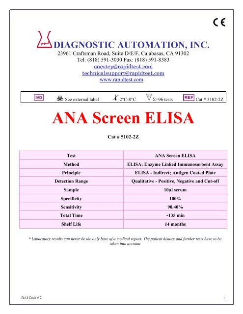
ANA Screen ELISA - Diagnostic Automation : Cortez Diagnostics
ANA Screen ELISA - Diagnostic Automation : Cortez Diagnostics
ANA Screen ELISA - Diagnostic Automation : Cortez Diagnostics
You also want an ePaper? Increase the reach of your titles
YUMPU automatically turns print PDFs into web optimized ePapers that Google loves.
The following components are not kit lot number dependent and may be used interchangeably with the<strong>ELISA</strong> assays: TMB, Stop Solution, and Wash Buffer.Note: Kit also contains1. Component list containing lot specific information is inside the kit box.2. Package insert providing instructions for use.STORAGE CONDITIONS1. Store the unopened kit between 2° and 8°C.2. Coated microwell strips: Store between 2° and 8°C. Extra strips should be immediately resealed withdesiccant and returned to proper storage. Strips are stable for 60 days after the envelope has beenopened and properly resealed and the indicator strip on the desiccant pouch remains blue.3. Conjugate: Store between 2° and 8°C. DO NOT FREEZE.4. Calibrator, Positive Control and Negative Control: Store between 2° and 8°C.5. TMB: Store between 2° and 8°C.6. Wash Buffer concentrate (10X): Store between 2° and 25°C. Diluted wash buffer (1X) is stable atroom temperature (20° to 25° C) for up to 7 days or for 30 days between 2° and 8°C.7. Sample Diluent: Store between 2° and 8°C.9. Stop Solution: Store between 2° and 25°C.QUALITY CONTROL1. Each time the assay is run the Calibrator must be run in triplicate. A reagent blank, Negative Control,and Positive Control must also be included in each assay.2. Calculate the mean of the three Calibrator wells. If any of the three values differ by more than 15%DAI Code # 2 3
from the mean, discard that value and calculate the mean using the remaining two wells.3. The mean OD value for the Calibrator and the OD values for the Positive and Negative Controlsshould fall within the following ranges:OD RangeNegative Control < 0.250Calibrator > 0.300Positive Control > 0.500a. The OD of the Negative Control divided by the mean OD of the Calibrator should be < 0.9.b. The OD of the Positive Control divided by the mean OD of the Calibrator should be > 1.25.c. If the above conditions are not met the test should be considered invalid and should berepeated.4. The Positive Control and Negative Control are intended to monitor for substantial reagent failure andwill not ensure precision at the assay cut-off.5. Additional controls may be tested according to guidelines or requirements of local, state, and/orfederal regulations or accrediting organizations.6. Refer to NCCLS document C24: Statistical Quality Control for Quantitative Measurements forguidance on appropriate QC practices.GENERAL PROCEDURE1. Remove the individual components from storage and allow them to warm to room temperature(20-25°C).2. Determine the number of microwells needed. Allow six Control/Calibrator determinations (one Blank,one Negative Control, three Calibrators and one Positive Control) per run. A Reagent Blank shouldbe run on each assay. Check software and reader requirements for the correct Controls/Calibratorconfigurations. Return unused strips to the resealable pouch with desiccant, seal, and return tostorage between 2° and 8°C.EXAMPLE PLATE SET-UP1 2A Blank Patient 3B Neg. Control Patient 4C Calibrator Etc.DCalibratorECalibratorFPos. ControlG Patient 1H Patient 23. Prepare a 1:21 dilution (e.g.: 10µL of serum + 200µL of Sample Diluent. NOTE: Shake WellBefore Use) of the Negative Control, Calibrator, Positive Control, and each patient serum.4. To individual wells, add 100µL of each diluted control, calibrator and sample. Ensure that the samplesare properly mixed. Use a different pipette tip for each sample.5. Add 100µL of Sample Diluent to well A1 as a reagent blank. Check software and reader requirementsfor the correct reagent blank well configuration.6. Incubate the plate at room temperature (20-25°C) for 60 to 65 minutes.7. Wash the microwell strips 5X.DAI Code # 2 4
A. Manual Wash Procedure:a. Vigorously shake out the liquid from the wells.b. Fill each microwell with Wash Buffer. Make sure no air bubbles are trapped in the wells.c. Repeat steps a. and b. for a total of 5 washes.d. Shake out the wash solution from all the wells. Invert the plate over a paper towel and tapfirmly to remove any residual wash solution from the wells. Visually inspect the plate to ensurethat no residual wash solution remains. Collect wash solution in a disposable basin and treatwith 0.5% sodium hypochlorite (bleach) at the end of the days run.B. Automated Wash Procedure:If using an automated microwell wash system, set the dispensing volume to 300-350µL/well. Setthe wash cycle for 5 washes with no delay between washes. If necessary, the microwell plate maybe removed from the washer, inverted over a paper towel and tapped firmly to remove any residualwash solution from the microwells.8. Add 100µL of the Conjugate to each well, including reagent blank well, at the same rate and in thesame order as the specimens were added.9. Incubate the plate at room temperature (20-25°C) for 30 to 35 minutes10. Wash the microwells by following the procedure as described in step 7.11. Add 100µL of TMB to each well, including reagent blank well, at the same rate and in the same orderas the specimens were added.12. Incubate the plate at room temperature (20-25°C) for 30 to 35 minutes.13. Stop the reaction by adding 50µL of Stop Solution to each well, including reagent blank well, at thesame rate and in the same order as the TMB was added. Positive samples will turn from blue toyellow. After adding the Stop Solution, tap the plate several times to ensure that the samples arethoroughly mixed.14. Set the microwell reader to read at a wavelength of 450nm and measure the optical density (OD) ofeach well against the reagent blank. The plate should be read within 30 minutes after the addition ofthe Stop Solution.CALCULATIONS/REPORTING RESULTSA. Calculations:1. Correction FactorA cutoff OD value for positive samples has been determined by the manufacturer and correlated to theCalibrator. The correction factor (CF) will allow you to determine the cutoff value for positive samples andto correct for slight day-to-day variations in test results. The correction factor is determined for each lot ofkit components and is printed on the Component List located in the kit box.2. Cutoff OD ValueTo obtain the cutoff OD value, multiply the CF by the mean OD of the Calibrator determined above.(CF x mean OD of Calibrator = cutoff OD value)3. Index Values or OD RatiosCalculate the Index Value or OD Ratio for each specimen by dividing its OD value by the cutoff OD fromstep 2.DAI Code # 2 5
Example:Mean OD of Calibrator = 0.793Correction Factor (CF) = 0.25Cut off OD = 0.793 x 0.25 = 0.198Unknown Specimen OD = 0.432Specimen Index Valueor OD Ratio= 0.432 / 0.198 = 2.18B. Interpretations:Index Values or OD ratios are interpreted as follows:Index Value or OD RatioNegative Specimens < 0.90Equivocal Specimens 0.91 to 1.09Positive Specimens > 1.10An OD ratio > 1.10 is interpreted as positive for IgG <strong>ANA</strong>. An OD ratio < 0.90 is interpreted as negativefor IgG <strong>ANA</strong>.Specimens with ratio values in the equivocal range are considered borderline for IgG <strong>ANA</strong>. Thesespecimens should be retested. Specimens which are repeatedly equivocal should be tested using analternative method such as the DAI <strong>ANA</strong> HEp-2 IFA test system.PRECAUTIONS1. For In Vitro <strong>Diagnostic</strong> Use.2. Normal precautions exercised in handling laboratory reagents should be followed. In case of contactwith eyes, rinse immediately with plenty of water and seek medical advice. Wear suitable protectiveclothing, gloves, and eye/face protection. Do not breathe vapor. Dispose of waste observing all local,state, and federal laws.3. The wells of the <strong>ELISA</strong> plate do not contain viable organisms. However, the strips should beconsidered POTENTIALLY BIOHAZARDOUS MATERIALS and handled accordingly.4. The human serum controls are POTENTIALLY BIOHAZARDOUS MATERIALS. Source materialsfrom which these products were derived were found negative for HIV-1 antigen, HBsAg. and forantibodies against HCV and HIV by approved test methods. However, since no test method can offercomplete assurance that infectious agents are absent, these products should be handled at theBiosafety Level 2 as recommended for any potentially infectious human serum or blood specimen inthe Centers for Disease Control/National Institutes of Health manual “Biosafety in Microbiological andBiomedical Laboratories”: current edition; and OSHA’s Standard for Bloodborne Pathogens (20).5. Adherence to the specified time and temperature of incubations is essential for accurate results. Allreagents must be allowed to reach room temperature (20-25°C) before starting the assay.Return unused reagents to refrigerated temperature immediately after use.6. Improper washing could cause false positive or false negative results. Be sure to minimize theamount of any residual wash solution; (e.g., by blotting or aspiration) before adding Conjugate orSubstrate. Do not allow the wells to dry out between incubations.7. The sample diluent, controls, wash buffer, and conjugate contain sodium azide at a concentration of0.1% (w/v). Sodium azide has been reported to form lead or copper azides in laboratory plumbingwhich may cause explosions on hammering. To prevent, rinse sink thoroughly with water afterdisposing of solution containing sodium azide.8. The Stop Solution is TOXIC. Causes burns. Toxic by inhalation, in contact with skin and if swallowed.DAI Code # 2 6
In case of accident or if you feel unwell, seek medical advice immediately.9. The TMB Solution is HARMFUL. Irritating to eyes, respiratory system and skin.10. The Wash Buffer concentrate is an IRRITANT. Irritating to eyes, respiratory system and skin.11. Wipe bottom of plate free of residual liquid and/or fingerprints that can alter optical density (OD)readings.12. Dilution or adulteration of these reagents may generate erroneous results.13. Reagents from other sources or manufacturers should not be used.14. TMB Solution should be colorless, very pale yellow, very pale green, or very pale blue when used.Contamination of the TMB with conjugate or other oxidants will cause the solution to change colorprematurely. Do not use the TMB if it is noticeably blue in color.15. Never pipette by mouth. Avoid contact of reagents and patient specimens with skin and mucousmembranes.16. Avoid microbial contamination of reagents. Incorrect results may occur.17. Cross contamination of reagents and/or samples could cause erroneous results18. Reusable glassware must be washed and thoroughly rinsed free of all detergents.19. Avoid splashing or generation of aerosols.20. Do not expose reagents to strong light during storage or incubation.21. Allowing the microwell strips and holder to equilibrate to room temperature prior to opening theprotective envelope will protect the wells from condensation.22. Wash solution should be collected in a disposal basin. Treat the waste solution with 10% householdbleach (0.5% sodium hypochlorite). Avoid exposure of reagents to bleach fumes.23. Caution: Liquid waste at acid pH should be neutralized before adding to bleach solution.24. Do not use <strong>ELISA</strong> plate if the indicator strip on the desiccant pouch has turned from blue to pink.25. Do not allow the conjugate to come in contact with containers or instruments that may havepreviously contained a solution utilizing sodium azide as a preservative. Residual amounts of sodiumazide may destroy the conjugate’s enzymatic activity.26. Do not expose any of the reactive reagents to bleach-containing solutions or to any strong odors frombleach-containing solutions. Trace amounts of bleach (sodium hypochlorite) may destroy thebiological activity of many of the reactive reagents within this kit.LIMITATIONS1. The <strong>ANA</strong> <strong>ELISA</strong> test is a diagnostic aid and by itself is not diagnostic. Test results should beinterpreted in conjunction with the clinical evaluation and the results of other diagnostic procedures.2. Positive <strong>ANA</strong> may be found in apparently healthy people. It is therefore imperative that the results beinterpreted in light of the patient’s clinical picture by a medical authority.3. SLE patients undergoing steroid therapy may have negative test results.4. Many commonly prescribed drugs may induce <strong>ANA</strong>.5. The DAI <strong>ANA</strong> <strong>Screen</strong> <strong>ELISA</strong> test system will not identify the specific type of <strong>ANA</strong> present in a positivespecimen. Positive specimens should be tested for individual autoantibodies using more specificreflex tests such as the DAI ENA Profile-6 <strong>ELISA</strong>, in combination with the DAI dsDNA <strong>ELISA</strong>.Alternatively, specific autoantibodies may be detected using a variety of methods includingimmunodiffusion, western blot or multiplexed fluorescent bead based assays such as the AtheNAMulti-Lyte <strong>ANA</strong> test system.REFERENCES1. Tan E, Cohen A, Fries J, et al: Special Article: The 1982 revised criteria for classification of systemiclupus erythematosus. Arthritis Rheum. 25:1271-1277, 1982.2. Beufels M, Kouki F, Mignon F, et al: Clinical significance of anti-Sm antibodies in systemic lupuserythematosus. Am. J. Med. 74:201-215, 1983.3. Sharp GC, Irwin WS, Tan EM, Holman H: Mixed connective tissue disease. An apparently distinctrheumatic disease syndrome associated with a specific antibody to an extractable nuclear antigenDAI Code # 2 7
(ENA). Am. J. Med. 52: 148-159, 1972.4. Infield JB, Brunner CB, Koffler DB: Serological studies in patients with systemic lupus erythematosusand central nervous system dysfunction. Arthritis Rheum. 21:289-294, 1978.5. Tan EM, Kunkel HG: Characteristics of a soluble nuclear antigen precipitating with sera of patientswith systemic lupus erythematosus. J. Immunol. 96:464-471, 1966.6. Maddison PJ, Mogavero H, Provost TT, Reichlin M: The clinical significance of autoantibodies tosoluble cytoplasmic antigen in systemic lupus erythematosus and other connective tissue diseases. J.Rheumatol. 6:189-192, 1979.7. Clark G, Reichlin M, Tomasi TB: Characterization of soluble cytoplasmic antigen reactive with serafrom patients with systemic lupus erythematosus. J. Immunol. 102:117, 1969.8. Alexander E, Arnett FC, Provost TT, Stevens MB: The Ro(SSA) and La(SSB) antibody system andSjögren’s syndrome. J. Rheum. 9:239-246, 1982.9. Alspaugh MA, Talal N, and Tan E: Differentiation and characterization of autoantibodies and theirantigens in Sjögren’s syndrome. Arthritis Rheum. 19:216-222, 1976.10. Marguerie C, Bunn CC, Beynon HL, et al: Polymyositis, pulmonary fibrosis and autoantibodies toaminoacyl-tRNA synthetase enzymes. Quart. J. Med. 77:1019-1038, 1990.11. Tan EM: Antinuclear antibodies: <strong>Diagnostic</strong> markers for autoimmune diseases and probes for cellbiology. Adv. Immunol. 44:93-151, 1989.12. Sontheimer RD, Thomas JR, Gilliam JN: Subacute cutaneous lupus erythematosus: A cutaneousmarker for a distinct lupus erythematosus subset. Arch. Derm. 115:1409-1415, 1979.13. Provost TT, Arnett FC, Reichlin M: Homozygous C2 deficiency, lupus erythematosus and anti Ro(SSA) antibodies. Arth. Rheum. In Press.14. LeRoy EC, Black CM, Fleishmajer R, et al: Scleroderma (systemic sclerosis): Classification, subsetsand pathogenesis. J. Rheumatol. 15:202-205, 1988.15. Weiner ES, Hildebrandt S, Senecal JL, et al: Prognostic significance of anticentromere antibodiesand anti-topoisomerase 1 antibodies in Raynaud’s disease. A prospective study. Arthritis Rheum.34:68-77, 1991.16. Mongey AB, Hess EV: Antinuclear antibodies and disease specificity. Advances in Int. Med. 36 (1):151-169, 1989.17. Procedures for the Handling and Processing of Blood Specimens. NCCLS Document H18-A, Vol. 10,No. 12, Approved Guideline, 1990.18. Procedures for the collection of diagnostic blood specimens by venipuncture. 2nd edition. ApprovedStandard (1984). Published by National Committee for clinical Laboratory Standards.19. Sturgess A: Review; Recently characterized autoantibodies and their clinical significance. Aust. N.Z.,J. Med. 22:279-289, 1992.20. U.S. Department of Labor, Occupational Safety and Health Administration: Occupational Exposure toBloodborne Pathogens, Final Rule. Fed. Register 56:64175-64182, 1991.Date Adopted Reference No.2005-08-23 DA-<strong>ANA</strong> <strong>Screen</strong>-2009DIAGNOSTIC AUTOMATION, INC.23961 Craftsman Road, Suite D/E/F, Calabasas, CA 91302Tel: (818) 591-3030 Fax: (818) 591-8383ISO 13485-2003Revision Date: 10/07/2009DAI Code # 2 8
CEpartner4U, 3951DB; 13.NL., Tel: +31(0)6.516.536.2DAI Code # 2 9


