Nanostructured Spinel ZnFe2O4 for the Detection of ... - ResearchGate
Nanostructured Spinel ZnFe2O4 for the Detection of ... - ResearchGate
Nanostructured Spinel ZnFe2O4 for the Detection of ... - ResearchGate
- No tags were found...

Create successful ePaper yourself
Turn your PDF publications into a flip-book with our unique Google optimized e-Paper software.
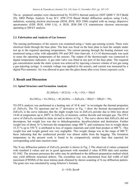
Sensors & Transducers Journal, Vol. 134, Issue 11, November 2011, pp. 107-119The as –prepared samples were characterized by TG/DTA <strong>the</strong>rmal analyzer (SDT Q600 V 20.9 Build20), XRD Philips Analytic X-ray B.V. (PW-3710 Based Model diffraction analysis using Cu-K αradiation), scanning electron microscope (SEM, JEOL JED 2300) coupled with an energy dispersivespectrometer (EDS JEOL 6360 LA), A JEOL JEM–200 CX transmission electron microscopeoperating at 200 kV analysis.2.2. Fabrication and Analysis <strong>of</strong> Gas SensorsThe sensing per<strong>for</strong>mance <strong>of</strong> <strong>the</strong> sensors was examined using a “static gas-sensing system. There wereelectrical feeds through <strong>the</strong> base plate. The heat was fixed on <strong>the</strong> base plate to heat <strong>the</strong> sample undertest up to <strong>the</strong> required operating temperatures. The current passing through <strong>the</strong> heating element wasmonitored using a relay with adjustable ON and OFF time intervals. A Cr-Al <strong>the</strong>rmocouple was usedto sense <strong>the</strong> operating temperature <strong>of</strong> <strong>the</strong> sensors. The output <strong>of</strong> <strong>the</strong> <strong>the</strong>rmocouple was connected todigital temperature indicators. A gas inlet valve was fitted at one port <strong>of</strong> <strong>the</strong> base plate. The requiredgas concentration inside <strong>the</strong> static system was achieved by injecting a known volume <strong>of</strong> test gas usinga gas-injecting syringe. A constant voltage was applied to <strong>the</strong> sensors, and current was measured by adigital Pico-ammeter. Air was allowed to pass into <strong>the</strong> glass dome after every Gases exposure cycle.3. Result and Discussion3.1. <strong>Spinel</strong> Structure and Formation Analysis2C 2 NH 3 O 2 + (9/2) O 2 → N 2 + CO 2 + 5H 2 OZn (NO 3 ) 3 + Fe (NO 3 ) 3 + 4C 2 H 5 NO 2 → ZnFe 2 O 4 + 8H 2 O + 10H 2 O + 5N 2TG-DTA analysis was per<strong>for</strong>med at a heating rate <strong>of</strong> 10 K min -1 to investigate <strong>the</strong> <strong>the</strong>rmal properties<strong>of</strong>. ZnFe 2 O 4. The TG spectrum and its 1 st derivative in Fig. 1 show <strong>the</strong> <strong>the</strong>rmal decomposition <strong>of</strong>ZnFe 2 O 4 is <strong>the</strong> curve indicates that <strong>the</strong> slight weight loss in ZnFe 2 O 4 powder due to little loss about14.66 at temperature up to 200 0 C in ZnFe 2 O 4 <strong>of</strong> moisture, carbon dioxide and nitrogen gas. The DTAcurve <strong>of</strong> ZnFe 2 O 4 recorded in static air and in shown in Fig. 1. The curve shown that ZnFe 2 O 4 did notdecompose, but weight loss was due to dehydrogenation, decarboxylation and denitration. Fur<strong>the</strong>rweight loss <strong>of</strong> about 16 % between <strong>the</strong> temperature range 400 0 C and continuous loss in weight about33.33 % up to 550 0 C is attributed to loss <strong>of</strong> organic materials and yield final product at 600 0 C. Thisweight loss and weight gained was very negligible. This weight change was in <strong>the</strong> range <strong>of</strong> 800 0 C<strong>the</strong>se indicating that <strong>the</strong> syn<strong>the</strong>sized powder was almost stable from <strong>the</strong> begging. The <strong>for</strong>mationtemperature in <strong>the</strong> present work is found to be comparatively similar than that reported <strong>for</strong>corresponding solid state reaction route.The X-ray diffraction pattern <strong>of</strong> ZnFe 2 O 4 powder is shown in Fig. 2 The observed d values comparedwith standard d values and are in good agreement with standard d value JCPDS data card number82-1042. The structure possesses <strong>the</strong> cubic may be attributed to <strong>the</strong> different preparation method whichmay yield different structural defects. The crystalline size was determined from full width <strong>of</strong> halfmaximum (FWHM) <strong>of</strong> <strong>the</strong> most intense peak obtained by shown scanning <strong>of</strong> X-ray diffraction pattern.The grain size was calculated by using Scherrer’s <strong>for</strong>mula.d = 0.9λ/ βcosθ109