Codium decorticatum
Codium decorticatum
Codium decorticatum
- No tags were found...

You also want an ePaper? Increase the reach of your titles
YUMPU automatically turns print PDFs into web optimized ePapers that Google loves.
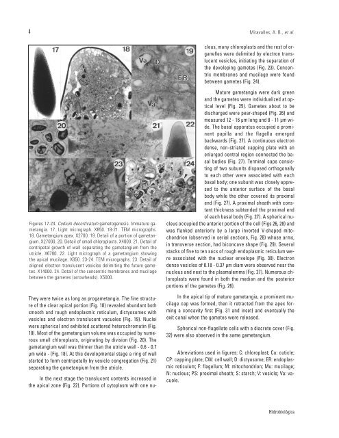
4Miravalles, A. B., et al.In the next stage the translucent contents increased inthe apical zone (Fig. 22). Portions of cytoplasm with one nucleus,many chloroplasts and the rest of organelleswere delimited by electron translucentvesicles, initiating the separation ofthe developing gametes (Fig. 23). Concentricmembranes and mucilage were foundbetween gametes (Fig. 24).Figures 17-24. <strong>Codium</strong> <strong>decorticatum</strong> gametogenesis. Immature gametangia.17. Light micrograph. X850. 18-21. TEM micrographs.18. Gametangium apex. X2700. 19. Detail of a portion of gametangium.X27000. 20. Detail of small chloroplasts. X4000. 21. Detail ofcentripetal growth of wall separating the gametangium from theutricle. X6700. 22. Light micrograph of a gametangium showingthe apical mucilage. X850. 23-24. TEM micrographs. 23. Detail ofaligned electron translucent vesicles delimiting the future gametes.X14000. 24. Detail of the concentric membranes and mucilagebetween the gametes (arrowheads). X5000.They were twice as long as progametangia. The fine structureof the clear apical portion (Fig. 18) revealed abundant bothsmooth and rough endoplasmic reticulum, dictyosomes withvesicles and electron translucent vacuoles (Fig. 19). Nucleiwere spherical and exhibited scattered heterochromatin (Fig.18). Most of the gametangium volume was occupied by numeroussmall chloroplasts, originating by division (Fig. 20). Thegametangium wall was thinner than the utricle wall - 0.6 - 0.7µm wide - (Fig. 18). At this developmental stage a ring of wallstarted to form centripetally by vesicle congregation (Fig. 21)separating the gametangium from the utricle.Mature gametangia were dark greenand the gametes were individualized at opticallevel (Fig. 25). Gametes about to bedischarged were pear-shaped (Fig. 26) andmeasured 12 - 16 µm long and 8 - 11 µm wide.The basal apparatus occupied a prominentpapilla and the flagella emergedbackwards (Fig. 27). A continuous electrondense, non-striated capping plate with anenlarged central region connected the basalbodies (Fig. 27). Terminal caps consistingof two subunits disposed orthogonallyto each other were associated with eachbasal body; one subunit was closely appresedto the anterior surface of the basalbody while the other covered its proximalend (Fig. 27). A proximal sheath with constantthickness subtended the proximal endof each basal body (Fig. 27). A spherical nucleusoccupied the anterior portion of the cell (Figs 26, 28) andwas flanked anteriorly by a large inverted V-shaped mitochondrion(observed in serial sections, Fig. 28) whose arms,in transverse section, had biconcave shape (Fig. 29). Severalstacks of five to ten sacs of rough endoplasmic reticulum wereassociated with the nuclear envelope (Fig. 30). Electrondense vesicles of 0.18 - 0.37 µm diam were observed near thenucleus and next to the plasmalemma (Fig. 27). Numerous chloroplastswere found in both the median and the posteriorportions of the gametes (Fig. 26).In the apical tip of mature gametangia, a prominent mucilagecap was formed, then it retracted from the apex forminga concavity first (Fig. 31 and inset) and eventually theexit canal when the gametes were released.Spherical non-flagellate cells with a discrete cover (Fig.32) were also observed in the same gametangium.Abreviations used in figures: C: chloroplast; Cu: cuticle;CP: capping plate; CW: cell wall; D: dictyosome; ER: endoplasmicreticulum; F: flagellum; M: mitochondrion; Mu: mucilage;N: nucleus; PS: proximal sheath; S: starch; V: vesicle; Va: vacuole.Hidrobiológica