PDF Lesson 2: Structure of the Mammalian Heart - Cary Academy
PDF Lesson 2: Structure of the Mammalian Heart - Cary Academy
PDF Lesson 2: Structure of the Mammalian Heart - Cary Academy

Create successful ePaper yourself
Turn your PDF publications into a flip-book with our unique Google optimized e-Paper software.
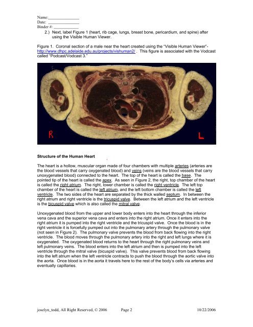
Name:_______________Date: _______________Binder #: ____________2.) Next, label Figure 1 (heart, rib cage, lungs, breast bone, pericardium, and spine) afterusing <strong>the</strong> Visible Human Viewer.Figure 1. Coronal section <strong>of</strong> a male near <strong>the</strong> heart created using <strong>the</strong> “Visible Human Viewer”-http://www.dhpc.adelaide.edu.au/projects/vishuman2/ . This figure is associated with <strong>the</strong> Vodcastcalled “Podcast/Vodcast 3.”<strong>Structure</strong> <strong>of</strong> <strong>the</strong> Human <strong>Heart</strong>The heart is a hollow, muscular organ made <strong>of</strong> four chambers with multiple arteries (arteries are<strong>the</strong> blood vessels that carry oxygenated blood) and veins (veins are <strong>the</strong> blood vessels that carryunoxygenated blood) connected to <strong>the</strong> heart. The top <strong>of</strong> <strong>the</strong> heart is called <strong>the</strong> base. Thepointed tip <strong>of</strong> <strong>the</strong> heart is called <strong>the</strong> apex. As seen in Figure 2, <strong>the</strong> right, top chamber <strong>of</strong> <strong>the</strong> heartis called <strong>the</strong> right atrium. The right, lower chamber is called <strong>the</strong> right ventricle. The left topchamber <strong>of</strong> <strong>the</strong> heart is called <strong>the</strong> left atrium, and <strong>the</strong> left bottom chamber is called <strong>the</strong> leftventricle. The two sides <strong>of</strong> <strong>the</strong> heart are separated by <strong>the</strong> thick walled septum. In between <strong>the</strong>right atrium and right ventricle is <strong>the</strong> tricuspid valve. Between <strong>the</strong> left atrium and <strong>the</strong> left ventricleis <strong>the</strong> bicuspid valve which is also called <strong>the</strong> mitral valve.Unoxygenated blood from <strong>the</strong> upper and lower body enters into <strong>the</strong> heart through <strong>the</strong> inferiorvena cava and <strong>the</strong> superior vena cava and enters into <strong>the</strong> right atrium. Once it enters into <strong>the</strong>right atrium it is pumped into <strong>the</strong> right ventricle and <strong>the</strong> tricuspid valve. Once <strong>the</strong> blood is in <strong>the</strong>right ventricle it is forcefully pumped out into <strong>the</strong> pulmonary artery through <strong>the</strong> pulmonary valve(not seen in Figure 2). The pulmonary valve prevents <strong>the</strong> blood from back flowing into <strong>the</strong> rightventricle. The blood moves through <strong>the</strong> pulmonary artery into <strong>the</strong> right and left lungs where it isoxygenated. The oxygenated blood returns to <strong>the</strong> heart through <strong>the</strong> right pulmonary veins andleft pulmonary veins. The blood enters into <strong>the</strong> left atrium and <strong>the</strong>n is pumped into <strong>the</strong> leftventricle through <strong>the</strong> mitral valve (bicuspid valve). This valve prevents blood from back flowinginto <strong>the</strong> left atrium when <strong>the</strong> left ventricle contracts to push <strong>the</strong> blood through <strong>the</strong> aortic valve into<strong>the</strong> aorta. Once blood is in <strong>the</strong> aorta it travels here to <strong>the</strong> rest <strong>of</strong> <strong>the</strong> body’s cells via arteries andeventually capillaries.joselyn_todd, All Right Reserved, © 2006 Page 2 10/22/2006