TIRF: Evanescent waves for high resolution membrane ... - Nikon
TIRF: Evanescent waves for high resolution membrane ... - Nikon
TIRF: Evanescent waves for high resolution membrane ... - Nikon
- No tags were found...

You also want an ePaper? Increase the reach of your titles
YUMPU automatically turns print PDFs into web optimized ePapers that Google loves.
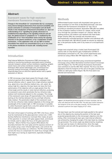
Abstract • Introduction • MethodsAbstract:<strong>Evanescent</strong> <strong>waves</strong> <strong>for</strong> <strong>high</strong> <strong>resolution</strong><strong>membrane</strong> fluorescence imagingChanges in the intracellular Ca 2+ concentration ([Ca 2+ ] i ) accompanymany diverse biological phenomena from neuronal excitability andmuscle contraction to activation of transcription and cell death.Thanks to the introduction of fluorescent intracellular Ca 2+ dyes ourunderstanding of Ca 2+ signalling has greatly advanced; it isestablished that depending on the signalling molecule and celltype, the global increase in cytoplasmic Ca 2+ is mediated by themobilisation of Ca 2+ from intracellular stores and by the openingchannels on the plasma <strong>membrane</strong>, or by a combination of bothmechanisms. In this application note, we use <strong>TIRF</strong> to monitorintracellular free calcium ion concentration [Ca 2+ ] i on or very closeto the plasma <strong>membrane</strong> of muscle cells including humanmyotubesIntroductionTotal Internal Reflection Fluorescence (<strong>TIRF</strong>) microscopy is amethod of monitoring <strong>membrane</strong> associated events includingvesicular transport, protein surface <strong>membrane</strong> targeting, synapticvesicle fusion and surface <strong>membrane</strong> receptor assembly. Thistechnique offers several advantages over conventional linescanning confocal microscopy, including the possibility ofacquiring data from an ultra thin optical section with a spatial<strong>resolution</strong> of 100 nm.MethodsDifferentiated human muscle cells (myotubes) were grown onglass coverslips 0.12 mm thick as described previously 1 and wereloaded with the fluorescent intracellular Ca 2+ dye, fluo-4,according to standard procedures. Ryanodine (500 μM) was addedto the incubation medium to block Ca 2+ release, which wouldoccur through the ryanodine receptor Ca 2+ channel. After theloading protocol, the medium was replaced with fresh Krebs-Ringer solution (KR); the coverslip was mounted onto athermostatically controlled perfusion chamber and individual cellswere stimulated with 100 mM KCl (made iso-osmotic by replacingNa + in the perfusion buffer, with K + ) by means of a microperfusionsystem.Images were acquired using a cooled, back-illuminated CCDcamera with on-chip electron gain multiplication (C9100-13Hamamatsu) connected to a PC. The current experiments werecarried out using a <strong>Nikon</strong> 60x <strong>TIRF</strong> objective (N.A. = 1.49).Cells of interest were identified using conventional brightfieldmicroscopy using a <strong>Nikon</strong> Biomedical inverted research microscopewith PFS (figure 1A). The PFS and epi-fluorescence illuminationwere switched on and the SRIC filter inserted. The focus was thenadjusted so that the footprint of the cell on the glass coverslip wasvisible as a dark grey outline (figure 1B); this focal plane wasselected and memorised.In <strong>TIRF</strong> microscopy a laser beam passes first though a <strong>high</strong>numerical aperture (N.A.) objective and then undergoes totalinternal reflection when it passes from a <strong>high</strong>-refractive medium(e.g., glass) into a low-refractive medium (e.g., cell, water). Byusing a <strong>high</strong> N.A. objective, the laser beam can leave the frontoptical plane of the objective with an angle greater than thecritical angle and this results in total internal reflection, producingan evanescent wave which penetrates the low refractive indexmedium (cell <strong>membrane</strong>) adjacent to the coverglass. The energyof the evanescent wave drops off exponentially in the z directionstarting at the interface between the low and <strong>high</strong> refractiveindex medium. The penetration depth of the evanescent waveinto the low refractive index medium depends on the energy andwavelength of the incident light. In <strong>TIRF</strong> microscopy it is possibleto collect fluorescent signals from a very thin optical section (up to100 nm) adjacent to the cell <strong>membrane</strong>.Figure 1: (A) Brightfield photomicrograph of human myotubes. (B) Thesame cells observed with the SRIC filter. The dark grey outline representsthe footprint of the cell on the glass coverslip. This focal plane wasselected to per<strong>for</strong>m time lapse imaging of Ca 2+ influx.The most critical aspect of working with <strong>TIRF</strong> is the focus, whichmust be maintained at the glass-specimen contact site. A SRIC(Surface Reflective Interference Contrast) filter makes all partsof the specimen in contact with glass appear black, and shouldbe initially used to adjust the focus at the glass-specimeninterface. <strong>Nikon</strong>’s Perfect Focus System (PFS) detects theboundary between the cover glass and the aqueous solution ofthe sample using a near infrared light beam, and controls thefocus using the boundary as a reference. The PFS continuouslycorrects the focus to compensate <strong>for</strong> small focus changes (drift)that might occur during an experiment, especially timelapseexperiments.The laser beam was first centred and made parallel to the opticalaxis of the objective by viewing the position of the laser spotprojected perpendicularly to the microscope’s objective onto theceiling. After minimising the laser spot, total internal reflectionwas achieved by changing the angle of the laser beam by rotatingthe goniometer. The correct angle was determined by visualisingthe laser spot by means of a Bertrand lens. Once the angleparameters were set with the goniometer, Ca 2+ influx could beassessed.