TIRF: Evanescent waves for high resolution membrane ... - Nikon
TIRF: Evanescent waves for high resolution membrane ... - Nikon
TIRF: Evanescent waves for high resolution membrane ... - Nikon
- No tags were found...

Create successful ePaper yourself
Turn your PDF publications into a flip-book with our unique Google optimized e-Paper software.
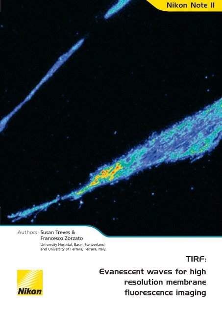
<strong>Nikon</strong> Note 11Authors: Susan Treves &Francesco ZorzatoUniversity Hospital, Basel, Switzerlandand University of Ferrara, Ferrara, Italy.<strong>TIRF</strong>:<strong>Evanescent</strong> <strong>waves</strong> <strong>for</strong> <strong>high</strong><strong>resolution</strong> <strong>membrane</strong>fluorescence imaging
Abstract • Introduction • MethodsAbstract:<strong>Evanescent</strong> <strong>waves</strong> <strong>for</strong> <strong>high</strong> <strong>resolution</strong><strong>membrane</strong> fluorescence imagingChanges in the intracellular Ca 2+ concentration ([Ca 2+ ] i ) accompanymany diverse biological phenomena from neuronal excitability andmuscle contraction to activation of transcription and cell death.Thanks to the introduction of fluorescent intracellular Ca 2+ dyes ourunderstanding of Ca 2+ signalling has greatly advanced; it isestablished that depending on the signalling molecule and celltype, the global increase in cytoplasmic Ca 2+ is mediated by themobilisation of Ca 2+ from intracellular stores and by the openingchannels on the plasma <strong>membrane</strong>, or by a combination of bothmechanisms. In this application note, we use <strong>TIRF</strong> to monitorintracellular free calcium ion concentration [Ca 2+ ] i on or very closeto the plasma <strong>membrane</strong> of muscle cells including humanmyotubesIntroductionTotal Internal Reflection Fluorescence (<strong>TIRF</strong>) microscopy is amethod of monitoring <strong>membrane</strong> associated events includingvesicular transport, protein surface <strong>membrane</strong> targeting, synapticvesicle fusion and surface <strong>membrane</strong> receptor assembly. Thistechnique offers several advantages over conventional linescanning confocal microscopy, including the possibility ofacquiring data from an ultra thin optical section with a spatial<strong>resolution</strong> of 100 nm.MethodsDifferentiated human muscle cells (myotubes) were grown onglass coverslips 0.12 mm thick as described previously 1 and wereloaded with the fluorescent intracellular Ca 2+ dye, fluo-4,according to standard procedures. Ryanodine (500 μM) was addedto the incubation medium to block Ca 2+ release, which wouldoccur through the ryanodine receptor Ca 2+ channel. After theloading protocol, the medium was replaced with fresh Krebs-Ringer solution (KR); the coverslip was mounted onto athermostatically controlled perfusion chamber and individual cellswere stimulated with 100 mM KCl (made iso-osmotic by replacingNa + in the perfusion buffer, with K + ) by means of a microperfusionsystem.Images were acquired using a cooled, back-illuminated CCDcamera with on-chip electron gain multiplication (C9100-13Hamamatsu) connected to a PC. The current experiments werecarried out using a <strong>Nikon</strong> 60x <strong>TIRF</strong> objective (N.A. = 1.49).Cells of interest were identified using conventional brightfieldmicroscopy using a <strong>Nikon</strong> Biomedical inverted research microscopewith PFS (figure 1A). The PFS and epi-fluorescence illuminationwere switched on and the SRIC filter inserted. The focus was thenadjusted so that the footprint of the cell on the glass coverslip wasvisible as a dark grey outline (figure 1B); this focal plane wasselected and memorised.In <strong>TIRF</strong> microscopy a laser beam passes first though a <strong>high</strong>numerical aperture (N.A.) objective and then undergoes totalinternal reflection when it passes from a <strong>high</strong>-refractive medium(e.g., glass) into a low-refractive medium (e.g., cell, water). Byusing a <strong>high</strong> N.A. objective, the laser beam can leave the frontoptical plane of the objective with an angle greater than thecritical angle and this results in total internal reflection, producingan evanescent wave which penetrates the low refractive indexmedium (cell <strong>membrane</strong>) adjacent to the coverglass. The energyof the evanescent wave drops off exponentially in the z directionstarting at the interface between the low and <strong>high</strong> refractiveindex medium. The penetration depth of the evanescent waveinto the low refractive index medium depends on the energy andwavelength of the incident light. In <strong>TIRF</strong> microscopy it is possibleto collect fluorescent signals from a very thin optical section (up to100 nm) adjacent to the cell <strong>membrane</strong>.Figure 1: (A) Brightfield photomicrograph of human myotubes. (B) Thesame cells observed with the SRIC filter. The dark grey outline representsthe footprint of the cell on the glass coverslip. This focal plane wasselected to per<strong>for</strong>m time lapse imaging of Ca 2+ influx.The most critical aspect of working with <strong>TIRF</strong> is the focus, whichmust be maintained at the glass-specimen contact site. A SRIC(Surface Reflective Interference Contrast) filter makes all partsof the specimen in contact with glass appear black, and shouldbe initially used to adjust the focus at the glass-specimeninterface. <strong>Nikon</strong>’s Perfect Focus System (PFS) detects theboundary between the cover glass and the aqueous solution ofthe sample using a near infrared light beam, and controls thefocus using the boundary as a reference. The PFS continuouslycorrects the focus to compensate <strong>for</strong> small focus changes (drift)that might occur during an experiment, especially timelapseexperiments.The laser beam was first centred and made parallel to the opticalaxis of the objective by viewing the position of the laser spotprojected perpendicularly to the microscope’s objective onto theceiling. After minimising the laser spot, total internal reflectionwas achieved by changing the angle of the laser beam by rotatingthe goniometer. The correct angle was determined by visualisingthe laser spot by means of a Bertrand lens. Once the angleparameters were set with the goniometer, Ca 2+ influx could beassessed.
Results• ConclusionResultsThe trace and the pseudo-coloured images show the changes in [Ca 2+ ] ioccurring in the myotube shown (figure 2). Changes in fluorescencewere obtained with the PFS ‘on’ and by illuminating fluo-4 loaded cellswith a sapphire laser (488 nm), thus only the fluorescence emitted atthe fixed focal plane was visible. The addition of 100 mM KCl (whichmimics depolarisation) caused a rapid influx of Ca 2+ independent of Ca 2+release mediated by the ryanodine receptor in intracellular Ca 2+ storesactivation, since the latter was blocked by incubation with 500 µMryanodine. In muscle cells, the depolarisation-induced Ca 2+ influx istermed ‘excitation coupled Ca 2+ entry’ or ECCE and is thought to bemediated by the dihydropyridine receptor L-type Ca 2+ channel 2,3 .Removal of KCl and addition of Krebs Ringer with 0.5 mM EGTAblocked Ca 2+ influx. The subsequent addition of 2 mM Ca 2+ to theextracellular medium also resulted in Ca 2+ influx, but the latter Ca 2+influx was not mediated by ECCE.Conclusion:Figure 2: Changes in fluo-4 fluorescence in human myotubes perfusedwith 100 mM KCl or after the re-addition of 2 mM CaCl 2 .<strong>TIRF</strong> technology is a powerful tool <strong>for</strong> studying <strong>membrane</strong>-associatedevents with <strong>high</strong> spatial <strong>resolution</strong>. In this study we have shown that<strong>TIRF</strong> can be used to monitor changes in [Ca 2+ ] occurring at or veryclose to the plasma <strong>membrane</strong> of human myotubes by using <strong>TIRF</strong> incombination with the fast Ca 2+ indicator, fluo-4. Similar studies can beper<strong>for</strong>med on other cell types 4 to gather in<strong>for</strong>mation on the role ofCa 2+ influx in normal and pathophysiological events.Copyright © <strong>Nikon</strong> Corporation. All Rights Reserved.
References • Special FeaturesYour <strong>Nikon</strong> Imaging Centre (NIC):www.nikonimagingcenters.comSusan TrevesAuthors’ BackgroundSusan Treves PhD is Research Group Leader at the Departments ofAnaesthesia and Biomedicine, Basel University Hospital, Basel,Switzerland and Assistant Professor, University of Ferrara School ofMedicine, Ferrara, Italy. Main research interests include calciumhomeostasis in excitable and non excitable cells and skeletal muscleexcitation-contraction coupling. E-mail: susan.treves@unibas.chFrancesco Zorzato MD, PhD is an Associate Professor at the School ofMedicine, University, of Ferrara, Ferrara, Italy. Main research interestsfocus on the biology and pathology of skeletal muscle excitation andcontraction coupling. Email: zor@unife.itReferences:Francesco Zorzato1. Ducreux S, Zorzato F, Müller C, Sewry C, Muntoni F, Quinlivan R, RestagnoG, Girard T, Treves S. Effect of ryanodine receptor mutations on interleukin-6 release and intracellular calcium homeostasis in human myotubes frommalignant hyperthermia-susceptible individuals and patients affected bycentral core disease. J Biol Chem. 2004;279(42):43838-46.2. Cherednichenko G, Hurne AM, Fessenden JD, Hui Lee E, Allen PD, BeamKG & Pessah IN.Co n<strong>for</strong>mational activation of Ca 2+ entry by depolarizationof skeletal myotubes. Proc. Natl. Acad. Sci. U.S.A. 2004;101: 15793-15798.3. Bannister RA, Pessah IN, & Beam KG. The skeletal L-type Ca 2+ current is amajor contributor to excitation-coupled Ca 2+ entry. J. Gen Physiol.2009;133:79-91.The <strong>Nikon</strong> Instruments organisation has established anumber of <strong>Nikon</strong> Imaging Centres to make available thelatest microscope developments to the researchers and tocreate an interaction between research and the <strong>Nikon</strong>product development. <strong>Nikon</strong> Imaging centres have beenestablished in:Heidelberg University, GermanyOx<strong>for</strong>d University, United KingdomMarie Curie Institute, FranceHarvard Medical School, USAUniversity Cali<strong>for</strong>nia San Francisco, USAHokkaido University, JapanSingapore Bioimaging Consortium, SingaporeNorthwestern University Chicago, USA<strong>TIRF</strong> is a powerful tool <strong>for</strong> studying<strong>membrane</strong>-associated events with <strong>high</strong> spatial<strong>resolution</strong>A critical aspect of <strong>TIRF</strong> is to maintain focus atthe glass-specimen contact site<strong>Nikon</strong>’s Perfect Focus System ensures focusduring timelapse experiments<strong>TIRF</strong> can be used to monitor changes inintracellular calcium concentration at, or veryclose, to the plasma <strong>membrane</strong> of humanmyotubes4. Ravier MA, Tsuboi T & Rutter GA. Imaging a target of Ca 2+ signaling:dense core granule exocytosis viewed by total internal reflectionfluorescence microscopy. Methods 2008; 46: 233-238.For more in<strong>for</strong>mation on <strong>Nikon</strong> <strong>TIRF</strong> systems go to:www.nikoninstruments.com/In<strong>for</strong>mation-Center/<strong>TIRF</strong>AsiaNIKON CORPORATION6-3, Nishiohi 1-chome, Shinagawa-ku, Tokyo 140-8601, Japanphone: +81-3-3773-8973 fax: +81-3-3773-8986www.nikon-instruments.jpEurope and AfricaNIKON INSTRUMENTS EUROPE B.V.Laan van Kronenburg 2 1183AS Amstelveen The Netherlandsphone: +31-20-44-96-230 fax: +31-20-44-96-298www.nikoninstruments.euAmericasNIKON INSTRUMENTS INC.1300 Walt Whitman Road, Melville, N.Y. 11747-3064, U.S.A.phone: +1-631-547-8500; +1-800-52-NIKON (within the U.S.A.only)fax: +1-631-547-0306www.nikoninstruments.comNIKON INSTRUMENTS (SHANGHAI) CO., LTD.phone: +86-21-5836-0050 fax: +86-21-5836-0030NIKON SINGAPORE PTE LTDphone: +65-6559-3618 fax: +65-6559-3668NIKON MALAYSIA SDN. BHD.phone: +60-3-78763887 fax: +60-3-78763387NIKON INSTRUMENTS KOREA CO., LTD.phone: +82-2-2186-8410 fax: +82-2-555-4415NIKON FRANCE S.A.S.phone: +33-1-45-16-45-16 fax: +33-1-45-16-00-33NIKON GMBHphone: +49-211-9414-0 fax: +49-211-9414-322NIKON INSTRUMENTS S.p.A.phone: +39-55-3009601 fax: +39-55-300993NIKON AGphone: +41-43-277-2860 fax: +41-43-277-2861NIKON UK LTD.phone: +44-20-8541-4440 fax: +44-20-8541-4584NIKON GMBH AUSTRIAphone: +43-1-972-6111-00 fax: +43-1-972-6111-40NIKON BELUXphone: +32-2-705-56-65 fax: +32-2-726-66-45NIKON CANADA INC.phone: +1-905-625-9910 fax: +1-905-625-0103Copyright © <strong>Nikon</strong> Corporation. All Rights Reserved. 2NN11-7-09