THE EVOLUTION OF HUMAN SKIN AND SKIN COLOR Nina G ...
THE EVOLUTION OF HUMAN SKIN AND SKIN COLOR Nina G ...
THE EVOLUTION OF HUMAN SKIN AND SKIN COLOR Nina G ...

You also want an ePaper? Increase the reach of your titles
YUMPU automatically turns print PDFs into web optimized ePapers that Google loves.
Annu. Rev. Anthropol. 2004. 33:585-623doi: 10.1l46/annurev.anthro.33.070203.143955Copyright © 2004 by Annual Reviews. All rights reservedFirst published online as a Review in Advance on June 21, 2004<strong>THE</strong> <strong>EVOLUTION</strong> <strong>OF</strong> <strong>HUMAN</strong> <strong>SKIN</strong> <strong>AND</strong><strong>SKIN</strong> <strong>COLOR</strong><strong>Nina</strong> G. JablonskiDepartment of Anthmpology. California Academy of Sciences, San Francisco,California 98103; email: njablonski@calacademy.orgKey Wordspigmentation, melanin, UV radiation, thermoregulation, race• Abstract Humans skin is the most visible aspect of the human phenotype. It isdistinguished mainly by its naked appearance, greatly enhanced abilities to dissipatebody heat through sweating, and the great range of genetically determined skin colorspresent within a single species. Many aspects of the evolution of human skin and skincolor can be reconstructed using comparative anatomy, physiology, and genomics.Enhancement of thermal sweating was a key innovation in human evolution that allowedmaintenance of homeostasis (including constant brain temperature) during sustainedphysical activity in hot environments. Dark skin evol ved pan pa^jw with the loss of bodyhair and was the original state for the genus Homo. Melanin pigmentation is adaptiveand has been maintained by natural selection. Because of its evolutionary lability, skincolor phenotype is useless as a unique marker of genetic identity. In recent prehistory,humans became adept at protecting themselves from the environment through clothingand shelter, thus reducing the scope for the action of natural selection on human skin.INTRODUCTIONWhen humans visualize a body, they see mostly skin. The skin is the body'sdirect interface with the physical environment, conveying a state of health andpersonal identity. The skin comprises a sheet-like investiture that protects thebody from attack by physical, chemical, and microbial agents. It is the organ thatregulates body temperature through control of surface blood flowand sweating anddetects critical information about the ambient environment and objects touched.The largest and most massive of the organs of the body, tbe skin of tbe averageadult human exceeds 2 m'^ yet is generally no thicker than 2 mm (Odiand 1991).The skin also provides a forum for advertising. It provides information about aperson's age, health, and some aspects of ancestry, and furnishes a placard uponwhich further information is placed through temporary and permanent decoration.Research on the evolution of human skin and skin color has not been commensuratewith the importance of skin in human evolution. Skin is generally notpreserved in the fossil record and so details of its evolution can be gained only from0084-6570/04/1021-0585$I4.00 585
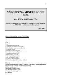
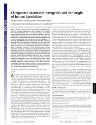
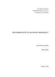
586 JABLONSKIcomparative anatomical and physiological evidence. Skin has also been overlookedas a topic of research interest in anthropology and human biology in recent decadesbecause of the social sensitivity surrounding discussions of skin color and becauseof the use and misuse of skin color in biological and social concepts of race.The goal of this review is to provide a comprehensive yet economical survey ofthe biology, evolution, and culture of human skin and skin color, with an emphasison new research—especially on the evolution of skin color. The review beginswith an overview of the basic biology of skin itself, followed by discussions of theevolution of skin and skin color, and of skin color and race.<strong>THE</strong> STRUCTURE <strong>AND</strong> FUNCTIONS <strong>OF</strong> <strong>HUMAN</strong> <strong>SKIN</strong>The skin serves as an effective physical barrier because its laminar structure rendersit relatively resistant to abrasion, puncture, and percutaneous absorption, andbecause its immune cells mount a first line of defense against pathogens comingin contact with the body. Lacking adequate protection from hair, human skin hasundergone numerous adaptive structural changes that give it strength, resilience,and sensitivity (Montagna 1981). The skin of humans, like that of all tetrapods,acts as a sun shield to protect the body from most solar UV radiation (UVR) andis the locus for the initiation of the important, UVR-driven process of vitamin Dproduction in the body.EpidermisThe laminar structure of human skin comprises two major tissue layers, a thinnerouter layer, the epidermis, and a thicker and more intemally complex inner layer,the dermis (Figure 1). Tbe epidermis is a stratified keratinizing epithelium witha smooth, abrasion-resistant surface that is interrupted only by hair follicles andthe pores of sweat glands. The barrier properties of the skin are predicated on theintegrity of the stratum comeum (Elias et al. 2003, Taylor 2002). Keratinocytes arethe principal cell type found in epidermis and are composed largely of filamentousproteins known as keratins, which are imbedded in an amorphous matrix. Theskin's elasticity and resistance to physical and chemical attack can be attributedto the high elastic modulus and unique amino acid composition of the keratinizedlayer of the epidermis (Marks 1991, Odiand 1991). The epidermis also containspopulations of three types of immigrant dendritic cells: melanocytes, Langerhanscells, and Merkel cells. Melanocytes produce the skin's primary pigment, melanin,and are discussed in greater detail below. Langerhans cells are specialized cellsof the immune system that present and respond to antigens coming in contactwith the skin, and Merkel cells are associated with nerve terminals that togetherfunction as slow-adapting mechanoreceptors for totich; they are most common onthe glabrous skin of tbe fingertips (Chu et al. 2003, Kripke & Applegate 1991,Lynn 1991, Odiand 1991). The epidermis is subdivided into four layers fromdeep to superficial; the stratum basale (the germinative layer of keratinocytesj, the
<strong>SKIN</strong> <strong>AND</strong> <strong>SKIN</strong> <strong>COLOR</strong> 587Melanocytes Keratinocytes Nerve endings FibroblastsFigure 1 Schematic rendering of a cross-section of human skin, showing itslaminar structure, main cell types, and appendages.
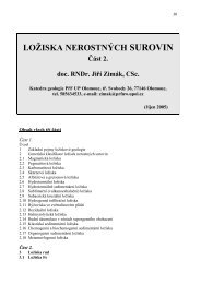
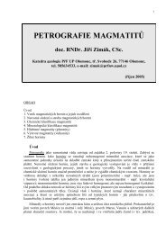
588 JABLONSKIStratum spinosum, the stratum granulosum, and the stratum comeum. The stratumcomeum consists of flattened, nonviable keratinocytes. In darkly pigmented orheavily tanned individuals, these keratinocytes contain specks of melanin "dust"(KoUias 1995a). The stratum comeum acts as a barrier to the unrestrained passageof water and solutes through the skin, defends against invasion by microorganismsand the penetration of toxic substances, and protects against most mechanicalinjury caused by friction, abrasion, pricks, or arthropod bites (Marks 1991). Tbesefunctions are successfully served despite the epidermis being in a constant state oftumover, as the outermost comified cells of the stratum comeum are shed as theyare replaced from below.Differences between human groups in epidermal stmcture and thickness havebeen reported, but most studies of this topic have been based on small sampleswith pooriy controlled experimental designs, as reviewed elsewhere (Taylor 2002).Considerable variation in epidermal thickness exists within human populations andis likely related to age and history of sun exposure. The stratum comeum of darklypigmented or heavily tanned people is more compact and consists of more comifiedcell layers than that of lightly pigmented people; these characteristics enhance thebarrier protection functions of the skin (Taylor 2002).In all primates, the epidermis of the volar surfaces of the hands and feet exhibitwell-developed epidermal ridges or dermatoglyphics, whicb impart greater resistanceagainst friction and help to insure secure purchase on locomotor substratesand on objects being gripped or manipulated. Dermatoglyphics are also found onthe ventral surfaces of the tails of prehensile-tailed New World monkeys and onthe knuckle pads of chimpanzees and gorillas (Ellis & Montagna 1962. Montagna1971).The melanocytes of the epidermis warrant close attention becau.se of theirrole in the production of the skin's primary pigment or chromophore, melanin.Melanocytes are specialized dendritic cells that reside in the stratum basale ofthe epidermis and in the matrix portion of the hair bulb. They originate in theneural crest as melanobiasts proliferate and migrate to the epidermis during theeighteenth week of embryonic development (Rawles 1948). Melanocytes producemelanins in specialized cytoplasmic organelles called melanosomes. which vary insize and degree of aggregation depending on skin type and pigmentation (Figure 2)(Szabo et al. 1969). The density of melanocytes varies over the surface of the body,and the number of active (melanin-producing) melanocytes varies with age andcan be increased by exposure to UVR (Halaban et al. 2003, Jimbow et al. 1991,Quevedo et al. 1975). The total number of melanocytes is relatively invariant fromone person to another, however, and is not related to variation between humangroups in skin pigmentation (Fitzpatrick et al. 1961, Jimbow et al. 1991. Robins1991, Young & Sheehan 2001). MacKintosh, following Wasserman, has recentlyadvanced the hypothesis that melanocytes. melanosomes, and melanin togetherfunction as part of the immune system against invading microorganisms and that themore darkly pigmented skins of the indigenous peoples of the tropics have evolvedprimarily to serve this function (MacKintosh 2(X)1; Wassermann 1965b, 1974).
<strong>SKIN</strong> <strong>AND</strong> <strong>SKIN</strong> <strong>COLOR</strong> 589IStratum corneum:Stratum granuiosumStratum spinosum,?55425iStratum basalejJ;""'©T*i Keratinocytes with^^^ laggregatations•'of smallmeianosomesLightly pigmented or untanned skinsStratum corneum(thickened, with melanin "dust")2 [Stratum spinosumKeratinocytes withmelanin granules^..Keratinocytes Iwith larger,non-aggregatedmeianosomesDarkly pigmented or heavily tanned skinFigure 2 Schematic rendering of cross-sections of lightly and darkly pigmentedhuman skin, showing diiferences in stratum corneum structure and in the size andaggregation of melanin-containing meianosomes.
590 JABLONSKIMelanocytes project their dendrites into keratinocytes where they then transfermature meianosomes (Figure 2). Melanosomes are ellipsoidal, membraneboundorganelles containing melanin. After melanosomes have been transferredto keratinocytes, they become aggregated and surrounded by a membrane in amelanosome complex (Jimbow et al. 1991, Szabo et al. 1969). In darkiy pigmentedskin, melanosomes are large and are not clumped in aggregations, whereasin lightly pigmented skin these organelles are smaller and aggregated (Szabo et al.1969). Intensity of skin coloration is determined by many factors; (a) The totalnumber of melanosomes in the keratinocytes and melanocytes, and their degreeof dispersion; (b) the rate of melanin production (melanogenesis); (c) the degreeof melanization of melanosomes; (e) the rate of transport and type of incorporationof melanosomes into keratinocytes; (f) the degradation of melanosomeswithin the keratinocytes; and (g) a person's chronological age because the numberof metabolically active melanocytes decreases over time (Halaban et al. 2003,Jimbow et al. 1976. Ortonne 1990, Parker 1981). Larger melanosomes breakdown more slowly in keratinocytes and contribute to higher levels of pigmentation(Sulaimon & Kitchell 2003).MELANIN PIGMENTATION <strong>AND</strong> ITS MEASUREMENT Human skin derives most ofits pigmentation from melanin, an extremely dense, virtually insoluble, high molecularweight polymer that is attached to a structural protein (Jimbow et al. 1991,Ortonne 2002, Parker 1981, Sulaimon & Kitchell 2003). Human skin contains thetwo types of melanin found in all mammals, the brownish-black eumelaiiin andthe reddish-yellow pheomelanin (Thody et al. 1991). Higher concentrations ofeumelanin characterize darker skin phenotypes including tanned skin. Concentrationsof pheomelanin in the skin vary considerably from individual to individualwithin any given human group, but pheomelanin-rich skin phenotypes are morecommon among red-haired northem Europeans, as well as East Asians and NativeAmericans (Rana et al. 1999, Thody et al. 1991). Melanin is synthesized byoxidation of tyrosine via the enzyme tyrosinase (Fitzpatrick et al. 1950, Jimbowct al. 1976, Ortonne 2002). Eumelanins and pheomclanins arise from a commonmetabolic pathway in which dopaquinone is tbe key intermediate (Ortonne 2002).As is discussed in greater detail below, production of melanins is regulated bypigmentation genes, hormones, and UVR (Fitzpatrick & Ortorme 2003, Sulaimon& Kitchell 2003, Thody & Smith 1977). A balance of many regulatory factors isessential for normal pigment production in the melanocyte, and derangements ofthese factors can lead to anomalies of cutaneous pigmentation such as albinismpiebald spotting, and various types of hyperpigmentation (Robins 1991, Sulaimon& Kitchell 2003, Thody & Smith 1977).The optical and chemical properties of melanins have been studied in detail(Ito 2003, Kollias et al. 1991, Ortonne 2002. Prota 1992c). but detailed chemicalcharacterization of the compounds has been difficult to obtain because melaninpolymers are composed of many different units connected through strong carboncarbonbonds (Ito 2003). The optical properties of natural melanin in vivo are
<strong>SKIN</strong> <strong>AND</strong> <strong>SKIN</strong> <strong>COLOR</strong> 591related to its abilities to absorb, scatter, and reflect light of different wavelengths(Kollias et al. 1991, Ortonne 2002). The melanins in human skin are a heterogeneousmixture of melanin polymers, precursors, and metabolites, characterizedby a continuous absorption capacity in the UV range and exponentiallydeclining absorption capacity from the UV to the visible range (Kollias 1995b,Sama & Swartz 1998). Natural protection against sunbuming (photoprotection)is due to the absorption and scattering of UVR by melanin (Kaidbey et al. 1979;Kollias 1995a,b). Both processes are influenced by the density and distributionof meianosomes within keratinocytes (Figure 2), with the larger, singly dispersedand heavily melanized meianosomes of darkly pigmented skin absorbing moreenergy than the smaller, less dense, and lightly melanized meianosomes of lightlypigmented skin (Kaidbey et al. 1979).Melanin was long considered to act as a passive screening filter against UVR,but it is by no means inert (Fitzpatrick et al. 1961). Photodegradation (photolysis)and/or oxidative polymerization of melanin may occur when it absorbs photons(Ortonne 2002). Recent evidence indicates that the photoprotective role of melaninin darkly pigmented skin may be augmented by its ability to scavenge oxygenderivedradicals (reactive oxygen species), such as superoxide anion and hydrogenperoxide, which are cytotoxic compounds generated hy the interaction of UV photonswith membrane lipids and other cellular components (Ortonne 2002, Prota1992c, Sulaimon & Kitchell 2003, Young & Sheehan 2001). At the physiologicallevel, the protective role of melanin pigmentation against UVR exposure derivesfrom its ability to prevent direct and indirect (oxidative) damage to DNA at wavelengthswhere it is most vulnerable (Cleaver & Crowley 2002, Kielbassa et al.1997, Shea &Panish 1991).Melanin pigmentation in human skin is considered as either constitutive skincolor or facultative skin color (Quevedo et al. 1975). Constitutive skin color isthe amount of genetically detertnined cutaneous melanin pigmentation that is generatedwithout any influence of solar radiation (Jimbow et al. 1976, Quevedoet al. 1975). Facultative skin color or "tan" constitutes the short-lived, immediate,and delayed tanning reactions elicited by exposure to UVR (Jimbow et al.1991, 1976; Quevedo et al. 1975). Lighter constitutive pigmentation is associatedwith a higher sunburn response, a lower tanning response, and a greatersusceptibility to skin cancers (Kollias et al. 1991, Sturm 2002, Wagner et al.2002).Objective and reproducible assessment of melanin pigmentation has long beena goal of anthropology and dermatology. In anthropology, verbal descriptions ofskin colors ("white," "yellow," "black," "brown," and "red") were replaced byCO lor-matching methods during the early twentieth century (Olivier 1960, vonLuschan 1897). The most popular of these methods was the von Luschan scale,based on the use of colored tablets or tiles of different colors and hues with whichthe colors of unexposed skin were matched. These and similar matching methodscould not be consistently reproduced, however, and were swiftly abandonedwhen reflectance spectrophotometry was introduced in the early 1950s (Lasker
592 JABLONSKI1954, Wassermann 1974). Reflectance spectrophotometry remains the method ofchoice for the objective study of skin pigmentation, color definition, and the spectralreflectance curves of skin because the incident light used and the distancebetween the light source and the subject are invariable and because subjectivefactors inherent in the visual matching methods are excluded (Wassemiann 1974).All instrumental approaches to skin color evaluation depend on the illuminationof the skin site by a standard light source at a fixed relative angle that minimizesthe reflected light from the stratum comeum. The detector collects light re-emittedby the skin site from a particular angle and with a chosen color filter (Kollias1995a). Because of the importance of assessing constitutive skin color on a partof the body that is not routinely exposed to sun, the inner (medial) surface oftbe upper arm has long been the standard reference site for studies of skin color.Portable reflectance spectrophotometers came into use with Weiner's (1951) study,with two types of instruments being commonly employed in anthropology duringthe latter part of the twentieth century. The instrument manufactured by theEvans Electroselenium Company (EEL) has been the most widely used, especiallyin studies of the skin colors of Old World peoples (Wassermann 1974).whereas that made by the Photovolt Corporation was more widely used in studiesof New World peoples. Unfortunately, tbe skin reflectance measurements obtainedby these two instruments are not directly comparable, requiring conversion formulaeto make them so (Lees & Byard 1978). Research is now underway that maymake possible the conversion of skin color assessments made by von Luschancolor tablets to values comparable with those derived from reflectance spectrophotometry(M. Henneberg, personal communication).In clinical medicine, constitutive skin color and skin sensitivity has been classifiedconunonly according to skin phototypes or sun-reactive skin types, fromType I (very sensitive, easily bumed, with little or no potential for tanning)to Type VT (insensitive, never bums, and deeply pigmented) (Fitzpatrick 1988,Fitzpatrick & Ortonne 2003, Jimbow et al. 1991). Skin type does not correspondwell to constitutive skin color, however, and has limited applicability with respectto the responses of moderately or deeply pigmented skin (Koilias et al. 1991, Prota1992c, Taylor 2002, Wagner et al. 2002, Westerhof et al. 1990). Despite theselimitations, skin phototyping has been widely embraced by many clinicians becauseassessments can be made without instrumentation. In recent years, highlysensitive diffuse reflectance spectrophotometers such as the DermaSpectrometerand the Datacolor Intemational Microflash as well as cbromaticity meters havebeen used increa.singiy to measure skin pigmentation and skin response to UVR(Kollias 1995a. Wagner et al. 2002).The photoprotective benefits of melanin have been assessed using several differentmeasures including minimal erythemal dose (MED), DNA damage, andincidence of skin cancer (Kollias et al. 1991). The MED represents the minimumamount of UVR necessary to bring about a slight visible reddening of lightly pigmentedskin. It is the easiest and most common method of assessing skin reactionsto UVR but is difficult to determine for deeply pigmented individuals in whom
<strong>SKIN</strong> <strong>AND</strong> <strong>SKIN</strong> <strong>COLOR</strong> 593visual redness is difficult to assess (Kaidbey et al. 1979, Ortonne 2002, Shono et al.1985).Exposure of human skin to UVR results in a profound alteration of the metabolism,structure, and function of epidermal cells. These activities include increasedactivation of melanocytes, augmentation of melanosome production, anincrease in the size of melanosome complexes incorporated within keratinocytes,and initiation of vitamin D synthesis (Parker 1981, Prota 1992a, Urbach 2001).The erythema response or sunburn reaction is related to constitutive skin color:Dark-skinned individuals can tolerate longer sun exposure than light-skinned individualscan. The skin of individuals with dark constitutive pigmentation exhibits asun protection factor (SPF) of 10-15, whereas that of moderately pigmented people(e.g., from the circum-Mediterranean) achieves an SPF of only 2.5 (Kaidbeyet al. 1979, Kollias et al. 1991, Ortonne 2002). In vitro studies of the reactions ofhuman melanocytes to UVR have shown that heavily pigmented melanocytes havea greater capacity to resume cell division after irradiation with short wavelengthUVR (UVB) than do their lightly pigmented counterparts, which suggests that theysuffered less damage to their DNA (Barker et ai. 1995). In contrast, UVB damagesthe immune system of the skin regardless of constitutive pigmentation by depletingboth heavily and lightly pigmented skin of Langerhans cells (Cleaver & Crowley2002, Kripke&Applegate 1991). The protective role of melanin in connection withskin cancer thus derives from its role in preventing damage to DNA in the first place,not in protecting against damage to the cutaneous immune system (Vermeer et al.1991). Tanning or facultative pigmentation induced by UVR is photoprotective tosome degree against the deleterious effects of further UVR exposure, but it does notsignificantly increase the SPF of individuals with light constitutive pigmentationor protect the DNA of their skin from UVR-induced damage (Kaidbey et al. 1979,Ortonne 2002). Although repeated exposure of tanned skin to UVR increases thenumber of metabolically active melanocytes and the intensity of melanogenesis(Lock-Anderson et al. 1998), the increased concentration of melanin in the tannedskin of inherently lightly pigmented people does not approach the photoprotectionconferred by natural melanin in intrinsically darker-skinned people (Kaidbey et al.1979). Individuals with lightly to moderately pigmented skin, who are repeatedlyexposed to UVR, experience premature aging (photoaging) of the skin, which ischaracterized by wrinkling and anomalies of pigmentation (Chung 2001, Fisheret al. 2(K)2, Kollias et al. 1991). This process is initiated by the photochemicalgeneration of reactive oxygen species causing degradation of structural proteinsin the dermis that confer strength and resiliency to the skin (Fisher et al. 2002).DermisThe dermis is a thick, dense fibroelastic connective tissue composed of collagenfibers, elastic fibers, and an interfibrillar gel composed of glycosaminoglycans,salts, and water. The primary cells of the dermis are collagen-rich fibroblasts.Collagen, which constitutes 77% of the fat-free dry weight of skin, largely accounts
594 JABLONSKIfor the tensile strength of the skin's fabric and for some of the ability of the dermisto scatter visible light (Kollias 1995a, Shea AParrisb 1991). Interwoven with thecollagen is a network of abundant elastic fibers that restore the skin to its normalconfiguration after stretching. The demiis is equally thick in people with dark orlight constitutive pigmentation (Taylor 2002).The dermis encloses a widely ramifying network of blood vessels, an extensivenerve network, sweat glands, and a pilosebaceous complex of hair follicles andsebaceous glands (Figure 1). Of these, only the sweat glands are addressed in detailin this review because of their importance in thermoregulation.The rich vascular supply of the skin is responsible for supplying the needs of thesweat glands, hair follicles, and rapidly multiplying epidermal cells in the sti-atumbasale. The density of cutaneous blood vessels varies throughout the body's surfaceand is related to temperature and blood pressure regulation and the relative amountsof intermittent physical pressure different parts of the body must withstand, withthe highest concentrations found in the skin covering the head, nipples, palms,soles, and ischial tuberosities (Edwards & Duntley 1939). The perineal skin offemale macaques, baboons, and chimpanzees is richly suffused with blood vessels(Montagna 1967, Montagna 1971) that create large sexual swellings advertisingthe female's state of reproductive receptivity and lifetime reproductive potential(Domb & Pagel 2001). The oxygenated and deoxygenated forms of hemoglobincarried in the skin's blood vessels are some of the skin's main pigments, witha person's skin color determined mainly by the skin's melanin and hemoglobincontent (Edwards & Duntley 1939). The erythema or strongly red appearance ofthe skin caused by exposure to UVR is the result of increases in the number anddiameter of vascular capillaries tiirough which blood is flowing and an increase inthe blood flow through each capillary (Kollias 1995a). Sunburned skin feels hot tothe touch because of the increased vascularization of the skin and the inflammatoryresponse mounted by the skin as it works to repair UVR-induced damage (Ryan1991, Shea &Parrish 1991).The nerve supply of the skin is highly complex because the skin is a majorsensory surface that contains varied types of receptors sending signals to thecentral nervous system about the extemal environment and the intemal state ofthe skin (Chu et al. 2003, Lynn 1991). These receptors include two types oftemperature sensors, diverse mechanoreceptors associated with both hairy andglabrous skin, and an important group of cutaneous sensory cells (nociceptiveafferents) specialized for the detection of tissue-threatening stimuli or the presenceof injury or inflammation (Lynn 1991). The glabrous skin of the hands andfeet of primates is densely packed with sensory nerve endings that permit highlysensitive tactile discrimination and exquisite differentiation of temperature andtexture (Chu et al. 2003. Lynn 1991. Martin 1990). These atu-ibutes greatiy enhancetiie manipulative functions of these appendages, especially the hand (Martin1990).Numerous hairs, which grow from hair follicles located in the dermis, are associatedwith mechanoreceptors and sebaceous glands. Hair performs a range of
•T>^<strong>SKIN</strong> <strong>AND</strong> <strong>SKIN</strong> <strong>COLOR</strong> 595functions from insulation, to protection against the sun, enhancement of cutaneoussensation to communication of emotion (through piloerection), and omamentation(Lavker et al. 2003; Montagna 1967, 1971; Wheeler 1984, 1985). Humansare unique among primates in possessing effectively naked skin, except on thescalp, the male chin, the axilla, and the groin. Although human skin bears millionsof hairs, most of them are so small as to be nearly invisible (Montagna1981).SWEAT GL<strong>AND</strong>S Human dermis contains two main types of sweat glands, eccrineand apocrine. Tbe former are widely distributed throughout the surface of the body,whereas the latter are concentrated in the axilla, perineum, and extemal auditorycanal. Eccrine glands are tubular in form (Figure 1) and lie in the outer portion ofthe dermis. They produce copious amounts of dilute, watery fluid expressed to thesurface of the skin through an individual pore. Humans have two to four millioneccrine glands on the surface of their bodies, with an average distribution rangingfrom ~150-340/cm2 (Folk & Semken 1991. Goldsmith 2003). Both apocrine andeccrine sweat glands are stimulated by the sympathetic division of the autonomicnervous system and produce sweat in response to thermal stimulation (thermalsweating). In contrast, the eccrine glands of the palms and soles respond only toemotional stimuli, whereas those of the face and axilla respond to both (Folk &Semken 1991, Zihlman & Cohn 1988).Considerable attention has been placed on comparisons of the quantity, structure,and function of sweat glands between human groups. The number of strictlycontrolled comparisons between members of different populations after equivalentperiods of deliberate acclimatization is quite small (Weiner 1977). The results ofmost rigorous comparative study of sweat gland densities in humans (Knip 1977)indicate that only small differences in the total number and average density of sweatglands exist between disparate human populations. As yet it has proven virtuallyimpossible to design studies that can determine conclusively whether differencesin sweating performance between human groups are due to genetic influences orenvironmental adaptations.The Skin in ThermoregulationDissipation of heat is the function that most conspicuously distinguishes humanskin from that of all other animals (Montagna 1981). The reasons for the evolutionof this unique capacity are discussed in the following section. Humans encounterheat stress more or less year round in equatorial areas and for varying lengthsof time in the rest of the world except for circumpolar and alpine environments.Heat stress is exacerbated by prolonged or rigorous exercise. Maintenance ofhomeostasis requires that the body's core temperature remain close to a neutralpoint, which varies from about 36.8 to 37.2^C, in order to permit unintermptedfunctioning of the temperature-sensitive cells of the human central nervous system.If the rates of production or loss of heat are excessively out of balance, core
596 JABLONSKItemperature can quickly increase or decrease to dangerous levels (Kraning 1991,Wenger 2003).Temperature regulation in humans includes involuntary (physiologic) and voluntary(behavioral) activity (Wenger 2003). Voluntary temperature regulation involvesthe conscious actions taken by people to maintain thermal comfort, includingthe seeking of shade and shelter, and the wearing or shedding of clothing.Involuntary temperature regulation in the skin has been studied in great detail inthe past 50 years by both physiologists and anthropologists, and only a superficialsummary of this corpus of work is presented here. Regulation of temperature by theskin is accomplished through its roles in (a) perceiving and transmitting its owntemperature to the centi'al nervous system; (b) regulating heat transfer betweenthe body's core and the skin through the cutaneous circulation; (c) serving as asuperficial casing through which body heat is conducted from the vascular layersto the surface; (d) acting as an interface for the loss or gain of heat to or from theenvironment by radiation, convection, or conduction; and (e) acting as a surface forthe spreading of sweat necessary for evaporative cooling (Frisancho 1981, Kraning1991). The relative role of the four avenues of heat loss (radiation, convection,conduction, and evaporation) depends on the interaction of the ambient temperatureand humidity (Chaplin et al. 1994; Frisancho 1981; Wenger 2003; Wheeler 1984.1991b). The ability of sweat glands to respond to heat stress is adversely affectedby sunburn (Pandolf et al. 1992). Protection of the integrity of sweat glands againstdamage caused by UVR, therefore, has been of great importance during the longcourse of human habitation of the tropics.Experimental studies and simulations undertaken to determine how thermalhomeostasis is maintained under the stressful environmental conditions of thetropics have shown that heat loss is maximized in people with a high ratio ofskin surface area to body weight, such as Nilotic tribespeople, the Kung San, andAustralian Aborigines (Frisancho 1981; Wheeler I991a,b, 1992), This relationshipsupports Allen's Rule in mammals, which states that mammals living in coldregions will minimize the size and surface area of their extremities, whereas thoseinhabiting hot areas will increase the relative size of appendages.The Role of the Skin in Vitamin D BiosynthesisSynthesis of vitamin D in the skin of vertebrates is the only unanimously agreedpositive effect of UVR exposure. Vitamin D3 is the form of vitamin D that is synthesizedby vertebrates, whereas vitamin DT is the primary form found in plants(Holick 2003). Vitamin D^ is more accurately characterized as a prosteroid hormonethan as a vitamin because, in mammals, it is derived from a cholesterol-likeprecursor (7-dehydrocholesteroI) found in the skin (Holick 2003). Vitamin D is aunique natural product thought to have first occurred on Earth as a photosyntheticproduct in marine phytoplankton more than 750 mya (Holick 1995). Although thephysiological role of vitamin D in plants and invertebrates is not clear, vitaminD was essential for the evolution of terrestrial vertebrates (Holick 1991, 1995).
<strong>SKIN</strong> <strong>AND</strong> <strong>SKIN</strong> <strong>COLOR</strong> 597Holick has reasoned that early tetrapods depended on vitamin D for the efficient useof scarce dietary calcium to preserve their rigid calcified skeletons (Holick 1995).Vitamin D can be synthesized only by a photochemical process, so early tetrapodscould only satisfy their body's vitamin D requirements by exposing themselvesto sunlight to photosynthesize vitamin D in their own skin or by ingesting foodscontaining vitamin D (Holick 1995).Vitamin D3 synthesized in the skin requires successive hydroxylations in theliver and kidney to be converted to its biologically active form, la, 25-dibydroxyvitaminD3 (Holick 1991, Jones et al. 1998). This functionally active form isimportant for the regulation of calcium and phosphorus metabolism, skeletal developmentand mineralization, the regulation of normal cell growth, and the inhibitionof cancer cell growth (Holick 1991, 2001). The production of vitamin D3is optimally stimulated by UVR wavelengths of 295-300 nm, in the UVB range(MacLaughlin et a!. 1982). High-energy UVB photons penetrate the skin and areabsorbed by the 7-dehydrocholesterol in the keratinocytes of the epidermis (especiallyof the strata basale and spinosum) and fibroblasts of the dermis, catalyzingthe formation of previtamin D3 (Holick 2001, Webb et al. 1988). Once formed intbe skin, previtamin D3 can undergo isomerization to vitamin D;* at body temperatureand then undergo further chemical conversions to la, 25-dihydroxyvitaminD3 The conversion of previtamin D^ or vitamin D3 to the functionally activeform is rate-limited, however. In the presence of biologically sufficient amountsof la, 25-dihydroxyvitamin D3 in the circulation, previtamin D3 and vitamin D3are transformed by UVA or UVB into a variety of inert byproducts, thus avertingoverproduction of the biologically active form and subsequent "vitamin D intoxication"(Holick 2001, Holick et al. 1981). This finding disproves the hypothesisthat dark constitutive skin pigmentation evolved in the tropics as an adaptation toprotect against the overproduction of la, 25-dihydroxyvitamin D3 (Loomis 1967).Melanin pigments are highly effective at absorbing and scattering the UVBwavelengths that catalyze vitamin D3 synthesis. Thus, high concentrations ofmelanin in the skin result in a decrease in the efficiency of conversion of 1-dehydrocholesterol to previtamin D3; pigmentation slows but does not preventcutaneous production of the vitamin (Holick et al. 1981, Webb et al. 1988). Individualswith very deep constitutive pigmentation often require 10 to 20 timeslonger exposure to sunlight than those of lighter pigmentation in order to promotean adequate synthesis of vitamin D3 (Holick et al. 1981). This finding explainswhy dark-skinned individuals living at high latitudes with low levels of environmentalUVB are at greater risk of vitamin D^-deficiency diseases than arelight-skinned people (Clemens et al. 1982, Holick 2001. Mitra & Bell 1997). Theevolutionary significance of this observation is discussed further below. The photoconversionof 7-dehydrocholesterol to previtamin D3 in the skin is also adverselyaffected by increasing age (Holick 1995), the wearing of clothing (Matsuoka etal.1992), and by the use of topical sunscreens, which block the UVB wavelengthsresponsible for both sunbum and vitamin D3 production (Holick 1997, Webb etal.1988).
598 JABLONSKI<strong>THE</strong> <strong>EVOLUTION</strong> <strong>OF</strong> MODERN <strong>HUMAN</strong> <strong>SKIN</strong>Reconstruction of the evolution of human skin relies on evidence provided bycomparative anatomy and physiology, as well as study of the evolution of thegenes and gene complexes that determine the function and pigmentation of skin.Using basic principles of historical morphology, one can reconstiuct the majorsteps in the evolution of human skin by utilizing a well-established phylogeny toexamine historical transformations of structure and function (Jablonski & Chaplin2000). This method leads to the reconstruction of the probable appearance of theskin in the last common ancestor of the human and chimpanzee lineages as beinglightly pigmented and covered with dark hair, like most catarrhine primates today(Jabionski & Chaplin 2000).The skin of modem humans is distinguished from that of other primates mainlyby its naked appearance, its greaUy enhanced abilities to dissipate body heatthrough sweating, and by the great range of genetically determined skin colorspresent within a single species. Most investigators have considered these attributesto be adaptations forged by natural selection.The Evolution of the Thermo regulatory Propertiesof Human SkinHuman skin is not hairless, but—as discussed above—the hairs over most of thebody's surface are so fine and present at a sufficiently low density tiiat the skinappears essentially naked. Explanations for the evolution of human hairlessnesshave been many, varied, and often highly creative. The most cogent explanationsare based on the importance of a functionally naked skin in maintaining bodytemperature in hot environments.Many animals, including primates, which live in hot environments, have heavycoats of insulating fur or feathers. In the heat caused by strong sunlight, suchinsulation reduces environmental heat gain (Folk & Semken 1991. Walsberg 1988).This is the case even for black coats, which absorb short-wave radiation near or atthe surface of the fur and reradiate large amounts of long-wave radiation before itreaches the skin (Dmi'el et al. 1980). Tbe effectiveness of fur insulation in reducingenvironmental heat gain is lessened by sweating. The most efficient evaporativecooling occurs at the skin's surface; in heavily furred animals, water vapor istransferred through the fur to the atmosphere (Folk& Semken 1991). If the fur iswet from sweating, however, maximum evaporation occurs at the surface of thefur. and heat from the blood vessels cannot be transferred as efficiently to the site ofevaporation (Folk & Semken 1991). Under these circumstances, much more watermust be used for evaporative cooling. Thermal sweating as a method of coolingbecomes more important as environmental temperatures rise or as activity levelsincrease because the lower gradient between core and environmental temperaturesrestricts the amount of heat loss that can be achieved by radiation, convection,and conduction (Frisancho 1981, Wheeler 1991b). Removal of excess heat is.
<strong>SKIN</strong> <strong>AND</strong> <strong>SKIN</strong> <strong>COLOR</strong> 599therefore, greatly facilitated by the loss of body hair because it increases thermalconductance and permits additional heat loss through sweating (Wheeler 1985,Zihlman & Cohn 1988).A strong case can be made for the evolutionary loss of apocrine sweat glandsin bumans because these sweat glands are most common in heavily furred animals(Folk & Semken 1991). The African apes exhibit a ratio of approximately 40%apocrine sweat glands to 60% eccrine; the great preponderance of eccrine sweatglands in modem humans probably evolved under the strong influence of naturalselection, following the loss of the apocrine component to sweating (Folk &Semken 1991, Montagna 1981, Zihlman & Cohn 1988). This process was probablypropelled by increases in body size and activity levels associated with modemlimb proportions and striding bipedalism, which occurred in the transition from theprimitive hominins of the late Miocene to the genus Homo of the Plio-Pleistocene(Chaplin et al. 1994; Folk & Semken 1991; Jablonski & Chaplin 2000; Montagna1981: Schwartz &Rosenblum 1981; Wheeler 1984. 1996).The importance of body cooling through the skin in modem humans has beenemphasized repeatedly by both physiologists and anthropologists because of theprimacy of preventing hyperthermia and attendant damage to the central nervoussystem (Cabanac & Caputa 1979, Falk 1990, Wheeler 1984. Zihlman & Cohn1988). The temperattu"e of the brain closely follows arterial temperature, requiringthat the temperature of the circulating blood be carefully regulated (Nelson &Nunneley 1998). This process became increasingly important as activity levels andbrain size increased in the genus Homo through the Pleistocene. Simulations andexperimental studies have confirmed that maintenance of stable core temperatureunder conditions of increased environmental beat load or exercise is best accomplishedvia recruitment of a whole-body cooling system, involving cooling ofperipheral blood vessels through sweating (Desmelle & Candas 2000, Nelson &Nunneley 1998). A recently mooted hypothesis that human hairlessness evolvedlate in human evolution as a result of the adoption of clothing and the need to reducethe load of extemal parasites (Pagel & Bodmer 2003) finds no support in light ofthe overwhelming evidence of the importance of hairlessness in themial sweatingand whole-body cooling in maintaining stable core temperature and homeostasis.The Evolution of Human Skin PigmentationRECONSTRUCTION <strong>OF</strong> <strong>SKIN</strong> <strong>COLOR</strong> IN EARLY HOMO The early members of thegenus Homo from the late Pliocene and Early Pleistocene of Africa exhibitedlarger bodies, relatively larger brains, and relative longer lower limbs than didtheir australopithecine predecessors (McHenry & Berger 1998; Ruff et al. 1993,1997). The higher activity levels and larger day ranges reconstructed for them(Wheeler 1991a, 1992) would have required that their skin be functionally nakedand endowed with a high density of eccrine sweat glands in order to facilitateheat loss (Jablonski & Chaplin 2000, Wheeler 1984). This situation created a newphysiological challenge for human skin: protection of a naked integument against
600 JABLONSKIUVR. Dense hairy coats protect the skin of mammals from UVR-induced damageto the skin because the hairs themselves absorb or reflect most short-wavelengthsolar radiation. In mammals with sparse coats of hair, however, 3%-5% of incidentUVR is transmitted to the skin (Walsberg 1988). Nonhuman mammals thatare active in hot, sunny environments exhibit sparse coats because they facilitatepassive heat loss; they also display highly melanized skin on their exposed (dorsal)surfaces to effectively block the UVR transmitted to the skin (Walsberg 1988). Thisevidence clearly indicates that hair loss in the human lineage was coupled withincreased melanization of the skin as activity levels in hot environments increased.The early members of the genus Homo, the ancestral stock from which all laterhumans evolved, were, thus, darkly pigmented (Jablonski & Chaplin 2000). Thisinterpretation has recently been supported by genetic evidence demonstrating thatstrong levels of natural selection acted about 1.2 mya to produce darkly pigmentedskin in early members of the genus Homo (Rogers et al. 2(X)4).Heavily pigmented skin does not, in fact, perceptibly increase the body's heatload under conditions of intense solar radiation (Baker 1958. Walsberg 1988).Tbis is because for half of the solar radiation reaching the Earth's surface—in theinfrared—there is essentially no difference in absorption between dark and lightskin (Baker 1958, Daniels 1964). This evidence negates tbe claim by Blum (1961)and others (Morison 1985) that heavily melanized pigmentation in humans couldnot be adaptive in the hot tropics because of the increased heat load caused bygreater amounts of absorbed solar radiation.<strong>SKIN</strong> PIGMENTATION IN MODERN <strong>HUMAN</strong> POPl^ATIONS Many of the aCCOUntSOftravelers and explorers from the fifteenth century onward include reports of theskin color of the peoples they encountered. As natural historians and humangeographers—mostly from Europe—ventured into Asia, Africa, Australia, and theAmericas and began to study the indigenous human populations in detail, mapsdepicting the worldwide distribution of human skin color were slowly assembled.The best known of these maps is that composed by the Italian geographer RenatoBiasutti, which was based on the von Luschan skin color scale. This map hasgained broad circulation in several widely distributed publications (Barsh 2003,Lewontin 1995. Roberts 1977, Walter 1971), despite tiie fact that, for areas witiino data, Biasutti simply filled in the map by extrapolation from findings obtainedin other areas (Robins 1991). A more accurate and exhaustive compilation of theskin colors of indigenous peoples based only on published skin reflectance measurementsis now available (Jablonski & Chaplin 2000). Both maps show similartrends, with darkly pigmented peoples found near the Equator and incrementallylighter ones found closer to the Poles. A larger percentage of people with dark skinis found in the Southem Hemisphere as compared with the Northem Hemisphere(Relethford 1997) because of a latitudinal bias in the distribution of land masses(Chaplin & Jablonski 1998).The data compiled by Jablonski and Chaplin also provide conclusive evidenceof sexual dimorphism previously observed in human skin pigmentation (Frost
<strong>SKIN</strong> <strong>AND</strong> <strong>SKIN</strong> <strong>COLOR</strong> 6011988, van den Berghe & Frost 1986), with females being consistently lighter thanmales in all populations studied (Jablonski & Chaplin 2000).One of the major problems encountered in assembling data on the distributionof human skin color in indigenous populations is determining exactly whatan indigenous population represents. For most anthropologists and human geographers,an arbitrary cutoff date of 1500 has been adopted to distinguish nativeor indigenous peoples from immigrant populations. This date is reasonable withrespect to the inauguration of the modem era of European colonization but fails torecognize tbe several major movements of human groups within continents (suchas the so-called Bantu expansion within Africa) that occurred before 1500. Thesemovements, along with European colonization and the increasingly rapid and distantmigrations of human populations through time, have fundamentally alteredthe human landscape established in prehistoric times. This has made the interpretationof geographically and biologically significant trends in human populationsmuch more difficult.ENVIRONMENTAL CORRELATES <strong>OF</strong> <strong>HUMAN</strong> <strong>SKIN</strong> <strong>COLOR</strong> The skin pigmentation ofindigenous human populations shows remarkable regularity in its geographic distribution.Darker skins occur in more tropical regions and lighter skins in temperate,although tbe gradient is less intense in the New World as compared tothe Old World. Even within Africa, the continent with the largest equatorial landmass, there is considerable heterogeneity of skin color, with the deepest colorsoccurring not in the lowest latitudes but in the open grasslands (Chaplin 2001,Roberts 1977). The strong latitudinal signal in skin color led most early workersto conclude that skin pigmentation represented an adaptation to sunlight orother solar-driven phenomena such as temperature. Walter (1958, 1971) was thefirst researcher to suggest that the pigmentation gradient observed was linked tothe intensity of UVR, and he established this relationship by calculation of correlationcoefficients between skin color (as measured on the von Luschan scale)and estimated UVR. The relationship between skin color and environment wasfurther explored by studies in which the relationship of skin color, as measuredby reflectance spectrometry, to latitude, temperature, and humidity was studiedby correlation and regression analyses (Roberts 1977, Roberts & Kahlon 1976).These analyses showed the dominant association of skin reflectance with latitude,which was then deduced to be an effect related to the intensity of UVR (Roberts1977, Roberts & Kahlon 1976).In recent years, studies of the relationships between morphological and physiologicalvariation and attributes of the physical environment have been advanced bytbe availability of remotely sensed data on levels of UVR, total solar radiation, temperature,humidity, precipitation, and other environmental variables at the Earth'ssurface. These data, which were not widely available to workers before 1990,have permitted correlation, regression, and other analyses of skin reflectance to beconducted against actual measurements, rather than estimates, of environmentalvariables (Chaplin 2001, 2004; Jablonski & Chaplin 2000).
602 JABLONSKIUsing data on the minimal erythemal dose of UVR (UVMED) at the Earth'ssurface collected by the NASA TOMS 7 satellite. Jablonski & Chaplin were able toestablish a conclusive correlation between latitude and annual average UVMED.and thence between annual average UVMED and skin reflectance (Jablonski &Chaplin 2000). This publication was followed by a study in which the influence ofminimum, maximum, and seasonal levels of UVR, as well as other directly measuredenvironmental variables, relative to skin reflectance were studied (Chaplin2001, Chaplin 2004). This study showed that skin reflectance was correlated withautumn levels of UVMED. and that skin reflectance could be almost fully modeledas a linear effect of this variable alone (Chaplin 2001. 2004). This studyalso showed that the relationship between summer levels of UVMED and skinreflectance appeared to reach a threshold past which higher levels of UVR werenot correlated with incrementally lower skin reflectance (darker pigmentation)(Chaplin 2001, 2004).Low reflectance values for human skin (dark pigmentation) are primarily a functionof UVMED (Jablonski & Chaplin 2000), with regression analysis demonstratingthat autumn UVMED levels have the strongest effect. This indicates that skincolor is more strongly correlated with UVA, which is consistenUy higher throughouttiie year at all latitudes, than with UVB (Chaplin 2001. 2004). MaximumUVMED had the next most significant effect (Chaplin 2001, 2004). Winter levelsof precipitation have the opposite effect, being positively correlated with highreflectance values (light pigmentation) (Chaplin 2001. 2004). Multiple regressionformulae relating skin reflectance to these environmental parameters can then beused to derive a map of predicted human skin colors, with the colors shown beingrealistic approximations of the tme color of skin (Chaplin 2001. 2004) (Figure 3.see color insert). This map depicts an idealized situation in which humans worldwideare assumed to have inhabited their respective regions for the same length.sof time, and have followed similar cultural practices that could affect skin color(e.g., diet, activity schedules, use of clothing and shelter).NATURAL SELECTION <strong>AND</strong> <strong>THE</strong> <strong>EVOLUTION</strong> <strong>OF</strong> <strong>HUMAN</strong> <strong>SKIN</strong> PIGMENTATION Thegeographical distiibution of human skin colors has invited many explanations,most of which have claimed melanin pigmentation to be an adaptation to someattribute of the physical environment that varies primarily by latitude. Ever sincethe harmful effects of UVR began to be appreciated by scientists, explanations forthe evolution of deeply melanized skin have centered on the importance of resistanceto sunbum, solar degeneration, and skin cancer (Daniels et al. 1972). Equallypopular has been the vitamin D hypothesis, which stated that lightly pigmentedskins were necessary outside of the tropics in order to permit vitamin D biosynthesisin the skin by low levels of UVR, whereas darkly pigmented skin affordedprotection against production of toxic doses of vitamin D in equatorial regions(Loomis 1967). Lightly pigmented skin has also been explained as an adaptationto resist cold injury, on the basis of experimental and epidemiological data thathave documented more severe injuries incurred by pigmented skin exposed to
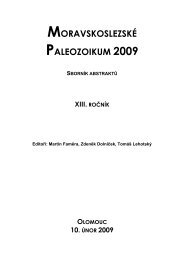
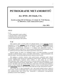
<strong>SKIN</strong> <strong>AND</strong> <strong>SKIN</strong> <strong>COLOR</strong> 603freezing conditions (Post et al. 1975, Steegmann 1967). Other explanations haveimputed highly melanized skin as providing effective concealment in habitats suchas tropical forests with differing light intensities and environmental illumination(Cowles 1959, Morison 1985), and still others have reasoned that tropical diseasesand parasites rather than tropical climate were the major selective forces leadingto the evolution of differential pigmentation in humans (MacKintosh 2001;Wassermann 1981, 1965a).Although adaptive explanations for human pigmentation have dominated theliterature, others have downplayed or discounted the role of adaptation by naturalselection. Some workers have emphasized the role of sexual selection, especiallyby way of explaining the lighter constitutive pigmentation of females relative tomales (Aoki 2002, Frost 1988). Deol claimed that differences in skin color betweenhuman populations were the pleiotropic byproducts of natural selection on otherfunctions of pigmentation genes (Deol 1975). Others have simply discouragedthe "amusing pastime" of adaptive reconstruction in the absence of data on thedifferential survival and reproduction of varying skin pigmentation phenotypes(Blum 1961, Lewontin 1995). Adaptive explanations "for" any given phenotypictrait require demonstration that the trait increases the real or probable reproductivesuccess of the organism. Although such evidence is often difficult to muster inthe case of traits bome by long-lived mammals, it is incumbent that adaptivereconstructions be tetbered by this responsibility.In the past, adaptive explanations for different levels of melanin pigmentationin human skin have suffered from an inability to demonstrate probable or realdifferences in survivorship and reproduction of different skin color phenotypesunder the same environmental conditions. Blum introduced this mode of criticalappraisal of competing hypotheses when he drew attention to the fact that dark skinpigmentation could not bave evolved primarily as adaptive protection against skincancer because such cancers rarely cause death during peak reproductive years(Blum 1961, Jablonski & Chaplin 2000). Other adaptive explanations for lightor dark skin pigmentation (e.g., protection against cold injury: camouflage) havesimilarly failed to demonstrate real or probable increases in reproductive successas a result of possession of these phenotypes.MELANIN AS A REGULATOR <strong>OF</strong> <strong>THE</strong> PENETRATION <strong>OF</strong> tmt INTO <strong>THE</strong> <strong>SKIN</strong> Recently,Jablonski & Chaplin (2000) published a new adaptive hypothesis for the evolutionof human skin pigmentation stating that melanin pigmentation evolved toregulate the penetration of UVR into the skin in order to prevent the photolysis ofphoto-labile compounds while permitting the photosynthesis of others. This hypothesiswas based on two equally important observations; (a) that the B vitaminfolate is destroyed by long wavelength UVR (UVA), and that folate deficienciescan markedly reduce individual reproductive success by adversely affecting celldivision; and (b) that vitamin D3 is synthesized in the skin by short wavelengthUVR (UVB) and that severe vitamin D deficiencies adversely affect reproductivesuccess by interfering with normal calcium metabolism (Jablonski & Chaplin
604 JABLONSKI2000). Natural selection has produced two opposing dines of skin pigmentation.The first is a cUne of photoprotection that grades from darkly pigmented skin atthe Equator to lightly pigmented skin near the Poles. The second is a cline ofvitamin D3 photosynthesis that grades from lightly pigmented near the Poles todarkly pigmented at the Equator. In the middle of the two clines we find peopleswith enhanced abilities to develop facultative pigmentation according to seasonalUVR levels.<strong>THE</strong> FOLATE HYPO<strong>THE</strong>SIS The potential importance of dark skin pigmentation inprotecting folate from UVR-induced photolysis was first recognized upon discoverythat folate undergoes photolysis in vitro when subjected to UVA (360 nm) andthat serum folate levels of human subjects dramatically declined when humansunderwent long-term exposure (minimum of 3 months) to the same wavelength,for 30-60 min once or twice a week (Branda & Eaton 1978). The potential significanceof the finding to the evolution of human skin pigmentation was echoedlater, but a causal mechanism was not mooted (Zihlman & Cohn 1988).Few nutrients compare with folate (folic acid) for its impact on health. Adequatefolate status is vital for the synthesis, repair, and expression of DNA. and thereforefor all processes involved in cell division and homeostasis (Kesavan et al. 2003,Lucock et al. 2003, Suh et al. 2001). The subtle infiuence of folate on the cell'sgenomic machinery has led to the realization that even marginal folate deficienciesmay have significance in developmental disorders and degenerative diseasesassociated with high morbidity and mortality (Lucock et al, 2003). Now that folatedeficiency is widely acknowledged as a risk factor for neural tube defects, recurrentearly pregnancy loss, and other complications of pregnancy, the maintenance ofadequate folate status in women of reproductive age has become a primary publichealth concem (Bower & Stanley 1989, Fleming & Copp 1998, Minns 1996, Suhet al. 2001), Folate's importance in spermatogenesis also highlights its importantrole in maintaining male reproductive competence (Cosentino et al. 1990. Mathuret al. 1977).The recognition of folate's pivotal roles in DNA synthesis and repair—and thusmost processes associated with reproductive success in both sexes—^has underlinedthe importance of protecting the body's folate stores from physical or chemicaldegradation. Because folate is susceptible to oxidative damage as a result of exposureto UVR and ionizing radiation (Branda & Eaton 1978, Hirakawa et al. 2002,Kesavan et al. 2003), the primary evolutionary function of melanin in regions receivinghigh annual UVR is to protect folate from photodegradation (Jablonski& Chaplin 2000). Photolysis of folate has been experimentally demonstrated at340 nm and 312 nm, in the UVA and near-UVA wavelengths (Hirakawa et al. 2002,Lucock et al. 2003). With skin refiectance being most closely correlated with autumnlevels of UVMED dominated by UVA, one can conclude that the longerwavelengths of UVR, which are capable of penetrating deep into the dermis ofthe skin, have been the most important agents of natural selection in connectionwith the evolution of skin pigmentation (Chaplin 2001) (Figure 4). The results of
<strong>SKIN</strong> <strong>AND</strong> <strong>SKIN</strong> <strong>COLOR</strong> 605UVA UVB UVC315-400nm 280-315nni 100-280nmVitamin D synthesis:1 a, 25-dihydroxyvitaminD,UVA UVB UVC315-400nm 280-315nm 100-280nmLightly pigmented or untanned skinFolic acid breakdown;Folate (folic acid)K'^
606 JABLONSKIa recent study (Gambichler et al. 2001) did not confirm the photolytic effect ofUVA on serum folate levels in a small number of human volunteers. This findingruns counter to the results of previous in vivo and in vitro studies demonstratingprofound photodegradation of folate upon exposure to UVR (Hirakawa et al. 2002,Lucock et al. 2003) and to X- and y-irradiation (Kesavan et al. 2003). A true andstatistically robust test of the folate hypothesis would require a case-control studyinvolving a large number of human volunteers experiencing long-term (once ortwice a week for a minimum of three months) exposure to UVR, with measurementof more labile folate species such as specific red cell folate coenzymes (Lucocket al. 2003).SiON PIGMENTATION <strong>AND</strong> VITAMIN D BIOSYN<strong>THE</strong>SIS In the millennia prior toabout 1.6 mya, the earliest members of the genus Homo appear to have beenrestricted in their distribution to the high-UVR regimes of equatorial Africa. Underthese environmental conditions, possession of highly melanized skin wascritical for survival. As populations of early Homo moved both northward andsouthward, they began to experience different schedules and intensities of UVRexposure.UVR levels at the Earth's surface are affected by latitude, altitude, season,moisture content, cloud cover, the depth of the ozone column, orbital parameters,and other factors (Hitchcock 2001. Madronich et al. 1998). Short wavelengthUVR (UVB, 280-315 nm) is more effectively absorbed by atmospheric ozonethan are longer wavelengths (UVA, 315-400). Thus, as one moves away from theEquator and the angle of solar elevation decreases, the thickness of the atmosphere(including the ozone layer), through which sunlight must pass, increases. Thisresults in a greater attenuation of UVR, especially of UVB, by scattering andabsorption by ozone, and consequently very low levels of UVB in high-latitudeecosystems (Caldwell et al. 1998). Very small increments or decrements of UVBlead to substantial biological effects (Madronich et al. 1998); thus, it is highlybiologically significant that regions north and south of 50° latitude receive onlytiny doses of UVB, and only then at the peak of summer (Caldwell et al. 1998,Chaplin 2001, Johnson et al. 1976, Neer 1985).As discussed earlier, deeply melanized skin confers excellent protection againstthe deleterious effects of UVR, but it also greatly slows the process of vitaminD3 synthesis in the skin. As hominins moved out of the tropics, their exposureto UVR—especially to vitamin D-inducing UVB—was dramatically reduced.Levels of UVR at the Earth's surface are not thought to have been appreciablydifferent in the Pleistocene as compared to today because similar conditions ofsolar emissivity and orbital parameters existed at the time, and similar levels ofUVR have been reconstructed from biological proxies (Rozema et al. 2002). Evenbefore remotely sensed data on UVB levels outside of the tropics were available,theorists surmised that early humans living in high latitudes with deeply pigmentedskin would have not been able to produce sufficient vitamin D3 in their skin to meettheir physiological demands and that strong selective pressure for depigmentation
<strong>SKIN</strong> <strong>AND</strong> <strong>SKIN</strong> <strong>COLOR</strong> 607of the skin had been exerted in order to facilitate photosynthesis of vitamin D3(Loomis 1967, Murray 1934, Neer 1975).Using known values of UVMED at the Earth's surface (Herman & Celarier1996) and the precise dosage of UVB necessary to catalyze vitamin D synthesis inhuman skin at a specific latitude (Webb et al. 1988), researchers can calculate theworldwide potential for vitamin D3 synthesis for lightly pigmented skin (Jablonski& Chaplin 2000) (Figure 5, see color insert). Zone 1 (shown without hachure inFigure 5) corresponds closely to the tropics and represents an area in which thereis adequate UVR throughout the year to catalyze vitamin D3 synthesis in theskin (Jabionski & Chaplin 2000). Zone 2 (area covered by vertical hachiu-e inFigure 5) represents the region in which there is insufficient UVR during al leastone month of the year to produce vitamin Di, and Zone 3 (cross-hatched area ofFigure 5) represents that in which there is insufficient UVR, as averaged over theentire year, to photosynthesize vitamin D^ in the skin (Jabionski & Chaplin 2000).The configuration of vitamin D synthesis zones for darkly pigmented skin differsmarkedly from this depiction, with Zone 1 being greatly reduced in area, and Zones2 and 3 significantly expanded because of the attenuation of UVB absorption bydark melanin pigmentation and concomitant prolongation of the length of UVBexposure required for vitamin D, biosynthesis (Jablonski & Chaplin 2000). Thisanalysis clearly demonstrates the profound impact of constitutive pigmentationon the potential for vitamin Di synthesis in the skin. An empirical demonstrationof this was recently provided by a school population of darkly pigmented andalbino children in South Africa, in which the former group of children requireda significantly higher dietary intake of vitamin D3 to attain the same levels ofvitamin D;, and plasma calcium than did the albinos {Cornish et al. 2000). Theimportance of the synthesis and physiological activity of vitamin D have beenfurther bom out by studies of the worldwide polymorphism in the vitamin E>bindingprotein (or group-specific component, Gc) that show a clear relationshipbetween the frequency of specific Gc alleles and levels of sunlight (OMIM 2003).Vitamin D^ insufficiency and deficiency can exert sinister effects on the bodythroughout life and have the demonstrated potential to reduce fitness when theyafflict children and adolescents. The most serious and notorious of the vitaminDi-deficiency diseases is rickets, caused by a failure of mineralization in thecartilaginous matrix of developing bones as a result of calcium and phosphatemalabsorption (Shaw 2003, Wharton & Bishop 2003). Comprehensive clinical descriptionsof rickets (Bereket 2003, Holick 1995, Shaw 2003, Wharton & Bishop2003) catalog the devastating osseous and nonosseous effects of the disease onchildren and adolescents, including the delayed closure of fontanelles, bowing ofthe lower limb bones, and narrowing of the pelvic outlet in females, which cancause obstructed labor and a high incidence of infant and matemal morbidity andmortality. Vitamin D3 deficiency in adults produce osteomalacia, a softening ofthe bone matrix, but inadequate vitamin Di status in pregnant women contributesto hypocalcemia and rickets in their babies (Wharton & Bishop 2003). The deleteriouseffects of vitamin D3 deficiency encompass a suite of problems affecting
608 JABLONSKIevolutionary fitness, including those involving the formation and maintenance ofthe skeleton, control of normal cell growth, inhibition of cancerous cell growth,and maintenance of normal immune system function (Grant 2002: Holick 1991,2001; Wharton & Bishop 2003). An important, but little reported consequence ofvitamin D deficiency in laboratory nuce and rats is a marked reduction in femalefertility and female reproductive failure apparently due to failure of vitamin D tointeract normally with its receptor on the ovary (Jones et al. 1998).An abundance of clinical and epidemiological evidence now supports the argumentthat depigmentation of the skin evolved in humans living outside of thetropics because of the importance of maintaining adequate vitamin Di productionin the skin for as long as possible throughout the year. Alterations in the functionof the vitamin D endocrine system in darkly pigmented people as a consequence ofdiminished exposure to sunlight result in vitamin D3 insufficiency and deficiency,as recently reviewed elsewhere (Mitra & Bell 1997, Wharton & Bishop 2003).These problems potentially afflict dark-skinned people who have migrated to orwho inhabit UVB-poor regions (e.g., northem Europe, the northem United States,or Canada) or darkly pigmented people living in sunny regions who habitually stayindoors or consistently wear concealing clothing when outdoors (Atiq et al. 1998,Bereket 2003, Brunvand & Haug 1993. Fogelman et al. 1995. Fonseca et al. 1984.Gessneret al. 1997, Hodgkin et ai. 1973, Holick 1995, Wharton & Bishop 2003).In these populations, vitamin D3 deficiency is exacerbated by breast feeding becauseof the low concentration of vitamin D? in human breast milk (Gessner et al.1997, Shaw 2003, Wharton & Bishop 2003).Vitamin Di insufficiency and deficiency also afflict lightly pigmented peoplewho are not exposed to sufficient sunlight because of occupation, advanced age, orhospitalization, or people who consistently wear protective clothing or sunscreenwhen outdoors (Holick 1995, 1997. 2001: Thomas et al. 1998). Rickets (knownto many as the English disease) was, in fact, first recognized as a disease of lightskinnedchildren living in dark, multistoried structures devoid of sunlight (Holick1991,1995).Brace (1963) argues that depigmentation of human skin occurred not as theresult of active selection for lighter pigmentation, but because of the relief ofselective pressure on pigmentary systems as humans populated increasingly highlatitudes where dark pigmentation was no longer required as a shield againstUVR. This structural reduction hypothesis is based on the "probable mutationeffect" whereby mutations in the genes controlling melanin pigmentation wouldaccumulate, leading to reduction of or failure to produce melanin (Brace 1963).A recent variation on this argument states that where natural selection for darkskin is sufficiently weak, a sexual preference for lighter skin could have driven theevolution of light skin (Aoki 2002, Ihara & Aoki 1999) (see below also). Thesearguments find limited support today with respect to the evolution of human skincoloration in light of the impressive body of recent clinical evidence cited abovethat attests to the many and highly significant functions of vitamin D3 in humans,which directly impact human health and reproductive competence.
<strong>SKIN</strong> <strong>AND</strong> <strong>SKIN</strong> <strong>COLOR</strong> 609Strong natural selection for vitamiti D^ production in human skin was likely apowerful factor influencing the evolution of skin pigmentation in human populationsat high latitudes. Preliminary study of the distribution of paleontological andarchaeological sites for the genus Homo in relationship to the vitamin D3 synthesiszones described above indicates that year-round hominin habitation of Zone3, i.e., latitudes generally higher than 50°, occurred only after human populationshad developed the technological competence to harvest fish, marine mammals, orother sources of food [such as reindeer lichen, or reindeer meat, organs, or milk(Bjom & Wang 2000)] rich in vitamin D3 (N. Jablonski, G. Chaplin & D. Tyler,manuscript in preparation). This capability is associated almost primarily withUpper Paleolithic peoples, living approximately 15,000-10,000 years ago. whoare known to have made extensive use of fish hooks, fish traps, nets, harpoons, andother implements for the harvesting of marine animals.SEXUAL DIMORPHISM IN <strong>HUMAN</strong> <strong>SKIN</strong> <strong>COLOR</strong> The observation that females exhibitlighter skin pigmentation than do males in all populations examined (Jablonski& Chaplin 2000, van den Berghe & Frost 1986) has invited speculation that thephenomenon may be due to infantile mimicry, sexual selection, or a combinationof both factors (Aoki 2002, Frost 1988, Ihara & Aoki 1999, van den Berghe &Frost 1986). These hypotheses are based on the observations that the attraction ofhuman infants and human females is partly due to their lighter pigmentation, andthat lighter-colored adult females are perceived as more feminine than are darkerfemales, and therefore are preferred as partners (Frost 1988). Jablonski & Chaplin(2000) have advanced the idea that sexual dimorphism in skin pigmentation isprimarily due to natural selection, on the basis of the need of females to maximizecutaneous vitamin D_i production in order to meet their absolutely higher calciumrequirements of pregnancy and lactation. Also, darker pigmentation may have beenthe object of natural selection in males because of the importance of maintainingoptimal levels of folate in order to safeguard sperm production, a process dependenton folate for DNA synthesis (G. Chaplin, personal communication). Sexualselection is thus considered to have played a role in increasing the disparity inskin color between the sexes in some societies through preference for more lightlypigmented females, but this was not its ultimate cause (Jablonski & Chaplin 2000).TANNING <strong>AND</strong> BLEACHING The temporary development of increased melanin pigmentationthrough exposure to UVR is called facultative pigmentation or tanning.Individuals with very light constitutive pigmentation (skin phototypes I and 11)never tan or tan minimally, whereas those with moderate to dark constitutive pigmentation(phototypes V and VI) tan profusely (Taylor 2002). Considerable variationin tanning potential exists even between people with ostensibly very similarlevels of constitutive pigmentation (Lee & Lasker 1959). Tanning develops in twostages (immediate and delayed) over the course of several hours or days, dependingon the wavelength and duration of UVR exposure (Ortonne 1990). Exposure toUVA causes tarming to develop quickly (Ortonne 1990), possibly as an adaptation
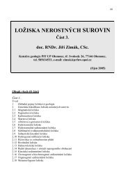
610 JABLONSKIto protect against photodegradation of essential biomoiecules. Facultative pigmentationis probably most important in areas such as the cu'Cum-Mediterraneanthat receive low levels of UVB but receive moderate levels of UVA that causephotodegradation of folate, DNA, and vitamin D3.The practice of recreational tanning has been eschewed by health care workersin the past 20 years because of the explosion in skin cancer rates due to increasedUVR exposure. A tanned skin is still viewed by many as fashionable or as a signof well-being, however, and this positive image has spurred the development of asimulated tanning industry in Europe, the Americas, and Australia (Brown 2001,Randle 1997).In many countries, however, tanned or dark skin does not connote membershipin a fashionable class, and the possession of light skin—especially amongwomen—was and still is viewed as highly desirable and indicative of higher socialstanding. In many Asian countries, most women practice sun avoidance diligently.In other countries where constitutive pigmentation is darker, skin-bleaching agents(including potent topical corticosteroids and hydroquinone formulations) have becomepopular (Tayior 2002).<strong>THE</strong> MULTIFACTORIAL DETERMINATION <strong>OF</strong> <strong>SKIN</strong> PIGMENTATION IN MODERN HU-MANS The evolution of skin pigmentation in humans has been determined bymany factors (Figiu^e 6). By far the most important of these is the UVR regimeof the environment because intensity of UVR has been the main selective factorinfluencing the evolution of melanin pigmentation in the skin. Through time,the number of factors influencing the evolution of human skin pigmentation hasincreased, and culture clearly has reduced the scope for the action of natural selectionon human skin. Cultural behaviors such as the wearing of clothes and theutilization of shelter have become more common through time and have affectedthe evolution of skin pigmentation in some populations because of their effects ofreducing an individual's UVR exposure. Related to this phenomenon is the lengthof time that a population has inhabited an area with a particular UVR regime andthe latitudinal distance traversed from the ancestral to the new homeland. Thereis certainly a considerable lag time between the time of settlement of an area andtime that a population reaches its "optimum" skin color for the UVR conditionsof the area. The length of that lag period for any population is not known butwould depend on the intensity of natural selection exerted on the population byenvironmental influences. In early prehistory, humans possessed a simpler materialculture, spent considerable time accumulating food, and had fewer cultural trappingsto buffer themselves against the environment. Under these conditions, naturalselection would have promoted mainly biological adaptations to the environment—including changes in skin coloration, body proportions, and regulation of thermalcooling. With increasing cultural competence over time, cultural solutions to theenvironmental challenges of sun, heat, and cold became preeminent. The oft-citedexample of the skin colors of the native populations of equatorial South America isworth revisiting in this connection. These populations have long been recognized
<strong>SKIN</strong> <strong>AND</strong> <strong>SKIN</strong> <strong>COLOR</strong> 611In evolutionary time:Environment:-UVradiation levels-PrecipitationMigration:- Length of migration- Recency of migrationOther cultural adaptations to climate:-Clothes wearing• Shade seeking- Shelter-Timing of daily activitiesDuring a human lifetime:Environmentalinfluences:• UV radiation exposure• Application of tanningor bleaching agentsDiet:-Vitamin D content- Foiate contentoEndogenousinfluences:- Genetic, esp.MCIR locus-AgeFigure 6The factors influencing human skin pigmentation, through evolutionarytime and during the course of a human Ufetime.
612 JABLONSKIas being more lightly pigmented than are their counterparts at similar latitudesand altitudes in the Old World (Frisancho 1981. Jablonski & Chaplin 2000). Thisfact is almost certainly due to the recency of populations' migration into SouthAmerica from Asia (within the past 10,000-15,000 years) and the fact that the immigrantpopulations into South America possessed many cultural behaviors andaccoutrements that protected them from high UVR exposure (Jablonski & Chaplin2000).Diet has also played a part in the evolution of human skin pigmentation invery recent human history, as is well illustrated by the Eskimo-Aleut peoplesof the northeast Asian and North American Arctic. Eskimo-Aleuts exhibit skinpigmentation darker than would be predicted on the basis of the UVMED in theirhabitats (Jablonski & Chaplin 2000). Several factors have likely contributed tothis phenomenon, including the relative recency of their migration to the far northfrom a lower-latitude Asian homeland and its implication that their skin colorhas not caught up with their current location. This is almost certainly not theentire story, however. The UVR regime of the latitudes in which Eskimo-Aleutsreside comprises almost exclusively UVA throughout the year, with virtually novitamin D-inducing UVB except for extremely small doses in the summer months(Chaplin 2001, Johnson et al. 1976). Habitation of this latitude (Figure 5, Zone 3)by humans would be impossible without reliance on a highly vitamin D-rich diet.The major components of the aboriginal Eskimo-Aleut diet—marine mammals,fish, and caribou—provide vitamin D3 in abundance. Much of the dietary vitaminD3 is stored in body fat (Mawer et al. 1972), denoting a possible evolutionaryconnection between the development of generous subcutaneous fat stores andvitamin D3 storage in these populations. With selection pressure on depigmentationapparently relaxed because of diet, Eskimo-Aleuts have evolved darker skin toprotect themselves from high levels of UVA as a result of direct solar irradiationand reflection from snow and ice. This scenario is supported by epidemiologicalstudies showing that departure from traditional diets in Eskimo-Aleut populationshas resulted in a high prevalence of vitamin D^-deficiency diseases, especiallyrickets (Gessner et al. 1997, Haworth & Dilling 1986, Moffatt 1995).<strong>THE</strong> GENETICS <strong>OF</strong> <strong>HUMAN</strong> <strong>SKIN</strong> <strong>COLOR</strong>ATION The Study of genetics of humanskin pigmentation has lagged considerably behind the study of the diversity andcausation of diverse human skin color phenotypes. This situation is now changingrapidly as comparative genomics, especially detailed studies of the genes regulatingcoat color pigmentation in mice (Barsh 1996, Sturm et al. 2001), begin topermit identification of the genes responsible for the pigmentation of human hair,skin, and eyes. Sixty of the 127 currently recognized pigmentation genes in themouse appear to have human orthologs (Bennett 2003).Human skin pigmentation has long been considered a polygenic trait that followsa quasi-Mendeiian pattern of inheritance (Brues 1975, Byard 1981, Byard& Lees 1981), with a few major genes of dramatic effect and additional modifiergenes (Sturm et al. 2001). Because pigmentation is a trait determined by the
<strong>SKIN</strong> <strong>AND</strong> <strong>SKIN</strong> <strong>COLOR</strong> 613synchronized interaction of various genes with the environment (John et al. 2003),determination of the relative roles of variant genes and varying environments hasproven extremely challenging (Sturm et al. 1998). Classical genetic studies of inheritanceof human skin coloration have shed little light on the molecular basisof skin color variation, beyond showing that interbreeding between light and darkskin color phenotypes produces offspring of intermediate pigmentation (Robins1991; Sturm etal. 1998, 2001). As a result of recent advances in the understandingof the chemistry and enzymology of the biosynthesis of melanins, the geneticregulation of the many steps in melanin production is now beginning to be understood.Among the numerous mutations affecting melanocyte function in humanpopulations are the F-gene and members of the TYRP and SILV gene families,which direct the assembly and maturation of melansomes within melanocytes(Sturm et al. 2001). Investigation of the influence of these genes on skin pigmentationphenotypes is just beginning (Akey et al. 2001), however, and it remainsto be demonstrated whether polymorphism in these gene systems correlates withpigmentary differences between populations.To date, the greatest scholarly attention has been focused on the melanocortin-1receptor (MCIR) gene, which is the human homologue of the Agouti locus thatin mice regulates the production of the eumelanin and pheomeianin pigments ofthe coat (Barsh 1996, Ranaet al. 1999). In humans, the synthesis of eumelanin isstimulated by the binding of a-melanotropin (tf-melanocyte-stimulating hormone)to the functional MCIR expressed on melanocytes (Scott et al. 2002). The MCIRappears to be one of the major genes involved in the determination of human hairand skin pigmentation, with MCIR polymorphisms in northem European populationsassociated with red hair and fair skin, reduced tanning ability, and high riskof melanoma and nonmelanoma skin cancer (Healy et al. 2001, Scott et al. 2002,Smith et al. 1998). The MCIR locus is characterized by high levels of polymorphismin light-skinned individuals outside of Africa and lower levels of variationin dark-skinned individuals within Africa (John et al. 2003, Rana et al. 1999).This is opposite the pattem observed in most other loci, where Africans are mostpolymorphic (Shriver et al. 1997). The observed pattem of variation in the MCIRsuggests that different selective pressures among individuals with dark and lightskin have shaped the genetic variation at this locus, with functional constraintsoperating to limit variation in African populations (John et al. 2003). The numerousMCIR polymorphisms in light-skinned individuals were originally thoughtto denote relaxation of selection for production of eumelanin outside of tropicallatitudes (Harding et al. 2000). A reinterpretation of these data indicates, however,that adaptive evolution for sun-resistant MCIR alleles began when humans firstbecame hairless in tropical Africa, and that buman movement into the less sunnyclimes of Eurasia favored any mutant MCIR allele that did not produce dark skin(Rogers et al. 2004). Recent study of the MCIR promoter function casts doubt onthe relaxation hypothesis and suggests instead the possible action of purifying ordiversifying selection on some MCIR variants in Asian and Europeans (Makovaet al. 2001). A study comparing populations in southem Africa of Bantu-language
614 JABLONSKIspeakers and San people showed some variation in MCIR sequences, but investigatorsconcluded that although some MCIR mutants are tolerated in Africa, thisgene has been the object of purifying selection and has played an important rolein the maintenance of dark pigmentation in Africans (John et al. 2003). The presenceof higher levels of MCIR variation in dark-skinned populations subjected tolower levels of UVR in southem Africa (as compared to equatorial Africa) supportsthe notion thai number and kinds of MCIR variants are strongly influencedby purifying selection (John et al. 2003). Further genetic studies of more Africanpopulations are needed to determine if the great diversity of skin color observedin populations in sub-Saharan Africa (Relethford 2000) can be related to specificpattems of MCIR or other polymorphisms that evolved in response to the region'sconsiderable heterogeneity of UVR and precipitation regimes (Chaplin 2001).The study of the genetics of human skin pigmentation is still in infancy, andmuch remains to be learned about the levels, effects, and interactions of polymorphismsin the loci influencing skin color phenotype. The production of eumelaninis under strong functional constraint as a result of natural selection in regions ofthe worid with high levels of UVR (Sturm et al. 2001), and there is increasingevidence that at least MCIR variation is an adaptive response to selection for differentalleles in different environments (Makova et al. 2001, Sturm et al. 2001).From what is known of the timing and nature of movements of groups of earlyHomo species and of Homo sapiens in prehistory, it appears that populations ofhumans have moved in and out of regions with different UVR regimes over thecourse of thousands of years. This finding would suggest that natural selectionwould have favored the evolution of dark and light skin pigmentation in disparateplaces at different times, resulting in the independent evolution of dark and lightskin phenotypes and possibly involving recurrent episodes of repigmentation anddepigmentation (Jablonski & Chaplin 2000). This phenomenon would have beenpronounced in the early history of the genus Homo (including the early history ofHomo sapiens) when cultural buffers against the environment were less effectiveand sophisticated.SiON <strong>COLOR</strong> <strong>AND</strong> RACE Skin color is the most obvious visible attribute of thehuman body. It has been the primary characteristic used to classify people intopurportedly genetically distinct geographic groups or "races." The biological basisof skin pigmentation in humans, however, strongly argues against its use as adiagnostic classificatory trait. Critical examination of the distribution of skin colorphenotypes in humans leads to the conclusion that skin pigmentation is adaptive,and its evolution in specific populations has been strongly influenced by the environmentalconditions (the UVR regimes, in particular) of specific places. Highlyadaptive phenotypic characteristics of organisms are of little use in classificationbecause they are subject to homoplasy (parallelism or convergent evolution) andare extremely labile. Emerging genetic evidence indicates that the evolution ofpigmentation genes has been driven by purifying and diversifying selection workingto produce adaptive responses in different environments (Makova et al. 2001,
<strong>SKIN</strong> <strong>AND</strong> <strong>SKIN</strong> <strong>COLOR</strong> 615Rogers et al. 2004, Sturm et al. 2001). This evidence indicates that sitnilar skincolors have evolved independently in human populations inhabiting similar environments.Darkly or lightly pigmented skin, therefore, provides evidence onlyabout the nature of the past environments in which people have lived, renderingskin pigmentation useless as a marker for membership in a unique group or "race."The continued social importance of skin color in human affairs refiects a highdegree of sensitivity to skin color, brought about by historic and complex culturalattitudes toward skin colors (Ehriich & Feldman 1969. Lewontin 1995, Parra et al.2003). The apparent existence of a difference between so-called human races andsubgroups is predicated on an exaggerated perception and heightened sensitivityto a visually obvious attribute of human appearance. The enormity of this bias isrevealed when the small amounts of actual genetic variation within purported racialgroups are revealed (Lewonfin 1995. Marks 2002). Overall, human populations areremarkably similar to one another, with the greatest fraction of human variationbeing accounted for by differences between individuals (Lewontin 1972, 1995;Marks 2002). This collective evidence militates that the concept of biologicalrace be abandoned and publicly disavowed (Lewontin 1995, Marks 2002, Muir1993). Race thus emerges as a cultural construct devoid of explanatory power anddestructive of human and social relations (Lewontin 1972, 1995; Muir 1993).PROSPECTUSThe past decade has witnessed a tremendous advance in the understanding of theevolution of human skin and especially skin color, largely as a result of two phenomena.First is availability of remotely sensed environmental data that permithypotheses about the adaptive value of properties of skin to be thoroughly tested.Second is the proliferation of studies of the molecular genetics of the skin colorthat are permitting new insight into the origins of skin color phenotypes and themechanisms by which they have evolved. Growth is anticipated in both of theseareas, and great potential exists for their interaction, in particular for the testing ofhypotheses of adaptation through the simultaneous and detailed study of pattemsof phenotypic, genotypic, and environmental variation, such as has been done inthe study of butterfly pigmentation (Watt et al. 2003). The study of the evolution ofhuman skin and skin color will also be advanced by the documentation of differentialsurvival of well-defined phenotypes and genotypes in different environmentalregimes through the use of epidemiological data, as has been undertaken recentlyin the study of geographic pattems of melanoma (Garland et al. 2003).Continued study of the evolution of human skin and skin color is importantnot only to our realization of a more complete picture of human evolution, butalso it is important because the skin is involved in so many aspects of human wellbeing.Many humans now live in regions far distant from their ancestral homelands,but they retain a covering of skin adapted to remote Pleistocene conditions. Asis evidenced by modem rates of skin cancer and vitamin D deficiencies, human
616 JABLONSKIbehavior and culture are not perfect buffers against the effects of these majortranslocations. An appreciation of the many roles of skin will improve human healthand attitudes toward diversity and will promote the fundamental understanding ofwhy it is that people look the way ±ey do.ACKNOWLEDGMENTSConversations and discussions with many colleagues in the months and yearsleading up to the writing of this review greatly enriched the content of this paper.These include Walter Alvarez, Carol Boggs, Carol Bower, C. Loring Brace, JimCleaver, Paul Ehrlich, Roberto Frisancho. Cedric Garland, Maciej Henneberg, thelate Gahriel Lasker, Charles Oxnard, Bill Nye, Lynn Rothschild, Donald Tyler, andWard Watt. I thank Rachel Wolf for helping to assemble references. Figures 1, 2,3, and 6 were produced by Jennifer Kane, whom I thank for her skillful renderingsand patient revisions. I thank George Chaplin for producing Figures 4 and 5, and formore than ten years worth of challenging and inspiring discussions on the evolutionof human skin and skin coloration. The spatial analyses and maps summarized herewere made possible by generous donations of geographical information systemssoftware to the California Academy of Sciences from Charles Convis and theConservation Program of the Environmental Systems Research Institute (ESRI).The Annual Review of Anthropology is online at http://anthro.annualreviews.orgLITERATURE CITEDAkey JM. Wang H, Xiong M, Wu H, Liu from fancy genes to complex traits. TrendsW, et al. 2(K)1. Interaction between the Genet. 12:299-305melanocortin-l receptor and P genes con- Barsh GS. 2003. What controls variation in hutributesto inter-individual variation in skin man skin color? Public Libr. Sci. Biol. Oct.pigmentation phenotypes in a Tibetan popu- 1 (1 ):E27lation. Hum. Genet. 108:516-20Bennett DC. 2003. IL-12. The colours of miceAokiK. 2002. Sexual selection as a cause of hu- and men—100 genes and beyond? Pigmentman skin colour variation: Darwin's hypoth- Celt Res. 16:576-77esis revisited. Ann. Hum. Biol. 29:589-608 Bereket A. 2003. Rickets in developing coun-AtiqM,SuriaA.QamaniddinNizamiS,Ahmed tries. In Vitamin D and Rickets, ed. ZI. 1998. Maternal vitamin-D deficiency Hochberg, pp. 220-32. Ba.sel: Kargerin Pakistan. Acta Obstet. Gynecol. Scand. Bjom LO. WangT. 2000. Vitamin Din an eco-77:970-73 logical context. Int. J. Circumpolar, HealthBaker PT. 1958. Racial differences in heat tol- 59:26-32erance. Am. J. Phys. Anthmpol. 16:287-305 Blum HF. 1961. Does the melanin pigment ofBarker D. Dixon K, Medrano EE. Smalara D, buman skin have adaptive value? Q. Rev.Im S. et al. 1995. Comparison of the re- Biol. 36:50-63sponsesofhuman melanocytes with different Bower C, Stanley FJ. 1989. Dietary folate as amelanin contents to ultraviolet B irradiation. risk factor for neural-tube defects: evidenceCancer Res. 55:4041-46from a case-control study in Westem Aus-Barsh G. 1996. Tbe genetics of pigmentation: tralia. Med. J. Aust. 150:613-19
<strong>SKIN</strong> <strong>AND</strong> <strong>SKIN</strong> <strong>COLOR</strong> 617Brace CL. 1963. Structural reduction in evolution.Am. Nat. 97:39^9Branda RF, Eaton JW. 1978. Skin color and nutrientphotolysis: an evolutionary hypothesis.Science 201:625-26Brown DA. 2(X)I. Skin pigmentation enhancers.See Goldsmith 1991, pp. 637-75Bnies AM. 1975. Rethinking human pigmentation.Am. J. Phys. Anthrvpol. 43:387-92Brunvand L, Haug E. 1993. Vitamin D deficiencyamongst Pakistani women in Oslo.Acta Obstet. Gynecol. Scand. 72:264-68Byard PJ. 1981. Quantitative genetics of humanskin color. Yearb. Phys. Anthropol. 24:123-37Byard PI, Lees FC. 1981. Estimating the numberofloci determining skin colour in a hybridpopulation. Ann. Hum. Biol. 8:49-58Cabanac M, Caputa M. 1979. Natural selectivecooling of the human brain: evidenceof its occurrence and magnitude. / Physiol.286:255-64Caldwell MM, Bjom LO, Bomman JF, FlintSD, Kulandaivelu G, et al. 1998. Effects ofincreased solar ultraviolet radiation on terres-Irial ecosystems. J. Photochem. Photobiol. B.46:40-52Chaplin G. 2001. The geographic distributionof environmental factors influencing humanskin colouration. M.Sc. thesis, ManchesterMetropolitan Univ.Chaplin G. 2004. Geographical distributionof environmental factors influencing skincoloration. Am. J. Phys. Anthropol. 123:InpressChaplin G, Jablonski NG. 1998. Hemisphericdifference in human skin color. Am. J. Phys.Anthrvpol. 107:221-24Chaplin G, Jablonski NG, Cable NT. 1994.Physiology, thermoregulation and bipedalism.J. Hum. Evol. 27:497-510Chu DH, Haake AR, Holbrook K, Loomis CA.2(X)3. The structure and development of skin.See Freedberg et al. 2003, pp. 58-88Chung JH. 2001. The effects of sunlight on theskin of Asians. See Goldsmith 1991, pp. 69-90Cleaver JE. Crowley E. 2002. UV damage.DNA repair and skin carcinogenesis. Front.Biosci. 1:1024-43Clemens TL, Henderson SL, Adams JS. HolickMF. 1982. Increased skin pigment reducesthe capacity of skin to synthesise vitamin D^.Lancet 1:74-76Comish DA, Maluleke V Mhlanga T. 2000.An investigation into a possible relationshipbetween vitamin D, parathyroid hormone,calcium and magnesium in a normally pigmentedand an albino rural black populationin the Northern Province of South Aftica.BioFactors 11:35-38Cosentino MJ, Pakyz RE, Fried J. 1990.Pyrimethamine: an approach to the developmentof a male contraceptive. Proc. Natl.Acad. Sci. C/5A 87:1431-35Cowles RB. 1959. Some ecological factorsbearing on the origin and evolution of pigmentin the human skin. Am. Nat. 93:283—93Daniels F. 1964. Man and radiant energy: solarradiation. In Adaptation to the Environment,ed. DB Dill, EF Adolph, CG Wilber, pp. 969-85. Washington, DC: Am. Physiol. Soc.Daniels F, Post PW, Johnson BE. 1972. Theoriesof the role of pigment in the evolutionof human races. In Pigmentation: Its Genesisand Biologic Control, ed. V Riley, pp. 13-22.New York: Appleton-Century-CroftsDeol MS. 1975. Racial differences in pigmentationand natural selection. Ann. Hum. Genet.London 3S:5Q\-3Desruelle AV, Candas V. 2000. Thermoregulatoryeffects of three different types of headcooling in humans during a mild hyperthermia.Eur J. Appl. Physiol. 81:33-39Dmi'el R. Prevulotzky A. Shkolnik A. 1980. Isa black coat in the desert a means of savingmetabolic energy? Nature 283:761-62Domb LG. Pagel M. 2001. Sexual swellings advertisefemale quality in wild baboons. Nature4\0:204-6Edwards EA, Duntley Q. 1939. The pigmentsand color of living human skin. Am. J. Anat.65:1-33Ehrlich PR, Feldman SS.\969. The Race Bomb:Skin Color, Prejudice, and Intelligence. New
618 JABLONSKIYork: Quadrangle/N.Y. Times Book Co. 207pp.Elias PM, Feingold KR, Flubr JW. 2003. Skinas an organ of protection. See Freedberg et al.2003, pp. 107-18Ellis RA, Montagna W. 1962. The skin ofprimates. VI. The skin of the gorilla (Gorillagorilla). Am. J. Phys. Anthropol. 20:79-93Falk D. 1990. Brain evolution in Homo\ the "radiator"theory. Behav. Brain Sci. 13:333-81Feldman D. Glorieux FH. Pike JW, eds. 1997.Vitamin D. San Diego: AcademicFisher GJ, Kang S, Varani J, Bata-Csorgo Z,Yinsbeng W. et al. 2002. Mechanisms of photoagingand chronological skin aging. Arch.Dermatol 138:1462-70Fitzpatrick TB. 1988. The validity and practicalityof sun reactive skin type I through VI.Arch. Dermatol. 124:869-71Fitzpauick TB, Becker SW Jr, Lemer AB,Montgomery H. 1950. Tyrosinase in bumanskin: demonstration of its presence and ofits role in human melanin formation. Science112:223-25Fitzpatrick TB. Seiji M. McGugan AD. 1961.Melanin pigmentation. New Engl. J. Med.265:328-32FitzpatrickTR, Ortonne J-P 2003, Nomial skincolor and general considerations of pigmentarydisorders. See Freedberg et al. 2003, pp.819-25Fleming A, Copp AJ. 1998. Embryonic folatemetabolism and mouse neural nibe defects.5denc
<strong>SKIN</strong> <strong>AND</strong> <strong>SKIN</strong> <strong>COLOR</strong> 619Herman J, Celarier E. 1996. TOMS Version7 UV-erythemal exposure: 1978-1993. Datadeveloped by NASA Goddard Space FlightCent. Ozone Process. TeamHirakawa K, Suzuki H, Oikawa S, KawanishiS. 2002. Sequence-specific DNA damage inducedby ultraviolet A-irradiated folic acidvia its photolysis product. Arch. Biochem.Biophys. 4lO:26l-2SHitchcock RT. 2001. Ultraviolet Radiation.Fairfax, VA: Am. Ind. Hyg. Assoc. 49 pp.Hodgkin P, Kay GH. Hine PM, Lumb GA,Stanbury SW. 1973. Vitamin-D deficiency inAsians at home and in Britain. Lancet 2:167-72Holick MF. 1991. Photobiology, physiology,and clinical applications for vitamin D. SeeGoldsmith 1991, pp. 928-56Holick MF. 1995. Environmental factors that influencethe cutaneous production of vitaminD. Am. J. Clin. Nutr. 61:638S^5Hotick MF 1997. Photobiology of vitamin D.See Feldman et al. 1997, pp. 33-39Holick MF. 2001. A perspective on tbe beneficialeffects of moderate exposure to sunlight:bone health, cancer prevention, mental healthand well being. See Goldsmith 1991. pp. 11-37Holick MF. 2003. Vitamin D: a millenium perspective.J. Cell Biochem. 88:296-307Holick MF, MacLaughlin JA, Doppelt SH.1981. Regulation of cutaneous previtaminDj photosynthesis in man: Skin pigment isnot an essential regulator. Science 211:590-93Diara Y, Aoki K. 1999. Sexual selection by malechoice in monogamous and polygynous humanpopulations. Theor. Popul. Biol. 55:77-93ItoS.2(X)3. A chemist's view of melanogenesis.Pigment Cell Res. 16:230-36Jablonski NG, Chaplin G. 2000. The evolutionof skin coloration. J. Hum. Evol. 39:57-106Jimbow K, Fitzpatrick TB. Wick MM. 1991.Biochemistry and physiology of melanin pigmentation.See Goldsmith 1991, pp. 873-909Jimbow K, Quevedo WC Jr, Fitzpatrick TB, SzaboG. 1976. Some aspects of melanin biology:1950-1975. J. Invest. Dermatol. 67:72-89John PR, Makova K, Li WH, Jenkins T. RamsayM. 2003. DNA polymorphism and selectionat the melanocortin-1 receptor gene in normallypigmented southem African individuals.Ann. N.Y Acad. Sci. 994:299-306Johnson FS, Mo T, Green AES. 1976. Averagelatitudinal variation in ultraviolet radiation atthe Earth's surface. Photochem. Photobiol.23:179-88Jones G, Stmngnell SA, DeLuca HF. 1998. Currentunderstanding of the molecular actionsof vitamin D. Physiol. Rev. 78:1193-231Kaidbey KH, Agin PP, Sayre RM, KligmanAM. 1979. Photoprotection by melanin—acomparison of black and Caucasian skin. Am.Acad. Dermatol. 1:249-60Kesavan V, Pote MS, Batra V, Viswanathan G.2003. Increased folate catabolism follovt-ingtotal body y-irradiation in mice. / Radiat.Res.44:l4\^HKielbassa C. Roza L. Epe B. 1997. Wavelengthdependence of oxidative DNA damage inducedby UV and visible Hght. Carcinogenesis\%:%\\-\6Knip AS. 1977. Ethnic studies on sweat glandcounts. In Physiological Variation and itsGenetic Basis, ed. JS Weiner, pp. 113-23.London: Taylor & FrancisKollias N. 1995a. The physical basis of skincolor and its evaluation. Clin. Dermatol.13:361-67Kollias N. 1995b. The spectroscopy of humanmelanin in pigmentation. In Melanin: ItsRote in Human Photoprotection, ed. LZeise,MR Chedekel, TB Fitzpatrick, pp. 11-22.Overland Park: ValdenmarKollias N, Sayre RM, Zeise L, Chedekel MR.1991. Photoproteclion by melanin. J. Photochem.Photobiol. B. 9:135-60Kraning KK. 1991. Temperature regulation andthe skin. See Goldsmith 1991, pp. 1085-95Kripke ML, Applegate LA. 1991. Alterations inthe immune response by ultraviolet radiation.See Goldsmith 1991, pp. 1222-39Lasker GW. 1954. Seasonal changes in skincolor. Am. J. Phys. Anthropol. 12:553-38
620 JABLONSKILavker RM, Bertoiino A, Freedberg IM, Sun T-T. 2003. Biology of hair follicles. See Freedberget al. 2003, pp. 148-58Lee MMC, La.sker GW. 1959. The sun-tanningpotential of human skin. Hum. Biol. 31:252-60Lees FC. Byard PJ. 1978. Skin colorimetry inBelize. I. Conversion formulae. Am. / Phys.Anthmpol. 4B:5\5-22Lewontin RC. 1972. The apportionment of humandiversity. Evot. Biol. 6:381-98Lewontin RC. 1995. Human Diversity:FreemanLock-Anderson J, Knudstorp ND, Wulf HC.1998. Faculative skin pigmentation in Caucasians:an objective biological indicator oflifetime exposure to ultraviolet radiation? Br.J. Dermatol. 138:826-32Loomis WF. 1967. Skin-pigment regulationof vitamin-D biosynthesis in man. Science157:501-6Lucock M. Yates Z, Gianville T, Leeming R,Simpson N, Daskalakis I. 2003. A criticalrole for B-vitamin nutrition in human developmentand evolutionary biology. Nutr. Res.23:1463-75Lynn B. 1991. Cutaneous sensation. See Goldsmitb1991, pp. 779-815MacKintosh JA. 2001. The antimicrobial propertiesof melanocytes. melanosomes andmelanin and the evolution of black skin. J.Theor.Biol.2\\.\m-\l>MacLaughlin JA, Anderson RR, Holick MF.1982. Spectral character of sunlight modulatesphotosynthesis of previtamin D%. andits photoisomers in human skin. Science216:1001-3Madronicb S, McKenzie RL, Bjom LO, CaldwellMM, 1998. Changes in biologically activeultraviolet radiation reaching the Earth'ssurface. / Photochem. Photobiol. B. 46:5-19Makova K. Ramsay M. Jenkins T, Li W-H.2001. Human DNA sequence variation in a6.6-kb region containing the melanocortin 1receptor promoter. Genetics 158:1253-68Marks J. 2002. What It Means to be 98%Chimpanzee: Apes. People, and Their Genes.Berkeley: Univ. Calif. Press. 312 pp.Marks R. 1991. Mechanical properties of theskin. See Goldsmith 1991Martin RD. 1990. Primate Origins and Evolution:A Phylogenetic Perspective. Princeton,NJ: Princeton Univ. Press. 8(M pp.Mathur U, DaUa SL, Mathur BB. 1977. Theeffect of aminopterin-induced folic acid deficiencyon spermatogenesis. Fertil. Steril.28:1356^0Matsuoka LY, Wortsman J, Dannenberg MJ,Hollis BW, Lu Z. Holick MF. 1992. Clothingprevents ultraviolet-B radiation-dependentphotosynthesis of vitamin Dj. J. Clin. Endocrinol.Metaboi 75:1099-103Mawer EB, Backhouse J, Holman CA, LumbGA, Stanbury S W. 1972. The distribution andstorage of vitamin D and its metabolites inhuman tissues. Clin. Sci. 43:413-31McHenry HM, Berger LR. 1998. Body proportionsin^u5/ra/opir/iecM.ja/aren.n5 and A.africanus and the origin of the genus Homo.J. Hum. Evol. 35:1-^22Minns RA. 1996. Folic acid and neural tube defects.Spinal Cord 34:460-65MiUTi D. Bell NH. 1997. Racial, geographic,genetic, and body habitus effects on vitaminD metabolism. See Feldman et al. 1997. pp.521-32Moffatt MEK. 1995. Current status of nutritionaldeficiencies in Canadian Aboriginalpeople. Can. J. Physiol. Pharmacol. 73:754-58Montagna W. 1967. Comparative anatomy andphysiology of the skin. Arch. Dermatol.96:357-63Montagna W. 1971. Cutaneous comparative biology.Arch. Dermatol. 104:577-91Montagna W. 1981. The consequences of havinga naked skin. Birth Defects: Orig. Artie.Ser. 17:1-7Morison WL. 1985. What is the functionof melanin? Arch. Dermatol. 121:1160-63Muir DE. 1993. Race: the mythic root of racism.Sociol. Inq, 63:339-50Murray FG. 1934. Pigmentation, sunlight, andnutritional disease. Am. Anthropol. 36:438-45
<strong>SKIN</strong> <strong>AND</strong> <strong>SKIN</strong> <strong>COLOR</strong> 621Neer RM. 1975. The evolutionary significanceof vitamin D, skin pigment, and ultravioletlight. Am. J. Phys. Anthropol. 43:409-16Neer RM. 1985. Environmental light: effects ofvitamin D synthesis and calcium metabolismin humans. Ann. N. Y. Acad. Sci. 4.53:14-20Nelson DA, Nunneley SA. 1998. Brain temperatureand limits on transcranial cooling inhumans: quantitative modeling results. Eur.J. Appl. Physiol. 78:353-59Odland GF 1991. Structure of the skin. SeeGoldsmith 1991, pp. 3-62Olivier G. 1960. Pratique Anthropolgique.Paris: Vigot Fr^resOnline Mendelian Inheritance in Man. 2003."139200 group-specific component; GC.In Online Mendelian Inheritance in Man.Betbesda, MD: Natl. Cent. Biotechnol. Inf.Ortonne J-P. 1990. Pigmentary changes of theageing skin. Br J. Dermatol. 122:21-28Ortonne J-P. 2(X)2. Photoprotective propertiesof skin melanin. Br. J. Dermatol. 146:7-10Pagel M, Bodmer W 2003. A naked ape wouldhave fewer parasites. Proc. R. Soc. London B270:S!17-19Pandolf KG, Gange RW, Latzka WA, Blank IH,Kraning KK, Gonzalez RR. 1992. Humanthemioregulatory responses during beat exposureafter artificially induced sunbum. Am.J. Physiol. 262:R610-16Parker F. 1981. The biology of pigmentation.Birth Defects 17:79-91Parra FC, Amado RC, Lambertucci JR, RocbaJ, Antunes CM, Pena SDJ. 2003. Color andgenomic ancestry in Brazilians. Proc. Natl.Acad. Sci. USA 100:177-82Post PW, Daniels F Jr, Binford RT Jr. 1975.Cold injury and the evolution of "white" skin.//Mm. BIO/. 47:65-80Prota G. 1992a. Melanin-producing cells. SeeProta 1992b, pp. 14-33Prota G, ed. 1992b. Melanins and Melanogenesis.San Diego: AcademicProta G. 1992c. Photobiology and photochemistryof melanogenesis. See Prota 1992b, pp.208-24Quevedo WC Jr, Fitzpatrick TB, Pathak MA.1975. Role of light in human skin colorvariation. Am. J. Phys. Anthropol. 43:393-408Rana BK, Hewett-Emmett D, Jin L, Chang BH,Sambuughin N, et al. 1999. High polymorphismat the human melanocortin 1 receptorlocus. Genetics 151:1547-57Randle HW. 1997. Suntanning: differences inperceptions throughout history. Mayo Clin.Proc. 72:461-66Rawles ME. 1948. Origin of melanophoresand their role in development of color pattemsin vertebrates, Physiol. Res. 28:383-408Relethford JH. 1997. Hemispheric differencein human skin color. Am. J. Phys. Anthropol.104:449-57Relethford JH. 2(XK). Human skin color diversityis highest in sub-Saharan African populations.Hum. Biol. 72:771-80Roberts DF. 1977. Human pigmentation: its geographicaland racial distribution and biologicalsignificance. / Soc. Cosmet. Chem. 28:329^2Roberts DF, Kahlon DPS. 1976. Environmentalcorrelations of skin colour. Ann. Hum. Biol3:11-22Robins AH. 1991. Biological Perspectiveson Human Pigmentation. Cambridge: CambridgeUniv. Press. 253 pp.Rogers AR, Iltis D, Wooding S. 2004. Geneticvariation at the MCIR locus and the timesince loss of human body hair. Curr Anthropol.45:105-1Rozema J, van Geel B. Bjom LO, Lean J,Madronicb S. 2002. Toward solving the UVpuzzle. Science 296:1621-22Ruff CB, Trinkaus E, Holliday TW. 1997.Body mass and encephalization in PleistoceneHomo. Nature 387:173-76Ruff CB, Trinkaus E, Walker A, Larsen CS.1993. Postcranial robusticity in Homo. I:Temporal trends and mechanical interpretation.Am. //\n//jTOpo/. 91:21-53Ryan TJ. 1991. Cutaneous circulation. SeeGoldsmith 1991, pp. 1019-84Sama T, Swartz HM. 1998. The physical propertiesof melanins. In The Pigmentary System:Physiology and Pathophysiology, ed. JJ
622 JABLON<strong>SKIN</strong>ordlund. RE Boissey, VJ Hearing, pp. 333-57. New York: Oxford Univ. PressSchwartz GG, Rosenblum LA. 1981. Allometryof primate hair density and the evolution ofhuman hairlessness. Am. J. Phys. Anthropol.55:9-12Scott MC, Suzuki I. Abdel-Malek ZA. 2002.Regulation of the human melanocortin 1 receptorexpression in epidermal melanocytesby paracrine and endocrine factors and byultraviolet radiation. Pigment Cell Res. 15:433-39Shaw NJ. 2003. Vitamin D deficiency rickets.In Vitamin D and Rickets, ed. Z Hochberg,pp. 93-104. Basel: KargerShea CR, Parrish JA. 1991. Nonionizing radiationand the skin. See Goldsmith 1991. pp.910-27Shono S. Imura M, Ota M, Ono S, Toda K. 1985.Tbe relationship of skin color, UVB-inducederythema, and melanogenesis. J. Invest. Dermatol.84:265-67Shriver MD. Jin L, Ferrell RE. Deka R. 1997.Microsatellite data support an early populationexpansion in Africa. Genome Res. 7:586-89Smitb RM, Heaiy E. Siddiqui S. 1998.Melanocortin 1 receptor variants in an Irishpopulation./ Invest. Dermatol. 111:119-22Steegmann AT Jr. 1967. Frostbite of the humanface as a selective force. Hum. Biol. 39:131-44Sturm RA. 2002. Skin colour and skin cancer—MCIR. the genetic link. Melanoma Res.12:405-16Sturm RA. Fox NF, Ramsay M. 1998. Humanpigmentation genetics: tbe difference is onlyskjn deep. BioEssays 20:712-21Sturm RA. Teasdale RD, Fox NF. 2001. Humanpigmentation genes: identification, structureand consequences of polymorphic variation.Gene 277:49-62Suh JR, Herbig AK. Stover PJ. 2001. New perspectiveson folate cataboisim. Annu. Rev,Nutr 21:255-&2Sulaimon SS, Kitchell BE. 2003. The biologyof melanocytes. Vet. Dermatol. 14:57-65Szabo G. Gerald AB. Pathak MA. FitzpatrickTB. 1969. Racial differences in the fate ofmelanosomes in human epidermis. Nature222:1081-82Taylor SC. 2002. Skin of color: biology, structure,function, and implications for dermatologicdisease. / Am. Acad. Dermatol.46:S41-62Thody AJ. Higgins EM. Wakamatsu K, Ito S,Burchill SA, Marks JM. 1991. Pheomelaninas well as eumelanin is present In human epidermis.J. Invest. Dermatol. 97:340-44Thody AJ. Smith AG. 1977. Hormones and skinpigmentation in tbe mammal. Int. J. Dermatol.16:657-64Thomas MK, Lloyd-Jones DM, Thadhani RI,Shaw AC, Deraska DJ, et al. 1998. HypovitaminosisD in medical inpatients. New Engl.J. Med 338:777-83Urbach F. 2001. The negative effects of solarradiation: a clinical overview. See Goldsmith1991. pp. 39-67van den Berghe PL, Frost P. 1986. Skin colorpreference, sexual dimorphism and sexualselection: acase of gene-culture coevolution?Ethnic Rac. Stud. 9:^1-] 13Vermeer M. Schmieder GJ. Yoshikawa T vanden Berg J-W, Metzman MS. et al. 1991.Effects of ultraviolet B light on cutaneous immuneresponses of humans with deeply pigmentedskin. / /nve.st. Dermatol. 97:729-34von Luschan F. 1897. Beitrage zur Volkerkundeder Deut.schen Schutzgebieten.Berlin: Deutsche BuchgemeinschaftWagner JK, Parra EJ, Norton HL. Jovel C,Shriver MD. 2(X)2. Skin responses to ultravioletradiation: effects of constitutive pigmentation,sex, and ancestry. Pigment Cell Res.15:385-90Walsberg GE. 1988. Consequences of skincolor and fur properties for solar heat gainand ultraviolet irradiance in two mammals./ Comp. Physiol. B 158:213-21Walter H. 1958. Der zusammenhang von hautfarbenverteilungund intensitat der ultraviolettenstrahlung. Homo 9:1-13Walter H. 1971. Remarks on tbe environmentaladaptation of man. Humangenetik 13:85-97
<strong>SKIN</strong> <strong>AND</strong> <strong>SKIN</strong> <strong>COLOR</strong> 623Wassermann GD. 1981. On the nature of thetheory of evolution. Philos. Sci. 48:416-37Wassemiann HP. 1965a. The circulation ofmelanin—its clinical and physiological significance.5. Af^. Med J. 39:711-16Wassermann HP. 1965b. Human pigmentationand environmental adaptation. Arch. Environ.Health 11:691-94Wassermann HP. 1974. Ethnic Pigmentation.New York: Biackwell Sci. 284 pp.Watt WB, Wheat CW, Meyer EH, Martin J-E2003. Adaptation at specific loci. VII. Naturalselection, dispersal and the diversityof molecular-functional variation pattemsamong butterfly species complexes (Colias:Lepidoptera, Pieridae). Mol. Ecol. 12:1265-75Webb AR, Kline L, Holick MF. 1988. Influenceof season and latitude on the cutaneoussynthesis of vitamin Dj: Exposure to wintersunlight in Boston and Edmonton will notpromote vitamin Di synthesis in human skin.J. Clin. Endocrinol. Metabol. 67:373-78Weiner JS. 1951. A spectrophotometer for measurementof skin colour. Man 51:152-53Weiner JS. 1977. Variation in sweating. In P/i>'5-iological Variation and its Genetic Basis, ed.JS Weiner, pp. 125-37. London: Taylor &FrancisWenger BC. 2003. Thermoregulation. SeeFreedberg et al. 2003, pp. 119-26Westerhof W, Estevez-Uscanga O. Meens J,Kammeyer A, Durocq M. Caho 1.1990. Therelation between constitutional skin colorand pbotosensitivity estimated from UVinducederythema and pigmentation doseresponsecurves./ Invest. Dermatol. 94:812-16Wharton B, Bishop N. 2003. Rickets. Lancet362:1389^00Wheeler PE. 1984. The evolution of bipedalityand loss of functional body hair in hominids./ Hum. Evol. 13:91-98Wheeler PE. 1985. The loss of functional bodyhair in man: the infiuence of thermal environment,body fomi and bipedality. J. Hum.Evol. 14:23-28Wheeler PE. 1991a. The influence of bipedalismon the energy and water budgets of earlyhominids. / Hum. Evol. 21:117-36Wheeler PE. 1991b. The thermoregulatory advantagesof hominid bipedalism in openequatorial environments: the contribution ofincreased convective heat loss and cutaneousevaporative cooling. / Hum. Evol. 21:107-15Wheeler PE. 1992. Tbe themioregulatory advantagesof large body size for bominidsforaging in savannah environments. J. Hum.Evol. 23:351-62Wheeler PE. 1996. The environmental contextof functional body hair loss in hominids (a replyto Amaral, 1996). J. Hum. Evol. 30:367-71Young AR, Sheehan J. 2001. UV-induced pigmentationin human skin. See Goldsmith1991, pp. 357-75Zihlman AL, Cohn BA. 1988. The adaptive responseof human skin to the Savanna. 3:397-409
<strong>SKIN</strong> <strong>AND</strong> <strong>SKIN</strong> <strong>COLOR</strong>C-IFigure 3 Human skin coloration as predicted from multiple regression formulae. See textfor discussion.Figure 5 The potential for synthesizing vitamin D3 in the skin relative to levels of annualaverage UVMED. The highest UVMED levels are indicated in deep violet, with incrementallylower levels indicated in shades of red. orange, yellow, green, and gray. Zone 1 {areawithout hachure enclosing the tropics) represents the region with adequate UVR throughoutthe year to catalyze vitamin D3 synthesis, Zone 2 (vertical hachure) represents the area inwhich there is insufficient UVR during at least one month of the year to produce vitamin D3.Zone 3 {cross-hatched area) represents the region in which there is insufficient UVR averagedover the entire year to photosynthesize vitamin D3. See text for further description.