View PDF - Johns Hopkins Pathology
View PDF - Johns Hopkins Pathology
View PDF - Johns Hopkins Pathology

Create successful ePaper yourself
Turn your PDF publications into a flip-book with our unique Google optimized e-Paper software.
Gynecological Endocrinology, February 2010; 26(2): 105–108PREGNANCYPostpartum thyroiditis and hypothalamo-hypophysial insufficiency inthe same woman with successive pregnancies: a case reportSIBEL ERTEK & GURBUZ ERDOGANGynecol Endocrinol Downloaded from informahealthcare.com by JHU John <strong>Hopkins</strong> University on 05/19/10For personal use only.Department of Endocrinology and Metabolic Diseases, Ufuk University Medical Faculty, Dr. Ridvan Ege Hospital, Ankara,Turkey(Received 13 April 2009; revised 24 May 2009; accepted 20 July 2009)AbstractObjective. Although the incidence of postpartum autoimmune disorders of endocrine glands are not rare, the presence oftwo different entities in the same patient with two different pregnancies is uncommon.Methods. We present a 35-year-old woman whose story starts with her first pregnancy when she was 29 years old, she hadthe diagnosis of postpartum thyroiditis with hypothyroidism. We followed up the patient when she had her second pregnancy.Results. When she was being followed up with levothyroxine replacement, 5 years later she had her second delivery afterwhich she had complaints of polydipsia, polyuria, weight loss and had the diagnosis of central diabetes insipitus and she hasstarted desmopressin treatment and 17 months later the delivery she again applied with amenorrhea, continuation of lactationlater she noticed oligomenorrhea, and her gonadotropin levels were found to be low as well as her TSH levels, although theL-thyroxine treatment dose was not changed. Dynamic tests of hypophysis revealed hypophyseal insufficiency and repeatedhypophyseal MRI was in concordance with lymphocytic hypophysitis which explains the pattern of endocrinologicalabnormalities after the second delivery.Conclusion. This case signals role of autoimmune mechanisms underlying the endocrinopathies seen after successivepregnancies of the same patient.Keywords: Postpartum, postpartum thyroiditis, hypophysitis, postpartum hypophysitis, lymphocytic hypophysitisIntroductionPostpartum thyroiditis is an autoimmune disease withvarying prevalence between 1.1% and 16.7% (meanprevalence rate 7.2%) and it commonly presents ashypothyroidism (43%) in previously euthyroid woman[1]. Lymphocytic infiltration of the thyroid is themost evident pathological feature and positive antithyroidperoxidase antibodies are frequently observed[2].Hypopituitarism arising postpartum may be causedfrequently either by lymphocytic hypophysitis or bySheehan syndrome [3]. Lymphocytic hypophysitis ispituicyte destruction with autoimmune basis, frequentin gestational and postpartum period [4].Together with the granulomatous hypophysitis andxanthomatous hypophysitis, lymphocytic hypophysitis,(also called autoimmune hypophysitis, AH),constitute primary chronic inflammatory diseases ofhypophysis. If the inflammation is limited in adenohypophysis,it is called lymphocytic adenohypophysitis(LAH). Other forms are lymphocyticinfundibulohypophysitis and lymphocytic panhypophysitis[5]. Most cases of LAH present in the lastmonth of pregnancy or within the first 2 months afterdelivery [5]. In the first case of LAH of the medicalliterature, Hashimoto thyroiditis was also present andthe authors emphasized the onset of autoimmunereaction to thyroid and pituitary antigens [6]. In therecent study of Manetti et al. [7] with 961 autoimmunethyroid disease patients, the frequency ofantipituitary antibody positivity was significantlyhigher than patients without autoimmune thyroiddisease. The link between these two disorders is nottotally clarified [8].Immune system changes and adaptations inpregnancy cause different effects on the courseof autoimmune diseases. Generally, pregnancyCorrespondence: Sibel Ertek, Department of Endocrinology and Metabolic Diseases, Ufuk University, Faculty of Medicine, Dr. Ridvan Ege Hospital, MevlanaBulvari 86-88 (Konya Yolu) 06520, Balgat, Ankara, Turkiye. Tel: þ90-312-204-40-00. Fax: þ90 312 204 40 55. E-mail: sibelertek@yahoo.itISSN 0951-3590 print/ISSN 1473-0766 online ª 2010 Informa UK Ltd.DOI: 10.3109/09513590903215532
106 S. Ertek & G. ErdoganGynecol Endocrinol Downloaded from informahealthcare.com by JHU John <strong>Hopkins</strong> University on 05/19/10For personal use only.improves the course of rheumatoid arthritis, Gravesdisease and Type1 diabetes mellitus, on the other sideworsens Wegener syndrome [9–12]. Lymphocytichypophysitis and postpartum thyroiditis are twounique examples of autoimmune diseases that thepregnancy is directly related with disease onset.Lymphocytic hypophysitis can also be accompaniedby other autoimmune diseases, like Addison disease,Type 1 diabetes mellitus, Graves disease, systemiclupus erythematosus, Sjogren syndrome, autoimmunehepatitis, primary biliary cirrhosis and as inother autoimmune diseases there may be relapsingand remitting course, and spontaneous recovery hasalso been reported [13].Searching the Embase and Pubmed both for the allcase reports and reviews about the co-existence ofthese two postpartum diseases in same patient, withkey words ‘postpartum hypophysitis’, ‘postpartumthyroiditis’, ‘lymphocytic hypophysitis’, ‘pregnancy’and ‘hypophysitis’ and ‘thyroiditis’, we found that itwas reported in the same pregnancy of a patient in thecase report of Bevan et al. [14], another report of bothdiseases was presented by Iwaoka in 2001 [15].In this article we present a case with a 9 years storyof follow-up during two successive pregnancies.Case presentationIn 1999, a 26-years-old female patient without anyhistory of thyroid disease has applied to Endocrinologyand Metabolic Diseases Department with complaintsof fatigue and increase in weight at her fifthmonth of postpartum period, during lactation. Herstory of menstruation was regular with age ofmenarche at 15 years, and she did not have anyproblem during her pregnancy and the labour. Onphysical examination she had diffuse goiter withoutpalpable nodules. On laboratory tests, free T4:9.59 ng/dl (Normal range:10–23 ng/dl), free T3:4.38 ng/dl (Normal range: 2.8–7 ng/dl), TSH (thyroid-stimulatinghormone): 18.6 mcIU/ml (Normalrange:0.27–4.20 mcIU/ml), TPO antibody: 365 IU/ml (Normal range: 0.01–34 IU/ml) and Thyroglobulinantibody: 1945 IU/ml (Normal range: 0.01–115 mIU/ml). She had the diagnosis of postpartumthyroiditis and L-thyroxine treatment was started.When she was being followed up with the diagnosisof permanent autoimmune thyroiditis with hypothyroidismafter post-partum period, under L-thyroxinereplacement, she had her second delivery at September2004. After the second delivery, she had thecomplaints of polydipsia, polyuria, prolonged lactation,weight loss, tiredness and she came toendocrinologist in January 2005 when these complaintsincreased. With water deprivation test she hadthe diagnosis of central diabetes insipitus. In herhypophyseal magnetic resonance (MR) imaging,hyperintensity of neurohypophysis was disappeared.She had started desmopressin spray with the diagnosisof central diabetes insipitus. Later, she hadnoticed that she had amenorrhea for 26 months afterdelivery, and 10 kg weight gain within last 6 monthsand her lactation continues although she was notgiving breast feeding. She applied to her gynaecologistand endocrinologist with these complaints inDecember 2006. The tests revealed mild hyperprolactinemiaand on February 2007 she had startedbromocriptin (Table I).At her control visit on December 2007, she had thecomplaint of oligomenorrhea and she was takingmedical treatment for hypothyroidism and centraldiabetes insipitus. Her bromocriptin treatment waschanged and cabergoline and lynesterol was started.She did not have complaint of galactorhea. Her testsfor anterior hypophysis had revealed a suspicion forhypophyseal insufficiency and she was hospitalizedfor dynamic tests and evaluation for hypophysealinsufficiency (Table I).Table I. Laboratory tests of the patient on December 2006, February and December 2007.Patient’s values(October 2006)Patient’s values(February 2007)Patient’s values(December 2007)Normal range*E 2 42.45 pg/ml 10 pg/ml 12.02 pg/ml 12.5–166*FSH 9.02 mIU/l 1.6 mIU/l 1.4 mIU/l 3.50–12.50*LH 7.6 mIU/ml 0.69 mIU/l 0.76 mIU/ml 2.40–12.6*Prolactin 363 mIU/ml 798 mIU/ml 371.4mIU/ml 102–496TSH 0.17 mIU/ml 0.012 mIU/ml 0.05 mIU/ml 0.27–4.20Free T4 1.35 ng/dl 1.44 ng/dl 1.38 ng/dl 0.93–1.71Anti-TG 360 IU/ml – – 0.01–115Anti-TPO 203 IU/ml – – 0.01–34Urine density 1025 – – 1005–1030ACTH – – 25 pg/ml 10–120 (morning)Cortisol – – 12 mcg/dl 6–25 (morning)GH – – 1.70 mIU/l Adult: 520IGF-1 – – 140 ng/ml 140–400*Normal range for follicular phase of the menstrual cycle.
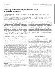
Gynecol Endocrinol Downloaded from informahealthcare.com by JHU John <strong>Hopkins</strong> University on 05/19/10For personal use only.Insulin hypoglycaemia test, CRH (corticotrophinreleasinghormone), LHRH (luteinizing hormonereleasinghormone) and TRH (thyrotropin-releasinghormone) tests were performed. The results of thetests are shown on Table II. As it is seen from thedynamic tests of hypophysis, the cortisol and growthhormone responses to hypoglycaemia, and responseof FSH (follicle-stimulating hormone) and LH(luteinizing hormone) to LHRH were not enough.It’s difficult to evaluate the TRH test, because thepatient was under L-thyroxine treatment and shedropped the medicine only on the day of test.The hypophyseal MR was repeated and focalthickening and contrast enhancement in the middleof infundibulum was reported and this was relevantfor lymphocytic hypophysitis (Figure 1).DiscussionThere are mainly three reasons of postpartumhypophyseal dysfunction; Sheehan syndrome, pituitarytumors and lymphocytic hypophysitis [5,14].Absence of obstetrical problem and the presence oflactation after delivery decrease the suspicions ofSheehan syndrome, meanwhile the hormone levelsand MR images were not relevant to this syndrome.In the presence of pituitary tumours the typicalpattern of endocrinological abnormalities in theadenohypophyseal tests are known to be typical (i.e.theoretically GH and gonadotropin deficienciesoccurs first, followed later by ACTH and TSHdeficiency) and the MR rules out the presence of anyhypophyseal tumour. Symmetrical enlargement andtypical thickening of the infundibular stalk on MRseries are typical features for lymphocytic hypophysitis.The mild hyperprolactinemia at the beginningof the story and presence of previous postpartumthyroiditis also support the diagnosis of this disease.Although the MR images resemble that of the ectopicbright spot of ectopic neurohypophysis, absence ofmusculoskeletal and midbrain anomalies and thepresence of neurohypophyseal dysfunction and clinicalstory in normal adult female in our case also givesa clue [16].HormonesPostpartum thyroiditis-hypophysitis 107Table II. Dynamic endocrine tests of the patient.BasalvaluesWithhypoglycaemia*Insulin hypoglycaemia test of the patientCortisol (mg/dl) 9.73 17.59ACTH (pg/ml) 25 12Growth_hormone (mIU/l) 2.18 2.48IGF-1 (ng/ml) 140 180Cortisol (mg/dl) ACTH (pg/ml)CRH test of the patient {0 min 14.72 18.0015 min 19.87 41.2230 min 23.10 21.9060 min 26.41 18.10Time FSH (mIU/l) LH (mIU/l)LHRH test { of the patientBeginning 2.37 0.9030 min 6.29 6.4260 min 8.18 7.0790 min 8.35 5.97Time TSH (mcIU/ml) PRL (ng/ml)TRH test x of the patientBeginning 0.03 16.8730 min 0.23 32.1060 min 0.20 20.5790 min 0.14 17.62*Hypoglycemia was accepted as blood glucose value below 40 mg/dl. Expected normal responses to hypoglycaemia were more than20 mIU/l for GH, and increase to at least 18–20 mg/dl for cortisol.{ Normally peak cortisol of 410 mg/dl should be observed within30–60 min.{ The normal response is five times increase in LH and two timesincrease in FSH within 20 min.x The patient stopped L-thyroxine intake only at the day of the test.Normal response should be 46 mIU/ml increase in TSH withinfirst 45 min and for this age of patient PRL should increasebetween 30 and 120 ng/ml in this test.Figure 1. MRI imaging of the patient in 2008 showing the central homogenous symmetrical bright contrast enhanced area in hypophysis, andintact sellar floor, typical for lymphocytic hypophysitis.
Gynecol Endocrinol Downloaded from informahealthcare.com by JHU John <strong>Hopkins</strong> University on 05/19/10For personal use only.108 S. Ertek & G. ErdoganThe diagnosis of lymphocytic hypophysitis is doneby tissue biopsy, but because it is not feasiblegenerally in the medical literature there are manypatients who had this probable diagnosis by clinicalpresentation and MRI findings.Anti-hypophyseal antibodies may be helpful for thediagnosis, but their significance is not clear. Sometimesalthough biopsy proven hypophysitis is present,antibodies may be negative [17,18]. Because thereare at least five types of cells in the pituitary gland,antibodies to different cell types, to different cellproteins or to different hormones may be the reason,and there are different techniques (like complementfixation, indirect immunoflorescence, immune blottingand enzyme-inked immunosorbent assay) todetect them. Autoantibodies to certain proteins maybe positive in 70% of the patients [19] but they canbe found in plasma of patients with other pituitarydiseases [20].Appropriate management of the lymphocytichypophysitis remains contraversal. All patientsshould have treatment for deficient hormones, andlong-term follow-up for new hormone deficiencies isnecessary. The efficacy of corticosteroid therapy isunclear with known side effects [4]. For our patient,we decided to have close follow-up without steroidtreatment.Declaration of interest: The authors report noconflicts of interest. The authors alone are responsiblefor the content and writing of the paper.References1. Stagnaro-Green A. Postpartum thyroiditis. J EndocrinolMetab 2002;87:4042–4047.2. Roti E, Uberti E. Post-partum thyroiditis a clinical update.Eur J Endocrinol 2002;146:275–279.3. Molitch ME. Pituitary diseases in pregnancy. Semin Perinatol1998;22:457–470.4. Molitch ME, Gillam MP. Lymphocytic hypophysitis. HormRes 2007;68(Suppl 5):145–150.5. Caturegli P, Newschaffer C, Olivi A, Pomper MG, Burger PC,Rose NR. Autoimmune hypophysitis. Endocr Rev 2005;26:599–614.6. Goudie RB, Pinkerton PH. Anterior hypophysitis and hashimoto’sdisease in a woman. J Pathol Bacteriol 1962;83:584–585.7. Manetti L, Lupi I, Morselli LL, Albertini S, Cosottini M,Grasso L, Genovesi M, Pinna G, Mariotti S, Bogazzi F, et al.Prevalance and functional significance of antipituitary antibodiesin patients with autoimmune and non-autoimmunethyroid diseases. J Clin Endocrinol Metab 2007;92:2176–2181.8. Ozawa Y, Shishiba Y. Recovery from lymphocytic hypophysitisassociated with painless thyroiditis: clinical implications ofcirculating antipituitary antibodies. Acta Endocrinol (Copenh)1993;128:493–498.9. Ostansen M, Villiger PM. Immunology of pregnancy-pregnancyas a remission inducing agent in rheumatoid arthritis.Transpl Immunol 2002;9:155–160.10. Amino N, Izumi Y, Hidaka Y, Takeoka K, Nakata Y, TatsumiKI, Nagata A, Takano T. No increase of blocking type antithyrotropinreceptor antibodies during pregnancy in patientswith Grave’s disease. J Clin Endocrinol Metab 2003;88:5871–5874.11. Ilic S, Jovanovic L, Wollitzer AO. Is the paradoxicalfirst trimester drop in insulin requirement due to an increasein C-peptide concentration in pregnant type 1 diabeticwomen? Diabetologia 2000;43:1329–1330.12. Auzary C, Huong DT, Wechsler B, Vauthier-Brouzes D,Piette JC. Pregnency in patients with Wegener’s granulomatosis:report of five cases in three women. Ann Rheum Dis2000;59:800–804.13. Matta MP, Kany M, Delisle MB, Lagarrigue J, Caron PH. Arelapsing-remitting lymphocytic hypophysitis. Pituitary 2002;5:37–44.14. Bevan JS, Othman S, Lazarus JH, Parkes AB, Hall R.Reversible adrenocorticotropin deficiency due to probableautoimmune hypophysitis in a woman with postpartumthyroiditis. J Clin Endocrinol Metab 1992;74:548–552.15. Iwaoka T. A case of hypopituitarism associated withHashimaoto’s thyroiditis and candidiasis: lymphocytichypophysitis or Sheehan’s syndrome? Endocr J 2001;48:585–590.16. Mitchell LA, Thomas PQ, Zacharin MR, Scheffer IE. Ectopicposterior pituitary lobe and periventricular heterotopia:cerebral malformations with the same underlying mechanism?Am Soc Neuroradiol 2002;23:1475–1481.17. Guay AT, Agnello V, Tronic BC, Gresham DG, FreidbergSR. Lymphocytic hypophysitis in a man. J Endocrinol Metab1987;64:631–634.18. Jensen MD, Handwerger BS, Scheithauer BW, Carpenter PC,Mirakian R, Banks PM. Lymphocytic hypophysitis withisolated corticotropin deficiency. Ann Intern Med 1986;105:200–203.19. Stromberg S, Crock P, Lernmark A, Hulting AL. Pituitaryautoantibodies in patients with hypopituitarism and theirrelatives. J Endocrinol 1998;157:475–480.20. De Bellis A, Colao A, Pivonello R, Savoia A, Battaglia M,Ruocco G, Tirelli G, Lombardi G, Bellastella A, Bizzarro A.Antipituitary antibodies in idiopathic hyperprolactinemicpatients. Ann N Y Acad Sci 2007;1107:129–135.