The DNA-encoded nucleosome organization of a eukaryotic genome
The DNA-encoded nucleosome organization of a eukaryotic genome
The DNA-encoded nucleosome organization of a eukaryotic genome
- No tags were found...

You also want an ePaper? Increase the reach of your titles
YUMPU automatically turns print PDFs into web optimized ePapers that Google loves.
Vol 458 |19 March 2009 |doi:10.1038/nature07667LETTERS<strong>The</strong> <strong>DNA</strong>-<strong>encoded</strong> <strong>nucleosome</strong> <strong>organization</strong> <strong>of</strong> a<strong>eukaryotic</strong> <strong>genome</strong>Noam Kaplan 1 *, Irene K. Moore 3 *, Yvonne Fondufe-Mittendorf 3 , Andrea J. Gossett 4 , Desiree Tillo 5 , Yair Field 1 ,Emily M. LeProust 6 , Timothy R. Hughes 5,7,8 , Jason D. Lieb 4 , Jonathan Widom 3 & Eran Segal 1,2Nucleosome <strong>organization</strong> is critical for gene regulation 1 . In livingcells this <strong>organization</strong> is determined by multiple factors, includingthe action <strong>of</strong> chromatin remodellers 2 , competition with sitespecific<strong>DNA</strong>-binding proteins 3 , and the <strong>DNA</strong> sequence preferences<strong>of</strong> the <strong>nucleosome</strong>s themselves 4–8 . However, it has been difficultto estimate the relative importance <strong>of</strong> each <strong>of</strong> thesemechanisms in vivo 7,9–11 , because in vivo <strong>nucleosome</strong> maps reflectthe combined action <strong>of</strong> all influencing factors. Here we determinethe importance <strong>of</strong> <strong>nucleosome</strong> <strong>DNA</strong> sequence preferences experimentallyby measuring the <strong>genome</strong>-wide occupancy <strong>of</strong> <strong>nucleosome</strong>sassembled on purified yeast genomic <strong>DNA</strong>. <strong>The</strong> resultingmap, in which <strong>nucleosome</strong> occupancy is governed only by theintrinsic sequence preferences <strong>of</strong> <strong>nucleosome</strong>s, is similar to in vivo<strong>nucleosome</strong> maps generated in three different growth conditions.In vitro, <strong>nucleosome</strong> depletion is evident at many transcriptionfactor binding sites and around gene start and end sites, indicatingthat <strong>nucleosome</strong> depletion at these sites in vivo is partly <strong>encoded</strong> inthe <strong>genome</strong>. We confirm these results with a micrococcal nucleaseindependentexperiment that measures the relative affinity <strong>of</strong><strong>nucleosome</strong>s for 40,000 double-stranded 150-base-pair oligonucleotides.Using our in vitro data, we devise a computational model<strong>of</strong> <strong>nucleosome</strong> sequence preferences that is significantly correlatedwith in vivo <strong>nucleosome</strong> occupancy in Caenorhabditis elegans.Our results indicate that the intrinsic <strong>DNA</strong> sequencepreferences <strong>of</strong> <strong>nucleosome</strong>s have a central role in determiningthe <strong>organization</strong> <strong>of</strong> <strong>nucleosome</strong>s in vivo.We sought to establish the extent to which the <strong>DNA</strong> sequencedetermines <strong>nucleosome</strong> <strong>organization</strong> in living cells. Our strategy,previously used by others for two yeast promoters 12 , was to comparein vivo <strong>nucleosome</strong> <strong>organization</strong> with that obtained by an in vitroassembly procedure using only purified <strong>nucleosome</strong>s and purified<strong>DNA</strong>. To obtain a <strong>genome</strong>-wide map <strong>of</strong> <strong>nucleosome</strong> occupancygoverned solely by <strong>nucleosome</strong> sequence preferences, we purifiedchicken erythrocyte histone octamers and assembled them on purifiedyeast genomic <strong>DNA</strong> by salt gradient dialysis 13 . We then isolatedmono<strong>nucleosome</strong>s by micrococcal nuclease digestion, and usedparallel sequencing to determine <strong>nucleosome</strong> positions. Weperformed two independent experiments, resulting in ,10,000,000<strong>DNA</strong> sequence reads that map uniquely to the yeast <strong>genome</strong>. Forcomparison to in vivo <strong>nucleosome</strong> positions, we isolated mono<strong>nucleosome</strong>sfrom living cells 5,7,9,10 , and obtained ,25,000,000 sequencereads from 6 independent experiments. For each map, we determinedthe average <strong>nucleosome</strong> occupancy at every base pair, calculatedas the log-ratio between the number <strong>of</strong> reads that cover that basepair and the <strong>genome</strong>-wide average coverage per base pair (seeMethods).<strong>The</strong> <strong>nucleosome</strong> <strong>organization</strong>s <strong>of</strong> the in vitro and in vivo maps arenotably similar, although not identical (Fig. 1), with a correlation <strong>of</strong>0.74 between the <strong>nucleosome</strong> occupancy per base pair (Fig. 2a). Onthe scale <strong>of</strong> individual <strong>nucleosome</strong>s, the in vitro data separate regionsthat are enriched in <strong>nucleosome</strong>s in vivo from regions depleted <strong>of</strong><strong>nucleosome</strong>s with high accuracy (Supplementary Fig. 1). Similarly,we found a significant correspondence between the positions <strong>of</strong>stable <strong>nucleosome</strong>s in the two maps (Supplementary Fig. 2). Thishigh degree <strong>of</strong> similarity between the maps indicates that <strong>nucleosome</strong>sequence preferences have a dominant role in determining in vivo<strong>nucleosome</strong> <strong>organization</strong>.<strong>The</strong> correlation between the maps is not uniform across the <strong>genome</strong>.We found a higher correlation between the maps at non-promoterintergenic regions located at ends <strong>of</strong> convergently transcribedgenes (0.83) and a lower correlation at promoter (0.69) and coding(0.69) regions. In addition, the depletion level in vivo relative to thatmeasured in vitro at coding regions increases with the expression level<strong>of</strong> the associated genes (Fig. 2b). <strong>The</strong>se results indicate that transcriptionfactors, chromatin regulators and active transcription influencethe resulting <strong>nucleosome</strong> <strong>organization</strong> in vivo.Because the <strong>nucleosome</strong> <strong>organization</strong> in vitro is determined onlyby the <strong>DNA</strong> sequence, we asked whether we could derive rules thatare predictive <strong>of</strong> <strong>nucleosome</strong> positioning and occupancy. For each<strong>of</strong> the 1,024 sequences <strong>of</strong> length 5 base pairs, we computed theaverage <strong>nucleosome</strong> occupancy <strong>of</strong> that sequence across all <strong>of</strong> itsinstances in the <strong>genome</strong>. We found a near perfect agreement (correlation<strong>of</strong> 0.98) between the average occupancy <strong>of</strong> these 5-base-pairsequences in vivo and in vitro (Fig. 3a). Many 5-base-pair sequencesshowed strong preferences for <strong>nucleosome</strong>-enriched or <strong>nucleosome</strong>depletedregions. For example, AAAAA has the lowest average<strong>nucleosome</strong> occupancy both in vivo and in vitro, consistent withthe reduced <strong>nucleosome</strong> affinity that poly(dA-dT) sequences havein vitro 14 , and with the <strong>nucleosome</strong> depletion observed over poly(dAdT)sequences in vivo 9,15 . Consistent with previous reports 4,5,11,16 ,wealso found clear ,10-bp periodicities <strong>of</strong> dinucleotides along the<strong>nucleosome</strong> length, both in vitro and in vivo (Fig. 3b, c). Notably,the dynamic range <strong>of</strong> these periodicities is greater in vitro, suggestingthat the fraction <strong>of</strong> <strong>nucleosome</strong>s positioned by these periodicmotifs in vitro is greater than that in vivo. This difference may bedue to the action <strong>of</strong> chromatin remodellers and transcription factorsin vivo, which may cause <strong>nucleosome</strong>s to deviate from the locationsdictated by the <strong>nucleosome</strong> sequence preferences. <strong>The</strong> higher1 Department <strong>of</strong> Computer Science and Applied Mathematics, 2 Department <strong>of</strong> Molecular Cell Biology, Weizmann Institute <strong>of</strong> Science, Rehovot 76100, Israel. 3 Department <strong>of</strong>Biochemistry, Molecular Biology, and Cell Biology, Northwestern University, 2153 Sheridan Road, Evanston, Illinois 60208, USA. 4 Department <strong>of</strong> Biology, Carolina Center for GenomeSciences, and Lineberger Comprehensive Cancer Center, University <strong>of</strong> North Carolina at Chapel Hill, Chapel Hill, North Carolina 27599, USA. 5 Department <strong>of</strong> Molecular Genetics,University <strong>of</strong> Toronto, Toronto, Ontario M5S 1A8, Canada. 6 Agilent Technologies Inc., Genomics—LSSU, 5301 Stevens Creek Boulevard, MS 3L/MT Santa Clara, California 95051,USA. 7 Terrence Donnelly Centre for Cellular & Biomolecular Research, 8 Banting and Best Department <strong>of</strong> Medical Research, 160 College Street, Toronto, Ontario M5S 3E1, Canada.*<strong>The</strong>se authors contributed equally to this work.362©2009 Macmillan Publishers Limited. All rights reserved
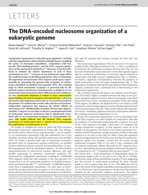
NATURE | Vol 458 |19 March 2009LETTERSGenesModelIn vitro dataYPD (in vivo)Ethanol (in vivo)Galactose (in vivo)Nucleosomeoccupancy4040404040Chromosome 14: 187000–2070002,000 bpSLA2 ATG2 ZWF1 NAR1 LAP3Genomic positionKEX2YTP1Figure 1 | <strong>The</strong> intrinsic <strong>DNA</strong>-<strong>encoded</strong><strong>nucleosome</strong> <strong>organization</strong> at a typical genomicregion. Shown are the four different maps <strong>of</strong><strong>nucleosome</strong> occupancy measured in this studyfor a typical 20,000-bp-long genomic region: thein vitro map, which reflects only the intrinsic<strong>nucleosome</strong> sequence preferences, and in vivoyeast maps for three different growth conditions(YPD, ethanol and galactose). Each track plotsthe measured <strong>nucleosome</strong> occupancy per basepair, computed by summing all <strong>of</strong> the<strong>nucleosome</strong> reads obtained in that experiment,and dividing that number by the average number<strong>of</strong> reads per base pair across the <strong>genome</strong>. <strong>The</strong> line<strong>of</strong> y 5 1 thus represents the <strong>genome</strong>-wide averageand is shown as a dashed orange line. <strong>The</strong> average<strong>nucleosome</strong> occupancy predictions from ourmodel are shown in blue.500 bpATG2ZWF1ModelIn vitro dataYPD (in vivo)Ethanol (in vivo)Galactose (in vivo)Nucleosomeoccupancy4040404040Genomic positionconcentration <strong>of</strong> <strong>nucleosome</strong>s in vivo relative to the concentrationused to create our in vitro map may also contribute to this difference,because higher <strong>nucleosome</strong> concentrations generally increase theaNormalized <strong>nucleosome</strong>occupancy in vivo (YPD)Number <strong>of</strong>base pairsb314R = 0.7450,000 12R = 0.3340,000100830,00020,00064210,0000–5–50 30–2 –2Normalized <strong>nucleosome</strong>occupancy in vitroTranscription level (log 2 )02.5Difference in normalized<strong>nucleosome</strong> occupancy oncoding regions (in vitro – in vivo)Figure 2 | In vitro and in vivo maps are highly similar. a, Shown is a densitydot plot comparison <strong>of</strong> the normalized <strong>nucleosome</strong> occupancy per base pairin the in vitro (x axis) and in vivo (y axis) maps (see Methods). Values abovezero indicate <strong>nucleosome</strong> enrichment relative to the <strong>genome</strong>-wide average.<strong>The</strong> colour <strong>of</strong> each point represents the number <strong>of</strong> base pairs that map tothat point in the graph. <strong>The</strong> Pearson correlation between the maps isindicated. b, Nucleosome depletion in vivo relative to in vitro over codingregions increases with the expression level <strong>of</strong> associated genes. Shown is a dotplot comparison between the expression level <strong>of</strong> every yeast gene (measuredin ref. 26) and the difference between the average normalized <strong>nucleosome</strong>occupancy <strong>of</strong> the coding region <strong>of</strong> that gene in the in vitro map comparedwith the in vivo map (that is, higher values indicate larger <strong>nucleosome</strong>depletion in vivo relative to in vitro). <strong>The</strong> Pearson correlation <strong>of</strong> the dot plotis indicated.©2009 Macmillan Publishers Limited. All rights reservedcontribution <strong>of</strong> non-specific binding, thus diminishing the contribution<strong>of</strong> the ,10-bp sequence periodicities. Nevertheless, theconservation <strong>of</strong> the ,10-bp dinucleotide periodicities and thenear-identity <strong>of</strong> 5-base-pair <strong>nucleosome</strong> occupancies demonstratethat <strong>nucleosome</strong>s have clear sequence preferences that are highlysimilar in vitro and in vivo.To test whether general sequence-based rules can be derived fromour in vitro data and be used to predict <strong>nucleosome</strong> occupancy invivo, we constructed a simple probabilistic model based on both theglobal preferences over sequences <strong>of</strong> length 5 and the positiondependentdinucleotide preferences 5,17 , which scores the <strong>nucleosome</strong>formation potential <strong>of</strong> every 147-bp sequence. Importantly, thismodel is learned only from in vitro <strong>nucleosome</strong> data, and thereforerepresents only <strong>nucleosome</strong> sequence preferences, whereas previousmodels 5–8,18 , which were learned from in vivo data, may also capturesequence preferences <strong>of</strong> other factors 7 , as well as indirect effects dueto chromatin remodelling activities. We tested the model in a crossvalidationscheme in which the <strong>nucleosome</strong> occupancy <strong>of</strong> each chromosomewas predicted using a model that was constructed from thedata from all other chromosomes. Our model has high correlations <strong>of</strong>0.89 and 0.75 with the in vitro and in vivo maps, respectively (Fig. 3d,e), and separates <strong>nucleosome</strong>-enriched regions from <strong>nucleosome</strong>depletedregions (Supplementary Fig. 3), indicating that the modelsuccessfully identified general predictive rules for the sequence preferences<strong>of</strong> <strong>nucleosome</strong>s.If <strong>nucleosome</strong> sequence preferences are important in other eukaryotes,then our model should also be predictive <strong>of</strong> their in vivo<strong>nucleosome</strong> <strong>organization</strong>. Indeed, we found a good (0.60) correlationbetween the <strong>nucleosome</strong> occupancy per base pair predicted by our363
LETTERS NATURE |Vol 458 |19 March 2009in vitro yeast-based model and that <strong>of</strong> in vivo <strong>nucleosome</strong> occupancyin C. elegans 19 (Fig. 3f). Moreover, our model classifies <strong>nucleosome</strong>enrichedregions from <strong>nucleosome</strong>-depleted regions in C. elegans withhigh accuracy (Supplementary Fig. 4), and the 5-base-pair sequencepreferences <strong>of</strong> the C. elegans in vivo map agree well with those <strong>of</strong> theyeast in vitro map (Fig. 3g). <strong>The</strong> poorer classification performance incomparison with yeast may indicate that factors other than the <strong>DNA</strong>sequence preferences make a greater contribution to <strong>nucleosome</strong><strong>organization</strong> in more complex eukaryotes. Alternatively, the poorerperformance may indicate that distinct sequence types are present inC. elegans for which our yeast in vitro data do not provide statistics.Nonetheless, our model is significantly correlated with the in vivo<strong>nucleosome</strong> <strong>organization</strong> across C. elegans.We next compared the <strong>DNA</strong>-<strong>encoded</strong> <strong>nucleosome</strong> <strong>organization</strong> <strong>of</strong>the in vitro map with <strong>nucleosome</strong> <strong>organization</strong> under growth conditionsthat cause substantial transcriptional changes relative to logphasegrowth in rich medium (that is glucose). In addition to ourmap obtained from yeast cells grown in rich medium, we also measuredthe <strong>nucleosome</strong> <strong>organization</strong> <strong>of</strong> yeast cells grown separately ingalactose, and in ethanol, and found that the overall <strong>nucleosome</strong>occupancy is very similar between all three in vivo maps, althoughlocalized differences are apparent (Fig. 1 and Supplementary Fig. 5).All three in vivo maps are highly correlated with the in vitro mapand show the sequence characteristics seen in vitro (SupplementaryFig. 6). <strong>The</strong>se results imply that intrinsic sequence preferences <strong>of</strong><strong>nucleosome</strong>s have a dominant role in determining <strong>nucleosome</strong><strong>organization</strong> in several growth conditions, with local, condition-specificchanges superimposed.To address concerns regarding biases that may be caused by thesequence specificity <strong>of</strong> micrococcal nuclease 20 and possible biases inparallel sequencing, we performed a different kind <strong>of</strong> in vitro experimentthat measures the relative <strong>nucleosome</strong> affinity <strong>of</strong> ,40,000 double-stranded150-bp oligonucleotides without the use <strong>of</strong> micrococcalnuclease or parallel sequencing. <strong>The</strong> resulting 5-base-pair <strong>nucleosome</strong>sequence preferences are in excellent agreement with thosediscovered in the <strong>genome</strong>-wide in vitro reconstitution (correlation<strong>of</strong> 0.83), and there is a good correlation (0.51) between the measuredoligonucleotide affinities and those predicted by the model constructedfrom the <strong>genome</strong>-wide in vitro map (Supplementary Fig.7). <strong>The</strong>se results are wholly independent <strong>of</strong> either micrococcal nucleaseor parallel sequencing, and thus confirm that the sequence specificitiesderived from our previous experiments were caused byintrinsic <strong>nucleosome</strong> preferences, rather than being an artefact <strong>of</strong>our experimental approach.Previous studies identified <strong>nucleosome</strong> depletion around transcriptionstart and stop sites 5–7,9–11 . However, because these studieswere based on in vivo data, it was not possible to determine whichmechanism accounted for the observed patterns. <strong>The</strong> in vitro and invivo maps show highly similar stereotypic <strong>nucleosome</strong> depletion attranslation end sites, indicating that this depletion is largely <strong>encoded</strong>by <strong>nucleosome</strong> sequence preferences (Fig. 4b and SupplementaryFig. 8). <strong>The</strong> two maps also show stereotypic <strong>nucleosome</strong> depletionaAverage normalized <strong>nucleosome</strong>occupancy on 5-mers in vitro0.50R = 0.98bDinucleotide frequency0.280.26In vitroAA/AT/TA/TTCC/CG/GC/GG–0.50.240.24–1.0 AAAAAATATA0.220.22–1.0 –1 0 0.5–60 –40 –20 0 20 40 60–60 –40 –20 0 20 40 60Average <strong>nucleosome</strong>Distance from <strong>nucleosome</strong> dyad (bp)Distance from <strong>nucleosome</strong> dyad (bp)occupancy on 5-mers in vivo (YPD)cDinucleotide frequency0.280.26YPDAA/AT/TA/TTCC/CG/GC/GGd Number <strong>of</strong> e Number <strong>of</strong> f Number <strong>of</strong> gbase pairsbase pairsbase pairs333100,000R = 0.89 R = 0.75 R = 0.6080,00060,00080,000060,000 040,000 060,00040,00040,00020,00020,00020,000Predicted normalized<strong>nucleosome</strong> occupancy–5 –5Predicted normalized<strong>nucleosome</strong> occupancy0 30 –5–5 0 3003Normalized <strong>nucleosome</strong>Normalized <strong>nucleosome</strong>Normalized <strong>nucleosome</strong>occupancy in vitrooccupancy in vivo (YPD)occupancy in vivo (C. elegans)Figure 3 | <strong>The</strong> in vitro sequence preferences <strong>of</strong> <strong>nucleosome</strong>s are highlysimilar to those <strong>of</strong> <strong>nucleosome</strong>-bound sequences in vivo and are predictive<strong>of</strong> <strong>nucleosome</strong> occupancy in C. elegans. a, Comparison <strong>of</strong> <strong>genome</strong>-widerelative <strong>nucleosome</strong> occupancy <strong>of</strong> <strong>nucleosome</strong>s over sequences <strong>of</strong> length 5.For the in vitro and in vivo maps <strong>of</strong> <strong>nucleosome</strong> occupancy, we separatelycomputed the average normalized <strong>nucleosome</strong> occupancy <strong>of</strong> each <strong>of</strong> the1,024 sequences <strong>of</strong> length 5, across all <strong>of</strong> its instances in the <strong>genome</strong>. Shownis a comparison between the distributions <strong>of</strong> these 5-base-pair sequences inboth maps. Also shown is the Pearson correlation between thesedistributions. b, Position-dependent sequence preferences <strong>of</strong> <strong>nucleosome</strong>sin the in vitro map. We aligned the individual <strong>nucleosome</strong> reads in the invitro <strong>nucleosome</strong> collection. Shown is the fraction (3-bp moving average) <strong>of</strong>AA/AT/TT/TA and CC/CG/GC/GG dinucleotides at each position <strong>of</strong> thealignment. c, Same as b, for the in vivo map. d, Shown is a density dot plotcomparison between the normalized <strong>nucleosome</strong> occupancy per base pair inthe in vitro map (x axis) and the normalized <strong>nucleosome</strong> occupancy per base364Predicted normalized <strong>nucleosome</strong>occupancy (C. elegans)©2009 Macmillan Publishers Limited. All rights reservedAverage <strong>nucleosome</strong> occupancyon 5-mers in vitro (yeast)0.50–0.5–1.0R = 0.65CGGCACCGGC–4 0–3 –1.0 –0.5 0 0.5Average <strong>nucleosome</strong> occupancyon 5-mers in vivo (C. elegans)pair predicted by our cross-validated computational model <strong>of</strong> <strong>nucleosome</strong>sequence preferences (y axis). Values above zero indicate <strong>nucleosome</strong>enrichment relative to the <strong>genome</strong>-wide average. <strong>The</strong> colour <strong>of</strong> each pointrepresents the number <strong>of</strong> base pairs that map to that point in the graph. <strong>The</strong>Pearson correlation between the maps is indicated. e, Same as d, comparingour model predictions to the in vivo map. f, In vitro <strong>nucleosome</strong> sequencepreferences on yeast genomic <strong>DNA</strong> are predictive <strong>of</strong> the in vivo <strong>nucleosome</strong><strong>organization</strong> in C. elegans. Same as d, comparing our model predictions andthe in vivo <strong>nucleosome</strong> occupancy map <strong>of</strong> C. elegans on chromosome 2(ref. 19). g, Comparison <strong>of</strong> yeast <strong>nucleosome</strong> sequence preferences in vitroand those <strong>of</strong> C. elegans in vivo. For each <strong>of</strong> the maps we separately computedthe average normalized <strong>nucleosome</strong> occupancy <strong>of</strong> every possible sequence <strong>of</strong>length 5. For C. elegans, we performed these computations onchromosome 2. Shown is a comparison <strong>of</strong> these 5-base-pair sequencedistributions between the yeast in vitro map and the in vivo map <strong>of</strong> C.elegans, along with the Pearson correlation between these distributions.
NATURE | Vol 458 |19 March 2009LETTERSat transcription start sites, indicating that this depletion is also partly<strong>encoded</strong> by <strong>nucleosome</strong> sequence preferences (Fig. 4a). However, thelevel <strong>of</strong> depletion around transcription start sites in vitro is smallerthan in vivo, indicating that transcription factors, chromatin remodellers,the transcription initiation machinery and other mechanismsalso contribute to the depletion. Another difference around start sites(Fig. 4a) is the longer-range ordering <strong>of</strong> <strong>nucleosome</strong>s into codingregions observed in the in vivo map. This may be partly explained bythe higher <strong>nucleosome</strong> concentration in vivo (see Methods), whichcauses increased ordering by statistical positioning 21 .Nucleosome depletion has also been reported around transcriptionfactor binding sites in vivo 7,9,11 . In both our in vivo and in vitro maps,<strong>nucleosome</strong> depletion was observed, on average, around thechromatin-immunoprecipitation-determined binding sites 22 <strong>of</strong> mosttranscription factors. <strong>The</strong> maps also agree on the degree <strong>of</strong> depletionaround binding sites (correlation <strong>of</strong> 0.62; Fig. 4c and SupplementaryFig. 9). <strong>The</strong>se results indicate that <strong>nucleosome</strong> depletion aroundregulatory factor binding sites is partly <strong>encoded</strong> in the <strong>genome</strong>’sintrinsic <strong>nucleosome</strong> <strong>organization</strong>, and that this intrinsic <strong>organization</strong>may facilitate transcription initiation and assist in directing transcriptionfactors to their appropriate sites in the <strong>genome</strong> 5,23 .Binding sites for the yeast factors Abf1 and Reb1 show the largestdeviation from the above agreement, with sites for both factors beingmore depleted in vivo than in vitro. Notably, both factors are highlyabundant and influence chromatin structure 24,25 , indicating that thedepletion around their sites in vivo may be attributable to their ownaction. <strong>The</strong> large <strong>nucleosome</strong> depletion over these factor sitesresulted in them being major components <strong>of</strong> a <strong>nucleosome</strong> occupancymodel created from in vivo data 7 . However, because the modelconstructed here is based solely on in vitro data, in which these siteshave only moderate depletion, they are not major components <strong>of</strong> ourmodel.In summary, we find marked similarities between the <strong>nucleosome</strong><strong>organization</strong> governed only by the <strong>DNA</strong> sequence preferences <strong>of</strong><strong>nucleosome</strong>s, and the <strong>organization</strong> <strong>of</strong> <strong>nucleosome</strong>s in vivo measuredunder different growth conditions. This result indicates that <strong>nucleosome</strong>sequence preferences are important determinants <strong>of</strong> <strong>nucleosome</strong><strong>organization</strong>s in vivo. Our analysis indicates that <strong>genome</strong>s mayuse their intrinsically <strong>encoded</strong> <strong>nucleosome</strong> <strong>organization</strong> to facilitatefunctions such as transcription factor binding and transcription.Despite the overall similarity between the in vitro and in vivo maps,there are differences, consistent with previous studies showing thataAverage normalized <strong>nucleosome</strong>occupancycAverage normalized <strong>nucleosome</strong> occupancyon binding sites in vitro10–1YPDIn vitroModelYPDIn vitroModel–2–2–500 0 500 –5000 500Distance from transcription start (bp)Distance from translation end (bp)0.50–1–2–3–3Enrichedin vitroDepletedin vitroR = 0.62Abf1 Reb1 Rap1Sum1Spt2Arr1Skn7Depletedin vivo–2–10Average normalized <strong>nucleosome</strong>occupancy on binding sites in vivoFigure 4 | <strong>The</strong> intrinsic <strong>nucleosome</strong> <strong>organization</strong> over transcripts andtranscription factor binding sites. a, For the in vitro and in vivo <strong>nucleosome</strong>occupancy maps, and for our model, shown is the normalized <strong>nucleosome</strong>occupancy per base pair around the transcription start site, averaged acrossall yeast genes. <strong>The</strong> long-range ordering <strong>of</strong> <strong>nucleosome</strong> occupancy which ispresent in the in vivo maps but not in the in vitro map may be partlyexplained by the lower <strong>nucleosome</strong> concentration in which the in vitroexperiment was carried out (see Methods), because higher <strong>nucleosome</strong>concentration in vivo is predicted to cause long-range ordering <strong>of</strong><strong>nucleosome</strong> arrays 21 . b, Same as a, but around translation end sites <strong>of</strong> genes(translation end was chosen because transcription end sites are poorlyannotated). <strong>The</strong> depletion around gene ends may be due to the presence <strong>of</strong>termination signals 27 , which disfavour <strong>nucleosome</strong> formation in vitro(Supplementary Fig. 8). <strong>The</strong> fact that these signals tend to occur in a specificAft2Enrichedin vivo0.5Average normalized<strong>nucleosome</strong> occupancy10–1–2Reb1–3–200 0 200Distance from binding site (bp)Skn7Rap1©2009 Macmillan Publishers Limited. All rights reservedbYPDIn vitro10–1Abf1Aft2Spt2Average normalized <strong>nucleosome</strong>occupancyorientation with respect to the direction <strong>of</strong> transcription 27 is consistent witha function in transcript processing, but does not exclude the possibility thatone or more <strong>of</strong> these motifs functions primarily to disfavour <strong>nucleosome</strong>s.c, Comparison <strong>of</strong> the <strong>nucleosome</strong> occupancy over transcription factorbinding sites between the in vitro and the YPD in vivo maps. For eachtranscription factor with at least 50 functional binding sites 22 , we computed,separately for the in vivo and in vitro maps, the average normalized<strong>nucleosome</strong> occupancy over its binding sites. Shown is a comparison <strong>of</strong> these<strong>nucleosome</strong> occupancies per factor, between the in vivo and in vitro maps,along with the Pearson correlation between them. For six factors taken fromdifferent regions <strong>of</strong> the plot, we also show the average normalized<strong>nucleosome</strong> occupancy around those factors’ binding sites, for both the invitro and the in vivo maps.365
LETTERS NATURE |Vol 458 |19 March 2009factors other than <strong>nucleosome</strong> sequence preferences contribute to<strong>nucleosome</strong> <strong>organization</strong> in vivo. Future studies will focus on understandinghow <strong>nucleosome</strong>s are remodelled locally and the function <strong>of</strong>such remodelling in transcriptional regulation.METHODS SUMMARYIn vivo maps from yeast <strong>nucleosome</strong> <strong>DNA</strong>s were prepared from log-phase cellsgrown in rich medium (YPD, six independent replicates) as described previously5 , as well as from cells grown in YP media supplemented with 2% galactose(three replicates) or 2.8% ethanol (four replicates) instead <strong>of</strong> glucose. <strong>The</strong> resulting<strong>DNA</strong>s were subjected to Illumina sequencing-by-synthesis. For the in vitromap, histone octamer was purified from chicken erythrocytes, assembled onpurified yeast genomic <strong>DNA</strong> by salt gradient dialysis 13 , digested with micrococcalnuclease and subjected to Illumina sequencing (two independent replicates). <strong>The</strong>resulting in vitro map has a lower concentration <strong>of</strong> <strong>nucleosome</strong>s along the <strong>DNA</strong>than obtained in vivo. This technical limitation was necessitated by our findingthat reconstitutions at the in vivo stoichiometry on long genomic <strong>DNA</strong> resultedin insoluble chromatin that was inaccessible to micrococcal nuclease. Wemapped the resulting reads to the <strong>genome</strong> and removed reads that mapped tomultiple genomic locations. We extended the <strong>nucleosome</strong> reads <strong>of</strong> each experimentto the average <strong>nucleosome</strong> length in that experiment (always between 140–170 bp). For each map, we then calculated the normalized <strong>nucleosome</strong> occupancyat every base pair as the log-ratio between the number <strong>of</strong> reads that coverthat base pair and the average number <strong>of</strong> reads per base pair across the <strong>genome</strong>.We then set the genomic mean in each sample to zero by subtracting the <strong>genome</strong>widemean from every base pair. <strong>The</strong> independent replicates for each experimenttype were in excellent agreement, so we averaged the replicates within each type.<strong>The</strong> resulting tracks are termed normalized <strong>nucleosome</strong> occupancy throughoutthe manuscript. <strong>The</strong> detailed formulation <strong>of</strong> our sequence-based model for<strong>nucleosome</strong> positioning is given in the Methods and is similar to that describedin ref. 17, except that it was learned using only the in vitro data. For our data,results and model, see http://genie.weizmann.ac.il/pubs/<strong>nucleosome</strong>s08/, andGEO accession number GSE13622.Full Methods and any associated references are available in the online version <strong>of</strong>the paper at www.nature.com/nature.Received 2 October; accepted 26 November 2008.Published online 17 December 2008.1. Kornberg, R. D. & Lorch, Y. Twenty-five years <strong>of</strong> the <strong>nucleosome</strong>, fundamentalparticle <strong>of</strong> the eukaryote chromosome. Cell 98, 285–294 (1999).2. Vignali, M., Hassan, A. H., Neely, K. E. & Workman, J. L. ATP-dependentchromatin-remodeling complexes. Mol. Cell. Biol. 20, 1899–1910 (2000).3. Korber, P., Luckenbach, T., Blaschke, D. & Horz, W. Evidence for histone eviction intrans upon induction <strong>of</strong> the yeast PHO5 promoter. Mol. Cell. Biol. 24,10965–10974 (2004).4. Satchwell, S. C., Drew, H. R. & Travers, A. A. Sequence periodicities in chicken<strong>nucleosome</strong> core <strong>DNA</strong>. J. Mol. Biol. 191, 659–675 (1986).5. Segal, E. et al. A genomic code for <strong>nucleosome</strong> positioning. Nature 442, 772–778(2006).6. Ioshikhes, I. P., Albert, I., Zanton, S. J. & Pugh, B. F. Nucleosome positionspredicted through comparative genomics. Nature Genet. 38, 1210–1215 (2006).7. Lee, W. et al. A high-resolution atlas <strong>of</strong> <strong>nucleosome</strong> occupancy in yeast. NatureGenet. 39, 1235–1244 (2007).8. Yuan, G. C. & Liu, J. S. Genomic sequence is highly predictive <strong>of</strong> local <strong>nucleosome</strong>depletion. PLOS Comput. Biol. 4, e13 (2008).9. Yuan, G. C. et al. Genome-scale identification <strong>of</strong> <strong>nucleosome</strong> positions in S.cerevisiae. Science 309, 626–630 (2005).10. Shivaswamy, S. et al. Dynamic remodeling <strong>of</strong> individual <strong>nucleosome</strong>s across a<strong>eukaryotic</strong> <strong>genome</strong> in response to transcriptional perturbation. PLoS Biol. 6, e65(2008).11. Albert, I. et al. Translational and rotational settings <strong>of</strong> H2A.Z <strong>nucleosome</strong>s acrossthe Saccharomyces cerevisiae <strong>genome</strong>. Nature 446, 572–576 (2007).12. Sekinger, E. A., Moqtaderi, Z. & Struhl, K. Intrinsic histone–<strong>DNA</strong> interactions andlow <strong>nucleosome</strong> density are important for preferential accessibility <strong>of</strong> promoterregions in yeast. Mol. Cell 18, 735–748 (2005).13. Thastrom, A., Bingham, L. M. & Widom, J. Nucleosomal locations <strong>of</strong> dominant<strong>DNA</strong> sequence motifs for histone–<strong>DNA</strong> interactions and <strong>nucleosome</strong> positioning.J. Mol. Biol. 338, 695–709 (2004).14. Anderson, J. D. & Widom, J. Poly(dA-dT) promoter elements increase theequilibrium accessibility <strong>of</strong> nucleosomal <strong>DNA</strong> target sites. Mol. Cell. Biol. 21,3830–3839 (2001).15. Iyer, V. & Struhl, K. Poly(dA:dT), a ubiquitous promoter element that stimulatestranscription via its intrinsic <strong>DNA</strong> structure. EMBO J. 14, 2570–2579 (1995).16. Ioshikhes, I., Bolshoy, A., Derenshteyn, K., Borodovsky, M. & Trifonov, E. N.Nucleosome <strong>DNA</strong> sequence pattern revealed by multiple alignment <strong>of</strong>experimentally mapped sequences. J. Mol. Biol. 262, 129–139 (1996).17. Field, Y. et al. Distinct modes <strong>of</strong> regulation by chromatin <strong>encoded</strong> through<strong>nucleosome</strong> positioning signals. PLOS Comput. Biol. 4, e1000216 (2008).18. Peckham, H. E. et al. Nucleosome positioning signals in genomic <strong>DNA</strong>. GenomeRes. 17, 1170–1177 (2007).19. Valouev, A. et al. A high-resolution, <strong>nucleosome</strong> position map <strong>of</strong> C. elegans revealsa lack <strong>of</strong> universal sequence-dictated positioning. Genome Res. 18, 1051–1063(2008).20. Horz, W. & Altenburger, W. Sequence specific cleavage <strong>of</strong> <strong>DNA</strong> by micrococcalnuclease. Nucleic Acids Res. 9, 2643–2658 (1981).21. Kornberg, R. D. & Stryer, L. Statistical distributions <strong>of</strong> <strong>nucleosome</strong>s: nonrandomlocations by a stochastic mechanism. Nucleic Acids Res. 16, 6677–6690 (1988).22. MacIsaac, K. D. et al. An improved map <strong>of</strong> conserved regulatory sites forSaccharomyces cerevisiae. BMC Bioinformatics 7, 113 (2006).23. Liu, X., Lee, C. K., Granek, J. A., Clarke, N. D. & Lieb, J. D. Whole-<strong>genome</strong>comparison <strong>of</strong> Leu3 binding in vitro and in vivo reveals the importance <strong>of</strong><strong>nucleosome</strong> occupancy in target site selection. Genome Res. 16, 1517–1528(2006).24. Lascaris, R. F., Groot, E., Hoen, P. B., Mager, W. H. & Planta, R. J. Different roles forAbf1p and a T-rich promoter element in <strong>nucleosome</strong> <strong>organization</strong> <strong>of</strong> the yeastRPS28A gene. Nucleic Acids Res. 28, 1390–1396 (2000).25. Raisner, R. M. et al. Histone variant H2A.Z marks the 59 ends <strong>of</strong> both active andinactive genes in euchromatin. Cell 123, 233–248 (2005).26. Nagalakshmi, U. et al. <strong>The</strong> transcriptional landscape <strong>of</strong> the yeast <strong>genome</strong> definedby RNA sequencing. Science 320, 1344–1349 (2008).27. Graber, J. H., Cantor, C. R., Mohr, S. C. & Smith, T. F. Genomic detection <strong>of</strong> newyeast pre-mRNA 39-end-processing signals. Nucleic Acids Res. 27, 888–894(1999).Supplementary Information is linked to the online version <strong>of</strong> the paper atwww.nature.com/nature.Acknowledgements We thank H. Kelkar for bioinformatics support in handling thesequencing files, P. Mieczkowski and J. McPherson for Illumina sequencing (librarypreparation and instrument operation), and the members <strong>of</strong> our respectivelaboratories for technical assistance, discussions and comments on themanuscript. <strong>The</strong> UNC sequencing facility is funded by the LinebergerComprehensive Cancer Center and University Cancer Research Funds. J.W.acknowledges the use <strong>of</strong> instruments at Northwestern University’s KeckBiophysics Facility. This work was supported by a grant from CIHR to T.R.H. andC. Nislow, a grant from the NIH to J.D.L., a grant from the NIH to J.W., and grantsfrom the European Research Council (ERC) and NIH to E.S. D.T. holds an NSERCpostgraduate scholarship. N.K. is a Clore scholar. E.S. is the incumbent <strong>of</strong> theSoretta and Henry Shapiro career development chair.Author Contributions This work was performed jointly in the Hughes, Lieb, Widomand Segal laboratories and with substantial contributions from each.Author Information This has been submitted to GEO under accession numberGSE13622. Reprints and permissions information is available at www.nature.com/reprints. Correspondence and requests for materials should be addressed to J.W.(j-widom@northwestern.edu) or E.S. (eran.segal@weizmann.ac.il).366©2009 Macmillan Publishers Limited. All rights reserved
doi:10.1038/nature07667METHODSParallel sequencing <strong>of</strong> yeast <strong>nucleosome</strong>s in vivo. Four samples <strong>of</strong> yeast <strong>nucleosome</strong><strong>DNA</strong>s (‘in vivo <strong>nucleosome</strong>s’) were prepared from log-phase cells grown inrich medium (YPD) as described previously 5 . Additionally, samples were preparedfrom cells grown in YP media supplemented with 2% galactose (twosamples) or 2.8% ethanol (two samples) instead <strong>of</strong> glucose. Five additionalsamples (two in YPD, one in galactose and two in ethanol) were prepared fromcells that were formaldehyde-crosslinked before isolating <strong>nucleosome</strong>s.Crosslinking was achieved with 1% formaldehyde for 15 min at 30 uC andstopped with 125 mM glycine. <strong>The</strong> cells were then washed, made into spheroplastswith lyticase as above, treated with micrococcal nuclease (MNase) asdescribed previously 12 , and nucleosomal <strong>DNA</strong>s isolated as described 10 . All <strong>of</strong>the resulting <strong>DNA</strong>s were subjected to Illumina/Solexa sequencing-by-synthesis.<strong>The</strong> results from crosslinked or uncrosslinked cells for a given growth mediumwere very similar; therefore, we averaged the replicates within each growthmedium (six YPD, four YPEtOH, three YPGal).Parallel sequencing <strong>of</strong> <strong>nucleosome</strong>s reconstituted in vitro on yeast genomic<strong>DNA</strong>. S. cerevisiae genomic <strong>DNA</strong> was purified from strain YLC8 (MATa ura3Dleu2D his3D met15D) using standard methods with additional steps to removecontaminating RNA (see Supplementary Information). Histone octamer waspurified from chicken erythrocytes using salt extraction and hydroxyapatitecolumn chromatography, as described previously 28 . Genomic <strong>DNA</strong> was reconstitutedinto <strong>nucleosome</strong>s under selective pressure for <strong>nucleosome</strong>-favouringsequences by salt gradient dialysis 13 , using 40 mg histone octamer plus 100 mg<strong>DNA</strong> in a 200 ml volume. This results in a lower concentration <strong>of</strong> <strong>nucleosome</strong>salong the <strong>DNA</strong> than obtained in vivo. This technical limitation was necessitatedby our finding that reconstitutions at the in vivo stoichiometry on this longgenomic <strong>DNA</strong> resulted in insoluble chromatin that was inaccessible to micrococcalnuclease. Two independent reconstitutions were carried out. <strong>The</strong> resulting<strong>nucleosome</strong>s were biochemically isolated by MNase digestion, using6 3 10 23 units MNase (Sigma Chemical Company) per 10 mg competitivelyreconstituted <strong>DNA</strong>, in 10 mM Tris, pH 8.0, and 1 mM CaCl 2 for 5 min at37 uC. <strong>DNA</strong> was extracted, and protected fragments <strong>of</strong> length ,147 bp wereisolated by polyacrylamide gel electrophoresis, and then extracted from thegel. Both samples were independently subjected to Illumina sequencing at theUNC high-throughput sequencing facility. We note that, because the chickenhistones represent a mixture <strong>of</strong> post-translationally modified and unmodifiedhistones as well as variant histones, there is a possibility that these modificationsor variants influence the measured <strong>nucleosome</strong> positions.Mapping and post-processing <strong>of</strong> parallel sequencing reads. We mapped readsresulting from sequencing experiments to the yeast <strong>genome</strong>, allowing at mostone mismatch and no gaps. To estimate the mean <strong>DNA</strong> fragment length in eachexperiment, we superimposed the <strong>nucleosome</strong> reads <strong>of</strong> one strand and examinedthe distribution <strong>of</strong> <strong>nucleosome</strong> reads <strong>of</strong> the opposite strand. As expected, thisdistribution showed a strong peak at ,140–170 bp for all experiments, withslight variations between experiments. We used the maximum <strong>of</strong> the peak asan estimation <strong>of</strong> the mean <strong>DNA</strong> fragment length and extended all <strong>nucleosome</strong>reads to this length. We defined repetitive regions as regions that were matchedby a read that mapped to more than one place in the <strong>genome</strong>. We excludedrepetitive regions and their 150-bp vicinity from our analyses. To obtain genomic<strong>nucleosome</strong> occupancy tracks we summed for each position all reads coveringit. We noticed that in some replicates from each <strong>of</strong> the three in vivoconditions (YPD, YPEtOH and YPGal), but not in either <strong>of</strong> the in vitro replicates,a small fraction <strong>of</strong> the mapped base pairs (in all cases less than 0.5%) werecovered by an aberrantly large number <strong>of</strong> reads (more than ten times the mediangenomic base pair coverage) that may potentially result from sequencing biases.For these regions, we set the coverage <strong>of</strong> these base pairs to equal ten times themedian genomic base-pair coverage. Next, for each map, we calculated theaverage <strong>nucleosome</strong> occupancy at every base pair by dividing the number <strong>of</strong>reads that cover that base pair by the average number <strong>of</strong> reads per base pair acrossthe <strong>genome</strong>. Finally, for each replicate we took the log <strong>of</strong> the ratio at every basepair and set the genomic mean to zero by subtracting the mean from every basepair. Thus, base pairs for which the occupancy value is below 0 showed a relativedepletion <strong>of</strong> <strong>nucleosome</strong>s in comparison with the <strong>genome</strong>-wide average, whereasbase pairs with values above 0 have a relative enrichment. <strong>The</strong> independentreplicates for each experiment type were in excellent agreement, so we averagedthe replicates within each type to create four <strong>nucleosome</strong> occupancy maps: threein vivo (YPD, YPEtOH, YPGal) and one in vitro. <strong>The</strong>se final tracks are termed thenormalized <strong>nucleosome</strong> occupancy. <strong>The</strong> data in Fig. 1 are presented without thelog transformation.Analysis <strong>of</strong> <strong>nucleosome</strong>s reconstituted on oligonucleotides. Two concernswith our experimental approach were the known sequence specificity <strong>of</strong> micrococcalnuclease 20 and the possibilities <strong>of</strong> biases introduced by the parallel<strong>DNA</strong>-sequencing strategy. We addressed both <strong>of</strong> these concerns simultaneouslywith an independent and different kind <strong>of</strong> experiment that obtains large-scalemeasurements <strong>of</strong> in vitro <strong>nucleosome</strong> sequence preferences without the use <strong>of</strong>micrococcal nuclease or parallel sequencing. In brief, we created a pool <strong>of</strong>,40,000 double-stranded oligonucleotides <strong>of</strong> length 150 bp, each flanked bycommon priming sites 29 , and combined the pool with limiting amounts <strong>of</strong>chicken histones to form <strong>nucleosome</strong>s. <strong>DNA</strong>s in reconstituted <strong>nucleosome</strong>s wereseparated from unincorporated <strong>DNA</strong>s by native gel electrophoresis, without theuse <strong>of</strong> any micrococcal nuclease. We then extracted from the gel the <strong>DNA</strong> thathad successfully competed to form <strong>nucleosome</strong>s, and used both parallel sequencingand microarrays to compare the nucleosomal <strong>DNA</strong>s to <strong>DNA</strong>s in the initialpool. For each sequence, we calculated the log-ratio between the reconstitutedfraction and the initial pool as a measure <strong>of</strong> the <strong>nucleosome</strong> affinity <strong>of</strong> thatsequence. Thus, a log-ratio above zero indicates a higher than average affinityto form <strong>nucleosome</strong>s, whereas a value below zero indicates a lower than averageaffinity. We note that this experiment is inherently noisy, as shown by a largevariation in the number <strong>of</strong> sequence reads in the unselected reference sample,which may be due to the use <strong>of</strong> two rounds <strong>of</strong> PCR in the procedure.Specifically, we created 197-mer oligonucleotides (containing 150-merunique sequences flanked by common priming sites) using a modification <strong>of</strong>the microarray-based method described previously 29 . Sequences were designedto represent tiled yeast genomic sequences (22,236 oligonucleotides), and asurvey <strong>of</strong> randomly generated sequences selected to represent a wide variety <strong>of</strong><strong>DNA</strong> sequence characteristics (nucleotide and dinuncleotide composition, periodicfeatures and <strong>DNA</strong> structural parameters 7 ). <strong>DNA</strong> obtained from the arraywas PCR-amplified and assembled into <strong>nucleosome</strong>s under selective pressure for<strong>nucleosome</strong>-favouring sequences by salt gradient dialysis 13 , using 6 mg histoneoctamer and 12 mg <strong>DNA</strong> in a 200-ml volume. <strong>The</strong> resulting <strong>nucleosome</strong>s wereseparated away from the remaining naked <strong>DNA</strong> by native polyacrylamide gelelectrophoresis (5% (w/v) polyacrylamide) in 1/33 TBE buffer (TBE is 90 mMTris, 90 mM boric acid, 2 mM EDTA, pH 8.3). <strong>The</strong> <strong>nucleosome</strong>-containing bandwas excised from the gel, and its <strong>DNA</strong> extracted by crushing and soaking into0.3 M NaOAc, 0.5 M NH 4 OAc, 0.1 mM EDTA and 0.1% (w/v) SDS, followed byethanol precipitation. For Illumina analysis, the pools before and after <strong>nucleosome</strong>assembly were re-amplified and NruI sites present in the primers werecleaved before sequencing from the ends. In our analyses, we used oligonucleotidesthat were sequenced at least once and at most 500 times in each experiment.For microarray analyses, the pools before and after assembly were re-amplifiedwith fluor-labelled primers, and hybridized to an Agilent array designed to detectthe individual sequences represented in the pools. Additional details are availablein the Supplementary Information.To remove any direct biases resulting from the primers in the computation <strong>of</strong>preferences over sequences <strong>of</strong> length 5 from these data (Supplementary Fig. 7),we omitted the primers altogether in the computation <strong>of</strong> 5-base-pair preferences(that is, all 5-base-pair sequences were extracted only from the variable oligonucleotideswithout the primers). This left open the possibility <strong>of</strong> indirect biasesin preferences <strong>of</strong> sequences <strong>of</strong> length 5 resulting from the primers; for example, ifone primer sequence attracted <strong>nucleosome</strong>s and if there were systematic biases incompositions <strong>of</strong> sequences <strong>of</strong> length 5 near that end <strong>of</strong> the designed oligonucleotides.However, half (,20,000) <strong>of</strong> the oligonucleotides were selected from theyeast <strong>genome</strong>, and the other half were designed in several unrelated ways (seeSupplementary Methods) and do not have such biases. <strong>The</strong> two approaches arein strong agreement regarding these preferences over sequences <strong>of</strong> length 5 basepairs, diminishing concerns regarding biases caused by the primers (the correlationsbetween the preferences computed from the first approach and the secondapproach are 0.81 and 0.83 for reading out the measurements <strong>of</strong> the secondapproach by parallel sequencing or microarrays, respectively; seeSupplementary Fig. 7).Data sets. <strong>The</strong> yeast <strong>genome</strong> sequence (May 2006 build) and gene and chromosomeannotations were obtained from SGD 30 . Yeast transcription start siteswere compiled from refs 31–33: for each gene, the transcription start site wastaken as that with the most sequence reads from refs 31 and 32, or from ref. 33when no sequencing data was available. Functional transcription factor <strong>DNA</strong>binding sites in yeast, defined as sites that are bound by their cognate transcriptionfactor were obtained from refs 22 and 34. Expression levels in yeast weretaken from ref. 26. <strong>The</strong> in vivo map <strong>of</strong> <strong>nucleosome</strong> <strong>organization</strong> in C. elegans wastaken from ref. 19; specifically, we used both the ‘adjusted <strong>nucleosome</strong> coverage’and ‘raw <strong>nucleosome</strong> coverage’, applying the same normalization method to thelatter as we did to our yeast <strong>nucleosome</strong> maps 19 .Classifying <strong>nucleosome</strong>-enriched regions from <strong>nucleosome</strong>-depleted regions.To test whether the two maps agree on the scale <strong>of</strong> individual <strong>nucleosome</strong>s, wetook the target map, and from it defined <strong>nucleosome</strong>-enriched regions as maximalconsecutive regions longer than 50 bp for which minimum occupancy atevery base pair is above some threshold, t e , and <strong>nucleosome</strong>-depleted regions as©2009 Macmillan Publishers Limited. All rights reserved
doi:10.1038/nature07667maximal consecutive regions longer than 50 bp for which maximum occupancyat every base pair is below some threshold, t d . We chose 50 bp as a compromisebetween partitioning as much <strong>of</strong> the <strong>genome</strong> as possible into <strong>nucleosome</strong>enrichedand <strong>nucleosome</strong>-depleted regions while still deriving each partitionfrom enough data to reduce noise that may arise when regions are too short (achoice <strong>of</strong> 25 bp yielded equivalent results). At various thresholds, we then scoredeach <strong>of</strong> the resulting <strong>nucleosome</strong>-enriched and <strong>nucleosome</strong>-depleted regions byits average occupancy in the predicting map. Note that at the thresholdt e 5 t d 5 0, the evaluation is done across the entire <strong>genome</strong> (typically ,97%,because regions ,50 bp are excluded), and the median length <strong>of</strong> the <strong>nucleosome</strong>-enrichedregions is typically 150–190 bp. If the predicting map were fullypredictive <strong>of</strong> the target map, then the predicting map occupancy score <strong>of</strong> every‘enriched’ region in the target map would be higher than that <strong>of</strong> every ‘depleted’region in the target map. A standard quantification <strong>of</strong> this predictive power is thereceiver operating characteristic curve, whose area under the curve is 1 for perfectperformance and 0.5 for random guessing.Sequence-based model for <strong>nucleosome</strong> positioning. We use the in vitro map torepresent the sequence preferences <strong>of</strong> <strong>nucleosome</strong>s by devising a probabilisticmodel, similar in formulation to that proposed in ref. 17, that assigns a score toevery 147-bp (<strong>nucleosome</strong>-length) sequence. Our model is based on both theposition-dependent and global preferences <strong>of</strong> sequences <strong>of</strong> length 5 that wecharacterized previously (Fig. 3). <strong>The</strong> first component, P N , represents the distributionover dinucleotides at each position along the <strong>nucleosome</strong> length, andthus captures the periodic signal <strong>of</strong> dinucleotides along the <strong>nucleosome</strong>. <strong>The</strong>second component, P L , represents the position-independent distribution <strong>of</strong><strong>nucleosome</strong>s over sequences <strong>of</strong> length 5, and thus captures sequences that aregenerally favoured or disfavoured by <strong>nucleosome</strong>s regardless <strong>of</strong> their detailedposition within the <strong>nucleosome</strong>. We chose to represent this component usingsequences <strong>of</strong> length 5, because this is the highest order k-mer for which our datahave sufficient statistics to robustly estimate each <strong>of</strong> the associated parameters.<strong>The</strong> final score that our model assigns to a 147-bp sequence S is then given by thelog-ratio <strong>of</strong> these two model components:Score(S)~ log P N(S)P L (S)P N,1 (S½1Š)P 147P N,i (S½iŠjS½i{1Š)i~2~ logP l (S½1Š)P 147P l (S½iŠjS½max (1,i{4)Š, ...,S½i{1Š)i~2where P N,i is the i-th component <strong>of</strong> the dinucleotide model component andrepresents the conditional probability distribution over nucleotides at positioni given the nucleotide that appeared at position (i 2 1), and P l is the positionindependentcomponent <strong>of</strong> the second component <strong>of</strong> our model (P L ). Note thatP N,1 is represented by a mononucleotide model over the nucleotide at the firstposition.We now describe in detail how each <strong>of</strong> the two components <strong>of</strong> our model wasderived. To estimate the position-specific dinucleotide component <strong>of</strong> ourmodel, P N , we took all <strong>of</strong> the <strong>nucleosome</strong> reads (a total <strong>of</strong> over 10,000,000) fromour collection <strong>of</strong> <strong>nucleosome</strong>-bound sequences, and first aligned these sequencesabout their centre. Each sequence was added twice to the alignment, once in itsoriginal form and once in its reverse complement form, to account for the tw<strong>of</strong>oldsymmetry in the <strong>nucleosome</strong> structure 35 . With each position i, we thenassociated a dinucleotide distribution, P N,i , which we estimated from the combineddinucleotide counts at alignment positions [i 2 2, i 2 1], [i 2 1, i] and[i, i 1 1] (the two end positions <strong>of</strong> the <strong>nucleosome</strong> are averaged with fewerpositions). Combining the dinucleotides at the two neighbouring positionssmoothes the resulting dinucleotide distribution at each position with a 3-bpmoving average, and is motivated by the experimental evidence that small 61-bpchanges in spacing <strong>of</strong> key <strong>nucleosome</strong> <strong>DNA</strong> sequence motifs can occur with arelatively small cost to the free energy <strong>of</strong> histone–<strong>DNA</strong> interactions 36 . To removesequence composition biases from this component, we normalized the distributionover each position separately, by dividing the final probability <strong>of</strong> everydinucleotide at each position by the average probability <strong>of</strong> that dinucleotideacross all positions, and finally normalized the resulting dinucleotide weightsto a probability distribution. We used this estimation procedure in the 127central positions <strong>of</strong> the <strong>nucleosome</strong>, and we forced a uniform distribution overthe 10 remaining positions at each end <strong>of</strong> the <strong>nucleosome</strong> pr<strong>of</strong>ile. This was doneto avoid biases in nucleotide distributions that may arise from the sequencespecificity <strong>of</strong> the micrococcal nuclease used to isolate the <strong>nucleosome</strong>, becausethis way we do not include statistics that are taken from the cut site <strong>of</strong> theð1Þnuclease. Note that our above construction produces a reverse complementsymmetrical distribution; that is, the probability <strong>of</strong> a sequence and its reversecomplement are equal by construction.<strong>The</strong> position-independent component <strong>of</strong> our model, P L , the purpose <strong>of</strong> whichis to represent sequences that are generally favoured or disfavoured regardless <strong>of</strong>their position within the <strong>nucleosome</strong>, assigns a score to each 147-bp sequence, asthe product <strong>of</strong> a position-independent Markov model, P l , <strong>of</strong> order 4. Thus, P ldefines a probability distribution over every one <strong>of</strong> the 1,024 possible sequences<strong>of</strong> length 5. We chose to model the distribution over sequences <strong>of</strong> length 5,because this is the highest order for which our data still provides sufficientstatistics to robustly estimate the value <strong>of</strong> each <strong>of</strong> the 1,024 parameters. Givena map <strong>of</strong> <strong>nucleosome</strong> positions, the probability <strong>of</strong> each sequence <strong>of</strong> length 5 isequal to the reciprocal <strong>of</strong> the average <strong>nucleosome</strong> occupancy <strong>of</strong> that sequenceacross all instances <strong>of</strong> the 5-base-pair-long sequence in the map, where thisreciprocal average occupancy is then scaled to a probability by dividing it bythe sum <strong>of</strong> all such reciprocal occupancies across all 5-base-pair-long sequences(we use the reciprocal <strong>of</strong> the average occupancy so that higher probabilitiescorrespond to disfavoured sequences). Thus, this Markov model, P l , includescontributions both from sequences that are disfavoured by <strong>nucleosome</strong>s andfrom sequences that are favoured by <strong>nucleosome</strong>s, because it models the distributionover all sequences <strong>of</strong> length 5, with the disfavoured sequences having arelatively high probability and the favoured sequences having a relatively lowprobability.<strong>The</strong> above probabilistic model assigns a <strong>nucleosome</strong> formation score to eachsequence <strong>of</strong> (<strong>nucleosome</strong>-length) 147 bp. We then followed the approach <strong>of</strong>ref. 5, and used the scores <strong>of</strong> this model to compute the <strong>genome</strong>-wide distributionover <strong>nucleosome</strong> positions, taking into account steric hindrance constraintsbetween neighbouring <strong>nucleosome</strong>s. <strong>The</strong>se computations can be doneefficiently using dynamic programming, and their output is a track <strong>of</strong> the average<strong>nucleosome</strong> occupancy per base pair, that is, the probability that each base pair inthe <strong>genome</strong> is covered by any <strong>nucleosome</strong>.In developing our model, we also separately evaluated the performance <strong>of</strong> theP N and P L components, and found that the performance <strong>of</strong> the P L componentalone is nearly the same as that <strong>of</strong> the full model (per-base-pair <strong>nucleosome</strong>occupancy correlation <strong>of</strong> 0.876 between a P L -only model and the in vitro map,compared with 0.880 for the full model using both P L and P N ), whereas theperformance <strong>of</strong> the P N component alone is highly predictive but slightly worsethan the full model (correlation <strong>of</strong> 0.820). Thus, for practical purposes <strong>of</strong> generatingpredictions, using only the P L component may suffice. Nevertheless, theP N component captures real periodic aspects <strong>of</strong> <strong>nucleosome</strong> sequence preferencesthat have been reported by several other groups 4,6,37 , and that we now findin the <strong>genome</strong>-wide in vitro <strong>nucleosome</strong> map in Fig. 3b. For these reasons, weretain the P N component as part <strong>of</strong> the model.For the synthetic oligonucleotide analysis (Supplementary Fig. 7), we used the<strong>nucleosome</strong> formation potential <strong>of</strong> equation (1). In all other analyses we convertedthis <strong>genome</strong>-wide <strong>nucleosome</strong> formation potential into <strong>genome</strong>-wide<strong>nucleosome</strong> occupancy per base pair, as described in ref. 5. We selected theconcentration and temperature parameters (t and b, respectively) in a crossvalidationmanner to be t 5 0.03, b 5 1 in yeast and t 5 0.1, b 5 1inC. elegans.Finally, to appropriately compare the model with log-transformed data, weapplied to the predictions <strong>of</strong> the model the same log normalization that wasapplied to the data.28. Feng, H. P., Scherl, D. S. & Widom, J. Lifetime <strong>of</strong> the histone octamer studied bycontinuous-flow quasielastic light scattering: test <strong>of</strong> a model for <strong>nucleosome</strong>transcription. Biochemistry 32, 7824–7831 (1993).29. Porreca, G. J. et al. Multiplex amplification <strong>of</strong> large sets <strong>of</strong> human exons. NatureMethods 4, 931–936 (2007).30. Cherry, J. M. et al. SGD: Saccharomyces <strong>genome</strong> database. Nucleic Acids Res. 26,73–79 (1998).31. Zhang, Z. & Dietrich, F. S. Mapping <strong>of</strong> transcription start sites in Saccharomycescerevisiae using 59 SAGE. Nucleic Acids Res. 33, 2838–2851 (2005).32. Miura, F. et al. A large-scale full-length c<strong>DNA</strong> analysis to explore the buddingyeast transcriptome. Proc. Natl Acad. Sci. USA 103, 17846–17851 (2006).33. David, L. et al. A high-resolution map <strong>of</strong> transcription in the yeast <strong>genome</strong>. Proc.Natl Acad. Sci. USA 103, 5320–5325 (2006).34. Harbison, C. T. et al. Transcriptional regulatory code <strong>of</strong> a <strong>eukaryotic</strong> <strong>genome</strong>.Nature 431, 99–104 (2004).35. Richmond, T. J. & Davey, C. A. <strong>The</strong> structure <strong>of</strong> <strong>DNA</strong> in the <strong>nucleosome</strong> core.Nature 423, 145–150 (2003).36. Ong, M. S., Richmond, T. J. & Davey, C. A. <strong>DNA</strong> stretching and extreme kinking inthe <strong>nucleosome</strong> core. J. Mol. Biol. 368, 1067–1074 (2007).37. Bolshoy, A., Ioshikhes, I. & Trifonov, E. N. Applicability <strong>of</strong> the multiple alignmentalgorithm for detection <strong>of</strong> weak patterns: periodically distributed <strong>DNA</strong> pattern asa study case. Comput. Appl. Biosci. 12, 383–389 (1996).©2009 Macmillan Publishers Limited. All rights reserved