Muscle Study Guides AXIAL MUSCULAR SYSTEM LAB Head ...
Muscle Study Guides AXIAL MUSCULAR SYSTEM LAB Head ...
Muscle Study Guides AXIAL MUSCULAR SYSTEM LAB Head ...
You also want an ePaper? Increase the reach of your titles
YUMPU automatically turns print PDFs into web optimized ePapers that Google loves.
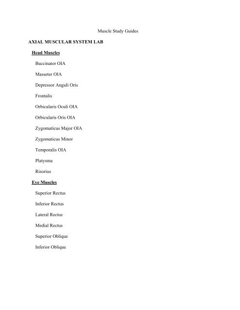
<strong>Muscle</strong> <strong>Study</strong> <strong>Guides</strong><strong>AXIAL</strong> <strong>MUSCULAR</strong> <strong>SYSTEM</strong> <strong>LAB</strong><strong>Head</strong> <strong>Muscle</strong>sBuccinator OIAMasseter OIADepressor Anguli OrisFrontalisOrbicularis Oculi OIAOrbicularis Oris OIAZygomaticus Major OIAZygomaticus MinorTemporalis OIAPlatysmaRisoriusEye <strong>Muscle</strong>sSuperior RectusInferior RectusLateral RectusMedial RectusSuperior ObliqueInferior Oblique
Torso <strong>Muscle</strong>sSternocleidomastoid OIAErector Spinae GroupSpinalisLongissimusIliocostalisIntertransversariiInterspinalisAbdominal <strong>Muscle</strong>sExternal ObliqueInternal ObliqueTransversus AbdominisRectus Abdominis OIAThorax <strong>Muscle</strong>sDiaphragmExternal IntercostalsInternal Intercostals
Arm / (model key)Subscapularis 5Supraspinatus 6Infraspinatus 7Teres major 9Teres minor 8Deltoid 10 OIACoracobrachialis15Biceps brachii (long12, short head13) 11 OIABrachialis14Brachioradialis19Extensor carpi radialis longus 20Extensor digitorum 30 OIAExtensor carpi ulnarisExtensor retinaculum (band of tissue) 38Pronator teres 22Supinator (deep) 34Flexor carpi radialis 23Palmaris longus 24Flexor carpi ulnaris 25Flexor retinaculum (band of tissue) 46Triceps brachii (long head17, medial head18, short head16) OIALegGluteus maximus 11 OIAGluteus medius 12Gluteus minimus (deep)Piriformis 13Obturator internus 15Tensor fasciae latae 18Iliotibial tract 19Rectus femoris 20 OIAVastus (lateralis, medialis, intermedius) 21, 23 22Sartorius 24 OIAAdductor longus 26 OIAAdductor magnus 28 OIA,Adductor brevis (deep) OIAGracilis 27 OIAPectineusBiceps femoris (long, short heads) L31, S32 OIASemitendinosous 30Semimembranosus 29Tibialis anterior 33Extensor hallucis longus 44Extensor digitorum longus 34Fibularis longus & brevis L35, B36Gastrocnemius (medial, lateral heads) 37,38 OIASoleus 39Tibialis posterior 40Popliteus 41Flexor hallucis longus 43Flexor digitorus longus 42Superior/inferior peroneal retinaculum 57, 58
TorsoTrapezius OIALevator scapulae OIARhomboid major and minor OIASerratus anterior OIAPectoralis majorPectoralis minorLatissimus dorsi OIAShoulder JointRotator Cuff TendonsSupraspinatus tendonInfraspinatus tendonSubscapularis tendonIliopsoas: psoas major 25, iliacusCAT MUSCLESPectoralis MajorPectoralis MinorClavotrapeziusAcromiotrapeziusSpinotrapeziusSubscapularisExternal ObliqueRectus abdominisSupraspinatusInfraspinatusTriceps Brachii (medial, lateral, long)Brachialis
BrachioradialisExtensor Carpi radialis longusExtensor digitorum communisExtensor digitorum lateralisExtensor Carpi ulnarisPalmaris longusFlexor carpi ulnarisFlexor carpi radialisGluteus maximusBiceps femorisTensor Fasciae lataeSartoriusSemitendinosusSemimembranosousRectous femorisVastas lateralisVastus medialisAdductor longusAdductor femorisGracilisSoleusGastrocnemiusTibialis Anterior
Skeletal <strong>Muscle</strong> Contraction and Sliding Filament TheoryStimulation:Action potential (AP) travels down a motor neuron to the neuromuscular junction.Acetylcholine (Ach), an chemical neurotransmitter, is released by the motor neuronand passed to receptors on the sarcolemma. This causes an AP to spread over theentire sarcolemma, and travels down the transverse tubules (t-tubules). T-tubulesbring the AP to the cell center and causes the sarcoplasmic reticulum (SR) to opencalcium channels, initiating a muscule contraction.Contraction:1. The rigor state: The released calcium ions bind to troponin, which changes theconformation of tryopomyosin and exposes the myosin-binding site on actin.The myosin heads form crossbridges. This rigor state occurs for only a very briefperiod.2. ATP binds to myosin causing myosin to dissociate from actin: The binding ofATP to the myosin head causes myosin to have a low affinity for actin.3. ATP hydrolysis: ATP is hydrolyzed into ADP and inorganic phosphate (Pi).Both products remain bound to the head.4. Myosin reattaches: The energy released from ATP causes myosin head to swingand bind weakly to a new actin molecule. Myosin has potential energy, like aspring and is ready to execute the power stroke.5. Pi release and power stroke: Pi is released from myosin causing myosin head toswing toward the M line and pushes the actin filament in the same direction.6. Release of ADP: myosin releases ADP and myosin head is back in the rigor state.Cycle is ready to begin again.7. The contracted position remains until the renewed binding of another ATPmolecule with the myosin heads. This rebinding causes a dislocation of the actinmyosincomplex. If ATP and calcium remain in the cell, contractions continuewith myosin heads re-cocking and reattaching to actin. A maximum potential iswhen myosin filaments butt from z-line to z-line.Relaxation:1. When the AP stops traveling down the motor neuron, Ach is no longer released at thesynapse. Acetylcholinesterase (AchE), an enzyme in the synaptic cleft, then breaksdown Ach, which then recycles back into the synapse. Without Ach bound to receptors,the AP traveling down the sarcolemma and t-tubles stop. The SR then takes up Ca2+from the sarcoplasm, which causes the troponin-tropomyosin complex to return to itsoriginal position, covering the binding sites on actin. Myosin heads can no longer formcrossbridges, so the sarcomeres return to their original positions with the help of thespring action from titin.


