Cardiovascular System - Health Science Technology Education
Cardiovascular System - Health Science Technology Education
Cardiovascular System - Health Science Technology Education

You also want an ePaper? Increase the reach of your titles
YUMPU automatically turns print PDFs into web optimized ePapers that Google loves.
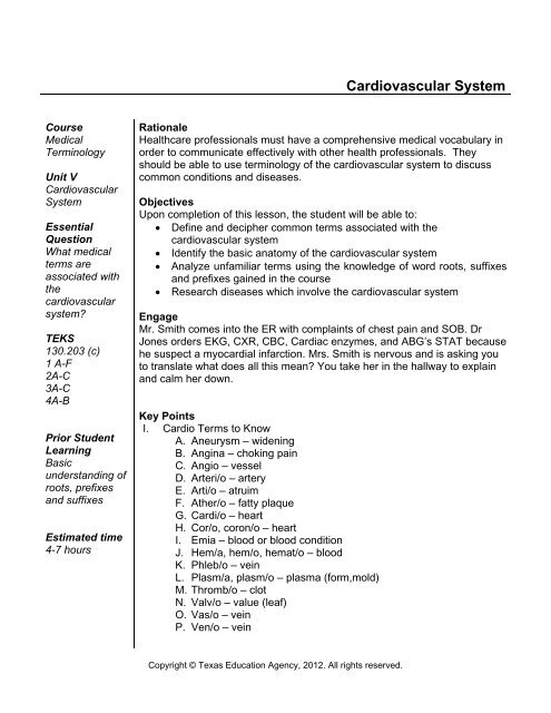
<strong>Cardiovascular</strong> <strong>System</strong>CourseMedicalTerminologyUnit V<strong>Cardiovascular</strong><strong>System</strong>EssentialQuestionWhat medicalterms areassociated withthecardiovascularsystem?TEKS130.203 (c)1 A-F2A-C3A-C4A-BPrior StudentLearningBasicunderstanding ofroots, prefixesand suffixesEstimated time4-7 hoursRationale<strong>Health</strong>care professionals must have a comprehensive medical vocabulary inorder to communicate effectively with other health professionals. Theyshould be able to use terminology of the cardiovascular system to discusscommon conditions and diseases.ObjectivesUpon completion of this lesson, the student will be able to:• Define and decipher common terms associated with thecardiovascular system• Identify the basic anatomy of the cardiovascular system• Analyze unfamiliar terms using the knowledge of word roots, suffixesand prefixes gained in the course• Research diseases which involve the cardiovascular systemEngageMr. Smith comes into the ER with complaints of chest pain and SOB. DrJones orders EKG, CXR, CBC, Cardiac enzymes, and ABG’s STAT becausehe suspect a myocardial infarction. Mrs. Smith is nervous and is asking youto translate what does all this mean? You take her in the hallway to explainand calm her down.Key PointsI. Cardio Terms to KnowA. Aneurysm – wideningB. Angina – choking painC. Angio – vesselD. Arteri/o – arteryE. Arti/o – atruimF. Ather/o – fatty plaqueG. Cardi/o – heartH. Cor/o, coron/o – heartI. Emia – blood or blood conditionJ. Hem/a, hem/o, hemat/o – bloodK. Phleb/o – veinL. Plasm/a, plasm/o – plasma (form,mold)M. Thromb/o – clotN. Valv/o – value (leaf)O. Vas/o – veinP. Ven/o – veinCopyright © Texas <strong>Education</strong> Agency, 2012. All rights reserved.
II. Introduction to the heartA. Fully formed by the 4th week of embryonic developmentB. A hollow muscular organ that acts as a double pumpC. A continuous pump – once pulsations begin, the heart pumpsendlessly until deathIII. Heart AnatomyA. General1. Size – approximately the size of a person’s fist2. Location – in the mediastinumB. Coverings – Pericardium (see the Pericardial Diagram)1. A double-layered sac2. Contains 10-20cc. of pericardial fluid to reduce the friction ofthe beating heart3. Parietal layer – a fibrous membrane; the outer layer4. Visceral layer – serous membrane; also called theepicardium; attached to the myocardiumC. The Heart Wall1. Myocardium – heart muscle; thicker on the left side of theheart2. Endocardium – the lining of the heart chambersD. Chambers1. Atriaa. The two upper chambers of the heartb. Have thin walls and a smooth inner surfacec. Responsible for receiving bloodd. The right atrium receives deoxygenated (oxygen poor)blood from the body through the superior and inferiorvena cavae. The left atrium receives oxygenated (oxygen rich) bloodfrom the lungs through the pulmonary veins2. Ventriclesa. The two lower chambers of the heartb. Have thicker walls and an irregular inner surfacec. Contain the papillary muscles and chordae tendineae(prevent the heart valves from turning inside out whenthe ventricles contract)d. The left wall is 3 times as thick as the right wall; formsthe apex of the hearte. Responsible for pumping blood away from the heartf. The right ventricle sends deoxygenated blood to thelungs via the pulmonary arteriesg. The left ventricle sends oxygenated blood to all parts ofthe body via the aorta3. Accessory StructuresCopyright © Texas <strong>Education</strong> Agency, 2012. All rights reserved.
a. Septum – the muscular wall dividing the heart into rightand left halvesb. Heart valves – prevents the backflow of bloodc. Papillary musclesd. Chordae tendineaeE. Great Vessels (see the External Heart Diagram)1. Superior and inferior vena cava – the largest veins in thebody; receive deoxygenated blood from all parts of the body2. Coronary sinus3. Pulmonary arteries – carry deoxygenated blood to the lungsfrom the right ventricle4. Pulmonary veins – carry oxygenated blood to the left atriumfrom the lungs5. Aorta – the largest artery in the body; carries oxygenatedblood to distribute to all parts of the bodyF. The Pathway of Blood Through the Heart and All Body Tissues(see the Internal Heart Diagram)1. Superior and inferior vena cava2. Right atrium3. Tricuspid valve4. Right ventricle5. Pulmonary semilunar valve6. Pulmonary arteries7. Lungs ( O 2 and CO 2 exchange - external respiration)8. Pulmonary veins9. Left atrium10. Bicuspid/Mitral valve11. Left ventricle12. Aortic semilunar valve13. Aorta – all parts of the body via the arteries14. Arterioles15. Capillaries of the individual tissues (O 2 and CO 2 exchange -internal respiration)16. Venules17. Veins18. Superior and inferior vena cavaG. <strong>Cardiovascular</strong> Circuits1. Pulmonary circuit – the transport of blood from the right sideof the heart to the lungs and then back to the left side of theheart2. <strong>System</strong>ic circuit – the transport of blood from the left side ofthe heart to all parts of the body and then back to the rightside of the heart3. Coronary circuit – the transport blood from the left side of theheart to the heart tissues and back to the right side of theCopyright © Texas <strong>Education</strong> Agency, 2012. All rights reserved.
heartH. Valves1. Tough fibrous tissues between the heart chambers andmajor blood vessels of the heart2. Gate-like structures to keep the blood flowing in onedirection and prevent the regurgitation or backflow of blood3. Atrioventricular valves – when ventricles contract, blood isforced upward and the valves close; attached by papillarymuscles and chordae tendineaea. Tricuspid valve – between the right atrium and the rightventricleb. Bicuspid/mitral valve – between the left atrium and theleft ventricle4. Semilunar Valves – three half-moon pockets that catch bloodand balloon out to close the openinga. Pulmonary semilunar valve – between the right ventricleand the pulmonary arteriesb. Aortic semilunar valve – between the left ventricle andthe aortic arch/aortaI. Cardiac Circulation (The Blood Supply to the Heart)1. Aorta –> coronary arteries –> capillaries in the myocardium–> coronary veins –> coronary sinus –> right atrium2. Blood in the chambers nourishes the endocardium3. The coronary circuit opens only during the relaxation phaseof the cardiac cycle4. Occlusion of the coronary artery – a myocardial infarction(heart attack) occurs if collateral circulation is inadequateIV. Heart PhysiologyA. Nerve Supply to the Heart1. Alters the rate and force of cardiac contraction2. Vagus nerve (parasympathetic nervous system) – slows theheart rate3. Sympathetic nerves – increase the heart rate4. Epinephrine/norepinephrine – increases heart rate5. Sensory (afferent) nerves – detect atria being stretched andlack of oxygen (changes the rate of contractions)6. Angina – chest pain due to a lack of oxygen in coronarycirculationB. Intrinsic Conduction <strong>System</strong> – Automaticity (see the HeartConduction <strong>System</strong> Diagram)1. Enables the heart to contract rhythmically and continuouslywithout motor nerve impulses2. Arrhythmia – myocardial cells leak sodium faster than the SAnode, causing an irregular heartbeat3. SA (sinoatrial) node – known as the pacemaker, locatedCopyright © Texas <strong>Education</strong> Agency, 2012. All rights reserved.
where the superior and inferior vena cava enter the rightatrium4. AV (atrioventricular) node – sends impulses to the ventricles5. Bundle of His/bundle branches – in the septum6. Purkinje fibers – in the heart wall to distribute nerve impulsesC. Cardiac Cycle – Generated in the Heart Muscle1. One (1) contraction (systole = 0.3 seconds) + one (1)relaxation (diastole = 0.5 seconds) at 75 beats per minute2. Initiation of contraction – impulse spreads out over both atriacausing them to contract together and force blood into bothventricles3. Impulses from the SA node are sent to the AV node(between the atria in the septum)4. Impulses from the AV node are sent to nerve fibers in theseptum (bundle of His) which transmits the impulse via theright and left bundle branches to the Purkinje fibers – causethe ventricles to contract together and force blood out of theaorta and pulmonary arteries, and into the body and thelungs5. The shift of ions along the conduction system = actionpotential6. Periods of rest = polarization7. Periods of activity = depolarization – when an impulse istransmitted; and repolarization – when a slow shift back topolarization occursD. EKG (see the EKG Diagram)1. Electrical changes during the cardiac cycle are recorded asan EKG2. To estimate heart rate using an EKG strip, count the numberof QRS complexes in a 6-second strip and multiply by 103. P wavea. Impulse received by the SA nodeb. The atria depolarize (contract)c. Enlarged P wave = enlarged atrium or stenosed AVvalve4. QRS complexa. Impulse passing through the ventricles (systole)b. The ventricles depolarize (contract)c. The atria repolarize (relax)d. Enlarged Q = myocardial infarctione. Enlarged R = enlarged ventricles5. T wavea. Repolarization of the ventricles (diastole)b. Elevated = K+ level too high6. PR intervala. 0.12 – 0.2 secondsCopyright © Texas <strong>Education</strong> Agency, 2012. All rights reserved.
. Too long = rheumatic heart disease or hardening of thearteries; conduction problem or delay at the AV node7. ST segmenta. Elevated = acute myocardial infarctionb. Depressed = insufficient oxygen to the heartE. Stroke Volume and Cardiac Output1. Cardiac out is the volume of blood pumped by the heart perminute, the function of heart rate, and stoke volume2. Stoke volume is the volume of blood, in milliliters (ml),pumped out of the heart with each beat3. Weak hearts have low stroke volume – they must pumpfaster to move an adequate amount of blood4. Well-trained athletes have good stroke volume – can pumpslower to move an adequate amount of bloodV. Overview of Blood VesselsA. General Composition and Function1. Allow for circulation of blood and other bodily fluids to all thebody’s cells2. Three layersa. Tunica adventitia – outer layer of tough fibrous tissueb. Tunica media – smooth muscle which allows vessels toconstrict and dilatec. Tunica intima – smooth, inner elastic layer (lumen =internal diameter)B. Arteries1. Carry blood away from the heart2. Thicker, to withstand pressure exerted during systole3. All but the pulmonary arteries carry oxygenated blood4. Aorta – the largest artery; 1 inch in diameter5. Arterioles – the smallest arteries6. Coronary arteries – the most important; supply blood to theheart musclea. Left and right main coronary arteryb. Left coronary artery –> left anterior descending –> leftcircumflex branchc. Right coronary artery –> right atrium and right ventricleC. Veins1. Carry blood toward the heart2. All but the pulmonary veins carry deoxygenated blood3. Layers are much thinner, and less elastic4. A series of internal valves that work against the flow ofgravity to prevent reflux5. Superior and inferior vena cava – the largest veins6. Venules – the smallest veinsD. CapillariesCopyright © Texas <strong>Education</strong> Agency, 2012. All rights reserved.
1. Tiny, microscopic vessels2. Walls are one cell layer thick3. Function – to transport and diffuse essential materials to andfrom the body’s cells and the bloodVI. PulseA. The pressure of the blood pushing against the wall of an artery asthe heart beats – during systoleB. Common pulse sites1. Temporal – at the side of the forehead2. Carotid – at the neck3. Brachial – the inner aspect of the forearm at the antecubitalspace (the crease of the elbow)4. Radial – at the inner aspect of the wrist on the thumb side5. Femoral – at the inner aspect of the upper thigh or groin6. Dorsalis pedis – at the top of the foot archVII. Blood PressureA. Systole – the maximum pressure formed during a ventricularcontractionB. Diastole – the minimum pressure during ventricular relaxation(atrial contraction)C. Measured in mm of HgD. BP = CO x PR (Blood Pressure = Cardiac Output x PeripheralResistance)E. Normal Ranges1. Systolic = 100–1402. Diastolic = 60–90F. Hypotension – systolic < 90G. Hypertension – systolic > 150 and/or diastolic > 90H. Must be lower in the pulmonary circuit to prevent fluid fromfiltering out into the alveoliI. Factors Affecting BP1. Cardiac output2. Peripheral resistance3. Blood volumeJ. Circulatory Shock1. Hypovolemic shock2. Vascular shock3. Cardiogenic shockVIII. Diagnostic Procedures for the <strong>Cardiovascular</strong> <strong>System</strong>A. History and Physical1. Checking for symptoms of disease2. Chest pain, shortness of breath, awareness of heartbeatCopyright © Texas <strong>Education</strong> Agency, 2012. All rights reserved.
(palpitation), fatigue, dizziness or loss of consciousness,edema, pain in the legs while walking (claudication)B. Electrocardiogram – a tracing of the electrical activity of the heartC. Phonocardiogram – an electrocardiogram with heart soundsD. Echocardiogram – ultrasound measures the size and movementof the heart structuresE. Doppler Ultrasound – measures blood flowF. Arteriography – radiopaque dye injected into and x-ray seriestaken of blood flowG. Cardiac Catheterization1. Right side of heart – a catheter threaded into a vein, then thevena cava, then the heart, then the pulmonary artery2. Left side of heart – a catheter threaded into an artery, thenthe left ventricle, then the aorta, then the coronary vessels3. X-rays taken during the procedure4. Dye is also injectedIX. Diseases of the <strong>Cardiovascular</strong> <strong>System</strong>A. Arteriosclerosis – hardening of the arteriesB. Atherosclerosis1. Fatty deposits on the walls of the arteries2. Causesa. Increased blood lipidsb. High blood pressurec. Smokingd. Obesitye. Physical inactivityf. TensionC. Hypertension1. 90% = essential hypertension – no specific cause2. 10% = a symptom of another disease, i.e. an adrenal tumoror kidney disease3. Increases the workload of the heart4. Leads to hypertrophy of the left ventricle, then heart failure5. Accelerates the development of atherosclerosisD. Ischemic Heart Disease1. The oxygen supply to the heart is inadequate2. Atherosclerosis is a major cause3. Can lead toa. Angina pectoris – a condition in which the coronaryarteries are temporarily blocked – reduced blood supplyto the heart – chest painb. Heart attack – cessation of normal cardiac contraction(cardiac arrest)c. Myocardial infarction – necrosis (death) of the heartmuscle due to severe, prolonged ischemiaCopyright © Texas <strong>Education</strong> Agency, 2012. All rights reserved.
d. Sudden death – the heart stops and ventricularfibrillation occursE. Cardiac Arrhythmias1. An abnormality in the rate, rhythm, or conduction of the heartbeatF. Bacterial Endocarditis1. An inflammation of the internal lining of the heart2. Also involves the heart valvesG. Valvular Heart Disease1. Involves abnormalities of the heart valves2. Especially the mitral and aortic valves3. The leading cause – rheumatic fever with a hypersensitivityreaction to streptococcus antigens4. Heart valves are scarred5. Treatment – valve replacementH. Congenital Heart Disease1. Defects in the heart that occurred during embryologic andfetal development2. Involves defective communication between the chambers,malformation of the valves, and malformation of the septa3. Cyanotic – the inability of the individual to get adequateblood oxygenation due to extensive cardiac abnormalitiesthat cause blood to be shunted away from lungs4. For example “Blue Babies” – a failure of the foramen ovaleto close or transposition of the great arteries or patent ductusarteriosusI. Congestive Heart Failure (CHF)1. Pumping action of the heart is diminished2. Fluid accumulates and is retained in the tissues3. Compensationsa. Increased heart rate, greater force of contractionb. Retention of fluid by the kidneysc. Enlargement of the heartJ. Cor Pulmonale1. Hypertrophy of the right ventricle due to hypertension inpulmonary circulation2. Increased BP in the lungs –a reduction in blood flow andincreased resistance in the lungs – pulmonary hypertension– increased pressure in the pulmonary arteries – bloodbacks up into the right ventricle – hypertrophyK. Peripheral Arterial Disease1. Decreased blood flow to the peripheral vesselsL. Varicose Veins1. Enlarged veins which can be inflamedM. Hemorrhoids1. Varicose veins of the rectal and anal areaCopyright © Texas <strong>Education</strong> Agency, 2012. All rights reserved.
N. Aneurysm1. A weak section in the wall of an artery that balloons out andrupturesO. Phlebitis1. Inflammation of a veinP. Thrombus1. A blood clot that stays where it is formedQ. Stroke (CVA)1. Brain infarct – caused by decreased oxygen supply to thebrain due to a blood clot or hemorrhageR. Raynaud’s Disease1. A circulation disorderS. Esophageal Varices1. Varicose veins of the esophagusT. Tetralogy of Fallot1. Four different heart defects that occur at birth which are lifethreatening to the fetusActivity1. Students should make flash cards of Cardio terms and practice puttingterms together with Prefixes and Suffixes to make new terms.2. Complete the <strong>Cardiovascular</strong> <strong>System</strong> Worksheet.3. Complete the Terminology Worksheet.4. Complete the <strong>Cardiovascular</strong> <strong>System</strong> Medical Terminology Worksheet5. Review media terms with the students using review games such as the“fly swatter game” or the “flash card drill” (See the Medical TerminologyActivities Lesson Plan -http://texashste.com/documents/curriculum/principles/medical_terminology_activities.pdf ).6. Research and report on diseases and disorders of the <strong>Cardiovascular</strong><strong>System</strong>.AssessmentSuccessful completion of ActivitiesMaterials<strong>Cardiovascular</strong> <strong>System</strong> WorksheetMedical Terms Worksheet<strong>Cardiovascular</strong> <strong>System</strong> Medical Terminology WorksheetKEY - <strong>Cardiovascular</strong> <strong>System</strong> WorksheetKEY - Medical Terms WorksheetKEY - <strong>Cardiovascular</strong> <strong>System</strong> Medical Terminology WorksheetAccommodations for Learning DifferencesFor reinforcement, the students will practice terms of the cardiovascularsystem using flash cards.Copyright © Texas <strong>Education</strong> Agency, 2012. All rights reserved.
For enrichment, the students will choose a disease related to thecardiovascular system and research the disease using the internet. Thestudents will share their findings with the class.National and State <strong>Education</strong> StandardsNational <strong>Health</strong>care Foundation Standards and Accountability Criteria<strong>Health</strong> care workers will know the various methods of giving and obtaininginformation. They will communicate effectively, both orally and in writing.TEKS130.203 (c)(1)(A) identify abbreviations, acronyms, and symbols;130.203 (c)(1)(B) identify the basic structure of medical words;130.203 (c)(1)(C) practice word-building skills;130.203 (c)(1)(D) research the origins of eponyms;130.203 (c)(1)(E) recall directional terms and anatomical planes related tobody structure;130.203 (c)(1)(F) define and accurately spell occupationally specific termssuch as those relating to the body systems, surgical and diagnosticprocedures, diseases, and treatments.130.203 (c)(2)(A) demonstrate appropriate verbal and written strategies suchas correct pronunciation of medical terms and spelling in a variety of healthscience scenarios;130.203 (c)(2)(B) employ increasingly precise language to communicate;130.203 (c)(2)(C) translate technical material related to the health scienceindustry.130.203 (c)(3)(A) examine medical and dental dictionaries and multimediaresources;130.203 (c)(3)(B) integrate resources to interpret technical materials;130.203 (c)(3)(C) investigate electronic media such as the Internet withappropriate supervision.130.203 (c)(4)(A) distinguish medical abbreviations used throughout thehealth science industry; and130.203 (c)(4)(B) translate medical abbreviations in simulated technicalmaterial such as physician progress notes, radiological reports, andlaboratory reports.College and Career Readiness StandardsEnglish/language artB.1 Identify new words and concepts acquired through study of theirrelationships to other words and concepts.B2. Apply knowledge of roots and affixes to infer the meanings of newwords.B3. Use reference guides to confirm the meanings of new words orCopyright © Texas <strong>Education</strong> Agency, 2012. All rights reserved.
concepts.Cross- Disciplinary standards-Foundational SkillsA2. Use a variety of strategies to understand the meanings of new wordsCopyright © Texas <strong>Education</strong> Agency, 2012. All rights reserved.
The <strong>Cardiovascular</strong> <strong>System</strong>Name _______________________________Period __________1. What is the protective membrane covering of the heart called?2. Which chambers of the heart receive blood from the veins?3. What chambers of the heart are known as pumping chambers?4. What is the name of the blood vessel that brings venous blood from the head, neck, andarms to the right atrium?5. What is the name of the blood vessel that brings venous blood from the abdomen and legsinto the right atrium?6. What is the name of the blood vessels that take deoxygenated blood from the right ventricleto the lungs?7. What is the name of the blood vessels that take oxygenated blood from the lungs to the leftatrium?8. The largest artery in the body is the .9. The valves are formed from the most inner heart layer, or the .10. The valve between the right atrium and the right ventricle is known as the. The valve between the left atrium and the left ventricle is knownas the .11. The valves between the ventricles and blood vessels are known as the .12. Complete flow of blood through the heart. Blood entering theatrium flows through the tricuspid valve and into the. From there, the deoxygenated blood flows past the pulmonarysemilunar valve and into the , and then into the lungs.Copyright © Texas <strong>Education</strong> Agency, 2012. All rights reserved.
Oxygenated blood leaves the lungs through the.and enters the13. What is the semilunar valve? .14. What is the pacemaker of the heart?15. List and describe the heart’s cardiac conduction system.a.b.c.d.16. a. What is systole?b. What is diastole?17. If the patient has an elevated blood pressure we say they have .18. What is the stroke volume?19. What is cardiac output?20. a. What vessel carries blood away from the heart?b. What vessel carries blood to the heart?c. What vessel is responsible for gas and nutrient exchange within each of the body’scells?21. Describe each of the following vessels:a. arteriesb. veinsc. capillaries22. What is a pulse?Copyright © Texas <strong>Education</strong> Agency, 2012. All rights reserved.
23. Identify the location of the following pulse points:a. What pulse is felt on the upper surface of the foot?b. What pulse is felt in the antecubital space?c. What pulse is felt in the groin?d. What pulse is found in the neck?e. What pulse is found on the wrist side of the arm?24. Answer the following questions about blood pressure.a. What is the first measurement of blood pressure?b. What does it measure?c. What is the second measurement of blood pressure?d. What does it measure?25. a. What circulation route takes deoxygenated blood to the lungs where it can pick upoxygen?b. What circulation route takes oxygenated blood through the body?Copyright © Texas <strong>Education</strong> Agency, 2012. All rights reserved.
WORKSHEET - <strong>Cardiovascular</strong> Review - KEY1. What is the protective membrane covering of the heart called? Pericardium2. What chambers of the heart receive blood from veins? Atria3. What chambers of the heart are known as pumping chambers? Ventricles4. What is the name of the blood vessel that brings venous blood from the head, neck, andarms to the right atrium? Superior Vena Cava5. What is the name of the blood vessel that brings venous blood from the abdomen and legsto the right atrium? Inferior Vena Cava6. What is the name of the blood vessels that take deoxygenated blood from the right ventricleto the lungs? Pulmonary arteries (which branch from the pulmonary trunk)7. What is the name of the blood vessels that take oxygenated blood from the lungs to the leftatrium? Pulmonary veins8. The largest artery in the body is the Aorta9. The valves are formed from the most inner heart layer or the Endocardium .10. The valve between the right atrium and the right ventricle is known as the Tricuspid Valve.The valve between the left atrium and the left ventricle is known as the Bicuspid Valve (alsocalled the Mitral Valve).11. The valves between the ventricles and blood vessels are known as the Pulmonary and AorticSemilunar Valves.12. Complete flow of blood through the heart. Blood entering the Right atrium flows through thetricuspid valve and into the Right ventricle. From there, the deoxygenated blood flows pastthe pulmonary semilunar valve and into the Pulmonary Arteries, and then into the lungs.Oxygenated blood leaves the lungs through the Pulmonary Veins and enters the Left atriumof the heart. Blood continues to flow through the Mitral/Bicuspid valve and into the Leftventricle. From there, blood will flow past the aortic semilunar valve and into the Aorta.13. What is a semilunar valve? Semilunar Valves: 3 half-moon pockets that catch blood andballoon out to close the opening14. What is the pacemaker of the heart? SA nodeCopyright © Texas <strong>Education</strong> Agency, 2012. All rights reserved.
15. List and describe the heart’s cardiac conduction system.a. SA (sinoatrial) node: known as the pacemaker, located where the superior and inferiorvena cava enter the right atriumb. AV (atrioventricular) node: sends impulses to the ventriclesc. Bundle of His/bundle branches: in the septumd. Purkinje fibers: in the heart wall to distribute nerve impulses16. a. What is systole? Maximum pressure formed during a ventricular contractionb. What is diastole? Minimum pressure during ventricular relaxation (atrial contraction)17. If the patient has an elevated blood pressure we say they have Hypertension.18. What is the stroke volume? Stroke volume is the volume of blood, in milliliters (ml), pumpedout of the heart with each beat19. What is cardiac output? Cardiac output is the volume of blood pumped by the heart perminute – the function of heart rate and stroke volume20. a. What vessel carries blood away from the heart? Arteriesb. What vessel carries blood to the heart? Veinsc. What vessel is responsible for gas and nutrient exchange within each of the body’scells? Capillaries21. Describe each of the following vessels:a. arteries carry blood away from the heart. Thicker to withstand the pressure exertedduring systole. All but the pulmonary arteries carry oxygenated bloodb. veins carry blood toward the heart. All but the pulmonary veins carrydeoxygenated blood. Layers are much thinner, less elastic. A series ofinternal valves that work against the flow of gravity to prevent reflux.c. capillaries tiny, microscopic vessels. Walls one cell layer thick. Function – to transportand diffuse essential materials to and from the body’s cells and the blood22. What is a pulse? The pressure of the blood pushing against the wall of an artery as the heartbeats during systole.23. Identify the location of the following pulse points:a. What pulse is felt on the upper surface of the foot? Dorsalis pedisb. What pulse is felt in the antecubital space? BrachialCopyright © Texas <strong>Education</strong> Agency, 2012. All rights reserved.
c. What pulse is felt in the groin? Femorald. What pulse is found in the neck? Carotide. What pulse is found on the wrist side of the arm? Radial24. Answer the following questions about blood pressure.a. What is the first measurement of blood pressure? Systoleb. What does it measure? Pressure as the ventricles contractc. What is the second measurement of blood pressure? Diastoled. What does it measure? Pressure remaining in the artery as the ventricles rest26. a. What circulation route takes deoxygenated blood to the lungs where it can pick upoxygen? Pulmonaryb. What circulation route takes oxygenated blood through the body? <strong>System</strong>icCopyright © Texas <strong>Education</strong> Agency, 2012. All rights reserved.
Medical Terminology WorksheetPlease write the meaning of the terms in the right column.Prefixes, Suffixes, and Root Wordsananti-apheresis-blast-critcyt/o-cyte-emiaerythr/oferrfibr/o-genhem/ohemat/o-ic-in-iskary/oleuk/olys/o-lysismacr/omegamon/omyel/o-ologist-ology-oma-osis-penia-phage-philiaplasm/o-plasty-poiesispolyproCopyright © Texas <strong>Education</strong> Agency, 2012. All rights reserved.
eticul/o-rrhagesepsisseptic-stasisthromb/oMedical TermsanemiaaplasticerythrocyteerythropoiesisferrousfibrinogenfibrinolysishematocrithematocytoblasthematologisthematologyhematomahematopoiesishemolytichemophiliahemorrhagehemostasisleukemialeukocyteleukocytosisleukopeniamacrophagemegakaryocytemonocytemyelofibrosisplasmapheresispolycythemiaproerythroblastreticulocytesepsissepticemiathrombocyteCopyright © Texas <strong>Education</strong> Agency, 2012. All rights reserved.
thrombocytopeniathrombolysisthromboplastinthrombosisMedicalAbbreviationsAIDSBPCBCCO 2CVADVTFBSGTTHBVHctHghgbHIBHIVmlmmO 2RBCS&SSOBstatWBCWNLCopyright © Texas <strong>Education</strong> Agency, 2012. All rights reserved.
KEY - Medical Terminology WorksheetPrefixes, Suffixes, and Root Wordsanwithoutantiagainst-apheresisremoval of-blastdeveloping cell-critto separatecyt/ocell-cytecell-emiablood conditionerythr/oredferrironfibr/ofiber-genproducinghem/obloodhemat/oblood-icpertaining to-inpertaining to-ispertaining tokary/obody, nucleusleuk/owhitelys/odestruction of-lysisdestruction ofmacr/olargemegalargemon/oonemyel/obone marrow (also spinal cord)-ologistone who studies, specialist-ologystudy of-omatumor, mass-osiscondition of-peniadeficiency of-phageeating-philialove, affection, affinityplasm/oplasma-plasty(surgical) repair-poiesismaking of/production ofpolymanyprobeforereticul/onetlikeCopyright © Texas <strong>Education</strong> Agency, 2012. All rights reserved.
-rrhagesepsisseptic-stasisthromb/oMedical Termsanemiaaplasticerythrocyteerythropoiesisferrousfibrinogenfibrinolysishematocrithematocytoblasthematologisthematologyhematomahematopoiesishemolytichemophiliahemorrhagehemostasisleukemialeukocyteleukocytosisleukopeniamacrophagemegakaryocytemonocyteburst forthinfectionpathogenicstanding stillclotwithout blood (generally used to describe a lack of red bloodcells)failing to develop into new tissue (Aplastic anemia is a termused to describe when red blood cells are notproduced by the bone marrow)red (blood) cellproduction of red (referring to the production of red bloodcells)pertaning to ironproducing fibers (which will be used in the blood clottingprocess)destruction of fibersto separate bloodblood-developing cell (this is the “stem” cell that is responsiblefor forming the three types of blood cells)one who specializes in the study of bloodthe study of bloodblood tumor or mass (bruise also called ecchymosis)the production of bloodthe destruction of bloodpertaining to an affinity or love of blood (has evolved to referto a number of blood coagulation disorders)blood bursting forthblood standing still (Refers to the stopping of the bleeding)“white blood” (cancer of the blood with many immature whiteblood cells)white (blood) cellcondition of white cells (used to refer to a high number ofwhite blood cells: higher than 10,000 WBC per mm 3 )deficiency of white cells (used to refer to a low number ofwhite cells; lower than 5,000 WBC per mm 3 )large eater (refers to the white blood cell, the monocyte, that isfound in the tissues and is an integral part in the immuneresponse).large, nucleated cell (will break apart to form the platelets orthrombocytes)one cell (refers to a specific type of WBC)Copyright © Texas <strong>Education</strong> Agency, 2012. All rights reserved.
myelofibrosisplasmapheresispolycythemiaproerythroblastreticulocytesepsissepticemiathrombocytethrombocytopeniathrombolysisthromboplastinthrombosisMedicalAbbreviationsAIDSBPCBCCO 2CVADVTFBSGTTHBVHctHghgbHIBHIVmlmmO 2RBCS&SSOBstatWBCWNLa condition of the fibers in the bone marrow (a conditioncaused when fibrous tissue replaces the bone marrow)removing plasmablood condition of many cells (refers to an overproduction ofall blood cell types)developing red cell that comes beforenet cell (refers to one of the stages of red blood celldevelopment)condition of infectioncondition of infected bloodclotting cell (platelets)deficiency of clotting cellsdestruction of a clotpertaining to forming a clotcondition of a clot (or clots)Acquired Immunodeficiency Syndromeblood pressurecomplete blood countcarbon dioxidecerebrovascular accident (a stroke)deep vein thrombosisfasting blood sugarglucose tolerance testhepatitis B virushematocritmercuryhemoglobinhaemophilus influenzae type Bhuman immunodeficiency virusmillilitermillimeteroxygenred blood cell(s)signs and symptomsshortness of breathimmediatelywhite blood cell(s)within normal limitsCopyright © Texas <strong>Education</strong> Agency, 2012. All rights reserved.
<strong>Cardiovascular</strong> <strong>System</strong> Medical Terminology WorksheetPlease write the meaning of the terms in the right column.Prefixes, Suffixes, Root Wordsa-ac-alangi/oaort/o-ararteri/o-aryather/oatri/obibradycalc/icardi/oclavicul/ocoronarydysech/o-ectomyelectr/o-emiaend/oepifemor/ofurc-gram-graph-grapher-graphyhem/ohemat/ohepat/ohomeohyperhypoCopyright © Texas <strong>Education</strong> Agency, 2012. All rights reserved.
interintraisch/o-itiskal/ilip/o-logymedi-megaly-metermy/onatri-odynia-ologist-ology-oma-osis-pathy-peniaperiphleb/o-plastypoly-rrhexis-sclerosis-scopesemisphygm/o-stasis-stenosisstern/osteth/o-stomysub-tachy-tensionthromb/otibi/o-tomyvalvevas/oCopyright © Texas <strong>Education</strong> Agency, 2012. All rights reserved.
ven/oventricleMedical Termsangiocardiographyangiomaangioplastyaortogramarteriorrhexisarteriosclerosisatherectomyatherosclerosisatrioventricularbifurcationbradycardiacardiaccardiodyniacardiologistcardiologycardiomegalycardiomyopathycoronarycoronary ischemiacoronary thrombosisechocardiogramelectrocardiogramelectrocardiographelectrocardiographyendarterectomyendocarditisendocardiumepicardiumfemoralhomeostasishypercalcemiahyperkalemiahyperlipidemia high blood levels offathypernatremiahypertensionCopyright © Texas <strong>Education</strong> Agency, 2012. All rights reserved.
hypocalcemiahypokalemiahyponatremiahypotensioninterventricularintravenousischemiamyocarditismyocardiumpericardiumpericarditispericardiostomyphlebitisphlebotomystethoscopesubclaviansphygmocardiographsphygmomanometertachycardiathrombophlebitisthrombosistibialvalvulitisvenogramMedical AbbreviationsavBPCBCCPRCVADNRDVTECGEKGERETAHgCopyright © Texas <strong>Education</strong> Agency, 2012. All rights reserved.
HRIVK+MINa+NCRPPERBCSAstatTPRVSVSSWBCCopyright © Texas <strong>Education</strong> Agency, 2012. All rights reserved.
KEY - <strong>Cardiovascular</strong> <strong>System</strong> Medical Terminology WorksheetPrefixes, Suffixes, Root Wordsa-ac-alangi/oaort/o-ararteri/o-aryather/oatri/obibradycalc/icardi/oclavicul/ocoronarydysech/o-ectomyelectr/o-emiaend/oepifemor/ofurc-gram-graph-grapher-graphyhem/ohemat/ohepat/ohomeohyperhypointerintraisch/owithout the absence ofpertaining topertaining tovesselaorta; largest arterypertaining toarterypertaining tofatty plaqueatriumtwoslowcalciumheartclavicle (collarbone)circling the heartbad, painful, difficultreflected soundremoval or excisionelectricityblood conditionwithinon, uponfemurbranch; forkedrecord a pictureinstrument that recordsone who recordsthe process of recording a picturebloodbloodliversameabovebelowbetweenwithindeficiency, blockageCopyright © Texas <strong>Education</strong> Agency, 2012. All rights reserved.
-itiskal/ilip/o-logymedi-megaly-metermy/onatri-odynia-ologist-ology-oma-osis-pathy-peniaperiphleb/o-plastypoly-rrhexis-sclerosis-scopesemisphygm/o-stasis-stenosisstern/osteth/o-stomysub-tachy-tensionthromb/otibi/o-tomyvalvevas/oven/oventricleinflammation ofpotassiumfatstudy ofmiddleenlargementinstrument that measuresmusclesodiumpainone who studies, specialiststudy ofmass, tumorcondition ofdiseasedeficiencyaroundveinsurgical repairmanyrupturehardeninginstrument to view or examinehalfpulsestanding stillnarrowing, constrictionchest, sternumchestcreate a new openingbelow, underfast, rapidpressureclottibia; lower leg boneto cut into, incisionstructure to permit one-way flowvesselveina small cavityCopyright © Texas <strong>Education</strong> Agency, 2012. All rights reserved.
Medical Termsangiocardiographyangiomaangioplastyaortogramarteriorrhexisarteriosclerosisatherectomyatherosclerosisatrioventricularbifurcationbradycardiacardiaccardiodyniacardiologistcardiologycardiomegalycardiomyopathycoronarycoronary ischemiacoronary thrombosisechocardiogramelectrocardiogramelectrocardiographelectrocardiographyendarterectomyendocarditisendocardiumepicardiumfemoralhomeostasisthe process of recording pictures (x-rays) of the heart andvesselstumor of the vesselsrepair of the vesselspicture (x-ray) of the aortarupture of an arteryhardening of an arteryremoval of the fatty plaquecondition of hardening (of a blood vessel) due to fattyplaquepertaining to the atria and the ventriclestwo branches (a blood vessel splits into two bloodvessels)slow heart beat (less than 60 beats per minute)pertaining to the heartpain in the hearta specialist of the heartstudy of the heartenlargement of the heartdisease of the heart musclecircling the heart (also used when referring to a heartattack or MI)lack of blood flow in the heart due to a blockagecondition of a blood clot within the heart's own bloodvesselsusing sound waves to visualize the heartrecording of the heart's electricity (electrical pattern)machine that records the heart's electricity (electricalpattern)the process of recording the heart's electricity (electricalpattern)removal from within an artery (used to describe theprocess of removing fatty plaque from an artery, such asthe carotid artery)inflammation within the heart (the inner lining of theheart).pertaining to the inner layer of the heartpertaining to upon the heart (the outer layer of the heart;also known as the visceral pericardium)pertaining to the femur"standing the same" (the body's ability to keep its internalenvironment constant)Copyright © Texas <strong>Education</strong> Agency, 2012. All rights reserved.
hypercalcemiahyperkalemiahyperlipidemia high blood levels offathypernatremiahypertensionhypocalcemiahypokalemiahyponatremiahypotensioninterventricularintravenousischemiamyocarditismyocardiumpericardiumpericarditispericardiostomyphlebitisphlebotomystethoscopesubclaviansphygmocardiographsphygmomanometertachycardiathrombophlebitisthrombosistibialvalvulitisvenogramhigh blood levels of calciumhigh blood levels of potassiumhigh blood levels of sodiumhigh blood pressurelow blood levels of calciumlow blood levels of potassiumlow blood levels of sodiumlow blood pressurepertaining to between the ventriclespertaining to within the veinsdeficiency of blood (to a muscle or an organ)inflammation of the heart musclepertaining to the heart muscle (the middle layer of theheart, composed of cardiac muscle)around the heartinflammation around the heart (an inflammation of themembranes surrounding the heart)formation of an opening in the pericardium (for drainageof extra fluid or blood)inflammation of a veinto cut (make an incision) into a veininstrument used to listen to sounds produced in the bodypertaining to below the clavicle (collarbone)machine used to record the pulse (usually the radial) withthe heartbeatinstrument used to measure the pulse (as in bloodpressure)rapid heartbeat (usually above 100 beats per minute)inflammation of a vein associated with a clotcondition of clottingpertaining to the tibiainflammation of the valvespicture (x-ray) of a vein or veinsMedical AbbreviationsavBPCBCatrioventricularblood pressurecomplete blood countCopyright © Texas <strong>Education</strong> Agency, 2012. All rights reserved.
CPRcardiopulmonary resuscitationCVAcerebrovascular accidentDNRdo not resuscitateDVTdeep vein thrombosisECGelectrocardiogramEKGelectrocardiogramERemergency roomETAestimated time of arrivalHgmercuryHRheart rateIVintravenousK+ potassiumMImyocardial infarctionNa+sodiumNCRno cardiac resuscitationPpulsePEpulmonary embolusRBCred blood cellSAsino-atrialstatimmediatelyTPRtemperature, pulse, respirationVSvital signsVSSvital signs stableWBCwhite blood countCopyright © Texas <strong>Education</strong> Agency, 2012. All rights reserved.