TRAUMA SESSION - All India Ophthalmological Society
TRAUMA SESSION - All India Ophthalmological Society
TRAUMA SESSION - All India Ophthalmological Society

Create successful ePaper yourself
Turn your PDF publications into a flip-book with our unique Google optimized e-Paper software.
<strong>TRAUMA</strong> <strong>SESSION</strong>5736. De Juan E, Sternberg P, Michels RG. Penetratingocular injuries; types of injuries and visual results.Ophthalmology 1983;90:1318-22.7. Esmaili B, Nanda SG, Schork A, Elner VM. Visualoutcome and ocular survival after penetratingtrauma. Ophthalmology 1995;102:393-400.8. May DR, Kuhn FP, Morris RE et al. Theepidemiology of serious eye injuries from theUnited States Eye Injury Registry. Graefes Arch ClinExp Ophthalmol 2000;238:153-7.9. Paul Stern berg JR; Prognosis and outcome forpenetrating ocular trauma. In: Shingleton BJ, HershPS, Kenyon KR,(eds). Eye Trauma, Mosby yearbook, 1991; pp.238-41.10. Sobaci G. Deadly weapon-related open-globeinjuries: outcome assessment by the oculartrauma classification system. Am J Ophthalmol2000;129:47-53.AUTHORS’S PROFILE:DR. DEBAJYOTI NANDA: M.B.B.S. (2003), Burdwan Medical College, The University ofBurdwan; M.S.(2nd year resident), Pt. J. N. M. Medical College, Pt. Ravi Shankar ShuklaUniversity, Raipur.E-mail: debajyotinanda@yahoo.co.inThe Aetiology of Perforating Ocular Injuries in Children andTheir Visual OutcomeDr. Debajyoti Nanda, Dr. A.K. Chandrakar, Dr. M.L. Garg, Dr. N. Pandey,Dr. Eesh Nigam(Presenting Author: Dr. Debajyoti Nanda)Perforating ocular injuries are a common causeof unilateral visual loss. Children account forbetween 20% and 50% of all ocular injuries. It isseen that most of the ocular injuries arepreventable. If the causes of injuries are knownthen appropriate strategies for prevention can beeasily taken. The aetiology of ocular injuries isdiffering from the adults. So it needs furtherevaluation.The principles of management of penetratingocular injury are same for children and adults.But the management is more difficult in cases ofchildren. The condition in developing countriesis more complicated. The possibility ofamblyopia in young children further complicatestreatment.The purpose of this study is to evaluate theaetiology of perforating ocular injuries inchildren and to evaluate their visual outcome.Materials and Methods<strong>All</strong> cases of perforating ocular injuries in children(age
574 AIOC 2009 PROCEEDINGSCause of InjuryTable-1Number of CasesInjury with wooden stick 44Stone injury 28Sharp object poked in own eye 17Hit with sharp object 16Injury with cow’s horn 04Blast injury 03Fall on a sharp object 02Motor vehicle accident 02Bicycle injury 02Miscellaneous 13Total 131Place of injuryTable-2Number of casesPlayground 62Home 56School 04Road 02Shop 01Unknown 06Total 131The most common place of injury wasplayground (47%) followed by home (43%).Insufficient data was available to determine thelocation of injury in 5% cases. The right eye wasmost commonly involved (52%). This notstatistically significant. Injuries involving corneaonly in (65%) cases or corneoscleral in (28%)cases. Wounds involving the sclera only in (7%)cases. The lens was involved in (52%) cases.There was uveal tissue prolapse in 60% cases. Thewound was less than 5mm in length in 42% cases,5-10 mm in 35% and more than 10 mm in 23%cases. Most commonly injuries occurred between3-6 pm.The overall outcome showed the final visualacuity achieved better or equal to 6/12 was only19% and less than 6/60 in 49%.12 wounds were self sealing. In 4 cases primarylens removal and in 28 cases secondary lensremoval was performed. Retinal detachment wasfound in 11 cases. Evisceration was performed in18 cases (14%). Endophthalmitis occur red in 7cases, which were treated with intravitrealantibiotics. Panophthalmitis occurred in 2 cases.There was no case of sympathetic ophthalmitis.DiscussionPerforating ocular injuries represent a significantcause of visual loss. There are only two largerecent studies of the aetiology of perforatingocular injuries in children (Soylu et al, 242 cases,Moreira et al, 146), one medium size study(Thompson et al, 72 cases) and two smallerstudies (Rudd et al, 46 cases, Alfaro et all, 30cases). With 131 cases this study is large sizestudy and in different country.The male preponderance of injuries in this studyof exactly two to one is similar to study done byThompson et al, but less than the findings ofother previous studies. The male predominanceof injuries may be a result of males and femalesbeing engaged in different activities withdifferent degree of risk of ocular injury.The high incidence of accidents in play ground(47%) is clearly of concern. In developingcountries like <strong>India</strong> children are not properlycared during playing. There is also lack of sportsground in developing countries. This alsoincreases the risk of injury. 43% injuries occur athome. It also signifies that children are not caredat home suitably. Only 3% accidents occur inschool. This signifies more attention is requiredduring playing and at home.There is slight predominance of right eye injury,although it is not statistically significant. Motorvehicle accidents are less than other study.Because in countries like <strong>India</strong> children are lessexposed to motor vehicle than the developedcountries. The more common cause of theseinjuries are also not similar to other studies,because in developing countries like <strong>India</strong>,children mostly play with various types ofwooden particles an are less exposed to moderninstruments.In this study only 19% of the children achieved6/12 or better visual acuity. Other paediatricstudies reported achieving 6/12 or better in 36%(Thompson et al), 43% (Alfaro et al), 46%(Moreira et al), 51% (Elder et al). This is becausemost of the time they present late to the tertiarylevel hospital and lack of proper primary levelcare. Lack of awareness of the parents alsocomplicates the figure.Prevention is very important to reduce thisocular morbidity. Children may play in theground under supervision of a senior person.
<strong>TRAUMA</strong> <strong>SESSION</strong>575They must be educated about the danger pointsof the games and precautions to avoid the injury.Many times they quarrel with each other andinjuries occur at that time. It may be prevented.Many times in developing countries both parentsare engaged in various types of work in farming.1. Punnonen E. Epidemiological and social aspects ofperforating eye injuries. Acta Ophthalmol1989;67:492-8.2. Blomdahl S, Staffan N. Perforating eye injury inStockholm population. An epidemiological study.Acta ophthalmol 1984;62:378-90.3. Moreira CA, Debert-Ribeiro M, Belfort R.Epidemiological study of eye injuries in Brazilianchildren. Arch Ophthalmol 1988;106:781-4.4. Soylu M, Demircon N, Yalaz M, et al. Etiology ofpediatric perforating injuries in Southern Turkey.Ophthalmic Epidemiol 1998;5:7-12.ReferencesFor this reason children are not properly cared athome. It may be prevented if they are left undersupervision of any senior family member. Last ofall primary level care should be improvedand they are educated for early referral ofthese cases.5. Rudd JC, Jaeger EA, Freitag SK, et al. Traumaticallyruptured globes in children. J Paediatr OphthalmolStrabismus 1994;31:307-11.6. Alfara DV, Chaudhry NA, Walkoner AF, et al.Penetrating eye injuries in young children. Retina1994;14:201-5.7. Elder M. Penetrating eye injuries in children of theWest Bank and Gaza strip. Eye 1993;7:429-32.8. Thompson CG, Kumar N, Billson FA, Martin F. Theaetiology of perforating ocular injuries in children.Br J Ophthalmol 2002;86:920-22.AUTHORS’S PROFILE:DR. MARY JOSEPH: M.B.B.S. (’83), St. John’s Medical College, Bangalore University; M.S.(’95), J.J.M. Medical College, Kuvempu University.Presently, Associate Professor, Dept. of Ophthalmology, St.John’s Medical College, Bangalore.E-mail: taurgo@vsnl.comA Reliable Pre Operative Prognostic Ocular Trauma ScoringSystem for Open Globe InjuriesDr. Mary Joseph, Dr. Rosina Thomas, Dr. Usha Vasu, Dr. Ashwin M.C.,Dr. Andrew K. Vasnaik(Presenting Author: Dr. Rosina Thomas)Ocular injuries account for over one millioncases of blindness and are the most commoncause of monocular loss of sight world wide. 1 Theterminology and classification of ocular traumaadopted by the International <strong>Society</strong> of OcularTrauma in 1997 has provided a clear definitionand standardization of ocular traumaterminology. 2 This has allowed us to grade theseverity of open globe injury, compare the resultsof a study with other studies using the sameterminology and predict the visual outcome inopen globe injuries with a fair amount ofaccuracy. Kuhn et al (2002) listed six key factorswhich can predict the outcome of an injured eye.These factors together form the Ocular TraumaScore (OTS) which aims to provide a singleprobability estimate of the vision that might beachieved six months after trauma. 3 A certain rawnumber was assigned to each factor and a scorecalculated.Studies published till date is retrospective; hencesome of the injury factors not recorded atpresentation has not been available for analysis.A prospective study was planned on <strong>India</strong>npatients with open globe injuries attending theEmergency medicine Department of a largetertiary care hospital of South <strong>India</strong>. The resultsof this study would stress the need of accuraterecording of injury factors at presentation inorder to predict visual recovery. There was also aneed for a simpler system of pre operativescoring to predict visual outcome.Prospective evaluation of open globe injuries andto determine the preoperative prognostic factors
576 AIOC 2009 PROCEEDINGSwhich correlate best to the final visual outcome.Also to establish a simplified preoperativescoring system based on these factors to predictthe visual outcome.Materials and Methods<strong>All</strong> patients with open globe injuries presentingto St Johns Medical College Hospital for a periodof 2 years between October 2002 and September2004.Inclusion CriteriaPatients who gave a written informed consentExclusion Criteria• Any patient of open globe injury with historyof previous ocular surgery, ocular trauma,major eye disease and uncooperative orcomatose patient.• A patient in whom visual acuity could not beassessed initially or subsequently.• The presence of a new injury during thefollow up period.The demographic data, epidemiological factors,injury factors, associated factors and surgicalfactors were noted in every patient in theproformaA complete ophthalmological evaluation wasdone and findings recorded.Ocular data such as initial visual acuity, grade,type and zone of injury and pupil response;retinal status, surgery details, final visual acuityat 6 months, follow up diagnosis and details ofsecond surgery if any were documented.Statistical Analysis: The statistical method usedwas univariate analysis with Pearsons Chisquared test. The factors which had a statisticallysignificant association with the final acuity weresubjected to a final scoring pattern to determinetheir collective importance in determining thevisual outcome.Outcome measure: The final visual acuity at sixmonth follow up after the primary surgicalprocedure. The visual acuity was grouped as6/18 or better (good visual outcome) and worsethan 6/18 (poor visual outcome) using the WHOcategorization. The effect of each of the injuryfactors, namely grade of injury, type of injury,pupil response and zone of injury and thecombined effect of all these factors on visualrecovery was statistically tested. The OTS andour scores were calculated using our data andtheir reliability in predicting outcome compared.Results49 patients qualified for the study and had therequired follow up.In our study, only 27 of 49 patients (55.1%)achieved a final visual acuity of 6/18 or better.This is inferior compared to western literaturebut comparable to <strong>India</strong>n studies.The 4 variables of the classification proposed byOcular Trauma classification group were: (1).Grade of injury – 1 to 5, (2). Type of injury - A, B,C, D and E, (3). Zone of injury – I, II and III, (4).Pupil - Positive/Negative.We compared the final visual acuity with each ofthese injury factors. Each were divided into twogroups and compared. This was because ofsparse counts in some cells.Grade of injury (Visual acuity at presentation):Of the 21 patients with a better grade of injury(grade 1 to 3), 18 patients had a VA of 6/18 orbetter versus 9 out of the 28 patients with grade4 and 5 injuries. This is statistically significant(p
<strong>TRAUMA</strong> <strong>SESSION</strong>577the initial score was good the final visual acuitybetter than 6/18 was seen in 25 (78.12%) patientsin the OTS group and in 26 (68.4%) in the SJMCgroup.OTSRaw Initial Final VA Final VAScore Score Grade Grade 3,4,1 and 2 No % & 5 No %0-441 1 12.5 7 87.545-65 2 3 33.33 6 66.6766-80 3 17 70.83 7 29.1781-91 4 3 100 0 092-100 5 5 100 0 0SJMC Pre operative scoring of an open globeinjury:A more simplified preoperative scoring of openglobe injury was made using each factorassociated with the ISOT classification. Eachfactor was assigned a score 0 or 1 and thecombination of these were then compared withthe final visual outcome. The score of the factorsare described below.ScoreFactor 0 1Grade 1-3 4 or 5Type B and C (Sharp Injury) A (Blunt injury)Zone I II and IIIPupil Negative PositiveCombined Score analysisPatients with score zero had good visualoutcome. While those with a score 4 had a poorvisual outcome. The combined effect of all fourvariables is statistically significant (p
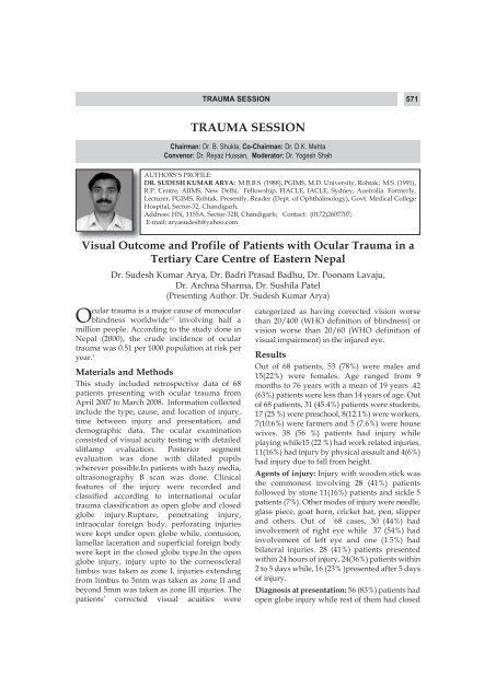
578 AIOC 2009 PROCEEDINGS1. World Health Organisation “blindness and VisualDisability – Part III of VII: OtherLeading Causes Worldwide” (February) Fact SheetN 144; World Health Organisation; 1997.2. Pieramici DJ,Sternberg P Jr,Aaberg Tm Sr,BridgesWZ Jr,et al “A system of classifying mechanicalinjuries of the eye (globe). The Ocular TraumaClassification Group”. Am J Ophthalmol1997;123:820-31.3. Kuhn F, Maisiak R, Mann L, Mester V, Morris R,Ocular emergencies are significant causevisual impairment in most parts of theworld. 1 The extent of vision loss from the traumaand other ocular emergencies ranges from mildto complete loss of vision. 2,3 Eye traumasconstitutes the bulk of eye emergency visits butthere are several other conditions which makethe reason for such visits.There have been studies about the clinicalfeatures and management of the eye emergencycases but data about the etiology andcircumstances of the cases seen in eye emergencyare meager if not lacking. 4 Researchers haveclassified etiology and have tried to define thepreventive steps for the various eye casualties.Such data are available in developed world 5 butvery few reports have come out of our country. 4Present study has tried to provide a fact file onocular emergencies in a tertiary eye care hospitalin eastern <strong>India</strong>. The aim of this study was to findout common conditions seen in the eyeemergency room. It has also tried to describe thetime distribution of the emergency visits bypatients with various conditions so that anassociation, if any could be established betweenthem.Materials and MethodsIt was a retrospective, observational and recordsbased study. The records from the emergencydepartment (ED) were analyzed from January2007 to December 2007. Weekdays were definedas from Monday to Saturday. Calculation wasReferencesOcular Emergencies: A Fact FileWitherspoon CD.” The Ocular Trauma Score(OTS)”. Ophthalmol Clin North Am. 2002;15:163-5.4. Sternberg P Jr, de Juan E Jr, Michels RG, AuerC.”Multivariate analysis of prognostic factors inpenetrating ocular injuries.” Am J Ophthalmol 1984;98:467-72.5. Pieramici DJ, Eong KG, Sternberg P Jr, MarshMJ.”The prognostic significance of a system forclassifying mechanical injuries of the eye (globe) inopen globe injuries.” J Trauma. 2003;54:750-4.Dr. Gautam Bhaduri, Dr. Rudra Prasad Ghosh, Dr. Chandra Nath ChaudhuriDr. Subhrangshu Sengupta, Dr. Kumar Saurabh, Dr. Swati Agarwal,Dr. Rajesh Kumar Mishra, Dr. Somnath Mukhopadhyay(Presenting Author: Dr. Rudra Prasad Ghosh)done for days and for different shifts like 2PM to8PM, 8PM to 2AM and 2AM to 8AM excludingthe ED visits during the morning hours whensimultaneous out-patient department (OPD) wasrunning. Separate calculations were done forSundays including the 8AM to 2PM shift whenOPD remains closed. Etiologies, age distributionof patients and temporal trends of visits were thekey features analyzed in this study.Statistical AnalysisData obtained were analyzed using SPSS 9.0.0,SPSS Inc. USA. Prevalence of common ocularconditions was enumerated and various charts,diagrams were prepared to show the results.ResultsIn the duration of one year, emergencydepartment (ED) at the center saw 4137 patients.Excluding the Sunday morning (8AM to 2PM)session, the number of patients visiting the EDwas 3709 while 428 patients attended theemergency service on Sunday morning session.The male female ratio of patients was 3.47:1. Onweekdays maximum number of patients arrivedduring 2PM to 8PM session while on Sundaysmaximum influx was seen during 8AM to 2PM.[Figure 1] The last three months of the year sawslightly more attendance (997; 26.88%) at the EDthan first (889; 23.96%) second (879; 23.69%) orthird (944; 25.45%) quarters.A maximum of 966 (23.35%) patients belonged to21-30 years age group followed by the age group
<strong>TRAUMA</strong> <strong>SESSION</strong>579of 31-40 years and < 10 years respectively (873;21.10% and 615; 14.86% respectively). Attendanceof children less than 10 years of age showed twinpeaks in the months of January and November.During different hours of weekday, children ofless than 10 years of age attended ED during 2PMto 8PM in maximum number (303; 49.26%).Ocular trauma (2711; 65.53%) was the mostcommon condition seen at the ED followed byinfective conditions (957; 23.13%) andmiscellaneous causes (469; 11.33%).Miscellaneous group included complicatedcataract, glaucoma, follow up cases and others.Foreign body injury (991; 23.95%) was theleading single cause of eye emergency visit. Blunttrauma (761; 18.39%) conjunctivitis (613; 14.81%)and globe rupture (350; 8.46%) followed in thatorder as other common causes. Foreign body andblunt trauma cases showed peak in the monthsof April and March respectively. Globe rupturecases had double peaks in the months of Marchand November. Most cases of conjunctivitis wereseen during autumn season i.e. in September andOctober. Cases of corneal ulceration also haddouble peaks in the months of June andDecember and there was an increasing trendtowards the end of the year. Most glaucoma caseswere seen in the months of March and Februaryrespectively.DiscussionPresent study has shown that ocular trauma was1. Parver L. Eye trauma. The neglected disorder. ArchOphthalmol 1986;104:1452-3.2. McGwin G Jr, Xie A, Owsley C. Rate of eye injury inthe United States. Arch Ophthalmol 2005;123:970-6.3. Nash EA, Margo CE. Patterns of emergencydepartment visits for disorders of the eye and ocularFigure-1: Number of patients on weekdaysReferencesthe most common condition seen at theemergency department (ED). Majority of patientswere aged between 21-30 years. Male to femaleratio was 3.47:1. These findings are in keepingwith those of Kaimbo et al 6 and Edward. 7 In thisstudy the maximum influx in ED was during lastthree months of the year. Increased outings andsocial contact in the festive season during thesemonths might be the reason. The under-10 agegroup as the third largest age group denotes thatits not that only outdoor activities are responsiblefor trauma but trauma or other emergentconditions can also develop at home.Like Verma et al 4 but unlike Tocino et al 8 we havefound that traumatic cases were the main reasonbehind emergency visits by patients. Tocino et al 8had found infective cases as the main cause.Similar to Verma et al 4 and Pokhrel-Loftus 9 fromAmerica; foreign body injury was the mostcommon condition among the ocular traumas.Present study has tried to elucidate a timedistribution of disease conditions seen at eyeemergency. The finding of more globe rupturecases during March and November stresses thatocular trauma is more common during the localfestive season (Holi and Durga pooja). Finding ofmaximum cases of conjunctivitis in autumnseason may help to keep the eye emergency wellprepared in advance. Time distribution ofcommon ocular emergency conditions will helpin proper planning and utilization of resources atthe emergency department (ED).adnexa. Arch Ophthalmol 1998;116:1222-6.4. Verma L. Arora R. Tewari H.K. Khosla P.K. Eyecasualty department. J. Ro Soc Medicine1994;87:217-8.5. Ngo CS, Leo SW Industrial accident-related ocularemergencies in a tertiary hospital in Singapore.
580 AIOC 2009 PROCEEDINGSSingapore Med J 2008;49:280-5.6. KaimboWA, Kaimbo DL, Spileers W. Ocularemergencies in Kinshasa (Democratic Republic ofCongo). Bull. Soc. belge Ophtalmol. 2002;284:49-3 .7. Edward RS. Ophthalmic emergencies in a districtgeneral hospital casualty department. Br JOphthalmol 1987;71:938-42.8. Tocino HS, Ferreiro AG, Cortiñas DI, Alonso JG,Muñoz MF.Epidemiological study of ocularemergency in general hospital. Arch Soc EspOftalmol. 2004;79:425-1.9. Pokhrel PK, Loftus SA. Ocular emergencies. AmFam Physician 2007;76:829-6.AUTHORS’S PROFILE:DR. SATYAN DEKA: M.B.B.S.(’91), Guwahati Medical College, Guwahati University; M.S.(‘97), Assam Medical College, Dibrugarh University; Vitreo-Retina fellowship (2000), MedicalResearch Foundation, Chennai; DNB (2004), National Board of Examination, New Delhi.Recipient of Young Investigators award at SERI, ARVO (2003), Singapore. Presently, Head,Vitreo-Retina Service of Sri Sankaradeva Nethralaya, Guwahati.E-mail: drsatyen@hotmail.comBomb Blast Injury: Clinical Scenario and Management Outcomein Ophthalmic (Posterior segment) PracticeDr. Satyen Deka, Dr. Harsha Bhattacharjee, Dr. Manab Jyoti Barman,Dr. Pareneeta Sachdev(Presenting Author: Dr. Satyen Deka)Ocular injury of bomb blast is becoming a newchallenge to the ophthalmic community asthere is no specific protocol to tackle thisupcoming threat. The eyeball measures 1/375 ofthe total body surface area and only 0.1% of theerect frontal silhouette. Despite this very smallarea of exposure, ocular blast injuries are seen incivilians and soldiers. The damage is usually dueto fragmentations that cause penetrating injuriesand to the explosive force that causes concussionof the eyeball. The fragments may come from theexplosive itself or from the environment. Theextent of injury from these penetrating fragmentsvaries according to their size and velocity, thedepth of penetration and the site of impact. Upto 10% of all blast survivors have significant eyeinjuries. These injuries involve perforations fromhigh-velocity projectiles, can occur with minimalinitial discomfort, and present for care days,weeks, or months after the event. Symptomsinclude eye pain or irritation, foreign bodysensation, altered vision, periorbital swelling orcontusions. Findings can include decreasedvisual acuity, hyphema, globe perforation,subconjunctival hemorrhage, foreign body, or lidlacerations. Present study was done to highlightthe clinical presentation and treatment outcomeof bomb blast injury with special reference toposterior segment trauma.Materials and MethodsA total of 14 patients of ocular blast injuryattending the vitreoretina clinic of a tertiary eyecare center of North East <strong>India</strong> during the periodof April 2006 to May 2007 were included in thisprospective, consecutive case series. Detailedophthalmic evaluation included-1. Relevant history of injury: Duration, anyprimary treatment done before referral,location and distance from blast site etc.2. Best corrected visual acuity (BCVA) usingSnellen’s chart or E chart. Perception of light(PL) and projection of rays (PR) checked ingrossly damaged eyes.3. Slit lamp biomicroscope evaluation, pupillaryevaluation, intraocular pressure (IOP) checkand indirect ophthalmoscopy done.4. B scanUSG done in cases where fundus wasnot visible and also intraocular foreign body(IOFB) suspected.5. VEP done if traumatic optic nerveinvolvement suggested by clinicalexaminationSurgical intervention included Primary repair upto full range of vitreous surgery including IOFBremoval and silicone oil or gas tamponade ifrequired. Patients were followed up on next day,
<strong>TRAUMA</strong> <strong>SESSION</strong>581one week and one month post operatively. Dataanalyzed using statistical test.ResultsThere were, in total, 14 cases (22 eyes) with ocularblast injuries. The ages of the casualties rangedbetween 7-70 years (mean age = 34.5 years).Cases attended to our center within 1-21 daysafter the blast injury. Binocular involvement wasseen in 8 cases (57.14%) out of 14 patients.Vitreous hemorrhage was present in all 22 eyes(100%), intraocular foreign body (IOFB) wasnoted in 8 eyes (36.36%), rhegmatogenous retinaldetachment was present in 5 eyes (22.72%), largescleral laceration in 3 (13.64%) andendophthalmitis in 2 (9%) eyes. Presenting visionranged from 6/24 to No PL and final visionranged from 6/6 to No PL. Surgical successachieved in >80% cases. Surgical procedure ofprimary sclerocorneal repair with or withoutforgein body removal done in 6 eyes. Sixteen eyesunderwent vitrectomy surgery with or withoutIOFB removal. Silicone oil or gas tamponaderequired in majority of cases. Significant visualrecovery noted in cases requiring Vitrectomysurgery. Repeat surgery was required forrecurrent retinal detachment and also to removethe silicone oil.Table-1: Presenting Vision in injured eyesVisual acuity No. %6/24 to 6/60 5 22.7
582 AIOC 2009 PROCEEDINGSinjuries of the iris, conjunctival and cornealforeign bodies, and corneal and/or scleralperforations were the most frequent types ofinjuries. In our study cases having posteriorsegment involvement were taken mainly and wealso observe high globe rupture incidence.Nearly half the cases with perforations also hadintra-ocular foreign bodies in a study by Boparaiet al. In our study the incidence of IOFB was1. Tariq FB et al. Frequency and causes of bilateralocular trauma. JCPSP 2007;17:679-82.2. Negussie Z. Blast Injuries of the Eye in Ethiopia. JComm Eye Health 1997;10:40-2.3. Boparai MS, Sharma RC. Ocular war injuries. Ind JOphthalmol 1984;32:277-9.For the management of rhegmatogenousretinal detachment, different surgicalmethods are available; and the choice of method,along with its technical specifications, variessignificantly between surgeons and centers.Relatively good anatomical success rates can beachieved with various techniques in cases ofrhegmatogenous detachment withoutcomplications, but following trauma, multiplefactors including the location of holes or tears,proliferative vitreoretinopathy, phakic status,retinal pigment epithelial viability and collateraltissue damage makes surgical results highlyunpredictable. Comparison of the surgicaltechniques to reach a conclusion regarding thesuitability of a particular type of surgery or stepof the surgery will be difficult to providetrustable results because of multiple factors. Butan overall view of a specific data can give somevaluable details when comparing. In this serieswe attempted to analyze and compare thetamponade effect of an agent (silicone oil andC3F8) to conclude which will provide betteranatomical success rate.References36.3% which is quite high. The study by Boparaiin 1984 indicated that lot of eyes were lost forwant of vitreous surgery.Surgical outcome wasencouraging (81.25%) in our study wherevitrectomies with multiple procedures wereperformed for 16 eyes. We found significantvisual recovery in our series. Conclusion: Ocularblast injury present in different mode and theresult of vitreoretinal surgery is encouraging.4. Quere MA, Bouchat J, Cornand G. Ocular blastinjuries. Am J Ophthalmol 1969;67:64-9.5. Bellows JG. Observations on 300 consecutive casesof ocular war injuries. Am J Ophthalmol1947;30:309-23.Comparison of Silicone Oil and C3F8 Tamponade in RetinalDetachment Associated with Closed Globe Injuries Managed withPars Plana VitrectomyDr. Ashad Sivaraman, Dr. Abhijith Datta, Dr. S. Natarajan, Dr. Priyanka Doctor,Dr. Dharmesh Kar, Dr. Soumen Mondal(Presenting Author: Dr. Abhijith Datta)Materials and MethodsA retrospective chart review was undertaken onpatients admitted with ocular and orbital traumato Aditya Jyot Hospital Pvt Ltd, a premiertertiary eye care centre in Mumbai, western<strong>India</strong>, over a period of six months.The study was approved by the Hospital EthicsCommittee and was approved by the localIndigenous representatives. Records wereidentified by computer search using equivalentcodes from the International Classification ofDisease (ICD) ninth and tenth revisions (ICD-9-CM: 1 January 1996–30 June 1999; ICD-10 AM 1July 1999–30 November 2002). Records from 38patients thus identified were reviewed for openorclosed-globe injury, as classified by the OcularTrauma Classification GroupData were recorded for patient demographics,injury demographics, initial assessment, surgicalintervention, initial and final best-correctedvisual acuity (VA) (from last recorded chart entryor at 6weeks review, whichever is later.) primaryand secondary repair, adjuvant treatment, andfollow-up. First data was classified on the basis
<strong>TRAUMA</strong> <strong>SESSION</strong>583of closed globe injuries and penetrating injuries.The closed globe injuries were analysed in detailagain for the initial visual acuity, anteriorsegment findings including corneal aberrations,anterior chamber flare and cells, hyphema, angleevaluation, iridodonesis, iridodialysis, traumaticmydriasis, lens subluxation, vitreous herniation,vitreous haemorrhage, retinal detachment, areaof detachment, retinal dialysis, pre retinal andretinal haemorrhage, Berlins odema, choroidalhaemorrhage, choroidal rupture.Those cases which underwent posterior segmentprocedure was done by a single surgeon who iswell experienced in managing such cases andanterior segment managed by 2 differentsurgeons who is also well experienced in doingsuch procedures. Vision was taken 6 weeks postoperatively and was not taken before that in allthe cases operated following ocular trauma.Grouping of data in certain categories wasnecessary for statistical analysis, which wasachieved by Windows excel.ResultsThere were 38 eyes of 38 patients who visited thehospital in 6 months with the history of oculartrauma. Out of the 38 cases 14 eyes were withclosed globe injuries and 24 eyes with open globeinjuries and there was only one female patientand all others were males which represented astatistically significant gender difference. Amongthe14 patients with closed globe injuries twowhere in above 51 years age group and two inbelow 20 years age group. Majority of thepatients, (11) were in the working age groupbetween 21 and 50 years. The right and left eyeswere affected approximately equally in ourseries.Five of the 38 patients who suffered eye traumawhere following cracker injury. 8 patients metwith the injury following road traffic accidentsand 2 got injured while traveling in crowdedtrain due to hit by hand of fellow passenger.There were 6 cases of sports injury and 5 of themwhere either playing or watching cricket. Therewere 4 patients who got injured by their ownbroken spectacle and 3 of them got it whileplaying.Injuries were significantly more common on thefestival season of Deepavali. There was noassociation with alcohol documented in any ofthe cases.Out of the 14 cases with blunt trauma, 3 weredue to road traffic accident, 4 where due tocracker injury, 4 due to sports injury, and 3 dueto other reasons.Of these 14 cases 9 developed RhegmatogenousRetinal detachment. These cases were manageddepending on the presence of vitreoushemorrhage, extent of the holes, presence ofproliferative vitreo retinopathy, duration ofretinal detachment etc.Scleral buckling and C3F8 tamponade was donein two cases. In four cases scleral buckling wasdone along with pars plana vitrectomy and C3F8tamponade. In two cases parsplana vitrectomyalone with C3F8 tamponade and in one casescleral buckling was done with parsplanavitrectomy and silicone oil tamponade.In all the nine cases in which retina was settledon the table without complications, retinaremained attached till the end of the follow up in5 eyes (55.6%) and developed re detachmentwithin 6 weeks of the procedure in 4 eyes(44.4%).Among the five anatomically successful cases(55.6%) following the first surgery itself, twocases underwent scleral buckling with pars planavitrectomy and C3F8 tamponade, two cases withscleral buckling and C3F8 tamponade and onecase with pars plana vitrectomy and silicone oiltamponade.In the four cases where re detachment occurred,2 cases underwent scleral buckling with parsplana vitrectomy and C3F8 tamponade andanother two cases with pars plana vitrectomyand C3F8 tamponade .<strong>All</strong> the cases with re detachment were subjectedto resurgery with in one week of noticing the redetachment. Among these, two cases in whichscleral buckling was not done in the first surgery,underwent scleral buckling and internaltamponade with silicone oil. In two cases wherescleral buckling was already done, pars planavitrectomy with silicone oil tamponade wasperformed. The surgeon was able to settle theretina on the table with removal of proliferativemembranes and internal drainage of sub retinalfluid. No new retinotomies were required in anyof these cases and endolaser was applied aroundthe tears before injecting silicone oil. After the
584 AIOC 2009 PROCEEDINGSsecond surgery retina was attached till the end offollow up in all these cases.On evaluation of the net result of surgicalintervention (including the first surgery andsecond surgery ) in this series as retinalreattachment rate it is found that followingsilicone oil tamponade the retinal reattachmentrate was 100% and following C3F8 tamponadewas 44.4%.DiscussionComparison of success rate in retinalreattachment sugery to arrive at a conclusion isdifficult because of the multiple factors involvedin deciding the outcomes. The factors includelocation of holes or tears, proliferative vitreoretinopathy, phakic status, retinal pigmentepithelial viability etc. The surgical interventionalso differs from case to case, surgeon to surgeonand from institution to institution which makecomparison still more difficult.In retinal reattachment surgeries the tamponadeused has lot of significance.The agent used fortamponade should have qualities like opticalclarity, long term chemical and Physicalstability, biological Inertness and shouldpermanently support retina against retinalpigment epithelium at all places. But till datesuch an ideal Vitreous substitute is not availableVarious substances have been tried as Vitreousreplacement, including Ringer Lactate Cleardonor Vitreous, cerebrospinal fluid, nonexpanding gases like air,O2,N2 and Expandinggases like Perfluorocarbon Gases.Now the commonly used agents are ringerlactate, air, expanding gases, Silicone Oil, PFCLetc. The short life of air in the eye has beenovercome with the use of expanding gases suchas SF6 and per fluorocarbon gases, havingvarious expansive capability and longevity.These gases have been used for restoration ofintra ocular volume, following vitrectomy forproliferative vitreoretinopathy, giant retinal tear,combined retinal detachment, macular hole,posterior breaks, etc. It has also been used totamponade breaks in pneumatic retinopexy, andin cases with ‘fishmouth’ phenomenon.We noted better anatomical success (100%)incases treated with Silicone oil as opposed toC3F8(44.4%) and this difference was statisticallysignificant. The retrospective nature was a minorlimitation; however, data that were lacking havebeen accounted for and do not impactsignificantly on the major findings of the study.These results are useful in adding to the body ofliterature describing trauma as a 'well-defineddisease state'. Information will also assist theophthalmologist in counselling of patients aboutprognosis and determining the most appropriatetransfer and surgical times as well as appropriatesurgical management.Silicone oil affords better tamponade and greaterchance of anatomical success in cases of retinaldetachment following closed globe injury.Multivariate analysis and larger case seriesevaluations required for furtherconfirmation.Analysis of 74 Open Globe Injuries (Needed Vitreo-RetinalIntervention) and Its Correlation with Ocular Trauma ScoreDr. Girish Arun Gadre, Dr. Vivek Som, Dr. Shreya Vaijwade, Dr. Preetam Dedia(Presenting Author: Dr. Girish Arun Gadre)Fifty years ago, there was very little to offer thepatient with severe injury of the posteriorsegment. Removal of the eye, in the case of severeopen globe injury, was often recommendedbecause of the perceived high-risk of sympatheticophthalmia. 1 During the last 2 decades, vitreoretinalsurgical techniques having become morerefined with wide angle viewing system,different tamponading agents, intravitrealantibiotics and IOFB removal techniques, madepossible, reconstruction and salvaging eyes withambulatory vision in many cases.It is critically important for patients andophthalmologist to have/reliable informationregarding expected outcome and prognosis ofinjured eye. Ferenc Kuhn et al 2 developed amethod by which we can predict functionaloutcome of a serious eye injury with reasonablecertainty. We correlated our analysis with OTSwhich was comparable with some differences.
<strong>TRAUMA</strong> <strong>SESSION</strong>585Materials and MethodsWe analyzed 74 eyes having open globe injuriesthat required posterior segment interventionand divided into four groups based onBirmingham Eye trauma terminology (BETT)3.This retrospective Observational study wasconducted at Vivekanand Netralaya, Miraj, fromJune 2006 to Dec 2007.Those patients who did notrequire posterior segment surgical interventionwere excluded.<strong>All</strong> patients underwent complete generalophthalmological examination prior to surgicalprocedure. Detailed history of trauma andprimary surgical intervention was done.Ultrasound B-scan, CT scan or X-ray orb it wereperformed in appropriate cases. In all casesStandard 3 port parsplana vitrectomy wasperformed with wide angle EIBOS system with20G cutter. Encircling band was placed in 22cases. Posterior vitreous detachment wasinduced in most cases except in fulminantendophthalmitis cases. Cataractous lens wasmanaged either by pars plana lensectomy withor without sparing anterior capsule; fewunderwent SICS. Retained intraocular foreignbodies were removed by IOFB forceps in 20 casesand intraocular magnet was used in two. <strong>All</strong>cases presented after 24 hours of trauma weregiven prophylactic vancomycin anddexamethasone at the end of surgery. Those caseswho had endophthalmitis received intravitrealVancomycin (1 mg) and ceftazidime+dexamethasone (2.25 mg) in clinically orlaboratory suspected bacterial endophthalmitisand amphotericin B in fungal endophthalmitis.Endolaser and appropriate endotamponade wasused in appropriate cases.Postoperatively patients were followed forminimum 6 months with detailed ocular exameach visit. Second intervention in the form ofresurgery for RD, silicon oil removal, secondaryIOL implantation, repeat intravitreal antibioticswas done in appropriate cases. None of ourpatients had undergone Optical keratoplasty.Each patient was given raw points as per OTScriteria and divided into BETT groups andTable-1Parameters PT (n=40) IOFB (n=22) PF (n=7) RPT (n=5)Age(yrs) 27.725 33.72 31.57 38.2M:F ratio 28:12 21:1 7:0 3:2Type of injury Thorn or wooden Hammer chisel or Iron wire or Sugar canecommonest stick 42.5% stone 68.18% needle 71.4% stick in 60%Average OTS 54.92% 55.09% 41.57% 38.4%endophthalmitis 12(30%) 11(50%) 2(28.5%) 1(20%)Retinal detachment 14(35%) 9(40%) 4(57%) 1(20%)Recurrent RD 3 1 1 0Preop Visual acuity < or = HM 34(85%) 19 (86.36%) 7(100%) 5(100%)>6/60 0 2 0 0Final visual acuity < or = HM 5(12.5%) 4(18.1%) 1(14%) 1(20%)>6/60 13(32.5%) 8(36.3%) 3(42.8%) 2(40%)Table-2: Comparison between our analysis and F.Khun et al as per OTS raw point scoreraw OTS No light Pl or hand 1/200-19/200 20/200-20/50 >or =20/40points perception motionKuhn Our Kuhn Our Kuhn Our Kuhn Our Kuhn Ouret al results et al results et al results et al results et al results0-44 1 74% 10.5% 15% 21.5% 7% 52.6% 3% 10.5% 1% 5.2%45-65 2 27% 0% 26% 27.3% 18% 38.6% 15% 30% 15% 4.5%66-80 3 2% 0% 11% 0% 15% 27.3% 31% 36.4% 41% 36.4%81-91 4 1% - 2% - 3% - 22% - 73% -92-100 5 0% - 1% - 1% - 5% - 94% -Bold letters are showing significant difference in our result and F Kuhn et al.
586 AIOC 2009 PROCEEDINGScompared with OTS criteria for calculating thelikelihood of final visual acuity and our finalvisual acuity.ResultsSeventy four patients were analyzed. Averageage 30.5, 40% were children below 18yrs. M: Fratio was 58:16. 54%(40) had penetrating injury(PT), 29.7%(22) had retained intraocular foreignbody (IOFB), 9.5% (7) had perforating injury (PF),6.75%(5) had globe rupture (RPT). Averageduration of first intervention from day of traumawas 10.04 days (range 4hrs to 52days).Commonest cause of injury was hammer chiselor stone in 32.43% (24), followed by thorn orwooden stick in 27%(20).35.13 %(26) presented with endophthalmitis,37.83% presented with retinal detachment. 6.7%(5) with traumatic aniridia, 62.16%(46) hadtraumatic cataract, 3 had posterior dislocationand 2 had IOL extrusion.DiscussionIn this study categorization and evaluation of theoutcome was based on the Ocular Trauma Scoreintroduced by Kuhn et al. the cases were groupedon the basis of the OTS calculated according tothe variables at presentation. In our study out of74 eyes none were included in OTS category 4and 5; this may be because of high incidence ofendophthalmitis and retinal detachment and1. de Juan E Jr et al. Penetrating ocular injuries. Typesof injuries and visual results. Ophthalmology1983;90:1318.2. Ferenc Kuhn et al. Birmingham Eye traumaterminology ( BETT): terminology and classificationof mechanical eye injuries. Ophthalmol clin N Am2002:139-43.3. Kuhn F, Maisiak R, MannnL et al. The oculartrauma score (OTS). Ophthalmolo Clinic of NorthAmerica 2002;15:163-5.Endophthalmitis following penetrating oculartrauma is fairly common entity and forms adistinct subset of intraocular infections .Manypoor initial visual acuity. In comparison to theOTS study and the results reported by MelihUnal, Ali Adayin et al. 4 the final visual outcomebased on the OTS category of our study showeda lower incidence of no PL and a higherpercentage of cases achieving 1/200-20/50 visualacuity whereas the percentage in the other twocategories were comparable. Hammer onmetal/stone accounted for 68.18% of IOFB in ourstudy which is comparable to studies of JacksonColeman et al. 5 and Percival 6 and Cridland et al 7in which hammer on metal accounted for 63%and 60% respectively.The majority of the patients were young malechildren / adults because of the prevalent socialworking pattern. Average interval betweeninjury and time of intervention was 10.04 days (4hrs – 52 days) which highlights the inequitableapproachability of the health facilities as well asthe lack of awareness among the people and thereferring GP about significance of earlyintervention with regards to the visual outcome.Hammer chisel injury while working withoutprotective glasses was the common injury.The present study was conducted to assess theprognostic value of OTS in our present workingscenario. However the findings and conclusionsshould be evaluated keeping in mind theshortcomings and limitations inherent toretrospective studies.References4. Melih Unal, et al. Validation of the OTS forintraocular foreign bodies in deadly weapon relatedopen globe injuries. Ophthalmic surgery, lasers andimaging 2008;39:121-4.5. Jackson Coleman et al. Management of IntraocularForeign Bodies; Ophthalmology 1987;12:1647-53.6. Percival SPB. A decade of intraocular foreignbodies. Br. J Ophthalmology 1972;56:454-61.7. Cridland N. et al Intraocular foreign bodies. Proc RSoc Med 1967;60:598-600.Post Traumatic Endophthalmitis : Profile, Management, andOutcomeDr. Rajkumar Sharma, Dr. Lokendra Tyagi, Dr. Meghna Dayal(Presenting Author: Dr. Lokendra Tyagi)factors like the age of the patient, mode of injury,virulent organism govern the visualoutcome.Several large studies have been done on
<strong>TRAUMA</strong> <strong>SESSION</strong>587postsurgical endophthhalmitis but fewer onposttraumatic endophthalmitis especially inpaedriatic age group.Herein we report our study, the paedriatictraumatic endophthalmitis in comparison toadult traumatic endophathalmitis regardingProfile and Outcome.Materials and MethodsWe retrospectively analyzed the medical recordsof 53 consecutive cases{paedriatic(n=27), andadult(n=26)} of post-traumatic endophthalmitismanaged at the RM Sahai Memorial Institute ofOphthalmology between Jan 2007- Mar 2008Paediatric age group was taken as ≤ 14 years.Information on demographic characteristics ofthe patients, the time of diagnosis ofendophthalmitis following the precipitatingevent, the presence of associated injuries, agentscausing trauma were retrieved from the medicalrecords. <strong>All</strong> patients had been subject to acomprehensive ocular examination atpresentation, which included visual acuityexamination at presentation and refraction(whenever feasible), slit lamp examination ofanterior segment, and posterior segmentexamination with an indirect ophthalmoscope. B-scan ultrasonography was performed in allcases,irrespective of media clarity, to assess theassociated injuries and foreign bodies. Thediagnosis of endophthalmitis was based onsubjective symptoms such as pain, anddiminished vision in the presence of objectivesigns such as lid edema, anterior chamberreaction, hypopyon, and/or vitreousexudates.Culture positivity was not taken as astringent criterion for diagnosis.The type of surgical intervention was decided bythe operating surgeon based on several factors,including the severity and duration of theinfection, type of predisposing injury, andpresence of associated injuries such as corneal orscleral tear, and ultrasonography results. <strong>All</strong>patients underwent vitrectomy and intravitrealinjection, with additional procedurecorneal/scleral tear repair,lensectomy, cryo,encirclage, silicon oil injection, IOFB removalwherever required. Functionally successfulvisual acuity was defined as >3/60.ResultsIn paedriatic age group the most common age ofpresentation was 7-10 years whereas in adults, itwas 31-35 years with predominance of malegender in both the groups.The most common mode of injury in childrenwas by vegetative material (wooden stick-7,babool thorn-5, bow and arrow-5) and in adults itwas metallic foreign body (n=9).Time interval between trauma and thepresentation to the hospital was more inpaedriatic age group(20-25days) as compared toadults (10 -15 days).Visual outcome was worse in case of children(VA3/60 in 22%, pthisical in14.8%), as compared to adults (3/60 in 34.6% and pthisical in 7.7%).DiscussionIn our study ambulatory vision (VA>3/60), wasachieved in 22% and 34.5% cases in children andadults respectively which was less than the otherstudies.Paediatric Adults>3/60 >3/60Subina Narang, Amod Gupta 45.95%et al IJO 2004;52:29-34Alfaro DV et al 66.7%BJO 1995 oct;79:888-891Abu-el-asar et al 44%EJO 1999 Jan-March;9:21-31Alfaro et al Retina 1994 25%Weinstein GS et al 23%Ann Ophth 1979 Jun;11:935-43OUR STUDY 22.2% 34.5%The accountable poor prognostic factors inchildren included poor presenting visual acuity,delayed presentation, vegetative mode of injury,rural setting and poor hygiene. Controlling ofinfection and attaining long term anatomical eyeintegrity means no less success.Prevention of trauma especially in children is ofparamount importance realizing the irreversiblenature of visual loss and psychosocial aspectsinvolved.
588 AIOC 2009 PROCEEDINGSClinico-Microbiological Profile and Visual Outcomes inPosttraumatic Endophthalmitis at A Tertiary Eye Care Centre inCentral <strong>India</strong>Dr. Sumeet Malhotra, Dr. Deepshikha Agrawal, Dr. Anil Kumbakar(Presenting Author: Dr. Sumeet Malhotra)Endophthalmitis is not only a dangerous anddevastating complication after intraocularsurgery but also after trauma.There arenumerous reports in the literature on spectrum,clinical profile, microbiology, anatomical andvisual outcomes in posttraumaticendophthalmitis but no reports from central<strong>India</strong>. 1, 2 We report the clinical andmicrobiological profile and visual outcomes ofpost-traumatic endophthalmitis at a tertiary eyecare centre in central <strong>India</strong>.Materials and MethodsA retrospective review of medical records of 38patients with different types of endophthalmitiswas done from Feburary 2005 to Feburary 2007.From the clinical records of the patients detailedhistory, duration and presenting symptoms,.visual acuity, anterior segment examination andvitreoretinal findings were recorded andanalysed through a computerized database.Standardized echography was done in patientswhere viewing of the fundus was not possibleand to detect extent of vitreous involvement,retinal detachment and choroidal detachment.<strong>All</strong> patients underwent vitreous biopsy andprompt parsplana vitrectomy, intraocular,andparenteral systemic antibiotics on the day ofpresentation. The intraocular antibiotics were acombination of vancomycin (1mg/0.1ml),ceftazidime (2.25mg/0.1ml) and dexamethasone(0.4mg/0.1ml). Lensectomy was done in patientswhere lenticular opacification interfered invitrectomy. Additional vitreoretinal surgicalintervention in traumatic endophthalmitispatients was decided by the operating surgeonbased on several factors, including the severityand duration of the infection, type ofpredisposing injury, and the presence ofassociated injuries such as corneal or scleral tearand ultrasonography results. Associated injuriessuch as corneal or scleral tears and intraocularforeign body (IOFB) were dealt at the time ofprimary surgical intervention. Secondarysurgical interventions like repeat intraocularantibiotics, repeat vitrectomy, cataract surgerywith or without intraocular lens implantationand retinal reattachment surgery wereperformed. Gram, giemsa and KOH stains weredone. Undiluted vitreous samples were plated onblood agar, thioglycollate chocolate agar,sabouraud’s dextrose agar, thioglycollate brothfor detection of bacteria and fungi in all thevitreous samples. Kirby bauerdisc diffusionmethod was performed for antibiotic sensitivityon culture positive cases.A positive culture wasdefined as growth of the same organism on oneor more solid media or growth on a single culturemedium of an organism detected on initialsmear.The favourable or good visual outcome wasdefined as visual acuity of 20/400 or better atfinal follow-up. The favourable anatomicaloutcome was defined as clear media withattached retina at 3 months follow-up, withvisibility of third grade retinalvasculature;unfavourable outcome was definedas failure to achieve clear media at 3 monthsfollow -up, or signs of phthisis bulbi or chronichypotony resulting in secondary changes.Fisher’s exact test was performed to test thesignificance of categorical variable.ResultsThe patients with traumatic endophthalmitis hadthe mean age of 21.68±14.83 (range 3-60 yrs) with34(89.4%) males and 21 (55.2%)right eyeinvolved. The mean duration of presentationafter injury was 7.9 days with 13 (34.2) patientspresenting to the hospital after 7 days and thecommonest type of injury was duringhammering an iron object. The presenting visualacuity was
<strong>TRAUMA</strong> <strong>SESSION</strong>589in 31(81.5%) patients. Thirteen cases were culturepositive for 15 isolates, The isolates identified inthe cultures were s.epidermidis in 5(33.3%),pseudomonas 4(26.6%), bacillus 3(20%),nocardia, diptheroids and streptococcus 1 (6.6%)each. The mean follow-up was 35weeks (range24-68) weeks. The common drugs to whichorganisms were sensitive were ofloxacin (n=11)and chloramphenicol (n=11). Susceptibility toceftazidime and tobramycin was poor. Onunivariate analysis with Fisher’s exact test therewas no association of improvement of visualacuity with presenting visual acuity (p=0.12),time of presentation of patient to the hospital(p=1), hypopyon (p=0.32),presence of IOFB(p=1.0), negative cultures (0.27),agent of injury,patient, age:16-49yrs, (p=0.3) and sex (p=1.0) ofpatient and lensectomy (p=1.0). Final visualacuity ≥ 20/400 was achieved in 24 (63%) of thepatients at 6 months follow-up. 5 patients hadinoperable retinal detachments, two had phthisisbulbi and one eye was eviscerated; others hadpersistent vitritis and corneal decompensation.DiscussionThere are numerous reports on traumaticendophthalmitis in the literature but nolandmark study like endophthalmitis vitrectomystudy (EVS). 3 There is also extensive work donein <strong>India</strong>n subcontinent on posttraumaticendophthalmitis especially from north and south<strong>India</strong> but no reports from central <strong>India</strong>. If wecompare our results with <strong>India</strong>n studies 4,5 onposttraumatic endophthalmitis our results arecomparable with these studies. Das et al 4 havereported achievement of final visual acuity of ≥1. Briton GS, Topping Tm, Hyndiuk RA, Aeberg TM,Reeser FH, Abrams GW. Posttraumaticendophthalmitis. Arch Ophthalmol 1984;102;547-50.2. Nobe JR, Gomez DS, Liggett P, Smith RE, Robin JB.Posttraumatic and postoperative endophthalmitis:a comparison of visual outcomes. Br Ophthalmol1987;71:614-7.3. Endophthalmitis Vitrectomy Study Group. Resultsof the Endophthalmitis Vitrectomy Study: arandomized trial of immediate vitrectomy andintravenous antibiotics for the treatment ofpostoperative bacterial endophthalmitis. ArchOphthalmol 1995;113:1479-96.4. Das T,Kunimoto DY, Sharma S, Jajali S, MajjiAB,Rao TN, Gopinathan U, Athmanathan S, and20/400 in 63.9% in their series. In an anotherstudy by Vedantham etal 5 a final visual acuity ofbetter than 20/200 was achieved in 32% of caseson last follow-up. In post traumaticendophthalmitis previous western studies 1,2,6have reported achievement of a final visualacuity of better than 20/400 in 9-50% of patients.In our study visual acuity of ≥ 20/400 wasachieved in 63% of cases and we could not findany factor which significantly affectedimprovement in visual acuity as compared toother studies. 4,5 The differences in success ratesin various studies may be due to regionalvariation in microbiological spectrum andvirulence of organism. Similar to the otherstudies Gram-positive organisms (Staphepidermidis) were the commonest organismsimplicated in posttraumatic endophthalmitis.Antibiotic susceptibility testing of positivecultures revealed that the organisms weresensitive to vancomycin but resistant toceftazidime which is commonly used asintravitreal antibiotic for treatment ofendophthalmitis. This finding is interesting anddifferent from the series reported byVendantham etal. 5 The organisms were sensitiveto chloramphenicol (n=11) and ofloxacin (n=11),these drugs can be tried in the management ofposttraumatic endophthalmitis.In conclusion the anatomical and visual outcomein posttraumatic endopthalmitis is not dismal;prompt and appropriate intervention can salvage63% of the eyes. Futher prospective studies areneeded to evaluate prognostic factors, newertreatments and antibiotics in the management ofposttraumatic endophthalmitis.Referencesthe Endophthalmitis Research Group..Relationshipbetween clinical presentation and the visualoutcomes in the postoperative and theposttraumatic endophthalmitis in south central<strong>India</strong>. <strong>India</strong>n J Ophthalmol 2005;53:5-16.5. Vedantham V, Nirmalan PK, Ramasamy K, PrakashK, Namperumalsamy P. Clinico-microbiologicalprofile and visual outcomes of post-traumaticendophthalmitis at a tertiary eye care center inSouth <strong>India</strong>. <strong>India</strong>n J Ophthalmol 2006;54:5-10.6. Affeldt JC, Flynn HW, Jr, Forester RK, MandelbaumS, Clarkson JG, Jarus GD. Microbialendophthalmitis resulting from ocular trauma.Ophthalmology 1987;94:407-13.
590 AIOC 2009 PROCEEDINGSVisual Outcome Following Ocular Trauma – A RetrospectiveCross Sectional StudyDr. Sulatha V Bhandary, Dr. Vijaya Pai H, Dr. A. G. Chinnappa, Dr. Ruchi Tayal,Dr. Neetha Kir, Dr. Nishit Dhoka(Presenting Author: Dr. Sulatha V Bhandary)Ocular injuries are one of the ill managedfields in rural and semi urban <strong>India</strong>. A majorreason for this may be lack of tertiary care centersor the lack of expertise in medical professionals.Ocular injuries are the leading cause of visualdisability and blindness in children. 1,2 Open-globeinjuries are among the most serious types ofocular trauma.Kuhn and associates 3 described the ocular traumascore (OTS), a simplified categorical system forstandardised assessment and visual prognosis inocular injuries, in a large series of patients. Theyclaimed OTS provides a single probabilityestimate an eye trauma patient will obtain aspecific visual range by six months after injury.But at the same time they also accepted that OTSis meant to be a continually evolving scoringsystem to be used by the clinician to facilitatepatient counseling, treatment, rehabilitation, andresearch.We attempted to conduct a retrospective study inassessing the visual outcome after ocular traumaand also tried to find out whether OTS wascomprehensive enough to predict long-termvisual outcome.Materials and MethodsWe conducted a retrospective review of allpatients who presented to the Kasturba Hospital,Manipal between January ’07 and December ’07.Age, gender, type of injury and past ocularhistory was determined from each patient’smedical record. Forty-four patients presentingwith ocular trauma, who underwent admissionin the hospital were included in the study. Ofwhich 35 patients underwent immediate surgicalcare. 3 patients had to undergo surgical treatmenton a delayed basis for cataract extraction and 1patient for vitrectomy with scleral fixated IOL. 5patients were managed conservatively. 6 patientshad to be excluded, as they lost for follow up. 3patients could not be scored on basis of OTS asinitial visual acuity could not be assessed.<strong>All</strong> patients had experienced only ocular traumawithout accompanying body trauma, and noneof the patients in our series had suffered bilateralinjuries.Patients underwent immediate surgical care ifrequired. Patients were given adequateastigmatism correction with spectacles once thewound healed and aphakics were corrected withcontact lenses/sec IOL.The type of injury was classified according toBirmingham Eye Trauma Terminology. 4 Anumerical value (raw number) was assigned toeach of the following variables: A) initial visualacuity, B) rupture, C) endophthalmitis, D)perforating injury, E) retinal detachment, F)afferent pupillary defect. The OTS wasdetermined by calculating the sum of the rawpoints: A+B+C+D+E+F. If a specific variable wasnot present, its value was zero. Similar to the OTSstudy group, presenting visual acuities weredivided into five categories (1 to 5) as 20/40 ,20/200–20/50, 1/200–19/200, LP/HM and NLPin our study (Table 1 and 2). Certain numericalvalues rendered to OTS variables were convertedto OTS categories. The lowest possible OTS scoreis zero, whereas the highest is 100.The distribution of the 44 eyes were studied andthe percentage of eyes following and notfollowing the OTS proposed final visual acuitywere calculated. The section of patients whosuccumbed to injuries at workplace andelsewhere were also noted.Results1) Patients were in the age group ranging from 2-60 yrs. 2) Mean age of 31.8yrs.Male: Female ratiowas 13.7:1. 3) Penetrating trauma scored overblunt trauma in incidence, being 61.36 %.4)Workplace injuries accounted for 45.45% of thetotal. Rest was accounted to road trafficaccidents, and household accidents.The final visual outcome at 2 months were alsograded in the ocular trauma score Visual Acuitygrade, and was found that 2 patients ended upwith No Perception of Light, 31.8% had Visual
<strong>TRAUMA</strong> <strong>SESSION</strong>591Table-1: Time of Presentation Pre Op VisionPost Op VisionTotalNLP LP-HM 1/200-19/200 20/200-20/50 >20/40 Didn’t R/WTOP 24HRS 0 6 1 1 5 0 13Total 2 13 6 4 13 6 44Post Op VisionAcuity of Hand Movements to Counting fingersClose to face, 5 patients had 1/60-5/60, 4 had6/60-6/18 and 29.5% had Visual acuity >6/12, 6patients lost follow up.Only 14 of our patients complied exactly to theOTS proposed final visual acuity(31.81%). 10patients fell in the category one above or belowthe proposed(22.72%). So, on the whole 54.54%patients complied to or fell among any categoryone above or below. As per the ocular traumascore the patients were graded into prognosiscategories, wherein we noted that majority fellinto group 2 (59.0%).DiscussionMen are more prone to ocular injuries thanwomen, more so because of the nature of theoccupation. Workplace injuries accounted for amajor chunk of injuries. So, we must enhanceworkplace safety standards, protective gear, andalso employee education in order to preventthem.In the paediatric population, OTS calculationsmight have limited value as predictors of visualoutcome, was already mentioned in previousstudies. 4 Initial visual acuity (major criteria inOTS) was found to be the best indicator of postop vision by numerous studies in the past.Paul sternberg and Eugene et al 7 in their study ofprognostic factors using multivariate analysis,Table-2: Ocular Trauma Score * Post Op VisionNLP LP-HM 1/200-19/200 20/200-20/50 >20/40 Didn’t R/WOTS Not Assessed 0 0 0 0 1 2 30-44 2 0 1 0 0 0 345-65 0 10 4 3 6 3 2666-80 0 3 1 0 2 1 781-91 0 0 0 1 4 0 5Total 2 13 6 4 13 6 44Totalsaid that an initial visual acuity of 20/800 orbetter was the most important factor associatedwith favorable prognosis. David Williams andWilliam Mieler 8 found a p value of
592 AIOC 2009 PROCEEDINGS1 Maltzman HA, Pruzon H, Mund ML. A survey ofocular trauma. Surv Ophthalmol 1976;21:285–90.2 Niiranen M, Raivio I. Eye injuries in children. Br JOphthalmol 1981;65:436-8.3 Kuhn F, Maisiak R, Mann L, et al. The ocular traumascore (OTS). Ophthalmol Clin N Am 2002;15:163–5.4 Kuhn F, Morris R, Witherspoon D, et al. Astandardized classification of ocular trauma.Ophthalmology 1996;103:240–3.5 Visual predictive value of ocular trauma score inchildren.Y B Unver, N Acar, Z Kapran, T Altan.Beyo lu Eye Research and Training Hospital,Istanbul, Turkey.References6 Esmaeli B.A et al. visual outcome and ocularsurvival after penetrating trauma. British Journal ofOphthalmology 2008;92:1122-4.7 Paul Sternberg Jr, Eugene De Juan et al. Multivariateanalysis of prognostic factors in penetrating ocularinjuries. AJO 1984;98:467-72.8 Williams DF, Mieler WF, Abrams GW, Lewis H.Results and prognostic factors in penetrating ocularinjuries with retained intraocular foreign bodies.Ophthalmology 1988;95:911-6.9 Barr CC. Prognostic factors in corneosclerallacerations. Arch Ophthalmology 1983;101:919-24.