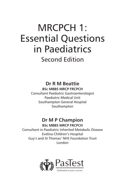
MRCPCH 1: Essential Questions in Paediatrics - PasTest
MRCPCH 1: Essential Questions in Paediatrics - PasTest
MRCPCH 1: Essential Questions in Paediatrics - PasTest
- No tags were found...
You also want an ePaper? Increase the reach of your titles
YUMPU automatically turns print PDFs into web optimized ePapers that Google loves.
ContentsContributorsIntroduction1. Cardiology 12. Child Development, Child Psychiatry and Community<strong>Paediatrics</strong> 133. Cl<strong>in</strong>ical Pharmacology and Toxicology 264. Dermatology 375. Emergency Medic<strong>in</strong>e 486. Endocr<strong>in</strong>ology 597. Ethics, Law and Governance 748. Gastroenterology and Nutrition 889. Genetics 10710. Haematology and Oncology 11711. Hepatology 12912. Immunology 14713. Infectious Diseases 15614. Metabolic Medic<strong>in</strong>e 16915. Neonatology 18216. Nephrology 19417. Neurology 20918. Ophthalmology 22019. Paediatric Surgery 22820. Respiratory Medic<strong>in</strong>e 24021. Rheumatology 25122. Statistics 265Index 279ivviii
11. CardiologyMultiple True–False <strong>Questions</strong>1.1 Muscular ventricular septal defects (VSDs) A do not require SBE prophylaxis B usually cause a heart murmur <strong>in</strong> the first day of life C if large, are closed by catheter procedures <strong>in</strong> 10% of cases D do not have conduction tissue runn<strong>in</strong>g on their <strong>in</strong>ferior marg<strong>in</strong> E if large, usually cause heart failure before the child is 4 days old1.2 Scimitar syndrome A is usually associated with a hypoplastic left lung B can be palliated by coil occlusion <strong>in</strong> the cardiac catheter laboratory C will show dextrocardia on the X-ray as a result of situs <strong>in</strong>versus D is associated with abnormal pulmonary arterial supply E is usually associated with abnormal radii1.3 On the first day of life, the follow<strong>in</strong>g may be found <strong>in</strong> neonateswith congenital heart disease: A a harsh pansystolic murmur with the diagnosis of ventricular septaldefect B severe cyanosis <strong>in</strong> unobstructed total anomalous pulmonary venousconnection C a harsh systolic murmur <strong>in</strong> transposition of the great arterieswithout associated defect D severe acidosis and poor pulses with hypoplastic left heart syndrome E severe cyanosis and acidosis <strong>in</strong> a baby with Down syndrome andatrioventricular septal defect1.4 The follow<strong>in</strong>g is true of persistent ductus arteriosus: A it is def<strong>in</strong>ed as persistence of ductal patency beyond 1 week afterthe date the baby should have been born B on auscultation, a cont<strong>in</strong>uous murmur <strong>in</strong> the right <strong>in</strong>fraclaviculararea is heard C it may present as heart failure with poor peripheral pulses D closure is usually undertaken <strong>in</strong> the catheter laboratory with coil ordevice at 1 year E if it is large, surgical ligation is recommended at 1–3 months
2CARDIOLOGY – QUESTIONS1.5 The follow<strong>in</strong>g statements about transposition of the greatarteries are true: A there is an association with coarctation of the aorta B arterial switch is the operation of choice, undertaken before2 weeks C the condition is detected antenatally <strong>in</strong> 50% of cases D presentation can occur upon closure of the ductus arteriosus E the arrangement of the coronary arteries is a major factor <strong>in</strong> determ<strong>in</strong><strong>in</strong>gthe success of the surgical repair1.6 The follow<strong>in</strong>g is true of Eisenmenger syndrome: A affected children are typically teenagers B it can be seen <strong>in</strong> children with Down syndrome C it is usually secondary to an untreated ventricular septal defect oratrioventricular septal defect D the pulmonary component of the second heart sound is quiet onauscultation E the ECG shows left ventricular hypertrophyBest of Five <strong>Questions</strong>1.7 You are asked to review an ECG of a baby on the <strong>in</strong>tensive careunit. The baby was well at birth, but soon became unwell andcyanosed. There was no heart murmur. ECG f<strong>in</strong>d<strong>in</strong>gs reveal asuperior axis, absent right ventricular voltages, and a largeP wave. What is the MOST likely diagnosis? A Complete atrioventricular septal defect B Tricuspid atresia C Critical pulmonary stenosis D Transposition of the great arteries E Total anomalous pulmonary venous connection (TAPVC)1.8 You are asked to see <strong>in</strong> cl<strong>in</strong>ic a 6-year-old girl with a diagnosis ofright atrial isomerism. Which one of the follow<strong>in</strong>g features wouldyou expect her to have? A Asplenia and a midl<strong>in</strong>e liver B Polysplenia C Two functional left lungs D T-cell deficiency E Trisomy 21
CARDIOLOGY – QUESTIONS 31.9 You are asked to review a child on the ward who is known tohave short stature and renal abnormalities. On exam<strong>in</strong>ation, shehas micrognathia and an ejection systolic murmur at the upperleft sternal edge. Her notes show that she has recently seen anophthalmologist. What is the MOST likely underly<strong>in</strong>g diagnosis? A Williams syndrome B DiGeorge syndrome C Alagille syndrome D Noonan syndrome E Left atrial isomerism1.10 A 1-day-old baby who is otherwise asymptomatic presents with aloud harsh heart murmur at the left sternal edge. There are nofeatures of heart failure present, the oxygen saturations arenormal, and the ECG performed by the resident specialityregistrar is reported to be normal. What is the MOST likelydiagnosis <strong>in</strong> this case? A Atrial septal defect B Small muscular ventricular septal defect C Large muscular ventricular septal defect D Pulmonary stenosis E Persistent ductus arteriosus1.11 A newborn baby presents cyanosed and unwell with a heartmurmur at the left sternal edge. The chest X-ray shows massivecardiomegaly with a dilated right atrium and reduced pulmonaryvascular mark<strong>in</strong>gs. You are <strong>in</strong>formed that the baby’s mother hasa history of bipolar depression and that she had been tak<strong>in</strong>glithium dur<strong>in</strong>g pregnancy. What is the MOST likely diagnosis? A Transposition of the great arteries B Tetralogy of Fallot C Tricuspid atresia D Ebste<strong>in</strong> anomaly E Pulmonary atresia, ventricular septal defect and collaterals
4CARDIOLOGY – QUESTIONS1.12 A 2-year-old boy presents with a murmur, heard <strong>in</strong> both systoleand diastole at the upper sternal edge, which disappears on ly<strong>in</strong>gdown. Physical exam<strong>in</strong>ation is otherwise normal. He is a well,asymptomatic child and there are no signs of cardiac failure. Youare told that his second cous<strong>in</strong> had a small ventricular septaldefect, which closed spontaneously, and that his uncle had aheart attack aged 45. What do you consider to be the BESTmanagement plan? A Refer for echocardiography and specialist op<strong>in</strong>ion from a consultantpaediatric cardiologist B Perform an ECG, chest X-ray and oxygen saturations, and then referfor echocardiography C Refer for genetic counsell<strong>in</strong>g and, possibly, gene-mapp<strong>in</strong>g studies D Reassure them that the murmur is <strong>in</strong>nocent E Say that you suspect the murmur is caused by a persistent arterialduct, which should be coil-occluded to avoid the development ofheart failure <strong>in</strong> the future1.13 You are asked to review a 4-month-old girl <strong>in</strong> cl<strong>in</strong>ic. Her ECGshows a short P–R <strong>in</strong>terval and giant QRS complexes.Echocardiography reveals evidence of hypertrophiccardiomyopathy. What is the MOST likely diagnosis? A Pompe disease B Lown–Ganong–Lev<strong>in</strong>e syndrome C Hurler syndrome D Noonan syndrome E Wolff–Park<strong>in</strong>son–White syndrome
Extended Match<strong>in</strong>g <strong>Questions</strong>1.14 Theme: Surgical procedures <strong>in</strong> paediatric cardiologyA Arterial switch procedureB Hemi-FontanC FontanD NorwoodE RastelliF Blalock–Taussig shuntG Pulmonary artery (PA) bandH Ductus arteriosus ligationI Coarctation of the aorta repairCARDIOLOGY – QUESTIONS 5From the list above, choose the most appropriate procedure for thechildren <strong>in</strong> the scenarios below. Each option may be used once, more thanonce, or not at all. 1. A 4-day-old baby who presented with absent femoral and brachialpulses, no heart murmur, and severe acidosis. ECG had revealedabsent left ventricular forces. 2. A 3-year-old child with complex cardiac problems, which were notsuitable for repair, <strong>in</strong>clud<strong>in</strong>g two separate ventricles. He had undergonea previous heart operation at the age of 7 months and hadoxygen saturations of 80–85%. His cardiologist felt that he requireda further operation, as there was <strong>in</strong>sufficient blood flow to thelungs, caus<strong>in</strong>g exercise limitation. 3. A severely cyanosed baby with tetralogy of Fallot and a loud heartmurmur at the upper left sternal edge, and a recent history ofsevere spells of cyanosis.
6CARDIOLOGY – QUESTIONS1.15 Theme: The sick newborn <strong>in</strong>fantA Pulmonary atresiaB Tetralogy of FallotC Coarctation of the aortaD Hypoplastic left heart syndromeE Transposition of the great arteriesF Interrupted aortic archG Obstructed total anomalous pulmonary venous connectionH Critical aortic stenosisI Ebste<strong>in</strong> anomalyChoose the most likely diagnosis from the histories and f<strong>in</strong>d<strong>in</strong>gs detailedbelow. Each option may be used once, more than once, or not at all. 1. A 6-day-old baby presents cyanosed, with a severe metabolic acidosis.On exam<strong>in</strong>ation, there is a large liver but no audible heartmurmur. ECG and chest X-ray were both reported to be normal. 2. A breathless baby with a cleft palate, absent left brachial andfemoral pulses, and a normal ECG. 3. A very unwell baby, with a loud heart murmur, a superior axis on theECG, and reduced pulmonary vascular mark<strong>in</strong>gs on the chest X-ray.
CARDIOLOGY – ANSWERS 7MT–F Answers1.1 Muscular ventricular septal defects (VSDs): DVentricular septal defects are the most common form of congenital heartdisease, compris<strong>in</strong>g 30% of the total number of cases. Muscular VSDs occur<strong>in</strong> the muscular part of the ventricular septum. Subacute bacterialendocarditis (SBE) prophylaxis is no longer <strong>in</strong>dicated, now only be<strong>in</strong>grequired <strong>in</strong> rare and specific cases. The pulmonary resistance is high atbirth, and hence there is little shunt between the two ventricles andtherefore no audible murmur <strong>in</strong> the first 24 hours. Only 25% of VSDsrequire cardiac surgery, and this is usually performed when the child is 3–5months of age. Very few patients have <strong>in</strong>terventional catheter closure,usually for smaller defects and at a later age. The conduction tissue islocated <strong>in</strong>feriorly <strong>in</strong> a perimembranous septal defect, which means thatsurgeons need to avoid that area when sutur<strong>in</strong>g a patch <strong>in</strong> place to closethe defect. If the VSD is large, patients present with symptoms of heartfailure after the first week of life and at that age have a right ventricularheave, a soft systolic murmur accompanied by an apical mid-diastolicmurmur, and a loud pulmonary second heart sound on exam<strong>in</strong>ation.1.2 Scimitar syndrome: B DScimitar syndrome is a form of anomalous pulmonary venous dra<strong>in</strong>age <strong>in</strong>which the ve<strong>in</strong>s from the lower right lung dra<strong>in</strong> <strong>in</strong>to the <strong>in</strong>ferior vena cava.The right lung itself is hypoplastic, and there is an associated dextrocardiadue to the heart mov<strong>in</strong>g over to the right side of the chest, but withnormal situs. Situs is the orientation of the organs, situs solitus be<strong>in</strong>gnormal, and situs <strong>in</strong>versus be<strong>in</strong>g mirror image. The arterial supply to thelung is from branches of the descend<strong>in</strong>g aorta. The right upper lobepulmonary ve<strong>in</strong> dra<strong>in</strong><strong>in</strong>g <strong>in</strong>to the <strong>in</strong>ferior vena cava may be seen as avertical l<strong>in</strong>e on a chest X-ray and is known as the ‘scimitar sign’. There maybe an atrial septal defect, and children can suffer with recurrent chest<strong>in</strong>fections, which may require right lower lobectomy.1.3 Congenital heart disease on the first day of life: D EBabies present<strong>in</strong>g with left-to-right shunt will have no murmur or symptomson the first day of life, because the pulmonary vascular resistance has yet tofall. Similarly, any common mix<strong>in</strong>g disease, such as atrioventricular septaldefect, can present with severe cyanosis on the first day of life, with highpulmonary vascular resistance, before breathlessness and heart failuredevelop at 1 week of age or more. All the obstructed left heart lesions, such
8CARDIOLOGY – ANSWERSas coarctation of the aorta and hypoplastic left heart syndrome, tend topresent with acidosis and weak pulses <strong>in</strong> the first few days of life.1.4 Persistent ductus arteriosus: D EThere is abnormal persistence of the ductus arteriosus beyond 1 monthafter the date the baby should have been born. Those children affected areusually asymptomatic and rarely develop heart failure. On auscultation, acont<strong>in</strong>uous ‘mach<strong>in</strong>ery’ or systolic murmur at the left <strong>in</strong>fraclavicular area isheard. The murmur is <strong>in</strong>itially systolic but, as the pulmonary vascularresistance falls, it becomes cont<strong>in</strong>uous <strong>in</strong> nature because there is acont<strong>in</strong>ual run-off of blood from the aorta to the pulmonary artery (as thepressure <strong>in</strong> the aorta is greater than <strong>in</strong> the pulmonary artery throughoutthe cardiac cycle). Other cl<strong>in</strong>ical features <strong>in</strong>clude bound<strong>in</strong>g pulses and widepulse pressure. If the duct is large, chest radiography can demonstratecardiomegaly and pulmonary plethora. Management is usually by closure <strong>in</strong>the cardiac catheter laboratory with a coil or device when the <strong>in</strong>fant is1 year of age. However, if the duct is large, surgical ligation can beundertaken when the <strong>in</strong>fant is aged 1–3 months. The presence of a ductusarteriosus <strong>in</strong> a pre-term baby is not congenital heart disease, but thesechildren have a higher <strong>in</strong>cidence of persistent ductus arteriosus.1.5 Transposition of the great arteries: A B D EIn this condition, the aorta usually arises anteriorly from the right ventricleand the pulmonary artery arises posteriorly from the left ventricle.Deoxygenated blood is therefore returned to the body, while oxygenatedblood goes back to the lungs. If these two parallel circuits were completelyseparate, the condition would be <strong>in</strong>compatible with life. These childrenhave high pulmonary blood flow and are very cyanosed, unless there is anatrial septal defect, a ductus arteriosus, or a ventricular septal defect,allow<strong>in</strong>g mix<strong>in</strong>g of the two circulations. Babies become cyanosed when theduct closes, thus reduc<strong>in</strong>g the mix<strong>in</strong>g between the systemic and pulmonarycirculations, but there is usually no murmur. Transposition of the greatarteries may be associated with ventricular septal defect, coarctation of theaorta or pulmonary stenosis. Management is to resuscitate the baby,followed by a balloon atrial septostomy (preferably via the umbilical ve<strong>in</strong>)at a cardiac centre <strong>in</strong> about 20% of cases. In the sick, cyanosed newbornbaby, a cont<strong>in</strong>uous <strong>in</strong>travenous <strong>in</strong>fusion of prostagland<strong>in</strong> E 1or E 2should becommenced to keep the duct open. Def<strong>in</strong>itive repair <strong>in</strong> the form of thearterial switch operation will usually be undertaken before the baby is2 weeks of age.
CARDIOLOGY – ANSWERS 91.6 Eisenmenger syndrome: A B CEisenmenger syndrome was first described <strong>in</strong> 1897, and occurs secondaryto a large left-to-right shunt (usually a ventricular septal defect oratrioventricular septal defect) <strong>in</strong> which the pulmonary hypertension leadsto pulmonary vascular disease (<strong>in</strong>creased resistance over many years).Eventually, the flow through the defect is reversed (right-to-left) so thechild becomes blue, typically at 10–15 years of age. There is not usually asignificant heart murmur. Eventually, they develop right heart failure. TheECG shows right ventricular hypertrophy and stra<strong>in</strong> pattern, with peakedP waves <strong>in</strong>dicat<strong>in</strong>g right atrial hypertrophy. Management is largelysupportive, as surgical closure is not possible when there is a right-to-leftshunt. They may be commenced on sildenafil or an endothel<strong>in</strong>-receptorantagonist on the advice of a specialist <strong>in</strong> pulmonary hypertension.Best of Five Answers1.7 B: Tricuspid atresiaTricuspid atresia is the condition <strong>in</strong> which there is no tricuspid valve andusually the right ventricle is very small. There is right-to-left shunt at atriallevel, as the blood cannot pass <strong>in</strong>to the right ventricle. Babies become verycyanosed when the ductus arteriosus closes if they are duct-dependent,and they usually have no heart murmur. Management options <strong>in</strong>clude aBlalock–Taussig shunt if the child is very blue, a pulmonary artery (PA) bandif they are <strong>in</strong> heart failure, a hemi-Fontan procedure after they reach 6months of age, and a Fontan procedure at 3–5 years of age. The ECG showsa superior axis, as the atrioventricular junction is located <strong>in</strong>feriorly, the Pwaves are large as a result of right atrial hypertension, and there is a smallright ventricle, reduc<strong>in</strong>g the forces visible on the ECG.1.8 A: Asplenia and a midl<strong>in</strong>e liverRight atrial isomerism is a multifactorial genetic defect. Right atrialisomerism is associated with asplenia, small-bowel malrotation, andcomplex heart disease with abnormalities of connection, <strong>in</strong> which thepulmonary ve<strong>in</strong>s always connect abnormally because there is nomorphological left atrium with which to connect. In left atrial isomerism,there is polysplenia, small-bowel malrotation (less common than <strong>in</strong> rightatrial isomerism), two left lungs and complex heart disease.
10CARDIOLOGY – ANSWERS1.9 C: Alagille syndromeAlagille syndrome is a genetic defect of the JAG-1 gene <strong>in</strong> 70% of cases.Features <strong>in</strong>clude peripheral pulmonary artery stenosis, a prom<strong>in</strong>entforehead, wide-apart eyes, small ch<strong>in</strong>, butterfly vertebrae, <strong>in</strong>trahepaticbiliary hypoplasia, embryotoxon (slit lamp for cornea), and renal andgrowth abnormalities. Posterior embryotoxon occurs when there is aprom<strong>in</strong>ent Schwalbe’s l<strong>in</strong>e visible just <strong>in</strong>side the temporal limbus. It occurs<strong>in</strong> approximately 15% of normal eyes and is visible through a clear corneaas a sharply def<strong>in</strong>ed, concentric white l<strong>in</strong>e or opacity anterior to the limbus.Williams syndrome is due to a 1.5-Mb deletion on chromosome 7 and leadsto typical facial features, behavioural abnormalities and cardiac features ofsupravalvar aortic stenosis and branch pulmonary artery stenosis. DiGeorgesyndrome is associated with conotruncal defects (tetralogy of Fallot,common arterial trunk and <strong>in</strong>terrupted aortic arch), typical facial features,cleft palate, absent thymus and absent parathyroids. Noonan syndrome isassociated with mutations <strong>in</strong> PTPN11, SOS1, KRAS or RAF1 genes, withcardiac features of hypertrophic cardiomyopathy, atrial septal defect,pulmonary stenosis and pulmonary hypertension.1.10 D: Pulmonary stenosisThose children with left-to-right shunts have no signs or symptoms on thefirst day of life. However, those with outflow obstruction have a murmurfrom birth. Pulmonary stenosis usually causes no cyanosis, and all neonateshave a dom<strong>in</strong>ant right ventricle, thus reveal<strong>in</strong>g no evidence of rightventricular hypertrophy.1.11 D: Ebste<strong>in</strong> anomalyEbste<strong>in</strong> anomaly is signified by an abnormal and regurgitant tricuspidvalve, which is set further down <strong>in</strong>to the right ventricle than normal. Theaffected child will be cyanosed at birth, with a pansystolic murmur oftricuspid regurgitation at the lower sternal edge. This congenital heartcondition has been associated with maternal <strong>in</strong>gestion of lithium.1.12 D: Reassure them that the murmur is <strong>in</strong>nocentThis is typical of a venous hum, an <strong>in</strong>nocent heart murmur. It may be easy tohear the venous blood flow return<strong>in</strong>g to the heart, especially at the uppersternal edge. This characteristically occurs <strong>in</strong> both systole and diastole, anddisappears when the child lies flat. Innocent murmurs are the most commonmurmurs heard <strong>in</strong> children, occurr<strong>in</strong>g <strong>in</strong> up to 50% of normal children. Theyare often discovered <strong>in</strong> children with a co-exist<strong>in</strong>g <strong>in</strong>fection or with anaemia.
CARDIOLOGY – ANSWERS 11Innocent murmurs all relate to a structurally normal heart and it is clearlyimportant to reassure the parents that their child’s heart is normal. Types of<strong>in</strong>nocent murmur <strong>in</strong>clude those caused by <strong>in</strong>creased flow across the branchpulmonary artery, Still’s murmur, and venous hums. The murmur should besoft (no thrill), systolic (diastolic murmurs are not <strong>in</strong>nocent) and short, neverpansystolic. The child is always asymptomatic. The murmur may changewith posture, as <strong>in</strong> venous hums.1.13 A: Pompe diseasePompe disease is a glycogen storage disorder that can affect the heart. Itresults <strong>in</strong> an autosomal recessive hypertrophic cardiomyopathy, and is rare.Glycogen accumulates <strong>in</strong> skeletal muscle, the tongue and diaphragm, andthe liver. The heart enlarges as glycogen is deposited <strong>in</strong> the ventricularmuscle. It is a progressive disease. The ECG reveals a short P–R <strong>in</strong>terval withgiant QRS complexes. Chest radiography shows an enlarged heart andoften congested lung fields. Treatment is largely supportive.EMQ Answers1.14 Surgical procedures <strong>in</strong> paediatric cardiology1. D: Norwood procedureThe Norwood procedure is used to palliate hypoplastic left heart syndrome.It is performed when the <strong>in</strong>fant is aged 3–5 days. The right ventricle is<strong>in</strong>tended to pump blood to the body, so the pulmonary artery is sewn ontothe aorta. The atrial septum is excised so that pulmonary venous blood canreturn to the right ventricle and, to ensure adequate pulmonary blood flow,either a Blalock–Taussig shunt is <strong>in</strong>serted or a conduit is constructed fromthe right ventricle to the pulmonary arteries. As there is a small leftventricle, there is little voltage from this chamber.2. C: Fontan procedureThe Fontan operation is a palliative procedure and is usually performedwhen the patient is 3–5 years of age. A channel is <strong>in</strong>serted to dra<strong>in</strong> bloodfrom the <strong>in</strong>ferior vena cava to the right pulmonary artery. Bluedeoxygenated blood then flows directly to the lungs and bypasses theheart. The oxygenated blood comes back from the lungs and is pumped bythe ventricle to the body.
12CARDIOLOGY – ANSWERS3. F: Blalock–Taussig shuntA Blalock–Taussig systemic-to-pulmonary shunt will <strong>in</strong>crease pulmonaryblood flow <strong>in</strong> the severely cyanosed baby with tetralogy of Fallot and arecent history of severe spells of cyanosis. Most of these children go on tohave elective surgical repair at 6–9 months to close the ventricular septaldefect and widen the right ventricular outflow tract.1.15 The sick newborn <strong>in</strong>fant1. G: Obstructed total anomalous pulmonary venous connectionIn total anomalous pulmonary venous connection, the pulmonary ve<strong>in</strong>s donot make the normal connection with the left atrium. Instead, they candra<strong>in</strong> upwards to the <strong>in</strong>nom<strong>in</strong>ate ve<strong>in</strong>, to the liver or to the coronary s<strong>in</strong>us.If the connection becomes obstructed, the baby will present at 1–7 days oflife with cyanosis, acidosis, breathlessness, collapse, and signs of rightheart failure, and will require emergency resuscitation, ventilation andsurgery.2. F: Interrupted aortic archInterrupted aortic arch can occur at any site from the <strong>in</strong>nom<strong>in</strong>ate artery asfar as distal to the left subclavian artery. It is a duct-dependent conditionand is associated with DiGeorge syndrome. In DiGeorge syndrome, there iscardiac disease, 22q11.2 gene deletion, absent thymus, absentparathyroids, abnormal facies, and cleft palate. Affected babies usuallypresent with absent left brachial and femoral pulses, and signs of heartfailure when the ductus arteriosus closes.3. I: Ebste<strong>in</strong> anomalyIn Ebste<strong>in</strong> anomaly, the tricuspid valve is abnormal: the posterior leaflet ofthe tricuspid valve orig<strong>in</strong>ates with<strong>in</strong> the right ventricular cavity, which isthus ‘atrialised’. A huge right atrium results from the <strong>in</strong>ability of the rightventricle to pump blood forward <strong>in</strong>to the pulmonary artery and thereforesometimes a palliative Blalock–Taussig shunt has to be <strong>in</strong>serted. Affectedbabies present with a loud heart murmur (tricuspid regurgitation), asuperior axis on the ECG, and reduced pulmonary vascular mark<strong>in</strong>gs onchest X-ray, with an enlarged cardiac silhouette.


