General cochlear anatomy - Faculty web pages
General cochlear anatomy - Faculty web pages
General cochlear anatomy - Faculty web pages

Create successful ePaper yourself
Turn your PDF publications into a flip-book with our unique Google optimized e-Paper software.
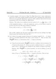
GENERAL COCHLEAR ANATOMYI. Scala VestibuliScala MediaScala TympaniII. Scala Media(a) Sensory Epithelia(b) Vascular System(c) Neural AnatomyFig. 12.A. The cochlea of the right ear showing the relative positions of thescala vestibuli, scala media, and scala tympani. Only the scalavestibuli communicates directly with the vestibule. The arrowsshow possible paths of soundB. A cross section of the <strong>cochlear</strong> tube.III.Sensory EpitheliumNote: Connective tissue faces the perilymph scala. ~ not quite true.Epithelial tissue faces the endolymph space.(a) Basement membrane usually separates connective fromepithelial tissue except at the stria-spiral ligament interface.- 13 -
Fig. 13.Cross section of the scala media showing the organ of Corti.IV.Basilar Membranepars tectapara pectinataground substance - fibersconnective tissue cellsconsists mostly of radial fiber - Note: However, strong longitudinalconnections via "glue-like" ground substance also few longit. fibers - fibercharacteristics change from base to apex.Apex radial fibers "seem" more or less independent. Fibers of the basilarmembrane run into the fibers of the spiral ligament laterally and into thefibers of the spiral limbus.- 14 -
Fig. 15. Detailed Anatomy of the Inner Hair Cell.VIII. (a) Inner hair cells - Apical Zone - cuticular plate, basal body area ~hairs/cell, arranged longitudinally. Accumulation of manyorganelles.Intermediate Zone -Perinuclear Zone -Basal Zone -IHC - larger than OHC - bottle shaped - nucleus positioneddifferently and whole morphology in general is different.- 16 -
VIII.IX.(b) OHC - cylindrical in shape - organelle description about thesame as for IHC. Stereocilia W shaped vertex points to lateralwall. Note IHC and OHC cuticular area - reticular laminajunctional complex. no possibility of intermixing endolymph intothe organ of Corti fluid spaces.Supporting Cells:(a) Cells with fibers, Dieters' cellsInner, outer pillar(b) Cells without fibers. Hensen cellsClaudius cellsBoettcher cellsFig. 16. Detailed Anatomy of the Outer Hair Cell.- 17 -
Peripheral Innervation:SUMMARY OF COCHLEAR INNERVATIONAll fiber loose myelin sheaths as they enter O of C.(1) Radial Fibers (a) to IHC and (b) tunnel crossing radialfibers to OHC.(2) Internal Spiral Bundle(3) Tunnel Spiral Fibers(4) Outer Spiral FibersAfferent and EfferentFig. 17.Schematic representation of different groups of nerve fibers in the organof Corti. The efferents are drawn in black. D : radial fibers to inner haircells; iS : internal spiral fibers; TS : tunnel spiral fibers; TR : tunnel radialfibers; B : basilar fibers; OS : outer spiral fibers.Efferent Innervation:Rasmussen: Olivo<strong>cochlear</strong> bundle brings an efferent supply to thecochlea from the contra and homolateral superior olives. Reachesthe cochlea with the vestibular nerve through the anastomosis ofOort.- 18 -
(1) Efferent endings in the Organ of Corti are characterized by:(1) large size; (2) synaptic vesicles; (3) postsynaptic cisterna.(2) Efferents generally contact OHC directly but direct contactwith IHC is rare.Fig. 18.Schematic representation of efferent synaptic connections in theorgan of Corti of the cat. At the outer hair cells (OH) synapticcontacts are almost exclusively with the sensory cell and at theinner hair cells (IH) only with the afferent dendrites (AD). E :efferent ending.Degeneration StudiesFig. 19.Outline of experimental lesions, which have been carried out oncats. la-lc show the possible sites of interruption of the olivo<strong>cochlear</strong>fibers; 2 the total transsection of the eighth nerve, and 3 aselective lesion of the <strong>cochlear</strong> nerve.- 19 -
By transsection of the entire olivo<strong>cochlear</strong> bundle; large nerve endingsdegenerate, as well as all inner and tunnel spiral fibers.Observations:(1) 1st row of OHC - most abundant efferent supply(2) In the base all three rows have an efferent supply but as apex isapproached, 2nd and 3rd rows lose their efferent supply. (6-8 endings/cellin the base).Estimated 40,000 endings + several hundred inner spiral fibersoriginate from the olivo<strong>cochlear</strong> bundle consisting of only about 500neurons.Only small % age of fibers passing through the habenular openingare efferent. (20-30 fibers pass through each opening).OHC Innervation:Fibers getting to the OHC cross the tunnel either at the middle levelor at the bottom as basilar fibers.All upper tunnel fibers are efferent. Since these disappear whenolivo<strong>cochlear</strong> bundle is cut. But this does not reduce number of outerspiral fibers.∴ Efferent innervation to OHC is predominantly radial.Selective transsection of the olivo<strong>cochlear</strong> bundle shows that 75% ofthe fibers originate from contra lat. and remainder from homo lat.superior olive.Iurato: shows contra lat. fibers (in rat) go to OHChomo lat. " " go to inner spiralfibers and OHCSpoendlin: shows only ~ 10% efferent in OHC arise from homo lat.bundle.∴ Contra later. fibers produce most efferent innervation to OHC andabout 1/2 to the inner spiral fibers, but homo lat. contribute other 1/2 ofinner spiral fiber and only a few to OHC.- 20 -
Synaptic Connection:At OHC, efferents make direct synapse with OHC. Synapse ischaracterized by synaptic visicles at presynaptic membrane and postsynaptic membrane differentiation.At IHC level efferents make direct contact with the afferentdendrites. The main demonstrated effect of the olivo<strong>cochlear</strong> fibers isinhibition → Anatomy shows: presynaptic inhibition in OHC and postsynaptic inhibition at the dendrited to IHCAfter efferent innervation is eliminated; what's left?(1) Radial fibers to inner H.C.(2) Basilar fibers(3) Outer spiral fibers*Spoendlin estimates 3-4000 efferents axons destined for the OHC, thisimplies that the majority of cohclear neurons lead to IHC i.e.: about 50,000!Q: Are upper tunnel radials efferent?Spoendlin says YesEldridge says MaybeAfferents enter in bundles of 20 through each habenular opening. Fibersdisperse between supporting cells of IHC. Majority continue til reachingan IHC; others spiral a little before becoming basilar. Out of 20 fibers only1 or 2 become basilar. Majority do not branch and end with a single nerveending at a single nerve ending at a single IHC. Synapse characterized bysynapitc bar.- 21 -
Fig. 20.Schema of the general innervation pattern of the organ of Corti.oH: outer hair cells; iH: inner hair cells; HA: habenular openings.Fig. 21.Schematic outline of the fiber distribution of the organ of Corti.Full thick lines: afferent fibers to the outer hair cells. Full thinlines: afferent fibers to the inner hair cells. Interrupted thick lines:efferent nerve fibers from the contralateral olivo<strong>cochlear</strong> bundle.Thin interrupted lines: efferent nerve fibers from the homolateralolivo<strong>cochlear</strong> bundle.- 22 -
Fig. 22. Schematic diagram summarizing known and hypothesized connections ofolivo<strong>cochlear</strong> neurons. Reflex circuits of a representative LOC neuron(right) and representative MOC neuron (left) are depicted. A <strong>cochlear</strong>nuclear multipolar cell, with dendrites extending into the granule cellregion, is shown hypothetically linking the periphery to the two kinds of OCneurons and receiving feedback in the form of a collateral from the MOCneuron. A projection by this multipolar neuron to OC neurons has yet to bedemonstrated.- 23 -
COCHLEAR VASCULATUREExperiments show that during exposure to noise, many of theperipheral blood vessels in the body contract. Vascular contraction is alsoapparent in the cochlea after noise exposure. It has been shown that theO 2 content in scala media is reduced and that a cochlea deprived of O 2 ismore susceptible to noise induced damage.The possible reasons for this sensitivity become apparent when oneconsiders the pathways of the cochlea vessels and the function of thevarious capillary beds.The spiral modiolar artery supplies the entire cochlea (Fig.over).Branches from this artery course over scala vestibuli to vascularize thelateral wall of the cochlea; another branch runs into the area of the spirallimbus and basilar membrane.The lateral wall contains an extremely vascularized area known asthe stria vascularis. The stria together with the cells of the outer sulcus arebelieved to be involved in maintaining the chemical composition of theendolymph. Alterations in the chemical composition of the endolymph leadto degeneration of the organ of Corti.The second group of vessels spiral beneath the basilar membrane,and are believed to supply O 2 to the organ of Corti. Similarly, loss ofsensory cells is often seen in areas with severe capillary occlusions.- 24 -
The Vascular Anatomy of the Cochlear in the Guinea Pig and In ManFig 23. Man, arterial system, schematic. OW = oval window, RW = round window,N VIII = acoustic nerve.- 25 -
Fig. 25.Guinea pig and man, spiral lamina and limbus, schematic. The vascular system is arcadicwith peripherally radiating arterioles, spirally running capillary vessels, and centrallyradiating venules. The region peripheral to the vessel of the basilar membrane is principallyavascular. The vessel of the basilar membrane is situated under the tunnel of Corti. The vellof the tympanic lip, which is located in the region of the habenula perforata, receives mostof the radiating vessels. Capillaries in the spiral limbus form irregular limbus vessels.Fig. 26.Guinea pig and man, external wall, radial section, schematic. Radiating arteriolespredominate in the scala vestibuli and supply spiral vascular systems; the vessel of the scalavestibuli, the vessel at the vestibular membrane, the stria vascularis, and the vessel of thespiral prominence. Collecting venules predominate in the scala tympani and drain spiralcapillary vessels and the capillary net in the scala vestibuli. A spiral vessel is formedbasally to the basilar membrane. Radiating arterioles and collecting venules connect with oneanother by arterio-venous anastomses lying exernally to the stria vascularis.- 26 -