Virtual reality-enhanced stroke rehabilitation ... - ResearchGate
Virtual reality-enhanced stroke rehabilitation ... - ResearchGate
Virtual reality-enhanced stroke rehabilitation ... - ResearchGate

Create successful ePaper yourself
Turn your PDF publications into a flip-book with our unique Google optimized e-Paper software.
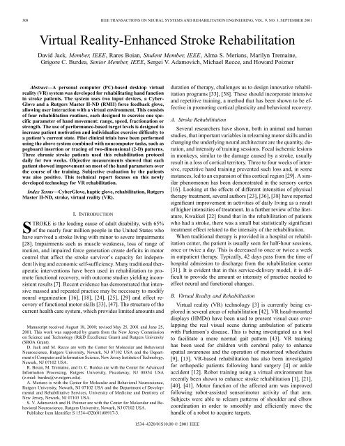
308 IEEE TRANSACTIONS ON NEURAL SYSTEMS AND REHABILITATION ENGINEERING, VOL. 9, NO. 3, SEPTEMBER 2001<strong>Virtual</strong> Reality-Enhanced Stroke RehabilitationDavid Jack, Member, IEEE, Rares Boian, Student Member, IEEE, Alma S. Merians, Marilyn Tremaine,Grigore C. Burdea, Senior Member, IEEE, Sergei V. Adamovich, Michael Recce, and Howard PoiznerAbstract—A personal computer (PC)-based desktop virtual<strong>reality</strong> (VR) system was developed for rehabilitating hand functionin <strong>stroke</strong> patients. The system uses two input devices, a Cyber-Glove and a Rutgers Master II-ND (RMII) force feedback glove,allowing user interaction with a virtual environment. This consistsof four <strong>rehabilitation</strong> routines, each designed to exercise one specificparameter of hand movement: range, speed, fractionation orstrength. The use of performance-based target levels is designed toincrease patient motivation and individualize exercise difficulty toa patient’s current state. Pilot clinical trials have been performedusing the above system combined with noncomputer tasks, such aspegboard insertion or tracing of two-dimensional (2-D) patterns.Three chronic <strong>stroke</strong> patients used this <strong>rehabilitation</strong> protocoldaily for two weeks. Objective measurements showed that eachpatient showed improvement on most of the hand parameters overthe course of the training. Subjective evaluation by the patientswas also positive. This technical report focuses on this newlydeveloped technology for VR <strong>rehabilitation</strong>.Index Terms—CyberGlove, haptic glove, <strong>rehabilitation</strong>, RutgersMaster II-ND, <strong>stroke</strong>, virtual <strong>reality</strong> (VR).I. INTRODUCTIONSTROKE is the leading cause of adult disability, with 65%of the nearly four million people in the United States whohave survived a <strong>stroke</strong> living with minor to severe impairments[28]. Impairments such as muscle weakness, loss of range ofmotion, and impaired force generation create deficits in motorcontrol that affect the <strong>stroke</strong> survivor’s capacity for independentliving and economic self-sufficiency. Many traditional therapeuticinterventions have been used in <strong>rehabilitation</strong> to promotefunctional recovery, with outcome studies yielding inconsistentresults [7]. Recent evidence has demonstrated that intensivemassed and repeated practice may be necessary to modifyneural organization [16], [18], [24], [25], [29] and effect recoveryof functional motor skills [33], [47]. The structure of thecurrent health care system, which provides limited amounts andManuscript received August 18, 2000; revised May 25, 2001 and June 25,2001. This work was supported by grants from the New Jersey Commissionon Science and Technology (R&D Excellence Grant) and Rutgers University(SROA Grant).D. Jack and M. Recce are with the Center for Molecular and BehavioralNeuroscience, Rutgers University, Newark, NJ 07102 USA and the Departmentof Computer and Information Science, New Jersey Institute of Technology,Newark, NJ 07102 USA.R. Boian, M. Tremaine, and G. C. Burdea are with the Center for AdvancedInformation Processing, Rutgers University, Piscataway, NJ 08854 USA(e-mail: burdea@vr.rutgers.edu).A. Merians is with the Center for Molecular and Behavioral Neuroscience,Rutgers University, Newark, NJ 07102 USA and the Department of Developmentaland Rehabilitative Services, University of Medicine and Dentistry ofNew Jersey, Newark, NJ 07103 USA.S. V. Adamovich and H. Poizner are with the Center for Molecular and BehavioralNeuroscience, Rutgers University, Newark, NJ 07102 USA.Publisher Item Identifier S 1534-4320(01)08917-3.duration of therapy, challenges us to design innovative <strong>rehabilitation</strong>programs [33], [38]. These should incorporate intensiveand repetitive training, a method that has been shown to be effectivein promoting cortical plasticity and behavioral recovery.A. Stroke RehabilitationSeveral researchers have shown, both in animal and humanstudies, that important variables in relearning motor skills and inchanging the underlying neural architecture are the quantity, duration,and intensity of training sessions. Focal ischemic lesionsin monkeys, similar to the damage caused by a <strong>stroke</strong>, usuallyresult in a loss of cortical territory. Three to four weeks of intensive,repetitive hand training prevented such loss and, in someinstances, led to an expansion of this cortical region [29]. A similarphenomenon has been demonstrated in the sensory cortex[16]. Looking at the effects of different intensities of physicaltherapy treatment, several authors [23], [36], [38] have reportedsignificant improvement in activities of daily living as a resultof higher intensities of treatment. In a further review of the literature,Kwakkel [22] found that in the <strong>rehabilitation</strong> of patientswho had a <strong>stroke</strong>, there was a small but statistically significanttreatment effect related to the intensity of the <strong>rehabilitation</strong>.When traditional therapy is provided in a hospital or <strong>rehabilitation</strong>center, the patient is usually seen for half-hour sessions,once or twice a day. This is decreased to once or twice a weekin outpatient therapy. Typically, 42 days pass from the time ofhospital admission to discharge from the <strong>rehabilitation</strong> center[31]. It is evident that in this service-delivery model, it is difficultto provide the amount or intensity of practice needed toeffect neural and functional changes.B. <strong>Virtual</strong> Reality and Rehabilitation<strong>Virtual</strong> <strong>reality</strong> (VR) technology [3] is currently being exploredin several areas of <strong>rehabilitation</strong> [42]. VR head-mounteddisplays (HMDs) have been used to present visual cues overlappingthe real visual scene during ambulation of patientswith Parkinson’s disease. This is being investigated as a toolto facilitate a more normal gait pattern [43]. VR traininghas been used for children with cerebral palsy to enhancespatial awareness and the operation of motorized wheelchairs[9], [13]. VR-based <strong>rehabilitation</strong> has also been investigatedfor orthopedic patients following hand surgery [4] or ankleaccident [12]. Robot training using a virtual environment hasrecently been shown to enhance <strong>stroke</strong> <strong>rehabilitation</strong> [1], [21],[40], [41]. Motor function of the affected arm was improvedfollowing robot-assisted sensorimotor activity of that arm.Subjects were able to relearn patterns of shoulder and elbowcoordination in order to smoothly and efficiently move thehandle of a robot to acquire targets.1534–4320/01$10.00 © 2001 IEEE
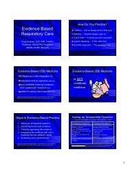
JACK et al.: VIRTUAL REALITY-ENHANCED STROKE REHABILITATION 309This technology provides the capability to create an environmentin which the intensity of feedback and training canbe systematically manipulated and <strong>enhanced</strong> in order to createthe most appropriate, individualized motor learning paradigm.The potential benefits of training in VR would be the abilityto increase the duration, frequency, and intensity of therapythat could be provided to patients by using semiautomated programs.Furthermore, lower cost personal computer (PC)-basedVR equipment is now available that will eventually allow<strong>rehabilitation</strong> stations to be placed in locations other than the<strong>rehabilitation</strong> center, such as a patient’s home. The Internetcan be used for data transfer, allowing a therapist to remotelymonitor progress and to modify the patient’s therapy program[4], [30]. The rate at which patients can relearn their motorskills, the extent of improvement, and the environment in whichthey are treated affect the duration, effectiveness, and cost ofpatient care. Therefore, developing new methods to accelerateand improve the level of motor retraining is a very importantsocietal consideration.VR-based <strong>rehabilitation</strong> systems have several other advantages,as well. VR <strong>rehabilitation</strong> exercises can be made to beengaging, such that the patient feels immersed in the simulatedworld. This is extremely important in terms of the patient motivation[30], which, in turn, is key to recovery. VR sensor technologycan also be used to fully quantify any progress made bythe patient, especially in terms of motor-control improvement.Although most neurologic recovery is attained by three monthsafter the <strong>stroke</strong> [17], several studies investigating the outcomeof treatment six months after the <strong>stroke</strong> have shown significantgains in dexterity, strength, and function [5], [32], [44].If VR-based <strong>rehabilitation</strong> of patients who experienced <strong>stroke</strong>years ago is proven successful, then treatment options becomeavailable past the traditional period of inpatient hospitalizationand <strong>rehabilitation</strong>.It has been shown, in normal subjects, that VR can be a beneficialenvironment for learning a motor task. Todorov et al.[39] used a VR system for table-tennis training, including virtualpaddles for the teacher and the subject, as well as a virtualball. Augmented feedback was used to indicate to the trainee themovement variables most relevant for successful performanceof the task. Results indicated that subjects who received the virtualenvironment training did better than subjects who receiveda comparable amount of training in a real environment. Anotherexperiment comparing VR training and real-world training ina pick-and-place task showed improvement in both groups, butthose trained in the real-world task did better [19]. This is notsurprising, because the VR group used low-resolution HMDsand gloves with no force feedback.Two patients with hemiplegia were trained in a virtual environmenton an upper-extremity-reaching task that progressedsequentially through six levels of difficulty [12]. Each subjectreceived 16 trials from 1 to 2 h duration. Both patients improvedin the task in the virtual environment and were able to progressto the sixth level of difficulty. However, only one of the subjectsshowed clinical and functional motor improvements; the secondshowed no improvements.In addition to sorting out the effects of motor training in avirtual environment, it is important to determine whether theFig. 1. The PC-based VR <strong>rehabilitation</strong> system. The user is wearing aCyberGlove connected to the interface unit on the right. Also shown is thehaptic control interface (HCI) for the RMII glove [14]. (© Association forComputing Machinery (ACM). Photo courtesy of ACM.)skills gained in that environment transfer to real-world conditions.Wilson et al. [45], [46] studied children with a varietyof disabilities and found that internal representations resultingfrom exploration of simulated space transferred to the real environment.However, although subjects trained on a motor task ina virtual environment demonstrated the ability to improve performanceon the task in that environment, the learning did notalways transfer to the real-world task [12], [19]. This conflictin findings could be reflective of differences in the learning requirementsof perceptual skills and motor skills, or it could bereflective of the current paucity of investigations into the useof VR for motor skill training. The experiments on motor-tasktraining and transfer of that task to the real-world environmentindicate that the effects of training in a virtual environment arenot fully understood, nor entirely conclusive.The literature investigating virtual <strong>reality</strong> as a tool for <strong>rehabilitation</strong>training does indicate potential benefits. These shouldbe more fully explored in order to ascertain the use of VR as anenhancement to traditional therapy. To that end, this technicalreport focuses primarily on the technological innovations for theuse of VR-<strong>enhanced</strong> <strong>rehabilitation</strong> of <strong>stroke</strong> patients. The clinicaldata for these patients is the focus of a companion article[27]. Section II presents the PC-based experimental system andSection III details the protocol used in pilot trials. Section IVdiscusses trial results and Section V concludes this report, offeringpossible directions for future research.II. EXPERIMENTAL SYSTEMThe experimental system used in this study consists of aPC-based <strong>rehabilitation</strong> workstation (running VR simulationexercises and a database), as well as a more traditional therapeuticstation.A. PC Rehabilitation WorkstationFig. 1 shows the PC system and interfaces used in this study.It consists of a Pentium II 400 MHz PC with a FireGL 4000graphics accelerator and two input–output gloves. These are theCyberGlove [20] (Immersion Co., San Jose, CA 95131) and theRutgers Master II-ND (RMII) force feedback glove prototype[30].
310 IEEE TRANSACTIONS ON NEURAL SYSTEMS AND REHABILITATION ENGINEERING, VOL. 9, NO. 3, SEPTEMBER 2001The gloves’ characteristics make each of them more suited forcertain hand <strong>rehabilitation</strong> exercises. The RMII force feedbackstructure limits the range of motion of the hand. The elasticityof the CyberGlove does not restrict the user’s movement, but itcannot provide an opposing force in the exercises. Thus, the CyberGloveis used in the VR exercises that primarily involve positionmeasurement of the patient’s fingers, and the RMII gloveis used in force-exertion exercises.1) CyberGlove: The CyberGlove is a sensorized structureworn on the hand. It has 18 embedded strain-gauge sensors thatmeasure the metacarpophalangeal (MCP) and proximal interphalangeal(PIP) joint angles of the thumb and fingers as wellas finger abduction and wrist flexion. The system uses only theMCP and PIP angles of the thumb and fingers.In order to minimize measurement errors due to hand-sizevariability, the glove is calibrated at the beginning of eachexperiment. Every hand joint is placed into two known positions,0 and 60 . From these measurements, two parameters(gain and offset) are obtained that determine the linear relationbetween the raw glove-sensor output (voltages) and the correspondinghand-joint angles being measured. The data sets areread through the serial port at a rate of 70 hand configurationsper second.2) RMII Glove: The RMII glove is an exoskeleton devicethat applies force to the user’s fingertips and uses noncontactposition sensors to measure the fingertip position in relation tothe palm. Lightweight custom pneumatic actuators are attachedto the tips of the thumb, index, middle, and ring fingers. Eachfinger actuator can apply up to 16 N of force when pressurizedat 100 psi. The air pressure is provided by a portable super-quietair compressor.Infrared sensors inside the actuators measure the displacementof the fingertip with respect to the exoskeleton base attachedto the palm. Hall-Effect sensors mounted at the base ofthe actuators measure their flexion and abduction angles withrespect to the base [30].The glove is connected to an HCI that reads RMII sensorsand controls the desired fingertip forces. In order to determinethe hand configuration corresponding to the values of the exoskeletonposition sensors, the joint angles of three fingers andthe thumb, as well as finger abduction, need to be estimated.This computation is based on a kinematic model similar to theone shown in Fig. 2 [11].The equations for the inverse kinematics areAdditionally, the following constraint equation can be imposedfor and [24]:The system is solved using least-squares linear interpolation.The calibration of the RMII glove consists of reading the sensorswhile the hand is completely opened. The values read arethe maximum piston displacement, minimum flexion angle, andneutral abduction angle. During the experiment, the calibrationof the RMII glove is performed before each session.Fig. 2. Finger kinematics [11]. (© Rutgers University. Reprinted bypermission.)Fig. 3. System software architecture. (© Rutgers University. Reprinted bypermission.)The actuator forces are controlled by the HCI at a rate of500 Hz [30]. This is done through a servo loop implemented insoftware on a Pentium 233-MHz embedded board of the HCI.By performing local computations on the embedded Pentium,the host PC is freed to perform mostly graphics computationsat a high frame rate needed in the simulations. Communicationbetween the host PC and the HCI is done on a serial line at arate of 38 400 baud. At this rate, the host receives 157 data sets(hand configurations) per second.B. Rehabilitation ExercisesThe overall software architecture organization for the PC <strong>rehabilitation</strong>workstation is shown in Fig. 3. The VR simulationsconsist of four exercises. Each of them concentrates on one particularparameter of the hand movement: range, speed, fractionation,and strength.The range-of-motion exercise is designed to improve the patient’sfinger flexion and extension. The patient is asked to flexthe fingers as much as possible and then open them as much aspossible. In the speed-of-motion exercise, the patient is askedto fully open the hand and then close it as fast as possible. The
JACK et al.: VIRTUAL REALITY-ENHANCED STROKE REHABILITATION 311The finger velocity in the speed of motion exercise is takenas the mean of the angular velocities of the MCP and PIP joints.The performance measure isspeed MCP speed PIPFinger fractionation or independence of finger movement isFig. 4. PC <strong>rehabilitation</strong> session structure. (© Rutgers University. Reprintedby permission.)fractionation exercise involves the use of the index, middle, ring,and small fingers. The goal of the exercise is to flex one fingeras much as possible while the others are kept open. The exerciseis executed separately for each of the four fingers. The strengthexercise is designed to improve the patient’s grasping. The fingersinvolved are the thumb, index, middle, and ring. The patientis asked to close the fingers against the forces applied to his fingertips.To reduce fatigue and tendon strain, the fingers are movedtogether and the thumb is moved alone for all exercises exceptfractionation. The exercise is executed separately for the thumbbecause, when the whole hand is closed, either the thumb or thefour fingers does not achieve full range of motion. Executingthe exercise for the index, middle, ring, and small fingers at thesame time is fine because, here, the fingers do not affect eachothers’ range of motion.The <strong>rehabilitation</strong> process is divided into sessions, blocks,and trials. A trial consists of one execution of each exercise.For instance, closing the thumb or fingers is a range-of-motiontrial. A block is a group of trials of the same type of exercise. Asession is a group of blocks, each of a different exercise. Fig. 4shows the components of a PC <strong>rehabilitation</strong> session.C. Performance EvaluationDuring each trial, the exercise parameters are estimated onlinein order to drive the graphics display and provide feedbackto the patient. After the trial has been completed, data collectedon the patient’s movements are low-pass-filtered at 8 Hz to reducesensor noise. The parameters are reevaluated and stored,along with the filtered data, into the database.The patient’s performance is calculated per trial and perblock. The block performance is the mean and the standarddeviation of the performances of the trials involved.For the range-of-motion and strength exercises, the flexionangle of the finger is considered to be the mean of the MCP andPIP joint angles. The performance measure isMCP PIP MCP PIPPassiveFingerRangeActiveFingerRangewhere ActiveFingerRange is the current average joint range ofthe finger being moved and PassiveFingerRange is the currentaverage joint range of the other three fingers combined. Movingone finger alone results in a measure of 100%, which decays tozero as more fingers are coupled in the movement. The patientis prompted to move only one finger while trying to keep theothers stationary. This is repeated four times for each finger.D. Baseline Test and Performance TargetsImplementing target-based exercises requires an initial testto evaluate the patient’s baseline movement. The evaluation testhas the same structure as that of the PC <strong>rehabilitation</strong> sessionpresented in Fig. 4.A special case during the baseline test is the strength exercise,which uses the RMII glove. As mentioned above, the rangeof movement in this glove is somewhat limited, so another setof range evaluations is performed to obtain the patient’s meanrange while wearing the RMII. The patient’s finger strength isestablished by doing a binary search of force levels and comparingthe range of movement at each level with the mean obtainedfrom the previous range test. If the range is at least 80%of that previously measured, the test is passed, and the force isincreased to the next binary level. If the test is failed, then theforce is decreased to the next binary level, and so on. Test forcesare applied until the maximal force level attainable by the patientis found.The set of targets for the first session is drawn from a normaldistribution around the mean and standard deviations given bythe initial evaluation baseline test. A normal distribution ensuresthat the majority of the targets will be within the patient’s performancelimits. However, the patient will find some new targetseasy or difficult depending on whether they came from the lowor high end of the target distribution. Initially, the target meansare set one standard deviation above the patient’s actual measuredperformance to obtain a target distribution that overlapsthe high end of the patient’s performance levels. After a blockis completed, the distribution of the patient’s actual performanceis compared to the preset target mean and standard deviations.If the mean of the patient’s actual performance is greater thanthe target mean, then that target is raised by one standard deviation.Otherwise, the target for the next session is lowered bythe same amount. To prevent the block targets from varying toolittle or too much between sessions, lower and upper boundsare placed upon their increments. These parameters allow thetherapist to choose how aggressively each training exercise willproceed. A high upper bound means that the targets for the nextsession are considerably higher than the previous ones. As thetargets change over time, they provide valuable information to
312 IEEE TRANSACTIONS ON NEURAL SYSTEMS AND REHABILITATION ENGINEERING, VOL. 9, NO. 3, SEPTEMBER 2001Fig. 5. The mean performance and target levels for the range of movement ofa control subject’s index finger. The y axis is labeled in degrees [14]. (© ACM.Photo courtesy of ACM.)Fig. 6.Range-of-motion VR exercise [14]. (© ACM. Photo courtesy of ACM.)the therapist as to how the patient is coping with the <strong>rehabilitation</strong>training.Fig. 5 shows a typical set of blocks gathered from a normalsubject [14]. The block targets and actual mean performance ofthe index finger during the range exercise are shown for foursessions taken over a two-day period. The first two columns arethe result of the initial subject evaluation, the target being setfrom the mean actual performance plus one standard deviation.As the exercises proceed, it can be seen how the targets werealtered based upon the subject’s performance. The block targetwas increased when the subject matched or improved upon thetarget level, or decreased otherwise.E. VR SimulationsFig. 7.Speed-of-motion VR exercise[14]. (© ACM. Photo courtesy of ACM.)For each of the exercises presented above, a VR simulationwas developed using the commercially available WorldToolKitgraphics library [8]. The simulations take the form of simplegames in which the patient performs a number of trials of a particulartask. The programs are designed to attract the patient’sattention and to challenge him to execute the tasks.The VR simulations are coupled to the sensing gloves and theperformance evaluation modules as shown in Fig. 3. During thetrials, the patient is shown a graphical model of his own hand,which is updated in real time to accurately represent the flexionof his fingers and thumb. The patient is informed of the fingersinvolved in the trial by highlighting the appropriate virtual fingertipsin green. The hand is placed in a virtual world that isacting upon the patient’s performance for the specific exercise.If the performance is higher than the preset target, then the patientwins the game. If the target is not achieved in less than oneminute, the trial ends.1) Range of Movement: The range-of-movement exercise isillustrated in Fig. 6 [14]. In this exercise, the patient moves awindow wiper to reveal an attractive landscape hidden behindthe fogged window. The higher the measured angular range ofmovement of the thumb or fingers, the more the wiper rotatesand clears the window. The rotation of the wiper is scaled sothat if the patient achieves the target range for that particulartrial, the window is cleaned completely.The fogged window consists of a two-dimensional (2-D)array of opaque square polygons placed in front of a largerpolygon mapped with a landscape texture. Upon detecting thecollision with the wiper, the elements of the array are madetransparent, revealing the picture behind it. Collision detectionis not performed between the wiper and the middle verticalband of opaque polygons because they always collide at thebeginning of the exercise. These elements are cleared when thetarget is achieved. To make the exercise more attractive, thetexture (image) mapped on the window is changed from trialto trial.2) Speed of Movement: The speed-of-movement exercise isdesigned as a catch-the-ball game, as illustrated in Fig. 7 [14].The patient competes against a computer-controlled opponenthand (on the left in the screen). On a “go” signal (green light ona traffic signal), the patient is required to close either the thumbor all the fingers together as fast as possible to catch a red ball.At the same time, the opponent hand also closes its thumb orfingers around its red ball. The angular velocity of the opponenthand goes from zero to the target angular velocity and then backto zero, following a sinusoid.If the patient surpasses the target velocity, then he beats theopponent (yellow) hand and gets to keep the red ball. Otherwise,the patient loses, and his ball falls, while the other red ball remainsin the opponent’s hand.
JACK et al.: VIRTUAL REALITY-ENHANCED STROKE REHABILITATION 313Fig. 8.ACM.)Finger-fractionation VR exercise [14]. (© ACM. Photo courtesy ofFig. 10. The digital performance meter shown to the user after every trial. Thetarget level is shown (white bar), as well as the actual performance of the user(black bars) [14]. (© ACM. Photo courtesy of ACM.)Fig. 9.ACM.)Strength-of-motion VR exercise [14]. (© ACM. Photo courtesy of3) Finger Fractionation: The fractionation exercise illustratedin Fig. 8 [14] shows a piano keyboard. As the activefinger is moved, the corresponding key on the piano is depressedand turns green. Nearing the end of the move, thefractionation measure is calculated online, and if it is greaterthan or equal to the trial target measure, then only that one keyremains depressed. Otherwise, other keys are depressed, andturn red to show which of the other fingers had been coupledduring the move. The goal of the patient is to move his hand sothat only one virtual piano key is depressed for each trial.4) Strength of Movement: In the strength exercise, a virtualmodel of the RMII glove is controlled by the patient, as shownin Fig. 9 [14]. The forces applied for each individual trial areagain taken from a normal distribution around the force levelfound in the initial evaluation. As each actuator on the RMII issqueezed, the graphical pistons start to fill from top to bottomin a green color, proportional to the percentage of the target thathad been achieved. The piston turns yellow and is completelyfilled if the patient manages to move the desired distance againstthat particular force level.Each piston has two fixed points: one in the palm, attached tothe base, and one attached to the fingertip. The virtual piston isimplemented with the same fixed points; the cylinder is a childnode of the palm graphical object, whereas the shaft is a childFig. 11. Database main tables. (© Rutgers University. Reprinted bypermission.)node of the fingertip graphical object. To implement the constraintof the shaft sliding up and down in the cylinder, for eachframe, the transformation matrices of both parts are calculatedin the reference frame of the palm. Then, the rotation of the partsis computed such that they point to one another.After every trial is completed for any of the previously describedsimulations, the patient is shown a graphical digital performancemeter similar to the one illustrated in Fig. 10 [14]. Thisvisualizes the target level and the patient’s actual performanceduring that exercise. It informs the patient of how his performancecompares with the desired one.F. Database DesignThe PC exercise data stored in files is subsequently loadedinto an Oracle database. In order to fit future developments,the database is designed in a modular fashion that maps to thereal-world system. A simplified diagram showing only the maindatabase tables is shown in Fig. 11.The PATIENTS table stores information about the conditionof the patient, prior <strong>rehabilitation</strong> training, and results of various
314 IEEE TRANSACTIONS ON NEURAL SYSTEMS AND REHABILITATION ENGINEERING, VOL. 9, NO. 3, SEPTEMBER 2001medical tests. The SESSIONS table contains information abouta <strong>rehabilitation</strong> session such as date, time, location, and handinvolved. The BLOCKS table stores the type of the exercise, theglove used (i.e., CyberGlove or RMII Glove), and the version ofthe data. The version of the data is linked to an auxiliary tablecontaining information about the data stored and the algorithmsused to evaluate it. For each exercise, there is a separate TRIALStable containing mainly control information about the status ofa trial. There are four DATA tables, one for each exercise. TheDATA tables store the sensor readings taken during the trials.For each exercise, there is a separate BASELINES table storingthe results of the initial evaluation.The main purpose of the database is to provide quick accessto the data. The targets and performances of the trials can alwaysbe computed from the stored sensor readings, but this approachwould be very slow due to the high amount of data to be processed.Because the calculation of the targets and performancesare always the same, it is a lot faster to do them once and thenjust access the stored results. The target and performance tablesin the lower right corner of Fig. 11 contain this information.A very frequent operation on the database is to find out towhom an entry belongs. For instance, one may need to knowwhich patient executed a certain trial. To speed up such queries,the keys of the tables on the top of the hierarchy are passed downmore than one level. Due to the large size of the DATA tables,the only foreign key passed to them is the trial key.The data access is provided through a user name and passwordassigned to each patient and member of the research team.To respect the patient’s privacy and to avoid potential undesirablemistakes in handling the data, all the data is stored intoa ROOT account. Only the database developers know the rootpassword. The database users (for now, the researchers, but, inthe future, this will include the patients) are granted only the appropriatereading and writing privileges [2].III. CLINICAL STUDYThe <strong>rehabilitation</strong> system described above has been tested onpatients during a two-week pilot study. All subjects were testedclinically, pre- and posttraining, using the Jebsen test of handfunction [15] and the hand portion of the Fugel-Meyer assessmentof sensorimotor recovery after <strong>stroke</strong> [6]. Grip strengthevaluation using a dynamometer was obtained pre-, intra-, andposttraining. In addition, subjective data regarding the subjects’affective evaluation of this type of computerized <strong>rehabilitation</strong>was also obtained pre-, intra-, and posttrial through structuredquestionnaires. Each subject was evaluated initially to obtain abaseline of performance in order to implement the initial computertarget levels. Subsequently, the subjects completed ninedaily <strong>rehabilitation</strong> sessions that lasted approximately five hourseach. These sessions consisted of a combination of VR-basedexercises using the PC-based system that alternated with noncomputerexercises. Cumulative time spent on the VR exercisesduring each day’s training was approximately 1–1.5 h per patient.The remainder of each daily session was spent on traditional<strong>rehabilitation</strong> exercises. Although a patient’s “good” armwas never restrained, patients were encouraged to use their impairedarms and were supervised in these activities by a physicalor occupational therapist. The latter exercises consisted ofseries of game-like tasks such as tracing 2-D patterns on paper,peg-board insertion, checkers, placing paper clips on paper, andpicking up objects with tweezers.A. Patient InformationThree subjects, two male and one female, ages 50–83, participatedin this study. They had sustained left hemisphere <strong>stroke</strong>sthat occurred between three and six years prior to the study. Allsubjects were right hand dominant and had had no therapy in thepast two years. Two of the subjects were independent in ambulationand one required the assistance of a walker. None of thesubjects was able to functionally use his or her hemiparetic righthand except as a minimal assist in a few dressing activities.B. Baseline Patient EvaluationEach VR-based exercise session consisted of four blocks of10 trials each. Multiple sessions were run each day for five daysfollowed by a weekend break and another four days. An individualblock concentrated on exercising one of the aforementionedparameters of range, speed, fractionation, or strength ofmovement. Similar to the evaluation exercises, the patients wererequired to alternate between moving the thumb alone and thenmoving all the fingers together for every exercise except fractionation.Most trials were started and stopped by the therapistpressing the spacebar, although there were a few patient-initiatedtrials. As mentioned previously, the patient had to attain acertain target level of performance in order to successfully completeevery trial. For a particular block of trials the first set oftargets were drawn from a normal distribution around the meanand standard deviation given by the initial evaluation baselinetest. A normal distribution ensured that the majority of the targetswould be within the patient’s performance limits, but thepatient would find some targets easy or difficult depending onwhether they came from the low or high end of the target distribution.Initially, the target means were set one standard deviationabove the patient’s actual measured performance to obtaina target distribution that overlapped the high end of the patient’sperformance levels.The four blocks of exercises were grouped in one session thattook 15–20 min to complete. The sessions were target-based,such that all the exercises were driven by the patient’s own performance.The targets for any particular block of trials were setbased on the performance in previous sessions. Therefore, nomatter how limited the patient’s movement actually was, if theirperformance fell within their parameter range then they successfullyaccomplished the trial. Each VR-based exercise sessionconsisted of four blocks (range of motion, speed, fractionation,strength) of 10 trials each of finger and thumb motions, or forfractionation only finger motion. The blocks were presented ina fixed order. Either three or four sessions were run each day forfive days followed by a weekend break and another four days.The VR interface and exercises evolved through a series ofpilot studies first on users with no hand deficits and, finally,with a user who had suffered a <strong>stroke</strong> but had nearly normalhand function. The exercises were initially designed to involvesingle-finger movement, but the number of trials per patient had
JACK et al.: VIRTUAL REALITY-ENHANCED STROKE REHABILITATION 315Fig. 12. Clinical study results for all patients. (A) Thumb range of movement. (B) Thumb angular velocity. (C) Index finger fractionation. (D) Thumb averagesession mechanical work.to be reduced significantly to counter fatigue. Moving to fourfingers and thumb exercises removed this difficulty.IV. DISCUSSION OF STUDY RESULTSThe great advantage for the therapist of using VR-based exercisesis the wealth of objective measures of a patient’s performance.Thus, the present study’s experimental data consist ofobjective measures, as well as subjective patient’s evaluations.Fig. 12(A) represents the change in thumb range of motionfor the three patients over the duration of the study. Data areaveraged across sessions within each day’s training. Calculationof improvements or decrements is based on the regressioncurves fit to the data. It can be seen that there is improvement inall three subjects, ranging from 16% in subject LE, who had theleast range deficit, to 69% in subject DK, who started with a verylow range of thumb motion of 38 . Fig. 12(B) shows that thethumb angular speed remained unchanged (an increase of 3%)for subject LE and improved for the other two subjects by 55%and 80%, patient DK again showing the largest improvement.Fig. 12(C) presents the change in finger fractionation, i.e., thepatients’ ability for individuated finger control. For patients MLand DK, this variable showed improvement of 11% and 43%, respectively.Subject LE showed a decrease of 22% over the ninedays. Finally, Fig. 12(D) shows the change in the average session’smechanical work of the thumb for the nine <strong>rehabilitation</strong>sessions. The three patients improved their daily thumb mechanicalwork capacity by 9–25%.The data shown in Fig. 12 are, by necessity, limited, becausesimilar measures were taken for the fingers as well. Full datasets have been submitted for publication in a companion clinicalpaper [27]. The data seem to indicate positive changes atthe level of physical hand parameters over this limited clinicalstudy.Fig. 13. Dynamometer readings for the three subjects before, during and at theend of trials for left (“good”) and right (affected) hands.Of all the VR exercises, the only one that required forceexertion was the piston-pushing exercise using the RMII glove.None of the noncomputer exercises required force exertionabove the minimum required to grasp a pen or a paper clip.Thus, if hand-grasping force improved, it was probably due toVR-based therapy. Fig. 13 shows the patients’ grasping forcesmeasured with a standard dynamometer at the start, midwayand at the end of therapy, for both the “good” (left) and affected(right) hands. It can be seen that all three patients improved theirgrasping force for the right hand, this improvement varyingfrom 13% for the strongest patient to 59% for the other two.This correlates somewhat with the 9–25% increase in thumb
316 IEEE TRANSACTIONS ON NEURAL SYSTEMS AND REHABILITATION ENGINEERING, VOL. 9, NO. 3, SEPTEMBER 2001Fig. 14.Daily thumb mechanical work during VR exercises.average session mechanical work ability shown in Fig. 12(D)for two of the patients. Patient LE had no improvement in his“good” hand, but did show 59% improvement in his right-handgrasping force. This improvement may be due to VR therapy.However, two of the patients had an improvement in theleft-hand grasping force as well. In particular, patient DK has aremarkably similar pattern in the change in grasping force forboth hands. This is suggestive of other factors influencing theirgrasping force capacity, such as self-motivation, confidence,and fatigue.If patient fatigue occurred, that may be correlated with thedrop in right-hand grasping force shown in Fig. 13 for patientDK between the middle and end of therapy. The total daily mechanicalwork (sum of thumb effort over all sessions in a day) isplotted in Fig. 14. Although the regression curve is positive forall three patients, daily values clearly plateau and then drop forpatient DK.An important question is whether the improvements seen inthe VR-based exercises transfer to changes in activities of dailyliving. The results of the dynamometer testing do suggest an increasedability for force development. This is a necessary componentof functional hand use. Additionally, the subjects showedchanges in the Jebsen test of hand function. This clinical measuretests the time it takes to pick up common household objectsof different sizes, weights, and configurations (e.g., beans,coins, food cans). All three subjects showed positive changeson the Jebsen test scores, with each subject showing improvementin a unique constellation of test items. None of the tasksthat were a part of the Jebsen battery was practiced during thenon-VR training activities. Anecdotally, Fig. 15 shows a patientbuttoning his shirt in the second week of the training period.This subject was unable to do this activity prior to his participationin the study.The changes that we found in the three patients could be dueto either the nature or intensity of the VR training or the natureor intensity of the real-world tasks. Because both were incorporatedinto the two-week training protocol, it is currently notclear whether these improvements were due to the VR-basedexercises, the real world tasks, or the combination of both. Constraint-induced(CI) movement therapy, an intervention that utilizesintensive practice of real-world tasks, has been reportedFig. 15. Subject DK buttoning his shirt. He was not able to perform thistask prior to the VR-<strong>enhanced</strong> <strong>rehabilitation</strong> training. (© Rutgers University.Reprinted by permission.)to improve the amount of use of that extremity [33], [35]. It is,therefore, quite reasonable to assume that both contributed, tosome degree. Subsequent experiments will be designed to investigatethis issue further and distinguish these possibilities. Itis conceivable that virtual <strong>reality</strong>-<strong>enhanced</strong> <strong>rehabilitation</strong> maybe an innovative way of applying CI therapy. This proceduremay be thought of as a particular form of shaping (see [34] fora discussion of shaping procedures).Subjective evaluation data from the patients was also positive.In a follow-up questionnaire, all three patients strongly agreedthat they wished the VR-based tasks had been part of their originalpost<strong>stroke</strong> therapy. All three agreed that their right handmotion improved and they felt that, with practice, it would improvemore. Two of the three patients strongly agreed that theywould be willing to continue undergoing the intensive trainingof this project.V. CONCLUSION AND FUTURE WORKVR technology has the potential to impact traditional <strong>rehabilitation</strong>techniques. A PC-based VR system for rehabilitatinghand function in <strong>stroke</strong> patients was developed. The system exercisesfour parameters of hand movement: range, speed, frac-
JACK et al.: VIRTUAL REALITY-ENHANCED STROKE REHABILITATION 317tionation, and strength. A novel performance-driven exerciseprogram was outlined, in which a patient’s own performancedictates future session targets.The VR rehab system was evaluated on three <strong>stroke</strong> patientsin an intensive therapy program. Typically, three or four sessionsof the four training exercises detailed here were run every day,five days a week, for a total of nine days followed, on the tenthday, by a reevaluation. Objective measurements revealed thateach patient showed improvement on most of the hand parametersover the course of the training. Independent dynamometermeasurements also showed significant grasp-force increases intwo of the three patients’ right hands. Two of the patients hadimprovements in their left (“good”) hands, as well. One patienthad no improvement in the left hand-grasping force, but didshow a 59% increase in right hand grasping force. Because theVR-based therapy was the only training that included a force exertionexercise, this result may be indicative of positive effects.The subjects showed improvement in functional activities ofdaily living, although is not possible, at this point, to distinguishthe contributions of the VR training and the real-world training.Further studies are planned to elucidate these distinctions andto quantify the overall clinical efficacy of VR-based therapy for<strong>stroke</strong> patients. VR <strong>rehabilitation</strong> may become an interestingand useful adjunct to traditional therapy by providing objectivequantification of the training process, as well as a motivatingway of using massed practice.A web interface to the Oracle database is being developed toprovide easy access for data retrieval and analysis. A left-handedRMII glove is under development to support patients with lefthandeddeficits. Also, other haptic devices for applying forcefeedback to the elbow and shoulder are under consideration.ACKNOWLEDGMENTThe authors wish to thank Dr. E. Taub at the University ofAlabama at Birmingham for his guidance in the implementationof CI therapy concepts into the non-VR tasks. A. Merians spenttwo weeks in Dr. Taub’s lab learning the CI techniques.REFERENCES[1] M. L. Aisen, H. I. Krebs, N. Hogan, F. McDowell, and B. T. Volpe,“The effect of robot-assisted therapy and rehabilitative training on motorrecovery following <strong>stroke</strong>,” Arch. Neurol., vol. 54, pp. 443–446, Apr.1997.[2] M. Ault, Oracle8i Administration and Management. New York: Wiley,2000.[3] G. Burdea and P. Coiffet, <strong>Virtual</strong> Reality Technology. New York:Wiley, 1994.[4] G. Burdea, V. Popescu, V. Hentz, and K. Colbert, “<strong>Virtual</strong> <strong>reality</strong>-basedorthopedic tele<strong>rehabilitation</strong>,” IEEE Trans. Rehab. Eng., vol. 8, pp.430–432, Sept. 2000.[5] M. Dam et al., “The effects of long-term <strong>rehabilitation</strong> therapy on post<strong>stroke</strong>hemiplegic patients,” Stroke, vol. 24, pp. 1186–1191, 1993.[6] P. W. Duncan, M. Propst, and S. G. Nelson, “Reliability of theFugl–Meyer assessment sensorimotor recovery following cerebrovascularaccident,” Phys. Therapy, vol. 63, no. 10, pp. 1606–1610, 1983.[7] P. Duncan, “Synthesis of intervention trials to improve motor recoveryfollowing <strong>stroke</strong>,” Stroke Rehab., vol. 3, no. 4, pp. 1–20, 1997.[8] Engineering Animation Inc. (2000) WorldToolKit. [Online]. Available:http://www.eai.com/products/sense8/worldtoolkit.html.[9] N. Foreman, P. Wilson, and D. Stanton, “VR and spatial awareness indisabled children,” Commun. ACM, vol. 40, no. 8, pp. 76–77, 1997.[10] M. Girone, G. Burdea, M. Bouzit, and J. Deutsch, “Orthopedic <strong>rehabilitation</strong>using the “Rutgers Ankle” interface,” in Proc. <strong>Virtual</strong> RealityMeets Medicine 2000: IOS Press, Jan. 2000, pp. 89–95.[11] D. Gomez, “A dextrous hand master with force feedback for virtual <strong>reality</strong>,”Ph.D. dissertation, Rutgers Univ., Piscataway, NJ, May 1997.[12] J. Deutsch, J. Latonio, G. Burdea, and R. Boian, “Rehabilitation of musculoskeletalinjury using the Rutgers ankle haptic interface: three casereports,” in Eurohaptics 2001, Birmingham, U.K., 2001.[13] D. Inma et al., “Teaching orthopedically impaired children to drive motorizedwheelchairs in virtual <strong>reality</strong>,” in Center Disabilities <strong>Virtual</strong> RealityConf., 1994.[14] D. Jack, R. Boian, A. Merians, S. Adamovich, M. Tremaine, M. Recce,G. Burdea, and H. Poizner, “A virtual <strong>reality</strong>-based exercise program for<strong>stroke</strong> <strong>rehabilitation</strong>,” in Proc. ASSETS 2000: 4th ACM SIGCAPH Conf.Assistive Technologies, Arlington, VA, 2000, pp. 56–63.[15] R. H. Jebsen, N. Taylor, R. B. Trieschman, M. J. Trotter, and L. A.Howard, “An objective and standardized test of hand function,” Arch.Phys. Med. Rehab., vol. 50, pp. 311–319, 1969.[16] W. Jenkins and M. Merzenich, “Reorganization of neocortical representationsafter brain injury: A neurophysiological model of the bases ofrecovery from <strong>stroke</strong>,” in Progress in Brain, F. Seil, E. Herbert, and B.Carlson, Eds. New York: Elsevier, 1987.[17] H. Jorgensen et al., “Outcome and time course of recovery in<strong>stroke</strong>—Parts I and II. The Copenhagen <strong>stroke</strong> study,” Arch. Phys. Med.Rehab., vol. 76, pp. 399–412, 1995.[18] Kopp, Kunkel, Muehlnickel, Villinger, Taub, and Flor, “Plasticity inthe motor system related to therapy-induced improvement of movementafter <strong>stroke</strong>,” Neuroreport, vol. 10, no. 4, pp. 807–810, Mar. 17, 1999.[19] J. Kozak et al., “Transfer of training from virtual <strong>reality</strong>,” Ergonomics,vol. 36, no. 7, pp. 777–784, 1993.[20] J. Kramer, P. Lindener, and W. George, “Communication system fordeaf, deaf-blind, or nonvocal individuals using an instrumented glove,”U.S. Patent 5 047 952, Sept. 10, 1991.[21] H. I. Krebs, N. Hogan, M. L. Aisen, and B. T. Volpe, “Robot-aided neuro<strong>rehabilitation</strong>,”IEEE Trans. Rehab. Eng., vol. 6, pp. 75–87, Mar. 1998.[22] K. G. Kwakkel et al., “Effects of intensity of <strong>rehabilitation</strong> after <strong>stroke</strong>,a research synthesis,” Stroke, vol. 28, no. 8, pp. 1550–1556, 1997.[23] P. Langhorne, R. C. Wagenaar, and C. Partridge, “Physiotherapy after<strong>stroke</strong>: More is better?,” Physiotherapy Res. Int., vol. 1, pp. 75–88, 1996.[24] J. W. Lee and K. Rim, “Maximum finger force prediction using a planarsimulation of the middle finger,” in Proc. Instrum. Mech. Eng., vol. 204,1990, pp. 160–178.[25] J. Liepert, W. H. Miltner, H. Bauder, M. Sommer, C. Dettmers, E. Taub,and C. Weiller, “Motor cortex plasticity during constraint-induced movementtherapy in <strong>stroke</strong> patients,” Neurosci. Lett., vol. 250, no. 1, pp. 5–8,1998.[26] J. Liepert, H. L. Bauder, W. Miltner, E. Taub, and C. Weiller, “Treatmentinducedcortical reorganization after <strong>stroke</strong> in humans,” Stroke, vol. 31,pp. 1210–1216, 2000.[27] A. Merians, D. Jack, R. Boian, M. Tremaine, G. Burdea, S. Adamovich,M. Recce, and H. Poizner, “<strong>Virtual</strong> <strong>reality</strong>-augmented <strong>rehabilitation</strong> forpatients post <strong>stroke</strong>: Three case studies,” Phys. Therapy, 2001, submittedfor publication.[28] National Stroke Association. (2000). [Online]. Available:http://www.<strong>stroke</strong>.org.[29] R. J. Nudo, “Neural substrates for the effects of rehabilitative trainingon motor recovery after ischemic infarction,” Science, vol. 272, pp.1791–1794, 1996.[30] V. Popescu, G. Burdea, M. Bouzit, M. Girone, and V. Hentz, “Orthopedictele<strong>rehabilitation</strong> with virtual force feedback,” IEEE Trans. Inform.Technol. Biomed., vol. 4, pp. 45–51, Mar. 2000.[31] P. Rijken and J. Dekker, “Clinical experience of <strong>rehabilitation</strong> therapistswith chronic diseases: A quantitative approach,” Clin. Rehab., vol. 12,no. 2, pp. 143–150, 1998.[32] P. Tangeman, D. Banaitis, and A. Williams, “Rehabilitation of chronic<strong>stroke</strong> patients: Changes in functional performance,” Arch. Phys. Med.Rehab., vol. 71, pp. 876–880, 1990.[33] E. Taub et al., “Technique to improve chronic motor deficit after <strong>stroke</strong>,”Arch. Phys. Med. Rehab., vol. 74, pp. 347–354, 1993.[34] E. Taub, J. E. Crago, L. D. Burgio, T. E. Groomes, E. W. Cook, 3rd,S. C. DeLuca, and N. E. Miller, “An operant approach to <strong>rehabilitation</strong>medicine: Overcoming learned nonuse by shaping,” J. Exp. Anal.Behav., vol. 61, no. 2, pp. 281–293, Mar 1994.[35] E. Taub and S. L. Wolf, “Constraint induced movement techniques tofacilitate upper extremity use in <strong>stroke</strong> patients,” Top. Stroke Rehab.,vol. 3, no. 4, pp. 38–61, 1997.
318 IEEE TRANSACTIONS ON NEURAL SYSTEMS AND REHABILITATION ENGINEERING, VOL. 9, NO. 3, SEPTEMBER 2001[36] E. Taub, “New discovery equals change in clinical practice.,” J. Rehab.Res. Develop., vol. 36, no. 3, pp. vii–viii, July 1999. Guest editorial.[37] E. Taub, G. Uswatte, and R. Pidikiti, “Constraint-induced movementtherapy: A new family of techniques with broad application to physical<strong>rehabilitation</strong>—A clinical review,” J. Rehab. Res. Develop., vol. 36, no.3, pp. 237–251, July 1999.[38] E. Taub, “Constraint-induced movement therapy and massed practice,”Stroke, vol. 31, no. 4, pp. 986–988, Apr. 2000.[39] E. Todorov, H. Shadmehr, and E. Bizzi, “Augmented feedback presentedin a virtual environment accelerates learning of a difficult motor task,”J. Motor Behav., vol. 29, no. 2, pp. 147–158, 1997.[40] B. T. Volpe, H. I. Krebs, N. Hogan, L. Edelsteinn, C. M. Diels, and M. L.Aisen, “Robot training <strong>enhanced</strong> motor outcome in patients with <strong>stroke</strong>maintained over 3 years,” Neurology, vol. 53, pp. 1874–1876, 1999.[41] B. T. Volpe, H. I. Krebs, N. Hogan, O. L. Edelstein, C. Diels, and M.Aisen, “A novel approach to <strong>stroke</strong> <strong>rehabilitation</strong>: Robot-aided sensorimotorstimulation,” Neurology, vol. 54, pp. 1938–1944, 2000.[42] P. Wann et al., “Rehabilitative environments for attention and movementdisorders,” Commun. ACM, vol. 40, no. 8, pp. 49–52, 1997.[43] S. Weghorst, “Augmented <strong>reality</strong> and Parkinson’s disease,” Commun.ACM, vol. 40, no. 8, pp. 47–48, 1997.[44] R. Werner and S. Kessler, “Effectiveness of an intensive outpatient <strong>rehabilitation</strong>program for postacute <strong>stroke</strong> patients,” Amer. J. Phys. Med.Rehab., vol. 75, pp. 114–120, 1996.[45] P. Wilson, N. Foreman, and M. Tlauka, “Transfer of spatial informationfrom a virtual to a real environment in physically disabled children,”Disability Rehab., vol. 18, no. 12, pp. 633–637, 1996.[46] P. Wilson, N. Foreman, and D. Stanton, “<strong>Virtual</strong> <strong>reality</strong>, disability and<strong>rehabilitation</strong>,” Disability Rehab., vol. 19, no. 6, pp. 213–220, 1997.[47] S. Wolf et al., “Forced use of hemiplegic upper extremities to reversethe effect of learned nonuse among chronic <strong>stroke</strong> and head injured patients,”Exp. Neurol., vol. 104, pp. 125–132, 1989.<strong>reality</strong>.David Jack (M’01) received the B.Eng. degreein mechanical engineering from the University ofGlasgow, Glasgow, U.K., in 1994 and the M.Sc.degree in information technology with an emphasison knowledge-based systems from the University ofEdinburgh, Edinburgh, U.K., in 1995. He is currentlypursuing the Ph.D. degree in computer science at theNew Jersey Institute of Technology, Newark.He is a Senior Software Engineer with Bear,Stearns, and Company, New York. His researchinterests include neural control, robotics, and virtualRares Boian (S’99) is pursuing the Ph.D. degree inelectrical and computer engineering at Rutgers University,Piscataway, NJ.His research interests include haptic virtual environments,virtual model deformations, hand-basedinteraction with virtual worlds, and applications ofvirtual <strong>reality</strong> in medicine.Marilyn Tremaine has received the Ph.D. degree.She is a Professor of Computer and InformationSystems at the New Jersey Institute of Technology(NJIT), Newark, where she heads the electronicarts habitat (eARTH) laboratory and chairs the jointprogram between Rutgers University and NJIT inHuman–Computer Interaction. Her research interestsinclude the development of multimodal interfacesused in telemedicine and telelearning. She is Chairof the Association for Computing Machinery SpecialInterest Group on Computer-Human Interaction(ACM-SIGCHI) and has chaired a number of ACM conferences.Grigore C. Burdea (S’87–M87–SM’90) receivedthe Ph.D. degree from New York University, NewYork, in 1987.Since 1988, he has been with the RutgersUniversity Electrical and Computer Engineering Department,Piscataway, NJ. Currently, he is Directorof the Human–Machine Interface Laboratory andAssociate Professor of Computer Engineering. Heis the author of the books <strong>Virtual</strong> Reality Technologyand Force and Touch Feedback for <strong>Virtual</strong> Reality,and served as the General Chair of the IEEE <strong>Virtual</strong>Reality 2000 International Conference.Sergei V. Adamovich received the M.S. degree fromthe Moscow Institute of Physics and Technology,Moscow, Russia, in 1983 and the Ph.D. degree inneurophysiology from the Institute for InformationTransmission Problems, Russian Academy ofSciences, Moscow, Russia, in 1988.He is currently a Research Associate at RutgersUniversity, Newark, NJ, and Research Scientist atthe Institute for Information Transmission Problems,Moscow, Russia. His research interests includeneuromuscular control, biomechanics and modelingof human movement, motor learning, and <strong>rehabilitation</strong>.Michael Recce received the B.Sc. degree in physicsfrom University of California, Santa Cruz, and thePh.D. degree in neurophysiology from UniversityCollege London, London, U.K., in 1994.He was in design engineering at Intel Corporationfor five years. He is currently an Associate Professorin the Biomedical Engineering, Computer Science,and Mathematical Sciences at the New JerseyInstitute of Technology, Newark. His research interestsinclude the neural basis for spatial navigationand motor control, virtual <strong>reality</strong>, development ofmathematical models, and robots.the brain.Alma S. Merians has received the Ph.D. degree.She is a licensed physical therapist (PT) andan Associate Professor and Chairperson of theDepartment of Developmental and RehabilitativeSciences at the University of Medicine and Dentistryof New Jersey (UMDNJ), Newark. Her researchinterests include cortical-level brain systems, neuralprocessing operations involved in the productionand control of skilled movement, and the studyof innovative training mechanisms to facilitate thereacquisition of motor skills lost through damage toHoward Poizner received the B.S. degree from theUniversity of Texas, Austin, in 1973 and the M.S. andPh.D. degrees from Northeastern University, Boston,MA, in 1975 and 1977, respectively.He was Associate Director of the CognitiveNeuroscience Laboratory at the Salk Institute forBiological Studies, La Jolla, CA, from 1977 to1989. He is currently Professor II at the Center forMolecular and Behavioral Neuroscience, RutgersUniversity, Newark, NJ. His research interestsinclude movement analysis in patients with brainsensory failure, and virtual <strong>reality</strong> <strong>rehabilitation</strong>s.