BXWI Brochure - Olympus Australia
BXWI Brochure - Olympus Australia
BXWI Brochure - Olympus Australia

Create successful ePaper yourself
Turn your PDF publications into a flip-book with our unique Google optimized e-Paper software.
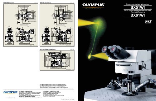
A dual commitment:Preventing vibration andprotecting living cell specimensOne design theme was central to the development of the new fixed stage microscopes from <strong>Olympus</strong> —achieve an evenhigher standard of stability and reliability in electro-physiological applications.The result is a wide range of advanced new features to avoid and prevent vibration. These innovationsinclude the introduction of a new observation method along with detailed analysis of operability andfurther refinements in image clarity. These improvementswork together to make patch clampoperations smoother and more efficient than ever before.Combined with the traditional excellence of UIS2 optics, the new <strong>Olympus</strong> fixed stage microscopesdefine new levels of quality in both performance and ease of use.BX51WI with Luigs & Neumann Accessories.An intermediate magnification changeris used to change magnification withoutchanging objectivesFront operation and reducedvibration for improved operabilityand stabilityIR-DIC and IR obliqueillumination condenser fordeep-section observationHigh N.A. objective offeringexcellent performance inIR-DIC observation,membrane potential imagingand fluorescence observationNewconceptFull-system physiologicalconfocal microscope,BX61WI with Z-axis motoris LSM readyRaising the objective andlowering the stage ofthe microscope enableseasier experimentation onsmall animalsFluorescence macro objectives formembrane potential observationA special bridge stage andX Y mover are available forthe microscope frame12
Interchanging low and high magnifications without ch anging objectives.A new concept in vibration-free design.A major concern for researchers conducting electro-physiology experiments is the vibration which occurs when switching objectivesand the resulting interference this can cause to the specimens and adjacent equipment.To solve this problem, <strong>Olympus</strong> introduces a new concept — the provision of an intermediate magnification changer in combinationwith the new High N.A. long working distance 20x objective that allows the user to switch between low and high magnificationswithout the need to switch objectives.New 20x objective (XLUMPLFLN20xW) N.A. 1.0; W.D.: 2.0 mmThe new 20x water immersion objective makes high-resolutionobservation possible with a wide range of intermediate magnificationlenses. Since exchanges between low and high magnification areperformed through the intermediate magnification changer, vibrationis reduced to a minimum and the usual concern about collisionsbetween objectives and patch clamp electrodes is eliminated.Simultaneous fluorescence and IR-DIC observationsWith the included 690 nm dichroic mirror in the WI-DPMC,fluorescence light is sent to the front port, and IR-DIC light is sent tothe back port allowing two cameras to image simultaneously with novibration introduced by light path selection.Variable magnification dual port (WI-DPMC)The WI-DPMC rear camera port includes a 2 position intermediatemagnification selector. A high magnification 4x intermediate lens isincluded and a (0.25x or 0.35x) low magnification lens is optional.High or low magnification selection is via a single lever with no clickstopsor detents allowing a specimen to be scanned and measuredwith minimal disturbance from vibration.775 nm and 900 nm IR-DIC compatible.*Available for 0.5x, 1x and 2x intermediate magnification lenses by special order.Variable Click-stopsAll click-stops, as when selecting between camera and observationmodes, can be adjusted to the point of no click and thus novibration.N.A.1.0XLUMPLFLN20xWObservation portFL/DICDual portWI-DPMCVideo portFL/DICDichroic mirrorIntermediatemagnificationchange leverAnalyzerIR-DIC portIR-DICExcitation lightFluorescence lightIR-DICTransmitted lightC mount4x lens0.35x(0.25x) lensLight path exchange leverMirror unit exchange turretTube lensMirror unitDIC Prism7× Intermediate magnification 0.35 x 80× Intermediate magnification 4 xXLUMPLFLN20xWN.A. 1.0CondenserDIC elementExamples usingintermediatemagnificationchanger WI-DPMC1/4 wave platePolarizerFilter turretIR filter(not to use with visiblelight DIC observations)IR-DIC observation, trigeminal motoneuron, Tomio Inoue Ph.D, Department of Oral Physiology, Showa University3 4 4
Front operation with no shock and no noise.A new concept in experimental operation.<strong>Olympus</strong> offers a wide choice of nosepieces for different applications.The new front operation system preventsinterference in patch clamping work.The design concept is simple and allowsfrequently performed operations likefocusing or filter exchange to be doneeasily at the front of the unit.Ample space is provided on both sides ofthe microscope frame and condenser, sothe necessary manipulation equipmentcan be positioned close to themicroscope.qwSwing nosepiece WI-SRE3Uniquie slim, compact design and front-to-back swingmotion permits objective changes without interfering withelectrodes and micromanipulators. Objective positioningincorporates a vibration-free counterspring mechanism.Slide nosepiece U-SLREThis nosepiece is designed for the attachment of onelarge diameter, low magnification fluorescence objective(XLFLUOR 2x/340 or 4x/340) and one objective withnormal (RMS) diamerter threads. Nosepiece motion is asimple horizontal slide.q Vibration-free shutterThe fluorescence shutter slides horizontallywith no detents and no vibration.ySingle position nosepieceWI-SNPXLU2Designed to accept the unique, large diameterXLUMPLFLN20xW objective.RMS adapter WI-RMSADThis adapter enables the attachment of an objective withRMS thread size to the WI-SNPXLU2.etuThe new swing-slide nosepiece preventsthe intrusion of air bubblesThis nosepiece features a new swing-slidemotion, whereby the objective swingsforward while being raised. As a result,the objective clears the walls of theperfusion chamber. This motion alsoprevents the trapping of air bubbles whenthe objective is lowered.w Mirror unit turret withadjustable click releaseThe click-stop on the 6 position turret canbe released with a precision screwdriver.rSwing-slide nosepiece WI-SSNPe Ample space around thecondenserFrame designed for ample space aroundthe condenser, making it easy to adjustNomarski DIC contrast, exchange filters,adjust the condenser's aperture stop andto easily switch between visible light,Nomarski DIC or IR-DIC.r Front focus knobs close tothe operator's handFine focus control is located at the front onboth sides of the microscope body.The knob on the right integrates bothcoarse and fine focus control.t Coarse focus lock leverWhen engaged at the desired position,the objective can be raised with the coarsefocus knob and then returned precisely toits original position.y A waterproofing sheetA waterproofing sheet, attached by thesupplied magnets, provides protectionagainst liquid overflow and spills.The sheet is large enough to protectthe frame, condenser and focusingmechanisms .u Remote power supply andhand switchThe remote TH4 power supply fortransmitted light is designed with nocooling fan to minimize electrical noise.Features on/off and intensity controls.Can also be used with the optional TH4-HShand switch providing light intensity andon/off control a maximal distance awayfrom the Faraday cage.Special bridge stage andmicroscope mover for the <strong>BXWI</strong>■ XY mover WI-XYMThe XY mover allows the movement ofthe microscope frame without movingeither the specimen or electrodes.Especially useful for multiple patchclampingexperiments, the XY moverhas convenient frontal controls.■ Bridge stage WI-XYSThis stage is designed for small animal,in-vivo observations; Stage height canbe lowered 50 mm by simply detaching the column spacers. Designed for use withthe XY mover (WI-XYM), the stage platform is compact requiring minimal deskspace. Stage top is pre-tapped and ferro-magnetic for flexibility in mountingmanipulators. * Some manipulators cannot be used, due to the size of the stage.Culture cell observationIX-SVL2 general purpose platformstage. Mounts for left or right handoperation. Provides stabile specimenX-Y movement.BX51WI+IX-SVL25 6
New functionality and solutions to meet a wide variety of needs.A powerful new concept.Experimenting with small animalsRaising the objective and lowering the stage toenable small animal experimentsThe arm height raising kit (WI-ARMAD) provides an additional 40 mmof clearance and is mounted between the microscope frame and thereflected light illuminator. Small animal experiments usually do notrequire transmitted light thus allowing the removal of the substagecondenser assembly. After removal, the stage may be lowered anadditional 50 mm, providing a total clearance increase of 90 mm.WI-ARMADMicroinjectionBX Stage and adapter for injection experimentsThe stage adapter WI-STAD is designed to allow the attachment ofa traditional microscope right or left hand stage to the WI frame.The compact design of the BX2 stage (U-SVRB-4, or U-SVLB-4)reduces the distance between the specimen and the manipulatorand creates a stable platform for injections.W.D.W.D.BX51WI+WI-STAD+U-SVRB-4W.D.18 mm18 mm40 mm18 mm40 mm50 mmNormal configuration 40 mm more clearance via WI-ARMAD Detaching the condenser assembly and lowering the stage by 50 mmprovides maximal clearanceMultiphoton Laser Scanning MicroscopePhotoactivationA variety of convenient units toadd light sources and control the lightLamphouse adapter U-LHADThis adapter allows the mounting of the dual port (U-DP) betweenthe microscope frame and lamp housing.Rectangular field stop U-RFSSDesigned for use with CCD cameras, prevents photobleaching ofthe specimen outside of the imaging area.Pinhole unit BX-RFSPOTLighting the cell via a pinhole allows experimentationon reaction to light. Optional MELLES GRIOT's ø16pinhole is used.U-LHADU-DPRemovable AS/FS(photo shown is BX-RFA)BX61WI — Built in Z-axis focusThe BX61WI frame incorporates a precise Z-axis focus with 0.01 µmstep size. Designed to incorporate the <strong>Olympus</strong> Fluoview scan unitand software, the BX61WI is ready for confocal z-stacks.Microscope frame includes programmable buttons for a widevariety of applications.Optional <strong>Olympus</strong> Fluoview series —Multiphoton Laser Scanning Microscope FV1000MPECombining the BX61WI with the FV1000MPE allows you to craft anoptimal system for the experiments you are performing, e.g. you can,in addition to multiphoton imaging, perform synchronization of laserlight stimulation and patch clamp signals.Faithful to the principles of multiphoton excitation imaging, theFV1000MPE allows bright, high-resolution observation of areas deepwithin cells and tissues without damaging the specimen.*FV1000MPE is a class 4 laser product.Configuration example: BX61WI+FV1000MPEU-RFSSBX-RFSPOT7 8
BX2WI SYSTEM DIAGRAMU-CMDPTSC-mount adapter forU-DPTSU-DPTSMulti double port tubeU-DPCADDual port tube withC-mountsU-PMTVC2XIR* 7U-PMTVC4XIR* 7C-mount camera portU-SPTSingle porttubeU-TVCAC* 7C-mount cameramagnificationchange unitU-TV0.35XC-2C-mount cameraport tube with0.35x lensU-TV0.5XC-3C-mount cameraport tube with0.5x lensU-TV0.63XCC-mount cameraport tube with0.63x lensU-TV1X-2Direct image camera portU-FMTF-mount adapterU-TMADT-mount adapterU-SMADSonymount adapterC-mount cameraU-BMADBayonetmount adapterU-CMAD3C-mount adapter* WHN10XWHN10X-HCROSSWHN10XEyepieceU-CT30Centering eyepiece*U-ETR-4* 1Erect image trinocular tube*U-TR30IRTrinocular tube for IR*U-TR30-2Trinocular tubeWI-DPMC* 6Double portmagnificationchange unit(DPMC4X included)DPMC0.25X0.25X adapter lens for WI-DPMCU-ANTAnalyzer for transmitted lightDPMC0.35X0.35X adapter lens for WI-DPMCU-CA* 3MagnificationchangerU-ECA* 3Magnificationchanger 2XU-FWOObservationfilter wheel(ø32 filter)U-DP* 2Dual portU-DP1XCDual port 1XC-mount camera(2/3" or less)U-TRU* 2 * 4Trinocularintermediate attachmentU-KPAIntermediate attachment forsimple polarizing observationWI-ANTIRIR analyzer for transmitted lightDPMC-ANAnalyzer for WI-DPMCDPMC-ANIRIR analyzer for WI-DPMCU-ANAnalyzer for reflected lightWI-ANIRIR analyzer for reflected lightWI-RSHReflected light shutterU-RFSSRectangularfield stopU-RLGADLight guideadapterFV5-FIRFiberLG-ULHADLamphouse adapterU-LH100XEAPO100 W xenon apolamphousingU-RX-TPower supply unit forxenon lampMirror unitBX-ARMArm for transmitted lightBX-URA2* 5Universal reflected lightilluminatorBX-RFA* 5BX fluorescence illuminatorBX-RFSPOTPinhole unitBX-RFAA* 5MotorizedfluorescenceilluminatorU-LHADLamphouse adapterU-FWRReflected filter wheelU-LH100HG100 W mercurylamphousingPetri dish plateU-SVL(R)B-4Mechanical stageWI-STAD* 8Stage adapterIX-SUSPStage plateIX-SVL2Mechanical stageWI-ARMADArm heightgaining KitFor nosepiece arm (WI-NPA)For frameBX51WIFFixed stage uprightmicroscope main bodyBX61WIFMotorized frameU-DP* 2Dual portU-LH100-3100 W halogenlamphousingU-LH100HGAPO100 W mercury apolamphousingU-RFL-TPower supply unit formercury lampU-SLRESlide nosepieceXLFLUOR2X/340XLFLUOR4X/340Fluorescence objectives forlow magnificationXL-CAPWater immersion lensfor XL objectiveWI-NPANosepiece armWI-SRE3Swing nosepieceWI-RMSADRMS adapterMPLN5XUMPLFLN10XWUMPLFLN20XWLUMPLFLN40XWLUMPLFLN60XWLUMFLN60XWObjectivesWI-SNPXLU2Single positioin nosepieceWI-DICTHRA2High resolutionDIC prismWI-DICT2DIC prism fortransmitted lightXLUMPLFLN20XW20X super high N.A.objectiveU-UCD8* 88 positionuniversalcondenserWI-TP137Quarter wavetint plateWI-SSNPSwing-slide nosepieceU-TLDDry top lensWI-OBCDLongworking distanceoblique condenserU-DIC10U-DIC10SU-DIC20U-DIC40U-DIC60Optical elementWI-FSHFixed stage adapterWI-DICDLongworking distanceDIC condenserWI-UCD WI-UCDLong working Swing-outdistance universal condensercondenserWI-DIC10HRWI-DIC20HRWI-DIC40HRWI-DIC60HRWI-DICXLU20HROptical element32BP775775 nmband pass filter32IR900900 nm filter32PO ø32 filterPolarizer32POIRIR polarizer*1 Slight vignetting may occur in the periphery of the field of view in combination with an additional intermediate attachment. *2 Slight vignetting may occur in the periphery of the field of view in combination with fluorescence illuminator. *3 Can be used with U-ETR-4 and U-TR30IR. Field of view is limited when using more than two intermediate tubes. *4 Sub port can accept U-TV1X-2, and 2/3" or less CCD cameras.*5 F.N. is 22 when fluorescence observation. *6 Use with fluorescent illuminator. *7 Acceptable camera adapters are U-TV1X-2, U-TVACA, U-PMTVC2XIR and U-PMTVC4X. *8 U-UCD8 cannot be used with WI-STAD.WI-XYMMicroscope moverManipulator45SCFHeat reflection filterWI-XYSBridge stageU-LH100IR100 W halogenlamphousing for IRU-RMTExtension cordBX-UCBControl boxTH4Externallight sourceBX61WIFU-IFFHFocus handleinterfaceMotorized unitTH4-HSHand switchU-FHFocus handle13 14