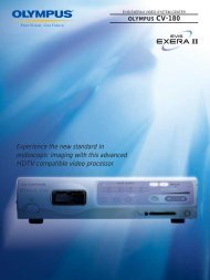
Sales Brochure: CV-160 EVIS EXERA Video System Center
Sales Brochure: CV-160 EVIS EXERA Video System Center
Sales Brochure: CV-160 EVIS EXERA Video System Center

You also want an ePaper? Increase the reach of your titles
YUMPU automatically turns print PDFs into web optimized ePapers that Google loves.
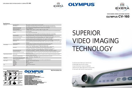
<strong>EVIS</strong> <strong>EXERA</strong> VIDEO SYSTEM CENTER OLYMPUS <strong>CV</strong> -<strong>160</strong><strong>EVIS</strong> <strong>EXERA</strong> VIDEO SYSTEM CENTEROLYMPUS <strong>CV</strong>-<strong>160</strong>SpecificationsObservationDocumentationImage storage and retrievalClassification(medical electrical equipment)Power supplySize<strong>Video</strong> signal outputWhite balanceStandard color chart outputDigital communication portColor tone adjustmentAutomatic gain control (AGC)ContrastIris mode selectionStructure enhancement settingEdge enhancement settingImage size selectionDefault resetFreeze screen displayRemote controlPatient dataPre-procedural patient dataRecalling and registeredscope information (scope ID function)Monitor outputMemorization of selected settingType of protection against electric shockDegree of protection against electricshock of applied partVoltageFrequencyInput currentFuse ratingDimensionsWeightVBS composite, Y/C, RGB; simultaneous output is possible.By pressing the white balance switch on the front panel, automatic white balance can be performed.After initialization, white balance settings are retained in memory for <strong>EVIS</strong> <strong>EXERA</strong> <strong>160</strong> Series scopes.The color bar image can be displayed using theColor Barkey on the keyboard.Ethernet (100BASE-TX)CHROMAcontrol: 7steps, Rcontrol: 7steps, Bcontrol: 7stepsThe image can be electrically amplified when the light is inadequate due to the distal end of the videoscope being toofar from the subject.Normal Contrast mode: Normal imageLow Contrast mode: The dark areas are lighter and the light areas are darker than in a normal image.High Contrast mode: The dark areas are darker and the light areas are lighter than in a normal image.Average: Normal observation Peak: When focusing on and/or observing a small bright area.The detailed patterns and edges of endoscopic images are emphasized electronically to increase image sharpness.The structure enhancement level can be switched betweenLow,MediumandHigh The edges of endoscopic images are emphasized electronically to increase image sharpness.The edge enhancement level can be switched betweenLow,MediumandHigh The size of endoscopic images can be changed using theImage Sizekey on the keyboard or endoscope switch.The following settings can be restored by pressing theRESETPrinter QTY.A stationary image can be displayed using the endoscope switch, keyboard or OVC switch.The following ancillary equipment can be controlled by the endoscope remote switch,the front panel and the keyboard. (Only the specified device types are valid.)Monitor VTR <strong>Video</strong> printer equipment Digital video cassette recorder Flushing pumpThe following data and modes can be displayed on the video monitor using the keyboard.1. ID number 2. Patient name 3. Sex, age 4. Date of birth 5. Date/time (built-in clock, stopwatch)6. Frame number 7. VTR condition 8. Picture quality selection 9. Physician 10. CommentsFor a maximum of 40 patients. 1. ID number 2. Patient name 3. Sex, age 4. Date of birth 5. PhysicianThe following scope-related data stored in the memory chip of the scope can be recalled and displayed on the screen.Scope Model, Serial No., Comments, Cumulative Uses, Check Period, Service Contract, Warranty Date, Owner,Customer ID No., ID Ver.Using the monitor output switch on the front panel, it is possible to select an image from the endoscope or ancillaryequipment for display on the monitor.The following settings on the front panel are retained even when the power is turned OFF.Color Enhancement White balance IrisClassType BF applied part(The applied part where classification types are not marked is Type BF applied part.)NTSC 100—120 V AC, PAL 220—240 V AC50/60 HzNTSC 1.0 A , PAL 0.5 A3.15 A , 250 V370 (W) 72 (H) 20 (D) mm8 kgSUPERIORVIDEO IMAGINGTECHNOLOGYAn advanced system featuring an impressive arrayof leading-edge video functions includingstructure enhancement, image size enlargement,scope ID function, digital image terminal, and much more,for high-quality, high-resolution image reproductionSan-Ei Building, 22-2, Nishi Shinjuku 1-chome, Shinjuku-ku, Tokyo, JapanPostfach 10 49 08, 20034 Hamburg / Wendenstrasse 14-16, 20097 Hamburg, Germany2 Corporate <strong>Center</strong> Drive, Melville, N.Y. 11747-3157, U.S.A.Keymed House, Stock Road, Southend-on-Sea, Essex SS2 5QH, England491B River Valley Road #12-01/04,Valley Point Office Tower,Singapore 248373Rm. 818, South Tower, Beijing Kerry Centre, No.1 Guanghua Road, Chaoyang District, Beijing, 100020, China117071, Moscow, Malaya Kaluzhskaya 19, bld. 1, fl.2, Russia1/104 Ferntree Gully Road, Oakleigh, VIC 3166, Australia
More features, more functions,and more performance make this innovative systemthe new standard for the 21st centuryDesigned specifically to maximizethe capabilities of our new line ofhigh-performance <strong>EVIS</strong> <strong>EXERA</strong><strong>160</strong> Series scopes, the <strong>CV</strong>-<strong>160</strong>video system center delivers highresolutionimages with improvedcolor reproduction capability anda larger display size.An improved video signalprocessing system enables clearobservation of minute details toensure more accurate examinationand treatment.Structure EnhancementStructure enhancementhighlights minute details.By electronically emphasizing minutedetails on the mucous membrane,the <strong>CV</strong>-<strong>160</strong>’s powerful structureenhancement circuitry makes endoscopicimages sharper without increasing noise.Unlike conventional edge enhancementfunctions, this system uses frequenciesspecifically suited to endoscopicimages to ensure more accurateobservation. Enhancement levels canbe selected by the user, allowing moreprecise control over image rendering.With enhancement, it is much easier toobserve minute tissue textures andsubtle color variations on the mucousmembrane surface.Full height expanded displaymode facilitates observation.The <strong>CV</strong>-<strong>160</strong> offers a full height mode thatuses the full vertical area of the monitorscreen to display images. The resultingexpanded display is much larger than waspossible with previous models and allowscloser examination of the image area.Full height display modeMedium display mode35% more compact design savesspace in your endoscopy suite.With their new, more compact designs, the<strong>CV</strong>-<strong>160</strong> and the CLV-<strong>160</strong> xenon light sourcecombine to form a system that’s a full 80 mmnarrower and 35% smaller than our previousmodels.The entire system can be installed on adedicated cart together with a video monitorand other ancillary equipment.Ergonomically designedfront panel and newlydesigned keyboard.Large membrane switches and indicators onthe front panel of the <strong>CV</strong>-<strong>160</strong> makeoperations clear and simple. The newlydesigned keyboard features a moreergonomic, user-friendly design and also fitssnugly on the dedicated cart.Dual-purpose system increasesendoscopy suite efficiency.The <strong>CV</strong>-<strong>160</strong> has been designed to eliminateredundancy and maximize the efficiency ofyour endoscopy suite. In addition to <strong>EVIS</strong><strong>EXERA</strong> <strong>160</strong> Series scopes, this versatilecenter can also accommodate <strong>EVIS</strong> <strong>EXERA</strong>video bronchoscopes, enabling you to usethe same system for both gastrointestinalendoscopy and bronchoscopy.<strong>CV</strong>-<strong>160</strong>Gastrointestinal<strong>Video</strong>scopes[ Ultra mini-CCD scope ]GIF-XP<strong>160</strong><strong>CV</strong>-140[ Q type scopes ]GIF-Q<strong>160</strong>CF-Q<strong>160</strong>AL/IGIF-Q140 etc.<strong>CV</strong>-100[ Other scopes ]GIF-<strong>160</strong>PCF-<strong>160</strong>AL/IGIF-140GIF-130GIF-100 etc.<strong>Video</strong>BronchoscopesBF-P<strong>160</strong>BF-<strong>160</strong>BF-1T<strong>160</strong>Scope ID function for moreefficient management and control.The <strong>CV</strong>-<strong>160</strong>’s scope ID function receivesdata from the memory chip built into theconnected <strong>EVIS</strong> <strong>EXERA</strong> <strong>160</strong> Series scopeand displays that information on themonitor. ID data includes model name andserial number, aswell as the numberof times the scopehas been connectedto the <strong>CV</strong>-<strong>160</strong>.The scope IDfunction alsoenables automaticcontrol of whitebalance.Memory chipbuilt into <strong>EVIS</strong> <strong>EXERA</strong><strong>160</strong> Series scopesScope information menuStart-up displayLevel 1 Level 7Level 4 Level 8Compatibility with<strong>EVIS</strong> 100/130/140 Series scopes.To ensure that your existing investment inendoscopic technology is not wasted, the<strong>CV</strong>-<strong>160</strong> is also compatible with all of our<strong>EVIS</strong> 100/130/140 Series gastrointestinalscopes.Digital image terminal for two-waycommunication with a PC.The <strong>CV</strong>-<strong>160</strong> features a digital imageterminal that will enable direct transfer ofendoscopic still images to a PC for storageusing dedicated interface software.