capsule experience - MG Lorenzatto
capsule experience - MG Lorenzatto
capsule experience - MG Lorenzatto

Create successful ePaper yourself
Turn your PDF publications into a flip-book with our unique Google optimized e-Paper software.
10<br />
Chapter 1<br />
THE OPTICAL DOME<br />
The optical dome and its relationship to the other optical<br />
components create two important advantages in <strong>capsule</strong><br />
endoscopy:<br />
a. Improved illumination efficiency.<br />
b. Favorable imaging geometry within the field of view.<br />
Another minor factor which may be contributing to image<br />
quality is the presence of a fluid interface between the optical<br />
dome and tissue at the time of examination improving the<br />
resolution of different anatomical structures such as villi.<br />
Finally, another advantage of the optical dome is that it lends<br />
itself to cleaning by the GI tract mucosa.<br />
Geometric and optical differences between image acquisition<br />
in wired and wireless endoscopy are compared in Figure 2A<br />
and Figure 2B. Figure 2A depicts the geometrical relationship<br />
for a push enteroscope inserted into the intestine. Geometrical<br />
relationship for the M2A ® <strong>capsule</strong> endoscope is depicted in<br />
Figure 2B. The standard endoscope (Figure 2A) includes an<br />
illumination source (2), and a lens (3), producing the field of<br />
illumination (4), and the field of view (5) shown in Figure 2A.<br />
Collapse of the intestinal wall (6) may obscure either the field<br />
of view or field of illumination. This technical problem is<br />
resolved in standard endoscopy by insufflating the intestine<br />
with air (7), resulting in the distancing of the intestinal muco<br />
from the tip of the endoscope, clearing both the field of view<br />
and the field of illumination. Air insufflation also enables the<br />
operator to orient the tip of the endoscope in the proper<br />
luminal direction, and to advance the endoscope tip accordingly.<br />
Figure 2B shows the wireless <strong>capsule</strong> endoscope (1) in the<br />
intestine. The <strong>capsule</strong> includes its illumination sources (2),<br />
and the lens (3), comprising a field of illumination (4), and a<br />
field of view (5) respectively. In order to prevent collapse of<br />
Physiological Endoscopy<br />
the intestinal wall (6), and resultant obscuring of either the<br />
field of illumination or the field of view, a specially designed<br />
optical dome (8) covers both the source of illumination and<br />
the lens. The space remaining between the dome and the<br />
intestinal wall may at times be occupied by fluid.<br />
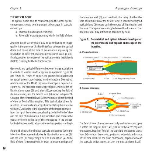
Figure 2. Geometrical and optical interrelationships for<br />
the enteroscope and <strong>capsule</strong> endoscope in the<br />
intestine.<br />
A - Push enteroscope<br />
2 - Illumination source<br />
1 - Endoscope inside<br />
intestine<br />
6 - Intestinal wall<br />
B - Capsule endoscope<br />
6 - Intestinal wall<br />
3 - Lens<br />
4 - Field of illumination 5 - Field of view<br />
3 - Lens<br />
1 - Wireless <strong>capsule</strong><br />
endoscope inside<br />
intestine<br />
7 - Air filling, due to insufflation<br />
8 - Optical dome<br />
The field of view of most commercially available endoscopes<br />
is within the range of 120˚-140˚, similar to the M2A ® <strong>capsule</strong><br />
endoscope. Depth of field of the standard endoscope starts<br />
from 3-5mm from the endoscope tip and extends to a distance<br />
of 100 mm. Unlike in the push endoscope, depth of view of<br />
the <strong>capsule</strong> endoscope starts on the optical dome itself.<br />
b<br />
2 - Illumination source<br />
4 - Field of illumination<br />
5 - Field of view<br />
a<br />
7 - Liquid filling,<br />
intestinal<br />
liquids