In vivo and In vitro Genotoxic Effects of Zerumbone
In vivo and In vitro Genotoxic Effects of Zerumbone
In vivo and In vitro Genotoxic Effects of Zerumbone

Create successful ePaper yourself
Turn your PDF publications into a flip-book with our unique Google optimized e-Paper software.
CARYOLOGIA Vol. 63, no. 1: 11-17, 2010<strong>In</strong> <strong>vivo</strong> <strong>and</strong> <strong>In</strong> <strong>vitro</strong> <strong>Genotoxic</strong> <strong>Effects</strong> <strong>of</strong> <strong>Zerumbone</strong>Al-Zubairi 1,3 Adel S., Ahmad Bustamam Abdul 1,2, *, Mohammed Yousif 1 ,Siddig Ibrahim Abdelwahab 1 , Manal Mohamed Elhassan 1 <strong>and</strong> Syam Mohan 11Laboratory <strong>of</strong> Cancer Research MAKNA-UPM, <strong>In</strong>stitute <strong>of</strong> Biosciences (IBS), University Putra Malaysia, Serdang,43400, Selangor DE, Malaysia.2Department <strong>of</strong> Biomedical Sciences, Faculty <strong>of</strong> Medicine & Health Sciences, University Putra Malaysia, Serdang,43400, Selangor D.E., Malaysia.3Department <strong>of</strong> Biochemistry <strong>and</strong> Molecular Biology, Faculty <strong>of</strong> Medicine <strong>and</strong> Health Sciences, University <strong>of</strong>Sana’a, Sana’a, Yemen.Abstract — <strong>Zerumbone</strong> (ZER) is derived from Zingiber zerumbet smith from the Zingiberaceae family. It hasbeen shown to have anti-cancer <strong>and</strong> apoptosis-inducing properties against various human tumour cells. The aim<strong>of</strong> our study was to assess the genotoxic effects <strong>of</strong> ZER in cultured human peripheral blood lymphocytes, ChineseHamster Ovary (CHO) cells <strong>and</strong> rat bone marrow polychromatic erythrocytes (PCEs) using micronucleustest (MN). All in <strong>vitro</strong> treatments were carried out in the absence <strong>of</strong> any exogenous metabolic activation system.Mitomycin C (MMC) was used as a positive control for in <strong>vitro</strong> treatments, while cisplatin was used as a positivemicronucleus inducer in rat bone marrow PCEs. ZER at high concentrations induced an apparent significantincrease in the frequency <strong>of</strong> micronuclei in <strong>vivo</strong> (1000 mg/kg b.w) <strong>and</strong> in <strong>vitro</strong> (40 <strong>and</strong> 80 µM) compared toconcurrent control values. Our in <strong>vivo</strong> <strong>and</strong> in <strong>vitro</strong> cytogenotoxicity studies suggest that high doses <strong>of</strong> ZER maybe genotoxic <strong>and</strong> cytotoxic.Keywords: CHO, genotoxicity, human peripheral blood lymphocytes, micronucleus, MNPCEs, zerumbone.INTRODUCTION*Corresponding authors: phone: +603-89462124; fax:+603-89462101; e-mail: abustamam@putra.upm.edu.my;adelalzubairi@hotmail.com.Zingiber zerumbet smith is used in local traditionalmedicine as a cure for a number <strong>of</strong> illnesses,locally known as ‘lempoyang’ wild gingerbelongs to Zingiberaceae family. It is native toSouth East Asia but has been widely cultivatedplant in village gardens throughout the tropical<strong>and</strong> subtropical area around the world <strong>and</strong>has naturalized in some areas for its medicinalproperties. <strong>In</strong> some Southeast Asian countries,the rhizomes <strong>of</strong> the plant are employed as traditionalmedicines for anti-inflammation, whilethe young shoots <strong>and</strong> inflorescence are used ascondiments. Scientific research towards Zingiberzerumbet proved that it contained a suppressiveeffect which was conducted by a bioactivecompound, zerumbone. It has been shown thatzerumbone is one <strong>of</strong> the most promising chemopreventiveagents against colon <strong>and</strong> skin cancer(MURAKAMI et al. 2004). It was reported to suppresscolonic tumour marker formation in rats<strong>and</strong> potentiated TRAIL-induced apoptosis inhuman HCT116 colon cancer cells (YODKEEREEet al. 2009). The compound was shown to inhibitthe proliferation <strong>of</strong> human colonic adenocarcinomacell lines in a dose-dependent manner,while the growth <strong>of</strong> normal human dermal <strong>and</strong>colon fibroblast was less affected (MURAKAMI etal. 2004). <strong>Zerumbone</strong> was further demonstratedto inhibit both azoxymethane-induced rataberrant crypt foci <strong>and</strong> phorbol ester-inducedpapilloma formation in mouse skin a furtherindication <strong>of</strong> its efficacy to prevent colon <strong>and</strong>skin cancers (MURAKAMI et al. 2004; TANAKA etal. 2001). Recently, Sung et al., (2009) reportedzerumbone as modulator for osteoclastogenesis
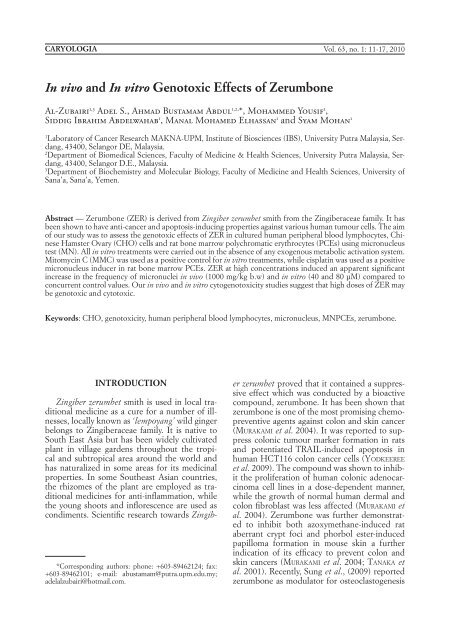
12AL-ZUBAIRI, ABDUL, YOUSIF, ABDELWAHAB, ELHASSAN <strong>and</strong> MOHANinduced by RANKL <strong>and</strong> breast cancer (SUNG etal. 2009).<strong>Genotoxic</strong>ity studies <strong>of</strong> both naturally occurring<strong>and</strong> synthetic substances are <strong>of</strong> great interestbecause <strong>of</strong> the widespread <strong>and</strong> <strong>of</strong>ten chronicuse <strong>of</strong> herbal remedies, <strong>of</strong> modern medicinalproducts, <strong>of</strong> food ingredients, as well as <strong>of</strong> otherhousehold <strong>and</strong> environmental chemicals. Manyplant products contain compounds known tocause various diseases or even death in animals<strong>and</strong> humans (DEARFIELD et al. 2002; AMES <strong>and</strong>GOLD 1997; RASKIN et al 2002; RATES 2001). Syntheticsubstances present as environmental pollutants,<strong>and</strong> toxicants may cause similar effects(AMES <strong>and</strong> GOLD 1997; RATES 2001). Many natural<strong>and</strong> synthetic compounds have been reportedto act as mutagens <strong>and</strong>/or carcinogens (VARGASet al. 1990).A variety <strong>of</strong> in <strong>vitro</strong> genotoxicity test systemshave been developed including the culturedmammalian cell systems such as human peripheralblood lymphocytes cells (PBL) or Chinesehamster ovary (CHO) cells (DEAN <strong>and</strong> DAN-FORD 1984; SCOTT et al. 1990), for the screening<strong>of</strong> potentially mutagenic, carcinogenic <strong>and</strong>/orteratogenic agents. The CBMN assay in humanlymphocytes uses cytochalasin-B, an inhibitor<strong>of</strong> actins polymerisation, which prevents cytokinesiswhile permitting nuclear division (FENECH<strong>and</strong> MORLEY 1985). As a result, binucleated(BN) cells are produced, which are scored forthe presence <strong>of</strong> MN (FENECH 1993). The rodentbone-marrow MN test is the most widely usedshort-term in <strong>vivo</strong> assay for the identification <strong>of</strong>genotoxic effects such as chromosome damage<strong>and</strong> aneuploidy associated with mutagenesis <strong>and</strong>carcinogenesis (VANDERKERKEN et al 1989).Taking into account the lack <strong>of</strong> informationabout in <strong>vivo</strong> <strong>and</strong> in <strong>vitro</strong> cytogenetic effects <strong>of</strong>zerumbone, we decided to provide some data onthe cytogenetic activity <strong>of</strong> this compound. Herewe report the results obtained on the cytogeneticeffects <strong>of</strong> zerumbone in <strong>vivo</strong> using rat bonemarrow erythrocyte micronucleus (MNPCE),in <strong>vitro</strong> chromosomal aberrations assay <strong>and</strong> micronucleustest using human lymphocytes (PBL)<strong>and</strong> micronucleus test in cultured CHO cell.MATERIALS AND METHODS<strong>Zerumbone</strong> extraction - <strong>Zerumbone</strong> was extractedin the laboratory <strong>of</strong> cancer research MAK-NA-UPM, University Putra Malaysia, from therhizomes <strong>of</strong> Zingiber zerumbet plant. The rhizomesobtained from the wet market in KualaLumpur, Malaysia. <strong>Zerumbone</strong> was extracted,isolated <strong>and</strong> purified using methanol extraction<strong>and</strong> column chromatography (CC) method. Theisolated <strong>and</strong> purified zerumbone crystals weresubjected to High Performance Liquid Chromatography(HPLC) <strong>and</strong> Liquid ChromatographyMass Spectrometry (LCMS) to confirm itspurity <strong>and</strong> molecular weight. Further, 13 C NMR<strong>and</strong> 1 H NMR analysis were conducted towardsthe zerumbone crystals to confirm its molecularstructure (Figure 1). A stock solution <strong>of</strong> zerumboneis prepared immediately before use in absoluteethanol (HmbG Chemicals).Chemicals - Mitomycin C (MMC), CytochalasinB (CB [CAS 4930-96-2] <strong>and</strong> Cisplatin [CAS15663-27-1] were obtained from Sigma, Geimsastain [CAS 67-56-1] <strong>and</strong> phytohaemagglutinin(PHA) were obtained from (Gibco, Germany)<strong>and</strong> Colcemid from (PAA Laboratories).Animals <strong>and</strong> their treatment for measurement <strong>of</strong>MNPCE - Male Sprague-Dawley rats (6-8 weeksold) weighing 170-200g were obtained from the<strong>In</strong>stitute <strong>of</strong> Medical Research, Kuala Lumpur.The rats were maintained in group per cage atroom temperature (25±1 C) <strong>and</strong> 12-h light: 12-hdark cycle <strong>and</strong> were given food <strong>and</strong> water ad libitum.The animals were acclimatised for at least5 days prior to dosing <strong>and</strong> were divided into fivegroups containing 3-6 rats each. Three dose levels<strong>of</strong> zerumbone (250, 500, 1000 mg/kg) weregiven intraperitoneal for 24h. Dose selection wasbased on preliminary experiments in which themaximum tolerable dose (MTD) was identifiedto be 1000 mg/kg body weight. An untreatedcontrol <strong>and</strong> a positive control (cisplatin 10 mg/kg) were also used to test the validity <strong>of</strong> the assay.The experiment complied with the guidefor Animal Care <strong>and</strong> Use Committee (ACUC),Faculty <strong>of</strong> Medicine <strong>and</strong> Health Sciences, UniversityPutra Malaysia.After 24h the animals were anesthetized withchlor<strong>of</strong>orm <strong>and</strong> sacrificed. For bone-marrowpreparations, both hind femora were isolated<strong>and</strong> the adherent muscle removed. The epiphyseswere cut <strong>of</strong>f <strong>and</strong> bone marrow cells wereflushed out with foetal bovine serum (PAA Laboratories).The suspension <strong>of</strong> bone marrow cells<strong>and</strong> foetal bovine serum was centrifuged for 10min at 1000 rpm. The resulting sediment wasre-suspended in foetal bovine serum. Bone marrowsmears were prepared from the resultingcell suspension. After air-drying <strong>and</strong> fixing for10 min in absolute methanol, slides were stainedwith Giemsa-staining method. The slides were
IN VIVO AND IN VITRO GENOTOXIC EFFECTS OF ZERUMBONE 13Fig. 1 — Molecular structure <strong>of</strong> zerumboneanalyzed in a blinded fashion using a Nikonlight microscope. Both normochromatic erythrocytes(NCE) <strong>and</strong> polychromatic erythrocytes(PCE) were scored for bone marrow activity<strong>and</strong> PCEs were scored for micronuclei (MN).A total <strong>of</strong> 2000 polychromatic erythrocytes werescored per animal for MNPCE <strong>and</strong> 200 erythrocyteswere counted for the PCE: NCE ratioaccording to the OECD guideline for testing <strong>of</strong>chemicals (mammalian erythrocyte micronucleustest), guideline No. 474 (OECD 1997). Forevery group <strong>of</strong> animals, the following parameterswere reported, the number <strong>of</strong> MN-containingcells/2000 PCE/animal, the number <strong>of</strong> PCE; thenumber <strong>of</strong> NCE <strong>and</strong> the NCE: PCE ratio.Micronucleus test in cultured human lymphocytes- Buffy coat (0.2 ml) was added to 5ml RPMI-1640 medium supplemented with 20% heat inactivatedfoetal bovine serum, antibiotics (penicillin<strong>and</strong> streptomycin) <strong>and</strong> L-glutamine (PAAlaboratories). Lymphocytes were stimulated byadding 2% phytohaemagglutinin (PHA) (Gibco).The cultured lymphocytes were incubatedfor 48h before treatment with zerumbone. Thecultures were incubated at 37°C for 72h <strong>and</strong>treated with zerumbone at 5, 10, 20, 40 <strong>and</strong>80µM during the last 24h. A control untreatedculture, MMC treated <strong>and</strong> ethanol treated cultureswere established as well. Cytochalasin-B(6µg/ml) was added to arrest cytokinesis at 44hafter the start <strong>of</strong> the culture. Then, the cells wereharvested by centrifugation (1000rpm, 10min),<strong>and</strong> the pellet was re-suspended in a pre-warmedhypotonic solution <strong>of</strong> 0.075M KCI for 5 min.Cells were re-centrifuged <strong>and</strong> fixed three timesin cold methanol: acetic acid (3:1). Slides wereprepared by dropping <strong>and</strong> air-drying. Finallyslides were stained with 5% Giemsa (pH 6.8) inphosphate buffer for 10 min, washed in distilledwater, dried at room temperature <strong>and</strong> mounted.Scoring - The induction <strong>of</strong> MN was determinedin 1000 binucleated cells with the cytoplasmwell preserved. <strong>In</strong> a blind test, using a Nikonmicroscope, cells containing 1 micronuclei werescored. The criterion for the identification <strong>of</strong>MN was according to Fenech (FENECH 1993). <strong>In</strong>each treatment, the numbers <strong>of</strong> mononucleated,binucleated, <strong>and</strong> polynucleated cells per 500cells were counted for cell cycle kinetic analysis<strong>and</strong> cytochalasin B proliferation index (CBPI) asdetermined in a blind test. Cells with well preservedcytoplasm, containing 1-4 nuclei, were
14AL-ZUBAIRI, ABDUL, YOUSIF, ABDELWAHAB, ELHASSAN <strong>and</strong> MOHANscored. The CBPI was calculated to determinepossible cytotoxic effects, according to OECDguideline number 487 (OECD 2004) using thefollowing formula:CBPI= MI + 2MII +3MIII+ 4MIV/Nwhere MI-MIV correspond to the numbers <strong>of</strong>cells with one, two, three <strong>and</strong> four nuclei <strong>and</strong>N is the total number <strong>of</strong> cells (SURALLES et al.1995).Micronucleus test (MN) in CHO - Chinese hamsterovary (CHO) cells were purchased fromECACC (UK). The cells grow as an adherentmonolayer in appropriate tissue culture vessels<strong>and</strong> were maintained in RPMI 1640 medium(PAA Laboratories, Germany) supplementedwith 10% foetal bovine serum (PAA Laboratories,Germany). The cells were incubated ina humidified tissue culture incubator at 37°C<strong>and</strong> 5% CO 2. The overnight cell cultures wereexamined under an inverted microscope. Duplicatecultures were prepared for each test substanceconcentration <strong>and</strong> controls. Control cultureswere h<strong>and</strong>led in a manner identical to thetreated ones. Mitomycin-C (Sigma) was used asa positive control. The treatment medium was 5ml <strong>of</strong> the cell culture medium with 10% foetalbovine serum, with the treatment concentrationor a control solution <strong>and</strong> the final concentrationswere 5, 10, 20, 40 <strong>and</strong> 80µM ZER. Thecells were cultured in the treatment medium for48h. After treatment, cells were washed twicewith 10 mL PBS, trypsinized with 0.05% trypsin<strong>and</strong> centrifuged for 5 min at 800 rpm. CHO cellswere then harvested <strong>and</strong> scored using the samemethod <strong>of</strong> human lymphocytes micronucleusmentioned above.Statistical analysis - Statistical analysis was performedusing SPSS 15. Rat bone marrow erythrocytesMN results were expressed as mean ±SE <strong>and</strong> were analyzed with one way analysis <strong>of</strong>variance, ANOVA, while the results from in <strong>vitro</strong>work were analyzed using Chi square analysis.All statistical tests were performed at the p
IN VIVO AND IN VITRO GENOTOXIC EFFECTS OF ZERUMBONE 15respectively. These results reveal that, ZER has agenotoxic activity only after treatment with highdrug concentrations (40 <strong>and</strong> 80 µM), inducingan increase in the frequency <strong>of</strong> binucleated cellswith MN (BNMN) when compared to the concurrentuntreated control cells (P
16AL-ZUBAIRI, ABDUL, YOUSIF, ABDELWAHAB, ELHASSAN <strong>and</strong> MOHANably elevated the frequencies <strong>of</strong> MN formationscompared to the concurrent controls, both inhuman lymphocyte when treated for 24h aswell as in CHO cells when treated for 48h. Thepercentage <strong>of</strong> structurally damaged cells in theMMC (positive control) treatment groups wasstatistically increased compared to the solventcontrol data indicating the responsiveness <strong>of</strong> thecells in this test system.Analysis <strong>of</strong> the frequency <strong>of</strong> occurrence <strong>of</strong>micronuclei in treated cells provides a comparativelyrapid <strong>and</strong> sensitive indication <strong>of</strong> bothchromosomal aberrations <strong>and</strong> chromosome lossthat lead to numerical chromosomal anomalies.Micronuclei are cytoplasmic chromatin masseswith the appearance <strong>of</strong> small nuclei that arisefrom chromosome lagging at anaphase or fromacentric chromosomal fragments. They providea quantifiable measure <strong>of</strong> recent DNA injury thatresult from when acentric fragments or wholechromosomes are left behind the main nucleusat telophase. An increase in the prevalence <strong>of</strong>MN in a population <strong>of</strong> cells indicates that chromosomedamage has occurred as a result <strong>of</strong> anexposure that caused either clastogenic or ananeuploidogenic effect (GONSEBATT et al. 1997;MAHATA et al. 2003). MN assay is a widely usedcytogenetic method to assess in <strong>vivo</strong> <strong>and</strong> in <strong>vitro</strong>chromosomal damage. However, the MN studyis the most reproducible one to show positiveeffects (ROBBIANO et al. 1998). Results <strong>of</strong> MNinduction in human lymphocytes revealed theability <strong>of</strong> this compound at high dose to act asaneugenic/clastogenic substance by inducingMN formation in human lymphocytes in <strong>vitro</strong>.The lethal dose (LD) <strong>of</strong> 2000 mg/kg b.w. wasdetermined in rat after intraperitoneal administration<strong>of</strong> ZER. <strong>In</strong> the present experimental conditions,a significant increase in rat bone marrowmicronuclei was recorded at the highest dose(1000 mg/kg b.w.). The induction <strong>of</strong> micronucleatederythrocytes following exposure to highdose <strong>of</strong> ZER indicates a potential for clastogenicity.<strong>In</strong> the present study, the observed inhibition<strong>of</strong> cell proliferation in the rat bone marrow illustratesthe cytotoxicity <strong>of</strong> ZER. These data indicatethe cytotoxic potential <strong>of</strong> ZER at higherexposure doses.Our results show that the increase in MN,<strong>and</strong> the decrease in the PCE:NCE ratio wasdose-dependent. We found that there is a linearrelationship between the ZER dose used <strong>and</strong> thefrequencies <strong>of</strong> micronuclei. It is concluded thatZER has a cytotoxic effect on bone marrow inrats, because the decrease in the ratio PCE:NCEwas observed at all doses compared with the control.Such a decreased ratio is <strong>of</strong>ten used as anindicator <strong>of</strong> bone marrow cytotoxicity or alterationsin erythropoiesis. <strong>In</strong> normal bone marrow,the PCE:NCE ratio is generally around 1:1.<strong>In</strong> conclusion our in <strong>vivo</strong> <strong>and</strong> in <strong>vitro</strong> cytogenotoxicitystudies suggest that high doses<strong>of</strong> zerumbone used in the present investigationmay be genotoxic <strong>and</strong> cytotoxic. It is importantto carry out more investigations using variouscytogenetic tests under different experimentalconditions to assess more the genotoxic effects<strong>of</strong> zerumbone.Acknowledgment — The authors would like toextend their utmost gratitude <strong>and</strong> appreciation toMOSTI (Ministry <strong>of</strong> Science, Technology <strong>and</strong> <strong>In</strong>novation)<strong>and</strong> The National Cancer Council Malaysia(MAKNA) for providing the research grants, IRPANo: 06-02-04-0720-EA001. The authors also wouldlike to convey their thanks to UPM for their providingadditional support <strong>of</strong> this work.TABLE 3 — Frequencies <strong>of</strong> micronucleus (MN) formation, cell cycle kinetics <strong>and</strong> CBPI on Human PBL culturestreated for 24h with different concentrations <strong>of</strong> zerumbone, mitomycin C <strong>and</strong> untreated control.Cell cycle kinetics aTreatment BN MNi %MNi M1 M2 M3 % BN CBPIZII (µM) 0 1000 4 0.40 170 251 79 50.2 1.81810 1000 4.0 0.40 185 201 114 40.2 1.85820 1000 4.0 0.40 180 207 113 41.4 1.86640 1000 16.0* 1.60 203 222 75 44.4 1.74480 1000 75.0** 7.50 400 65 35 13.0 1.270EtOH 1000 3.0 3.00 175 248 77 49.6 1.804MMC (µg/ml) 1.2 1000 100** 10.00 384 82 34 16.4 1.300The numbers <strong>of</strong> mononucleated (M1), binucleated (M2), <strong>and</strong> polynucleated (M3) cell per 500 cells were quantitated for cellcycle kinetic analysis.*P < 0.05, significantly different from control.
IN VIVO AND IN VITRO GENOTOXIC EFFECTS OF ZERUMBONE 17REFERENCESAMES B.N. <strong>and</strong> GOLD L.S., 1997 — Environmentalpollution, pesticides <strong>and</strong> the prevention <strong>of</strong> cancer:misconceptions. FASEB Journal, 11: 1041-1052.DEAN B.J. <strong>and</strong> DANFORD N., 1984 — Assays for the detection<strong>of</strong> chemically-induced chromosome damagein cultured mammalian cells. <strong>In</strong>: Venitt S, ParryJM (eds.), Mutagenicity Testing: A Practical Approach,p 187-232IRL Press, Ltd., Oxford.DEARFIELD K.L., CIMINO M.C., MCCARROLL N.E.,MAUER I. <strong>and</strong> VALCOVIC L.R. 2002 — <strong>Genotoxic</strong>ityrisk assessment: a proposed classification strategy.Mutation Research, 521: 121-135.FENECH M. <strong>and</strong> MORLEY A.A., 1985 — Measurement<strong>of</strong> micronuclei in lymphocytes. Mutation Research,147: 29-36.FENECH M., 1993 — The cytokinesis-block micronucleustechnique: A detailed description <strong>of</strong> the method<strong>and</strong> its application to genotoxicity studies in humanpopulations. Mutation Research, 285: 35-44.GONSEBATT M.E., VEGA L. <strong>and</strong> SALAZA A.M., 1997 —Cytogenetic effects in human exposure to arsenic.Mutation Research, 386: 219-228.KOSHIMIZU K., OHIGASHI H., TOKUDA H., KONDO A.<strong>and</strong> YAMAGUCHI K., 1998 — Screening <strong>of</strong> edibleplants against antitumor promoting activity. CancerLetters, 39: 247-257.MAHATA J., BASU A., GHOSHAL S., SARKAR J.N., ROYA.K., PODDAR G., NANDY A.K., BANERJEE A., RAYK., NATARAJAN A.T., NILSSON R. <strong>and</strong> GIRI A.K.,2003 — Chromosomal aberrations <strong>and</strong> sister chromatidexchanges in individuals exposed to arsenicthrough drinking water in West Bengal, <strong>In</strong>dia. MutationResearch, 534: 133-143.MURAKAMI A., TAKHASIH D., KINOSHITA T., KOSHIMIZAK., KIM H.W., YOSHIHIRO A., NAKAMURA Y., JI-WAJINDA S., TERAO J. <strong>and</strong> OHIGASHI H., 2002 —<strong>Zerumbone</strong>, a Southeast Asian ginger sesquiterpene,markedly suppresses free radical generation,pro-inflammatory protein production <strong>and</strong> cancercell proliferation accompanied by apoptosis: the alpha,beta-unsaturated carbonyl group is a prerequisite.Carcinogenesis, 23(5): 795-802.MURAKAMI A., TANAKA T., LEE J.Y., SURH Y.J., KIMH.W., KAWABATA K., NAKAMURA Y., JIWAJINDA S.<strong>and</strong> OHIGASHI H., 2004 — <strong>Zerumbone</strong>, a sesquiterpenein subtropical ginger, suppresses skin tumorinitiation <strong>and</strong> promotion stages in ICR mice. <strong>In</strong>ternationalJournal <strong>of</strong> Cancer, 110: 481-490.OECD (Organization for Economic Co-operation<strong>and</strong> Development) — <strong>In</strong> <strong>vitro</strong> mammalian chromosomeaberration test. 1997 OECD Guidelinefor the Testing <strong>of</strong> Chemicals 473.OECD (Organization for Economic Co-operation<strong>and</strong> Development)., 2004 — <strong>In</strong> <strong>vitro</strong> MicronucleusTest. OECD Guideline for the Testing <strong>of</strong> Chemicals487.PRATT W.B., RUDDON R.W., ENSMINGER W.D. <strong>and</strong>MAYBAUM J., 1994 — The Anticancer Drugs. seconded., 265-284. Oxford University Press, NewYork.RASKIN I., RIBNICKY D.M., KOMARNYSKY S., ILIC N.,POULEV A., BERISJUK N., BRINKER A., MORENOD.A., RIPOLL C., YAKOBY N., O’NEAL J.M., CORN-WELL T., PASTOR I. <strong>and</strong> FRIDLENDER B., 2002 —Plants <strong>and</strong> human health in the twenty-first century.Trends in Biotechnology, 20: 522-531.RATES S.M.K., 2001 — Plants as source <strong>of</strong> drugs. Toxicon,39: 603-613.ROBBIANO L., MERETO E., MORANDO A.M., PASTOREP. <strong>and</strong> BRAMBILLA G., 1998 — <strong>In</strong>creased frequency<strong>of</strong> micronucleated kidney cells in rats exposed tohalogenated anaesthetics. Mutation Research, 413:1-6.SCOTT D., DEAN B.J., DANFORD N.D. <strong>and</strong> KIRKLANDD.J., 1990 — Metaphase chromosome aberrationassays in <strong>vitro</strong>. <strong>In</strong>: Kirkl<strong>and</strong> DJ (ed.), Basic MutagenicityTests: UKEMS Recommended Procedures,p 62-86. UKEMS/University <strong>of</strong> CambridgePress, Cambridge.SUNG B., MURAKAMI A., OYAJOBI B.O., AGGARWALB.B., 2009 — <strong>Zerumbone</strong> abolishes RANKL-inducedNF-KB activation, inhibits osteoclastogenesis,<strong>and</strong> suppresses human breast cancer inducedbone loss in athymic nude mice. Cancer Research,69: 1477-1484.SURALLES J., XAMENA N., CREUS A., CATALAN J., NORP-PA H. <strong>and</strong> MARCOS R., 1995 — <strong>In</strong>duction <strong>of</strong> micronucleiby five pyrethroid insecticides in whole-blood<strong>and</strong> isolated human lymphocyte cultures. MutationResearch, 341: 169-184.TANAKA T., SHIMIZU M., KOHNO H., YOSHITANI S.,TSUKIO Y., MURAKAMI A., SAFITRI R., TAKAHASHID., YAMAMOTO K., KOSHIMIZU K., OHIGASHIH. <strong>and</strong> MORI H., 2001 — Chemoprevention <strong>of</strong>azoxymethane-induced rat aberrant crypt foci by dietaryzerumbone isolated from Zingiber zerumbet.Life Science, 69: 1935-1945.VARGAS V.M.F., MOTTA V.E.P., LEITAO A.C. <strong>and</strong> HER-IQUES J.A.P., 1990 — Mutagenic <strong>and</strong> genotoxic effects<strong>of</strong> aqueous extracts <strong>of</strong> Achyroline satureoidesin prokaryotic organisms. Mutation Research, 240:13-18.VANDERKERKEN K., VANPARYS P., VERSCHAEVE L. <strong>and</strong>KIRSCH-VOLDERS M., 1989 — The mouse bone marrowmicronucleus assay can be used to distinguishaneugens from clastogens. Mutagenesis, 4: 6-11.WEIJL N.I., CLETON F.J. <strong>and</strong> OSANTO S., 1997 — Freeradicals <strong>and</strong> antioxidants in chemotherapy-inducedtoxicity. Cancer Treatment Reviews, 23: 209-224.YODKEEREE S., SUNG B., LIMTRAKUL P. AND AGGAR-WAL B.B., 2009 — <strong>Zerumbone</strong> Enhances TRAIL-<strong>In</strong>duced Apoptosis through the <strong>In</strong>duction <strong>of</strong> DeathReceptors in Human Colon Cancer Cells: Evidencefor an Essential Role <strong>of</strong> Reactive Oxygen Species.Cancer Research, 69: 6581-6589.Received February 6 th 2009; accepted April 1 th 2010