surgical treatment of subluxation and dislocation of the hips in ...
surgical treatment of subluxation and dislocation of the hips in ...
surgical treatment of subluxation and dislocation of the hips in ...
Create successful ePaper yourself
Turn your PDF publications into a flip-book with our unique Google optimized e-Paper software.
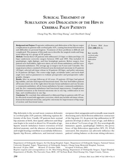
C.T. Wu, S.C. Huang, <strong>and</strong> C.H. ChangSURGICAL TREATMENT OFSUBLUXATION AND DISLOCATION OF THE HIPS INCEREBRAL PALSY PATIENTSChung-T<strong>in</strong>g Wu, Shier-Chieg Huang, 1 <strong>and</strong> Chia-Hsieh Chang 2Background <strong>and</strong> Purpose: Progressive <strong>subluxation</strong> <strong>and</strong> <strong>dislocation</strong> <strong>of</strong> <strong>the</strong> hip are majorcomplications <strong>in</strong> patients with cerebral palsy (CP), caus<strong>in</strong>g functional deterioration<strong>and</strong> difficulties <strong>in</strong> personal hygiene. Treatment <strong>of</strong> <strong>the</strong>se problems is difficult <strong>and</strong>complicated. The purpose <strong>of</strong> this study was to describe <strong>the</strong> <strong>surgical</strong> results <strong>and</strong> longtermfollow-up <strong>in</strong> a group <strong>of</strong> CP patients.Methods: Twenty-three CP patients with subluxated (15 <strong>hips</strong>) or dislocated <strong>hips</strong> (12<strong>hips</strong>) underwent corrective surgery between 1985 <strong>and</strong> 1993. This <strong>in</strong>cluded 11quadriplegic, eight diplegic, <strong>and</strong> four hemiplegic patients. Before surgery, fourpatients were bed-ridden, eight were sitters, six were house-ambulators, <strong>and</strong> five werecommunity-ambulators. The average age at surgery was 8 years <strong>and</strong> 5 months. The<strong>surgical</strong> procedures consisted <strong>of</strong> femoral varus derotational osteotomy <strong>in</strong> 21 patients(25 <strong>hips</strong>), selected s<strong>of</strong>t tissue release <strong>in</strong> 18 patients (22 <strong>hips</strong>), <strong>and</strong> pelvic osteotomy<strong>in</strong> 18 patients (20 <strong>hips</strong>). The center–edge angle, acetabular <strong>in</strong>dex, <strong>and</strong> neck–shaftangle were used as parameters to evaluate preoperative <strong>and</strong> postoperative radiographicchanges.Results: After an average follow-up <strong>of</strong> 4.8 years, 19 patients (22 <strong>hips</strong>) had ga<strong>in</strong>edhip stability, <strong>and</strong> also had improved functional status. The four bed-ridden patientsall became sitters; six <strong>of</strong> <strong>the</strong> eight sitters became house-ambulators <strong>and</strong> one becamea community-ambulator; all six house-ambulators became community-ambulators,<strong>and</strong> <strong>the</strong> five community-ambulators had functional improvement. Complications<strong>in</strong>cluded nonunion at <strong>the</strong> femoral osteotomy site <strong>in</strong> one hip, re<strong>dislocation</strong> <strong>in</strong> two<strong>hips</strong>, <strong>and</strong> re<strong>subluxation</strong> <strong>in</strong> one hip.Conclusions: We conclude that subluxated or dislocated <strong>hips</strong> <strong>in</strong> patients with CP canbe effectively treated with aggressive correction, which may <strong>in</strong>clude s<strong>of</strong>t tissue release,femoral derotational osteotomy, <strong>and</strong> pelvic osteotomy for improvement <strong>of</strong> hip range<strong>of</strong> motion <strong>and</strong> functional status.(J Formos Med Assoc2001;100:250–6)Key words:cerebral palsy<strong>subluxation</strong><strong>dislocation</strong><strong>hips</strong>urgeryHip deformity is <strong>the</strong> second most common deformity<strong>in</strong> cerebral palsy (CP) patients, follow<strong>in</strong>g equ<strong>in</strong>us deformity<strong>of</strong> <strong>the</strong> foot. Usually, CP patients have normalhip anatomy at birth, <strong>the</strong>n spastic adductors <strong>and</strong> iliopsoasbeg<strong>in</strong> to be noted at about 6 to 18 months <strong>of</strong> age.Spastic muscle imbalance fosters retention <strong>of</strong> fetalskeletal anatomy, <strong>and</strong> <strong>the</strong> absence <strong>of</strong> normal motion<strong>and</strong> weight bear<strong>in</strong>g contribute to acetabular deficiency.Spastic hip flexors, adductors, <strong>and</strong> <strong>in</strong>ternal rotatorsoverpower <strong>the</strong>ir antagonists <strong>and</strong> eventually cause muscleshorten<strong>in</strong>g <strong>and</strong> a fixed flexion-adduction contracture<strong>of</strong> <strong>the</strong> hip jo<strong>in</strong>t [1]. In general, hip <strong>subluxation</strong> <strong>in</strong> CPpatients develops at about 3 years <strong>of</strong> age, <strong>and</strong> <strong>dislocation</strong>at about 7 to 8 years <strong>of</strong> age [2]. For neurologicallyimmature, spastic, quadriplegic children, <strong>the</strong> prevalence<strong>of</strong> hip <strong>dislocation</strong> approaches 50% [3]. If leftuntreated, this situation can adversely <strong>in</strong>fluence <strong>the</strong>patient’s sitt<strong>in</strong>g balance or decrease sitt<strong>in</strong>g tolerance.Department <strong>of</strong> Orthopedic Surgery, En Chu Kong Hospital, Taipei, 1 Department <strong>of</strong> Orthopedic Surgery, National TaiwanUniversity Hospital, Taipei, 2 Department <strong>of</strong> Orthopedic Surgery, Chang Gung Memorial Hospital, Taoyuan.Received: 19 June 2000. Revised: 27 October 2000. Accepted: 6 February 2001.Repr<strong>in</strong>t requests <strong>and</strong> correspondence to: Dr. Shier-Chieg Huang, Department <strong>of</strong> Orthopedic Surgery, Taipei Hospital,Department <strong>of</strong> Health, The Executive Yuan, ROC, 127 Su-Yuan Road, Hs<strong>in</strong>-Chuang City, Taipei County, Taiwan.250J Formos Med Assoc 2001 • Vol 100 • No 4
Surgery for Hip Dislocation <strong>in</strong> Cerebral PalsyAn unbalanced trunk can fur<strong>the</strong>r compromise <strong>the</strong>patient’s upright posture <strong>and</strong> decrease <strong>the</strong> ability touse <strong>the</strong> upper extremities, necessitat<strong>in</strong>g custom seat<strong>in</strong>gmodifications. W<strong>in</strong>dblown posture might be acquired<strong>in</strong> severe cases. Pa<strong>in</strong> is also a major issue <strong>in</strong> upto 50% <strong>of</strong> untreated hip <strong>dislocation</strong>s [4]. Per<strong>in</strong>ealhygiene is difficult, <strong>and</strong> pressure sores may appear, add<strong>in</strong>gto <strong>the</strong> patient’s misery <strong>and</strong> <strong>the</strong> cost <strong>of</strong> care [1, 5].Prophylactic <strong>treatment</strong> aimed at normal development<strong>of</strong> <strong>the</strong> acetabulum should be performed as soonas <strong>the</strong> deformity is diagnosed. Various k<strong>in</strong>ds <strong>of</strong> <strong>surgical</strong>procedures, <strong>in</strong>clud<strong>in</strong>g s<strong>of</strong>t tissue release <strong>and</strong> boneprocedures [6, 7], have been advocated. S<strong>in</strong>ce CPpatients have a wide variety <strong>of</strong> cl<strong>in</strong>ical manifestationsdepend<strong>in</strong>g on causative factors <strong>and</strong> tim<strong>in</strong>g, many proceduresmay be comb<strong>in</strong>ed accord<strong>in</strong>g to <strong>the</strong> adaptivechanges that have occurred around <strong>the</strong> hip jo<strong>in</strong>ts. Thepurpose <strong>of</strong> this study was to describe <strong>the</strong> results <strong>of</strong>aggressive, comprehensive <strong>treatment</strong> <strong>of</strong> <strong>subluxation</strong><strong>and</strong> <strong>dislocation</strong> <strong>of</strong> <strong>the</strong> <strong>hips</strong> <strong>in</strong> a series <strong>of</strong> CP patients.Materials <strong>and</strong> MethodsA total <strong>of</strong> 23 CP patients underwent surgery for correction<strong>of</strong> subluxated or dislocated <strong>hips</strong> at National TaiwanUniversity Hospital dur<strong>in</strong>g <strong>the</strong> period from 1985to 1993. There were n<strong>in</strong>e boys <strong>and</strong> 14 girls. Twenty-twopatients had spastic CP, <strong>and</strong> one had mixed spastica<strong>the</strong>toidCP (Case 23). Eleven patients were quadriplegic,eight diplegic, <strong>and</strong> four hemiplegic. The righthip was <strong>in</strong>volved <strong>in</strong> 10 patients, left <strong>in</strong> n<strong>in</strong>e, <strong>and</strong> both <strong>in</strong>four. There were 15 subluxated <strong>and</strong> 12 dislocated <strong>hips</strong>.Walk<strong>in</strong>g ability, range <strong>of</strong> motion (ROM), <strong>and</strong> pa<strong>in</strong>level were recorded preoperatively <strong>and</strong> postoperatively.Before surgery, five patients were communityambulators, six patients house-ambulators, eight patientssitters, <strong>and</strong> four patients were bed-ridden. Allpatients had decreased abduction <strong>and</strong> extension <strong>of</strong> <strong>the</strong><strong>hips</strong>. Five patients had hip pa<strong>in</strong>.The preoperative <strong>and</strong> f<strong>in</strong>al radiographic evaluationwas based on <strong>the</strong> center–edge angle (CEA), acetabular<strong>in</strong>dex (AI), <strong>and</strong> neck–shaft angle (NSA). For <strong>the</strong> 15subluxated <strong>hips</strong>, <strong>the</strong> CEA was –4° on average (range,–40°–30°), <strong>the</strong> AI was 28° (range, 19°–38°), <strong>and</strong> <strong>the</strong>NSA was 156° (range, 114°–140°). For <strong>the</strong> 12 dislocated<strong>hips</strong>, <strong>the</strong> CEA was –78° on average (range, –150°to–35°), <strong>the</strong> AI was 33° (range, 25°–43°), <strong>and</strong> <strong>the</strong> NSA was170° (range, 150°–197°).Surgery was suggested when <strong>the</strong> adductor musclecontracture prohibited 30° <strong>of</strong> hip abduction, or when45° flexion contracture <strong>of</strong> <strong>the</strong> hip was present, or bothoccurred toge<strong>the</strong>r. The goal <strong>of</strong> <strong>the</strong> operation was toimprove <strong>the</strong> hip ROM, <strong>in</strong>crease abduction to at least45°, <strong>and</strong> decrease flexion contracture to no more than30°. The mean age <strong>of</strong> patients at <strong>the</strong> time <strong>of</strong> surgery was7 years 5 months (range, 1 yr 8 mo–14 yr 5 mo). Release<strong>of</strong> s<strong>of</strong>t tissue <strong>in</strong>clud<strong>in</strong>g adductors, iliopsoas, <strong>and</strong>hamstr<strong>in</strong>gs, was only used <strong>in</strong> comb<strong>in</strong>ation with o<strong>the</strong>rprocedures. Varus derotational osteotomy (VDO) wasperformed <strong>in</strong> all but two patients with deformity <strong>of</strong> <strong>the</strong>proximal femur. This procedure was <strong>in</strong>dicated when amarked break <strong>in</strong> <strong>the</strong> Shenoton l<strong>in</strong>e, a CEA <strong>of</strong> 10° orless, <strong>and</strong> <strong>in</strong>creased coxa valga <strong>and</strong> anteversion werepresent, as advocated by Mubarak et al for three-level<strong>treatment</strong> for CP <strong>hips</strong> [8]. Open reduction was alsoperformed for <strong>hips</strong> <strong>in</strong> which concentric reductioncould not be obta<strong>in</strong>ed by closed means. In cases withhigh <strong>dislocation</strong> or severe jo<strong>in</strong>t contracture, femoralshorten<strong>in</strong>g was performed. The subtrochanteric osteotomywas fixed with a 90° blade plate. Pelvic osteotomywas performed <strong>in</strong> all patients with acetabulardysplasia (AI > 30°). All but five patients underwent thisprocedure. The Dega procedure was used <strong>in</strong> five patients(Fig. 1), Salter osteotomy <strong>in</strong> five, Pembertonosteotomy <strong>in</strong> four (Fig. 2), <strong>and</strong> triple osteotomy <strong>in</strong> four.The choice <strong>of</strong> pelvic osteotomy was based on <strong>the</strong> age <strong>of</strong><strong>the</strong> patient <strong>and</strong> <strong>the</strong> severity <strong>of</strong> hip deficiency; Salterosteotomy was <strong>in</strong>dicated for children aged between 18months <strong>and</strong> 6 years old, <strong>and</strong> Pemberton osteotomy orSteel osteotomy for children aged 18 months to 10years old with more than 10–15° correction <strong>of</strong> AI. Degaosteotomy was <strong>in</strong>dicated for older patients.Student’s t-test was used to compare radiographicvalues before <strong>and</strong> after surgery.ResultsThe average follow-up period was 4.8 years (range, 2.1–8.5 yr). Both cl<strong>in</strong>ical <strong>and</strong> radiologic results werefavorable. ROM <strong>of</strong> <strong>the</strong> hip jo<strong>in</strong>ts improved <strong>in</strong> all patients.Pa<strong>in</strong> disappeared <strong>in</strong> all five patients. Walk<strong>in</strong>g abilityimproved <strong>in</strong> 10 patients. The four bed-ridden patientscould all sit postoperatively. Of <strong>the</strong> eight sitters, onlyone did not improve; six became house-ambulators,<strong>and</strong> one became a community-ambulator. All six houseambulatorsbecame community-ambulators. The fivecommunity-ambulators reta<strong>in</strong>ed <strong>the</strong>ir good functionalstatus after surgery.Radiographically, <strong>in</strong> <strong>the</strong> 15 subluxated <strong>hips</strong>, <strong>the</strong> meanCEA was 32° (range, 5°–50°), <strong>the</strong> AI was 19.0° (range, 5°–30°), <strong>and</strong> <strong>the</strong> NSA was 137° (range, 130°–160°); <strong>the</strong> CEA<strong>and</strong> AI were significantly improved compared with preoperativevalues (p < 0.005). For <strong>the</strong> 12 dislocated <strong>hips</strong>, <strong>the</strong>CEA was 21° (range, 0°–65°), <strong>the</strong> AI was 21° (range, 10°–J Formos Med Assoc 2001 • Vol 100 • No 4 251
C.T. Wu, S.C. Huang, <strong>and</strong> C.H. ChangFig. 1. A) Case 19: a 13-year-old boywith left hip <strong>subluxation</strong> with erosion<strong>of</strong> <strong>the</strong> femoral head. B) Varusderotational osteotomy <strong>and</strong> Degaosteotomy were performed. C) Six yearslater, remodel<strong>in</strong>g <strong>of</strong> <strong>the</strong> acetabulum<strong>and</strong> femoral head is evident, <strong>and</strong> <strong>the</strong>boy felt no hip pa<strong>in</strong>.A B CABC35°), <strong>and</strong> <strong>the</strong> NSA was 130° (range, 100°–152°). Thesechanges were also all statistically significant (p < 0.001).Complications developed <strong>in</strong> five patients. Case 4had nonunion at <strong>the</strong> varus osteotomy site, which resolvedafter replat<strong>in</strong>g <strong>and</strong> bone graft<strong>in</strong>g. Her right hip,<strong>in</strong>itially normal, progressively developed <strong>dislocation</strong> 2years later. Varus derotational osteotomy was performedto stabilize <strong>the</strong> hip. A similar situation occurred <strong>in</strong> Case11. Re<strong>dislocation</strong> <strong>of</strong> <strong>the</strong> hip developed <strong>in</strong> two patients(two <strong>hips</strong>) <strong>and</strong> re<strong>subluxation</strong> <strong>in</strong> one (one hip).Re<strong>dislocation</strong> <strong>of</strong> <strong>the</strong> hip <strong>in</strong> Case 6 (Fig. 3), whichhappened after removal <strong>of</strong> <strong>the</strong> spica, was treated byopen reduction, spica cast<strong>in</strong>g for 6 weeks, <strong>and</strong> anabduction brace for 3 months. Re<strong>dislocation</strong> <strong>of</strong> <strong>the</strong>right hip <strong>in</strong> Case 22, which developed progressively3 months after surgery, was treated by closed reduction,Staheli slotted acetabular augmentation, spica castfor 6 weeks, <strong>and</strong> abduction brace for 3 months. Re<strong>subluxation</strong><strong>of</strong> <strong>the</strong> hip <strong>in</strong> Case 12 was treated by VDO,Chiari osteotomy with shelf augmentation, <strong>and</strong> spicacast for 6 weeks. All <strong>of</strong> <strong>the</strong> patients still had residual<strong>subluxation</strong> at <strong>the</strong> f<strong>in</strong>al follow-up.The Table summarizes <strong>the</strong> cl<strong>in</strong>ical characteristics,<strong>surgical</strong> procedures, <strong>and</strong> outcomes <strong>in</strong> all patients.DiscussionFig. 2. A) Case 20: an 8-year-old boy with bilateral hip <strong>subluxation</strong>.B) Bilateral hamstr<strong>in</strong>g release, open reduction, varus derotationalosteotomy, <strong>and</strong> Pemberton ostotomy were performed. C) Three yearslater, a good result is seen <strong>in</strong> both <strong>hips</strong>.252The natural course <strong>of</strong> untreated subluxated or dislocated<strong>hips</strong> <strong>in</strong> CP patients <strong>of</strong>ten leads to pa<strong>in</strong>, decrease<strong>in</strong> walk<strong>in</strong>g ability, severe limitation <strong>in</strong> sitt<strong>in</strong>g, <strong>and</strong><strong>in</strong>creased difficulty <strong>in</strong> per<strong>in</strong>eal care [4, 9, 10]. Theetiology <strong>of</strong> pa<strong>in</strong> is believed to be excessive pressure onJ Formos Med Assoc 2001 • Vol 100 • No 4
Surgery for Hip Dislocation <strong>in</strong> Cerebral PalsyA B C DFig. 3. A) Case 6: an 8-year-oldgirl with left hip <strong>dislocation</strong>.B) S<strong>of</strong>t tissue release, openreduction, varus derotationalosteotomy, <strong>and</strong> Pembertonosteotomy were performed. C)Progressive hip <strong>subluxation</strong> isnoted 2 months after operation; asecond operation was performedwith open reduction, hip spica,<strong>and</strong> prolonged abduction brace.D) Eight years later, a satisfactoryresult was observed. The platewas <strong>the</strong>n removed.<strong>the</strong> femoral head as well as <strong>the</strong> loss <strong>of</strong> articular cartilage<strong>and</strong> deformity <strong>of</strong> <strong>the</strong> head. Patients with pa<strong>in</strong>ful dislocatedor subluxated <strong>hips</strong> have previously been shown tobenefit from <strong>surgical</strong> <strong>treatment</strong> [11, 12], <strong>and</strong> thisf<strong>in</strong>d<strong>in</strong>g is supported by <strong>the</strong> results <strong>of</strong> our series.The pathologic factors lead<strong>in</strong>g to hip deformity<strong>in</strong>clude spastic muscle imbalance, non-ambulation,coxa valga, femoral head anteversion, <strong>and</strong> acetabulardysplasia [1]. If <strong>the</strong> hip <strong>subluxation</strong> is left untreated,<strong>the</strong>re is a 10% to 18% annual lateral migration <strong>of</strong> <strong>the</strong>femoral head [13]. Bagg et al reported that, if leftuntreated, two-thirds <strong>of</strong> <strong>the</strong>ir CP patients rema<strong>in</strong>ed <strong>hips</strong>ubluxated, <strong>and</strong> one-sixth <strong>of</strong> <strong>the</strong> <strong>hips</strong> became dislocated[14]. Rapid deterioration <strong>of</strong> <strong>the</strong> deformity wasnoted, especially when <strong>the</strong> CEA was less than 0°. Cookeet al reported that, <strong>in</strong> <strong>the</strong> radiographic evaluation <strong>of</strong>462 CP patients, AI was <strong>the</strong> most powerful s<strong>in</strong>gle predictor<strong>of</strong> hip <strong>dislocation</strong> [15]. They suggested thatradiographic screen<strong>in</strong>g for CP hip <strong>dislocation</strong> shouldbe performed by measurement <strong>of</strong> <strong>the</strong> AI at 2 <strong>and</strong> 4 years<strong>of</strong> age.Deformity <strong>of</strong> <strong>the</strong> hip jo<strong>in</strong>t has been studied bycomputerized tomography (CT). Us<strong>in</strong>g three-dimensional(3-D) images, Abel et al found that acetabularro<strong>of</strong> development is <strong>the</strong> factor most strongly associatedwith <strong>the</strong> degree <strong>of</strong> hip <strong>subluxation</strong> [16]. They demonstratedthat <strong>the</strong> majority <strong>of</strong> CP <strong>hips</strong>, especially amongnon-ambulatory quadriparetic patients, weresubluxat<strong>in</strong>g <strong>in</strong> a posterosuperior direction <strong>in</strong> associationwith flexion <strong>and</strong> adduction contracture <strong>of</strong> <strong>the</strong>femur. They recommended that acetabular reconstructionsdesigned to obta<strong>in</strong> anterior coverage (eg, Salteror double osteotomy) should not be employed.However, some authors found o<strong>the</strong>rwise. Gugenheimet al reported that an anterior deficiency <strong>in</strong> <strong>the</strong> acetabulum<strong>in</strong> congenital <strong>and</strong> paralytic hip <strong>in</strong>stability wasdemonstrated by transpelvic CT [17]. Kim <strong>and</strong> Wengerfound, us<strong>in</strong>g a 3-D CT technique, that <strong>the</strong> location <strong>of</strong>acetabular deficiency <strong>in</strong> patients with neuromusculardisease was posterior (37%), anterior (29%),midsuperior (15%), <strong>and</strong> mixed (19%) (anterosuperior,posterosuperior, <strong>and</strong> global) [18]. They concludedthat <strong>the</strong> choice <strong>of</strong> pelvic osteotomy depended on <strong>the</strong>nature <strong>of</strong> <strong>the</strong> acetabular deficiency, which should beconfirmed by 3-D CT or arthrographic methods. Salterosteotomy did not cause any complication <strong>in</strong> any <strong>of</strong> ourfive patients. This procedure is contra<strong>in</strong>dicated onlywhen <strong>the</strong> acetabular deficiency is <strong>in</strong> a posterosuperiordirection, because it was designed to obta<strong>in</strong> betteranterior coverage. As mentioned earlier, Pembertonosteotomy is be<strong>in</strong>g <strong>in</strong>creas<strong>in</strong>gly used <strong>in</strong> our hospital;although its technical dem<strong>and</strong> is equivalent to that <strong>of</strong>Salter osteotomy, more angular correction can beachieved, <strong>and</strong> k-wire fixation is not needed, whichmeans fewer p<strong>in</strong>-related problems.In this series, mild <strong>subluxation</strong> <strong>of</strong> <strong>the</strong> hip jo<strong>in</strong>ts wastreated by comb<strong>in</strong>ed s<strong>of</strong>t tissue release <strong>and</strong> VDO. S<strong>of</strong>ttissue release alone was not used because it leads torecurrence <strong>of</strong> <strong>dislocation</strong> or <strong>subluxation</strong> <strong>in</strong> manypatients, especially <strong>in</strong> quadriplegic patients [12, 19].Better results are obta<strong>in</strong>ed when corrective surgery isdone before <strong>the</strong> development <strong>of</strong> acetabular dysplasia.Sharrard et al reported that, <strong>of</strong> 49 patients withsubluxated <strong>hips</strong>, 12 rema<strong>in</strong>ed subluxated after a s<strong>of</strong>ttissue procedure, <strong>and</strong> only 13 were normal. Moreover,three <strong>of</strong> four dislocated <strong>hips</strong> rema<strong>in</strong>ed subluxated[12]. Samilson et al reported that 25% <strong>of</strong> patients whohad adductor release later experienced hip <strong>dislocation</strong>[5]. However, when <strong>the</strong> hip was already dislocatedprior to s<strong>of</strong>t tissue release, <strong>the</strong>y found <strong>the</strong> results wereconsistently unfavorable, as did Sherk et al [19].For our patients, severe hip <strong>in</strong>stability was stabilizedby aggressive, comb<strong>in</strong>ed procedures that <strong>in</strong>cluded s<strong>of</strong>ttissue release, VDO, <strong>and</strong> pelvic osteotomy. Houkom etal found that hip <strong>dislocation</strong> <strong>in</strong> children younger than6 years old is best treated with comb<strong>in</strong>ed s<strong>of</strong>t tissueJ Formos Med Assoc 2001 • Vol 100 • No 4 253
C.T. Wu, S.C. Huang, <strong>and</strong> C.H. ChangTable. Cl<strong>in</strong>ical characteristics, <strong>surgical</strong> procedures, <strong>and</strong> outcomesSex Age Side Type Hip Walk<strong>in</strong>g Pa<strong>in</strong> Ad Fle Ham OR VDO Pelvic Duration Complication Subsequent Walk<strong>in</strong>g Pa<strong>in</strong>(yrs + status ability operation abilitymos)1 M 5+6 R, L Quad D, S Sit N B B N N B N 7+2 N N Sit N2 F 3+7 R Quad D Lie N B N N R R N 26+1 N Sit N3 F 4+10 R Quad S Sit N R R N N R Salter 6+7 N N House ambul N4 F 6+2 R Di D Sit N B L L N 5+4 Nonunion Osteosyn<strong>the</strong>sis House, NR <strong>dislocation</strong> VDO (80.05) Ambul5 F 1+8 L Quad D Lie N N N N L N Salter 5+3 N N Sit N6 F 8+3 L Di D Sit N B B B L L Pemberton 8+6 Re<strong>dislocation</strong> Open reduction Comm N(76.04) ambul7 M 3+5 R Quad S Lie N N N N R R Salter 5+2 N N Sit N8 F 9+3 R Quad S Sit N B N N N R Pemberton 5+1 N N House ambul N9 M 4+9 L Quad S Lie N B N B N L N 5+10 N N Sit N10 F 3+4 L Quad D Sit N N N N L L Salter 4+2 N N House ambul N11 F 9+3 L Di D Comm N N N N L L Triple 7+10 R <strong>dislocation</strong> Open, VDO, Comm Nambul triple (83.07) ambul12 M 7+5 L Quad D House N N N N L L Triple 4+1 Subluxation VDO, Chiari, Comm Nambul shelf (80.07) ambul13 F 7+1 R Quad D Sit N R N N R R Triple 4+1 N N House ambul N14 M 7+3 L Di S Comm N B B B N N Pemberton 4+2 N N Comm Nambul ambul15 M 7+6 R, L Di S, S Sit N B B B N B N 4+1 N N House ambul N16 F 9+1 R Hemi S Comm N R R N N R Dega 4+1 N N Comm Nambul ambul17 M 14+5 R Di S House Y R N N N R Dega 3+1 N N Comm Nambul ambul18 F 8+9 R Hemi D Comm N N N R R R Dega 3+10 N N Comm ambul Nambul19 M 13+10 L Hemi S House Y L N N N L Dega 4+7 N N Comm ambul Nambul20 M 8+6 R, L Di S, S House Y N N B B B Pemberton 3+10 N N Comm ambul Nambul21 F 6+10 R Hemi S Comm N R R B N R Salter 2+3 N N Comm ambul Nambul22 F 13+10 R, L Quad D, D House Y B B N B B Dega 2+1 R re<strong>dislocation</strong> Closed reduction, Comm ambul Nambul Staheli (82.12)23 F 11+8 L Di S House Y L L N N L Triple 2+2 N N Comm ambul NambulAd = adductor release; Fle = flexor release; Ham = hamstr<strong>in</strong>g release; OR = open reduction;VDO = varus derotational osteotomy; M = male; R = right; L = left; Quad = quadriplegia;D = <strong>dislocation</strong>; S = <strong>subluxation</strong>; N = no; B = bilateral; F = female; ambul = ambulatory; Di = diplegia; Comm = community; Hemi = hemiplegia.254J Formos Med Assoc 2001 • Vol 100 • No 4
elease <strong>and</strong> bone reconstruction [20]. Based on <strong>the</strong>study <strong>of</strong> <strong>the</strong> anatomy <strong>of</strong> hip deformity <strong>in</strong> CP patients,Bleck advocated a comb<strong>in</strong>ed approach consist<strong>in</strong>g <strong>of</strong>bilateral adductor release, obturator neurectomy, iliopsoasrelease, open reduction, VDO (with femoralshorten<strong>in</strong>g as necessary), <strong>and</strong> iliac osteotomy [11]. Hecautioned that <strong>the</strong> articular cartilage <strong>of</strong> <strong>the</strong> femoralhead must appear satisfactory at <strong>the</strong> time <strong>of</strong> capsulotomy<strong>in</strong> order to proceed with relocation <strong>of</strong> <strong>the</strong> femoralhead. For patients younger than 10 years old, he recommendeda Pemberton osteotomy; for older patients, aChiari osteotomy. Samilson et al also found <strong>the</strong> Chiariosteotomy to be beneficial for older patients withacetabular dysplasia [5], but Sherk et al warned thathip extension contracture or knee flexion contracturemust be corrected or <strong>the</strong> results could be compromised[19]. Hips with severe, globally <strong>in</strong>sufficient acetabulummay be better treated with a shelf augmentation ora comb<strong>in</strong>ed Chiari <strong>and</strong> shelf procedure because <strong>of</strong> <strong>the</strong>greater flexibility <strong>in</strong> cover<strong>in</strong>g <strong>the</strong> femoral head [21].Song <strong>and</strong> Carroll suggested that <strong>the</strong> <strong>in</strong>cidence <strong>of</strong>re<strong>subluxation</strong> or re<strong>dislocation</strong> is related to <strong>the</strong> preoperativelack <strong>of</strong> coverage <strong>of</strong> <strong>the</strong> femoral head, <strong>and</strong> <strong>the</strong>unstable hip with a lack <strong>of</strong> coverage <strong>of</strong> greater than70% needs a comb<strong>in</strong>ed VDO <strong>and</strong> acetabular procedure[22]. The Staheli acetabular augmentation is not<strong>in</strong>tended to be used as a prophylactic procedure, nor isit for <strong>the</strong> hip-at-risk or mildly subluxated hip <strong>in</strong> veryyoung patients [23]. Pelvic osteotomy must be selected<strong>in</strong> accordance with <strong>the</strong> deformity <strong>and</strong> dysplasia <strong>of</strong> <strong>the</strong>acetabulum [18, 24].Because <strong>of</strong> asymmetric <strong>in</strong>volvement or muscleimbalance, <strong>dislocation</strong> <strong>of</strong> <strong>the</strong> contralateral hip maydevelop, as was <strong>the</strong> case <strong>in</strong> two <strong>of</strong> our patients. Ano<strong>the</strong>r<strong>in</strong>terest<strong>in</strong>g f<strong>in</strong>d<strong>in</strong>g <strong>of</strong> Carr <strong>and</strong> Gage was that unilaterals<strong>of</strong>t tissue release should be avoided due to <strong>the</strong> deleteriouseffect on <strong>the</strong> contralateral hip [9]. However, <strong>in</strong>our series, s<strong>of</strong>t tissue release was performed when <strong>the</strong>jo<strong>in</strong>t contracture became <strong>the</strong> major concern, so contralaterals<strong>of</strong>t tissue release was not rout<strong>in</strong>ely performed,<strong>and</strong> fur<strong>the</strong>r <strong>in</strong>vestigation <strong>of</strong> its effect on <strong>the</strong> contralateralhip is needed. We believe that re<strong>dislocation</strong> <strong>and</strong>residual <strong>subluxation</strong> are due to <strong>in</strong>adequate correction<strong>and</strong> exist<strong>in</strong>g dysplasia. Review<strong>in</strong>g <strong>the</strong> possible reasonsfor postoperative complications <strong>in</strong> our series, we foundthat <strong>in</strong> Case 22, <strong>in</strong>adequate VDO <strong>and</strong> more severeacetabular dysplasia <strong>of</strong> <strong>the</strong> right hip was noted whencompared with <strong>the</strong> left side. A fluoroscopic exam<strong>in</strong>ationdur<strong>in</strong>g surgery would have enabled us to avoid thiscomplication. We th<strong>in</strong>k that <strong>the</strong> development <strong>of</strong> postoperativehip <strong>subluxation</strong> <strong>in</strong> one patient was ma<strong>in</strong>lydue to <strong>in</strong>adequate postoperative protection, because<strong>the</strong> immediate postoperative radiographic evaluationwas satisfactory. In short, overcorrection or hyperconta<strong>in</strong>mentmay be beneficial. However, <strong>the</strong>re is stillSurgery for Hip Dislocation <strong>in</strong> Cerebral Palsycontroversy about <strong>the</strong> efficacy <strong>of</strong> prolonged brac<strong>in</strong>g orcast<strong>in</strong>g. Miller et al found a much lower <strong>in</strong>cidence <strong>of</strong>postoperative fracture if patients were mobilized byphysical <strong>the</strong>rapy immediately after surgery [25]. Inconclusion, when hip <strong>subluxation</strong> or <strong>dislocation</strong> isconfirmed, early adequate s<strong>of</strong>t tissue release <strong>and</strong> comb<strong>in</strong>edbone reconstruction procedures are necessaryto ma<strong>in</strong>ta<strong>in</strong> hip stability, which helps to improve <strong>the</strong>functional status <strong>of</strong> CP patients.References1. Gamble JG, R<strong>in</strong>sky LA, Bleck EE: Established hip <strong>dislocation</strong>s<strong>in</strong> children with cerebral palsy. Cl<strong>in</strong> Orthop 1990;253:90–9.2. Pritcheff JW: The untreated unstable hip <strong>in</strong> severe cerebralpalsy. Cl<strong>in</strong> Orthop 1983;173:169–72.3. Rang M, Douglas G, Bennet GC, et al: Seat<strong>in</strong>g forchildren with cerebral palsy. J Pediatr Orthop 1981;1:279–87.4. Cooperman DR, Bartucci E, Dietrick E, et al: Hip <strong>dislocation</strong><strong>in</strong> spastic cerebral palsy: long-term consequences.J Pediatr Orthop 1987;7:268–76.5. Samilson RL, Tsou P, Aamoth G, et al: Dislocation <strong>and</strong><strong>subluxation</strong> <strong>of</strong> <strong>the</strong> hip <strong>in</strong> cerebral palsy: pathogenesis,natural history <strong>and</strong> management. J Bone Jo<strong>in</strong>t Surg Am1972;54:863–73.6. H<strong>of</strong>fer MM, Ste<strong>in</strong> GA, K<strong>of</strong>fman M, et al: Femoral varusderotationosteotomy <strong>in</strong> spastic cerebral palsy. J Bone Jo<strong>in</strong>tSurg Am 1985;67:1229–35.7. Ko JY, Shih CH, Mubarak SJ, et al: Comb<strong>in</strong>ed osteotomy<strong>and</strong> s<strong>of</strong>t tissue release for spastic dislocated hip. J OrthopSurg ROC 1996;13:50–8.8. Mubarak SJ, Valencia FG, Wenger DR: One-stage correction<strong>of</strong> <strong>the</strong> spastic dislocated hip: use <strong>of</strong> pericapsularacetabuloplasty to improve coverage. J Bone Jo<strong>in</strong>t Surg Am1992;74:1347–57.9. Carr C, Gage JR: The fate <strong>of</strong> <strong>the</strong> nonoperated hip <strong>in</strong>cerebral palsy. J Pediatr Orthop 1987;7:262–7.10. Lonste<strong>in</strong> JE, Beck K: Hip <strong>dislocation</strong> <strong>and</strong> <strong>subluxation</strong> <strong>in</strong>cerebral palsy. J Pediatr Orthop 1986;6:521–6.11. Bleck EE: The hip <strong>in</strong> cerebral palsy. Orthop Cl<strong>in</strong> North Am1980;11:79–104.12. Sharrard WJ, Allen JM, Heaney SH: Surgical prophylaxis<strong>of</strong> <strong>subluxation</strong> <strong>and</strong> <strong>dislocation</strong> <strong>of</strong> <strong>the</strong> hip <strong>in</strong> cerebralpalsy. J Bone Jo<strong>in</strong>t Surg Br 1975;57:160–6.13. Onimus M, Allamel G, Manzone P, et al: Prevention <strong>of</strong>hip <strong>dislocation</strong> <strong>in</strong> cerebral palsy by early psoas<strong>and</strong> adductors tenotomies. J Pediatr Orthop 1991;11:432–5.14. Bagg MR, Farber J, Miller F: Long-term follow-up <strong>of</strong> hip<strong>subluxation</strong> <strong>in</strong> cerebral palsy patients. J Pediatr Orthop1993;13:32–6.15. Cooke PH, Cole WG, Carey RP: Dislocation <strong>of</strong> <strong>the</strong> hip <strong>in</strong>cerebral palsy: natural history <strong>and</strong> predictability. J BoneJo<strong>in</strong>t Surg Br 1989;71:441–6.J Formos Med Assoc 2001 • Vol 100 • No 4 255
C.T. Wu, S.C. Huang, <strong>and</strong> C.H. Chang16. Abel MF, Wenger DR, Mubarak SJ, et al: Quantitativeanalysis <strong>of</strong> hip dysplasia <strong>in</strong> cerebral palsy: a study <strong>of</strong>radiographs <strong>and</strong> 3-D reformatted images. J Pediatr Orthop1994;14:283–9.17. Gugenheim JJ, Gerson LP, Sadler C, et al: Pathologicmorphology <strong>of</strong> <strong>the</strong> acetabulum <strong>in</strong> paralytic <strong>and</strong> congenitalhip <strong>in</strong>stability. J Pediatr Orthop 1982;2:397–400.18. Kim HT, Wenger DR: Location <strong>of</strong> acetabular deficiency<strong>and</strong> associated hip <strong>dislocation</strong> <strong>in</strong> neuromuscular hipdysplasia: three-dimensional computed tomographicanalysis. J Pediatr Orthop 1997;17:143–51.19. Sherk HH, Pasquariello PD, Doherty J: Hip <strong>dislocation</strong> <strong>in</strong>cerebral palsy: selection for <strong>treatment</strong>. Dev Med ChildNeurol 1983;25:738–46.20. Houkom JA, Roach JW, Wenger DR, et al: Treatment <strong>of</strong>acquired hip <strong>subluxation</strong> <strong>in</strong> cerebral palsy. J Pediatr Orthop1986;6:285–90.21. Roye DP Jr, Chorney GS, Deutsch LE, et al: Femoral varus<strong>and</strong> acetabular osteotomies <strong>in</strong> cerebral palsy. Orthopedics1990;13:1239–43.22. Song HR, Carroll NC: Femoral varus derotation osteotomywith or without acetabuloplasty for unstable <strong>hips</strong> <strong>in</strong>cerebral palsy. J Pediatr Orthop 1998;18:62–8.23. Zuckerman JD, Staheli LT, McLaughl<strong>in</strong> JF: Acetabularaugmentation for progressive hip <strong>subluxation</strong> <strong>in</strong> cerebralpalsy. J Pediatr Orthop 1984;4:436–42.24. Gordon JE, Capelli AM, Strecker WB, et al: Pembertonpelvic osteotomy <strong>and</strong> varus rotational osteotomy <strong>in</strong> <strong>the</strong><strong>treatment</strong> <strong>of</strong> acetabular dysplasia <strong>in</strong> patients who have staticencephalopathy. J Bone Jo<strong>in</strong>t Surg Am 1996;78:1863–71.25. Miller F, Girardi H, Lipton G, et al: Reconstruction <strong>of</strong> <strong>the</strong>dysplastic spastic hip with peri-ilial pelvic <strong>and</strong> femoralosteotomy followed by immediate mobilization. J PediatrOrthop 1997;17:592–602.256J Formos Med Assoc 2001 • Vol 100 • No 4


