Capillary PS-DVB Monolithic Column of 500-µm i.d. for ... - Cromlab
Capillary PS-DVB Monolithic Column of 500-µm i.d. for ... - Cromlab
Capillary PS-DVB Monolithic Column of 500-µm i.d. for ... - Cromlab

You also want an ePaper? Increase the reach of your titles
YUMPU automatically turns print PDFs into web optimized ePapers that Google loves.
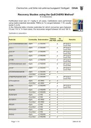
Application Note 529<strong>Capillary</strong> <strong>PS</strong>-<strong>DVB</strong> <strong>Monolithic</strong> <strong>Column</strong> <strong>of</strong><strong>500</strong>-µm i.d. <strong>for</strong> Peptide and Protein Separationsin Top-Down Proteomics StudiesIntroductionPolymeric monolithic stationary phases <strong>of</strong>fer analternative to the classical microparticulate sorbents,bringing important advantages to sample analysis. Incontrast to the traditional stationary phases, whichconsist <strong>of</strong> packed particles the monolithic separationmedium is made <strong>of</strong> a continuous, rigid polymeric rodwith a porous structure. The lack <strong>of</strong> intraparticular voidvolume improves mass transfer and separation efficiency,allowing <strong>for</strong> fast, high-quality separations.Polystyrene-divinylbenzene (<strong>PS</strong>-<strong>DVB</strong>) capillarymonolithic columns with internal diameters (i.d.) <strong>of</strong>100–200 µm have successfully been applied in combinationwith ESI and MALDI MS <strong>for</strong> the separation andidentification <strong>of</strong> peptides and proteins.The wide dynamic concentration range in whichproteins are present in complex biological matrices,necessitates the use <strong>of</strong> large sample amounts. Top-downand bottom-up proteomics LC separations there<strong>for</strong>erequire columns with enough sample capacity. Here theapplication <strong>of</strong> <strong>PS</strong>-<strong>DVB</strong> capillary monolithic columnswith an i.d. <strong>of</strong> <strong>500</strong> µm is described <strong>for</strong> separation <strong>of</strong>peptides and proteins. The chromatographic per<strong>for</strong>manceis demonstrated with separations <strong>of</strong> standard peptide andprotein samples as well as proteins from an Eschericiacoli cell lysate.ExperimentalLC system: UltiMate 3000<strong>Column</strong>: <strong>PS</strong>-<strong>DVB</strong> monolithic column<strong>500</strong>-µm i.d. × 5 cm (P/N 164087)Solvent A: Water, 0.05% TFASolvent B: MeCN, 0.04% TFAGradient: 5–35% MeCN in 7.5 min <strong>for</strong> peptides15–50% MeCN in 20 min <strong>for</strong> proteinsFlow Rate: 25 µL/minTemperature: 60 ºCSamples: (1) Cytochrome c digest, 8 pmol injected(2) Standard proteins, 0.05 µg injected(3) Ion-exchange fraction <strong>of</strong> E.coli celllysateDetection: UV, 214 nm, 45-nL flow cellApplication Note 529
ResultsPeptidesTo determine the per<strong>for</strong>mance <strong>of</strong> the <strong>500</strong>-µm i.d.<strong>PS</strong>-<strong>DVB</strong> monolithic column <strong>for</strong> peptide separations, adigest <strong>of</strong> cytochrome c digest was injected. The separationwas per<strong>for</strong>med with a steep gradient <strong>of</strong> 5–35%MeCN in 7.5 min and is shown in Figure 1. Peak widthat half height was between 0.08 and 0.10 min <strong>for</strong> thetryptic peptides. Under these conditions the peakcapacity <strong>for</strong> peptides <strong>of</strong> this monolithic column is approximately50. The peak capacity is calculated with the<strong>for</strong>mula P = 1 + T g/w, where P is the peak capacity, T gis the gradient time and w is the average peak width at13.4% <strong>of</strong> the peak height. Shallower gradients can beapplied to increase the peak capacity up to around 150<strong>for</strong> a gradient time <strong>of</strong> 90 min.ProteinsThe <strong>PS</strong>-<strong>DVB</strong> monolithic columns can also be used<strong>for</strong> protein separations using the same mobile phases.To elute proteins, the MeCN concentration must be increasedcompared to the elution <strong>of</strong> peptides. In Figure 2,a chromatogram is shown <strong>for</strong> the separation <strong>of</strong> 8 standardproteins. All proteins were baseline separated in 20 minwith peak widths between 0.1 and 0.2 min at half height.The peak capacity <strong>for</strong> proteins is approximately 60 <strong>for</strong> a20 min gradient.The injected amount was 25 ng <strong>for</strong> the standardproteins. The maximum loading capacity <strong>for</strong> proteinsallowing a 10% increase in peak width at half-heightwas around 10 pmol.Proteins From an E. coli Cell LysateTo demonstrate the suitability <strong>of</strong> the <strong>500</strong>-µm i.d.<strong>PS</strong>-<strong>DVB</strong> monolithic column <strong>for</strong> more complex samples,an ion-exchange fraction <strong>of</strong> an E. coli total cell lysatewas separated on the column. Approximately 2 µg in50 µL ion-exchange buffer was loaded onto the column.The chromatogram is shown in Figure 3.Several proteins were fractionated in a 384 well plate<strong>for</strong> further identification. The procedure involved mobilephase evaporation, in-well digestion and LC-MS/MSanalysis. The protein identification results are shown inTable 2.50mAU<strong>Column</strong>: <strong>Monolithic</strong> <strong>500</strong> µm × 5 cmEluent: H 2O, 0.04 TFATemperature: 60 °CFlow Rate: 25 µL/minInj. Volume: 1 µLDetection: UV, 214 µm–100 2.5 5.0 7.5 10.0 12.5 15.0MinutesFigure 1. Separation <strong>of</strong> a cytochrome c digest separated on amonolithic capillary column <strong>of</strong> <strong>500</strong>-µm i.d. × 5 cm, gradient5–35% MeCN in 7.5 min, sample amount 8 pmol, flow rate25 µL/min.<strong>Column</strong>: <strong>Monolithic</strong> <strong>500</strong> µm × 5 cmEluent: H 2O, 0.04 TFATemperature: 60 °CFlow Rate: 25 µL/minInj. Volume: 1 µLDetection: UV, 214 µm302371mAU45–100 5 10 15 20 25 30MinutesFigure 2. Separation <strong>of</strong> eight standard proteins (Table 1) on amonolithic capillary column (<strong>500</strong>-µm i.d. × 5 cm). Gradient15–50% MeCN in 20 min. Injected amount 0.025 µg <strong>of</strong> eachprotein, flow rate 25 µL/min.Table 1. Peak Parameters <strong>for</strong> Eight StandardProteins Separated on <strong>PS</strong>-<strong>DVB</strong> <strong>Monolithic</strong> <strong>Column</strong>s17.522997Peak # Protein Retention Peak Width atTime (min) 50% Height (min)1 Ribonuclease 6.0 0.112 Cytochrome c 9.1 0.113 Lysozyme 10.2 0.094 Transferrin 12.1 0.165 BSA 13.4 0.166Peaks:81. Ribonuclease2. Cytochrome c3. Lysozyme4. Transferrin5. BSA6. Carbonic anhydrase7. β-lactoglobulin8. Ovalbumin229986 Carbonic anhydrase 15.2 0.097 β-lactoglobulin 15.8 0.128 Ovalbumin 18.3 0.19<strong>Capillary</strong> <strong>PS</strong>-<strong>DVB</strong> <strong>Monolithic</strong> <strong>Column</strong> <strong>of</strong> <strong>500</strong>-µm i.d. <strong>for</strong> Peptide and Protein Separationsin Top-Down Proteomics Studies
150mAU<strong>Column</strong>: <strong>Monolithic</strong> <strong>500</strong> µm × 5 cmEluent: H 2O, 0.04 TFATemperature: 60 °CFlow Rate: 20 µL/minInj. Volume: 1 µLDetection: UV, 214 µm–200 5 10 15 20 25 30Minutes22999Figure 3. Separation <strong>of</strong> E. coli proteins (ion-exchange fraction)on a monolithic capillary column (<strong>500</strong>-µm i.d. × 5 cm).Gradient 10–50% MeCN in 20 min, flow rate 20 µL/min, injectedamount approximately 2 µg.Conclusions<strong>PS</strong>-<strong>DVB</strong> monolithic columns <strong>of</strong> <strong>500</strong>-µm i.d. allow<strong>for</strong> fast and efficient separations <strong>of</strong> peptides and proteinswith peak width at half-height between 0.1 and 0.2 min.Peak capacities <strong>for</strong> proteins are approximately 60 usinga 20 min gradient and 50 <strong>for</strong> peptides using a steepgradient in 7.5 min and can be increased by usingshallower gradients.The relatively high sample capacity <strong>of</strong> the capillary<strong>PS</strong>-<strong>DVB</strong> monolithic columns allows <strong>for</strong> micropreparativefractionation <strong>of</strong> proteins and their identification.In an alternative approach, this column can be directlycoupled to ESI-TOF-MS instruments, with or withoutpostcolumn splitting.Table 2. Protein Identification Results from theSeparation Shown in Figure 3 Separated on<strong>Capillary</strong> <strong>PS</strong>-<strong>DVB</strong> <strong>Monolithic</strong> <strong>Column</strong>sIdentified ProteinSequenceCoverage (%)Chain A, Yoda From Escherichia Colicrystallized with zinc ions 51High-affinity branched-chain amino acidtransport protein 43Putative adhesin 41High-affinity leucine-specific transport system;periplasmic binding protein 38Phosphoglycerate mutase 1 34Transaldolase B 51Fructose-biphosphate aldolase, class II 18Phosphoserine aminotransferase 49Alkyl hydroperoxide reductase C22 protein 455-methyltetrahydropteroyltri-glutamatehomocysteinemethyltransferase 8Aspartate-semialdehyde dehydrogenase 24Phosphoserine aminotransferase 8UltiMate is a trademark <strong>of</strong> Dionex Corporation.Passion. Power. Productivity.Dionex Corporation1228 Titan WayP.O. Box 3603Sunnyvale, CA94088-3603(408) 737-0700North AmericaSunnyvale, CA (408) 737-8522 Westmont, IL (630) 789-3660Houston, TX (281) 847-5652 Atlanta, GA (770) 432-8100Marlton, NJ (856) 596-0600 Canada (905) 844-9650EuropeAustria (43) 1 616 51 25 Belgium (32) 3 353 4294Denmark (45) 36 36 90 90 France (33) 1 39 30 01 10Germany (49) 6126 991 210 Italy (39) 06 66 51 5052The Netherlands (31) 161 43 43 03Switzerland (41) 62 205 99 66United Kingdom (44) 1276 691722Asia PacificAustralia (61) 2 9420 5233 China (852) 2428 3282India (91) 22 28475235 Japan (81) 6 6885 1213Korea (82) 2 2653 2580Application Note 529 www.dionex.com LPN 1775 05/06©2006 Dionex Corporation