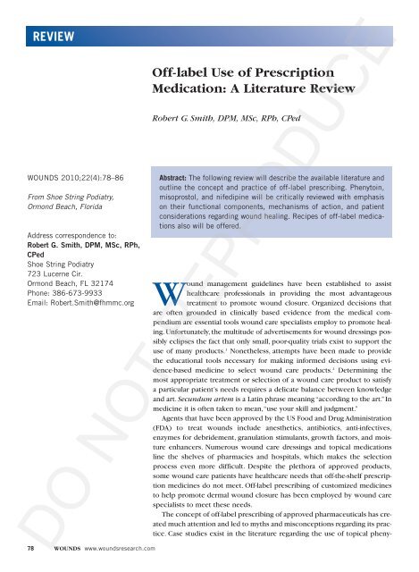
Off-label Use of Prescription Medication: A Literature Review - Wounds
Off-label Use of Prescription Medication: A Literature Review - Wounds
Off-label Use of Prescription Medication: A Literature Review - Wounds
Create successful ePaper yourself
Turn your PDF publications into a flip-book with our unique Google optimized e-Paper software.
SmithThe practice <strong>of</strong> <strong>of</strong>f-<strong>label</strong> prescribing <strong>of</strong> drugs andmedical devices plays an important therapeutic role inmany disease states, including psychiatric disorders,human immunodeficiency virus (HIV), and diseases inchildren and other patient populations that are underservedby approved medicines. When medications anddevices are used <strong>of</strong>f-<strong>label</strong> for medical treatment, their primarypurpose is to benefit the individual patient. Withinthe scope <strong>of</strong> medical practice, the clinician’s primarypurpose and intention when using a product <strong>of</strong>f-<strong>label</strong> isto enhance the wellbeing <strong>of</strong> the patient with an expectation<strong>of</strong> success. It is clear that rapid developments inmedical science <strong>of</strong>ten outpace the speed with which regulatoryauthorities like the FDA can move to implementnew legislation. 5 Physicians may safely prescribe anydrug <strong>of</strong>f-<strong>label</strong> provided he or she practices a standard <strong>of</strong>care based on sound evidence. 6,7 Williams 8 states that <strong>of</strong>f<strong>label</strong>drug use may constitute the current standard <strong>of</strong>care, and failure to provide or discuss <strong>of</strong>f-<strong>label</strong> therapywith patients may be considered malpractice.Uniformly accepted specific guidance to assist cliniciansattempting to make decisions about the appropriateness<strong>of</strong> such prescriptions does not exist. Given thelack <strong>of</strong> FDA approval for <strong>of</strong>f-<strong>label</strong> uses, many <strong>of</strong>f-<strong>label</strong> uses<strong>of</strong> drugs are published in the literature as case reports,which allows other clinicians the opportunity to learn anduse these treatments on a greater number <strong>of</strong> patients. 3One might surmise that these case studies are not giventhe same degree <strong>of</strong> scientific scrutiny that are required for<strong>label</strong>ed indications from drug manufacturers. 7 Gazarian etal 4 outline consensus recommendations for evaluatingappropriateness <strong>of</strong> <strong>of</strong>f-<strong>label</strong> use <strong>of</strong> medicines.Compounding pharmacists create alternate methods<strong>of</strong> delivery like transdermal gels, ointments, or solutions<strong>of</strong> approved medications to provide for patients withunique health needs; thus, compounding pharmacistscreate <strong>of</strong>f-<strong>label</strong> medications. While the FDA and FederalTrade Commission (FTC) enforce laws that prohibit makingunsubstantiated claims regarding safety and efficacyin pharmacists’ marketing materials, <strong>of</strong>f-<strong>label</strong> dispensing<strong>of</strong> approved medications is not considered malpractice.When a licensed practitioner writes a prescription for acompounded drug, the medication is normally needed assoon as possible. As a matter <strong>of</strong> public health policy, it isclear that individual compounded medications should beexempted from the FDA approval process becauserequiring approval would be neither cost-effective nor inthe best interest <strong>of</strong> the patient. State pharmacy boardsregulate compounded medications and are not subject to80 WOUNDS www.woundsresearch.comfederal laws designed to regulate mass-produced drugs.Therefore, pharmacists should observe the same principlesand levels <strong>of</strong> evidence when dispensing <strong>of</strong>f-<strong>label</strong>medications that a physician applies when prescribing<strong>of</strong>f-<strong>label</strong> drugs.<strong>Medication</strong> Classes and Wound Healing<strong>Medication</strong>s may exert their effects by either assistingor interfering with the specific phases <strong>of</strong> wound healing.The effect <strong>of</strong> a particular medication on the wound healingprocess may depend on its mechanism <strong>of</strong> action,dosage, and route <strong>of</strong> administration in relation to the specificphase <strong>of</strong> the wound healing process. 10 Specific medications<strong>of</strong> interest to the wound care specialists includemisprostol, nifedipine, and phenytoin.A search pr<strong>of</strong>ile was compiled using key terms to performBoolean logic electronic searches to identify relativeprimary literature citations from 1967 to 2009(Figure 1). Further, a manual review <strong>of</strong> citations’ referenceslists and bibliographies was undertaken to gatherany additional information that might have led to furthermaterial for this review. Citations (n = 317) werereviewed and evaluated as defined by the search limita-Figure 1. Citations (n = 317) were reviewed andevaluated as defined by the search limitations.Accounting for duplication, 35% (n = 110) <strong>of</strong> thecitations were reviewed for significance and relevancefor inclusion in this review.DO NOT REPRODUCE
tions. Accounting for duplication, 35% (n = 110) <strong>of</strong> thecitations were reviewed for significance and relevancefor inclusion for this review.Misoprostol. Misoprostol [15-deoxy-16-hydroxy-16-methyl-PGE1] is a synthetic prostaglandin E1 analog withan additional ester group at C1. 11 As a therapeutic agentmisoprostol embodies various pharmacological featuresthat contribute to its pharmacodynamic actions. It seemsto inhibit the acid secretion by means <strong>of</strong> direct action onparietal cells. The inhibition <strong>of</strong> adenylate cyclase may bedependent on guanosine-5'-triphosphate (GTP). 12 The significantcytoprotective actions <strong>of</strong> misoprostol are relatedto several mechanisms, which include: increased secretion<strong>of</strong> bicarbonate; decrease in the volume and pepsincontent <strong>of</strong> gastric secretions; enhancement <strong>of</strong> mucosalblood flow as a result <strong>of</strong> direct vasodilatation; stabilization<strong>of</strong> tissue lysozymes/vascular endothelium; andimprovement <strong>of</strong> mucosal regeneration capacity. 13Misoprostol’s potential pharmacological benefits incutaneous wound healing may be explained by reviewingthe role <strong>of</strong> prostaglandins in wound healing. Products<strong>of</strong> the earliest cellular events are activated upon tissueinjury. Phospholipase stimulate the release <strong>of</strong> arachidonicacid, ultimately leading to the production <strong>of</strong>prostaglandins, leukotrienes, and other factors. Histaminereleased from platelets and circulating mast cells increasesvascular permeability and indirectly stimulates vasodilationthrough the production <strong>of</strong> prostaglandins E1 andE2. Prostaglandins cause vasodilation through activation<strong>of</strong> the adenylate cyclase pathway via the production <strong>of</strong>cyclic adenosine monophosphate.Several investigations have been conducted evaluatingpharmaceutical developed prostaglandin E1 (PGE-1),misoprostol, and its action on wounds. 14–19 Milio et al 14performed one <strong>of</strong> the first, and one <strong>of</strong> the few humaninvestigations that focused on the efficacy <strong>of</strong> intravenousPGE-1 in the treatment <strong>of</strong> lower extremity venous ulcers.This randomized, single-blinded, placebo controlled trial<strong>of</strong> 87 consecutive patients observed healing time forvenous ulcers. 14 The treatment group (n = 44) received60-mg <strong>of</strong> PGE-1 diluted in 250-mL <strong>of</strong> saline solution,while the placebo group (n = 43) received just 250-mL <strong>of</strong>saline solution over a 20-day span. Also, both groups weretreated with elastic bandaging and local therapy. Themain outcome <strong>of</strong> the study was the percentage <strong>of</strong> ulcerrecovery at the conclusion <strong>of</strong> the 120-day period <strong>of</strong>observation and time to healing. 14 The investigators concludedthat the study demonstrated that treatment withPGE-1 is effective on lower extremity venous ulcers;Smithhowever, the authors concluded that the mechanismwith which PGE-1 favors healing <strong>of</strong> venous ulcersremains to be verified. 14The remaining investigations were conducted as animalobservations <strong>of</strong> surgically induced wounds. 15–19 deOliveira et al 15 investigated the influence <strong>of</strong> misoprostolon healing <strong>of</strong> colonic anastomoses in Wistar rats particularlyregarding changes in collagen levels. The animalsreceived misoprostol intragastrically at a dosage <strong>of</strong> 200-mcg/kg body weight diluted in 0.9% saline twice daily bymeans <strong>of</strong> a stomach tube. Their results demonstrated thatmisoprostol administration increased the hydroxyprolineconcentration on postoperative day 14 withoutinterfering in the inflammatory response, which was verifiedby histopathological study. Three investigations centeredon topical application <strong>of</strong> PGE-1 and skin flap viability.16–18 All <strong>of</strong> these investigations observed a beneficialeffect <strong>of</strong> topical application <strong>of</strong> prostaglandins as definedby augmented skin flap viability. 16–18 A final animal observationalinvestigation focused on the application <strong>of</strong> misoprostolpowder suspended in saline in a 1:1000 concentrationon a surgically prepared wound. 19 This investigation<strong>of</strong>fered data concluding that when applied daily toan acute surgical wound, topically applied misoprostolresults in decreased healing time. 19 Additionally, theauthors <strong>of</strong>fered the assumption that prostaglandins exerttheir greatest effect in the inflammatory phase <strong>of</strong> woundhealing, while acknowledging that their investigationwas preliminary and called upon further studies to validatethe use <strong>of</strong> misoprostol in chronic wounds. 19Nifedipine. Nifedipine is 3,5-pyridinedicarboxylicacid, 1,4-dihydro-2,6-dimethyl-4-(2-nitrophenyl)-, dimethylester used as an antianginal medication that belongs to aclass <strong>of</strong> pharmacological agents known as calcium channelblockers. Nifedipine is a calcium ion influx inhibitorthat inhibits the transmembrane influx <strong>of</strong> calcium ionsinto both cardiac muscle and smooth muscle withoutchanging serum calcium concentrations. Recalling thatcellular calcium metabolism appears to regulate extracellularmatrix production as well as other critical stepsin wound healing 20,21 leads to the inference that nifedipineas a calcium channel blocker could potentially be anattractive agent to study with regard to its influence onthe wound healing process. The report by Lupo et al 22describing the dose-dependent antioxidant activity <strong>of</strong>nifedipine and other calcium channel blockers whencompared to α-tocopherol further validates the influence<strong>of</strong> these drugs on the wound-healing process.Several investigations have evaluated nifedipine andDO NOT REPRODUCEVol. 22, No. 4 April 2010 81
Smithits influence on the wound healing process. 20–29 An evendistribution <strong>of</strong> both animal and human investigations arepresent in the literature and include case reports, retrospectivestudies, and prospective, randomized, blindedstudies. 23–29 These investigations have reviewed the topicalapplication <strong>of</strong> nifedipine on surgically preparedwounds, 23–25 a chronic wound, 26 a hypertrophic wound, 26and anal fissures. 27–29A review <strong>of</strong> the studies <strong>of</strong> the animal wound modelsreveals similar investigational methods were used. 23–25 Allthree investigations used healthy Wistar rats (150 g–250g). 23–25 One investigation evaluated the co-founder <strong>of</strong> diabetesby comparing their study group <strong>of</strong> diabetic ratswith normal rats. 23 Surgically-applied incisional and excisionalwounds were evaluated and topical nifedipine wasapplied to the wounds. 23–25 An interesting aspect was thattwo investigations examined calcium channel blockerapplication in the wounds <strong>of</strong> animals treated with systemicdexamethasone. 24,25Corticosteroids affect cells by altering gene expressionafter crossing the cell membrane where they bind tocytoplasmic receptors and translocate into the cellnucleus. The inhibitory effect on gene expression overmany cells allows for corticosteroids to affect almostevery phase <strong>of</strong> wound healing. 10 The degree <strong>of</strong> geneexpression inhibition is related to the particular corticosteroid’spotency. The most prominent effects are seenwhen corticosteroids are administered during the earlyinflammatory phase. 10 Collectively, the findings andresults <strong>of</strong> these three animal studies are similar. 23–25The effects <strong>of</strong> nifedipine were evaluated in the threedifferent phases <strong>of</strong> the wound-healing process. Topicalnifedipine significantly improved the inflammatoryphase, 23–25 significantly improved the process during onlythe maturation phase in diabetic rats, 23 and did not affectthe proliferation phase. 23–25 Nifedipine enhanced normalhealing by increasing the tensile strength <strong>of</strong> 10-day-oldgranulation tissue. There was no significant change inhydroxyproline level, collagen level, or glycosaminoglycancontent in the granulation tissue. 25 This finding validatesthe tissue model findings <strong>of</strong> Lee and Ping 20 whoconcluded that the cellular calcium metabolism appearsto regulate extracellular matrix production and thathypertrophic disorders <strong>of</strong> wound healing may respondto therapy with calcium antagonist drugs. Finally, theapplication <strong>of</strong> topical nifedipine was able to overcomesteroid depression <strong>of</strong> wound healing. 24,25Torsiello and Kopacki 26 described their experiencewith transdermal nifedipine in two patients. The first82 WOUNDS www.woundsresearch.compatient was a 43-year-old woman with a nonhealing rightheel wound. The formula used for this wound was 8%nifedipine in pluronic lecithin organogel administeredby the patient twice daily. The authors used wound sizeas an outcome indicator for healing time. They observeda 6- to 8-week healing time with the therapeutic inventioncompared to previous accounts for the same woundin which a healing time <strong>of</strong> 4 to 5 months was observed.The second case report described an 8-year-old boy witha linear hypertrophic scar contracture. A nifedipine 2%pluronic lecithin organogel formulation was appliedtwice daily. After surgical invention and applications <strong>of</strong>transdermal nifedipine complete wound healingoccurred in 3 to 4 weeks. The authors acknowledged thatthe exact mechanism <strong>of</strong> action <strong>of</strong> the influence <strong>of</strong> transdermalnifedipine on the wound-healing process remainsto be determined. The authors believe that the samepharmacological action <strong>of</strong> smooth muscle relaxationoccurs in the skin vasculature and creates a localizedperipheral vasodilatation and increases flow to the localizedarea, which accelerates epithelialization and possiblymicrovascular neogenesis.The last two human accounts focus on the use <strong>of</strong> topicalnifedipine as nonsurgical treatment <strong>of</strong> chronic analfissures. 27,28 One report was a retrospective chart reviewwith telephone follow-up <strong>of</strong> 88 patients. The other reportwas a prospective, randomized, double-blinded study <strong>of</strong>110 consecutive patients. 27,28 Both investigations concludedthat chronic anal fissures can be simply and effectivelytreated with topical nifedipine with eitherinjectable botulinum toxin 27 or as a compounded formulawith lidocaine. 28 The treatment was demonstrated tobe safe and effective with a low recurrence rate. Perrottiet al 28 chose healing <strong>of</strong> the chronic anal fissure as the primaryoutcome measure defined as epithelialization orformation <strong>of</strong> a scar observed under anoscopy. Their controlgroup received 1.5% lidocaine and 1% hydrocortisoneacetate and the treatment group received 1.5% lidocaineand 0.3% nifedipine applied every 12 hours for 6weeks. Clinical healing was achieved in 55 patients in thenifedipine group at 42 days. The final account is a criticalnarrative <strong>of</strong> the clinical investigation by Perrotti et al. 28Merenstein and Rosenbaum 29 demonstrated the effectiveness<strong>of</strong> topical nifedipine by highlighting the 46patients who were in the control group and did notexperience healing were <strong>of</strong>fered nifedipine treatment,resulting in a 82% (n = 38) healing rate. Also, none <strong>of</strong> thepatients in the nifedipine group experienced systemicside effects—only one patient in the secondary treat-DO NOT REPRODUCE
ment group experienced slight local hyperemia, whichimproved when treatment was completed. 29 Finally,Merenstein and Rosenbaum emphasized that this topicalformulation is an extremely safe, well tolerated, and effectivetreatment that has the potential to provide clinicianswith a reliable nonsurgical method for treating chronicanal fissures.Phenytoin. Phenytoin sodium is an antiepilepticdrug. Phenytoin sodium is related to the barbiturates inchemical structure, but has a five-membered ring. Thechemical name is sodium 5,5-diphenyl-2, 4-imidazolidinedione.It has been reported that phenytoin’s main actionin wound healing is its modification <strong>of</strong> collagen remodelingby decreasing collagenase activity. 10 Also, collagensynthesis is not significantly affected by phenytoin,although it does inhibit wound contraction. 30 Finally,Spaia et al 31 report that studies historically have shownthat phenytoin stimulates fibroblast proliferation,decreases collagenase activity, increases epidermal andkeratinocyte growth factor receptors and collagen disposition,accelerates initial inflammatory responses,induces new vessel formation, speeds the decrease <strong>of</strong>microbial colonies thus decreasing bacterial contamination,reduces wound exudate, and improves healing. 31–37 Alimitation <strong>of</strong> Spaia et al’s case report is that the exactamount <strong>of</strong> phenytoin applied to this patient’s wound wasnot quantified as it is only referred to as “1–2 ampoules<strong>of</strong> a commercial preparation.” 31In order to explore this pharmacological action, systematicsearches <strong>of</strong> PubMed, Medline, Cinahl, and theCochrane library were performed. The medical literatureis replete with citations describing the clinical effect <strong>of</strong>topical phenytoin on wound healing. Given that thiscompound has been rigorously investigated, a summary<strong>of</strong> published accounts to emphasize and report on themost important points is presented. 35–37Bhatia and Prakash 35 <strong>of</strong>fer a thorough review on topicalphenytoin for wound healing. This review beginswith a historical perspective on the use <strong>of</strong> topical phenytoinover time. The review recounts animal studies, cellcultures, case reports, retrospective studies, and prospectivecontrolled comparison human observational findings.35 The types <strong>of</strong> wounds investigated with regard totopical phenytoin and its effects on healing in thisreview include abscesses, ballistic wounds, decubitusulcers, diabetic foot ulcers, pressure ulcers, surgicalinduced, tropic ulcers, and venous stasis ulcers withregard to topical phenytoin and its affects on healing. 35The authors acknowledge that despite the extensiveSmithinvestigation <strong>of</strong> topical phenytoin for wound healing thebest method <strong>of</strong> delivery <strong>of</strong> topical phenytoin remainsunknown. 35 The authors explore the safety issues by statingthat side effects from topical phenytoin are rare andthen provide detailed accounts <strong>of</strong> adverse effects includinggeneralize rash and hypertrophic granulation tissueboth <strong>of</strong> which resolved with stoppage <strong>of</strong> treatment. 35Finally, the issue <strong>of</strong> phenytoin’s systemic absorption isevaluated. This review concludes that the systemicabsorption <strong>of</strong> topical phenytoin is not significiant. Theauthors further explain that most studies that have monitoredserum phenytoin levels during topical applicationhave shown undetectable levels. 35Younes et al 36 conducted an intriguing observationalinvestigation that is relevant to cutaneous wound healingand is pivotal to wound healing events associated withindividuals who have diabetes. These researchers testedwound bed preparation with 10% phenytoin ointmentand observed split-thickness skin graft survival. Sixteenpatients with diabetes and large, lower extremity footulcers underwent wound bed preparation with phenytoinointment followed by split-thickness skin graft application.The product was compounded with phenytoinpowder and levigated with white petroleum to a finalconcentration <strong>of</strong> 10% (W/W). Younes et al <strong>of</strong>fered a qualifyingstatement to their observations remarking thatcomparison <strong>of</strong> their results to those <strong>of</strong> other groupsusing various growth factors was not possible because <strong>of</strong>the different methodologies and different ways <strong>of</strong> measuringoutcomes that were used. Finally, in order to minimizethe potential risk <strong>of</strong> wound sepsis due to thepatient’s co-morbidities and ulcer size, the authors recommendedthat adequate surgical debridement andbroad spectrum antibiotics be initiated before anywound therapy.Shaw et al 37 <strong>of</strong>fered results from their systematicreview <strong>of</strong> the clinical effect <strong>of</strong> topical phenytoin onwound healing. A total <strong>of</strong> 347 references were locatedthrough database, journal, and handsearching; however,only 14 randomized controlled trials were suitable forreview. 37 Two <strong>of</strong> these papers were <strong>of</strong> high quality usingthe Van Tudler (VT) method and methodologicallysound: Pai et al 39 who performed a double-blind study <strong>of</strong>topical phenytoin treatment <strong>of</strong> diabetic ulcers; andSimpson et al 40 who investigated the use <strong>of</strong> diphenylhydantoinin the treatment <strong>of</strong> venous stasis ulcers. 37–40 Theremaining 12 papers scored between 10–14 with the VTmethod and were deemed to be low to moderate quality,providing at best level 2 (n = 9) and level 3 (n = 2)DO NOT REPRODUCEVol. 22, No. 4 April 2010 83
Smithevidence for the effectiveness <strong>of</strong> phenytoin in the treatment<strong>of</strong> wounds. 37 Shaw et al point out in thesedescribed 14 studies, the sample size ranged from 30 to102, which is typical <strong>of</strong> many wound care studies andwas not directly related to the VT score. 37 Also, the randomizationprocess was inadequately described in many<strong>of</strong> the studies, with subjects <strong>of</strong>ten assigned to treatmentgroups rather than being randomized through a trulyindependent process. 37 Another finding <strong>of</strong> this reviewwas that 12 out <strong>of</strong> 14 studies reported a statistically significantreduction in wound area in the phenytoin-treatedgroup when compared with the control groups invarious wounds. A final acknowledgement made in thisreview indicated that it is easy to be critical <strong>of</strong> thedesign <strong>of</strong> the studies reviewed but presenting solutionsto the highlighted problems associated with the particulardesign is much more difficult. 37Clinical ApplicationIt is the duty and responsibility <strong>of</strong> both the woundcare specialists and the dispensing pharmacist to makecertain that any <strong>of</strong>f-<strong>label</strong> use <strong>of</strong> a drug is supported bycredible, evidence-based medicine. It was the purpose <strong>of</strong>this review to <strong>of</strong>fer data regarding the <strong>of</strong>f-<strong>label</strong> use <strong>of</strong>misoprostol, nifedipine, and phenytoin. Formulas doexist and are presented in Table 1 for compounding topicalpreparations such as powders, creams, and gelsusing one if not all <strong>of</strong> the medications reviewed. Someformulas incorporate either metronidazole or gentamicinas anti-infectives or as odor controlling substance orlidocaine as a local anesthetic. Questions that presentthemselves are: “How was the effective concentration <strong>of</strong>each component <strong>of</strong> the each formula decided upon?”and “Are the concentrations based on animal studies, cellcultures, or anecdotal information from wound careexperts in the field?”Several limitations must be acknowledged whenapplying some <strong>of</strong> the literature citations to clinical practice.The foremost is that wound healing rates in rats maynot be directly comparable to humans because normalrates are not equivalent between species. Levenson et al 41reported that incisions in rats may heal more slowly thanwound models predict. 41 Secondly, chronic woundsexhibit significant differences from acute wounds inboth immunohistochemistry and histopathology.Therefore, it would be difficult to apply the clinical findings<strong>of</strong> surgically induced wounds to chronic wounds.Thirdly, in many <strong>of</strong> the literature accounts, one cannotexactly determine the amount <strong>of</strong> active drug applied84 WOUNDS www.woundsresearch.comdirectly to the wound. A drug-specific concern regardingphenytoin is the added expense <strong>of</strong> ordering and monitoringdrug levels to insure that a patient does notbecome toxic.Despite the fact that some studies rely on measures <strong>of</strong>statistical power to legitimize data, it remains that many<strong>of</strong> these studies have small sample sizes. This phenomenonis typical <strong>of</strong> many wound care studies, and mayimpart some statistical bias in the results.It is an optimistic outlook that experts from woundhealing societies will develop policy documents to outlineprocedures for documenting and dispensing <strong>of</strong>f<strong>label</strong>medications for wound healing. Criteria should bedeveloped and embraced by wound care specialists andguidelines should be <strong>of</strong>fered for prescribing appropriate<strong>of</strong>f-<strong>label</strong> medications. These criteria should <strong>of</strong>fer validatedand uncontested data in support <strong>of</strong> particular <strong>of</strong>f-<strong>label</strong>medications. Available literature and existing compoundedrecipes 42–47 should attest to the drug’s safe and effectiveuse in treating a particular type <strong>of</strong> wound.ConclusionThe concept <strong>of</strong> <strong>of</strong>f-<strong>label</strong> prescribing <strong>of</strong> approvedpharmaceuticals to treat cutaneous wounds has generatedsubstantial attention. Studies have reported the use <strong>of</strong>topical misoprostol, nifedipine, and phenytoin to treatwounds. The purpose <strong>of</strong> this article is in part to serve asa foundation and a review <strong>of</strong> the available literature outliningthe concept and practice <strong>of</strong> <strong>of</strong>f-<strong>label</strong> prescribing.In addition, specific medications were reviewed with anemphasis on their mechanisms <strong>of</strong> action and patient considerationsin the context <strong>of</strong> wound healing. Finally,details and compounding recipes <strong>of</strong> these medicationscan be found throughout the literature.References1. Vermeulen H, Ubbink DT, Goossens A, de Vos R, LegemateDA. Systematic review <strong>of</strong> dressings and topical agents forsurgical wounds healing by secondary intention. Br JSurg. 2005;92(6):665–672.2. Aung BJ. Using evidence-based medicine to selectedwound care products. Podiatry Manage.2007;26(9):213–216.3. Bennett WM. <strong>Off</strong>-<strong>label</strong> use <strong>of</strong> approved drugs: therapeuticopportunity and challenges. J Am Soc Nephrol.2004;15(3):830–831.4. Gazarian M, Kelly M, McPhee JR, Graudins LV, Ward RL,Campbell TJ. <strong>Off</strong>-<strong>label</strong> use <strong>of</strong> medicines: consensus recommendationsfor evaluating appropriateness. Med JDO NOT REPRODUCE
Aust. 2006;185(10):544–548.5. Beck JM, Azari ED. FDA, <strong>of</strong>f-<strong>label</strong> use, and informed consent:debunking myths and misconceptions. Food DrugLaw J. 1998;53(1):71–104.6. Tomaszewski C. <strong>Off</strong>-<strong>label</strong>: just what the doctor ordered. JMed Toxicol. 2006;2(3):87–88.7. Radley DC, Finkelstein SN, Stafford RS. <strong>Off</strong>-<strong>label</strong> prescribingamong <strong>of</strong>fice-based physicians. Arch Intern Med.2006;166(9):1021–1026.8. Williams GA. What are the legal issues regarding the use<strong>of</strong> <strong>of</strong>f-<strong>label</strong> drugs? Retina Today. 2007(Jan);43–47.9. McGrath MH, Hundahl SA. The spatial and temporal quantification<strong>of</strong> my<strong>of</strong>ibroblasts. Plast Reconstr Surg.1982;69(6):975–985.10. Karukonda SR, Flynn TC, Boh EE, McBurney EI, Russo GG,Millikan LE. The effects <strong>of</strong> drugs on wound healing: part2. Specific classes <strong>of</strong> drugs and their effects on healingwounds. Int J Dermatol. 2000;39(5):321–333.11. Hoogerwerf WA, Pasricha PJ. Agents used for control <strong>of</strong>gastric acidity and treatment <strong>of</strong> peptic ulcers and gastroesophagealreflux disease. In: Goodman & Gilman’sThe Pharmacological Basis <strong>of</strong> Therapeutics. 10th ed.Harman JG, Limbird LE, eds. New York, NY: McGraw-Hill;2001:1011–1012.12. Shimizu N, Nakamura T. Prostaglandins as hormones. DigDis Sci. 1985;30(11 Suppl):109S–113S .13. Wilson DE. Antisecretory and mucosal protective actions<strong>of</strong> misoprostol. Potential role in the treatment <strong>of</strong> pepticulcer disease. Am J Med. 1987;83(1A):2–8.14. Milio G, Minà C, Cospite V, Almasio PL, Novo S. Efficacy <strong>of</strong>the treatment with prostaglandin E-1 in venous ulcers <strong>of</strong>the lower limbs. J Vasc Surg. 2005;42(2):304–308.15. de Oliveira PG, Soares EG, Aprilli F. Influence <strong>of</strong> misoprostol, a synthetic prostaglandin E1 analog, on the healing<strong>of</strong> colonic anastomoses in rats. Dis Colon Rectum.1994;37(7):660–663.16. Eskitascioglu T, Gunay GK. The effects <strong>of</strong> topical prostacylinand prostaglandin E1 on flap survival after nicotineapplication in rats. Ann Plast Surg. 2005;55(2):202–206.17. Sawada Y, Sugawara M, Hatayama I, Sone K. A study <strong>of</strong> topicaland systemic prostaglandin E1 and survival experimentalskin flaps. Br J Plast Surg. 1993;46(8):670–672.18. Asai S, Fukuta K, Torii S. Topical administration <strong>of</strong>prostaglandin E1 with iontophoresis for skin flap viability.Ann Plast Surg. 1997;38(5):514–517.19. Mahoney J, Ponticello M, Nelson E, Ratz R. Topical misoprostoland wound healing in rats. WOUNDS.2007;19(12):334–339.20. Lee RC, Ping JA. Calcium antagonists retard extracellularSmithmatrix production in connective tissue equivalent. J SurgRes. 1990;49(5):463–466.21. Johnson H Jr, Parham M, Davis E, Wise L. Preliminary study<strong>of</strong> the protective effect <strong>of</strong> the calcium channel blocker,nifedipine on adriamycin- induced tissue injury. J InvestSurg. 1991;4(3):313–322.22. Lupo E, Locher R, Weisser B, Vetter W. In vitro antioxidantactivity <strong>of</strong> calcium antagonists against LDL oxidationcompared with alpha-tocopherol. Biochem Biophys ResCommun. 1994;203(3):1803–1808.23. Ebadi A, Cheraghali AM, Qoshoni H, Hossain E. Healingeffect <strong>of</strong> topical nifedipine on skin wounds <strong>of</strong> diabeticrats. DARU. 2003;11(1):19–22.24. Bhaskar HN, Udupa SL, Udupa AL. Effect <strong>of</strong> nifedipine andamlodipine on wound healing in rats. Indian J PhysiolPharmacol. 2004;48(1):111–114.25. Bhaskar HN, Saraswathi UL, Laxminarayana UA. Effect <strong>of</strong>nifedipine and amlodipine on dead space wound healingin rats. Indian J Exp Biol. 2005;43(3):294–296.26. Torsiello MJ, Kopacki MH. Transdermal nifedipine forwound healing case reports. Int J Pharm Comp.2000;4(5):356–358.27. Tranqui P, Trottier DC, Victor C, Freeman JB. Nonsurgicaltreatment <strong>of</strong> chronic anal fissure: nitroglycerin and dilatationversus nifedipine and botulinum toxin. Can J Surg.2006;49(1):41–45.28. Perrotti P, Bove A, Antropoli C, et al. Topical nifedipinewith lidocaine ointment vs. active control for treatment<strong>of</strong> chronic anal fissure: results <strong>of</strong> a prospective, randomized,double-blind study. Dis Colon Rectum.2002;45(11):1468–1475.29. Merenstein D, Rosenbaum D. Is topical nifedipine effectivefor chronic anal fissure? Patient oriented evidencethat matters. J Fam Pract. 2003;52(3):190–191.30. Zitelli JA. Wound healing by secondary intention. In:Roenigk RK, Roenigk HH, eds. Dermatologic SurgeryPrinciples and Practice. New York, NY: Marcel Dekker.1996:101–130.31. Spaia S, Eleftheriadis T, Pazarloglou M, et al. Phenytoin efficacyin treating the diabetic foot ulcer <strong>of</strong> a haemodialysispatient. Nephrol Dial Transplant. 2004;19(3):753.32. Modéer T, Andersson G. Regulation <strong>of</strong> epidermal growthfactor receptor metabolism in gingival fibroblasts byphenytoin in vitro. J Oral Pathol Med.1990;19(4):188–191.33. DaCosta ML, Regan MC, al Sader M, Leader M, Bouchier-Hayes D. Diphenylydantoin sodium promotes early andmarked angiogenesis and results in increased collagendeposition and tensile strength in healing wounds.DO NOT REPRODUCEVol. 22, No. 4 April 2010 85
SmithSurgery. 1998;123(3):287–293.34. Lodha SC, Lohiya ML, Vyas MC, Bhandari S, Goyal RR,Harsh MK. Role <strong>of</strong> phenytoin in healing <strong>of</strong> large abscesscavities. Br J Surg. 1991;78(1):105–108.35. Bhatia A, Prakash S. Topical phenytoin for wound healing.Dermatol Online J. 2004;10(1):5.36. Younes N, Albsoul A, Badran D, Obedi S. Wound bedpreparation with 10-percent phenytoin ointmentincreases the take <strong>of</strong> split-thickness skin graft in large diabeticulcers. Dermatol Online J. 2006;12(6):5.37. Shaw J, Hughes CM, Lagan KM, Bell PM. The clinical effect<strong>of</strong> topical phenytoin on wound healing: a systematicreview. Br J Dermatol. 2007;157(5):997–1004.38. van Tulder MW, Assendelft WJ, Koes BW, Bouter LM.Method guidelines for systematic reviews in theCochrane Collaboration back <strong>Review</strong> Group for SpinalDisorders. Spine. 1997;22(20):2323–2330.39. Pai MR, Sitaraman N, Kotian MS. Topical phenytoin in diabeticulcers: a double blind controlled trial. Indian J MedSci. 2001;55(11):593–599.40. Simpson GM Kunz E, Slafta J. <strong>Use</strong> <strong>of</strong> sodium diphenylhydantoinin treatment <strong>of</strong> leg ulcers. N Y State J Med.1965;65:886–888.41. Levenson SM, Geever EF, Crowley LV, Oates JF 3rd, BerardCW, Rosen H. The healing <strong>of</strong> rat skin wounds. Ann Surg.1965;161(2):293–308.42. Meece J. Misprostol 0.024% and phenytoin 5% diabeticcream. Int J Pharm Comp. 2003;7(3):171.43. Allen L. Lidociane HCl 2% Misprostol 0.024% and phenytoin5% topical powder. Int J Pharm Comp.2004;8(4):298.44. Allen L Metronidazole 2%, Misprostol 0.024% and phenytoin5% topical gel. Int J Pharm Comp. 2004;8(4):299.45. Allen L. Lidocaine HCl 2%, misprostol 0.003%, phenytoin2.5% topical gel. Int J Pharm Comp. 2000;4(4):301.46. Allen L. Gentamicin Sulfate 0.2%, misprostol 0.024%, andphenytoin 5% topical gel. Int J Pharm Comp.2004;8(6):472.47. Meece J. Five compounds for treating diabetes-relatedconditions. Int J Pharm Comp. 2003;7(3):170–174.48. Collins ME, Walton JP, Dwyer AL. <strong>Prescription</strong> compoundsfor wound care. Available at:http://www.healthwayrx.com/faxdocuments/woundcare.htm.Accessed: September 4, 2009.DO NOT REPRODUCE86 WOUNDS www.woundsresearch.com


