PSORIASIS â THE RELATIONSHIP BETWEEN NAIL CHANGES ...
PSORIASIS â THE RELATIONSHIP BETWEEN NAIL CHANGES ...
PSORIASIS â THE RELATIONSHIP BETWEEN NAIL CHANGES ...

Create successful ePaper yourself
Turn your PDF publications into a flip-book with our unique Google optimized e-Paper software.
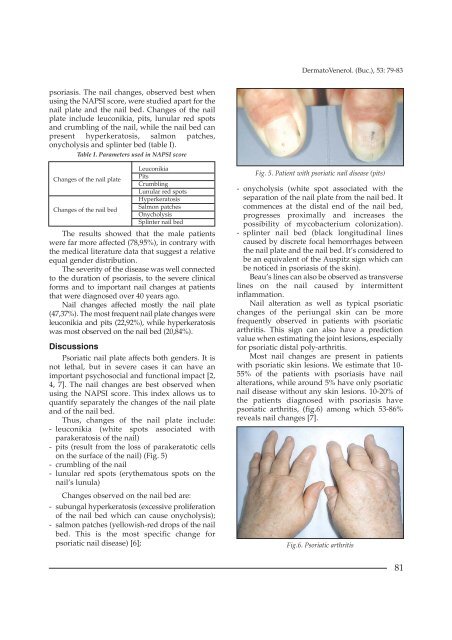
DermatoVenerol. (Buc.), 53: 79-83psoriasis. The nail changes, observed best whenusing the NAPSI score, were studied apart for thenail plate and the nail bed. Changes of the nailplate include leuconikia, pits, lunular red spotsand crumbling of the nail, while the nail bed canpresent hyperkeratosis, salmon patches,onycholysis and splinter bed (table I).Table I. Parameters used in NAPSI scoreChanges of the nail plateChanges of the nail bedLeuconikiaPitsCrumblingLunular red spotsHyperkeratosisSalmon patchesOnycholysisSplinter nail bedThe results showed that the male patientswere far more affected (78,95%), in contrary withthe medical literature data that suggest a relativeequal gender distribution.The severity of the disease was well connectedto the duration of psoriasis, to the severe clinicalforms and to important nail changes at patientsthat were diagnosed over 40 years ago.Nail changes affected mostly the nail plate(47,37%). The most frequent nail plate changes wereleuconikia and pits (22,92%), while hyperkeratosiswas most observed on the nail bed (20,84%).DiscussionsPsoriatic nail plate affects both genders. It isnot lethal, but in severe cases it can have animportant psychosocial and functional impact [2,4, 7]. The nail changes are best observed whenusing the NAPSI score. This index allows us toquantify separately the changes of the nail plateand of the nail bed.Thus, changes of the nail plate include:- leuconikia (white spots associated withparakeratosis of the nail)- pits (result from the loss of parakeratotic cellson the surface of the nail) (Fig. 5)- crumbling of the nail- lunular red spots (erythematous spots on thenail’s lunula)Changes observed on the nail bed are:- subungal hyperkeratosis (excessive proliferationof the nail bed which can cause onycholysis);- salmon patches (yellowish-red drops of the nailbed. This is the most specific change forpsoriatic nail disease) [6];Fig. 5. Patient with psoriatic nail disease (pits)- onycholysis (white spot associated with theseparation of the nail plate from the nail bed. Itcommences at the distal end of the nail bed,progresses proximally and increases thepossibility of mycobacterium colonization).- splinter nail bed (black longitudinal linescaused by discrete focal hemorrhages betweenthe nail plate and the nail bed. It’s considered tobe an equivalent of the Auspitz sign which canbe noticed in psoriasis of the skin).Beau’s lines can also be observed as transverselines on the nail caused by intermittentinflammation.Nail alteration as well as typical psoriaticchanges of the periungal skin can be morefrequently observed in patients with psoriaticarthritis. This sign can also have a predictionvalue when estimating the joint lesions, especiallyfor psoriatic distal poly-arthritis.Most nail changes are present in patientswith psoriatic skin lesions. We estimate that 10-55% of the patients with psoriasis have nailalterations, while around 5% have only psoriaticnail disease without any skin lesions. 10-20% ofthe patients diagnosed with psoriasis havepsoriatic arthritis, (fig.6) among which 53-86%reveals nail changes [7].Fig.6. Psoriatic arthritis81