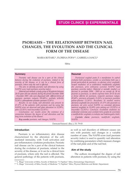
PSORIASIS â THE RELATIONSHIP BETWEEN NAIL CHANGES ...
PSORIASIS â THE RELATIONSHIP BETWEEN NAIL CHANGES ...
PSORIASIS â THE RELATIONSHIP BETWEEN NAIL CHANGES ...

You also want an ePaper? Increase the reach of your titles
YUMPU automatically turns print PDFs into web optimized ePapers that Google loves.
DermatoVenerol. (Buc.), 53: 79-83psoriasis. The nail changes, observed best whenusing the NAPSI score, were studied apart for thenail plate and the nail bed. Changes of the nailplate include leuconikia, pits, lunular red spotsand crumbling of the nail, while the nail bed canpresent hyperkeratosis, salmon patches,onycholysis and splinter bed (table I).Table I. Parameters used in NAPSI scoreChanges of the nail plateChanges of the nail bedLeuconikiaPitsCrumblingLunular red spotsHyperkeratosisSalmon patchesOnycholysisSplinter nail bedThe results showed that the male patientswere far more affected (78,95%), in contrary withthe medical literature data that suggest a relativeequal gender distribution.The severity of the disease was well connectedto the duration of psoriasis, to the severe clinicalforms and to important nail changes at patientsthat were diagnosed over 40 years ago.Nail changes affected mostly the nail plate(47,37%). The most frequent nail plate changes wereleuconikia and pits (22,92%), while hyperkeratosiswas most observed on the nail bed (20,84%).DiscussionsPsoriatic nail plate affects both genders. It isnot lethal, but in severe cases it can have animportant psychosocial and functional impact [2,4, 7]. The nail changes are best observed whenusing the NAPSI score. This index allows us toquantify separately the changes of the nail plateand of the nail bed.Thus, changes of the nail plate include:- leuconikia (white spots associated withparakeratosis of the nail)- pits (result from the loss of parakeratotic cellson the surface of the nail) (Fig. 5)- crumbling of the nail- lunular red spots (erythematous spots on thenail’s lunula)Changes observed on the nail bed are:- subungal hyperkeratosis (excessive proliferationof the nail bed which can cause onycholysis);- salmon patches (yellowish-red drops of the nailbed. This is the most specific change forpsoriatic nail disease) [6];Fig. 5. Patient with psoriatic nail disease (pits)- onycholysis (white spot associated with theseparation of the nail plate from the nail bed. Itcommences at the distal end of the nail bed,progresses proximally and increases thepossibility of mycobacterium colonization).- splinter nail bed (black longitudinal linescaused by discrete focal hemorrhages betweenthe nail plate and the nail bed. It’s considered tobe an equivalent of the Auspitz sign which canbe noticed in psoriasis of the skin).Beau’s lines can also be observed as transverselines on the nail caused by intermittentinflammation.Nail alteration as well as typical psoriaticchanges of the periungal skin can be morefrequently observed in patients with psoriaticarthritis. This sign can also have a predictionvalue when estimating the joint lesions, especiallyfor psoriatic distal poly-arthritis.Most nail changes are present in patientswith psoriatic skin lesions. We estimate that 10-55% of the patients with psoriasis have nailalterations, while around 5% have only psoriaticnail disease without any skin lesions. 10-20% ofthe patients diagnosed with psoriasis havepsoriatic arthritis, (fig.6) among which 53-86%reveals nail changes [7].Fig.6. Psoriatic arthritis81
DermatoVenerol. (Buc.), 53: 79-83Fig.7 a, b – Patient with severe form of psoriatic nail disease, with mixed alteration of the nail bed and of the nail plateFig.8 a, b. Important palmo-plantar nail changes in a patient with psoriasis vulgarisLikewise, around half of the patients withpsoriasis in our study group (47,53%) presentedassociated nail changes, among which a quarter(26,31%) were diagnosed with psoriasis over 40years ago (Fig. 7 a, b).Joint alteration in the study group patients isfar less important (5, 26%) then it is specified inmedical literature (10-20%). This lack of similaritymight be explained by a misdiagnosis of psoriaticarthritis.The pathology of psoriatic nail disease is notcompletely understood, but it implies the associationof genetic, environmental and immunologicalfactors (T-cell mediated inflammatoryreaction). Recent studies proved the connectionbetween psoriasis and some subtypes of HLAantigens:Cw6, B13, Bw57, Cw2, Cw11 and B27 [1].Psoriatic nail disease can be a part of theclinical features during the evolution of psoriasis,related to the severity of the disease, or it can be aclinical form of psoriasis, where only the nail isaffected (Fig. 8 a,b). Also, nail alteration inpsoriasis can be associated with onycomicosis orparonychia. The differential diagnosis mightinclude nail lichen planus, pityriasis rubra pilaris,alopecia areata, punctate keratoses [1].The final diagnosis is established by nailbiopsy which reveals hyperkeratosis, increase inthe granular layer, hemorrhages in stratumcorneum, epidermal papillomatous hyperplasiaand spongiosis.The therapy methods used so far do notinsure complete healing, but improve the psychosocialimpact of the nail disorder.When treating psoriatic nail disease, one canuse:- occlusive dressing with topical corticosteroids(anti-inflammatory effect, inhibiting polinuclearmigration and modifying capilar permeability)- occlusive dressing with 5-Fluorouracil solution1% or cream 5% (improves pits and subungalhyperkeratosis)82
DermatoVenerol. (Buc.), 53: 79-83- topical treatment with Methotrexate in colodiumbase.- PUVA therapy might improve skin lesions aswell as nail changes. Long term treatment caninduce nail discoloration [5].- local monthly injections with Triamcinolon [3]- systemic therapy used when treating psoriaticskin lesions might improve nail changes aswell.- surgical or chemical nail ablation.The method of treatment is to be chosendepending on the clinical form, length of thedisease and associated disorders [2, 4, 7].ConclusionsAs mentioned in the medical literature,psoriatic nail changes are more common inpatients with medium or severe forms whichhave evolved for a long period of time.In our study, nail changes are present in47,37% of the patients with psoriasis, whopresent medium or severe forms.Our results prove the existence of arelationship between the degree of nail alterationand a long term evolution of the disease.By using NAPSI score, the study shows thatthe nail plate is slightly more affected (52,09%)than the nail bed, due to pits (22,92%) andleuconikia (22,92%).The authors consider that using the NAPSIscore to quantify nail changes can be a useful andprestigious method when diagnosing psoriaticnail disease.Bibliography1. Bikowski JB – Psoriatic nail disease: diagnosis andtreatment options. Cutis July 1999; 64: 1-9.2. Calvert HT, Smith MA, Wells RS – Psoriasis andthe nails. Br J Dermatol 1963 Nov; 75: 415-8.3. de Berker DA, Lawrence CM – A simplifiedprotocol of steroid injection for psoriatic naildystrophy. Br J Dermatol 1998 Jan; 138 (1): 90-54. Farber EM, Nall L: Nail psoriasis. Cutis 1992 Sep;50(3): 174-8.5. Handfield-Jones SE, Boyle J, Harman RR – LocalPUVA treatment for nail psoriasis. Br J Dermatol1987 Feb; 116(2): 280-1.6. Kouskoukis CE, Scher RK, Ackerman AB – The„oil drop“ sign of psoriatic nails. A clinical findingspecific for psoriasis. Am J Dermatopathol 1983 Jun;5(3): 259-62.7. Scher RK – Psoriasis of the nail. Dermatol Clin 1985Jul; 3(3): 387-94.Intrat în redacþie: 7 mai 200883