You also want an ePaper? Increase the reach of your titles
YUMPU automatically turns print PDFs into web optimized ePapers that Google loves.
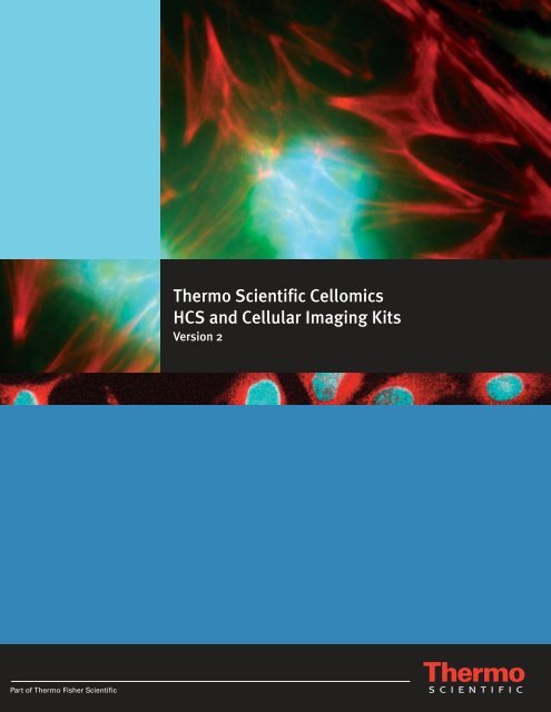
Thermo Scientific <strong>Cellomics</strong>HCS and Cellular Imaging KitsVersion 2
Menu of Thermo Scientific <strong>Cellomics</strong> HCS Reagent Kits, Accessories and Related ProductsKit Category . . . . . . . . . . . . . . . . . . . . . . . . . . . . . . . . . . . . . . . . . . . . PageCell Signaling and Transcription Factor Kits . . . . . . . . . . 1-2Beta Catenin Activation . . . . . . . . . . . . . . . . . . . . . . . . . . . . . . . . . . . . . . 1c-Jun ActivationERK MAPK ActivationPhospho-JNK Detection . . . . . . . . . . . . . . . . . . . . . . . . . . . . . . . . . . . . . . 2PKA ActivationPKCα ActivationTranscription Factor Activation – HIF-1 alpha, Phospho-CREB and FOXO3aTranscription Factor Activation – Phospho-CREBTranscription Factor Activation – HIF-1 alphaTranscription Factor Activation – FOXO3aTranscription Factor Activation: Smad3 and Phospho-CREBTranscription Factor Activation: Smad3Cytotoxicity and Apoptosis Kits. . . . . . . . . . . . . . . . . . . . . . 2-3Caspase 3 Activation . . . . . . . . . . . . . . . . . . . . . . . . . . . . . . . . . . . . . . . . 2Caspase 9 ActivationCell ViabilityCleaved PARP Detection . . . . . . . . . . . . . . . . . . . . . . . . . . . . . . . . . . . . . 3Cytochrome C DetectionLC3B DetectionPoly-Ubiquitin DetectionLC3B and Poly-Ubiquitin Multiplex DetectionMultiparameter Apoptosis IMultiparameter Cell Death DetectionMultiparameter Cytotoxicity 1Multiparameter Cytotoxicity 2 containing both Lysosomeand Mitochondrial ProbesMultiparameter Cytotoxicity 2 containing Lysosome ProbeMultiparameter Cytotoxicity 2 containing Mitochondrial ProbeMultiparameter Cytotoxicity 3Genotoxicity, DNA Damage and Repair Kits. . . . . . . . . . . 4-5Ku70/80 Activation. . . . . . . . . . . . . . . . . . . . . . . . . . . . . . . . . . . . . . . . . . 4MDM2 and p53 Detection – Orange p53 and Green MDM2MDM2 Detection – Orange MDM2Micronucleusp38 MAPK Activationp53 and p21 Activation Kit containing Orange p21 Probeand Green p53 Probep53 Activation Kit containing Orange p53 Probep21 Activation Kit containing Orange p21 ProbePhospho-ATMPhospho-ATM and p53 ActivationPhospho-Chk2 ActivationPhospho-H2AX Activation . . . . . . . . . . . . . . . . . . . . . . . . . . . . . . . . . . . . 5Phospho-p53 and p53 Activation containing Orange Phospho-p53Probe and Green p53 ProbePhospho-p53 Activation containing Orange Phospho-p53 ProbeInflammation and Cell Stress Kits. . . . . . . . . . . . . . . . . . . . . 5ATF-2 ActivationCell MotilityCHOP/GADD153 DetectionCOX 2 ActivationFOXO1A ActivationHeme Oxygenase 1 ActivationHeme Oxygenase 1 and Phospho-p38 ActivationKit Category . . . . . . . . . . . . . . . . . . . . . . . . . . . . . . . . . . . . . . . . . . . . PageInflammation and Cell Stress Kits (continued) . . . . . . . . . . 6-8Hsp27 and Phospho-Hsp27 Detection . . . . . . . . . . . . . . . . . . . . . . . . . . . 6Phospho-Hsp27 DetectionHsp60 and Hsp90β DetectionHsp60 DetectionHsp70 and Hsp90α DetectionHsp70 DetectionHsp90α DetectionImmunophilin FKBP52 DetectioniNOS ActivationMnSOD InductionMnSOD and Phospho-H2AX InductionNFκB ActivationNFκB and c-Jun Activation Kit containing Orange NFκBand Green c-Jun Duplex Dyes. . . . . . . . . . . . . . . . . . . . . . . . . . . . . . . 7NFκB and c-Jun Activation Kit containing Orange NFκB ProbeNFAT-1 ActivationOxidative Stress IPhospho-AKT ActivationPhospho-4E-BP1 DetectionPhospho-c-jun and Phospho-JNK Multiplex DetectionPhospho-GSK-3 DetectionPhospho-S6 DetectionPKA and Phospho-CREB ActivationSTAT 1 Activation . . . . . . . . . . . . . . . . . . . . . . . . . . . . . . . . . . . . . . . . . . . 8STAT 2 ActivationSTAT 3 ActivationCell Cycle and Cell Proliferation Kits. . . . . . . . . . . . . . . . . . 8BrdU and Ki67 Cell Proliferation – Multiplex Kit ContainingGreen BrdU & Orange Ki67 ProbesBrdU Cell Proliferation – Singleplex Containing Orange BrdUKi67 Cell Proliferation – Singleplex Containing Orange Ki67 ProbeCell Cycle ICyclin B1 ActivationMultiplex Mitosis-Apoptosis KitMitotic IndexPhospho-mTOR ActivationPhospho-PLK1 ActivationPhospho-Rb ActivationCell Morphology and Phenotypic Change Kits. . . . . . . . . . 9Cytoskeletal Rearrangement – Whole Cell Stain, F-actin and TubulinCytoskeletal Rearrangement – Whole Cell Stain and F-actinCytoskeletal Rearrangement – Whole Cell Stain and TubulinNeurite OutgrowthSynaptogenesis KitAccessory Reagents . . . . . . . . . . . . . . . . . . . . . . . . . . . . . . . . 9Whole Cell Stain ToolboxRabbit Antibody Detection WCS ToolboxMouse Antibody Detection WCS ToolboxWhole Cell Stain BlueWhole Cell Stain GreenWhole Cell Stain OrangeWhole Cell Stain Red
Thermo Scientific <strong>Cellomics</strong> HCS Reagent KitsNew kits expand your ability to realize the full potential of Cellular ImagingLarge packsizes available—Custom packingavailableThe use of appropriate combinations of fluorescent probes,antibodies and reagents can help realize the full potentialof powerful cellular imaging. Thermo Scientific <strong>Cellomics</strong>HCS Reagent Kits provide just such a combination in aneasy-to-use kit, with methods and reagents to prepare imaging-qualitysamples for automated cell-based assays.These kits are an integral part of the Thermo Scientific <strong>Cellomics</strong>Total Solution HCS Platform, allowing a wide rangeof biologies to be investigated with ease. Each kit is designedfor a specific biology and rigorously validated andoptimized for HCS/HCA.All kits use a fixed end-point assay based on immunofluorescencedetection in cells grown on standard high-density microplates.The primary antibodies selected for each kit are specific for theirtargets and have minimal cross-reactivity with other targets.Thermo Scientific <strong>Cellomics</strong> Cell Signalingand Transcription Factor KitsHighlights:• Customized components and bulk quantities available• Flexible – available in multiplex or singleplexconfigurations• Optimized protocol and reagent preparations includedfor reproducible results• Validated on Thermo Scientific <strong>Cellomics</strong> ArrayScanInstruments, but the kits also work on otherHCS platforms and with fluorescence microscopes• Wide range of targets for cell signaling, toxicity andcell phenotype changesBeta-Catenin ActivationOur Beta-Catenin Activation Kit enables measurement of the intracellular distribution ofβ-catenin and its translocation to the nucleus. The kit contains a polyclonal anti-β-cateninrabbit primary antibody, a goat anti-rabbit secondary antibody conjugated to the ThermoScientific DyLight 549 Fluorophore and other reagents and buffers required for immunofluorescencelabeling of β-catenin HCS assays.Signaling through the Wnt pathway via the nuclear translocation of the key transcriptionfactor β-catenin is critical for cell growth and differentiation during development. β-cateninis located mostly in the adherens junctions of epithelial cells associated with E-cadherin, butalso in cytoplasmic pools complexed with APC. In the absence of Wnt signaling, β-cateninis phosphorylated by both casein kinase 1a (CK1a) and glycogen synthase kinase 3b(GSK3b), thus targeting this protein for ubiquitination and subsequent proteolysis.Product # Description Pkg. Size U.S. Price8403601 Beta Catenin Activation 1 x 96 $ 2348403602 5 x 96 $ 519c-Jun Activationc-Jun is activated through phosphorylation in the activation domain by Jun-N-terminalkinases (JNKs). Phosphorylated Jun family members then form homodimers or heterodimericcomplexes with Fos, composing the AP-1 transcription factor, which migratesinto the nucleus.Product # Description Pkg. Size U.S. PriceK01-0003-1 c-Jun Activation 5 x 96 $ 435ERK MAPK ActivationMAP kinases mediate signal transduction from growth hormones, heat shock, UV radiation,osmolarity or cytokines, to alter transcription factor activity in the nucleus. Aberrantand deregulated functioning of MAP kinases can initiate and support carcinogenesis.Insulin and IGF-1 also activate a mitogenic MAP kinase pathway that may be importantin acquired insulin resistance associated with type 2 diabetes.Product # Description Pkg. Size U.S. PriceK01-0007-1 ERK MAPK Activation 5 x 96 $ 5441
Phospho-JNK DetectionOur Phospho-JNK Detection Kit enables quantitation of phosphorylated Jun N-terminal kinase (phospho-JNK) expression in the nucleus. Phospho-JNK, alsoreferred to as stress-activated protein kinase 1 (SAPK), is measured directlyusing a fixed end-point assay based on immunofluorescence detection in cellsgrown on standard high-density microplates. This kit contains an anti-phospho-JNK primary antibody (rabbit monoclonal), a goat anti-rabbit secondary antibodyconjugated to Thermo Scientific DyLight 549 Fluorophore and other reagentsand buffers required for immunofluorescence staining for HCS assays.Product # Description Pkg. Size U.S. Price8404001 Phospho-JNK Detection 1 x 96 $ 2328404002 5 x 96 $ 519PKA ActivationOur PKA Activation Kit enables detection and measurement of activated cAMPdependentprotein kinase A (PKA) in stimulated cells for high-content screening.After stimulation, activated PKA redistributes from the perinuclear region (orperinuclear spots) to the cytoplasm and nucleus, producing a more diffusestaining. This assay measures PKA redistribution using antibody staining andis applicable across many cell types.Product # Description Pkg. Size U.S. Price8404301 PKA Activation 1 x 96 $ 2328404302 5 x 96 $ 519PKCα ActivationThe Protein Kinase C (PKC) family is a group of related signal transduction proteins,and their activation is one of the earliest events leading to a variety ofcellular responses, including cytokine secretion, differentiation, proliferation,muscle contraction and modulation of membrane excitability. PKCα is believedto play an important role in the development and growth of cancer cells.Product # Description Pkg. Size U.S. PriceK09-0001-1 PKCα Activation 5 x 96 $ 435Transcription Factor IOur Transcription Factor Activation HCS Reagent Kits are for the simultaneousquantitation of HIF-1 alpha, Phospho-CREB and FOXO3a expression in thesame cell. The multiplex kits contain primary antibodies toward HIF-1 alpha,phosphorylated CREB, FOXO3a (rabbit polyclonal, mouse monoclonal, and goatpolyclonal, respectively) and secondary antibodies conjugated with DyLight 549(orange), DyLight 488 (green) and DyLight 649 (red) Fluorophores.The singleplex kits contain the same primary antibodies and DyLight 549-Conjugated Secondary Antibodies. In cancer cells, rapid proliferation oftenresults in a competition for nutrients and oxygen. Three key transcription factorsplay a pivotal role in determining cancer cell fate: HIF, CREB and FOXO3a. Allrespond to both oxidative stress that occurs in rapidly proliferating cells andto growth factor signaling through PI3 kinase (PI3K) and AKT, also known asprotein kinase B (PKB). Hypoxia inducible factor 1 (HIF-1) and the cyclic AMPresponse element binding protein (CREB) promote cell proliferation and enhancemetabolism; however, FOXO3a (FKHRL1), a member of the Forkhead family oftranscription factors, induces cell cycle arrest or apoptosis.Product # Description Pkg. Size U.S. Price8401401 Transcription Factor Activation – 1 x 96 $ 3128401402 HIF-1 alpha, Phospho-CREB and FOXO3a 5 x 96 $ 6968401501 Transcription Factor Activation – 1 x 96 $ 2328401502 Phospho-CREB 5 x 96 $ 5198401601 Transcription Factor Activation – 1 x 96 $ 2348401602 HIF-1 alpha 5 x 96 $ 5198401701 Transcription Factor Activation – 1 x 96 $ 2348401702 FOXO3a 5 x 96 $ 519Transcription Factor II Smad3 and Phospho-CREBOur multiplexed Smad3 and Phospho-CREB Reagent Kits contain optimizedreagents for the simultaneous detection and quantitation of Smad3 andPhospho-CREB activation in the same cell using different compounds. Thesekits allow direct measurements of Smad3 and Phospho-CREB in the nucleus.The primary antibodies are specific for their targets and have minimal crossreactivitywith other targets. Two kit versions are available in two package sizes:• A multiplexed kit for detecting Smad3 and Phospho-CREB simultaneously.This kit contains primary antibodies against Smad3 and Phospho-CREB (rabbitpolyclonal and mouse monoclonal, respectively) and secondary antibodiesconjugated with DyLight 488 (green) and DyLight 549 (orange) Fluorophores.• A Smad3 singleplex kit that contains a rabbit polyclonal antibody againstSmad3 and a DyLight 549-Conjugated Secondary Antibody.Product # Description Pkg. Size U.S. Price8402001 Transcription Factor Activation: 1 x 96 $ 3128402002 Smad3 and Phospho-CREB 5 x 96 $ 6968402101 Transcription Factor Activation: Smad3 1 x 96 $ 2328402102 5 x 96 $ 535Thermo Scientific <strong>Cellomics</strong> Cytotoxicityand Apoptosis KitsCaspase 3Our Caspase 3 Activation HCS Reagent Kit contains optimized reagents for thedetection and quantitation of caspase 3 activation (cleaved) in the cells. The primaryantibody is specific for cleaved caspase 3 from human, mouse and rat and does notrecognize full-length caspase 3 or other caspases.The secondary antibody is conjugated to DyLight 549 Fluor (orange). Caspasesare intracellular cysteine proteases that are important in apoptotic cell death ina variety of cell lines. Caspase 3 can be activated by two different pathways:mitochondrial apoptosis and Fas ligand-mediated apoptosis.Product # Description Pkg. Size U.S. Price8402201 Caspase 3 Activation 1 x 96 $ 2418402202 5 x 96 $ 535Caspase 9Our Caspase 9 Activation Reagent Kit contains optimized reagents for thedetection and quantitation of caspase 9 activation in cells. This kit allowsdirect in-cell measurements using a fixed end-point assay based on immunofluorescencedetection in cells grown on standard high-density microplates.The primary antibody is specific for human cleaved caspase 9 and does notcross-react with total caspase 9 or other caspases.The secondary antibody is conjugated to DyLight 549 Fluor (orange). Caspasesare intracellular cysteine proteases that are important in apoptotic cell death ina variety of cell lines. Caspase 9 (ICE-LAP6, Mch6) is a member of the mitochondrialpathway of apoptosis.Product # Description Pkg. Size U.S. Price8402301 Caspase 9 Activation 1 x 96 $ 2348402302 5 x 96 $ 535Cell ViabilityThe basis of our Viability Assay are Thermo Scientific DeadDye and ThermoScientific VitalDye Proprietary Fluorescent Probes that enable quantification ofthe number of live and dead cells, as well as their relative percentages andlive/dead ratio, on any standard microplate format.Product # Description Pkg. Size U.S. PriceK02-0001-1 Cell Viability 5 x 96 $ 4352
Cleaved PARP DetectionOur Cleaved PARP Detection Kit enables detection and quantitation of cleavedPARP in the nuclei. The kit contains a primary monoclonal antibody that detectsonly the cleaved portion of human PARP, a goat anti-mouse secondary antibodyconjugated to DyLight 549 Fluorophore and other reagents and buffers that arerequired for immunofluorescence staining for HCS assays.Poly (ADP-ribose) polymerase (PARP) cleavage is an important marker ofcaspase 3-mediated apoptosis. PARP is a 116 kDa nuclear protein involved inrepair of DNA nicks induced by various stressors and is one of the substratesfor caspase 3, which cleaves PARP into an 85 kDa fragment during apoptosis.In human PARP, cleavage occurs at Asp214 and Gly-215 residues, leading toformation of 89 and 24 kDa fragments. Cleavage of PARP correlates withDNA fragmentation and other morphological changes making it a critical markerof apoptosis.Product # Description Pkg. Size U.S. Price8402701 Cleaved PARP Detection 1 x 96 $ 2348402702 5 x 96 $ 519Cytochrome C DetectionOur Cytochrome C Detection Kit measures release of cytochrome c from mitochondria,a key early step in apoptosis or programmed cell death. The kitcontains a mouse monoclonal anti-cytochrome c primary antibody, a secondaryantibody conjugated to DyLight 549 Fluorophore, and other reagents and buffersrequired for immunofluorescent detection of cytochrome c for HCS assays.Product # Description Pkg. Size U.S. Price8405601 Cytochrome C Detection 1 x 96 $ 2348405602 5 x 96 $ 519LC3B and Poly-Ubiquitin Multiplex DetectionOur LC3B and Poly-Ubiquitin Detection Kits enable quantitation of LC3B proteinon autophagic vesicles and ubiquitin on polyubiquitinated proteins. Autophagyand the ubiquitin proteasome system constitute two major intracellular proteindegradation pathways that are essential during cell starvation, differentiation,aging and death. LC3B participates in the process of mammalian autophagy,also characterized as a caspase-independent programmed cell death.Autophagy is the catabolic process of sequestering organelles and long-livedproteins in autophagic vesicles, which are eventually digested by lysosomalmachinery. The ubiquitin proteasome system controls the stability of numerousproteins that regulate progression through the cell cycle and apoptosis such ascyclins, cyclin-dependent kinases, tumor suppressors and NFκB. Once theseproteins are tagged with a single ubiquitin molecule, other ligases are signaledto attach additional ubiquitin. The result is a polyubiquitinated protein that isdirected to the proteasome, a large protein complex, where it is degraded.Product # Description Pkg. Size U.S. Price8407601 LC3B Detection 1 x 96 $ 2348407602 5 x 96 $ 5198407701 Poly-Ubiquitin Detection 1 x 96 $ 2348407702 5 x 96 $ 5198407801 LC3B and Poly-Ubiquitin Multiplex Detection 1 x 96 $ 3128407802 5 x 96 $ 675Multiparameter Apoptosis 1Apoptosis is a critical process in the life and death of a cell. Interruption of theprocess can cause many diseases. Key criteria for determining whether a cell isundergoing apoptosis include morphological changes in the appearance of thecell, as well as alteration in biochemical and molecular markers. The patterns ofapoptotic signals are similar, but the details of the pathway vary significantlydepending on the cell type and apoptotic inducer.Product # Description Pkg. Size U.S. PriceK04-0001-1 Multiparameter Apoptosis I 5 x 96 $ 544Multiparameter Cell Death DetectionOur Multiparameter Cell Death Detection Kits for high-content screening (HCS)enable simultaneous quantitation of LC3B protein on autophagic vesicles,cytochrome c localization and its release from mitochrondria, cell membraneintegrity and its permeability and nuclear morphology and DNA content. Thesecellular properties are measured directly using fixed end-point assay and fluorescencedetection in cells grown on standard high-density microplates. The kitscontain highly specific primary antibodies, DyLight-conjugated SecondaryAntibodies and other reagents and buffers required to stain cells for HCS assays.Product # Description Pkg. Size U.S. Price8408001 Multiparameter Cell Death Detection 1 x 96 $ 3128408002 5 x 96 $ 675Multiparameter Cytotoxicity 1The Multiparameter Cytotoxicity 1 HCS Reagent Kit is an in vitro assay tool thatallows users to rapidly acquire information on the changes in the followingcellular properties: (1) nuclear morphology/size, (2) cell membrane permeability,(3) lysosomal mass-pH and (4) cell density (number of cells per field). This kitdiffers from the Multiparameter Cytoxicity 2 Kit in that the dyes are supplied as acocktail as well as not measuring mitochondrial potential.Product # Description Pkg. Size U.S. PriceK02-0002-1 Multiparameter Cytotoxicity 1 5 x 96 $ 544Multiparameter Cytoxicity 2Our Multiparameter Cytoxicity 2 HCS Reagent Kits contain optimized reagentsfor simultaneous detection and quantitation in the same cell of changes innuclear morphology and size, changes in the cell membrane’s permeabilitystatus, changes in cell density (number of cells per field) from the compoundtoxicity, and changes induced to EITHER the lysosome’s mass or pH OR themitochondria’s transmembrane potential.Product # Description Pkg. Size U.S. Price8400001 Multiparameter Cytotoxicity 2 1 x 96 $ 3218400002 containing both Lysosome and Mitochondrial Probes 5 x 96 $ 6968400101 Multiparameter Cytotoxicity 2 1 x 96 $ 2348400102 containing Lysosome Probe 5 x 96 $ 5158400201 Multiparameter Cytotoxicity 2 1 x 96 $ 2418400202 containing Mitochondrial Probe 5 x 96 $ 535Multiparameter Cytoxicity 3Our Multiparameter Cytotoxicity 3 Kit enables simultaneous measurement of sixorthogonal cell-health parameters: cell loss, nuclear morphology, DNA content,cell membrane permeability, mitochondrial membrane potential changes andcytochrome c localization and release from mitochondria. The kit contains aHoechst dye, cell permeability dye, mitochon-drial membrane potential dye,and a mouse monoclonal antibody against cytochrome c and a goat anti-mouseDyLight 649-conjugated Secondary Antibody, and various other essentialreagents and buffers.Product # Description Pkg. Size U.S. Price8408102 Multiparameter Cytotoxicity 3 5 x 96 $ 7993
Thermo Scientific <strong>Cellomics</strong> Genotoxicity,DNA Damage and Repair KitsKu70/80 ActivationOur Ku 70/80 Activation Kit contains optimized reagents for the detectionand quantitation of Ku70/80 in the nuclei of cells. The kit contains a primarymonoclonal antibody specific for human Ku70/80 heterodimers, a goat antimousesecondary antibody conjugated to DyLight 549 Fluorophore and otherreagents and buffers required for immunofluorescence labeling of Ku70/80 forHCS analysis.The Ku70/80 heterodimer plays an important role in DNA double-strand break(DSB) repair. During DSB repair by non-homologous end joining (NHEJ), Ku70/80 binds with high affinity to DNA ends of a DSB and then recruits thecatalytic domain of DNA protein kinase (DNAPK). The formation of DNAPKcomplex at the site of DSBs results in the recruitment of other repair proteins toligate the broken ends. Recently, Ku70/80 was identified as having a role in theATM-dependent activation of ATR during DNA DSB damage response.Product # Description Pkg. Size U.S. Price8403101 Ku70/80 Activation 1 x 96 $ 2328403102 5 x 96 $ 519MDM2 and p53Our MDM2 and p53 Detection HCS Reagent Kits are for the simultaneousquantitation of MDM2 and p53 expression in the same cell. These kits allowdirect measurements of MDM2 and p53 in the nucleus using a fixed end-pointassay based on immunofluorescence detection in cells grown on standard highdensitymicroplates.The orange p53 and green MDM2 multiplex kits contain primary antibodiestoward p53 and MDM2 (rabbit polyclonal and mouse monoclonal, respectively)and secondary antibodies conjugated with DyLight 488 (green) and DyLight 549(orange) Fluorophores. The orange MDM2 singleplex kits contain a mousemonoclonal primary antibody toward MDM2 and a DyLight 549-ConjugatedSecondary Antibody.Product # Description Pkg. Size U.S. Price8401801 MDM2 and p53 Detection – 1 x 96 $ 3218401802 Orange p53 and Green MDM2 5 x 96 $ 6758401901 MDM2 Detection – Orange MDM2 1 x 96 $ 2328401902 5 x 96 $ 519MicronucleusMicronucleus (MN) formation is a hallmark of genetic toxicity; as such, micronucleiare used as indicators of genotoxicity caused by drug candidates orenvironmental toxins. The in vitro micronucleus assay is among a set ofgenetic toxicology assays wherein cultured cells are treated and scored formicronucleus induction.Product # Description Pkg. Size U.S. PriceK11-0001-1 Micronucleus 5 x 96 $ 435p38 MAPK Activationp38 MAP kinase (MAPK, Mitogen-activated protein kinase), also known as aCDC-2-related protein kinase or CSBP (cytokine suppressive anti-inflammatorydrug binding protein), regulates many cellular processes, including inflammation,cell differentiation, cell growth and death. p38 MAPK is activated inresponse to a variety of extracellular stimuli, including osmotic shock, cytokines,LPS and anisomycin.Product # Description Pkg. Size U.S. PriceK01-0004-1 p38 MAPK Activation 5 x 96 $ 544p53 and p21 DetectionOur Multiplexed p53 and p21 Detection HCS Reagent Kits are for the simultaneousquantitation of p53 and p21 expression in the same cell. These kits allowdirect measurements in the nucleus.Product # Description Pkg. Size U.S. Price8400601 p53 and p21 Activation Kit containing 1 x 96 $ 3218400602 Orange p21 Probe and Green p53 Probe 5 x 96 $ 6968400801 p53 Activation Kit containing 1 x 96 $ 2348400802 Orange p53 Probe 5 x 96 $ 5198400901 p21 Activation Kit containing 1 x 96 $ 2348400902 Orange p21 Probe 5 x 96 $ 519Phospho-ATM ActivationOur Phospho-ATM Activation Kit contains optimized reagents for the detectionand quantitation of phosphorylated ATM (Ser1981) in the nuclei. The kit containsa primary monoclonal antibody that detects only the phosphorylated formof human ATM, a goat anti-mouse secondary antibody conjugated to DyLight549 Fluorophore and other reagents and buffers required for immunofluorescencestaining for HCS assays.Ataxia telangiectasia mutated kinase (ATM, 350 kDa) is involved in cell cyclecheck-point signaling and DNA repair. Mutation in the ATM gene leads to ataxiatelangiectasia, an autosomal recessive human disease. ATM is auto phosphorylatedat Ser1981 upon induction of DNA double-strand breaks (DSBs) leading torapid check-point signaling. ATM kinase has several identified targets includingH2AX, BRCA1, NBS1, Chk1, Chk2 and p53.Product # Description Pkg. Size U.S. Price8403001 Phospho-ATM Activation 1 x 96 $ 2328403002 5 x 96 $ 519Phospho-ATM and p53 ActivationOur Phospho-ATM and p53 Activation Kit contains optimized reagents for thedetection and quantitation of phosphorylated ATM (Ser1981) and p53 in thenucleus. The kit contains a monoclonal antibody that detects only the phosphorylatedform of human ATM, anti-p53 polyclonal antibody, DyLight-conjugatedSecondary Antibodies and various other reagents and buffers for immunofluorescencestaining for HCS assays.Product # Description Pkg. Size U.S. Price8405701 Phospho-ATM and p53 Activation 1 x 96 $ 2348405702 5 x 96 $ 519Phospho-Chk2 (Thr68) ActivationOur Phospho-Chk2 Activation Kit contains optimized reagents for the detectionand quantitation of phosphorylated Chk2 (Thr68) in the nuclei. The kit contains aprimary monoclonal antibody that detects only the phosphorylated form ofhuman Chk2, a goat anti-mouse secondary antibody conjugated to DyLight 549Fluorophore and other reagents and buffers required for immunofluorescencestaining for HCS assays.Chk1 and Chk2 are kinases involved in DNA damage-induced cell-cycle checkpointsignaling. Chk2 is phosphorylated by ATM kinase in response to DNAdamage, and Chk2 activation results in cell-cycle inhibition by p53 phosphorylationand other downstream targets. Phosphorylation at Thr68 is a prerequisitefor the subsequent activation step, which is caused by Chk2 autophosphorylationon residues Thr383 and Thr387 in the kinase domain activation loop.Product # Description Pkg. Size U.S. Price8402801 Phospho-Chk2 Activation 1 x 96 $ 2348402802 5 x 96 $ 5194
Phospho-H2AX ActivationOur Phospho-H2AX Activation Kit contains optimized reagents for the detectionand quantitation of phosphorylated H2AX (Ser139) in the nucleus. The kit containsa primary monoclonal antibody that detects only the phosphorylated formof human H2AX, a goat anti-mouse secondary antibody conjugated to DyLight549 Fluorophore and other reagents and buffers required for immuno-fluorescencestaining for HCS assays.The nucleosome is made of four core histone proteins (H2A, H2B, H3 and H4).H2AX belongs to an H2A family of histones. DSNA damage induction by variousagents leads to rapid phosphorylation of H2AC at Ser139 (also known asGamma H2AX) by ATM, ATR or DNA protein kinase leading to formation of DNAfoci at the site of DNA double-strand breads (DSBs). Phosphorylated H2AXhelps in recruiting the proteins responsible for double-strand break repair.Product # Description Pkg. Size U.S. Price8402901 Phospho-H2AX Activation 1 x 96 $ 2348402902 5 x 96 $ 519Phospho-p53 and p53 DetectionOur Multiplexed phospho-p53 and p53 Detection HCS Reagent Kits are for thesimultaneous quantitation of phospho-p53 and p53 expression in the same cell.These kits allow direct measurements in the nucleus.Product # Description Pkg. Size U.S. Price8400501 Phospho-p53 and p53 Activation 1 x 96 $ 3218400502 containing Orange Phospho-p53 Probe 5 x 96 $ 696and Green p53 Probe8400701 Phospho-p53 Activation 1 x 96 $ 2348400702 containing Orange Phospho-p53 Probe 5 x 96 $ 519CHOP/GADD153 DetectionOur CHOP/GADD153 Detection Kit is for the direct quantitation of CHOP/GADD153expression in the nucleus. Expression of mutant proteins disrupts protein foldingin the endoplasmic reticulum (ER), causes ER stress and activates a signalingnetwork called the unfolded protein response (UPR). Depending on the capacityof the ER to repair the protein folding process, the UPR either increases ordecreases the biosynthetic capacity of the secretory pathway through upregulationof ER chaperone expression or by attenuating the global protein synthesisand increasing pro-apoptotic mechanisms.Product # Description Pkg. Size U.S. Price8403901 CHOP/GADD153 Detection 1 x 96 $ 2348403902 5 x 96 $ 519COX-2 ActivationOur COX-2 Activation Kit is for detecting and measuring cytoplasmic inductionof the COX-2 enzyme. This kit contains a monoclonal mouse anti-COX-2antibody, a goat anti-mouse secondary antibody conjugated to DyLight 549Fluorophore and other reagents and buffers required for immunofluorescencedetection of COX-2 for HCS assays.This assay is primarily for immune cells, specifically macrophages, which arekey mediators of the innate immune response and cytokine production. Oncestimulated with lipopolysaccharide (LPS) and interferon gamma (IFNγ), or otherharmful stimuli, immune cells defensively produce cytokines, prostaglandins,chemokines and reactive amines. Prostaglandins (PGs), produced by the cyclooxygenase(COX) enzymes, are critical in the immune response to inducevasodilation, vasoconstriction, pain and fever.Product # Description Pkg. Size U.S. Price8403701 COX 2 Activation 1 x 96 $ 2328403702 5 x 96 $ 519Thermo Scientific <strong>Cellomics</strong> Inflammationand Cell Stress KitsATF-2 ActivationThe Activating Transcription Factor-2 (ATF-2) Kit responds to the inflammatorycytokines and cellular stressors, including genotoxicity and ischemia/reperfusion.ATF-2 is activated through threonines 69 and 71 by members of the SAPKfamily. Once activated, ATF-2 forms complexes with Jun family or other ATFfamily members. These complexes bind to the cAMP Response Element found inthe promoters of many genes, stimulating gene transcription.Product # Description Pkg. Size U.S. PriceK01-0010-1 ATF-2 Activation 5 x 96 $ 435Cell MotilityCell motility is central to a number of biological and pathological processes,including cancer cell invasion and metastasis, inflammation, angiogenesis,wound repair, and embryonic development. Cell movement occurs via theconcerted activities of cell adhesion molecules, the actin cytoskeleton and anextensive network of signaling molecules.Product # Description Pkg. Size U.S. PriceK08-0001-1 Cell Motility 5 x 96 $ 551FOXO1A ActivationOur FOXO1A Activation Kit measures activation of FOXO1A, a transcription factorinvolved in the initiation of cell arrest and apoptosis. The kit contains a polyclonalrabbit FOXO1A antibody, a goat anti-rabbit secondary antibody conjugated toDyLight 549 Fluorophore and other reagents and buffers required for immunofluorescentdetection of FOXO1A for HCS assays.Product # Description Pkg. Size U.S. Price8407201 FOXO1A Activation 1 x 96 $ 2348407202 5 x 96 $ 519Heme Oxygenase 1 ActivationOur Heme Oxygenase 1 Activation Kit measures activation of heme oxygenase 1in cells. Heme oxygenase 1 is a microsomal enzyme that catalyses the oxidationof heme to antioxidants, biliveridin and carbon monoxide and protects cells fromwide variety of stress conditions through activation of p38 MAPK. Carbonmonoxide produced during oxidation of heme by heme oxygenase 1 activatesp38 MAPK, which confers tissue protection through inhibition of cytokine production.Heme oxygenase 1 can be induced by oxidative stress, hypoxia, heatshock, heavy metals and cytokines.Product # Description Pkg. Size U.S. Price8405801 Heme Oxygenase 1 Activation 1 x 96 $ 2348405802 5 x 96 $ 519Heme Oxygenase 1 and Phospho-p38 ActivationOur Heme Oxygenase 1 and Phospho-p38 Activation Kit contains reagents for measuringactivation in cells for HCS assays. The kit contains a rabbit polyclonal antibodythat detects only phosphorylated p38, a mouse monoclonal antibody for heme oxygenase1, DyLight-conjugated Secondary Antibodies and various other reagents andbuffers required for immunofluorescence detection.Product # Description Pkg. Size U.S. Price8405901 Heme Oxygenase 1 and Phospho-p38 Activation 1 x 96 $ 3128405902 5 x 96 $ 6755
Hsp27 and Phospho-Hsp27 DetectionOur Hsp27 and Phospho-Hsp27 Detection Kits are for simultaneous quantificationof nuclear DNA content, heat shock protein 27 (Hsp27) and phosphorylatedHsp27. These kits allow direct measurements of Hsp27 modulation and Hsp27phosphorylation using a fixed end-point assay based on immunofluorescencedetection in cells grown on standard high-density microplates. The DNA bindingdye, DAPI, enables nuclear size and morphology determination and cell cyclephase identification by DNA content. The primary antibodies are specific fortheir targets and have minimal cross-reactivity. The anti-phospho-Hsp27 antibodydetects at Ser78.Product # Description Pkg. Size U.S. Price8406001 Hsp27 and Phospho-Hsp27 Detection 1 x 96 $ 3128406002 5 x 96 $ 6758406101 Hsp27 and Phospho-Hsp27 Detection 1 x 96 $ 2348406102 5 x 96 $ 5198406201 Phospho-Hsp27 Detection 1 x 96 $ 2348406202 5 x 96 $ 519Hsp60 and Hsp90-beta DetectionOur Hsp60 and Hsp90b Detection Kits are for simultaneous quantification ofnuclear DNA, heat shock protein 60 (Hsp60) and heat shock protein 90b(Hsp90b). Heat shock proteins (HSP) are essential for protein folding, proteinsynthesis, cellular stress defense and many other functions. Cellular stressincreases the HSP levels in cells by transcriptional regulation through HSF-1,STAT1, ATF3 and c Jun. This cellular response is critical for cellular homeostasis.Induction of HSP is closely correlated with substance cytotoxicity andlipophilicity given to the cell.Product # Description Pkg. Size U.S. Price8406701 Hsp60 and Hsp90β Detection 1 x 96 $ 3128406702 5 x 96 $ 6758406801 Hsp60 Detection 1 x 96 $ 2348406802 5 x 96 $ 519Hsp70 and Hsp90-alpha DetectionOur Hsp70 and Hsp90a Detection Kits are for simultaneous quantification ofnuclear DNA content, heat shock protein 70 (Hsp70) and heat shock protein 90α(Hsp90α). These kits allow direct measurements using a fixed end-point assaybased on immunofluorescence detection in cells grown on standard high-densitymicroplates. The DNA binding dye, DAPI, enables nuclear size and morphologydetermination and cell cycle phase identification by DNA content. The primaryantibodies are specific for their targets and have minimal cross-reactivity.Product # Description Pkg. Size U.S. Price8406301 Hsp70 and Hsp90α Detection 1 x 96 $ 3128406302 5 x 96 $ 6758406401 Hsp70 Detection 1 x 96 $ 2348406402 5 x 96 $ 5198406501 Hsp90α Detection 1 x 96 $ 2348406502 5 x 96 $ 519Immunophilin FKBP52 DetectionOur Immunophilin FKBP52 Detection Kit is for the simultaneous quantification ofnuclear DNA content and FK506-binding protein 52 (FKBP52). This kit detectsinducible FKBP52 (also known as FKBP4, FKBP59, Hsp56, Hsp59) protein in thecell. FKBP52 is a large immunophilin that binds to the immunosuppressive drugFK506 and has peptidyl-prolyl cis-trans isomerase (PPIase) activity, which isinhibited by the binding of FK506. Immunophilins are enriched in the central andperipheral neurons and reports indicate that FKBP52 has neurotrophic activity.Product # Description Pkg. Size U.S. Price8406601 Immunophilin FKBP52 Detection 1 x 96 $ 2348406602 5 x 96 $ 519iNOS ActivationOur iNOS Activation Kit measures cytoplasmic induction of the iNOS enzyme viaactivation of one of the many inflammatory pathways.The kit contains a monoclonal mouse anti-iNOS antibody, a goat anti-mousesecondary antibody conjugated to DyLight 549 Fluorophore and the other reagentsand buffers required for immunofluorescence labeling of iNOS for HCS assays.This assay is based on the elicitation of the immune response. After stimulationwith lipopolysaccharide (LPS) and interferon gamma (IFNγ), or other harmfulstimuli, immune cells produce cytokines, chemokines and oxidative radicals as adefensive mechanism. Nitric oxide synthase (NOS) is responsible for generationof nitric oxide (NO) through oxidizing L-arginine to L-citrulline. Nitric oxide actsas a signaling molecule in the cardiovascular system, as well as an inflammatorymolecule, and can react with other species to form more potent radicalssuch as peroxynitrite and lipid peroxides.There are three forms of NOS:• eNOS expressed in endothelial cells• nNOS, expressed in neuronal cells• iNOS, expressed in macrophagesInduction of iNOS through inflammatory mediators results in protein nitration,apoptosis induction, oxidative stress, DNA damage and respiration inhibition.Product # Description Pkg. Size U.S. Price8403801 iNOS Activation 1 x 96 $ 2348403802 5 x 96 $ 519MnSOD InductionOur MnSOD Induction Kit measures induction of manganese superoxidedismutase (MnSOD), an enzyme that reduces cellular oxidative stress via adismutation reaction of superoxide. The kit contains a polyclonal rabbit MnSODantibody, a goat anti-rabbit secondary antibody conjugated to DyLight 549Fluorophore, and other reagents and buffers required for immunofluorescentdetection of MnSOD for HCS assays.Product # Description Pkg. Size U.S. Price8407001 MnSOD Induction 1 x 96 $ 2348407002 5 x 96 $ 519MnSOD and Phospho-H2AX InductionOur MnSOD and Phospho-H2AX Induction Kit measures the production of oxidativeDNA damage using manganese superoxide dismutase (MnSOD) and theDNA damage sensor, phospho-histone 2AX. The kit contains a polyclonal rabbitanti-MnSOD antibody, a mouse monoclonal anti-phospho-H2AX antibody, secondaryantibodies conjugated to a DyLight Fluorophore and other reagents andbuffers required for immunofluorescent detection of MnSOD and phospho-H2AXfor HCS assays.Product # Description Pkg. Size U.S. Price8407301 MnSOD and Phospho-H2AX Induction 1 x 96 $ 3128407302 5 x 96 $ 675NFκB ActivationNuclear factor kappa B (NFκB) transcription factor plays an important role formany physiological processes and responses such as cell proliferation, cellsurvival, cellular responses to stress and immune response. Normally, NFκB ispresent in the cytoplasm as a complex with members of the lκB inhibitor family.Both the size of this complex and lκB masking of the nuclear localizationsequence on NFκB prevent it from entering the nucleus.Product # Description Pkg. Size U.S. PriceK01-0001-1 NFκB Activation 5 x 96 $ 4416
NFκB and c-Jun ActivationOur Multiplexed NFκB and c-Jun Activation HCS Reagent Kits are for thesimultaneous quantification of NFκB and c-jun activation in the same cell.These kits allow direct measurements of NFκB and phospho-c-jun translocationfrom the cytoplasm to the nucleus. These kits can be used for a wide range ofapplications, including cancer, inflammation and diabetes researchProduct # Description Pkg. Size U.S. Price8400301 NFκB and c-Jun Activation Kit 1 x 96 $ 3218400302 containing Orange NFκB & Green c-Jun Duplex Dyes 5 x 96 $ 6968400401 NFκB and c-Jun Activation Kit 1 x 96 $ 2348400402 containing Orange NFκB Probe 5 x 96 $ 519NFAT-1 ActivationNuclear factor of activated T cells (NFAT) is a family of transcription factorsimplicated in multiple biological processes, including cytokine gene expression,cardiac hypertrophy and adipocyte differentiation. NFAT1 (also known asNFATc2 or NFATp) is a 154 kDa member of this family that is regulated by thecalcium-dependent phosphatase calcineurin.Product # Description Pkg. Size U.S. PriceK01-0011-1 NFAT-1 Activation 5 x 96 $ 444Oxidative Stress IOxidative Stress is one of the most important cytotoxicity mechanismsinvestigated using HCS. Our Oxidative Stress I Kit uses a method to quantifychemically induced oxidative stress by measuring the amount of DNA bound toethidium, a product of dihydroethidium (DHE) oxidation. The method’s principleis that reactive oxygen species convert non-fluorescent dihydroethidium to fluorescentethidium that intercalates into DNA.Product # Description Pkg. Size U.S. Price8401001 Oxidative Stress I 1 x 96 $ 2418401002 5 x 96 $ 535Phospho-AKT ActivationOur Phospho-AKT Activation Kit measures phosphorylation of the 1, 2 and 3isoforms of AKT, a key kinase involved in regulation of cell proliferation. The kitcontains a polyclonal rabbit phospho-AKT antibody, a goat anti-rabbit secondaryantibody conjugated to DyLight 649 Fluorophore, Whole Cell Stain Green andother reagents and buffers required for immunofluorescence detection of phospho-AKTfor HCS assays.AKT is activated through the PI3 kinase pathway by growth factors, cytokines,mitogens and hormones. After the signal has been transduced through themembrane by receptor tyrosine kinases, PI3 kinase phosphorylates AKT atSer473 and Thr308 residues. When phosphorylated, activated AKT phosphorylateskey proteins involved in metabolism, protein synthesis, apoptosis,transcription factor regulation and the cell cycle, including MDM2, FOXO,BAD, GSK-3b, and mTOR. Alterations in AKT signaling lead to uncontrolled cellproliferation, and genetic mutations in the PI3 kinase signaling pathway areprominent in colon, breast and prostate cancers.Product # Description Pkg. Size U.S. Price8404102 Phospho-AKT Activation 5 x 96 $ 519Phospho-4E-BP1 DetectionOur Phospho-4E-BP1 Detection Kit enables quantitation of phosphorylated4E-BP1 protein (phospho-4E-BP1). Downstream effectors of mTOR-induced translationalcontrol include ribosomal protein S6 kinase (S6K) and the eukaryoticinitiation factor 4E (eIF4E)-binding protein (4E-BP1). The 4E-BP1 (eIF4E-bindingprotein 1, also called PHAS-I) is a small heat- and acid-stable phosphoproteinwhose phosphorylation is enhanced by growth factors, serum stimulation andinsulin in a PI3 kinase-dependent manner. 4E-BP1 undergoes phosphorylation atseveral sites leading to its release from eIF4E and allowing eIF4E to bind eIF4Gand form initiation complexes that facilitate upregulation of protein synthesis.Product # Description Pkg. Size U.S. Price8405301 Phospho-4E-BP1 Detection 1 x 96 $ 2348405302 5 x 96 $ 519Phospho-c-jun and Phospho-JNK Multiplex DetectionOur Phospho-c-jun and Phospho-JNK Detection Kit enables quantitation ofphosphorylated c-jun (phospho-c-jun) and phosphorylated Jun N-terminal kinase(phospho-JNK) in the nucleus. Phospho-JNK, also referred to as stress activatedprotein kinase 1 (SAPK), and phospho-c-jun are measured directly using a fixedend-point assay based on immunofluorescence detection in cells grown on standardhigh-density microplates. This kit contains an anti-phospho-c-jun primaryantibody (mouse monoclonal), anti-phospho-JNK primary antibody (rabbit monoclonal),DyLight Fluor-conjugated Secondary Antibodies and other reagents andbuffers required for immunofluorescence staining for HCS assays.Product # Description Pkg. Size U.S. Price8407901 Phospho-c-jun and Phospho-JNK 1 x 96 $ 3128407902 Multiplex Detection 5 x 96 $ 675Phospho-GSK-3 DetectionOur Phospho-GSK-3 Detection Kit measures phosphorylation of the α(Ser21)and β(Ser9) isoforms of glycogen synthase kinase-3 (GSK-3), a kinase involvedin glycogen metabolism, translation regulation and Wnt signaling. The kit containsa polyclonal rabbit phospho-GSK-3 antibody, a goat anti-rabbit secondaryantibody conjugated to DyLight 649 Fluorophore, Whole Cell Stain Green andvarious other reagents and buffers required for immunofluorescent detection ofphospho-GSK-3 for HCS assays.Product # Description Pkg. Size U.S. Price8407101 Phospho-GSK-3 Detection 1 x 96 $ 2348407102 5 x 96 $ 519Phospho-S6 DetectionOur Phospho-S6 Detection Kit enables quantitation of phosphorylated S6 protein(phospho-S6) of the mammalian 40S ribosomal subunit in the cytoplasm. Phospho-S6 is measured directly using a fixed end-point assay based on immunofluorescencedetection in cells grown on standard high-density microplates. This kit contains ananti-phospho-S6 primary antibody (rabbit monoclonal), a goat anti-rabbit secondaryantibody conjugated to DyLight 549 Fluorophore and other reagents andbuffers required for immunofluorescence staining for HCS assays.Product # Description Pkg. Size U.S. Price8405201 Phospho-S6 Detection 1 x 96 $ 2348405202 5 x 96 $ 519PKA and Phospho-CREB ActivationOur PKA and Phospho-CREB Activation Kit enables simultaneous detectionand measurement of activated protein kinase A (PKA) and phosphorylatedcAMP response element-binding (CREB) in stimulated cells. After stimulation,PKA is activated and redistributes from the perinuclear region (or perinuclearspots) to diffuse staining in the cytoplasm and nucleus. Once activated, PKAphosphorylates CREB, resulting in translocation of CREB to the nucleus. Thisassay measures PKA redistribution and CREB phosphorylation.Product # Description Pkg. Size U.S. Price8404701 PKA and Phospho-CREB Activation 1 x 96 $ 2347
Thermo Scientific <strong>Cellomics</strong> CellMorphology and Phenotypic Change KitsThermo Scientific <strong>Cellomics</strong> AccessoryReagentsCytoskeletal RearrangementOur Cytoskeletal Rearrangement HCS Reagent Kits are for the simultaneousquantitation of DNA content, cell morphology and the intracellular arrangementof microfilaments and microtubules in the same cell. These kits allow directmeasurements of cell and nuclear morphology, F-actin, and microtubule changesusing a fixed end-point assay based on immunofluorescence detection in cellsgrown on standard high-density microplates.The primary antibody is specific for its target and has minimal cross-reactivitywith other targets. The intracellular meshwork of the cytoskeleton is responsiblefor maintaining cell shape, cell movement, cytokinesis and organelleorganization. The cytoskeleton network also facilitates proper function of otherproteins by direct binding, transporting, repositioning and sequestering theseproteins. The structure of cytoskeleton is controlled by cytoskeleton-associatedproteins in response to the external signaling. Therefore, defects in the abilityto regulate the dynamics of cytoskeletal structure are likely to cause detrimentaleffects on other cell function.Product # Description Pkg. Size U.S. Price8402401 Cytoskeletal Rearrangement – 1 x 96 $ 3128402402 Whole Cell Stain, F-actin and Tubulin 5 x 96 $ 6758402501 Cytoskeletal Rearrangement – 1 x 96 $ 2328402502 Whole Cell Stain and F-actin 5 x 96 $ 5358402601 Cytoskeletal Rearrangement – 1 x 96 $ 2348402602 Whole Cell Stain and Tubulin 5 x 96 $ 515Neurite OutgrowthEfforts in CNS drug discovery research are focused on the identification ofcompounds that affect the growth of neurites. Drugs that promote nerve growthhave potential curative effect in a wide variety of diseases and traumas thatresult in neuropathy and nerve injury, including stroke, spinal cord injuries andneurodegenerative illnesses such as Parkinson’s and Alzheimer’s disease.Product # Description Pkg. Size U.S. PriceK07-0001-1 Neurite Outgrowth 5 x 96 $ 544Synaptogenesis KitThe Synaptogenesis Kit enables simultaneous detection of neuronal population,neurite, pre-synaptic vesicle, post-synaptic puncta and synapse using a fixedend-point assay based on immunofluorescence detection in cells grown on standardhigh-density microplates. The molecular network between synapses controlssynaptic signal transmission and plasticity and regulates neuronal growth,differentiation and death. To understand the relationship between synaptic activityand neuropathophysiology and the molecular mechanism involved insynaptogenesis and synapse regulation, the microstructure of synaptic junctionshas been extensively studied. The modulation of neurite and synaptic structuresin neurons are closely related to the pathological process of neurological diseasesor in neurodevelopment.This kit has been optimized with the ArrayScan ® HCS Reader using the NeuronalProfiling BioApplication Software Module, which identifies the synapse measuredby colocalization of the pre-synaptic marker with the post-synaptic marker.Thus, automated plate-handling, focusing, cell image acquisition/processing, anddata analysis/management are combined in one HCA system to assay for testcompounds. In addition to HCS instruments, cells labeled by the kit reagents canbe viewed and analyzed by other fluorescence microscopes.Product # Description Pkg. Size U.S. Price8408402 Synaptogenesis Kit 5 x 96 $ 1,1008408403 50 x 96 $ 8.999HCS Reagent ToolboxOur HCS Reagent Kits enable fluorescent detection of any rabbit or mouseprimary antibody, together with stains, to characterize nuclear and whole cellmorphology.Our Toolbox Kits consist of essential cell staining reagents validated and optimizedfor HCS assays. Toolbox Kits contain Whole Cell Stain (WCS) Green andHoechst Dye (for nuclear staining), as well as other reagents and buffers necessaryfor immunofluorescence labeling and detection. The Rabbit and MouseAntibody Detection WCS Toolbox Kits also contain a secondary antibody fordetecting target-specific antibodies and are formatted for use with any mouseor rabbit antibody specific for the target of interest. Cells stained using thesekits also can be imaged using fluorescence or confocal microscopy. These kitsprovide the critical reagents and protocol necessary to simultaneously detecttwo or three staining parameters associated with most HCS applications.Product # Description Pkg. Size U.S. Price8404901 Whole Cell Stain Toolbox 1 x 96 $ 1988404902 5 x 96 $ 4268405001 Rabbit Antibody Detection WCS Toolbox 1 x 96 $ 1908405002 5 x 96 $ 4268405101 Mouse Antibody Detection WCS Toolbox 1 x 96 $ 1908405102 5 x 96 $ 426Whole Cell StainsOur Whole Cell Stains provide excellent staining for HCS assays and fluorescencemicroscopy. These stains are intense, highly photostable and match theoutput wavelengths of common fluorescence instrumentation. Effective imageanalysis in HCS cell-based assays and fluorescence microscopy requires fluorescentlabeling of the entire cell. In these assays, the cellular primary object isused to identify and count individual cells and define the cell region in whichthe image analysis is applied. The primary object might be a major cellularcomponent, such as the nucleus, a large organelle or the whole cell. When thewhole cell is the primary object, high-quality <strong>Cellomics</strong> Whole Cell Stainseffectively distinguish intact cells from bordering cells.Product # Description Pkg. Size U.S. Price8403501 Whole Cell Stain Blue 1 x 96 $ 408403502 5 x 96 $ 1588403201 Whole Cell Stain Green 1 x 96 $ 408403202 5 x 96 $ 1588403301 Whole Cell Stain Orange 1 x 96 $ 398403302 5 x 96 $ 1588403401 Whole Cell Stain Red 1 x 96 $ 398403402 5 x 96 $ 1589
Contact InformationBelgium and Europe,the Middle Eastand Africa DistributorsTel: +32 53 85 71 84FranceTel: 0 800 50 82 15The NetherlandsTel: 076 50 31 880GermanyTel: 0228 9125650United KingdomTel: 0800 252 185SwitzerlandTel: 0800 56 31 40Email: perbio.euromarketing@thermofisher.comwww.thermo.com/perbioUnited StatesTel: 815-968-0747 or 800-874-3723Customer Assistance E-mail:Pierce.CS@thermofisher.comwww.thermo.com/hcs© 2009 Thermo Fisher Scientific Inc. All rights reserved.These products are supplied for laboratory or manufacturingapplications only. All trademarks are property of Thermo FisherScientific Inc. and its subsidiaries.#1601843 09/09


