Equine Placenta – Marvelous Organ and a Lethal Weapon
Equine Placenta – Marvelous Organ and a Lethal Weapon
Equine Placenta – Marvelous Organ and a Lethal Weapon

You also want an ePaper? Increase the reach of your titles
YUMPU automatically turns print PDFs into web optimized ePapers that Google loves.
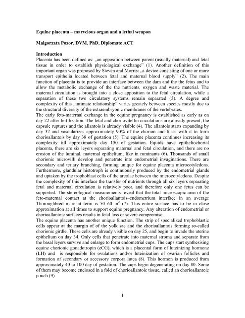
<strong>Equine</strong> placenta <strong>–</strong> marvelous organ <strong>and</strong> a lethal weaponMalgorzata Pozor, DVM, PhD, Diplomate ACTIntroduction<strong>Placenta</strong> has been defined as: „an apposition between parent (usually maternal) <strong>and</strong> fetaltissue in order to establish physiological exchange” (1). Another definition of thisimportant organ was proposed by Steven <strong>and</strong> Morris: „a device consisting of one or moretransport epithelia located between fetal <strong>and</strong> maternal blood supply” (2). The mainfunction of placenta is to provide an interface between the dam <strong>and</strong> the the fetus <strong>and</strong> toallow the metabolic exchange of the the nutrients, oxygen <strong>and</strong> waste material. Thematernal circulation is brought into a close apposition to the fetal circulation, while aseparation of these two circulatory systems remain separated (3). A degree <strong>and</strong>complexity of this „intimate relationship” varies greately between species mostly due tothe structural diversity of the extraembryonic membranes of the vertebrates.The early feto-maternal exchange in the equine pregnancy is established as early as onday 22 after fertilization. The fetal <strong>and</strong> choriovitellin circulations are already present, thecapsule ruptures <strong>and</strong> the allantois is already visible (4). The allantois starts exp<strong>and</strong>ing byday 32 <strong>and</strong> vascularizes approximately 90% of the chorion <strong>and</strong> fuses with it to formchorioallantois by day 38 of gestation (5). The equine placenta continues increasing itscomplexity till approximately day 150 of gestation. Equids have epitheliochorialplacenta, there are six leyers separating maternal <strong>and</strong> fetal circulation, <strong>and</strong> there are noerosion of the luminal, maternal epithelium, like in ruminants (6). Thous<strong>and</strong>s of smallchorionic microvilli develop <strong>and</strong> penetrate into endometrial invaginations. There aresecondary <strong>and</strong> teriary branching, forming unique for equine placenta microcotyledons.Furthermore, gl<strong>and</strong>ular histotroph is continuously produced by the endometrial gl<strong>and</strong>s<strong>and</strong> uptaken by the trophoblast cells of the areolae between the microcotyledons. Despitethe complexity of this interface the transfer of nutrients through all six leyers separatingfetal <strong>and</strong> maternal circulation is relatively poor, <strong>and</strong> therefore only one fetus can besupported. The stereological measurements reveal that the total microscopic area of thefeto-maternal contact at the chorioallantois<strong>–</strong>endometrium interface in an averageThoroughbred mare at term is 50<strong>–</strong>60 m 2 (7). This entire surface has to be in closeapproximation at all times to support equine pregnancy. Any alteration of endometrial orchorioallantoic surfaces results in fetal loss or severe compromise.The equine placenta has another unique function. The strip of specialized trophoblasticcells appear at the margin of of the yolk sac <strong>and</strong> the chorioallantois forming so-calledchorionic girdle. These cells are already visible on day 25, <strong>and</strong> begin to invade the uterineepithelium on day 34. Only cells that penetrate into maternal stroma <strong>and</strong> separate fromthe basal leyers survive <strong>and</strong> enlarge to form endometrial cups. The cups start synthesisingequine chorionic gonadotropin (eCG), which is a placental form of luteinizing hormone(LH) <strong>and</strong> is responsible for ovulations <strong>and</strong>/or luteinization of ovarian follicles <strong>and</strong>formation of secondary or accessory corpora lutea (8). This hormon is produced fromapproximately 40 to 100 day of gestation. The cups begin degenerating on day 80. Someof them may become enclosed in a fold of chorioallantoic tissue, called an chorioallantoicpouch (9).1
And finally, the utero-placental tissues play a major role in producting <strong>and</strong> transportingvarious hormones. Some steroid precursors, such as cholesterol, are transported frommaternal to fetal circulation <strong>and</strong> metabolized within the fetal <strong>and</strong> placental tissues. Themajor products of these chemical reactions are progestagens <strong>and</strong> estrogens, which aresecreted back to the maternal circulation (10). The feto-placental unit is essentiall insupporting pregnancy by production of progestagens, such as 5αDHP (5α-pregnan-3,20-dione). This hormon circulate in maternal plasma during the second half of thepregnancy, but it is also ruturned to the fetus, especially near term. The total progestagenconcentration in maternal plasma increases rapidly during the last two weeks ofpregnancy thanks to the increased level of delivery of the substrates, doubling of the fetaladrenal gl<strong>and</strong>s, <strong>and</strong> increased expression of the progestagenic enzymes in the fetaladrenals <strong>and</strong> placenta.The final decline in progestagen concentration occurs usually soonbefore parturition. Other hormons produced by the equine placenta are: estrogens <strong>and</strong>relaxin. The precursor for estrogens, DHEA (dehydroepi<strong>and</strong>rostenone), is produced byenlarging fetal gonads, regardles of fetal sex. DHEA is excreted to the umbilical artery,transported into the placenta <strong>and</strong> aromatased into estrogens <strong>–</strong> estrone, estradiol 17-α <strong>and</strong>estradiol 17-β. Relaxin is produced mainly by the placenta. This hormon reaches its highconcentration in maternal circulation by day 80 <strong>and</strong> remains high until it increases againduring parurition, as a response to rising concentrations of oxytocin.Summarizing, the equine placenta is a very complex, marvelous organ which acts in asynchrony with fetal development <strong>and</strong> its needs, as well as with a dam <strong>and</strong> her ability tosupport her pregnancy. Unfortunately, things not always go, as planned, <strong>and</strong> equineplacenta may become a deadly trap for the fetus <strong>and</strong>/or a silent killer for the mare.Evaluation of equine placenta post partumThe fetal part of the equine placenta (fetal membranes) is usually expelled within 30minutes after delivery of the foal. A delay in expulsion of the fetal membranes beyond 3hours is considered abnormal in the mare <strong>and</strong> may have serious consequences, such asmetritis/laminitis/septicemia complex. It is important to remember that even a very smallpiece of chorioallantois retained in mare’s uterus is just as lethal as fetal membranesretained as a whole. Therefore, a thorough examination of the fetal membranes that werepassed post partum is so crucial for planning both foal’s <strong>and</strong> mare’s care. <strong>Placenta</strong>consists of maternal <strong>and</strong> fetal part, <strong>and</strong> only fetal part is expelled in the horse. However,many horsemen, farmers, animal scientists, as well as veterinarians, refer to this tissues asjust the placenta. Therefore, we will use this term from now on in this paper as theequivalent to the fetal membranes.The best time to evaluate equine placenta is immidiately after foaling, <strong>and</strong> placentaexpulsion. <strong>Placenta</strong> should be protected from tearing <strong>and</strong> excessive contamination withfeces, shavings, straw or s<strong>and</strong>. Therefore, any freely hanging from the mare’s vulvafragment of the placenta should be tied immediately after foaling. Farm crew should betrained to collect expelled placenta <strong>and</strong> to place it in a clean backet or trash bag forevaluation. Preparing an outline of the examination <strong>and</strong> a form, which has to be filled outis very helpful in collecting <strong>and</strong> saving good records.<strong>Equine</strong> placenta is usually expelled intact, inside out <strong>–</strong> with the chorionic surface inside<strong>and</strong> the allantoic surface facing outside. The exception to this rule is a prematureplacental separation, when the entire chorioallantoic sac comes out with the chorionic2
surface outermost. The chorioallantois has a similar shape as the gravid uterus, with thegravid horn significantly bigger than another, non-gravid one. Normal placental weight atterm is approximately 11% of the weight of the foal, which translates into approximately2.2-6.4 kg (11, 12). The best way of checking for a completeness is arranging the entireplacenta into so-called „lazy F position” (Fig. 1).Figure 1.<strong>Placenta</strong> in „lazy F position”; left - chorionic side out; right <strong>–</strong> allantoic side outCompleteness of chorioallantois is determined first, with a special attention to the tips ofboth horns. The most common site of placental tears is a tip of the non-gravid horn due tomuch thinner wall than the gravid horn. This horn is expelled last which makes it evenmore prone for being torn <strong>and</strong> retained. The tip of the non-gravid horn may be shreddedbut complete. This poses a challenge for a person examining the placenta. One should putall pieces together, like a puzzle, to make sure that, in fact, the entire chorioallantois waspassed. After checking for the completeness, both sides of the chorioallantois areexamined. The chorionic side should be red, <strong>and</strong> has a velvet-like surface. The gravidhorn may have a brighter color that the non-gravid horn, which may appear darker thanthe gravid horn, or even brownish color. The gravid horn is usually larger, smoother <strong>and</strong>thicker than the non-gravid horn. Its tip has an obvious edema (Fig. 2). In contrast, thenon-gravid horn is often, but not always, shorter, much thinner, <strong>and</strong> appears wrinkled dueto the numerous allantoic vessels, which hold this horn’s shape in place (13).Figure 2Chorioallantois - tips of horns; edematous gravid horn <strong>and</strong> thin non-gravid horn: left <strong>–</strong>chorionic side out; right <strong>–</strong> allantoic side out3
One has be really careful in interpreting color changes, since even a short storage ortransportation may cause dramatic changes in color patterns on the chorionic side. Patchybrownish to tan discoloration is often present in normal equine placentas (Fig. 3).Figure 3Normal chorioallantois with patchy discoloration due to short storageThere are small avillous areas on the tips of both horns, which are facing the oviductalpapillae in the uterus (Fig. 4). The body of the uterus is relatively thin. There are alwaysat least two avillous areas on the chorionic surface of this part of the equine placenta. Oneis located around the attachement of the umbilical cord <strong>and</strong> another one is at the internalcervical os <strong>and</strong> is called cervical star (Fig. 4). The cervical star brakes during parturitioninitiating the second stage labor. This area is also affected first in a process of ascendingplacentitis.Figure 4Avillous areas: left <strong>–</strong> oviductal papilla; middle - cervical star; right <strong>–</strong> fold on the largeallantoic vesselSmall avillous areas are also present at the previous location of the endometrial cups.Occassionally, we can see the longitudinal avillous areas, which are overlying largebranches of the umbilical vessels (Fig. 4) (14). These vessels have limited elasticity <strong>and</strong>cause “in utero” folding of the chorioallantoic membrane (15).The fetal foot PAD4
(placental area of degeneration) can be also found on the chorionic surface of the gravidhorn, close to the tip, as a result of chronic pressing of the fetal foot against the placentaduring the last trimester of the pregnancy (14). Any other areas with no villi or withsignificant discoloration should be a reason for concern.Figure 5Chorioallantois <strong>–</strong> allantoic side: left <strong>–</strong> allantoic vesicles; middle <strong>–</strong> allantoic pouches; right- hippomaneAllantoic side of the equine placenta should be smooth <strong>and</strong> transparent. Accumulation offluid in allantoic stroma may lead to the formation of allantoic vesicles, which may befound around the blood vessels. (Fig. 5) Allantoic pouches have different origin. Theycontain necrotic remains of the endometrial cups which slough from the endometrialsurface. They are usually present near the attachment of the umbilical cord. Characteristicfeatures that are often found in the allantoic cavity are hippomanes (Fig. 5). There arealso called “allantoic calculi”. They contain lipids, cellular debris, degenerated bloodcells <strong>and</strong> irregular mineralized material (11). Their significance is unknown.There are many big arteries <strong>and</strong> veins on this side that provide the blood supply to thechorion <strong>and</strong> the chorionic villi. There are three vascular patterns that can be identified onthe allantoic side, with type I being the most commonly seen among the St<strong>and</strong>ardbred(STD) mares (16). Pattern II is consistent with changing location of the fetus from onehorn where implantation took place to another one, where the fetus is present at term.Pattern III is considered abnormal <strong>and</strong> is associated with having a co-twin, which diedspontaneously or was reduced early. The umbilical cord should be attached on the dorsalside at the base of one of the horns. It consists of one vein <strong>and</strong> two arteries on the fetalside <strong>and</strong> two arteries <strong>and</strong> two veins on the maternal side (Fig. 6). The length of the cordvaries, but the large study on 143 Thoroughbred mares showed that the mean length is 55cm, with a broad range from 32 - 90 cm (14). The urachus connects the fetal urinarybladder <strong>and</strong> the allantoic cavity. This structure runs within loose stromal tissue of theamniotic section of the cord (14). A remnant of the yolk sac is a normal feature of theequine placenta <strong>and</strong> is often present along the allantoic portion of the cord (Fig. 6).5
Figure 6Umbilical cord: left <strong>–</strong> normal architecture; right <strong>–</strong> remnant of the yolk sacThe allantoamnion is a large, translucent sac attached to the umbilical cord (Fig. 7). Thismembrane contains numerous blood vessels, which are tortuous <strong>and</strong> prominent (14,15).The amnion itself is not vascular, but the allantoic side provides necessaryvascularization to the allantoamnion.Figure 7Normal allantoamnion: left <strong>–</strong> overview; right <strong>–</strong> amniotic plaguesThere may be amniotic plagues present on the amniotic surface of the allantoamnion <strong>and</strong>on the amniotic part of the umbilical cord. These features are focal areas of squamousmetaplasia (Fig. 8). Their surface may become keratinized <strong>and</strong> they look like small hornlikerugose growths (14).<strong>Placenta</strong>l abnormalitiesA variety of placental abnormalities can be identified during thorough examination.Areas of placental separation are initially bright red or bruise-like as a result of congestedvessels in microcodyledons (14). This is especially obvious when a so-called red-bagdelivery occurs <strong>and</strong> the entire fetus is born within the intact chorioallantoic sac. Thecervical star does not rupture <strong>and</strong> may be very thick. One should consider fescuetoxicosis as a cause of this scenario. However, a much more common reason for placental6
separation in mares is ascending bacterial placentitis. It starts from thickened, congestedchorioallantois at the cervical star, which appears bright red on evaluation, but later in theprocess this area become necrotic, avillous, pale to cream color (Fig. 8). This diseaseaffects mostly the cervical star area <strong>and</strong> the uterine body. An obvious line of demarcationbetween the affected <strong>and</strong> healthy tissues is often seen.Figure 8Ascending placentitis: left <strong>–</strong> bright-red changes at cervical star; middle : very thick, redchanges at cervical star; right <strong>–</strong> pale/cream color changes at cervical starOccasionally, the process may extend to the allantoic side, where neovascularisation <strong>and</strong>chorionic adenomatous hyperplasia occurs, which can be identified as raised, ward-likelesions (17). The fetus is severely compromised by decreased nourishment <strong>and</strong>oxygenation. Another form of placentitis is caused by the Nocardia-form bacteria, whichcolonize the ventral aspect of the base of the gravid horn, as well as the junction of thehorn with the uterine body. Large avillous area can be found on these aspects of thechorioallantois covered with large amount of mucoid-purulent material (Fig. 9).Surprisingly, despite this severe condition, foals born from mares with Nocardia-formplacentis often survive.Figure 9Nocardia-form placentitis: left <strong>–</strong> purulent material expelled during parturition; middle <strong>–</strong>placenta with typical lesions; right <strong>–</strong> adenomatous hyperplasia7
Fungal placentitis is rare in mares. Infection with filamentous fungi, such as Aspergillussp., may mimic bacterial placentitis, since the area at the cervical star is mainly affected(18). This region becomes very thick <strong>and</strong> leathery preventing it from rupturing duringfoal delivery (Fig. 10). This type of mycotic placentitis may be quite extensive <strong>and</strong>involves the allantoic side as well, where hyperplastic changes are often found. Othertypes of mycotic placentis may appear as multifocal granulomas (Histioplasma sp.), or asdiffuse, necrotizing, proliferating inflammation (C<strong>and</strong>ida sp.).Figure 10Mycotic placentitis: left <strong>–</strong> thick cervical star; middle <strong>and</strong> right - diffuse, necrotizing,proliferating inflammationIf any of these abnormalities are identified, veterinarian who is taking care of the foal <strong>and</strong>the mare should be immediately notified. Prompt intervention <strong>and</strong> appropriate treatmentmay be warranted, even necessary to save the foal <strong>and</strong> to help the mare in preparing herreproductive tract for next pregnancy. Furthermore, swabs should be taken from thechorionic surface for bacterial <strong>and</strong>/or fungal culture <strong>and</strong> sensitivity testing. Makingimpression slides <strong>and</strong> taking small samples of tissues for histopathologic evaluation willadd even more information.More extensive disease may affect allantoamniotic membrane as well. In such a case theallantoamnion is significantly thickened <strong>and</strong> not transparent. The vessels become moreprominent, convoluted <strong>and</strong> congested. If the fetus is stressed, meconium is released <strong>and</strong>the allantoamnion is stained yellow or green (Fig. 11).Figure 11Allantoamnion: left <strong>–</strong> thickened membrane; right - meconium-stained allantoamnion8
Umbilical cord has to be inspected as well. The attachment site is localized first.Abnormal attachment is occasionally observed, which is often consistent with aninappropriate fixation position of the conceptus in the uterus, or from a disorientation ofthe conceptus post fixation. Ventral cord attachments may results in the prolongedgestation length, intrauterine growth retardation, <strong>and</strong> inability of the foal to adjust to theoutside environment (19).The length of the cord is measured <strong>and</strong> the number of twists iscounted <strong>and</strong> recorded. The extensive length of the cord may contribute to thedevelopment of torsion of the umbilical cord <strong>and</strong> significant restriction of blood flowfrom the placenta to the fetus <strong>and</strong> vice versa (14). More than four or five twists of thecord are enough to occlude the umbilical vessels. They become enlarged <strong>and</strong> congested.The urachus may be also occluded <strong>and</strong> distended when the amniotic segment of theumbilical cord is twisted (Fig. 12). Occasionally the umbilical cord wraps around thehind limb of the fetus, causing fetal hypoxia.Figure 12Umbilical cord lesions: left <strong>–</strong> umbilical cord torsion; middle <strong>–</strong> umbilical cord wrappedaround the hind limb of the fetus; right <strong>–</strong> dilated urachusThe fetus often dies acutely, which results in abortion. Rarely, the enlarged <strong>and</strong> ossifiedremnant of the yolk sac compresses the vessels of the umbilical cord causing fetal demise<strong>and</strong> abortion. Significant dilations of the umbilical vessels may be also found on theallantoic side of the placenta. These lesions have been associated with an extensivepressure in these vessels. However, clinical significance of this feature has not beendetermined yet (13).We can conclude that a thorough evaluation of the equine placenta has an extraordinaryvalue to the veterinarian who is taking care of the foal <strong>and</strong> the mare.Retained placentaRetained placenta in a mare is a very serious condition. It occurs in approximately 2-10.5% of foalings in mares, but its incidence is higher in draft mares, after dystocia,cesarean section, prolonged gestation, or hydrops condition (20). Stillbirths <strong>and</strong> lack ofnursing <strong>and</strong> oxytocin release may also contribute to the retained placenta. There is noconsensus on the etiology of this problem. However, many believe that an area ofchorioallantois, which is attached to the tip of the non-gravid horn, fails to separate postpartum.It can be partially explained by more branched <strong>and</strong> larger villi <strong>and</strong> more foldingof the placenta present in the non-gravid than in the gravid horn. Therefore, there is a9
tendency for this part of the chorioallantois to be retained more often than any otherfragments of the equine placenta.Figure 13Retained non-gravid horn of chorioallantois: first left <strong>–</strong> chorioallantois with missing tip ofthe non-gravid horn; second left <strong>–</strong> small weight attached to the placental tag; first right <strong>–</strong>large-volume lavage of mare’s uterus; second right <strong>–</strong> retrieved placental tagUnfortunately, retention of even a very small fragment of the chorioallantois is just asdeadly as retention of the entire equine placenta (Fig. 13). The retained tissues undergoquick autolysis, followed by a massive bacterial growth, severe inflammation, <strong>and</strong>absorption of toxins, leading to endotoxemia <strong>and</strong> laminitis. The consequences of theuntreated retained placenta or its fragment in the horse often include metritis, laminitis,septicemia or death (Fig.14).Figure 14Mare with metritis/laminitis/septicemia complex due to retained placenta10
Regardless of the amount of retained tissue, treatment has to be implementedimmediately. Administration of oxytocin is a treatment of choice for retained placenta<strong>and</strong> should be initiated as early as three hours post partum. Injection of oxytocinpromotes uterine contractions <strong>and</strong> facilitates a release of microvilli from endometrialcrypts. There are several methods to administer oxytocin. Frequent boluses of 10-20 IUIV or 5-40 IU IM, every 2-4 hrs, are usually given in the first 3-6 hours. Administrationof higher doses of oxytocin is contraindicated, as it leads to a development of a strongcrump of the uterus, which can hold retained tissue in place preventing expulsion. If thismethod is not effective, a slow infusion of 30-100 IU in 1 or 3 L saline over 30 to 60minutes, followed by 5-10 minutes of walking, can be implemented (21). Mares usuallytolerate this procedure quite well. However, some may show signs of significantdiscomfort. If the mare is hospitalized, 40-60 IU in 5 L of lactated Ringer's solution canbe given as a slow IV drip.Re-distention of the chorioallantois or Burn’s technique is also a valid option ifchorioallantois is intact (no tears). During this method the chorioallantois is distendedwith large amounts (up to 12 L) of a weak (
Affected mares have to be monitored closely for any signs of systemic disease orlaminitis through daily observation <strong>and</strong> a routine blood work. Foal heat breeding afterretained placenta is not recommended.<strong>Equine</strong> placenta is a marvelous organ, which nourish equine fetus throughout the entirepregnancy. Furthermore, it is an open book when evaluated post partum. It conveyspriceless information to a clinician who is taking care of the foal <strong>and</strong> the mare. However,it can also be a deadly weapon if it does not function properly. The fetus is trapped “inutero” <strong>and</strong> often dies as a result. When retained in the uterus post partum, placenta is asilent killer of the mare. It works like a Petri dish for deadly bacteria, which do the dirtywork causing metritis/laminitis/septicemia complex.References:1. Mossman HW. Comparative morphogenesis of the fetal membranes <strong>and</strong>accessory uterine structures Contrib Embryol Carnegie Inst1937;26:129<strong>–</strong>246.2. Steven DH <strong>and</strong> Morris G Development of the foetal membranes. In: Steven DH(ed) Comparative <strong>Placenta</strong>tion, New York: Academic Press, 1975, pp.214-67.3. Enders AC <strong>and</strong> Blankership T. Comparative placental structure. Advanced DrugDelivery Review 1999;38:3-15.4. Enders AC <strong>and</strong> Liu IKM. Lodgment of the equine blastocyst in the uterus fromfixation through endometrial cup formation. J Reprod Fertil Suppl 1991;44:427-38.5. Steven DH. <strong>Placenta</strong>tion in the mare. J Reprod Fertil 1982; 31:41-5.6. Mossman HW. Vertebrate fetal membranes: comparative ontogeny <strong>and</strong>morphology; evolution; phylogenetic significance; basic functions; researchopportunities. Macmillan, Houndmills, Basingstoke, Hampshire <strong>and</strong> London1987.7. Wilsher S <strong>and</strong> Allen WR. The effects of maternal age <strong>and</strong> parity on placental <strong>and</strong>fetal development in the mare. <strong>Equine</strong> Vet J 2003;35:476-83.8. Urwin VE <strong>and</strong> Allen WR. Pituitary <strong>and</strong> chorionic gonadotrophic control ofovarian function during early pregnancy in equids. J Reprod Fertil Suppl1982;32:371-81.9. Clegg MT, Boda JM, Cole HH. The endometrial cups <strong>and</strong> allantochorionicpouches in the mare with emphasis on the source of equine gonadotrophin.Endocrinology. 1954 Apr;54:448-63.10. Ousey J. Endocrinology of Pregnancy. In: <strong>Equine</strong> Reproduction. Second Edition.McKinnon AO, EL Squires, WE Vaala, DD Varner (Eds). Wiley-Blackwell,Ames, Iowa. 2011, pp 2222-2233.11. Schlafer DH. Postmortem examination of the equine placenta, fetus <strong>and</strong> neonate,methods <strong>and</strong> interpretation of findings. AAEP Proceedings 200412. Knottenbelt DC. <strong>Equine</strong> stud farm medicine <strong>and</strong> surgery. Elsevier 2003.13. Wester N. Associations between placental parameters, mare <strong>and</strong> foal bodycondition score at birth, <strong>and</strong> foal health in the first month of life. Doctoral thesis,Faculty of Veterinary Medicine, University of Utrecht, 2010.12
14. Schlafer DH. Examination of the placenta. In: <strong>Equine</strong> Reproduction. SecondEdition. McKinnon AO, EL Squires, WE Vaala, DD Varner (Eds). Wiley-Blackwell, Ames, Iowa. 2011, pp 99-110.15. Rossdale PD <strong>and</strong> Ricketts SW. Evaluation of the fetal membranes at foaling.<strong>Equine</strong> vet Educ Manual 2002;5:78-82.16. Whitehead AE, Chenier TS, Foster RA. <strong>Placenta</strong>tion characteristics ofSt<strong>and</strong>ardbred mares. AAEP Proceedings 2005;51:215-20.17. Christensen BW. Parturition. In: <strong>Equine</strong> Reproduction. Second Edition.McKinnon AO, EL Squires, WE Vaala, DD Varner (Eds). Wiley-Blackwell,Ames, Iowa. 2011, pp 2272-5.18. Hong CB, Donahue JM, Giles RC Jr, Petrites-Murphy MB, Poonacha KB,Roberts AW, Smith BJ, Tramontin RR, Tuttle PA, Swerczek TW. Etiology <strong>and</strong>pathology of equine placentitis. J Vet Diagn Invest 1993;5:56-63.19. Wilsher S, Ousey J, Allen WR Abnormal umbilical cord attachment sites in themare: a review illustrated by three case reports. <strong>Equine</strong> Vet J. 2009;41:930-9.20. Threlfall WR. Retained fetal membranes. In: <strong>Equine</strong> Reproduction. SecondEdition. McKinnon AO, EL Squires, WE Vaala, DD Varner (Eds). Wiley-Blackwell, Ames, Iowa. 2011, pp 2520-9.21. Troedsson M, Spensley M, Fahning M. Retained fetal membranes. In: CurrentTherapy in <strong>Equine</strong> Medicine 4, Ed. Robinson NE, W.B. Saunders Comp.,Philadelphia 1997, 560-1.13