Hip Dislocation After Modular Unipolar Hemiarthroplasty
Hip Dislocation After Modular Unipolar Hemiarthroplasty
Hip Dislocation After Modular Unipolar Hemiarthroplasty
Create successful ePaper yourself
Turn your PDF publications into a flip-book with our unique Google optimized e-Paper software.
The Journal of Arthroplasty Vol. 24 No. 5 2009<strong>Hip</strong> <strong>Dislocation</strong> <strong>After</strong> <strong>Modular</strong><strong>Unipolar</strong> <strong>Hemiarthroplasty</strong>Christopher C. Ninh, MD,* Anil Sethi, MD,y Mohammed Hatahet, MD,*Clifford Les, PhD,* Massimo Morandi, MD,* and Rahul Vaidya, MD, FRCSCyAbstract: An institutional review board–approved retrospective review of hipfractures in elderly patients treated with a modular unipolar implant was carried outto identify factors predisposing to dislocation of a hemiarthroplasty. The mainoutcome measure evaluated was dislocation vs nondislocation. Two hundredseventeen patients underwent the surgery, and 174 were available for review at6 weeks and 144 at 1 year. The incidence of dislocation was 6%. The average time ofdislocation after surgery was 19.3 days. Clinical factors significant for dislocation weremale sex and mental disease. Radiographic factors in dislocated hips included asmaller femoral neck and contralateral femoral neck offset. The center edge anglewas also smaller in the dislocated patients. These patients had a higher mortality rate.Key words: unipolar, hemiarthroplasty, dislocation, fracture neck femur, elderly.© 2009 Elsevier Inc. All rights reserved.<strong>Hemiarthroplasty</strong> of the hip is an accepted treatmentof acute, displaced femoral neck fractures in theelderly. Some reports have indicated that even inminimally displaced and impacted fractures hemiarthroplastyis superior to internal fixation [1]. Theadvantages are immediate weight bearing, no risk ofavascular necrosis or nonunion, and decreased rateof reoperation compared with internal fixation[2,3]. The disadvantages are that the native femoralhead and neck are injured and replaced with animplant. The surgery is more extensive with greatersurgical exposure and a greater amount of bloodloss. There is also a higher perioperative mortalityalthough the patients are usually older with greatercomorbidities [2-5].From the *Department of Orthopedic Surgery, Henry Ford Hospital,Detroit, Michigan; and †Department of Orthopedic Surgery, DetroitReceiving Hospital and University Health Center, Detroit, Michigan.Submitted December 10, 2007; accepted February 29, 2008.No benefits or funds were received in support of the study.Reprint requests: Anil Sethi, MD, Department of OrthopedicSurgery, Detroit Receiving Hospital, UHC 6A, 4201 St AntoineBlvd, Detroit, MI 48201.© 2009 Elsevier Inc. All rights reserved.0883-5403/08/2405-0017$36.00/0doi:10.1016/j.arth.2008.02.019Although reports of dislocation after total hiparthroplasty abound in literature, they are sparse inthe setting of a hemiarthroplasty. Early dislocation isa severe complication of hemiarthroplasty with amortality rate of 65% to 75% at 6 months [6].Factors that predispose a hemiarthroplasty todislocation are technical, anatomical, and medicalcomorbidities. Cognitive impairment has beenreported as a cause for dislocation [7]. The technicalfactors cited are surgical approach, excessive lengthof the residual neck, and smaller femoral neck offset.The anatomical factors include acetabular measurementsindicative of hip dysplasia and shorterpatients [8]. The purpose of this article is to analyzethe radiographic and clinical data in determining thefactors predisposing to dislocation after unipolarmodular hemiarthroplasty of the hip in intracapsularfracture neck femur in the elderly.Materials and MethodsAn institutional review board–approved retrospectivereview was undertaken on 217 patientswho sustained a femoral neck fracture and underwenthemiarthroplasty with a unipolar modular768
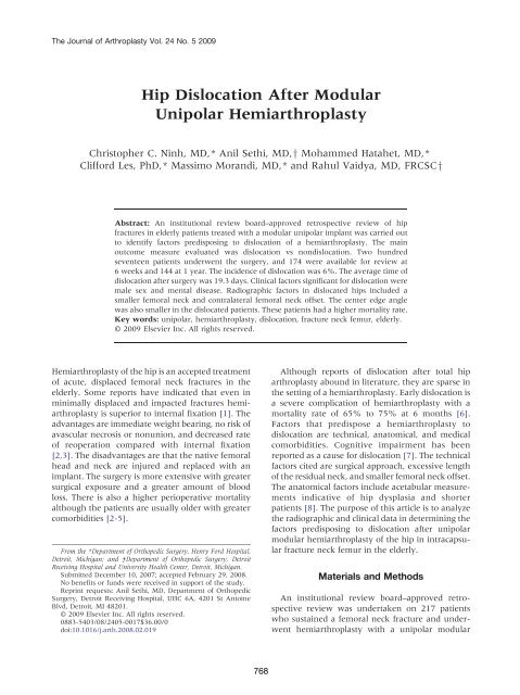
<strong>Hip</strong> <strong>Dislocation</strong> <strong>After</strong> <strong>Modular</strong> <strong>Unipolar</strong> <strong>Hemiarthroplasty</strong> Ninh et al 769Table 1. Anatomical measurements in dislocated andnondislocated hipsMeasurementsin Dislocated<strong>Hip</strong>sMeasurements inNondislocated<strong>Hip</strong>sTotal 11 83FemoralMean 29.86 35.86Neck Offset SD 7.59 7.42Contralateral Mean 29.02 37.27Femoral SD 6.85 7.53Neck OffsetRatio of Femoral Mean 1.05 0.98Neck Offset SD 0.29 0.21Residual Femoral Mean 15.92 16.57NeckSD 8.67 5.67Prosthesis Femoral Mean 139.64 137.9Neck shaft Angle SD 7.59 5.63Acetabular Index Mean 36.55 35.83SD 3.3 4.43Center Edge Angle Mean 30.36 37.39of Wiberg SD 5.29 9.15prosthesis from January 2000 to August 2004. Allpatients included in the study presented with adisplaced fracture and were older than 60 years.Their residential status was not a determining factorfor inclusion in the study. Patients with undisplacedor minimally displaced fractures were excludedfrom the study. Also patients with rheumatoidarthritis, osteoarthritis, and fractures secondary totumors were excluded. The age ranged from 61 to95 years with a mean age of 77.3 years and an SD of11.8 years. One hundred forty-nine patients werefemales (69%) and 68 were males (31%). Onehundred ten patients sustained a left-sided femoralneck fracture, and 107 had a right-sided fracture. Anevaluation of the mental status using the minimentalstatus examination showed impairment in48 patients (22%).All patients were evaluated radiographically andtreated with a cemented unipolar modular hemiarthroplasty(Conquest FX <strong>Hip</strong> System, Smith andNephew, Memphis, Tenn). The operation was donewithin 24 hours of admission unless other comorbiditiesdelayed the procedure. A posterior surgicalapproach (Moore/Kocher-Langenbeck) [9,10] wasused on 174 patients, and in 43 patients, the lateralapproach (Hardinge) [9,11] was chosen. The surgicalapproach was entirely the operating surgeons'choice. Standard anteroposterior and lateral radiographsof the hip were taken postoperatively, andpatients were allowed to weight bear from the dayafter surgery. All patients were advised to limit therange of motion of the hip to less than 90° of flexionand 45° of external and internal rotation and to avoidadduction for first 6 weeks after surgery. Mentallyimpaired patients had additional hip precautionswith the placement of an abduction pillow in theoperating room before bed transfer and used pillowsto maintain abduction while in bed. The mainoutcome measure evaluated was dislocation vsFig. 1. A, Femoral neck offset—distance from a vertical line of the midshaft femur to the center of the prosthesis head.B, Contralateral femoral neck offset—distance from vertical line of the midshaft femur to the center of the nativecontralateral head.
770 The Journal of Arthroplasty Vol. 24 No. 5 August 2009nondislocation. Two hundred seventeen patientsunderwent the surgery, and 174 were available forreview at 6 weeks and 144 at 1 year.Radiographic data of 11 dislocated patients werecompared to 83 random patients in the nondislocatedgroup (Table 1). The radiographic measurementsincluded femoral neck offset [12] of thefractured and contralateral side (Fig. 1), femoralneck offset ratio (Fig. 1), residual femoral necklength [8] (Fig. 2), acetabular index [13] (Fig. 3),center edge angle of Wiberg [14] (Fig. 4), andprosthesis neck-femur shaft angle [7] (Fig. 5).The clinical factors included age, sex, surgicalapproach, presence of mental impairment, andmortality rate at 1 year (Tables 2 and 3). Clinicaldata were collected using an electronic computerbasedmedical records tracking system. Radiographicdata were analyzed on the electronic public accesscomputer system (EPACS, Stentor, Brisbane, Calif).StatisticsA series of χ 2 analyses, with Yates continuitycorrections, was used to evaluate the proportion ofsurgical approach, sex, and mental impairment withrespect to the dislocation rate. The analyses weredone separately for 6-week follow-up and for 12-month follow-up.A series of Mann-Whitney rank sum tests wereused to evaluate whether the patient age or any ofthe radiographic measurements differed significantlybetween patients who had dislocated vspatients who had no dislocated hips. The analyseswere done separately for 6-week follow-up and for12-month follow-up (P = .05).A backward, stepwise logistic regression wasperformed on the data at 6-week follow-up and at12-month follow-up. Briefly, in these analyses, theoutput variable was the presence or absence of adislocation. The surgical approach, sex, age, mentalstatus, and all of the radiographic measurementswere presented to the model as predictor variables.Predictor variables with a P N .20 were removedfrom the model, and the logistic regression wasrepeated. At that point, predictor variables with aP N .15 were removed from the model, and so on,until all predictor variables remaining in the modelhad a P b .05.ResultsAmong 144 patients followed up at 1 year, therewere 11 dislocations. In 10 patients, hip dislocationoccurred within the first 6 weeks of follow-up(Table 2). For these patients, the average time ofdislocation after surgery was 19.3 days (range, 7-42 days). Median time of dislocation was 19 days.One patient had the hip dislocated at 364 daysafter surgery.Isolated Effect of AgeAt 6 weeks (n = 185), there was no demonstrableassociation between dislocation and age (P = .80;power = 0.05). Again at 12 months (n = 144), therewas no demonstrable correlation between dislocationand age (P = .81; power = 0.05).Isolated Effect of Surgical ApproachAt 6 weeks (n = 185), there was no demonstrablecorrelation between dislocation and surgicalapproach (P = .069; power = 0.06). A similarobservation was made at 12 months (n = 144),with no demonstrable correlation between dislocationand surgical approach (P = .82; power = 0.05).Fig. 2. Residual femoral neck length—distance fromprosthesis collar to lesser trochanter.Isolated Effect of SexAt 6 weeks (n = 185), males were overrepresentedamong the patients who had dislocated hips (P =.01). At 12 months (n = 144), males were
<strong>Hip</strong> <strong>Dislocation</strong> <strong>After</strong> <strong>Modular</strong> <strong>Unipolar</strong> <strong>Hemiarthroplasty</strong> Ninh et al 771Fig. 3. Acetabular index angle—measured as the angle between the roof and a horizontal line (line from the teardropparallel to interischial line).overrepresented among the patients who had dislocatedhips (P = .03).Isolated Effect of Mental ImpairmentAt 6 weeks (n = 185), there was no demonstrablecorrelation between dislocation and mental impairment.However, there was statistical trend towardmore dislocations among the mentally impaired (P =.07). At 12 months (n = 144), those with mentalimpairment were overrepresented among thepatients who had dislocated hips (P = .02).Fig. 4. Center edge angle of Wiberg—measured as theangle between a vertical line and a line from the centerof the prosthesis head to the lateral edge of theacetabular roof.Fig. 5. Prosthesis neck-femur shaft angle—line from tip ofprosthetic head bisecting the neck and vertical line of themidshaft femur.
772 The Journal of Arthroplasty Vol. 24 No. 5 August 2009Table 2. Clinical data in dislocated hips6-Wk Follow-UpIsolated Effect of Femoral Neck OffsetAt 6 weeks (n = 114), there were no demonstrabledifferences between dislocated and nondislocatedpatients (P = .13; power = 0.24). At 12 months (n =94), there was a significant difference betweendislocated and nondislocated patients (P = .02;dislocated, 29.9 ± 7.6; nondislocated, 35.9 ± 7.4).Isolated Effect of Contralateral FemoralNeck OffsetAt 6 weeks (n = 114), there was a significantdifference between dislocated and nondislocatedpatients (P = .001; dislocated, 29.1 ± 7.2; nondislocated,36.7 ± 8.4).At 12 months (n = 94), there was a significantdifference between dislocated and nondislocatedpatients (P = .002; dislocated, 29.0 ± 6.9; nondislocated,37.3 ± 7.5).Isolated Effect of Ratio of Femoral Neck Offsetto Contralateral Femoral Neck OffsetAt 6 weeks (n = 114), there was no demonstrabledifferences between dislocated and nondislocatedpatients (P = .43; power = 0.14).At 12 months (n = 94), there was no demonstrabledifference between dislocated and nondislocatedpatients (P = .61; power = 0.05).Isolated Effect of Residual Femoral Neck1-Y Follow-UpTotal 10 11Approach Posterior, 8 Posterior, 9Nonposterior, 2 Nonposterior, 2Sex Male, 7 Male, 7Female, 3 Female, 4Mental Disease 5 6At 6 weeks (n = 114), there was no demonstrabledifferences between dislocated and nondislocatedpatients (P = .66; power = 0.05). At 12 months (n =94), there was no demonstrable difference betweendislocated and nondislocated patients (P = .89;power = 0.05).Isolated Effect of Acetabular Index AngleAt 6 weeks (n = 114), there was no demonstrabledifferences between dislocated and nondislocatedpatients (P = .85; power = 0.05). At 12 months (n =94), there was no demonstrable difference betweendislocated and nondislocated patients (P = .67;power = 0.05).Isolated Effect of Center Edge Angle of WibergAt 6 weeks (n = 114), there was a significantdifference between dislocated and nondislocatedpatients (P = .001; dislocated 29.0 ± 2.8°; nondislocated,37.6 ± 8.7°).At 12 months (n = 94), there was a significantdifference between dislocated and nondislocatedpatients (P = .012; dislocated, 30.4 ± 5.3°; nondislocated,37.4 ± 9.1°).Isolated Effect of Prosthesis Neck-FemoralShaft AngleAt 6 weeks (n = 114), there was no demonstrabledifferences between dislocated and nondislocatedpatients (P = .87; power = 0.05).At 12 months (n = 94), there was no demonstrabledifference between dislocated and nondislocatedpatients (P = .57; power = 0.05).Combined Effects of Clinical andRadiographic FactorsAt 6-week follow-up (n = 114), the remainingpredictor variables were as follows:• Center edge angle of Wiberg (P = .006; oddsratio, 0.772; 95% confidence interval [CI],0.642-0.927). Higher values of center edgeangle of Wiberg were associated with lowerrates of dislocation.• Sex (P = .014; odds ratio, 8.644; 95% CI, 1.544-48.378). Males had higher rates of dislocation.At 12-month follow-up (n = 94), the remainingpredictor variables were as follows:• Center edge angle of Wiberg (P = .010; oddsratio, 0.829; 95% CI, 0.719-0.957). Highervalues of center edge angle of Wiberg wereassociated with lower rates of dislocation.• Mental impairment (P = .010; odds ratio,8.177; 95% CI, 1.671-40.023). Mentalimpairment was associated with higher ratesof dislocation.NondislocationTable 3. Clinical data in dislocated hips6-WkFollow-Up1-YFollow-UpNoFollow-UpTotal 174 133 33Approach Posterior, 139 Posterior, 106 Posterior, 27Nonposterior,35Nonposterior,27Nonposterior,6Sex Male, 48 Male, 36 Male, 13Female, 126 Female, 97 Female, 20Mental Disease 33 25 10
<strong>Hip</strong> <strong>Dislocation</strong> <strong>After</strong> <strong>Modular</strong> <strong>Unipolar</strong> <strong>Hemiarthroplasty</strong> Ninh et al 773Mortality RateMortality rate was 36.4% for patients withdislocated hemiarthroplasty vs 16.8% for patientswithout hemiarthroplasty dislocation.At 12 months postoperatively, no associationcould be demonstrated between mortality rate anddislocation (P = .452; power = 0.109).DiscussionPostoperative dislocation of a hemiarthroplasty isa serious complication with a mortality rate of 60%to 75% at 6 months [7,8,15]. The rate of dislocationis increased by various factors such as age, medicalcondition, mental disorder, surgical approach, andprosthetic malposition [16]. The published incidenceof dislocation of the hemiarthroplasty ranges from1% to 15% [6,15,17].We evaluated radiographic data for anatomicalvariability of patients and technical factors ofsurgery. Our data clearly show that dislocatedhemiarthroplasties have a lower center edge angleof Wiberg compared to the nondislocated groupsuggesting an inherent instability within the hip.Femoral neck offset in a modern hemiarthroplasty isa factor that can be modified technically to increasethe stability of the hip by adjusting the femoral neckcomponent, head size, and placement of the femoralstem prosthesis. We measured the ratio of femoralneck offset to the contralateral neck offset and notedthat it was close to or equal to 1.0 in all patients(Table 1). We observed that patients with low offsethips were more inherently unstable and henceprone to dislocations.Although the patient population in our study waspredominately females, the rate of hemiarthroplastydislocation at 1 year follow-up for males was 4 timeshigher than that of females (16.3% vs 4.0%,respectively). There was a positive statistical significancebetween sex and the center edge angle ofWiberg (P = .02). In this study, we observed thatmale patients had a lower mean center edge angle ofWiberg compared to females (33.3° vs 38.1°). Wehypothesize that males' hips dislocated morebecause of colinearity secondary to hip dysplasiathat was prevalent in our male patient population.Mental impairment was associated with higherrates of dislocation. Our data show that mentalimpairment was present in 54.5% patients withdislocation vs 18.8% in nondislocated patients.Controversy exists over the applicability of endoprostheticarthroplasty of the femoral head as compared toother treatments for femoral neck fractures inpatients with mental impairment [18-21]. Reportsshow that there is an unacceptably high dislocationrate of 37% and mortality rate of 75% in patientswith neurologic impairment [2,16,18,22,23]. Theinability to adhere to strict hip precautions and aflexion and adduction contracture predisposes thesepatients to higher rates of dislocations. However, acomprehensive review in patients with Parkinsondisease did not substantiate these findings [24].Furthermore, the studies performed involved nonmodularendoprosthesis.The incidence of dislocation with the posteriorapproach (9/139) was not any higher than with theuse of the lateral approach (2/35). The posteriorapproach has been indicated in many studies ashaving a higher rate of dislocation [8,25,26]. Adislocation rate of 0.9% with the anterior approachvs 14% by a posterior approach [15] has beenreported earlier. Another study found that the rateof dislocation was 16% for posterior approach and3.2% for the direct lateral approach [8]. In a studycomparing the dislocation rate after posterior andlateral approaches in more than 3500 patients, itwas observed that the overall dislocation rate for theposterior approach was 9.0%, whereas that for thedirect lateral was 3.3% [26]. Interestingly, it wasnoted that the results were not statistically significant,and the dislocation rates were dependent uponthe operative experience and seniority of thesurgeons. Researchers have advocated the posteriorapproach after reports of no dislocations in a seriesof 115 patients with posterior approach hemiarthroplasties[27]. Further in a series of 235 patients,the authors showed no statistically significantincidence of dislocation after an anterior or posteriorapproach [28]. The posterior capsule and posteriorsoft tissues are strong stabilizers of the hip andshould not be compromised. The disruption of theposterior stabilizers while performing the posteriorapproach has been stated to increase the instabilityof the hip prosthesis ultimately leading to failure anddislocation. Significant stresses are imposed on theposterior stabilizers when the hip is in a flexedposition as when getting out of a chair. However,most of the above studies used a Thompson orAustin Moore prosthesis that is not modular. Inaddition, those studies failed to mention posteriorcapsule or posterior soft tissue repair, which is nowcommonly performed.Femoral neck fractures are associated with a highrate of mortality. Patients are often old and havemany associated comorbidities. An earlier reportstates that patients operated on beyond 5 days ofadmission had a 1 year mortality rate of 35% and a6% to 11% mortality rate at 1 year if operated onwithin 2 to 5 days of hospital admission [4,5]. It has
774 The Journal of Arthroplasty Vol. 24 No. 5 August 2009been reported that the medical condition of thepatient will frequently deteriorate if hip fracturesurgery is delayed [29]. Therefore, surgery shouldbe performed as soon as possible after admission.Although not statistically significant, in ourstudy, the mortality rate at 1-year follow-up was3 times higher in patients who had a dislocation ofthe hemiarthroplasty. Patients with hip fractureshave mortality rate greater than that expected innormal population [5,9]. Furthermore, patientssustaining a dislocation have an even greater mortalityrate [2,9,15].In our retrospective review, we found that thefactors predisposing to dislocation of the hemiarthroplastyare multifactorial. Mental impairmentand the male sex are significant factors that mayincrease the incidence of dislocation. Radiographicfactors such as femoral neck offset,contralateral femoral neck offset, and centeredge angle were also contributing factors forhemiarthroplasty dislocation.References1. Hui AC, Anderson GH, Choudhry R, et al. Internalfixation or hemiarthroplasty for undisplaced fracturesof the femoral neck in octogenarians. J Bone JointSurg Br 1994;76:891.2. Lu-Yao GL, Keller RB, Littenberg B, et al. Outcomesafter displaced fractures of the femoral neck. A metaanalysisof one hundred and six published reports.J Bone Joint Surg Am 1994;76:15.3. Sikorski JM, Barrington R. Internal fixation versushemiarthroplasty for the displaced subcapital fractureof the femur. A prospective randomised study. J BoneJoint Surg Br 1981;63-B:357.4. Kenzora JE, Magaziner J, Hudson J, et al. Outcomeafter hemiarthroplasty for femoral neck fractures inthe elderly. Clin Orthop Relat Res 1998:51.5. Kenzora JE, McCarthy RE, Lowell JD, et al. <strong>Hip</strong>fracture mortality. Relation to age, treatment, preoperativeillness, time of surgery, and complications.Clin Orthop Relat Res 1984:45.6. Blewitt N, Mortimore S. Outcome of dislocation afterhemiarthroplasty for fractured neck of the femur.Injury 1992;23:320.7. Khatod M, Barber T, Paxton E, et al. An analysis of therisk of hip dislocation with a contemporary total jointregistry. Clin Orthop Relat Res 2006;447:19.8. Pajarinen J, Savolainen V, Tulikoura I, et al. Factorspredisposing to dislocation of the Thompson hemiarthroplasty:22 dislocations in 338 patients. ActaOrthop Scand 2003;74:45.9. Canale ST, Campbell WC. Campbell's operativeorthopaedics. St. Louis, MO: Mosby; 2003.10. Moore AT. The self-locking metal hip prosthesis.J Bone Joint Surg Am 1957;39-A:811.11. Hardinge K. The direct lateral approach to the hip.J Bone Joint Surg Br 1982;64:17.12. Pellicci P, Tria AJ, Garvin KL. Orthopaedic knowledgeupdate. <strong>Hip</strong> and knee reconstruction 2. Rosemont, Ill.:American Academy of Orthopaedic Surgeons; 2000.13. Sharp I. Acetabular dysplasia. J Bone Joint Surg1961;43B:268.14. Wiberg G. Studies on dysplastic acetabular andcongenital subluxation of the hip joint: with specialreference to the complication of osteoarthritis. ActaChir Scand Suppl 1939;58:1.15. Chan RN, Hoskinson J. Thompson prosthesis forfractured neck of femur. A comparison of surgicalapproaches. J Bone Joint Surg Br 1975;57:437.16. Lung HR. The role of prosthetic replacement of thehead of the femur as primary treatment for subcapitalfractures. Injury 1971;3:107.17. D'Arcy J, Devas M. Treatment of fractures of thefemoral neck by replacement with the Thompsonprosthesis. J Bone Joint Surg Br 1976;58:279.18. Coughlin L, Templeton J. <strong>Hip</strong> fractures in patients withParkinson's disease. Clin Orthop Relat Res 1980:192.19. Hunter GA. A comparison of the use of internalfixation and prosthetic replacement for fresh fracturesof the neck of the femur. Br J Surg 1969;56:229.20. Hunter GA. The rationale for internal fixation andagainst hemiarthroplasty. In: Hungerford DS, editor.The hip : proceedings of the eleventh open scientificmeeting of The <strong>Hip</strong> Society, 1983, in memory of SirJohn Charnley. St. Louis, MO: C.V. Mosby Company;1983. p. 34.21. Hunter GA, Krajbich IJ. The results of medialdisplacement osteotomy for unstable intertrochantericfractures of the femur. Clin Orthop Relat Res1978:140.22. Garcia Jr A, Neer II CS, Ambrose GB. Displacedintracapsular fractures of the neck of the femur. 1.Mortality and morbidity. J Trauma 1961;1:128.23. Rothermel JE, Garcia A. Treatment of hip fractures inpatients with Parkinson's syndrome on levodopatherapy. J Bone Joint Surg Am 1972;54:1251.24. Staeheli JW, Frassica FJ, Sim FH. Prosthetic replacementof the femoral head for fracture of the femoralneck in patients who have Parkinson disease. J BoneJoint Surg Am 1988;70:565.25. Keene GS, Parker MJ. <strong>Hemiarthroplasty</strong> of the hip—the anterior or posterior approach? A comparison ofsurgical approaches. Injury 1993;24:611.26. Unwin AJ, Thomas M. <strong>Dislocation</strong> after hemiarthroplastyof the hip: a comparison of the dislocation rateafter posterior and lateral approaches to the hip. AnnR Coll Surg Engl 1994;76:327.27. Salvati EA, Wilson Jr PD. Long-term results offemoral-head replacement. J Bone Joint Surg Am1973;55:516.28. Wood MR. Femoral head replacement followingfracture: an analysis of the surgical approach. Injury1980;11:317.29. Neer II CS. The surgical treatment of the fractured hip.Surg Clin North Am 1950;31:499.


