Automated Breast Volume Scanning 3D Ultrasound of the Breast
Automated Breast Volume Scanning 3D Ultrasound of the Breast
Automated Breast Volume Scanning 3D Ultrasound of the Breast
You also want an ePaper? Increase the reach of your titles
YUMPU automatically turns print PDFs into web optimized ePapers that Google loves.
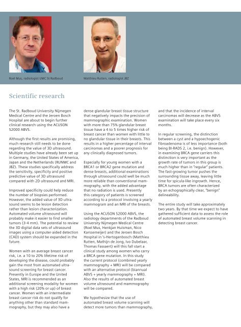
Roel Mus, radiologist UMC St RadboudMatthieu Rutten, radiologist JBZScientific researchThe St. Radboud University NijmegenMedical Centre and <strong>the</strong> Jeroen BoschHospital are about to begin fur<strong>the</strong>rclinical research using <strong>the</strong> ACUSONS2000 ABVS.Although <strong>the</strong> first results are promising,much research still needs to be doneregarding <strong>the</strong> value <strong>of</strong> <strong>3D</strong> ultrasound.Multiple studies have already been set upin Germany, <strong>the</strong> United States <strong>of</strong> America,Japan and <strong>the</strong> Ne<strong>the</strong>rlands (RUNMC andJBZ). These studies specifically address<strong>the</strong> sensitivity, specificity and positivepredictive value <strong>of</strong> <strong>3D</strong> ultrasoundcompared with 2D ultrasound and MRI.Improved specificity could help reduce<strong>the</strong> number <strong>of</strong> biopsies performed.However, <strong>the</strong> added value <strong>of</strong> <strong>3D</strong> ultrasoundseems to be lesion detectionra<strong>the</strong>r than lesion characterization.<strong>Automated</strong> volume ultrasound willprobably make it easier to find smallertumors (3-4 mm). The potential to review<strong>the</strong> <strong>3D</strong> digital data sets <strong>of</strong> ultrasoundimages using a computer-aided detection(CAD) system should be expanded in <strong>the</strong>future.Women with an average breast cancerrisk, i.e. a 10 to 20% lifetime risk <strong>of</strong>developing <strong>the</strong> disease, could probablygain <strong>the</strong> most from automated ultrasoundscreening for breast cancer.Presently in Europe and <strong>the</strong> UnitedStates, MRI is recommended as anadditional screening modality for womenwith a high risk (20% or up) <strong>of</strong> breastcancer. Women with an intermediatebreast cancer risk do not qualify foranything o<strong>the</strong>r than standard mammography,but <strong>the</strong>y may also have adense glandular breast tissue structurethat negatively impacts <strong>the</strong> precision <strong>of</strong>mammographic examination. Womenwith more than 75% glandular breasttissue have a 4 to 5 times higher risk <strong>of</strong>breast cancer than women with little tono glandular tissue in <strong>the</strong>ir breasts. Thisresults in a higher percentage <strong>of</strong> intervalcarcinomas and a poorer prognosis forany clinically diagnosed tumors.Especially for young women with aBRCA1 or BRCA2 gene mutation anddense breasts, additional examinationsthrough ultrasound could well be muchmore reliable than conventional mammography,with <strong>the</strong> added advantagethat no radiation is used. Presentlythis category <strong>of</strong> patients is screenedaccording to a protocol involving a yearlymammogram and an MRI <strong>of</strong> <strong>the</strong> breasts.Using <strong>the</strong> ACUSON S2000 ABVS, <strong>the</strong>radiology departments <strong>of</strong> <strong>the</strong> RadboudUniversity Nijmegen Medical Centre(Roel Mus, Henkjan Huisman, NicoKarssemeijer) and <strong>the</strong> Jeroen BoschHospital in ‘s-Hertogenbosch (MatthieuRutten, Mathijn de Jong, Ivo Dubelaar,Thomas Fassaert) will this fall start aclinical study among women who carrya BRCA gene mutation. In this study<strong>the</strong> current protocol (combined yearlymammography + MRI) will be comparedwith an alternative protocol (biannualABVS + yearly mammography + MRI).Also <strong>the</strong> results <strong>of</strong> automated breastvolume ultrasound and mammographywill be compared.We hypo<strong>the</strong>size that <strong>the</strong> use <strong>of</strong>automated breast volume scanning willdetect more tumors than mammography,and that <strong>the</strong> incidence <strong>of</strong> intervalcarcinomas will decrease as <strong>the</strong> ABVSexamination will take place every sixmonths.In regular screening, <strong>the</strong> distinctionbetween a cyst and a hypoechogenicfibroadenoma is <strong>of</strong> less importance (bothbeing BI-RADS 2, i.e. benign). However,in examining BRCA gene carriers thisdistinction is very important as <strong>the</strong>growth rate <strong>of</strong> tumors in this group ismuch higher than in “regular” patients.The fast-growing tumor pushes <strong>the</strong>surrounding tissue away, leaving littletime for spicula-like ingrowth. Hence,BRCA tumors are <strong>of</strong>ten characterizedby an echographically clear, “benign”delineability.The entire study will take approximatelytwo years. By that time we expect to havega<strong>the</strong>red sufficient data to assess <strong>the</strong> role<strong>of</strong> automated breast volume scanning indetecting breast cancer.





![WalkAway plus Technical Specifications [41 KB] - Siemens Healthcare](https://img.yumpu.com/51018135/1/190x253/walkaway-plus-technical-specifications-41-kb-siemens-healthcare.jpg?quality=85)
